cutaneous leishmaniasis in pakistan: a neglected disease
TRANSCRIPT
RESEARCH Open Access
Cutaneous Leishmaniasis in Pakistan: aneglected disease needing one healthstrategyBehzad Kayani1, Shakera Sadiq1, Hamad Bin Rashid2, Naseer Ahmed3, Altaf Mahmood4, Muhammad Shakeel Khaliq4,Rubab Maqsood1, Haroon Rashid5, Saima Hasan1, Muhammad Hassan Mushtaq1, Ubaid-ur-Rehman Zia1 andMamoona Chaudhry1*
Abstract
Background: Cutaneous Leishmaniasis (CL) is a neglected tropical disease, which mainly affects poor communities.It is one of the major vector-borne disease and endemic in Pakistan.
Methods: A case-control study to evaluate potential risk factors of human-CL was conducted in Khewra region,District Jhelum, Pakistan from January–April 2014. Case data about 90 cases registered during October 2012 toNovember 2013 was retrieved from Municipal Hospital. Controls were matched (1,1 ratio) on the date ofregistration with cases from same hospital. Both cases and controls were invited to participate and data wascollected in a face-to-face interview. A prospective study of canine leishmaniasis (canine-CL) was also conducted atCivil Veterinary Hospital in the same area. Suspected dogs with skin ulceration signs were included in the study andblood samples were collected. Statistical analyses were conducted to determine association between variousparameters and outcome of interest.
Results: The ages of cases ranged from 1 to 76 years (median = 15 years) and proved to be protective factor i.e.increase in each year in age reduced the likelihood of being infected with human-CL [Odds Ratio (OR) = 0.4, 95%Confidence Interval (CI) = 0.25–0.76]. People sleeping outsides in an open area were more likely to become a case(OR = 8.7, 95% CI = 2.90–26.37) than a control. Poor sanitary condition inside the house (OR = 3.3, 95% CI 1.03–10.56)and presence of other animals in house (livestock, poultry) (OR = 3.6, 95% CI = 1.07–12.12) also identified as riskfactors of high significance. The proportion of positive dogs with canine-CL was 21.05% and was significantlyassociated with human-CL cases in the same area (p < 0.05).
Conclusions: We concluded that adopting self-protections measures against sand-fly, and maintaining goodhygiene may lower the risk of human-CL. One-Health Strategy is suggested to control leishmaniasis in human anddog population.
Keywords: Leishmaniasis, Sand-fly, Cutaneous, Vector-borne, Risk factors, Case-control, Pakistan, Zoonosis
© The Author(s). 2021 Open Access This article is licensed under a Creative Commons Attribution 4.0 International License,which permits use, sharing, adaptation, distribution and reproduction in any medium or format, as long as you giveappropriate credit to the original author(s) and the source, provide a link to the Creative Commons licence, and indicate ifchanges were made. The images or other third party material in this article are included in the article's Creative Commonslicence, unless indicated otherwise in a credit line to the material. If material is not included in the article's Creative Commonslicence and your intended use is not permitted by statutory regulation or exceeds the permitted use, you will need to obtainpermission directly from the copyright holder. To view a copy of this licence, visit http://creativecommons.org/licenses/by/4.0/.The Creative Commons Public Domain Dedication waiver (http://creativecommons.org/publicdomain/zero/1.0/) applies to thedata made available in this article, unless otherwise stated in a credit line to the data.
* Correspondence: [email protected] of Epidemiology and Public Health, University of Veterinary andAnimal Sciences, Lahore, PakistanFull list of author information is available at the end of the article
Kayani et al. BMC Infectious Diseases (2021) 21:622 https://doi.org/10.1186/s12879-021-06327-w

BackgroundCutaneous Leishmaniasis (CL) is a parasitic diseasetransmitted via the bite of female sand-flies belonging tothe genera Phlebotomus in the Old World and Lutzo-myia in the New World. It is a skin disease ranging fromself-healing lesions to single or large skin ulcers and iscaused by protozoan parasites of the genus Leishmania[1]. Leishmania major is a main cause of CL in humansin an area that stretches from India through CentralAsia, the Middle East, to North and West Africa [2, 3].The epidemiology of leishmaniases is dynamic and the
conditions of transmission are continually changing de-pending on change in environment, demography, humanbehavior, socioeconomic status, and immunogenic pro-file of affected human populations [2, 4]. Among themost important zoonotic diseases, leishmaniasis is amajor concern for public health [5]. In terms of burdenof diseases, it is estimated to be the third most import-ant vector-borne disease. Despite this fact, it is one ofthe “neglected diseases”. The tropical and sub-tropicalparts of the world are endemic with leishmaniasis [3].The disease occurs in 88 countries of the world with 70being endemic. Afghanistan, Algeria, Brazil, Pakistan,Peru, Saudi Arabia, and Syria are the countries where90% of the cases occur [6]. According to an estimate, 1.3million new cases and 20,000 to 30,000 deaths occur an-nually [5]. The disease is disfiguring skin affliction as re-ported by U.S Centre of Disease Control and Prevention(CDC). Leishmaniasis can be divided into two formsbased on epidemiology of disease: zoonotic which in-cludes animal reservoir hosts in the transmission cycleof the disease, and anthroponotic, in which humans areconsidered to be the sole source of infection for thesand-fly vector [7].In Pakistan, leishmaniasis has been reported in human
and animal population [8, 9]. Human-CL is endemic inseveral parts of Pakistan and is the second most preva-lent vector-borne disease in the country after malaria[10]. There are 37 out of 70 species of the sand-fly in-habitant in Pakistan, which can transmit disease tohealthy hosts [11]. Endemic areas of disease in Pakistaninclude areas of Baluchistan, Interior Sindh, SouthPunjab and Khyber Pakhtunkhwa [11–14]. Currently,the progression of the disease is a public health issueand represents a challenge for health professionals. Epi-demiological studies might help planning for effectivestrategies to control human-CL. Several factors such asclimatic and environmental changes, the movement ormigration of infected people, animal reservoirs and fe-male infected sand-flies play important role in the trans-mission of leishmaniasis [15].In Pakistan, cases of human-CL have been reported
from different districts of Punjab province, however, datais scant about the identification of risk factors specifically
in District Jhelum. Few studies have been carried out inBaluchistan, Khyber Pakhtunkhwa and Azad Kashmir [11,12, 16]. In the present study, we aimed to quantify riskfactors associated with human-CL in Khewra region ofDistrict Jhelum, Pakistan, with an objective to inform pol-icy makers for evidence-based disease control recommen-dations to prevent future outbreaks. To study thepresence of zoonotic risk of human-CL, we also con-ducted a prospective study in dogs, suspected for canineleishmaniasis (canine-CL) in the same geographical area.
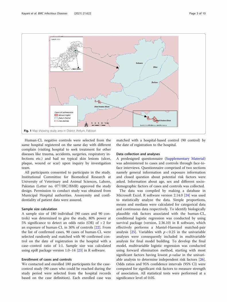
MethodsStudy areaThe study was conducted in Khewra region, Tehsil PindDadan Khan, District Jhelum (Fig. 1). The district is ad-ministratively divided into four tehsils namely Jhelum,Dina, Sohawa and Pind Dadan Khan. Khewra region isdivided into two union councils: Khewra no.1 andKhewra no.2 with a population of around 35,000 [17].The area is surrounded by the famous Salt Range. It islocated at 32°38′ 60″ N 73° 1′ 0″ E. Khewra City is alsoknown as “The Kingdom of Salt” because of its rock salt,which is 98% pure and natural source of salt in Pakistan.Khewra Salt Mine is the second largest salt mine in theworld [18, 19]. Previously several outbreaks of human-CL in local population have been reported from this areabetween 2012 and 2013 [20].
Study designCase-control study in humanA case-control study was designed to evaluate the riskfactors associated with human-CL between January toApril 2014 in residents of Khewra region, DistrictJhelum. Patient records from outpatient and inpatientclinics of Municipal Hospital, Pind Dadan Khan, DistrictJhelum, were retrieved and reviewed for case selection.
Definition of case and controlHuman-CL positive cases were diagnosed by medicalphysicians at the Municipal Hospital. A case was definedas a person having at least one leishmania lesion (pres-ence of a skin ulcer with typical raised edges and de-pressed centre or a skin plaque-a circumscribed, nodularor palpable skin lesion) and/or a typical scar (a typicalCL scar develop when a papule appears after biting ofsand-fly, which may enlarge to become an indolent ul-cerated nodule or plaque, and after self-healing of theplague, a depressed scar is left on skin) [21]. The trainedmedical officers used clinical diagnosis (leishmania le-sion or/and CL scar) followed by confirmatory micros-copy (impression smear) to confirm leishmaniasis. Casepatients were visited in their house after getting theirinformation from hospital records.
Kayani et al. BMC Infectious Diseases (2021) 21:622 Page 2 of 10
Human-CL negative controls were selected from thesame hospital registered on the same day with differentcomplain (visiting hospital to seek treatment for otherdiseases like trauma, accidents, surgeries, respiratory in-fections etc.) and had no typical skin lesions (ulcer,plaque, wound or scar) upon inquiry by investigationteam.All participants consented to participate in the study.
Institutional Committee for Biomedical Research atUniversity of Veterinary and Animal Sciences, Lahore,Pakistan (Letter no. 077/IRC/BMR) approved the studydesign. Permission to conduct study was obtained fromMunicipal Hospital authorities. Anonymity and confi-dentiality of patient data were assured.
Sample size calculationA sample size of 180 individual (90 cases and 90 con-trols) was determined to give the study, 80% power at5% significance to detect an odds ratio (OR) of > 2 foran exposure of human-CL in 30% of controls [22]. Fromthe list of confirmed cases, 90 cases of human-CL wereselected randomly and matched with 90 confirmed con-trol on the date of registration in the hospital with acase–control ratio of 1:1. Sample size was calculatedusing epiR package version 1.0–14 [23] in R software.
Enrollment of cases and controlsWe contacted and enrolled 180 participants for the case-control study (90 cases who could be reached during thestudy period were selected from the hospital recordsbased on the case definition). Each enrolled case was
matched with a hospital-based control (90 control) bythe date of registration to the hospital.
Data collection and analysesA predesigned questionnaire (Supplementary Material)was administered to cases and controls through face-to-face interviews. Questionnaire comprised of two sectionsnamely general information and exposure informationand closed question about potential risk factors wereasked. Information about age, sex and different socio-demographic factors of cases and controls was collected.The data was compiled by making a database in
Microsoft Excel. R software version 2.14.0 [24] was usedto statistically analyze the data. Simple proportions,means and medians were calculated for categorical dataand continuous data respectively. To identify biologicallyplausible risk factors associated with the human-CL,conditional logistic regression was conducted by usingsurvival package (version, 2.36.10) in R software, whicheffectively performs a Mantel-Haenszel matched-pairanalysis [25]. Variables with p < 0.25 in the univariableanalyses were consequently included in multivariableanalysis for final model building. To develop the finalmodel, multivariable logistic regression was conductedusing forward elimination method, starting with mostsignificant factors having lowest p-value in the univari-able analysis to determine independent risk factors [26].Odds ratios and 95% confidence intervals (95% CI) werecomputed for significant risk factors to measure strengthof association. All statistical tests were performed at asignificance level of 0.05.
Fig. 1 Map showing study area in District Jhelum, Pakistan
Kayani et al. BMC Infectious Diseases (2021) 21:622 Page 3 of 10
QGIS version 2.14.3 (available at https://www.qgis.org/en/site/forusers/download.html#) was used to visualizethe spatial distributions of cases in Khewra region,District Jhelum.
Prospective study in dogsA prospective study of canine-CL in pet dogs attendingCivil Veterinary Hospital from Khewra region, was con-ducted from January–April 2014. Owners of all sus-pected dogs with skin lesions (dermatitis, alopecia,cutaneous ulcerations, weight loss, ocular or nasal le-sions) [27, 28] attending the government veterinary hos-pital were requested to participate in study. Only thosedogs were included whose owners consented to partici-pate in the study. Peripheral blood samples were col-lected by a trained veterinarian from suspected dogs andthin dry smears were made using leishman’s stain. Theamastigotes of Leishmania were detected by using acompound microscope [29].
Data analysisData was statistically analyzed by using R software ver-sion 2.14.0 [24]. Chi square test was used to asses anyassociation between canine-CL, area, and presence ofany positive human-CL case. Proportion of canine-CLwas calculated.
ResultsThe human-CL cases enrolled in current study were reg-istered in Municipal Hospital, Pind Dadan Khan, DistrictJhelum from October 2012 to November 2013. All of
them visited hospital after the development of the le-sions. Therefore, the date of the sand-fly bite was not ac-curately known by the cases and the time of exposure tohuman-CL could not be specified.
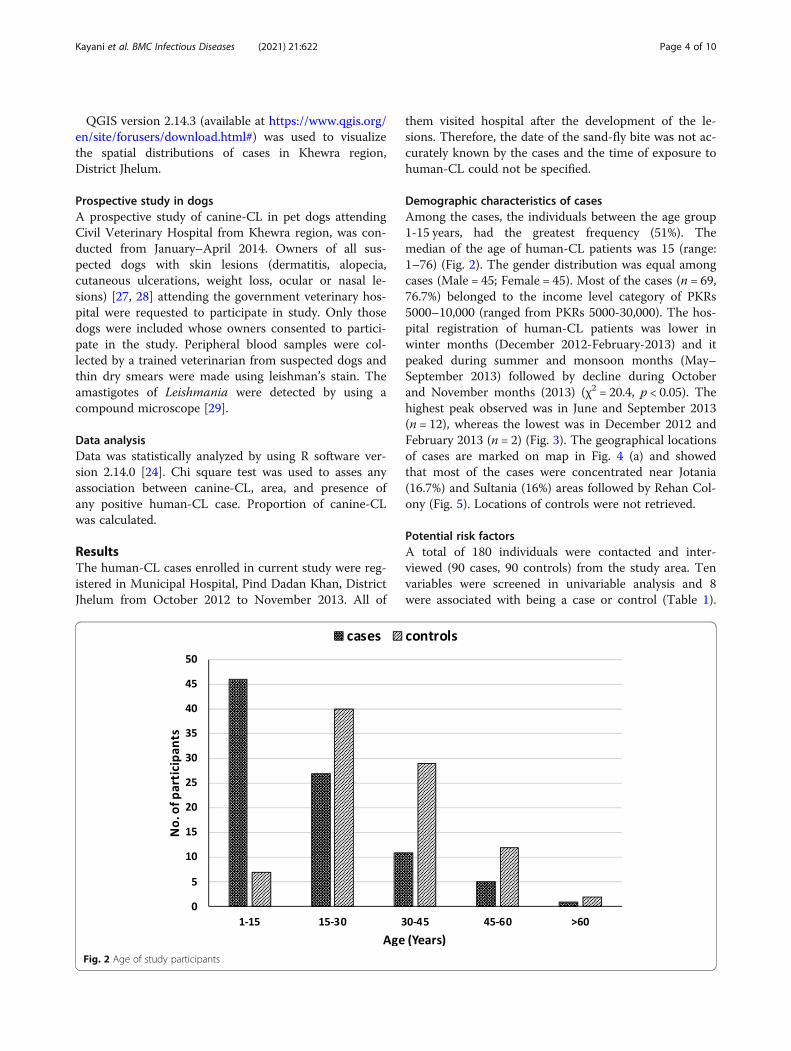
Demographic characteristics of casesAmong the cases, the individuals between the age group1-15 years, had the greatest frequency (51%). Themedian of the age of human-CL patients was 15 (range:1–76) (Fig. 2). The gender distribution was equal amongcases (Male = 45; Female = 45). Most of the cases (n = 69,76.7%) belonged to the income level category of PKRs5000–10,000 (ranged from PKRs 5000-30,000). The hos-pital registration of human-CL patients was lower inwinter months (December 2012-February-2013) and itpeaked during summer and monsoon months (May–September 2013) followed by decline during Octoberand November months (2013) (χ2 = 20.4, p < 0.05). Thehighest peak observed was in June and September 2013(n = 12), whereas the lowest was in December 2012 andFebruary 2013 (n = 2) (Fig. 3). The geographical locationsof cases are marked on map in Fig. 4 (a) and showedthat most of the cases were concentrated near Jotania(16.7%) and Sultania (16%) areas followed by Rehan Col-ony (Fig. 5). Locations of controls were not retrieved.
Potential risk factorsA total of 180 individuals were contacted and inter-viewed (90 cases, 90 controls) from the study area. Tenvariables were screened in univariable analysis and 8were associated with being a case or control (Table 1).
Fig. 2 Age of study participants
Kayani et al. BMC Infectious Diseases (2021) 21:622 Page 4 of 10

Poor sanitary conditions, presence of other Leishmaniainfected persons in the house, sleeping outsides in openareas, other animal on premises, and house type, gender(being female), using protections like insecticide sprays,bed nets, screens etc. and age (in years) were selected formultivariable analysis based on selection criteria (p <0.025). Two variables ‘income of the participant’ and‘keeping a dog’ having p > 0.25 were excluded from
further analysis. One variable namely ‘other human-CLpatient in house’ was excluded from analysis due toinsufficient number of discordant pairs (Table 1).In the final multivariable model, four variables were
identified as significantly associated with the human-CLin Khewra residents (Table 2). Cases keeping other ani-mals in house (livestock, poultry) were 3.6 times (95%CI: 1.07–12.12, p < 0.05) more likely to have human-CL
Fig. 3 Distribution of reported cases of human-CL per month
Fig. 4 Locations of enrolled cases of human-CL (a) and canine-CL (b) in Khewra Region, District Jhelum
Kayani et al. BMC Infectious Diseases (2021) 21:622 Page 5 of 10

compared to controls. Similarly, cases having poor sani-tation conditions at home were more likely to havehuman-CL (OR: 3.3, 95% CI 1.03–10.56, p < 0.05) ascompared to controls. The odds of being diagnosed withhuman-CL were 8.7 time more in cases who slept out-side in open area (95% 2.90–26.37, p < 0.001) when com-pared to exposure in controls. The increasing ageshowed to have decreased the likelihood of human-CL0.4 times (95% CI: 0.25–0.76, p < 0.005) (Table 2).
Prospective study in dogsDuring the study period (January–April 2014), 15 bloodsamples of the dogs brought to Civil Veterinary Hospitalwith skin lesions were collected. A dry thin stainedsmear was made from the blood sample for the detec-tion of amastigote forms of Leishmania in macrophagesof the dogs. Leishmania amastigotes forms were foundin 4 out of 15 samples (21%). The presence of positivedogs was significantly associated (p < 0.001) with thepositive cases of human-CL in the same area. The spatialdistribution of the canine-CL cases with human-CLcases is shown in Fig. 4 (b). The dogs from Jotania,Islamganj and Karimpura areas were tested positive forcanine leishmaniasis (Fig. 5). The status of the dogs fromother areas was unknown.
DiscussionThe epidemiological triad of CL is complex with variousepidemiological risk factors associated with host, agentand environment. Early recognition of these risk factorsmay prevent the further transmission to susceptible
population. Results of our study support the findings ofother studies from Pakistan that CL cases are increasingin the local human and dog population in Pakistan [9,12, 13, 30, 31] suggesting that a one-health approachwould be needed to reduce the disease burden.Published literature about risk factors for human-CL
in Pakistan in generally sparse or obsolete [8, 32, 33].The current study was aimed to determine the risk fac-tors associated with human-CL that prevailed in thelocal environment and detection of canine-CL in dogs inKhewra region, District Jhelum. After extensive literaturereview, age, sex, income, keeping dog, keeping other ani-mals (livestock, poultry), poor sanitary conditions, sleep-ing outside in open areas, using protections andpresence of other Leishmania infected persons in thehouse were included as risk factors [2, 12, 30, 34–36].Seasonal pattern of transmission is useful to establish
disease surveillance and control activities [37]. Human-CL patients registered in the current study, visited thehospital over a period of 12 months. There was a signifi-cant increase in the patient visits to the hospital duringsummer and monsoon months (May–September 2013).This reflects seasonal activity of sand-fly vector duringsummer and rainy season. Jhelum experience monsoonseason from June to September which brings heavy rain,while the dry season in this region is from November toJanuary. Varied transmission patterns have been re-ported by various studies suggesting seasonal trends indifferent geographical locations [37–39].Previous studies have reported clustering of leishman-
iasis at household level [33, 38, 40]. In current study,
Fig. 5 Number of human-CL cases according to areas and canine-CL cases
Kayani et al. BMC Infectious Diseases (2021) 21:622 Page 6 of 10

among all cases, 59% (n = 53) confirmed the presence ofother human-CL patient in their house or in the neigh-bors. Presence of an infected person in the household in-creases the risk of getting infected because sand-flieshave limited fly zone and they remain in same vicinityand could bite multiple hosts living at the same place[32, 38, 40]. L. tropica transmission has also been knownto be characterized by clustering of cases [32].In the current study, we identified a set of risk factors
that might significantly contribute to the web of
causation of human-CL in the region. Age (increasing inyears) was identified as a protective factor (OR < 1). Themajority of the cases in our study were children andyoung people < 15 years of age and human-CL was lessreported in adults compared to children age groups.Increasing each year in age reduced the likelihood ofhuman-CL 0.4 times. This could be correlated to theoutdoor activities of the children (playing outdoorgames) with minimum precautions to cover their body,which might have exposed them to bites of sand-flies,while adults adopt more precautions during outdooractivity [38, 41, 42].Keeping other animals in house (livestock, poultry)
showed association with CL (OR > 1). Presence of otheranimals e.g. livestock and poultry, could attract vector ofCL due to presence of barn and dried dung, and mayexpose residents to the female sand-flies. Previously,presence of sand-flies was reported to be associated with
Table 1 Risk factors for cutaneous leishmaniasis based on univariable analysis
Variables Response Case numbers (%) Control numbers (%) OR 95% CI p-value
Age (years) 1–15 46 (51.1) 7 (7.9) Ref < 0.009*!
15–30 27 (30) 40 (44.4) 0.11 0.03–0.34
30–45 11 (12.2) 29 32.2 0.07 0.02–0.25
45–60 5 (5.6) 12 (13.3) 0.08 0.02–0.35
> 60 1 (1.1) 2 (2.2) 0.02 0.01–3.26
Sex Male 45 (50) 18 (20) 0.25 0.12–0.52 0.0002*
Female 45 (50) 72 (80)
Income (PKRs) 5000–10,000 69 (76.7) 60 (66.7) Ref 0.225
10,000–20,000 16 (17.8) 21 (23.3) 0.64 0.29–1.44
20,000–30,000 4 (4.4) 8 (8.9) 0.46 0.13–1.59
> 30,000 1 (1.1) 1 (1.1) 0.68 0.37–12.26
Dog in house No 82 (91.1) 83 (92.2) 1.17 0.39–3.47 0.782
Yes 8 (8.9) 7 (7.8)
Other animals on premises No 44 (48.8) 67 (74.4) 3.10 1.57–6.1 0.00114*
Yes 46 (51.1) 23 (25.5)
Poor sanitary conditions No 16 17.8 43 47.8 4.37 2.03–9.43 <.00017*
Yes 74 (80.2) 47 (52.2)
Protection used Yes 15 16.7 26 28.9 0.48 0.22–1.01 0.0535*
No 75 (83.3) 64 (71.1)
House type Concrete 44 (48.9) 57 (63.3) 1.93 1.01–3.68 0.0461*
Mud 46 (51.1) 33 (36.7)
Other CL patients in house No 37 (41.1) 86 (95.6) 50 6.91–361.9 0.000107$
Yes 53 (58.9) 4 (4.4)
Sleeping Outside in open area No 6 (6.7) 62 (68.9) 12.2 4.90–30.36 0.0000056*
Yes 84 (93.3) 28 (31.1)*Factors selected for multivariable analysis!p-value bases on likelihood ratio test$Excluded from analysis due to inadequate discordant pairs
Table 2 Risk factors in final multivariable logistic model
Variables Odds ratio 95% CI p-value
Age (years) 0.4 0.25–0.76 0.003
Other animals in house 3.6 1.07–12.12 0.039
Poor sanitary conditions 3.3 1.03–10.56 0.045
Sleeping outside in open area 8.7 2.90–26.37 0.0001
Kayani et al. BMC Infectious Diseases (2021) 21:622 Page 7 of 10

cattle and cattle blood was found in Phlebotomus tobbifemales [41, 43, 44].Patients reporting poor sanitary condition were 3.3
times more likely to diagnosed with human-CL comparedto those with better condition. Almost 80% of the cases re-ported unsatisfactory sanitary condition at home, whichincluded, open toilets, open sewage, mud floors and un-hygienic livings. Poor sanitary conditions provide suitablehabitat for sand-flies to breed and spread human-CL [45,46]. Studies have consistently shown more cases ofhuman-CL among poor, neglected populations, who arelikely to be less educated and mostly unemployed [41, 47].Furthermore, most of cases (76.7%) in current study,belonged to families having an income of PKRs 10,000 orless. Families with lower income level have less resourcesto adopt protective measure and awareness about the pro-tections against diseases, consequently increased risk ofexposure to sand-flies and Leishmania infection [2]. Inter-ventions such as poverty alleviation and improving livingcondition might aid significantly in controlling human-CLtransmission in the region [21].Sleeping outside in open air increased the odds of
human-CL, probably due to their exposure to the sand-fly bites during the sleeping time as one cannot protectoneself. Sleeping outdoor in open space during summermonths is very common in Pakistan. Our findings cor-roborated with the results of other studies that sleepingoutside is a risk factor for CL [3, 41, 47, 48]. In Pakistan,May–September are hot and humid months and people,especially in villages prefer to sleep outside the rooms inopen air. Sand-fly activity is also increased through Juneand July, with peak in August. Entomological studies in-dicated nocturnal activity of sand-flies starts at the be-ginning of the night, and is strongly associated withrelative humidity rather than with temperature [3, 41,47]. Our data also supported the speculation that themost appropriate transmission period of CL is duringthe hot and humid nights from July to September.During the period of 3 months, 15 suspected dogs for
CL were brought to the local veterinary hospital. Theirblood samples were taken and tested for presence ofLeishmania protozoa. Among 15 suspected, 21% (n = 4)were detected positive for canine-CL. Although dogs areconsidered major reservoir for L. infantum, the possibilityof clinical canine disease and their potential as secondaryhosts for L. major should be investigated in endemic areasfor human L. major infection [28, 34]. Our results showeda significant association between the areas of reportedcases of human-CL and canine-CL positive dogs. Areaswith high burden of human-CL cases had presence ofcanine-CL positive dogs.The findings of the study have some limitations due to
case-control nature of the design as it is difficult to es-tablish the temporal causality in case-control studies.
Furthermore, these study designs are prone to selectionbias and recall bias. Future investigations based on co-hort study design would be more appropriate to ascer-tain the causal relationship between risk factors andoutcome.
ConclusionsPakistan has a diverse landscapes and climates that mayaffect the transmission of Leishmania in the country.The key risk factor identified in present study may beextrapolated to design an early preparedness responsefor human-CL outbreaks at human-animal interface.The current study also provides initial evidence for thepresence of canine-CL in Khewra region, DistrictJhelum.
Supplementary InformationThe online version contains supplementary material available at https://doi.org/10.1186/s12879-021-06327-w.
Additional file 1.
AcknowledgmentsWe gratefully acknowledge the assistance of staff at Municipal Hospital, andCivil Veterinary Hospital, Pind Daden Khan. We would also like to thank studyparticipants and owners of dogs for providing data and samples.
Authors’ contributionsConceived and designed the study: MC, MHM, HBR and BK. Collected data:BK and NA. Analyzed the data: MC, SS, SH, URZ, RM and MSK. Drafted paper:MC, BK, HBR, MHM, SS, SH, URZ, MSK, AM, HR, RM. All authors approved thedraft.
FundingThere was no funding available for this study.
Availability of data and materialsThe data gathered and generated during the current study are availablefrom the corresponding author (Mamoona Chaudhry) on reasonable request.
Declarations
Ethics approval and consent to participateThe study protocol and consent procedure were approved by theInstitutional Review Committee for Biomedical Research, (Reference No. 077/IRC/BMR) (For Humans) and Advanced Study and Research Board (ASRB) ofUniversity of Veterinary and Animal Sciences, Lahore, Pakistan (for Animals).For human subjects, all participants and their attendees were briefed aboutthe purpose of research, interview and questions, voluntary participation andother aspects of the study. Confidentiality of data were maintained duringstudy and analysis. All procedures and methods were carried out accordingto Helsinki Declaration 2013. A trained medical doctor or paramedic staffregistered with Pakistan Medical and Dental Council (PMDC) collected thesamples from human subjects.For animal patients, a trained veterinary staff, registered with PakistanVeterinary Medical Council (PVMC), collected the blood samples from dogaccording to standard guidelines and procedures. All suspected dogs wereprovided with adequate water and food. Animals were properly restrainedand a trained staff collected peripheral blood from brachial vein. Wefollowed guidelines published by University of Minnesota InstitutionalAnimal Care and Use Committee, USA.For human participants, Informed consent was obtained from the individualpatients and the parents (in case of minors). For animal cases, informedconsent was obtained from the owner of the animal before taking samplesand data.
Kayani et al. BMC Infectious Diseases (2021) 21:622 Page 8 of 10

Consent for publicationNot applicable as no human identifiable images are provided in themanuscript.
Competing interestsAll authors declare that they have no competing interest.
Author details1Department of Epidemiology and Public Health, University of Veterinary andAnimal Sciences, Lahore, Pakistan. 2Department of Clinical Medicine andSurgery, University of Veterinary and Animal Sciences, Lahore, Pakistan.3District Health Development Center, Jhelum, Pakistan. 4Directorate ofAnimal Disease, Diagnostic, Reporting, and Surveillance, Livestock and DairyDevelopment Department, Government of Punjab, Lahore, Pakistan.5Department of Microbiology, Faculty of Veterinary Sciences, IslamiaUniversity Bahawalpur, Bahawalpur, Pakistan.
Received: 8 March 2021 Accepted: 16 June 2021
References1. Killick-Kendrick R. The biology and control of Phlebotomine sand flies. Clin
Dermatol. 1999;17(3):279–89. https://doi.org/10.1016/S0738-081X(99)00046-2.2. Alvar J, Yactayo S, Bern C. Leishmaniasis and poverty. Trends Parasitol. 2006;
22(12):552–7. https://doi.org/10.1016/j.pt.2006.09.004.3. Reithinger R, Dujardin J-C, Louzir H, Pirmez C, Alexander B, Brooker S.
Cutaneous leishmaniasis. Lancet Infect Dis. 2007;7(9):581–96. https://doi.org/10.1016/S1473-3099(07)70209-8.
4. Leta S, Dao THT, Mesele F, Alemayehu G. Visceral Leishmaniasis in Ethiopia:an evolving disease. PLoS Negl Trop Dis. 2014;8(9):e3131. https://doi.org/10.1371/journal.pntd.0003131.
5. Leishmania fact sheet number 375. 2016. [http://www.who.int/mediacentre/factsheets/fs375/en/]. Accessed 10 Dec 2016.
6. Desjeux P. Leishmaniasis: current situation and new perspectives. CompImmunol Microbiol Infect Dis. 2004;27(5):305–18. https://doi.org/10.1016/j.cimid.2004.03.004.
7. Desjeux P. Leishmaniasis: public health aspects and control. Clin Dermatol.1996;14(5):417–23. https://doi.org/10.1016/0738-081X(96)00057-0.
8. Munir MA, Rab MA, Iqbal J, Ghafoor A, Khan MA, Burney MI. A Review of theStatus of Leishmaniasis in Pakistan from 1960–1986. In: Hart DT, editor.Leishmaniasis: the current status and new strategies for control. Boston:Springer US; 1989. p. 47–56.
9. Durrani AZ, Durrani HZ, Kamal N, Mehmood N. Prevalence of cutaneousLeishmaniasis in humans and dogs in Pakistan. Pak J Zool. 2011;43(2):263–71.
10. Ejaz A, Raza N, Iftikhar N. Recurrent cutaneous leishmaniasis presenting assporotrichoid abscesses: a rare presentation near Afghanistan border.Dermatol Online J. 2007;13(2):15.
11. Khan SJ, Muneeb S. Cutaneous leishmaniasis in Pakistan. Dermatol Online J.2005;11(1):4.
12. Afghan AK, Kassi M, Kasi PM, Ayub A, Kakar N, Marri SM. Clinicalmanifestations and distribution of cutaneous Leishmaniasis in Pakistan. JTrop Med. 2011;2011:359145.
13. Rowland M, Munir A, Durrani N, Noyes H, Reyburn H. An outbreak ofcutaneous leishmaniasis in an Afghan refugee settlement in north-WestPakistan. Trans R Soc Trop Med Hyg. 1999;93(2):133–6. https://doi.org/10.1016/S0035-9203(99)90285-7.
14. Hussain M, Munir S, Khan TA, Khan A, Ayaz S, Jamal MA, et al. Epidemiologyof cutaneous Leishmaniasis outbreak, Waziristan, Pakistan. Emerg Infect Dis.2018;24(1):159–61. https://doi.org/10.3201/eid2401.170358.
15. Kassi M, Kassi M, Afghan AK, Rehman R, Kasi PM. Marring leishmaniasis: thestigmatization and the impact of cutaneous leishmaniasis in Pakistan andAfghanistan. PLoS Negl Trop Dis. 2008;2(10):e259. https://doi.org/10.1371/journal.pntd.0000259.
16. The only option for cutaneous leishmaniasis treatment in KhyberPakhtunkhwa. 2019. [https://www.msf.org/only-option-cutaneous-leishmaniasis-treatment-pakistan]. Accessed 13 Nov 2020.
17. Khewra:city in Punjab, Pakistan. 2017. [https://www.citypopulation.de/Pakistan-Punjab.html]. Accessed 20 Jan 2020.
18. Khaliq F. Khewra mines: a salt wonder for tourists. In: DAWN; 2019.19. Salt Range. 2019. [http://www.tdcp.gop.pk/page.php?pid=260]. Accessed 10
Nov 2020.
20. Anonymous. Weekly Bullitin: DEWS, Pakistan. In: Islamabad; 2013.21. Neglected Tropical Diseases. 2020. [http://www.emro.who.int/neglected-
tropical-diseases/information-resources-leishmaniasis/cl-factsheet.html].Accessed 30 Nov 2020.
22. Schlesselman JJ. Case-control studies : design, conduct, analysis. NewYorkUnited States of America: Oxford University Press Inc; 1982.
23. Stevenson M. Package ‘epiR’. In., 0.9–46 edn; 2013.24. Team RDC. R: a language and environment for statistical computing. In: R 2.
14.0 GUI 1.42 edn. Vienna: R Foundation for Statistical Computing; 2011.25. Therneau T, Lumley T. Survival: Survival analysis, including penalised
likelihood. In: R package version 2.36–10; 2011.26. Dohoo I, Martin W, Stryhn H. Veterinary epidemiologic research.
Charlottetown: AVC Inc.; 2003.27. Sasani F, Javanbakht J, Samani R, Shirani D. Canine cutaneous leishmaniasis.
J Parasit Dis. 2016;40(1):57–60. https://doi.org/10.1007/s12639-014-0444-4.28. Baneth G, Nachum-Biala Y, Shabat Simon M, Brenner O, Gaier S, Rojas A,
et al. Leishmania major infection in a dog with cutaneous manifestations.Parasit Vectors. 2016;9(1):246. https://doi.org/10.1186/s13071-016-1541-2.
29. Moody AH, Chiodini PL. Methods for the detection of blood parasites. ClinLab Haematol. 2000;22(4):189–201. https://doi.org/10.1046/j.1365-2257.2000.00318.x.
30. Alvar J, Vélez ID, Bern C, Herrero M, Desjeux P, Cano J, et al. Boer md, theWHOLCT: Leishmaniasis worldwide and global estimates of its incidence.PLoS One. 2012;7(5):e35671. https://doi.org/10.1371/journal.pone.0035671.
31. Ayub S, Gramiccia M, Khalid M, Mujtaba G, Bhutta RA. Cutaneousleishmaniasis in Multan: species identification. J Pak Med Assoc. 2003;53(10):445–7.
32. Brooker S, Mohammed N, Adil K, Agha S, Reithinger R, Rowland M, et al.Leishmaniasis in refugee and local Pakistani populations. Emerg Infect Dis.2004;10(9):1681–4. https://doi.org/10.3201/eid1009.040179.
33. Kolaczinski J, Brooker S, Reyburn H, Rowland M. Epidemiology ofanthroponotic cutaneous leishmaniasis in Afghan refugee camps inNorthwest Pakistan. Trans R Soc Trop Med Hyg. 2004;98(6):373–8. https://doi.org/10.1016/j.trstmh.2003.11.003.
34. Kent A, Ramkalup P, Mans D, Schallig H. Is the dog a possible reservoir forcutaneous Leishmaniasis in Suriname? J Trop Med. 2013;2013:324140.
35. Oryan A, Akbari M. Worldwide risk factors in leishmaniasis. Asian Pac J TropMed. 2016;9(10):925–32.
36. Oryan A, Alidadi S, Akbari M: Risk Factors Associated With Leishmaniasis.Trop Med Surg. 2014;2(3):e118.
37. Siriwardana Y, Zhou G, Deepachandi B, Akarawita J, Wickremarathne C,Warnasuriya W, et al. Trends in recently emerged Leishmania donovaniinduced cutaneous Leishmaniasis, Sri Lanka, for the first 13 years. BiomedRes Int. 2019;2019:4093603.
38. Ullah K, Khan NH, Sepulveda N, Munir A, Wahid S. Assessing incidencepatterns and risk factors for cutaneous Leishmaniasis in Peshawar region,Khyber Pakhtunkhwa, Pakistan. J Parasitol. 2016;102(5):501–6. https://doi.org/10.1645/15-919.
39. Akhlagh A, Salehzadeh A, Zahirnia AH, Davari B: 10-Year Trends inEpidemiology, Diagnosis, and Treatment of Cutaneous Leishmaniasis inHamadan Province, West of Iran (2007–2016). Front Public Health. 2019;7(27). https://doi.org/10.3389/fpubh.2019.00027.
40. Reithinger R, Mohsen M, Leslie T. Risk factors for anthroponotic cutaneousLeishmaniasis at the household level in Kabul, Afghanistan. PLoS Negl TropDis. 2010;4(3):e639. https://doi.org/10.1371/journal.pntd.0000639.
41. Votypka J, Kasap OE, Volf P, Kodym P, Alten B. Risk factors for cutaneousleishmaniasis in Cukurova region, Turkey. Trans R Soc Trop Med Hyg. 2012;106(3):186–90. https://doi.org/10.1016/j.trstmh.2011.12.004.
42. Bhutto AM, Soomro FR, Katakura K. Leishmaniasis in Sindh, Pakistan:outbreak and review of literature. J Pak Assoc Dermatol. 2008;18:212–9.
43. Velo E, Paparisto A, Bongiorno G, Di Muccio T, Khoury C, Bino S, et al.Entomological and parasitological study on phlebotomine sandflies incentral and northern Albania. Parasite. 2005;12(1):45–9. https://doi.org/10.1051/parasite/2005121045.
44. Svobodová M, Alten B, Zídková L, Dvorák V, Hlavacková J, Mysková J, et al.Cutaneous leishmaniasis caused by Leishmania infantum transmitted byPhlebotomus tobbi. Int J Parasitol. 2009;39(2):251–6. https://doi.org/10.1016/j.ijpara.2008.06.016.
45. Müller GC, Kravchenko VD, Rybalov L, Schlein Y. Characteristics of restingand breeding habitats of adult sand flies in the Judean Desert. J VectorEcol. 2011;36(s1):S195–205. https://doi.org/10.1111/j.1948-7134.2011.00131.x.
Kayani et al. BMC Infectious Diseases (2021) 21:622 Page 9 of 10

46. Yaghoobi-Ershadi MR, Akhavan AA, Zahraei-Ramazani AV, Abai MR, EbrahimiB, Vafaei-Nezhad R, et al. Epidemiological study in a new focus of cutaneousleishmaniasis in the Islamic Republic of Iran. East Mediterr Health J. 2003;9(4):816–26.
47. Ngere I, Gufu Boru W, Isack A, Muiruri J, Obonyo M, Matendechero S, et al.Burden and risk factors of cutaneous leishmaniasis in a peri-urbansettlement in Kenya, 2016. PLoS One. 2020;15(1):e0227697. https://doi.org/10.1371/journal.pone.0227697.
48. Bashaye S, Nombela N, Argaw D, Mulugeta A, Herrero M, Nieto J, et al. Riskfactors for visceral leishmaniasis in a new epidemic site in Amhara region,Ethiopia. Am J Trop Med Hyg. 2009;81(1):34–9. https://doi.org/10.4269/ajtmh.2009.81.34.
Publisher’s NoteSpringer Nature remains neutral with regard to jurisdictional claims inpublished maps and institutional affiliations.
Kayani et al. BMC Infectious Diseases (2021) 21:622 Page 10 of 10