culture at high density improves the ability of human ... · pdf fileculture at high density...
TRANSCRIPT

of May 10, 2018.This information is current as
Mycobacterial Growthof Human Macrophages to Control Culture at High Density Improves the Ability
HanceGrodet, Vladimir Pelicic, Brigitte Gicquel and Allan J. Neio Boechat, Francine Bouchonnet, Marcel Bonay, Alain
http://www.jimmunol.org/content/166/10/6203doi: 10.4049/jimmunol.166.10.6203
2001; 166:6203-6211; ;J Immunol
Referenceshttp://www.jimmunol.org/content/166/10/6203.full#ref-list-1
, 33 of which you can access for free at: cites 55 articlesThis article
average*
4 weeks from acceptance to publicationFast Publication! •
Every submission reviewed by practicing scientistsNo Triage! •
from submission to initial decisionRapid Reviews! 30 days* •
Submit online. ?The JIWhy
Subscriptionhttp://jimmunol.org/subscription
is online at: The Journal of ImmunologyInformation about subscribing to
Permissionshttp://www.aai.org/About/Publications/JI/copyright.htmlSubmit copyright permission requests at:
Email Alertshttp://jimmunol.org/alertsReceive free email-alerts when new articles cite this article. Sign up at:
Print ISSN: 0022-1767 Online ISSN: 1550-6606. Immunologists All rights reserved.Copyright © 2001 by The American Association of1451 Rockville Pike, Suite 650, Rockville, MD 20852The American Association of Immunologists, Inc.,
is published twice each month byThe Journal of Immunology
by guest on May 10, 2018
http://ww
w.jim
munol.org/
Dow
nloaded from
by guest on May 10, 2018
http://ww
w.jim
munol.org/
Dow
nloaded from

Culture at High Density Improves the Ability of HumanMacrophages to Control Mycobacterial Growth1
Neio Boechat,* Francine Bouchonnet,* Marcel Bonay,* Alain Grodet,* Vladimir Pelicic,†
Brigitte Gicquel,† and Allan J. Hance2*
The mechanisms through which granuloma formation helps control mycobacterial infection are poorly understood, but it ispossible that the accumulation of macrophages at high density at sites of infection promotes the differentiation of macrophages intocells with improved mycobactericidal activity. To test this possibility, varying numbers of monocytes were cultured in 96-wellplates for 3 days, infected withMycobacterium bovisbacillus Calmette-Guerin, and mycobacterial number was assessed 7 daysafter infection based on the measurement of luciferase activity expressed by a mycobacterial reporter strain or by counting CFU.Mycobacterial growth was optimal in cultures containing 53 104 cells/well, but increasing the number of cells to 23 105 cells/wellresulted in complete inhibition of mycobacterial growth. This effect could not be explained by differences in mycobacterial uptake,multiplicity of infection, acidification of the extracellular medium in high density cultures, enhanced NO production, or paracrinestimulation resulting from secretion of cytokines or other proteins. The morphology of cells cultured at high density was strikinglydifferent from that of monocytes cultured at 5 3 104 cells/well, including the appearance of numerous giant cells. The bacteriostaticactivity of monocyte-derived macrophages was also dependent on cell number, but fewer of these more mature cells were requiredto control mycobacterial growth. Thus, the ability of human macrophages to control mycobacterial infection in vitro is influencedby the density of cells present, findings that may help explain why the formation of granulomas in vivo appears to be a key eventin the control of mycobacterial infections. The Journal of Immunology,2001, 166: 6203–6211.
Pathogenic mycobacteria survive and proliferate withinmononuclear phagocytes, cells capable of eliminatingmost internalized organisms. The ability of the host re-
sponse to increase the mycobactericidal activity of macrophages isclearly important in the development of an effective response. Nu-merous in vivo studies have shown that lymphocytes play an es-sential role in increasing the mycobactericidal activity of macro-phages. For example, the depletion of CD4 T cells in humansinfected with HIV-1 and the depletion of either CD4 or CD8 Tcells in murine models leads to a severe impairment in the controlof mycobacterial growth (1–4). A number of cytokines are pro-duced by T cells in the course of responses to mycobacterial in-fection, and several of these mediators have been shown to beessential for the development of mycobactericidal activity in vivo.Thus, inactivation of genes coding for IFN-g or its receptor,TNF-a or TNF receptors, IL-12, and IL-18 have all been shown toseriously impair host immunity in murine models (5–11), and theidentification of mutations in the genes coding for IFN-g and IL-12receptors in individuals with increased sensitivity to mycobacterialinfection have confirmed the absolute requirement for these cyto-kines in the control of mycobacterial infection in humans (12–15).
The mechanisms through which T cells increase the mycobac-tericidal activity of macrophages remain controversial. Studiesevaluating murine macrophages in vitro have shown that cytokinesproduced by T cells, in particular the combination of TNF-a andIFN-g, can directly stimulate macrophage bactericidal activity, atleast in part through the induction of NO synthetase activity (16–18). Numerous attempts to demonstrate similar effects of thesecytokines on human macrophages have failed. Whether used in-dividually or in combination, TNF-a, IFN-g, and IL-12 have notbeen found to reproducibly improve the mycobactericidal activityof human macrophages (19–22). Taken together, these findingssuggest that exposure to T cell-derived cytokines alone is not suf-ficient for the development of potent mycobactericidal activity inhuman macrophages. Several recent studies have suggested thatcell-cell interactions between T cells and macrophages may alsocontribute to the development of mycobactericidal activity(22–24).
In addition, lymphocytes may act indirectly, by fostering theformation of well-developed immune granulomas, which are aconstant feature of effective antimycobacterial responses in bothexperimental models and humans. Granulomas are composed of acentral core of monocyte-derived cells expressing characteristicmorphologic features (epithelioid macrophages and giant cells)that is surrounded by and infiltrated with activated CD41 andCD81 T cells. Cytokine secretion by T cells is required for gran-uloma formation in vivo, because depletion of T cells or inactiva-tion of signaling by IFN-g, TNF-a, and IL-12 have all been shownto impair and/or delay granuloma formation (3–10, 25, 26), and theextent of these abnormalities correlates with the overall impair-ment in mycobactericidal activity observed.
The mechanisms through which granuloma formation helpscontrol mycobacterial infection are poorly understood, but it ispossible that the accumulation of macrophages at high density atsites of infection promotes the differentiation of macrophages into
*Institut National de la Sante et de la Recherche Medicale, Unite 82, Institut Nationalde la Sante et de la Recherche Medicale, Hopital Bichat-Claude Bernard, Paris,France; and†Unite de Genetique Mycobacterienne, Institut Pasteur, Paris, France
Received for publication October 16, 2000. Accepted for publication March 8, 2001.
The costs of publication of this article were defrayed in part by the payment of pagecharges. This article must therefore be hereby markedadvertisementin accordancewith 18 U.S.C. Section 1734 solely to indicate this fact.1 This work was supported in part by a grant from Sidaction/Fondation pour la Re-cherche Medicale. N.B. was supported by a fellowship from the Brazilian Council ofResearch.2 Address correspondence and reprint requests to Dr. Allan J. Hance, Institut Nationalde la Sante et de la Recherche Medicale Unite 552, IMEA-Institut National de laSante et de la Recherche Medicale, Hopital Bichat-Claude Bernard, 46 rue HenriHuchard, 75018 Paris, France. E-mail address: [email protected]
Copyright © 2001 by The American Association of Immunologists 0022-1767/01/$02.00
by guest on May 10, 2018
http://ww
w.jim
munol.org/
Dow
nloaded from

cells with improved mycobactericidal activity. To test this possi-bility, we have evaluated the effect of macrophage density andmaturation on the intracellular growth ofMycobacterium bovisbacillus Calmette-Guerin (BCG),3 and demonstrate that culture athigh density considerably improves the ability of human macro-phages to control mycobacterial proliferation. The evaluation ofthe mechanisms involved suggests that differences in their differ-entiated state, not the production of autocrine/paracrine mediators,are chiefly responsible for the improved bacteriostatic activity ofthese cells.
Materials and MethodsPurification and culture of human monocytes
PBMC were isolated from leukapheresis concentrates obtained fromhealthy volunteers by centrifugation on Lymphoprep (Nycomed Pharma,Oslo, Norway). Monocytes were then purified by counterflow centrifugalelutriation (27), using a J2-21 ME centrifuge and a JE-6B rotor (BeckmanInstruments, Palo Alto, CA). Monocytes had a viability of.95% and apurity of at least 92% in all experiments. For some experiments, monocytesthat had been frozen at280°C in 10% DMSO were used.
To initiate experiments, monocytes were resuspended in complete me-dium (IMDM (Sigma, St. Louis, MO) supplemented with 2 mML-glu-tamine (Life Technologies, Gaithersburg, MD), 200 U/ml penicillin G, 1mg/ml kanamycin (Sigma), and 20% human AB serum (Institut JacquesBoy, Reims, France)). Cells were cultured in 96-well flat-bottom plateswith opaque sides and transparent bottoms (EG&G Wallac, Turku, Fin-land) at concentrations ranging from 13 104 to 23 105 cells/well in a finalvolume of 200ml medium. In most experiments, cells were maintained at37°C in 95% air, 5% CO2 for 3 days before infection. In some experiments,cells were cultured for 10 days before infection, in which case medium wasreplaced after 7 days of culture.
Infection of monocytes
The construction of the mycobacterial reporter strain used in these studieshas previously been described (19). Briefly, to obtain this strain,M. bovisBCG 1173 P2 (Pasteur Institute, Paris, France) was transformed with theplasmid pPV12, which contains the mycobacterial origin of replicationfrom pAL5000, a kanamycin resistance gene, and the luciferase geneLuc1from Photinus pyralisunder control of the mycobacterial promoter Pan.Mycobacteria were grown at 37°C with gentle shaking in 7H9 liquid me-dium supplemented with Middlebrook ADC enrichment mixture (Difco,Detroit, MI) and 20mg/ml kanamycin to an OD600 of '1, resuspended bypipeting, diluted with an equal volume of 40% glycerol, and stored as1.2-ml aliquots at280°C. On the day of infection, stocks were thawed, 1.2ml complete medium without serum were added, and aggregates were dis-persed by three cycles of sonication (50 W, 30 s, 0°C) using a Vibracellsonicator (Sonics and Materials, Danbury, CT) followed by vortexing (5 s).Mycobacteria were pelleted by centrifugation (18003 g for 10 min), re-suspended in 3 ml complete medium without serum, and centrifuged (403g for 5 min) to pellet residual aggregates. The top 2.5 ml of the suspensionwere removed, and mycobacteria were quantified using a disposable count-ing chamber (Kova Slide 10; Hycor Biomedical, Irvine, CA). In most ex-periments, mycobacteria were diluted to 23 103 mycobacteria/ml. In ex-periments evaluating the effect of changes in the multiplicity of infection,suspensions containing a 4-fold higher and 4-fold lower concentration oforganisms were also used. Most mycobacteria in the suspension were in-dividual organisms, although a small proportion of aggregates containingfewer than five mycobacteria were also present. Essentially 100% of theorganisms were viable as assessed using live/dead BacLight viability stain(Molecular Probes, Eugene, OR).
To infect cells, culture medium was removed, and 25ml of the myco-bacterial suspension were added (e.g., 53 104 mycobacteria/well), fol-lowed by fresh complete medium with or without other additions as indi-cated below to a final volume of 200ml. At different times after infection,175 ml medium were removed from each well, and the plates were eitherfrozen at220°C (for measurement of luciferase activity) or used imme-diately for the evaluation of CFU. In some experiments, wells were washedthree times with 200ml warmed complete medium before evaluating lu-ciferase activity.
Evaluation of mycobacterial growth
Determination of luciferase activity.Mycobacterial number was assessedby measuring luciferase activity produced by the mycobacterial reporterstrain as previously described (19). Briefly, 96-well plates where thawed,and 75ml lysis reagent (Promega, Madison, WI) were added to lyse themacrophages and permeabilize the mycobacteria. Luminescence was thenmeasured during a 50-s interval using a EG&G Berthold MicroLumat LB-96P luminometer, 1.6 s after automatic injection of 100ml of the substratesolution containing luciferin and ATP (Luciferase Assay Reagent; Pro-mega). In all experiments, results for each experimental condition, ex-pressed as relative light units (RLU (3)/well, are the means of triplicatedeterminations.CFU. After removal of 175ml medium from each well, 50ml 7H9Middlebrook medium containing 0.1% SDS and 20mg/ml kanamycin(37°C) were added. After a 30-min incubation at 37°C, 75ml PBS con-taining 20% BSA was added, and the contents of each well were resus-pended by vigorous pipeting. Three serial 10-fold dilutions were preparedusing 7H9 Middlebrook medium, and 75-ml aliquots were plated in trip-licate on petri dishes containing 7H10 medium supplemented with Middle-brook OADC enrichment mixture (Difco) and 20mg/ml kanamycin. Plateswere incubated at 37°C for 3 wk before colonies were counted. Results areexpressed as CFU/well, and in all experiments are the average of triplicatevalues for each experimental condition.
When mycobacterial number was evaluated in parallel 24 h after infec-tion based on CFU and RLU, a CFU:RLU ratio of 9.26 3.7 (n5 8) wasobserved; this ratio was not different for cultures containing 53 104 and2 3 105 cells/well. As indicated above, cultures were infected with;5 3104 mycobacteria/well. As expected, luciferase activity 24 h after infectionwas;5000 RLU/well in all experiments, and the activity was similar incultures containing 53 104 and 23 105 cells/well.
Evaluation of morphology and cell number
Cytologic evaluation was performed on cells grown under conditions iden-tical with those used to evaluate mycobacterial growth. To evaluate cellmorphology and viability, culture medium was removed and replaced withsolution containing 50 ng/ml propidium iodide and 3mg/ml acridine or-ange in PBS, and cultures were examined under epifluorescent illuminationusing an inverted Zeiss fluorescent microscope. To identify mycobacteria,culture medium was removed, and cells were fixed with 10% formalin for30 min, washed with 100ml PBS, and stained for 20 min with 30ml 5mg/ml acridine orange and 0.7mg/ml propidium iodide in PBS. Afterwashing with PBS, the bottom of each well was removed using a hollowsteel punch, and the disc was attached to a microscope slide using SuperGlue-3. Slides were immersed in methanol, dried, restained with 25ml ofthe fluorescent dye solution, and rinsed successively with PBS and meth-anol. The slides were examined under epifluorescent illumination using aLeitz DMRD microscope camera system (Leica, Wetzlar, Germany). DNAcontent of cultures was determined using the method of Labarca and Pai-gen (28).
Modification of culture medium
In some experiments, the pH of culture medium (initially pH 7.7) wastitrated with 1 M HCl to pH values between 6.4 and 7.5 after completeequilibration with 5% CO2. To produce macrophage-conditioned medium,purified human macrophages were cultured in 96-well plates at 23 105
cells/well in 200ml complete medium. After 7 days, the medium wasremoved, centrifuged at 6003 g for 10 min to remove residual cells,titrated to pH 7.5 by the addition of 1 N NaOH, filtered through a 0.22-mmpore size membrane (Millex; Millipore, Bedford, MA), and stored at220°C. Medium conditioned by Jurkat cells (TIB-152; American TypeCulture Collection (ATCC)) and A549 lung carcinoma cells (CCL-185;ATCC) was produced by similar techniques. Cell number was adjustedsuch that the pH of the conditioned medium after 7 days of culture wassimilar to that of macrophage-conditioned medium (pH 6.5–6.6). In someexperiments, conditioned medium was evaluated after dialysis (Slide-A-Lyser; Pierce, Rockford, IL) against complete medium without serum, afterpassage through filtration membranes with 3 or 100 kDa cutoffs (Cen-triprep concentrator; Amicon, Beverly, MA), in which case both the filtrateand the 10-fold concentrated retentate were tested, or after passage througha 1-ml C18 hydrophobic chromatographic cartridge (Waters, Milford, MA).To evaluate the effect ofN-iminomethylornithine hydrochloride (L-NIL;Cayman Chemical, Ann Arbor, MI) on myocobacterial growth, a stocksolution containing 4mg/ml was prepared, and aliquots were added to thecultures to produce a final concentration of 5–125mg/ml. Solutions wereprepared contemporaneously from anhydrous solid at the time of addition.
3 Abbreviations used in this paper: BCG, bacillus Calmette-Guerin;L-NIL, N-imi-nomethylornithine hydrochloride; MOI, multiplicity of infection; RLU, relative lightunits.
6204 MACROPHAGE DENSITY AND MYCOBACTERIAL GROWTH
by guest on May 10, 2018
http://ww
w.jim
munol.org/
Dow
nloaded from

Statistical methods
All results are expressed as mean6 SD unless otherwise indicated. Com-parisons between groups were made by ANOVA. Posttest comparisons(performed only ifp , 0.05) were made using the Newman-Keuls test.
ResultsEffect of macrophage numbers on mycobacterial growth
Monocytes were cultured for 3 days in 96-well plates, infectedwith a suspension ofM. bovis BCG, and mycobacterial numberwas assessed 7 days after infection based on the measurement ofluciferase activity expressed by the mycobacterial reporter strain.Mycobacterial growth was highly dependent on the number of hu-man monocytes present in the cultures (Fig. 1A). In the absence ofmonocytes, little mycobacterial growth was observed. In culturescontaining ,5 3 103 macrophages/well, the viability of theheavily infected cells was poor. Thus, most mycobacteria wereextracellular, because either they were deposited in the spaces be-tween cells or released from dying cells, and no significant growthwas seen. Mycobacterial growth was first observed in cultures con-taining 13 104 monocytes, and increased progressively as mono-cyte number was increased. Maximal mycobacterial growth wasalways observed in cultures containing either 23 104 (n 5 3) or5 3 104 cells/well (n5 5) in the eight experiments in which thesetwo concentrations were compared. In cultures containing moremonocytes, however, mycobacterial growth was partially (13 105
cells/well) or almost totally suppressed (23 105 cells/well) in allexperiments.
To further explore this phenomenon, the kinetics of mycobac-terial growth was evaluated in cultures containing numbers ofmacrophages that were optimal (53 104 cells/well) or inhibitory(2 3 105 cells/well) for growth (Fig. 1B). In cultures containing5 3 104 cells/well, progressive mycobacterial growth was ob-served, resulting in a 15-6 5-fold increase in RLU between 1 and
7 days. For cultures containing 23 105 cells/well, modest myco-bacterial growth was observed at day 4 (2.5-6 0.7-fold increase inRLU). Between days 4 and 7, however, luciferase activity de-creased in six of nine experiments, resulting in an overall 1.4-60.4-fold increase in RLU between 1 and 7 days. When mycobac-teria were cultured in the absence of macrophages, no net growthwas seen at 7 days.
To confirm the important effect of macrophage density onmycobacterial growth, a second technique was used to evaluatemycobacterial number. When mycobacterial growth in culturescontaining 53 104 and 2 3 105 cells/well was assessed bythe measurement of CFU, the results were comparable to thoseobtained using the luciferase assay (Fig. 2). When mycobac-teria were cultured in the absence of macrophages, the number ofCFU decreased progressively in all four experiments (data notshown).
Differences in mycobacterial uptake or the multiplicity ofinfection (MOI) do not explain the reduced mycobacterialgrowth in high density cultures
When cultures were infected for 24 h and washed three times be-fore measuring luciferase activity, RLU in washed cultures repre-sented 766 13% (53 104 cells/well) and 746 11% (23 105
cells/well) of the activity detected in unwashed cultures (n 5 6,difference not significant). Furthermore, after washing, total RLUat day 1 were similar for cultures containing 53 104 and 23 105
cells/well. Thus, as previously described (20), most mycobacteriawere cell associated in these cultures, and the subsequent differ-ences in mycobacterial growth could not be explained by reduceduptake of mycobacteria in cultures containing 23 105 cells/well.
In experiments described thus far, cultures were infected withthe same quantity of mycobacteria, irrespective of cell number.Therefore, it was important to demonstrate that the 4-fold increasein the number of mycobacteria/cell (MOI) found when comparingcultures containing 53 104 and 23 105 cells/well did not explainthe differences in mycobacterial growth observed. We found, how-ever, that despite varying the MOI over a 16-fold range, culturescontaining 53 104 cells/well were permissive for mycobacterialgrowth, whereas cultures containing 23 105 cells/well were ableto control mycobacterial growth (Fig. 3). Thus, when the numberof mycobacteria used to infect cultures containing 23 105 cells/
FIGURE 1. Effect of cell number on the growth ofM. bovisBCG incultured human macrophages.A, Monocytes were purified by elutriation,cultured at the indicated number per well for 3 days, and infected withM.bovisBCG. Seven days after infection, culture medium was removed andthe luciferase activity (RLU) expressed by the mycobacterial reporter strainwas measured by luminometry. Dashed line, RLU/well for cultures con-taining BCG only evaluated 1 day after infection. Results, expressed asRLU, are the mean6 SD for triplicate determinations from a one of twoexperiments that gave similar results.B, Cultures containing 53 104 cells/well (F), 2 3 105 cells/well (f), prepared as described above, or no mono-cytes (E) were infected withM. bovisBCG reporter strain, and luciferaseactivity was measured after the indicated time in culture. Results are themean6 SD for nine independent experiments performed using cells ob-tained from different individuals.
FIGURE 2. Comparison of mycobacterial growth as assessed by mea-surement of luciferase activity and enumeration of CFU. Cultures contain-ing 5 3 104 macrophages/well (F,f) or 2 3 105 macrophages/well (E,M), prepared as described above, were infected with theM. bovis BCGreporter strain. After the indicated time in culture, the number of myco-bacteria present were assessed by measuring luciferase activity (RLU,M,f) and colony forming units (CFU,E, F). Results are presented as themean6 SEM for four independent experiments performed using mono-cytes from different individuals.
6205The Journal of Immunology
by guest on May 10, 2018
http://ww
w.jim
munol.org/
Dow
nloaded from

well was increased 4-fold compared with that used to infect cul-tures containing 53 104 cells/well, resulting in the same MOI forthese cultures, high density cultures, but not low density cultures,could still control mycobacterial growth (compare solid bars inFig. 3). Similarly, when the number of mycobacteria used to infectcells containing 53 104 cells/well was decreased 4-fold comparedwith that used to infect cultures containing 23 105 cells/well, lowdensity cultures were still permissive for mycobacterial growth(compare hatched bars in Fig. 3), again indicating that the effect ofcell number on mycobacterial growth is independent of MOI.
Effect of extracellular pH on mycobacterial growth in culturedmacrophages
Macrophages cultured at 23 105 cells/well progressively acidifiedthe culture medium, an effect that occurred to a much lesser extentwhen cells were cultured at 53 104 cells/well (Fig. 4A). To eval-uate whether such acidification of the extracellular medium couldcontribute to the ability of macrophages to control mycobacterialproliferation, cultures containing 53 104 cells/well were infectedfor 24 h, after which the culture medium was replaced with me-dium previously adjusted to pH values between 6.8 and 7.7. Forcells cultured initially at pH 6.8, the pH after 7 days was as low asthat observed in cultures containing 23 105 cells/well (Fig. 4B).Nevertheless, acidification of the extracellular medium had littleimpact on the growth of mycobacteria at 4 or 7 days in macro-phages cultured at 53 104 cells/well, whereas mycobacterialgrowth was controlled in cultures containing 23 105 cells/well.Thus, changes in the pH of the extracellular medium could notaccount for the differences in mycobacterial growth in low andhigh density cultures.
Morphology and viability of cultured macrophages
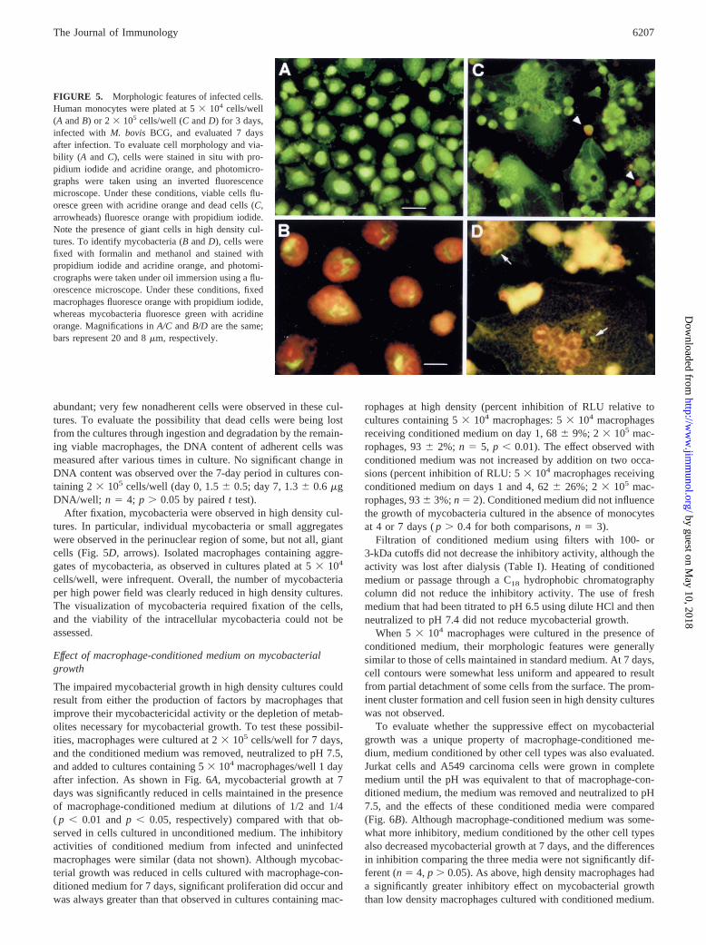
When monocytes were plated at 53 104 cells/well, the cells rap-idly formed a monolayer in which most cells were firmly attached
to the surface. After infection, most cells remained adherent, al-though a small number of cell clusters were observed. The cellsprogressively increased in size, producing an essentially confluentmonolayer at 7 days (Fig. 5A). At this time, essentially 100% of thecells were viable as assessed by their ability to exclude propidiumiodide. After fixation and staining, small clusters of mycobacteriacould be observed in most cells 7 days after infection (Fig. 5B).Although not quantified, the number of mycobacteria per cellclearly increased during the week after infection.
Several differences were observed when monocytes were platedat 2 3 105 cells/well. First, clusters of cells were prominent im-mediately after plating and increased in size after infection. Cellfusion, resulting in giant cell formation, was evident 4 days afterinfection; by 7 days after infection, large numbers of giant cellscontaining 5–15 nuclei were present (Fig. 5C). Despite the low pHof the extracellular medium,.90% of cells in high density cul-tures remained viable after 7 days of culture, although small num-bers of dead cells were always present (orange nuclei in Fig. 5C).Subsequently, the viability of high density cultures declined, and at10 days numerous dead cells were present. Cells with condensednuclei typical of apoptotic cells were observed but were never
FIGURE 3. Effect of MOI on the growth of mycobacteria in macro-phages cultivated at high and low density. Macrophages were cultivated at5 3 104 cells/well (left) or 2 3105 cells/well (right) for 3 days. For eachexperiment, serial 2-fold dilutions of the original suspension of theM.bovisBCG were prepared, and cells were infected with the indicated rel-ative number of mycobacteria. The number of mycobacteria indicated as“1” corresponds to the amount generally used in these studies (i.e., 3–53103 RLU/well corresponding to an MOI of;1:1 in cultures containing 53104 cells/well). Each group of three bars shows mycobacterial numberassessed by measurement of RLU after 1 day (left bar), 4 days (middlebar), and 7 days (right bar). Results are presented as the mean6 SD forthree experiments (mycobacterial numbers 1, 2, and 4) or one experiment(mycobacterial number 1/4). Cultures in which macrophages cultivated at5 3 104 and 23 105 cells/well were infected at the same MOI are indicatedby results shown with solid and hatched bars. FIGURE 4. Effect of extracellular pH on the growth of mycobacteria in
cultured macrophages.A, Macrophages were cultivated at 53 104 cells/well (M) or 2 3105 cells/well (f) for 3 days, infected withM. bovisBCG,and pH of the extracellular medium was measured at the indicated timesafter infection. Results are the mean6 SD for five independent experi-ments.B, Macrophages were cultivated at 53 104 cells/well (M) or 2 3105 cells/well (f) for 3 days and infected with theM. bovisBCG reporterstrain, and 1 day after infection medium was replaced with culture mediumadjusted to the indicated initial pH. Mycobacterial growth was assessed atthe indicated times by evaluation of luciferase activity. The final pH of theculture medium is indicated by the values inside the bars. Results are themean6 SD for three experiments performed using cells from differentindividuals.
6206 MACROPHAGE DENSITY AND MYCOBACTERIAL GROWTH
by guest on May 10, 2018
http://ww
w.jim
munol.org/
Dow
nloaded from
abundant; very few nonadherent cells were observed in these cul-tures. To evaluate the possibility that dead cells were being lostfrom the cultures through ingestion and degradation by the remain-ing viable macrophages, the DNA content of adherent cells wasmeasured after various times in culture. No significant change inDNA content was observed over the 7-day period in cultures con-taining 23 105 cells/well (day 0, 1.56 0.5; day 7, 1.36 0.6 mgDNA/well; n 5 4; p . 0.05 by pairedt test).
After fixation, mycobacteria were observed in high density cul-tures. In particular, individual mycobacteria or small aggregateswere observed in the perinuclear region of some, but not all, giantcells (Fig. 5D,arrows). Isolated macrophages containing aggre-gates of mycobacteria, as observed in cultures plated at 53 104
cells/well, were infrequent. Overall, the number of mycobacteriaper high power field was clearly reduced in high density cultures.The visualization of mycobacteria required fixation of the cells,and the viability of the intracellular mycobacteria could not beassessed.
Effect of macrophage-conditioned medium on mycobacterialgrowth
The impaired mycobacterial growth in high density cultures couldresult from either the production of factors by macrophages thatimprove their mycobactericidal activity or the depletion of metab-olites necessary for mycobacterial growth. To test these possibil-ities, macrophages were cultured at 23 105 cells/well for 7 days,and the conditioned medium was removed, neutralized to pH 7.5,and added to cultures containing 53 104 macrophages/well 1 dayafter infection. As shown in Fig. 6A, mycobacterial growth at 7days was significantly reduced in cells maintained in the presenceof macrophage-conditioned medium at dilutions of 1/2 and 1/4( p , 0.01 andp , 0.05, respectively) compared with that ob-served in cells cultured in unconditioned medium. The inhibitoryactivities of conditioned medium from infected and uninfectedmacrophages were similar (data not shown). Although mycobac-terial growth was reduced in cells cultured with macrophage-con-ditioned medium for 7 days, significant proliferation did occur andwas always greater than that observed in cultures containing mac-
rophages at high density (percent inhibition of RLU relative tocultures containing 53 104 macrophages: 53 104 macrophagesreceiving conditioned medium on day 1, 686 9%; 23 105 mac-rophages, 936 2%; n 5 5, p , 0.01). The effect observed withconditioned medium was not increased by addition on two occa-sions (percent inhibition of RLU: 53 104 macrophages receivingconditioned medium on days 1 and 4, 626 26%; 23 105 mac-rophages, 936 3%;n 5 2). Conditioned medium did not influencethe growth of mycobacteria cultured in the absence of monocytesat 4 or 7 days (p . 0.4 for both comparisons,n 5 3).
Filtration of conditioned medium using filters with 100- or3-kDa cutoffs did not decrease the inhibitory activity, although theactivity was lost after dialysis (Table I). Heating of conditionedmedium or passage through a C18 hydrophobic chromatographycolumn did not reduce the inhibitory activity. The use of freshmedium that had been titrated to pH 6.5 using dilute HCl and thenneutralized to pH 7.4 did not reduce mycobacterial growth.
When 53 104 macrophages were cultured in the presence ofconditioned medium, their morphologic features were generallysimilar to those of cells maintained in standard medium. At 7 days,cell contours were somewhat less uniform and appeared to resultfrom partial detachment of some cells from the surface. The prom-inent cluster formation and cell fusion seen in high density cultureswas not observed.
To evaluate whether the suppressive effect on mycobacterialgrowth was a unique property of macrophage-conditioned me-dium, medium conditioned by other cell types was also evaluated.Jurkat cells and A549 carcinoma cells were grown in completemedium until the pH was equivalent to that of macrophage-con-ditioned medium, the medium was removed and neutralized to pH7.5, and the effects of these conditioned media were compared(Fig. 6B). Although macrophage-conditioned medium was some-what more inhibitory, medium conditioned by the other cell typesalso decreased mycobacterial growth at 7 days, and the differencesin inhibition comparing the three media were not significantly dif-ferent (n5 4, p . 0.05). As above, high density macrophages hada significantly greater inhibitory effect on mycobacterial growththan low density macrophages cultured with conditioned medium.
FIGURE 5. Morphologic features of infected cells.Human monocytes were plated at 53 104 cells/well(A andB) or 2 3105 cells/well (CandD) for 3 days,infected with M. bovis BCG, and evaluated 7 daysafter infection. To evaluate cell morphology and via-bility (A and C), cells were stained in situ with pro-pidium iodide and acridine orange, and photomicro-graphs were taken using an inverted fluorescencemicroscope. Under these conditions, viable cells flu-oresce green with acridine orange and dead cells (C,arrowheads) fluoresce orange with propidium iodide.Note the presence of giant cells in high density cul-tures. To identify mycobacteria (BandD), cells werefixed with formalin and methanol and stained withpropidium iodide and acridine orange, and photomi-crographs were taken under oil immersion using a flu-orescence microscope. Under these conditions, fixedmacrophages fluoresce orange with propidium iodide,whereas mycobacteria fluoresce green with acridineorange. Magnifications inA/C andB/D are the same;bars represent 20 and 8mm, respectively.
6207The Journal of Immunology
by guest on May 10, 2018
http://ww
w.jim
munol.org/
Dow
nloaded from

Effect ofL-NIL on mycobacterial growth in macrophagescultured at high density
To evaluate whether NO production was required for the improvedmycobacteriostatic activity of macrophages cultured at high den-sity, the effect of theL-NIL, an inducible NO synthase inhibitor,was evaluated. The addition of 5mg/ml L-NIL on the day of in-fection did not increase mycobacterial growth at 7 days (RLU/wellat 7 days: control, 38706 1326;L-NIL, 4140 6 1878 RLU/well;p . 0.2). Increasing the concentration ofL-NIL to 125 mg/ml oradding 5mg/ml L-NIL every 48 h after infection also failed toincrease mycobacterial growth (data not shown).
Effect of macrophage maturity on the control of mycobacterialgrowth
In the experiments described thus far, monocytes were infectedafter 3 days of culture in vitro. To determine whether the matura-tion of these cells influenced the relationship between cell numberand their ability to control mycobacterial growth, monocytes fromthe same individuals cultured for 3 and 10 days before infectionwere compared. As described above, monocytes plated at 23 104
and 53 104 cells/well, but not cells plated at 23 105 cells/well,were permissive for mycobacterial growth (Fig. 7A). Monocytescultured for 10 days before infection exhibited progressive differ-entiation into large macrophage-like cells that were firmly adher-ent to the culture wells. Interestingly, when cultured at 23 104
cells/well, these macrophage-like cells were still permissive formycobacterial growth, but cultures containing 53 104 differenti-ated macrophages controlled mycobacterial growth as well as cul-tures containing 23 105 monocytes infected 3 days after isolation( p . 0.2 at 4 and 7 days comparing the two populations shown inFig. 7, A andB).
DiscussionThis study demonstrates that the ability of human monocyte-de-rived macrophages to control mycobacterial infection in vitro ishighly dependent on the density at which these cells are present.
FIGURE 6. Effect of conditioned medium on mycobacterial growth inhuman macrophages.A, One day after infection, medium was removed andreplaced with fresh medium (2) or the indicated dilution of macrophage-conditioned medium (e.g., medium recovered from macrophages main-tained at 23 105 cells/well for 7 days and neutralized to pH 7.5).B,Macrophages were cultivated at 53 104 cells/well (M,s, o, p) or 2 3105
cells/well (f) for 3 days and infected with theM. bovis BCG reporterstrain. One day after infection, medium was removed and replaced withfresh medium (2) or a 1/2 dilution of medium conditioned by macrophages(M), Jurkat cells (J), or A549 carcinoma cells (A). Mycobacterial growthwas assessed at the indicated times by evaluation of luciferase activity.Results are the mean6 SD for four experiments performed using cellsfrom different individuals.
Table I. Effect of pretreatment of macrophage-conditioned medium onits ability to inhibit mycobacterial growth in human macrophagesa
Pretreatment% Inhibition of
Mycobacterial Growth n
None 72.66 11.8 7
100-kDa filtration 2Filtrate 71.26 15.6Retentate 82.86 2.6
3-kDa filtration 2Filtrate 68.86 20.8Retentate 84.76 2.1
Dialysis 15.86 12.4 4Heating (95°C for 15 min) 70.2 1Hydrophobic chromatography (C18) 77.16 6.2 2
a Conditioned medium was obtained from macrophages cultivated for 7 days at2 3 105 cells/well and neutralized to pH 7.5. After the indicated pretreatments, a 1/2dilution of the conditioned medium was added to cultures containing 53 104 mac-rophages/well, 1 day after infection with theM. bovisBCG reporter strain. Results areexpressed as mean6 SD for the percent inhibition of mycobacterial growth comparedwith that of infected cultures not treated with conditioned medium.
FIGURE 7. Effect of maturation on the ability of macrophages to con-trol mycobacterial replication. Monocytes from a given donor were purifiedby elutriation and frozen in aliquots. Either 3 days before infection (A) or10 days before infection (B), aliquots were thawed and cultured at 23 104
(E), 5 3 104 (F), or 2 3 105 (f) cells/well. At the indicated times afterinfection, mycobacterial growth was assessed by evaluation of luciferaseactivity. Because of the high metabolic activity of differentiated macro-phages, cultures containing 23 105 cells/well had poor viability after 10days of culture, and mycobacterial growth could not be evaluated. Resultsare the mean6 SD for four experiments performed using cells from threeindividuals.
6208 MACROPHAGE DENSITY AND MYCOBACTERIAL GROWTH
by guest on May 10, 2018
http://ww
w.jim
munol.org/
Dow
nloaded from

When plated at a density that results in a single monolayer of cellsafter 7 days of culture, we found that monocytes were permissivefor growth of M. bovis BCG, findings consistent with previousreports (29, 30). When the same cells were plated at a 2- to 4-foldhigher density, however, mycobacterial growth was strongly in-hibited, and, in some experiments net mycobactericidal activitywas observed. These findings demonstrate that human monocyteshave the intrinsic capacity to develop in vitro into cells capable ofcontrolling mycobacterial infection.
The role of a number of possible mechanisms through whichchanges in cell density could influence mycobactericidal activitywere investigated. Cell density did not change mycobacterial up-take by the cultured macrophages. Furthermore, the effect of mac-rophage density on mycobacterial growth was insensitive to themultiplicity of infection over a 16-fold range. Thus, neither ofthese factors could explain the observed effect.
The control of intraphagosomal pH is also thought to be animportant determinant of mycobacterial survival in macrophages.Mycobacterial growth can be directly influenced by an acidic en-vironment, and acidification can increase their sensitivity to oxi-dant-mediated killing (31, 32). Virulent mycobacteria have beenreported to inhibit the acidification of phagosomes through exclu-sion of vesicular proton-ATPase complexes (33). Acidification ofthe extracellular milieu can also decrease the intracellular pH ofmacrophages (34), and we found that macrophages cultured at highdensity did acidify the extracellular medium to a pH as low as 6.5after 7 days. Several observations suggested, however, that thischange did not account for improved mycobacteriostasis. First,despite acidification of the culture medium to similar levels, mac-rophages cultured at lower density remained permissive for my-cobactericidal growth. In addition, the viability of high densitymacrophages remained excellent despite the low extracellular pH,suggesting that these cells maintained a physiologic intracellularpH via energy-dependent transport mechanisms. Similarly, thebactericiostatic activity of macrophages cultured at high densitycould not be attributed to the preferential generation of NO bythese cells, because treatment with the inducible NO synthase in-hibitor L-NIL did not influence their ability to control mycobacte-rial replication. Consistent with these observations, prior studieshave shown that although human macrophages express inducibleNO synthase after mycobacterial infection (35, 36), the levels ofNO produced are low, and the inhibition of NO production byhuman macrophages does not reproducibly increase mycobacterialgrowth (36, 37). Increased apoptosis of macrophages infected withmycobacteria has been described and in some but not all cases hasbeen associated with mycobacterial killing (38–41). In this study,the viability of macrophages cultured at high density was excellentat 7 days, few detached cells were present, and the DNA contentof the cultures was stable during the 7-day culture period. Thesefindings indicate that macrophage apoptosis or ingestion of in-fected apoptotic cells by neighboring uninfected macrophages isunlikely to account for the reduced mycobacterial growth seen inhigh density cultures.
Two observations in our study suggested that the differentiatedstate of macrophages have an important bearing on their ability tocontrol mycobacterial infection and that both the time in cultureand the density of cells present could influence this differentiationprocess. The mycobacteriostatic activity of both freshly isolatedmonocytes and macrophages that had been cultured for 10 daysbefore infection was dependent on the number of cells present inthe cultures, but fewer of the more differentiated macrophage-likecells were required to control mycobacterial proliferation. Priorstudies have shown that the maturation in vitro of human mono-cytes before infection can modify their capacity to control myco-
bacterial replication. In these studies, however, an increase (42), aprogressive decrease (43), or an initial decrease followed by anincrease (44) in the permissiveness for mycobacterial infection hasbeen observed as a function of the time in culture. The importanteffect of cell density was not evaluated in these studies and mayaccount, at least in part, for the discrepant findings. We also ob-served that the morphologic features of monocytes cultured at highdensity were strikingly different from those of cells cultured at lowdensity. In high density cultures, macrophages were larger, cellaggregates were prominent during the first few days of culture, andlater times the progressive appearance of giant cells was observed.These changes occurred to a similar extent in uninfected cultures,indicating that mycobacterial infection was not responsible. Byrd(45) previously observed an association between giant cell forma-tion and the ability of cultured monocytes to inhibit mycobacterialproliferation. As in his study, we observed that mycobacteria ac-cumulated in the perinuclear area of giant cells, which may restrictthe invasion of adjacent cells (45). A number of signals have beendescribed that increase giant cell formation in cultured macro-phages (46–48). Conditioned medium from high density macro-phage cultures did not induce giant cell formation in low densitycultures, suggesting that paracrine stimulation by cytokines wasnot sufficient for the induction of giant cell formation seen in thesestudies. Culture of cells at high density resulted in the early for-mation of cell aggregates, compatible with the idea that prolongedmembrane contact may have been important.
It should be emphasized that the relationship between increasedmycobacteriostatic activity and macrophage differentiation ob-served in these studies does not prove that the processes are caus-ally related. Furthermore, giant cell formation was not a feature ofeither low density cultures treated with conditioned medium ormacrophages cultured at 53 104 cells/well for 10 days beforeinfection, although these populations were able, respectively, topartially or completely inhibit mycobacterial replication. Thus, thistype of differentiation, although possibly important, was not indis-pensable for improved mycobacteriostatic activity. Future studiesevaluating the effect of inhibiting macrophage differentiation onmycobacteriostatic activity may prove informative. In this regard,we have recently found that when monocytes are cultured withtype I IFNs, they persist as small “monocyte-like” cells for up to7 days. Under these conditions, monocytes are very permissive tomycobacterial growth, even when cultured at high density (F. Bou-chonnet, N. Boechat, M. Bonay, M. Vokurka, and A. J. Hance,manuscript in preparation).
An interesting finding in the current studies was the observationthat when low density macrophages were maintained in the pres-ence of medium conditioned by high density macrophages, intra-cellular mycobacterial growth was reduced. This finding is com-patible with the possibility that high density cultures releasefactor(s) that can stimulate mycobacteriostatic activity or depletemetabolites necessary for mycobacterial growth. The observationthat medium conditioned by other cell types also decreased my-cobacterial growth in low density macrophages suggests that thiseffect is not specific for macrophages. Furthermore, modificationof the extracellular milieu did not appear to completely explain theincreased mycobacteriostatic activity of cells cultured at high den-sity, because low density macrophages cultured in conditioned me-dium inhibited mycobacterial growth less well than macrophagescultured at high density, and cells cultured in conditioned medium,unlike high density cultures, did not inhibit mycobacterial growthat 4 days.
The ability of conditioned medium to enhance mycobacteriosta-tic activity was not decreased by heating, passage through a mem-brane with a 3-kDa cutoff or passage through a C18 hydrophobic
6209The Journal of Immunology
by guest on May 10, 2018
http://ww
w.jim
munol.org/
Dow
nloaded from

column, whereas the activity was completely lost by dialysis. Inaddition, the 10-fold concentrated retentate obtained by ultrafiltra-tion of conditioned medium did not contain increased activity.These findings argue strongly against a role for soluble secretedproteins or lipid mediators in this phenomenon. Thus, althoughcultured macrophages are secrete large quantities of cytokines, in-cluding IFN-g, TNF-a, and IL-12 (22, 23, 30, 49, 50), our resultsare compatible with prior studies indicating that these cytokinesare not sufficient to induce strong mycobactericidal activity in oth-erwise permissive human macrophages. Low molecular mass sub-stances produced by macrophages, and other cell types can modifythe activity of macrophages and improve their resistance to intra-cellular pathogens, (e.g., ATP and nonheme iron) (20, 51), and therelease of such agents could explain our findings. Attempts to iso-late a putative stimulatory factor by ion exchange chromatographywere unsuccessful, but the buffers required for these studies provedto be toxic for macrophages, which complicated this endeavor(data not shown). Alternatively, it is the possibility that substancesthat deactivate macrophages or that are required for optimal my-cobacterial growth had been depleted by the metabolically activecells (e.g., iron, adenosine, amino acids) (52–56). The activity ofconditioned medium was completely lost after dialysis against se-rum-free medium, indicating that the culture medium, not humanserum, would have to be the source of such a metabolite. Further-more, because conditioned medium diluted 4-fold with fresh me-dium retained some activity, this metabolite would have to bepresent in suboptimal amounts in fresh medium. Further studieswill be required to identify the substances responsible for the ef-fects observed with conditioned medium and to determine whethermacrophages within granulomas can “condition” their environ-ment in a similar fashion.
Taken together, these results indicate that the ability of humanmacrophages to control mycobacterial infection in vitro is stronglyinfluenced by the density of cells present and indicate that celldensity and the time in culture before infection can influence thedifferentiated state of these cells and their capacity to modify theexternal milieu. The requirement for both monocytes and moremature macrophages to be present at a critical density to suppressmycobacterial proliferation offer insights into why the formation ofgranulomas in vivo appears to be a key event in the control ofmycobacterial infections. The availability of this model, in whichthe same cells are either permissive or resistant to mycobacterialinfection, should be useful in further characterizing the cellularevents that are indispensable for the development of mycobacte-ricidal activity.
AcknowledgmentsWe thank Dr. Sitthy (Hopital St. Louis, Paris, France) for help in obtainingnormal human leukocytes.
References1. Havlir, D. V., and P. F. Barnes. 1999. Tuberculosis in patients with human im-
munodeficiency virus infection.N. Engl. J. Med. 340:367.2. Flynn, J. L., M. M. Goldstein, K. J. Triebold, B. Koller, and B. R. Bloom. 1992.
Major histocompatibility complex class I-restricted T cells are required for re-sistance toMycobacterium tuberculosisinfection. Proc. Natl. Acad. Sci. USA89:12013.
3. Ladel, C. H., S. Daugelat, and S. H. E. Kaufmann. 1995. Immune response toMycobacterium bovisbacille Calmette Guerin infection in major histocompati-bility complex class I- and II-deficient knock-out mice: contribution of CD4 andCD8 T cells to acquired resistance.Eur. J. Immunol. 25:377.
4. Caruso, A. M., N. Serbina, E. Klein, K. Triebold, B. R. Bloom, and J. L. Flynn.1999. Mice deficient in CD4 T-cells have only transiently diminished levels ofIFN-g, yet succumb to tuberculosis.J. Immunol. 162:5407.
5. Flynn, J. L., J. Chan, K. J. Triebold, D. K. Dalton, T. A. Stewart, andB. R. Bloom. 1993. An essential role for interferong in resistance toMycobac-terium tuberculosisinfection.J. Exp. Med. 178:2249.
6. Cooper, A. M., D. K. Dalton, T. A. Stewart, J. P. Griffin, D. G. Russell, andI. M. Orme. 1993. Disseminated tuberculosis in interferong gene-disrupted mice.J. Exp. Med. 178:2243.
7. Kamijo, R., J. Le, D. Shapiro, E. A. Havell, S. Huang, M. Aguet, M. Bosland, andJ. Vilcek. 1993. Mice that lack the interferon-g receptor have profoundly alteredresponses to infection with bacillus Calmette-Guerin and subsequent challengewith lipopolysaccharide.J. Exp. Med. 178:1435.
8. Flynn, J. L., M. M. Goldstein, J. Chan, K. J. Triebold, K. Pfeffer,C. J. Lowenstein, R. Schreiber, T. W. Mak, and B. R. Bloom. 1995. Tumornecrosis factor-a is required in the protective immune response againstMyco-bacterium tuberculosisin mice. Immunity 2:561.
9. Bean, A. G. D., D. R. Roach, H. Briscoe, M. P. France, H. Korner,J. D. Sedgwick, and W. J. Britton. 1999. Structural deficiencies in granulomaformation in TNF gene-targeted mice underlie the heightened susceptibility toaerosolMycobacterium tuberculosisinfection, which is not compensated for bylymphotoxin.J. Immunol. 162:3504.
10. Cooper, A. M., J. Magram, J. Ferrante, and I. M. Orme. 1997. Interleukin 12(IL-12) is crucial to the development of protective immunity in mice intrave-nously infected withMycobacterium tuberculosis. J. Exp. Med. 186:39.
11. Sugawara, I., H. Yamada, H. Kaneko, S. Mizuno, K. Takeda, and S. Akira. 1999.Role of interleukin-18 (IL-18) in mycobacterial infection in IL-18-gene-disruptedmice. Infect. Immun. 67:2585.
12. Newport, M. J., C. M. Huxley, S. Huston, C. M. Hawrylowicz, B. A. Oostra,R. Williamson, and M. Levin. 1996. A mutation in the interferon-g-receptor geneand susceptibility to mycobacterial infection.N. Engl. J. Med. 335:1941.
13. Jouanguy, E., F. Altare, S. Lamhamedi, P. Revy, J. F. Emile, M. Newport,M. Levin, S. Blanche, E. Seboun, A. Fischer, and J.-L. Casanova. 1996. Inter-feron-g-receptor deficiency in an infant with fatal bacille Calmette-Guerin infec-tion. N. Engl. J. Med. 335:1956.
14. Altare, F., A. Durandy, D. Lammas, J.-F. Emile, S. Lamhamedi, F. Le Deist,P. Drysdale, E. Jouanguy, R. Doffinger, F. Bernaudin, et al. 1998. Impairment ofmycobacterial immunity in human interleukin-12 receptor deficiency.Science280:1432.
15. de Jong, R. F. Altare, I.-A. Haagen, D. G. Elferink, T. de Boer,P. J. C. van BredaVriesman, P. J. Kabel, J. M. T. Draaisma, J. T. van Dissel,F. P. Kroon, et al. 1998. Severe mycobacterial andSalmonellainfections in in-terleukin-12 receptor-deficient patients.Science 280:1435.
16. Flesch, I. E., and S. H. E. Kaufmann. 1990. Activation of tuberculostatic mac-rophage functions byg interferon, interleukin-4, and tumor necrosis factor.Infect.Immun. 58:2675.
17. Chan, J., K. Tanaka, D. Carroll, J. Flynn, and B. R. Bloom. 1993. Effects of nitricoxide synthase inhibitors on murine infection withMycobacterium tuberculosis.Infect. Immun. 63:736.
18. MacMicking, J., R. J. North, R. Lacourse, R. S. Mudgett, S. Shah, andC. F. Nathan. 1997. Identification of NOS2 as a protective locus against tuber-culosis.Proc. Natl. Acad. Sci. USA 94:5243.
19. Bonay, M., F. Bouchonnet, V. Pelicic, B. Lagier, M. Grandsaigne, D. Lecossier,A. Grodet, M. Vokurka, B. Gicquel, and A. J. Hance. 1999. Effect of stimulationof human macrophages on intracellular survival ofMycobacterium bovisbacillusCalmette-Guerin: evaluation with a mycobacterial reporter strain.Am. J. Respir.Crit. Care Med. 159:1629.
20. Byrd, T. F. 1997. Tumor necrosis factora (TNFa) promotes growth of virulentMycobacterium tuberculosisin human monocytes. Iron-mediated growth sup-pression is correlated with decreased release of TNFa from iron-treated infectedmonocytes.J. Clin. Invest. 99:2518.
21. Warwick-Davies, J., J. Dhillon, L. O’Brien, P. W. Andrew, and D. B. Lowrie.1994. Apparent killing ofMycobacterium tuberculosisby cytokine-activated hu-man monocytes can be an artifact of a cytotoxic effect on the monocytes.Clin.Exp. Immunol. 96:214.
22. Silver, R. F., Q. Li, W. H. Boom, and J. J. Ellner. 1998. Lymphocyte-dependentinhibition of growth of virulentMycobacterium tuberculosisH37Rv within hu-man monocytes: requirement for CD41 T cells in purified protein derivative-positive, but not in purified protein derivative-negative subjects.J. Immunol.160:2408.
23. Bonecini-Almeida, M., S. Chitale, I. Boutsikakis, J. Geng, H. Doo, Suhui He, andJ. L. Ho. 1998. Induction of in vitro human macrophage anti-Mycobacteriumtuberculosisactivity: requirement for IFN-g and primed lymphocytes.J. Immu-nol. 160:4490.
24. Yoneda, T., and J. J. Ellner. 1998. CD41 T cell and natural killer cell-dependentkilling of Mycobacterium tuberculosisby human monocytes.Am. J. Respir. Crit.Care Med. 158:395.
25. Kindler, V., A.-P. Sapppino, G. E. Grau, P.-F. Piguet, and P. Vassalli. 1989. Theinducing role of tumor necrosis factor in the development of bactericidal gran-ulomas during BCG infection.Cell 56:7.31.
26. Wakeham, J., J. Wang, J. Magram, K. Croitoru, R. Harkness, P. Dunn,A. Zganiacz, and Z. Xing. 1998. Lack of both types 1 and 2 cytokines, tissueinflammatory responses, and immune protection during pulmonary infection byMycobacterium bovisbacille Calmette-Guerin in IL-12-deficient mice.J. Immu-nol. 160:6101.
27. Wahl, L. M., I. L. Katona, R. L. Wilder, C. C. Winter, B. Haroui, I. Seher, andS. M. Wahl. 1984. Isolation of human mononuclear cell subsets by counterflowcentrifugal elutriation (CEE). I. Characterization of B-lymphocyte, T-lymphocyteand monocyte-enriched fractions by flow cytometric analysis.Cell. Immunol.85:373.
28. Labarca, C., and K. Paigen. 1980. A simple, rapid, and sensitive DNA assayprocedure.Anal. Biochem. 102:344.
6210 MACROPHAGE DENSITY AND MYCOBACTERIAL GROWTH
by guest on May 10, 2018
http://ww
w.jim
munol.org/
Dow
nloaded from

29. Molloy, A., P. A. Meyn, K. D. Smith, and G. Kaplan. 1993. Recognition andestruction of bacillus Calmette-Guerin-infected human monocytes.J. Exp. Med.177:1691.
30. Silver, R. F., Q. Li, and J. J. Ellner. 1998. Expression of virulence ofMycobac-terium tuberculosiswithin human monocytes: virulence correlates with intracel-lular growth and induction of tumor necrosis factora but not with evasion oflymphocyte-dependent monocyte effector functions.Infect. Immun. 66:1190.
31. Jackett, P. S., V. R. Aber, and D. B. Lowrie. 1978. Virulence and resistance tosuperoxide, low pH and hydrogen peroxide among strains ofMycobacteriumtuberculosis. J. Gen. Microbiol. 104:37.
32. Piddington, D. L., A. Kashkouli, and N. A. Buchmeier. 2000. Growth ofMyco-bacterium tuberculosisin a defined medium is very restricted by acid pH andMg21 levels.Infect. Immun. 68:4518.
33. Sturgill-Koszycki, S., P. H. Schlesinger, P. Chakrabgorty, P. L. Haddix,H. L. Collins, A. K. Fok, R. D. Allen, S. L. Gluck, J. Heuser, and D. G. Russell.1994. Lack of acidification inMycobacteriumphagosomes produced by exclu-sion of the vesicular proton-ATPase.Science 263:678.
34. Mellergard, P., Y. Ou-Yang, and B. K. Siesjo. 1994. Relationship between intra-and extracellular pH in primary cultures of rat astrocytes.Am. J. Physiol. 267:C581.
35. Nicolson, S., M. Bonecini- Aleida, J. R. Lapa e Silva, C. Nathan, Q. Xie,R. Mumford, J. R. Weidner, J. Calaycay, J. Geng, N. Boechat, et al. 1996. In-ducible nitric oxide synthase in pulmonary alveolar macrophages from patientswith tuberculosis.J. Exp. Med. 183:2293.
36. Jagannath, C., J. K. Actor, and R. L. Hunter, Jr. 1998. Induction of nitric oxidein human monocytes and monocyte cell lines byMycobacterium tuberculosis.Nitric Oxide 2:174.
37. C. Aston, W. N. Rom, A. T. Talbot, and J. Reibman. 1998. Early inhibition ofmycobacterial growth by human alveolar macrophages is not due to nitric oxide.Am. J. Respir Crit. Care Med. 157:1943.
38. Molloy, A., P. Laochumroonvorapong, and G. Kaplan. 1994. Apoptosis, but notnecrosis, of infected monocytes is coupled with killing of intracellular bacillusCalmette-Guerin.J. Exp. Med. 180:1499.
39. Laochumroonvorapong, P., S. Paul, K. B. Elkon, and G. Kaplan. 1996. H2O2
induces monocyte apoptosis and reduces viability ofMycobacterium avium-M.intracellulare within cultured human monocytes.Infect. Immun. 64:452.
40. Fratazzi, C., R. D. Arbeit, C. Carini, and H. G. Remold. 1997. Programmed celldeath ofMycobacterium aviumserovar 4-infected human macrophages preventsthe mycobacteria from spreading and induces mycobacterial growth inhibition byfreshly added, uninfected macrophages.J. Immunol. 158:4320.
41. Keane, J., H. G. Remold, and H. Kornfeld. 2000. VirulentMycobacterium tu-berculosisstrains evade apoptosis of infected alveolar macrophages.J. Immunol.164:2016.
42. Carvalho de Sousa, J. P., and N. Rastogi. 1992. Comparative ability of humanmonocytes and macrophages to control the intracellular growth ofMycobacte-rium avium and Mycobacterium tuberculosis: effect of interferon-g and indo-methacin.FEMS Microbiol. Immunol. 4:329.
43. Paul, S., P. Laochumroonvorapong, and G. Kaplan. 1996. Comparable growth ofvirulent and avirulentMycobacterium tuberculosisin human macrophages invitro. J. Infect. Dis. 174:105.
44. Douvas, G. S., E. M. Berger, J. E. Repine, and A. J. Crowle. 1986. Naturalmycobacteriostatic activity in human monocyte-derived adherent cells.Am. Rev.Respir. Dis. 134:44.
45. Byrd, T. F. 1998. Multinucleated giant cell formation induced by IFN-g/IL-3 isassociated with restriction of virulentMycobacterium tuberculosiscell to cellinvasion in human monocyte monolayers.Cell. Immunol. 188:89.
46. Enelow, R. I., G. W. Sullivan, H. T. Carper, and G. L. Mandell. 1992. Inductionof multinucleated giant cell formation from in vitro culture of human monocyteswith interleukin-3 and interferon-g: comparison with other stimulating factors.Am. J. Respir. Cell Mol. Biol. 6:57.
47. Ohgimoto, S., N. Tabata, S. Suga, M. Nishio, H. Ohta, M. Tsurudome,H. Komada, M. Kawano, N. Watanabe, and Y. Ito. 1995. Molecular character-ization of fusion regulatory protein-1 (FRP-1) that induces multinucleated giantcell formation of monocytes and HIV gp160-mediated cell fusion. FRP-1 and4F2/CD98 are identical molecules.J. Immunol. 155:3585.
48. DeFife, K. M., C. R. Jenney, A. K. McNally, E. Colton, and J. M. Anderson.1997. Interkeukin-13 induces human monocyte/macrophage fusion and macro-phage mannose receptor expression.J. Immunol. 158:3385.
49. Zhang, M., J. Gong, Y. Lin, and P. F. Barnes. 1998. Growth of virulent andavirulentMycobacterium tuberculosisstrains in human macrophages.Infect. Im-mun. 66:794.
50. Wallis, R. S., and J. J. Ellner. 1994. Cytokines and tuberculosis.J. LeukocyteBiol. 55:676.
51. Lammas, D. A., C. Stober, C. J. Harvey, N. Kendrick, S. Panchalingam, andD. S. Kumararatne. 1997. ATP-induced killing of mycobacteria by human mac-rophages is mediated by purinergic P2Z(P2X7) receptors.Immunity 7:433.
52. Douvas, G. S., M. H. May, J. R. Pearson, E. Lam, L. Miller, and N. Tsuchida.1994. Hypertriglyceridemic serum, very low density lipoprotein, and iron en-hanceMycobacterium aviumreplication in human macrophages.J. Infect. Dis.170:1248.
53. Xaus, J., M. Mirabet, J. Lloberas, C. Soler, C. Lluis, R. Franco, and A. Celada.1999. IFN-g up-regulates the A2B adenosine receptor expression in macrophages:a mechanism of macrophage deactivation.J. Immunol. 162:3607.
54. Pfefferkorn, E. 1984. Interferon-g blocks the growth ofToxoplasma gondiiinhuman fibroblasts by inducing the host cell to degrade tryptophan.Proc. Natl.Adad. Sci. USA 81:908.
55. Taylor, M. W., and G. Feng. 1991. Relationship between interferon-g, indoleam-ine 2,3-dioxygenase, and tryptophan catabolism.FASEB J. 5:2516.
56. Harth, G., and M. A. Horwitz. 1999. An inhibitor of exportedMycobacteriumtuberculosisglutamine synthetase selectively blocks the growth of pathogenicmycobacteria in anexic culture and in human monocytes: extracellular proteins aspotential novel durg targets.J. Exp. Med. 189:1425.
6211The Journal of Immunology
by guest on May 10, 2018
http://ww
w.jim
munol.org/
Dow
nloaded from