culturable diversity of halophilic bacteria in foreshore soils · culturable diversity of...
TRANSCRIPT

Culturable diversity of halophilic bacteria in foreshore soils
Aarzoo Irshad1, Irshad Ahmad2, Seung Bum Kim1
1Department of Microbiology, School of Bioscience and Biotechnology,
Chungnam National University, Yuseong, Daejeon, Republic of Korea.2Biology Department, King Fahd University of Petroleum and Minerals,
Dhahran, Saudi Arabia.
Submitted: January 15, 2013; Approved: September 9, 2013.
Abstract
Halophilic bacteria are commonly found in natural environments containing significant concentra-
tion of NaCl such as inland salt lakes and evaporated sea-shore pools, as well as environments such as
curing brines, salted food products and saline soils. Dependence on salt is an important phenotypic
characteristic of halophilic bacteria, which can be used in the polyphasic characterization of newly
discovered microorganisms. In this study the diversity of halophilic bacteria in foreshore soils of
Daecheon, Chungnam, and Saemangeum, Jeonbuk, was investigated. Two types of media, namely
NA and R2A supplemented with 3%, 5%, 9%, 15%, 20% and 30% NaCl were used. More than 200
halophilic bacteria were isolated and BOX-PCR fingerprinting analysis was done for the typing of
the isolates. The BLAST identification results showed that isolated strains were composed of 4 phyla,
Firmicutes (60%), Proteobacteria (31%), Bacteriodetes (5%) and Actinobacteria (4%). Isolates were
affiliated with 16 genera and 36 species. Bacillus was the dominant genus in the phylum Firmicutes,
comprising 24% of the total isolates. Halomonas (12%) and Shewanella (12%) were also found as the
main genera. These findings show that the foreshore soil of Daecheon Beach and Saemangeum Sea
of Korea represents an untapped source of bacterial biodiversity.
Key words: culturable diversity, halophilic bacteria, foreshore soil.
Introduction
Halophiles are basically salt-loving organisms that
inhabit hypersaline environments. Halophilic bacteria can
be classified according to their salt requirement and growth
pattern. Slight halophiles show optimum growth at 2-5%
NaCl, moderate halophiles at 5-20% NaCl, and extreme
halophiles show 20-30% NaCl respectively. Many halo-
philes and halotolerant microorganisms can grow over a
wide range of salt concentrations with occasionally de-
pending on environmental and nutritional factors for the
growth and tolerance (DasSarma, 2001; DasSarma and
Arora, 2002; Das et al., 2006).
Moderately halophilic bacteria are a group of halo-
philic microorganisms able to grow optimally in media
containing 5-20% NaCl (Ventosa et al., 1998). Apart from
their ecological importance, moderately halophilic bacteria
have great potential for its use in biotechnology.
Moderately halophilic bacteria form a diverse group of mi-
croorganisms adapted to the hypersaline environments.
Most species of moderately halophilic bacteria keep their
intracellular ionic concentrations at low levels while syn-
thesizing or accumulating organic solutes to provide os-
motic equilibrium of the cytoplasm with the surrounding
medium (De Bruijn, 1992; Ghasemi et al., 2011a; 2011b;
Oren, 1999). Some species of moderately halophilic bacte-
ria are used for the degradation of polluting industrial resi-
dues or toxic chemicals and for enhanced oil-recovery
processes (Birbir and Ilgaz, 1996; Ventosa and Nieto,
1995). Moreover, moderately halophilic bacteria produce
extracellular salt tolerant enzymes of great interest for bio-
technological processes (Birbir and Ilgaz, 1996; Li and Yu,
2012; Onishi et al., 1991; Shafiei et al., 2012). Extracellular
proteins, such as those secreted into the medium and proba-
Brazilian Journal of Microbiology 45, 2, 563-571 (2014) Copyright © 2014, Sociedade Brasileira de Microbiologia
ISSN 1678-4405 www.sbmicrobiologia.org.br
Send correspondence to I. Ahmad. Department of Biotechnology, Sarhad University of Science and Information Technology, Peshawar, Khyber
Pakhtunkhwa, Pakistan. E-mail: [email protected].
Research Paper

bly those in the periplasmic space, are exposed to external
saline environments and must adapt to variable salinities.
They constitute a heterogeneous group of microorganisms
including species belonging to various genera, such as
Halomonas and Salinivibrio, which have been studied with
respect to their ecology, physiology, biochemistry and ge-
netics (Ventosa et al., 1998; Ventosa, 2006).
Extremely halophilic bacteria grow optimally at salt
concentrations from above 20% (w/v) to saturation (Ven-
tosa, 2006). They have the capability of producing hydro-
lytic enzymes such as amylase and lipase. The potential
importance of extremely halophiles in various industrial ar-
eas such as the leather industry and food preservation is evi-
dent (Birbir and Ilgaz, 1996; Mellado and Ventosa, 2003).
Halophilic bacteria provide a high potential for bio-
technological applications for at least two reasons: 1. their
activities in natural environments with regard to their par-
ticipation in biogeochemical processes of C, N, S, and P,
the formation and dissolution of carbonates, the immobili-
zation of phosphate, and the production of growth factors
and nutrients and 2. their nutritional requirements are sim-
ple. Most of them can grow at high salt concentrations,
minimizing the risk of contamination. Moreover, several
genetic tools developed for the non halophilic bacteria can
be applied to the halophiles, and hence their genetic manip-
ulation seems feasible (Rodriguez-Valera, 1993; Ventosa
et al., 1998).
Current research is focused on microorganisms
adapted to live in saline environment. Foreshore soil is en-
riched with salts and nutrients which provide a conducive
environment for the growth of halophilic microorganisms.
Therefore samples were collected from the foreshore soil
(Daecheon Beach, Chungnam Province and Saemangeum
Sea, Jeonbuk Province in South Korea) to determine the
culturable bacterial diversity associated with marine envi-
ronment.
Materials and Methods
The chemical reagents used in this research work
were of analytical grade and purchased from Sigma,
Merck, Acros and Fisher scientific chemical companies.
Collection of samples
The sampling sites are located in Chungnam and
Jeonbuk Provinces in Korea. In Chungnam Province, the
samples were collected from the foreshore soil of Dae-
cheon Beach (Site A). In Jeonbuk Province samples were
collected from the saline sea water (Site B) and foreshore
soil of Saemangeum Sea (Site C). 25 samples each from sa-
line sites A and C were collected at 10 cm depth with the
help of sterilized soil corers and were transferred individu-
ally into sterile tubes (50 mL, Corning, USA). Similarly
25 samples from site B were collected 10 cm below the wa-
ter surface with 50 mL sterile pipettes and transferred to
200 mL sterile bottles. The collected samples were put in an
icebox, and then transported to the laboratory and pro-
cessed immediately.
Isolation of halophilic bacteria
The procedure of heat treatment used was previously
described by (Cho et al., 2006; Seong, 1992). For the isola-
tion of halophilic bacteria, 10 mL of sterile 1/4 strength
Ringer’s solution (2.25 g sodium chloride, 0.105 g potas-
sium chloride, 0.12 g calcium chloride, and 0.05 g sodium
bicarbonate in 1 l distilled water) was added to 1 gram soil
sample. The tenfold diluents were shaken on reciprocal
shaker for 20 min and then incubated at 50 °C for 15 min.
Aliquots (0.2 mL) of each dilution were spread onto NA
medium contained (g/L): beef extract (3.0), peptone (5.0),
agar (15.0) and R2A medium contained (g/L): yeast extract
(0.5), proteose peptone (0.5), casamino acids (0.5), dex-
trose (0.5), soluble starch (0.5), sodium pyruvate (0.3),
dipotassium phosphate (0.3), magnesium sulphate (0.05),
agar (15.0). The NA and R2A media plates were supple-
mented with 3%, 5%, 9%, 15%, 20% and 30% NaCl. Inocu-
lated plates were incubated at 30 °C for at least one week.
The colonies of halophilic bacteria were selected and
subcultured using the same media.
DNA extraction and purification
DNA was extracted from the halophilic bacteria using
the following protocol. The bacterial cells were taken into
an Eppendorf tube containing 100 �L of STES buffer
(500 mM NaCl, 200 mM Tris-Cl (pH 7.6) and 10 mM
EDTA, 1% SDS) and glass beads. The mixture was vor-
texed for 5 min using a TOMY micro tube mixer (TOMY,
Japan) and then 200 �L of TE buffer (pH 8.0) was added.
DNA was purified through phenol/chloroform/isoamyl al-
cohol (25:24:1) extraction. RNA was removed by RNase A
treatment at 37 °C for 3 h. The purified DNA was precipi-
tated with 0.1 volumes of 3 M sodium acetate and 2 vol-
umes of cold 95% ethanol, and then centrifuged at 12,000 g
for 20 min at room temperature. The supernatant was re-
moved and the pellet was washed with 70% ethanol, dried
in air and then resuspended in 50 �L of TE buffer. The re-
suspended DNA was stored at -20 °C until required.
Box-PCR analysis
The 22-mer BOXAIR primer
(5’-CTACGGCAAGGCGACGCTGACG-3’) was used to
generate BOX-PCR profiles (10, 24). One PCR reaction
(reaction volume 25 �L) contained 2.5 �L 10x buffer, 2 �L
dNTP, 3 �L DMSO 100% , 2 �L primer (BOXA1R,
0.3 �g �L–1, Pharmacia), 0.25 �L Taq polymerase and 1 �L
purified DNA.
The Box-PCR consisted of an initial denaturation step
at 94 °C for 7 min, which was followed by 35 cycles of
94 °C for 1 min, 60 °C for 1 min, 65 °C for 8 min, and then
three cycles at 65 °C for 16 min. The PCR amplification
564 Irshad et al.

products were detected by electrophoresis on 2% agarose
gels in 0.5x Tris-borate-EDTA (TBE) buffer, which were
stained with ethidium bromide (EtBr 1.25 mg/L), visual-
ized under UV light and the image was printed.
PCR amplification and sequencing of 16S rDNA
PCR amplification of 16S rDNA was carried out us-
ing two universal primers, 27f
(5’-AGAGTTTGATCMTGGCTCAG-3’) and 1492r
(5’-GGYTACCTTGTTACGACTT-3’), which were also
used for sequencing. The PCR consisted of an initial dena-
turation step at 95 °C for 3 min, which was followed by
30 cycles of 95 °C for 1 min, 55 °C for 40 min and 72 °C for
1 min. Sequencing was performed using the service of
Solgent Co. (Korea).
Phylogenetic analysis
The 16S rDNA sequences of the isolates were com-
pared with the sequences available by the BLAST search in
the NCBI, GenBank database
(http://www.ncbi.nlm.nih.gov). The sequences were proof-
read, edited and merged into comparable sequences using
the PHYDIT program version 3.0 (available at
http://plaza.snu.ac.kr/~jchun/phydit/). The sequences were
aligned together with those of reference taxa retrieved from
public databases. The evolutionary distances were gener-
ated based on parameter model (Jukes and Cantor, 1969)
and phylogenetic trees were constructed by using the
neighbor-joining method (Saitou and Nei, 1987).
Results
Isolation and viable counts of halophilic bacteria
For the specific isolation of halophilic bacteria, NA
and R2A media supplemented with 3%, 5%, 9%, 15%, 20%
and 30% NaCl were used. Maximum number of halophilic
bacterial colonies was observed on 3%, 5% and 9% NaCl
media plates. The highest CFU counts (2.3x10⁸, 1.8x10⁸,
and 1.1 x 10⁸ CFU/g) were obtained from 3%, 5% and 9%
NaCl respectively (Figure 1). On 15% and 20% NaCl less
growth of halophilic bacteria was obtained. There was no
growth on 30% NaCl media plates. Our isolates did not
show any growth on media without NaCl (data not shown).
Most halophilic bacteria were isolated from site C (112 iso-
lates) followed by site A (66 isolates), and relatively less
number of halophilic bacteria were isolated from site B
(22 isolates). Among 200 isolates 108 were obtained on
R2A medium while 92 were obtained on NA medium.
These results showed that site C (Saemangeum Sea) of Ko-
rea represents an untapped source of halophilic bacterial di-
versity. Most of the isolates were moderately halophilic
bacteria. Two hundred halophilic bacteria were isolated
and subjected to further molecular characterization.
Box-PCR analysis
The BOX-PCR products from 200 halophilic bacte-
rial strains were developed on agarose gel (2%), and the
patterns were analyzed by Gel Compare II software. The
numbers of recognizable bands were between 4 and 12 for
Diversity of halophilic bacteria 565
Figure 1 - Concentration of halophilic bacteria on different media.

each strain. From the cluster analysis with the fingerprint
data, 80 distinct groups could be recognized (Figure 2). On
the basis of variation in banding pattern 80 strains were se-
lected for sequencing and phylogentic analysis. The major-
ity of the strains analyzed had different BOX patterns,
confirming the high discriminative power of the BOX-PCR
fingerprint technique and reporting high diversity of halo-
philic bacteria in marine environment.
Phylogenetic analysis using 16S rDNA
In the sequenced strains Phylogenetic analysis re-
vealed that the isolated strains were composed of 4 phyla,
Firmicutes (60.4%), Proteobacteria (31.4%), Bacteroidetes
(4.8%) and Actinobacteria (3.2%) (Table 1). Among 200
isolates, 16S rDNA sequences of 80 halophilic bacterial
strains were sequenced, in which 47 strains showed diver-
sity (Tables 2-3). The phylogenetic relationships between
the isolated 16S rRNA gene sequences compared to those
of representative species are illustrated in Figures 3 and 4.
All of the isolates were affiliated with 16 genera. The iso-
lates belonging to Firmicutes were assigned to the genera
Bacillus, Halobacillus, Staphylococcus, Planococcus,
Exiguobacterium and Lysinibacillus. Isolates that belong to
the phylum Proteobacteria were assigned to the genera
Halomonas, Shewanella, Psychrobacter, Marinobacter,
Pseudomonas, Amphritea and Rhodobacter. The isolates of
the phylum Bacteroidetes were assigned to the genera
Cyclobacterium and Gramella. Bacterial strains of the phy-
lum Actinobacteria were assigned to the genus Leifsonia.
566 Irshad et al.
Figure 2 - Dendrogram showing variation in the banding pattern of 200
halophilic bacterial strains.
Table 1 - Taxonomic composition of halophilic isolates from three sites.
Phylum Genus Proportion of isolates (%)
Daecheon Saemangeum
Soil Soil Seawater
Actinobacteria Leifsonia 0 0 4
Bacteroidetes Gramella 2 0 0
Cyclobacterium 3 0 0
Firmicutes Bacillus 6 16 2
Halobacillus 4 4 0
Lysinibacillus 0 5 0
Staphylococcus 4 3 0
Planococcus 3 5 0
Exiguobacterium 3 5 0
Proteobacteria Halomonas 8 4 0
Shewanella 2 10 0
Psychrobacter 0 2 0
Marinobacter 2 0 0
Pseudomonas 0 1 0
Rhodobacter 0 1 0
Amphritea 1 0 0
Diversity of halophilic bacteria 567
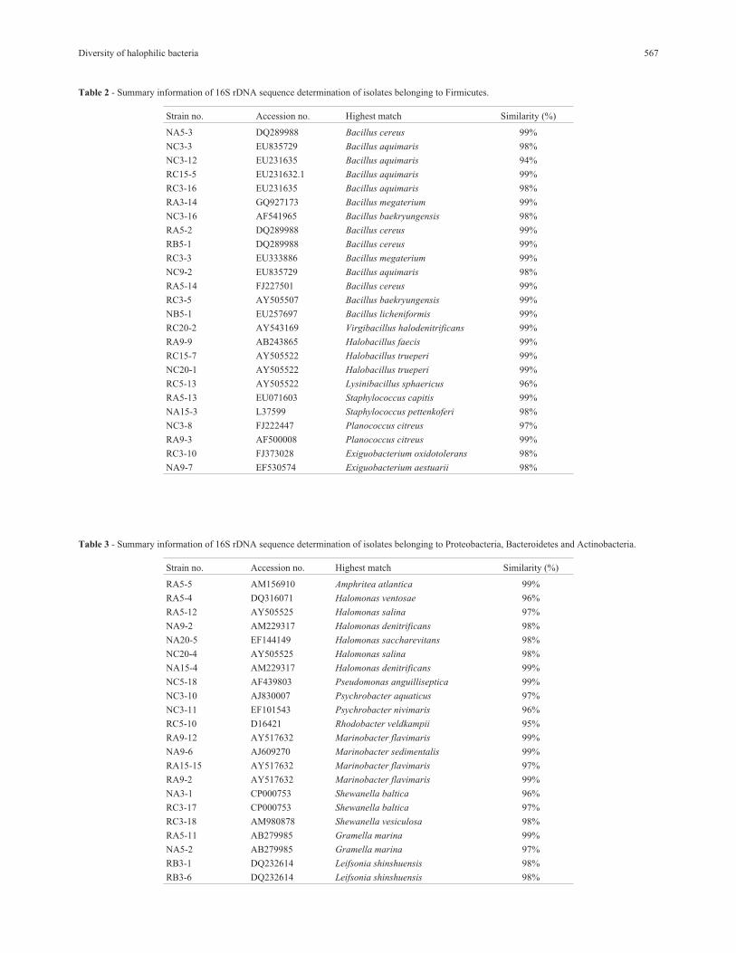
Table 2 - Summary information of 16S rDNA sequence determination of isolates belonging to Firmicutes.
Strain no. Accession no. Highest match Similarity (%)
NA5-3 DQ289988 Bacillus cereus 99%
NC3-3 EU835729 Bacillus aquimaris 98%
NC3-12 EU231635 Bacillus aquimaris 94%
RC15-5 EU231632.1 Bacillus aquimaris 99%
RC3-16 EU231635 Bacillus aquimaris 98%
RA3-14 GQ927173 Bacillus megaterium 99%
NC3-16 AF541965 Bacillus baekryungensis 98%
RA5-2 DQ289988 Bacillus cereus 99%
RB5-1 DQ289988 Bacillus cereus 99%
RC3-3 EU333886 Bacillus megaterium 99%
NC9-2 EU835729 Bacillus aquimaris 98%
RA5-14 FJ227501 Bacillus cereus 99%
RC3-5 AY505507 Bacillus baekryungensis 99%
NB5-1 EU257697 Bacillus licheniformis 99%
RC20-2 AY543169 Virgibacillus halodenitrificans 99%
RA9-9 AB243865 Halobacillus faecis 99%
RC15-7 AY505522 Halobacillus trueperi 99%
NC20-1 AY505522 Halobacillus trueperi 99%
RC5-13 AY505522 Lysinibacillus sphaericus 96%
RA5-13 EU071603 Staphylococcus capitis 99%
NA15-3 L37599 Staphylococcus pettenkoferi 98%
NC3-8 FJ222447 Planococcus citreus 97%
RA9-3 AF500008 Planococcus citreus 99%
RC3-10 FJ373028 Exiguobacterium oxidotolerans 98%
NA9-7 EF530574 Exiguobacterium aestuarii 98%
Table 3 - Summary information of 16S rDNA sequence determination of isolates belonging to Proteobacteria, Bacteroidetes and Actinobacteria.
Strain no. Accession no. Highest match Similarity (%)
RA5-5 AM156910 Amphritea atlantica 99%
RA5-4 DQ316071 Halomonas ventosae 96%
RA5-12 AY505525 Halomonas salina 97%
NA9-2 AM229317 Halomonas denitrificans 98%
NA20-5 EF144149 Halomonas saccharevitans 98%
NC20-4 AY505525 Halomonas salina 98%
NA15-4 AM229317 Halomonas denitrificans 99%
NC5-18 AF439803 Pseudomonas anguilliseptica 99%
NC3-10 AJ830007 Psychrobacter aquaticus 97%
NC3-11 EF101543 Psychrobacter nivimaris 96%
RC5-10 D16421 Rhodobacter veldkampii 95%
RA9-12 AY517632 Marinobacter flavimaris 99%
NA9-6 AJ609270 Marinobacter sedimentalis 99%
RA15-15 AY517632 Marinobacter flavimaris 97%
RA9-2 AY517632 Marinobacter flavimaris 99%
NA3-1 CP000753 Shewanella baltica 96%
RC3-17 CP000753 Shewanella baltica 97%
RC3-18 AM980878 Shewanella vesiculosa 98%
RA5-11 AB279985 Gramella marina 99%
NA5-2 AB279985 Gramella marina 97%
RB3-1 DQ232614 Leifsonia shinshuensis 98%
RB3-6 DQ232614 Leifsonia shinshuensis 98%

Bacillus was the dominant genus in the phylum Firmicutes,
comprising 24% of the total isolates and exhibiting 94-99%
16S rDNA sequence similarity to published species (Ta-
ble 1). The second dominant genera were Halomonas and
Shewanella in the phylum Proteobacteria, representing 6%
of the isolated strains and showing 97-99% 16S rDNA se-
quence similarity to known species. The third dominant ge-
nus was Psychrobacter, representing only 3% of the
strains.
Discussion
A study of halophilic bacterial diversity was carried
out on foreshore soils of Daecheon Beach, Chungnam, and
foreshore soils and seawater of Saemangeum Sea, Jeonbuk.
The diversity of slightly, moderately, and extremely halo-
philic bacteria were studied using two types of media con-
taining 3%, 5%, 9%, 15%, 20% and 30% NaCl. Different
colonies grown on media were selected and purified for the
investigation of halophilic bacterial diversity. The highest
568 Irshad et al.
Figure 3 - Phylogenetic tree based on the 16S rDNA sequences showing the relationship among the representatives of Firmicutes. The strains isolated in
this study are indicated in bold.

Diversity of halophilic bacteria 569
Figure 4 - Phylogenetic tree based on the 16S rDNA sequences showing the relationship among the representatives of Proteobacteria. The strains isolated
in this study are indicated in bold.

number of halophilic bacteria was found in sample C, and
the lowest number of halophilic bacteria was found in sam-
ple B. None of the isolates grew in the media with 30%
NaCl. The optimal concentration of NaCl was determined
to be 3% and 5%, and thus all of the isolates were consid-
ered to be moderate halophiles. No exceptions to this desig-
nation were found among the isolates. These results are in
line with the observation that the halophilic bacterial diver-
sity was markedly decreased as the salt concentration of the
media increased. The slightly and moderately halophilic
bacteria were more abundant than the extremely halophilic
bacteria. These results are in good agreement with those re-
ported by (Quesada et al., 1982; Rodriguez-Valera, 1988)
in that saline soil appeared to yield largely moderately
halophilic and halotolerant bacteria rather than the ex-
tremely halophilic bacteria. These phenomena are mainly
due to the level of salt content in foreshore soil, which does
not support the growth of extremely halophilic bacteria.
Moreover, the variation in the average counts of
slightly halophilic bacteria was more correlated to the mod-
erately halophilic bacteria than the extremely halophilic
bacteria. An explanation for this phenomenon is that the
slightly and moderately halophilic bacteria colonize differ-
ent niches to the extremely halophilic bacteria (Oren, 1999;
Oren, 2002a; 2002b). The levels of salt requirements for
growth of the slightly and moderately halophilic bacteria
are closer than those required for the extremely halophilic
bacteria. Also the growth of extremely halophilic bacteria
requires relatively high NaCl (at least 9%) and the majority
of them require magnesium ion (Mg2+) for growth while the
growth of slightly and moderately halophilic bacteria is not
generally dependent on this cation (Grant et al., 2001).
In the present study Box-PCR technique was em-
ployed for the rapid molecular typing of 200 halophilic bac-
terial isolates. Box-PCR is based on the observation that
outwardly facing oligonucleotide primer, complementary
to interspersed repeated sequences, enables the amplifica-
tion of differently sized DNA fragments, consisting of
DNA sequences lying in between these elements (De
Bruijn, 1992; Georghiou et al., 1994; Judd et al., 1993).
The majority of the strains analyzed had different BOX pat-
terns, confirming the high discriminative power of the
BOX-PCR fingerprint technique and reporting high diver-
sity of halophilic bacteria in marine environment.
The BLAST identification results showed that iso-
lated strains were composed of 4 phyla, Firmicutes (60%),
Proteobacteria (31%), Bacteriodetes (5%) and Actino-
bacteria (4%). Isolates were affiliated with 16 genera and
36 species. Bacillus was the dominant genus in the phylum
Firmicutes comprising (24%) of the total isolates. The
dominance of Bacillus spp. is in line with the previous stud-
ies (Quesada et al., 1982; Rodriguez-Valera, 1988; Zahran,
1997).
The aim of this study was to evaluate halophilic bac-
terial diversity in foreshore soil. To our knowledge, this is
the first extensive report on halophilic bacterial diversity
from foreshore soil of South Korea.
References
Birbir M, Ilgaz A (1996) Isolation and identification of bacteria
adversely affecting hide and leather quality. J Soc Leath
Technol Chem 80:147-153.
Cho SH, Han J, Seong CN, Kim SB (2006) Phylogenetic diversity
of acidophilic sporoactinobacteria isolated from various
soils. J Microbiol 44:600-606.
DasSarma S, Arora P (2001) Halophiles. In: Encyclopaedia of life
science. Nature Publishing Group, London, pp 1-9.
DasSarma S, Arora P (2002) Halophiles. In: Encyclopedia of Life
Sciences. Nature Publishing Group, London, 8:458-466.
Das S, Lyla PS, Khan SA (2006) Marine microbial diversity and
ecology: importance and future perspectives. Curr Sci
90:1325-1335.
De Bruijn FJ (1992) Use of repetitive (repetitive extragenic palin-
dromic and enterobacterial repetitive intergenic consensus)
sequences and the polymerase chain reaction to fingerprint
the genomes of Rhizobium meliloti isolates and other soil
bacteria. Appl Environ Mocrobiol 58:2180-2187.
Delgado-García M, Valdivia-Urdiales B, Aguilar-González CN,
Contreras-Esquivel JC, Rodríguez-Herrera R (2012) Halo-
philic hydrolases as a new tool for the biotechnological in-
dustries. J Sci Food Agric 92:2575-2580.
Georghiou P, Doggett A, Kielhofner M, Stout J et al. (1994). J
Clin Microbiol 19:2989-2994.
Ghasemi Y, Rasoul-Amini S, Ebrahiminezhad A, Kazemi A,
Shahbazia M, Talebniaa N (2011a) Screening and Isolation
of Extracellular Protease Producing Bacteria from the
Maharloo Salt Lake. Iran J Pharm Sci 7:175-180.
Ghasemi Y, Rasoul-Amini S, Kazemi A, Zarrini G, Morowvat
MT, Kargar M (2011b) Isolation and Characterization of
Some Moderately Halophilic Bacteria with Lipase Activity.
Mikrobiologiia 80:477-481.
Grant WD, Kamekura M, McGenity TJ, Ventosa A (2001) Class
III. Halobacteria class nov. In: Boone, D.R., Castenholz,
R.W. (eds). Bergey’s Manual of Systematic Bacteriology,
Springer, New York, pp 294
Judd AK, Schneider M, Sadowsky MJ, de Bruijn FJ (1993) Use of
repetetive sequences and the polemerase chain reaction
technique to classify genetically related Bradyrhizoium
japonicum serocluster 123 strains. Appl Environ Microbiol
59:1702-1708.
Jukes TH, Cantor CR (1969) Evolution of protein molecules. In:
Munro, H.N. (ed). Mammalian protein metabolism. Aca-
demic Press, New York, pp 21-132.
Li X, Yu HY (2012) Characterization of an organic solvent-
tolerant �-amylase from a halophilic isolate,
Thalassobacillus sp. LY18. Folia Microbiol 57:447-453.
Martin B, Humbert O, Camara M, Guenzi E, Walker J, Mitchell T,
Andrew P, Prudhomme M, Hakenbeck, R (1992) A highly
conserved repeated DNA element located in the chromo-
some of Steptococcus pneumonia. Nucleic Acids Res
20:3479-3483.
Mellado ME, Ventosa A (2003) Biotechnological potential of
moderately and extremely halophilic microorganisms. In:
Barredo, J.L. (ed). Microorganisms for health care, food and
enzyme production, Research Signpost, Kerala, pp 233-256.
570 Irshad et al.

Onishi H, Yokoi H, Kamekura M (1991) An application of a
bioreactor with flocculated cells of halophilic Micrococcus
varians sub sp. Halophilus which preferentially adsorbed
halophilic nuclease H to 5- nucleotide production. In: Rodri-
guez-valera, F. (ed). General and Applied Aspects of Halo-
philic Microorganisms. Plenum Press, New York, pp 341-
349.
Oren A (1999) Bioenergetics aspects of halophilism. Microbiol
Mol Biol Rev 63:334-348.
Oren A (2002a) Halophilic microorganisms and their environ-
ments: Kluwer Academic Publishers, Dordrecht, Nether-
lands, pp 575.
Oren A (2002b) Diversity of halophilic microorganisms: environ-
ments, phylogeny, physiology, and applications. J Ind
Microbiol Biotechnol 28:56-63.
Quesada E, Ventosa A, Rodriguez-Valera F, Ramos-Cormenzana
A (1982) Types and properties of some bacteria isolated
from hypersaline soils. J Appl Microbiol 53:155-161.
Rodriguez-Valera F (1988) Characteristics and microbial ecology
of hypersaline environments. In: Rodriguez-Valera, F. (ed).
Halophilic Bacteria, CRC Press, Boca Raton, pp 3-30.
Rodriguez-Valera F (1993) Introduction to saline environments.
In: Vreeland, R.H., Hochstein, L.I. (eds). The Biology of
Halophilic Bacteria, CRC Press, Boca Raton, pp 1-12.
Saitou N, Nei M (1987) The neighbor joining method: a new
method for constructing phylogenetic trees. Mol Biol Evol
4406-425.
Seong CN (1992) Numerical taxonomy of acidophilic and neutron
tolerant actionmycetes isolated from acid soil in Korea.
Ph.D. thesis, Seoul National University, Seoul, Republic of
Korea.
Shafiei M, Ziaee AA, Amoozegar MA (2012) Purification and
characterization of a halophilic-amylase with increased ac-
tivity in the presence of organic solvents from the moder-
ately halophilic Nesterenkonia sp. strain F. Extremophiles
16:627-635.
Ventos A, Nieto JJ (1995) Biotechnological applications and po-
tentialities of halophilic microorganisms. World J Microbiol
Biotechnol 11:85-94.
Ventosa A, Nieto JJ, Oren A (1998) Biology of moderately halo-
philic aerobic bacteria. Microbiol Mol Biol Rev 62:504-544.
Ventosa A (2006) Unusual microorganisms from unusual habi-
tats: hypersaline environments. In: Logan, N.A., Lappin-
Scott, H.M., Oyston, P.C.F. (eds). Prokaryotic diversity-
mechanism and significance. Cambridge University Press,
Cambridge, pp 223-253.
Versalovic J, Koeuth T, Lupski JR (1991) Distribution of repeti-
tive DNA sequences in eubacteria and application to finger-
printing of bacterial genomes. Nucleic Acids Res 19:6823-
6831.
Zahran HH (1997) Diversity, adaptation and activity of the bacte-
rial flora in saline environments. Biol Fertil Soil 25:211-
223.
All the content of the journal, except where otherwise noted, is licensed under a
Creative Commons License CC BY-NC.
Diversity of halophilic bacteria 571