ctls by blocking atp binding to tap cowpox virus protein
TRANSCRIPT
of April 4, 2018.This information is current as
CTLs by Blocking ATP Binding to TAPCowpox Virus Protein CPXV012 Eludes
WiertzJan Lebbink, Maaike E. Ressing and Emmanuel J. H. J.Klaus Früh, Jacques J. Neefjes, Antoinette Killian, Robert Martijn Koorengevel, Jan W. Drijfhout, Elisabeth Kremmer,Wouter F. van Leeuwen, Jennine Grootens, Daniëlle Horst, Rutger D. Luteijn, Hanneke Hoelen, Elisabeth Kruse,
ol.1400964http://www.jimmunol.org/content/early/2014/07/13/jimmun
published online 14 July 2014J Immunol
average*
4 weeks from acceptance to publicationFast Publication! •
Every submission reviewed by practicing scientistsNo Triage! •
from submission to initial decisionRapid Reviews! 30 days* •
Submit online. ?The JIWhy
Subscriptionhttp://jimmunol.org/subscription
is online at: The Journal of ImmunologyInformation about subscribing to
Permissionshttp://www.aai.org/About/Publications/JI/copyright.htmlSubmit copyright permission requests at:
Email Alertshttp://jimmunol.org/alertsReceive free email-alerts when new articles cite this article. Sign up at:
Print ISSN: 0022-1767 Online ISSN: 1550-6606. Immunologists, Inc. All rights reserved.Copyright © 2014 by The American Association of1451 Rockville Pike, Suite 650, Rockville, MD 20852The American Association of Immunologists, Inc.,
is published twice each month byThe Journal of Immunology
by guest on April 4, 2018
http://ww
w.jim
munol.org/
Dow
nloaded from
by guest on April 4, 2018
http://ww
w.jim
munol.org/
Dow
nloaded from

The Journal of Immunology
Cowpox Virus Protein CPXV012 Eludes CTLs by BlockingATP Binding to TAP
Rutger D. Luteijn,* Hanneke Hoelen,* Elisabeth Kruse,* Wouter F. van Leeuwen,*
Jennine Grootens,* Danielle Horst,* Martijn Koorengevel,† Jan W. Drijfhout,‡
Elisabeth Kremmer,x Klaus Fr€uh,{ Jacques J. Neefjes,‖ Antoinette Killian,†
Robert Jan Lebbink,* Maaike E. Ressing,*,1 and Emmanuel J. H. J. Wiertz*
CD8+ CTLs detect virus-infected cells through recognition of virus-derived peptides presented at the cell surface by MHC class I
molecules. The cowpox virus protein CPXV012 deprives the endoplasmic reticulum (ER) lumen of peptides for loading onto newly
synthesized MHC class I molecules by inhibiting the transporter associated with Ag processing (TAP). This evasion strategy allows
the virus to avoid detection by the immune system. In this article, we show that CPXV012, a 9-kDa type II transmembrane
protein, prevents peptide transport by inhibiting ATP binding to TAP. We identified a segment within the ER-luminal domain of
CPXV012 that imposes the block in peptide transport by TAP. Biophysical studies show that this domain has a strong affinity for
phospholipids that are also abundant in the ER membrane. We discuss these findings in an evolutionary context and show that
a frameshift deletion in the CPXV012 gene in an ancestral cowpox virus created the current form of CPXV012 that is capable of
inhibiting TAP. In conclusion, our findings indicate that the ER-luminal domain of CPXV012 inserts into the ER membrane,
where it interacts with TAP. CPXV012 presumably induces a conformational arrest that precludes ATP binding to TAP and, thus,
activity of TAP, thereby preventing the presentation of viral peptides to CTLs. The Journal of Immunology, 2014, 193: 000–000.
Nucleated cells express MHC class I (MHC I) molecules attheir cell surface to present peptides to CD8+ CTLs.Recognition of virus-derived peptides by CTLs results in
the elimination of virus-infected cells, allowing control of virusspread.The majority of peptides presented by MHC I at the surface of
infected cells results from proteasomal degradation of proteinswithin the cytosol (1). These peptides need to cross the endo-plasmic reticulum (ER) membrane for loading onto MHC I in the
ER lumen. Translocation of peptides over the ER membrane ismediated by the transporter associated with Ag processing (TAP),a heterodimeric ABC transporter composed of two subunits: TAP1and TAP2. Each subunit contains a cytosolic nucleotide bindingdomain (NBD) that facilitates the binding and hydrolysis of ATPto drive peptide translocation over the ER membrane. In addition,the TAP1/TAP2 heterodimer contains a transmembrane (TM)domain composed of 9 TM helices of TAP1 intertwined with 10TM helices of TAP2 (2, 3). TAP forms the center of the MHCclass I peptide-loading complex (PLC) and recruits tapasin,a chaperone that binds newly synthesized, “empty” MHC I to thePLC (4, 5). This arrangement facilitates efficient peptide loadingof MHC I. In addition, the chaperones calreticulin and ERp57further stabilize MHC I in the PLC. Once a peptide is loadedonto the peptide-binding domain of MHC I, the MHC I/peptidecomplex dissociates from the PLC and travels via the Golginetwork to the cell surface, where it displays its content to cy-totoxic T cells (6).Large DNA viruses, such as herpesviruses, adenoviruses, and
poxviruses, have acquired gene products that allow for specificinterference with MHC I function at various stages during itssynthesis, assembly, and transport to the cell surface (7). TAPappears to be an attractive target for these viral immune-evasionstrategies, because multiple herpesvirus family members encodespecific inhibitors for this transporter. The viral TAP inhibitorsshow no structural homology and use different strategies to in-terfere with TAP-mediated peptide transport. ICP47 of HSV-1 andHSV-2 acts as a high-affinity competitor for binding of peptides tothe cytosolic peptide-binding domain of TAP (8–10). The humanCMV protein US6 alters ATP binding to TAP by diminishing ATPbinding to TAP1 and promoting ATP binding to TAP2 (11–13).The UL49.5 proteins of most varicelloviruses restrict conforma-tional alterations of TAP that are necessary to drive peptidetransport (14, 15). In addition, UL49.5 of bovine herpesvirus(BHV)-1 and BHV-5 induces degradation of TAP1 and TAP2,
*Department of Medical Microbiology, University Medical Center Utrecht, 3584 CXUtrecht, the Netherlands; †Department of Membrane Biochemistry and Biophysics,Utrecht University, 3584 CH Utrecht, the Netherlands; ‡Department of Immunohe-matology and Blood Transfusion, Leiden University Medical Center, 2333 ZA Lei-den, the Netherlands; xHelmholtz Center Munich, German Research Center forEnvironmental Health, Institute of Molecular Immunology, 81377 Munich,Germany; {Vaccine and Gene Therapy Institute, Oregon Health and Science Univer-sity, Beaverton, OR 97006; and ‖Department of Cell Biology, The Netherlands Can-cer Institute, 1066 CX Amsterdam, the Netherlands
1Current address: Department of Molecular Cell Biology, Leiden University MedicalCenter, Leiden, the Netherlands.
Received for publication April 14, 2014. Accepted for publication June 13, 2014.
This work was supported by Veni Grant 916.10.138 from the Netherlands Organiza-tion for Scientific Research and Marie Curie Career Integration Grant PCIG-GA-2011-294196 (both to R.J.L.), Netherlands Scientific Organization Vidi Grant917.76.330 (to M.E.R.), and a grant from the Dutch Diabetes Research Foundation(to E.J.H.J.W.).
Address correspondence and reprint requests to Prof. Emmanuel J.H.J. Wiertz, De-partment of Medical Microbiology, University Medical Center Utrecht, P.O. Box85500, 3508 GA Utrecht, the Netherlands. E-mail address: [email protected]
Abbreviations used in this article: BHV, bovine herpesvirus; CD, circular dichroism;CPXV, cowpox virus; DOPC, 1,2-dioleoyl-sn-glycero-3-phosphocholine; DOPS, 1,2-dioleoyl-sn-glycero-3-phospho-L-serine; EHV, equid herpesvirus; ER, endoplasmicreticulum; IRES, internal ribosomal entry site; LUV, large unilaminar vesicle; MES,2-(N-morpholino)ethanesulfonic acid; MHC I, MHC class I; MHC II, MHC class II;MJS, MelJuSo; NBD, nucleotide binding domain; NP-40, Nonidet P-40; PLC,peptide-loading complex; TAP, transporter associated with Ag processing; TfR, trans-ferrin receptor; TM, transmembrane.
Copyright� 2014 by The American Association of Immunologists, Inc. 0022-1767/14/$16.00
www.jimmunol.org/cgi/doi/10.4049/jimmunol.1400964
Published July 14, 2014, doi:10.4049/jimmunol.1400964 by guest on A
pril 4, 2018http://w
ww
.jimm
unol.org/D
ownloaded from

whereas UL49.5 of equid herpesvirus (EHV)-1 and EHV-4 inter-feres with ATP binding to the NBDs of TAP (14). BNLF2a ofEBV blocks both peptide and ATP binding to TAP (16). Inhibitionof TAP-mediated peptide transport was considered to be unique toherpesviruses until the recent identification of the cowpox virus(CPXV) protein CPXV012 as an effective inhibitor of peptidetransport by TAP (17, 18).CPXVs are members of the Orthopoxvirus genus, which com-
prises several clinically and economically important pathogens,including variola virus (the causative agent of smallpox), camel-pox virus, and monkeypox virus. CPXVs have the largest genomeof all orthopoxviruses and encode an elaborate arsenal of im-mune-evasion proteins (19). Two of these proteins, CPXV012 andCPXV203, prevent cytotoxic T cell recognition by interfering withMHC I–mediated Ag presentation. CPXV203 inhibits Ag presen-tation by retaining MHC I/peptide complexes in the ER (20).CPXV203 acts through a KDEL-related KTEL ER-retention motifencoded in its C-terminal domain (20). Deletion of CPXV203from CPXV only partially restored MHC I surface expression,suggesting the presence of an additional mechanism that is activein MHC I suppression. Indeed, CPXV012 was identified as thesecond CPXV protein to interfere with MHC I Ag presentation(17, 18). Deletion of both CPXV012 and CPXV203 fully restoredMHC I surface levels. Ex vivo, T cells isolated from CPXV-infectedmice produced IFN-g upon stimulation with CPXVD012D203-infected cells, in contrast with wild-type CPXV-infected cells (18).Furthermore, in vivo experiments showed that mice infected withthe CPXVD012D203 strain survived, whereas mice infectedwith the wild-type virus succumbed to virus infection (18).CPXV012 specifically abrogates TAP-mediated peptide trans-
location through an unknown mechanism, thereby depriving theER of peptides (17, 18). In this article, we show that CPXV012inhibits TAP through its ER-luminal domain by interfering withATP binding to the cytosolic NBDs of TAP. We define the regionswithin the CPXV012 ER-luminal domain that are critical for in-hibition of TAP function and downregulation of MHC I expres-sion. The ER-luminal domain of CPXV012 adopts an a-helicalstructure in the presence of phospholipids and displays a strongaffinity for ER membranes. The ER-luminal domain of CPXV012binds to TAP and arrests the TAP complex in a conformationalstate that is unable to acquire energy (ATP), thereby inhibiting thetranslocation of peptides into the ER. Additionally, we provide anexplanation for the emergence of the functional ER domain ofCPXV012 in evolution, via the acquisition of a frameshift muta-tion in the ancestral protein of CPXV012.
Materials and MethodsCell lines
The human melanoma cell line MelJuSo (MJS) (21) was maintained inRPMI 1640 medium (Invitrogen), supplemented with 10% FCS (PAALaboratories), 2 mM L-glutamine, 100 U/ml penicillin, and 100 mg/mlstreptomycin (complete medium). HEK293T cells used for lentivirusproduction were maintained in DMEM complete medium (Invitrogen).The retrovirus packaging cell line wNX-A was maintained in IMDM(Invitrogen) complete medium. MJS cells stably transfected with TAP1-eGFP were obtained from E. Reits (Academic Medical Center, Amster-dam, The Netherlands) (22).
Lentivirus and retrovirus production and transduction
A plasmid encoding codon-optimized CPXV012 from CPXV strainBrighton Red with a FLAG-tag at the N terminus (CPXV012) was used asa template for PCR amplification with the primers 59-CGGGATCCAC-CATGGACTACAA-39 and 59-GCCTCGAGTCATCAGATGATGC-39. Theresulting PCR product was cloned into the pLZRS retroviral vector. EBV-derived BNLF2a with a C-terminal HA tag (BNLF2a) was cloned into theretroviral vector pLZRS, as described previously (16). Genes were cloned
upstream of an internal ribosomal entry site (IRES) to allow coexpressionof the truncated nerve growth factor receptor (DNGFR) as a marker.
CPXV012 and CPXV012 variants were cloned into the lentiviral vectorpLV upstream of IRES and eGFP, as described previously (17).
For the production of replication-deficient recombinant retrovirus,pLZRS vectors were transfected into wNX-A packaging cells. MJS cellswere transduced with retrovirus, as described previously (23). Cells wereFACS sorted to achieve expression of the gene of interest in all cells. Forthe production of replication-deficient recombinant lentivirus, the lentiviralvector and third-generation packaging vectors pVSV-G, pMDL, and pRSVwere cotransfected into HEK293T cells. Lentivirus-containing super-natants were harvested after 3 d of virus production and used to transduceMJS cells by spin infection at 1000 3 g for 2 h at 33˚C in the presence of3.2 mg/ml polybrene.
Cells transduced with retrovirus or lentivirus were sorted by flowcytometry (FACSAria II; BD Biosciences).
CPXV012 variants
CPXV012 deletion and substitution variants were generated and cloned intothe pLV-IRES eGFP lentiviral vector. For cloning purposes, a silent mu-tation resulting in an XhoI restriction site was introduced into CPXV012using primers 1 and 2 (Table I). The following primers (Table I) were usedto generate the CPXV012 deletion and substitution variants: primers 2 and3 for CPXV012-Dcyt, primers 4 and 5 for CPXV012-DER, primers 2 and 6for CPXV012 soluble, primers 7–10 for TfR-CPXV012, primers 2 and 11for CPXV012-Ala1, primers 2 and 12 for CPXV012-Ala2, primers 4 and13 for CPXV012-Ala3, primers 4 and 14 for CPXV012-Ala4, primers 4and 15 for CPXV012-Ala5, primers 2 and 16 for CPXV012-Ala6, andprimers 2 and 17 for CPXV012-Ala7.
To ensure proper ER localization of the CPXV012-soluble variant, wereplaced the endogenous signal sequence of CPXV012 with the (cleavable)human CD8 signal sequence (24), because CPXV012 is a type II proteinwith the TM domain acting as a signal sequence. The chimeric proteintransferrin receptor (TfR)-CPXV012 was constructed, in which the ER-luminal domain of CPXV012 was fused to amino acid residues 58–88 ofthe type II TfR. This region in TfR includes part of the cytosolic tail (aa58–67) and the TM domain (aa 68–88) (25). The two cysteine residuesflanking the TfR TM domain were replaced by alanine residues to removea potential localization and dimerization signal (25). All variants wereproduced from a bicistronic transcript, encoding CPXV012 followed by anIRES and eGFP.
Ab generation
The CPXV012-specific rat IgG2a mAb 2A8 was generated by immunizingLou/C rats with OVA-coupled peptide encompassing CPXV012 aa 28–44(sequence: CSMEHGYFQEGISRFKI).
Abs
The following primary and directly conjugated Abs were used: mouse anti-HLA class I HC HC10 mAb, PE-conjugated mouse anti–HLA-A/B/C W6/32 mAb (AbD Serotec; #MCA81PE), PE-conjugated mouse anti–HLA-DRmouse G46-6 mAb (BD Pharmingen; #347401), mouse anti-calnexin AF8mAb (kindly provided by M. Brenner, Harvard Medical School, Boston,MA), mouse anti-FLAG M2 mAb (Sigma-Aldrich; #F1804), rabbit anti-tapasin gp48-C (provided by P. Cresswell, Yale School of Medicine, NewHaven, CT), rat anti-tapasin 7F6 mAb (provided by R. Tampe, JohannWolfgang Goethe University, Frankfurt/Main, Germany), mouse anti-TAP1 148.3 mAb (provided by R. Tampe), mouse anti-TAP2 435.4 mAb(provided by P. van Endert, Hospital Necker, Paris, France), rat anti-CPXV012 2A8 mAb, rat anti-GFP RQ2 mAb (MBL; #D153-3), mouseanti-GFP 1E4 mAb (Enzo; #ADI-SAB-500), mouse anti-CD71 (TfR)H68.4 (Invitrogen; #1368xx), and biotin-conjugated mouse anti-NGFRmAb (BD; #557195).
The following secondary Abs were used: HRP-conjugated rabbit anti-ratIgG (DAKO, #P0450), HRP-conjugated goat anti-rat IgG L-chain specific(Jackson ImmunoResearch, #112-035-175), HRP conjugated goat anti-mouse IgG L-chain specific (Jackson ImmunoResearch, #115-035-174),HRP-conjugated goat anti-mouse IgG (Bio-Rad; #172-1011), and allo-phycocyanin-conjugated streptavidin (BD Pharmingen; #554067).
Coimmunoprecipitation and immunoblotting
Cells were lysed in 0.5%Nonidet P-40 (NP-40) buffer (0.5%NP-40, 50 mMTris-HCl [pH 7.5], and 5 mMMgCl2) or 1% digitonin buffer (1% digitonin,50 mM Tris-HCl [pH 7.5], 5 mM MgCl2, and 150 mM NaCl) in thepresence of 10 mM leupeptin and 1 mM 4-(2-aminoethyl) benzenesulfonylfluoride. To remove nuclear fractions, cell lysates were centrifuged at
2 COWPOX VIRUS PROTEIN CPXV012 BLOCKS ATP BINDING TO TAP
by guest on April 4, 2018
http://ww
w.jim
munol.org/
Dow
nloaded from
12,000 3 g at 4˚C for 20 min. For coimmunoprecipitation experiments,postnuclear digitonin lysates were incubated with protein G-Sepharosebeads (GE Healthcare) and the Abs indicated. After overnight incubationat 4˚C, samples were washed four times in 0.5% digitonin buffer, andbound fractions were resuspended in TE buffer (20 mM Tris [pH 6.8] and1 mM EDTA).
To prepare samples for SDS-PAGE separation, coimmunoprecipitationsamples and NP-40 postnuclear lysates were boiled in Laemmli samplebuffer at 95˚C for 5 min. After SDS-PAGE, proteins were transferred topolyvinylidene difluoride membranes (GE Healthcare) and incubated withAbs specific for the indicated proteins, followed by HRP-coupled sec-ondary Abs. Bound Abs were visualized by incubating polyvinylidenedifluoride membranes with ECL (Thermo Scientific Pierce) and exposureto Amersham Hyperfilm (GE Healthcare).
Flow cytometry
MHC I andMHC class II (MHC II) cell surface expression was measured byflow cytometry. All washing and Ab-incubation steps were performed inPBS, supplemented with 0.5% BSA and 0.02% sodium azide. For MHC Icell surface expression, cells were incubated with PE-conjugated W6/32mAb. For MHC II cell surface expression, cells were incubated with PE-conjugated G46-6 Ab. Cells were fixed using 1% formaldehyde in PBSsupplemented with 0.5% BSA and 0.02% sodium azide, and flow cytome-tric parameters were recorded using a FACSCanto II (BD Biosciences).Analysis of flow cytometry data was performed using FlowJo software(TreeStar).
Peptides
Synthetic peptides were prepared by normal Fmoc chemistry using pre-loaded Tentagel resins, using PyBop/N-methylmorpholine for in situ ac-tivation and 20% piperidine in N-methylpyrrolidinon for Fmoc removal(26). Couplings were performed for 75 min. After final Fmoc removal,peptides were cleaved with trifluoroacetic acid/H2O 19:1 containing ad-ditional scavengers when C or W residues were present in the peptidesequence. Peptides were isolated by ether/pentane precipitation. Peptideswere checked for purity using reversed-phase HPLC–mass spectrometryand for integrity using MALDI-TOF mass spectrometry, showing thecalculated molecular masses.
Labeling of peptides used for the peptide-transport assay was performedwith 5-(iodoacetamide) fluorescein (Fluka Chemie) at pH 7.5 (Na-phosphate in water/acetonitrile 1:1 v/v). The labeled peptides weredesalted over Sephadex G-10 and further purified by C18 reversed-phase HPLC. The molecular mass of the labeled peptides was checked
using MALDI-TOF mass spectrometry. All other peptides were synthe-sized with an N-terminal biotin moiety and a C-terminal amide. Cysteineswere replaced by the isosteric a-amino-n-butyric acid.
Peptide-transport assay
The peptide-transport assay was performed as described (27). Control MJScells or MJS cells expressing CPXV012 wild-type or CPXV012 variantswere permeabilized with streptolysin O (Murex Diagnostics) and incu-bated with the fluorescein-coupled peptide CVNKTERAY (N-linked gly-cosylation site underlined) in the presence or absence of 10 mM ATP at37˚C for 10 min. Cells were subsequently lysed in Triton X-100 buffer (1%Triton X-100, 500 mM NaCl, 2 mM MgCl2, 50 mM Tris-HCl [pH 8]) andcentrifuged at 12,000 3 g at 4˚C for 20 min to remove nuclei and debris.To evaluate peptide transport into the ER, postnuclear lysates were incu-bated with ConA–Sepharose beads (GE Healthcare), and glycosylatedpeptides were isolated from the lysate. After washing the beads with thefirst-wash buffer (0.1% Triton X-100, 500 mM NaCl, 2 mM MgCl2,50 mM Tris-HCl [pH 8]) and with the second-wash buffer (500 mM NaCl,50 mM Tris-HCl [pH 8]), peptides were eluted from beads using elutionbuffer (500 mM mannopyranoside, 10 mM EDTA, 50 mM Tris-HCl[pH 8]), and fluorescent peptides were detected using a FLUOstarOmega (BMG LABTECH) fluorescence plate reader.
ATP-binding assay
The ATP-binding assay was performed as described previously (16).Briefly, control MJS cells or MJS cells expressing BNLF2a or CPXV012were lysed in 1% digitonin lysis buffer, as described for coimmunopre-cipitation and immunoblotting experiments. Cell lysates were centrifugedat 12,0003 g at 4˚C for 20 min to remove nuclei. Postnuclear lysates wereincubated with hydrated C-8 ATP-agarose beads (13 mM final concentra-tion; Sigma-Aldrich) at 4˚C for 2 h. Unbound supernatant was collected,and ATP-bound proteins were eluted from the beads with 500 mM EDTA(ATP-bound fraction). The ATP-bound fraction and unbound supernatantwere analyzed for protein content by immunoblotting with the Abs indi-cated.
Peptide-binding assay
Peptide binding was evaluated as described previously (16). MJS cellsstably transfected with TAP1-eGFP and transduced with control retrovirusor retroviruses expressing BNLF2a or CPXV012 were homogenized usinga cell cracker (European Molecular Biology Laboratory; chamber 8.020 mm,ball 8.006 mm) to obtain TAP-containing microsomes. These microsomeswere incubated with the 125I-labeled peptide 5PS2 (ERYDKSE-[BPA]-L)
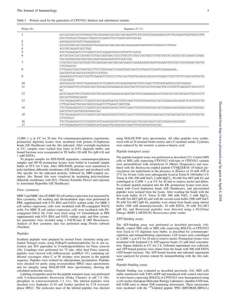
Table I. Primers used for the generation of CPXV012 deletion and substitution variants
Primer No. Sequence (59–39)
1 ACCGCGGCCACCATGGACTACAAGGACGACGACGACAAGTTCATCATGCGGGAGAGCATCTACAGAGTGATGATCGTGATCCTGTACCTGAGCCTGATCTCGAGCTTCCTGGTCATCTGCAG
2 AGTGAGCGCTGTCTAGAGGATC3 ACCGCGGCCACCATGGACTACAAGGACGACGACGACAAGAGAGTGATGATCGTGATCCTGTAC4 ACCGTCAGATCGCCTGG5 ATCTAGAGGATCCCCGGGTCATCAGAAGTAGCCGTGTTCCATGC6 ACCGCGGCCACCATGGCCTTACCAGTGACCGCCTTGCTCCTGCCGCTGGCCTTGCTGCTCCACGCCGCCAGGCCGGAC
TACAAGGACGACGACGACAAGCAGGAAGGCATCAGCCGG7 CCGCGGCCACCATGGACTACAAGGACGACGACGACAAGCCAAAAAGGGCAAGTGGAAGTATCGCCTATGGGACTATTG
CTGTGATCG8 CTTGAACCGGCTGATGCCTTCCTGATAGCCCAAGTAGCCAATCATAAATCCAATCAAGAAAAA,
GACGATCACAGCAATAGTCCCATGGC9 GGAAGGCATCAGCCGGTTCAAGATCTGCCCCTACCACTGGTACAAGCAGCACATGAGCCTGCTGTTCCGGCGGTACTA
CCACAAGC10 AGTGAGCGCTGTCTAGAGGATCCCCGGGTCATCAGATGATGCTGTCCAGCTTGTGGTAGTACCGCCGGAAC11 ACTCGAGCTTCCTGGTCATCTGCAGCATGGAACACGGCTACTTCGCCGCTGCAGCTGCCCGGTTCAAGATCTGCCCCT
AC12 ACTCGAGCTTCCTGGTCATCTGCAGCATGGAACACGGCTACTTCCAGGAAGGCATCAGCGCCGCTGCAGCTGCCCCCT
ACCACTGGTACAAGC13 TTCTAGAGGATCCCCGGGTCATCAGATGATGCTGTCCAGCTTGTGGTAGTACCGCCGGAACAGCAGGCTCATGTGCTG
CTTGGCAGCTGCAGCGGCGCAGATCTTGAACCGGCTGA14 TTCTAGAGGATCCCCGGGTCATCAGATGATGCTGTCCAGCTTGTGGTAGTACCGCCGGAACAGCAGGGCAGCTGCAGC
GGCGTACCAGTGGTAGGGGCAG15 TTCTAGAGGATCCCCGGGTCATCAGATGATGCTGTCCAGCTTGTGGTAGTAGGCAGCTGCAGCGGCGCTCATGTGCTG
CTTGTACC16 TTCTAGAGGATCCCCGGGTCATCAGATGATGCTGTCGGCAGCTGCAGCGGCCCGCCGGAACAGCAGGC17 TTCTAGAGGATCCCCGGGTCATCAAGCTGCAGCGGCCAGCTTGTGGTAGTACCGC
The Journal of Immunology 3
by guest on April 4, 2018
http://ww
w.jim
munol.org/
Dow
nloaded from

containing a photoactivatable bisphenol A cross-linker (28). After incu-bation on ice for 15 min, the microsomes were washed with PBS, andpeptide was covalently linked to binding partners by UV exposure for 10min on ice using a UVP device (Blak-Ray B-100A) with a mercury lamp(PAR38; Sylvania). Microsomes were lysed in 0.5% CHAPS buffer (0.5%CHAPS, 100 mM Tris-HCl [pH 8], 5 mM MgCl2), and TAP1-eGFP wasisolated using the anti-GFP RQ2 mAb and Protein G beads. Samples wereseparated by SDS-PAGE, as described above, and 125I-labeled peptidebinding was visualized by phosphoimaging. A fraction of the unlabeledmicrosomes was used for immunoblotting with the anti-GFP 1E4 primaryAb and HRP-conjugated anti-mouse IgG secondary Ab.
Preparation of large unilaminar vesicles and circulardichroism
Preparation of large unilaminar vesicles (LUVs) and circular dichroism(CD) spectrum recording were performed as described previously (29). LUVscomposed of a mixture of 1,2-dioleoyl-sn-glycero-3-phosphocholine(DOPC) and 1,2-dioleoyl-sn-glycero-3-phospho-L-serine (DOPS) (AvantiPolar Lipids) in a 7:3 molar ratio were prepared from 5-mM stock sol-utions of DOPC and DOPS in chloroform in a glass tube. The solvent wasevaporated with nitrogen gas, and the resulting lipid film was kept ina vacuum desiccator for 20 min. Lipid films were hydrated in 10 mM 2-(N-morpholino)ethanesulfonic acid (MES) buffer (pH 6.2) at a lipid concen-tration of 5 mM for 30 min. The lipid suspensions were freeze-thawed for20 cycles at temperatures of approximately 2196 and 40˚C and subse-quently extruded 20 times through 0.2-mm pore size filters (Anotop 10;Whatman, Maidstone, U.K.). A total of 625 mM LUVs and/or 20 mMpeptides diluted in 10 mM MES buffer (pH 6.2) were used for measure-ments. CD spectra were recorded on a Jasco 810 spectropolarimeter (Jasco,Easton, MD) over a wavelength range of 200–250 nm. Measurements wererecorded every 1 nm at a scan rate of 20 nm/min at room temperature incuvettes with a path length of 1.0 mm. Each reported spectrum is theaverage of five independent scans.
Langmuir monolayer
Peptide-induced changes in the surface pressure of a monomolecular layer(monolayer) of phospholipids at a constant surface area were measuredusing the Wilhelmy plate method (29). A Teflon trough was filled with5.0 ml PBS (pH 7.4). The buffer below the monolayer (subphase) was con-tinuously stirred during the measurements. Lipid monolayers were spreadfrom a 1-mM stock solution in chloroform at the air–buffer interface togive an initial surface pressure of 35 mN/m, before 25 ml of a 55-mMfreshly prepared stock solution of peptide in DMSO was injected into thesubphase, resulting in a final peptide concentration of 0.3 mM.
ResultsCPXV012 interacts with an intact MHC class I PLC
CPXV protein CPXV012 impairs MHC I surface expression byinhibiting TAP-mediated peptide transport (17, 18). However, themolecular mechanism that underlies this TAP inhibition is un-known. To study the biochemical and functional characteristics ofCPXV012, the viral protein was equipped with an N-terminalFLAG tag and expressed in the human melanoma cell line MJSusing lentiviral transduction. As expected, TAP-mediated trans-port was inhibited in cells expressing FLAG-CPXV012 (Fig. 1A,upper panel). Furthermore, expression of FLAG-CPXV012 re-duced MHC I surface expression, whereas MHC class II expres-sion remained unaffected (Fig. 1A, lower panels), indicating thatthe FLAG-tagged CPXV012 is functional and selective for MHC Iin MJS cells.To test whether CPXV012 influences the composition of the
PLC, individual PLC constituents were isolated from cells lysed inthe presence of digitonin to preserve the interactions within thePLC (Fig. 1B). CPXV012 was found to interact with the complexand did not seem to disturb the overall integrity of the PLC, be-cause similar amounts of TAP1, TAP2, and tapasin were coiso-lated in the presence and absence of CPXV012 (Fig. 1B, comparelanes 1 and 2 and lanes 5 and 6). In the presence of CPXV012, thePLC was enriched for MHC I (compare lanes 1 and 2 and lanes 5
FIGURE 1. CPXV012 interacts with an intact PLC. (A) TAP-mediated
peptide transport is affected by CPXV012 (upper panel). Transport of a flu-
orescently labeled peptide was measured in MJS cells expressing control
vector or CPXV012. To distinguish between active transport and passive
diffusion, transport was measured in the presence and absence of ATP. Active
transport in cells expressing CPXV012 was compared with transport in
control cells expressing eGFP vector only, which was set at 100%. Mean 6SEM from three independent experiments is shown. CPXV012 specifically
decreases MHC I surface expression (lower panels). Flow cytometric analysis
of MHC I and MHC II surface expression on the human melanoma cell line
MJS stably expressing CPXV012 (black line) or control vector (gray line).
The W6/32 mAb was used for detection of MHC I, and the HLA-DR–spe-
cific G46-6 mAb was used for the detection of MHC II. Flow cytometry
results are representative of at least three independent experiments. (B)
CPXV012 interacts with the PLC. TAP1 (mAb 148.3), TfR (mAb H68.4),
tapasin (Tpsn; mAb gp48-C), or MHC I (mAb HC10) were immunoprecip-
itated from digitonin lysates of MJS cells expressing CPXV012 (+ lanes) or
control vector (- lanes). Immunoprecipitated proteins were immunoblotted
for TAP1 (mAb 148.3), TAP2 (mAb 435.4), tapasin (mAb 7F6), MHC I (mAb
HC10), CPXV012 (mAb 2A8), and TfR (mAb H68.4). Lanes 9 and 10 show
input levels of indicated proteins prior to immunoprecipitation. A shorter ex-
posure time was used for lanes 7 and 8 of the immunoblot for MHC I, as well
as for lanes 9 and 10 of the immunoblots for TAP1, tapasin, MHC I, and
CPXV012. Immunoblots are representative of three independent experiments.
4 COWPOX VIRUS PROTEIN CPXV012 BLOCKS ATP BINDING TO TAP
by guest on April 4, 2018
http://ww
w.jim
munol.org/
Dow
nloaded from

and 6). In agreement with this, more TAP1, TAP2, and tapasincoimmunoprecipitated with MHC I in CPXV012-expressing cellscompared with control cells (compare lanes 7 and 8). This sug-gests that peptide loading of MHC I is obstructed by CPXV012,thus delaying the release of MHC I from the PLC.The interactions between CPXV012 and the components of the
PLC were specific, because TfR, an unrelated TM protein, andMHC II (data not shown) were not coisolated. Conversely, isolationof TfR did not yield any PLC proteins (Fig. 1B, lanes 3 and 4).In conclusion, CPXV012 interacts with the PLC in the ER.
CPXV012 does not affect the organization of the core componentsof the PLC (TAP1, TAP2, and tapasin); MHC I appears to accu-mulate in the PLC, likely as the result of diminished peptide supply.
CPXV012 interferes with ATP binding to TAP
Peptide translocation by TAP can be blocked by viral TAPinhibitors at different stages during the peptide-translocation cycle(30). This cycle initiates with the binding of peptide to the cyto-solic peptide-binding domain of TAP, which allows subsequentATP binding to the cytosolic NBDs (31). ATP binding and hy-drolysis at the NBDs cause conformational rearrangements withinthe transporter that facilitate translocation of the peptides over theER membrane (31).We investigated whether CPXV012 affects peptide binding
to TAP. An 125I-labeled peptide linked to a photoactivatable cross-linker was incubated with microsomes derived from MJS TAP1-eGFP cells. The cells expressed CPXV012 or the EBV-derivedTAP inhibitor BNLF2a, which was previously shown to inhibitpeptide binding to TAP (16). After UV cross-linking, TAP1-eGFPwas isolated from cell lysates, and 125I-labeled peptide/proteincomplexes were visualized using phosphoimaging.In cells expressing CPXV012, radiolabeled peptides were cross-
linked to TAP1-eGFP (Fig. 2A, lane 1), indicating that CPXV012does not prevent peptide binding to TAP. As previously reported,125I-labeled peptides were not cross-linked to TAP1-eGFP in cellsexpressing BNLF2a (Fig. 2A, lane 2), but they were able to cross-link to TAP1-eGFP in control cells (Fig. 2A, lane 3) (16).The next step in the peptide-translocation cycle, ATP binding
to TAP, was investigated using an ATP-binding assay (Fig. 2B).Agarose beads coated with immobilized ATP were incubated withdigitonin lysates of control cells, cells expressing CPXV012, orcells expressing the TAP inhibitor BNLF2a (16). In addition topeptide binding, BNLF2a reduces ATP binding to TAP (16). Pull-down of ATP-agarose beads yields an ATP-bound fraction and anATP-unbound supernatant fraction. In control cells, the ATP-bound fraction contained TAP1, TAP2, and tapasin (Fig. 2B,lane 1). In contrast, in the ATP-bound fractions of cells expressingCPXV012 or BNLF2a, the amounts of these PLC proteins werereduced (Fig. 2B, compare lanes 2 and 3 with lane 1), whereasthese PLC components were abundantly present in the unboundsupernatant of CPXV012- and BNLF2a-expressing cells (Fig. 2B,lanes 5 and 6). These results suggest that CPXV012 inhibits theTAP transport cycle by interfering with ATP binding to TAP.
The ER-luminal domain of CPXV012 is required for inhibitionof TAP
CPXV012 is a type II TM protein consisting of a cytosolic domain,a TM domain, and an ER-luminal domain (Fig. 3A) (17). To es-tablish the contribution of these domains to TAP inhibition,CPXV012 variants were generated lacking the cytosolic domain(CPXV012-Dcyt), the ER-luminal domain (CPXV012-DER), orthe cytosolic and TM domains (i.e., consisting of only the ER-luminal domain [CPXV012-soluble)] (Fig. 3A). To test the func-tionality of the CPXV012 variants, their effect on MHC I surface
expression was assessed by flow cytometry (Fig. 3B, 3C). Asexpected, MHC I surface expression was reduced on cells ex-pressing wild-type CPXV012 compared with control cells. MHC Isurface levels also were decreased on cells expressing CPXV012lacking its cytosolic tail (CPXV012-Dcyt), although not as effi-ciently as for wild-type CPXV012. MHC I surface levels were notreduced on cells expressing CPXV012 lacking its ER-luminal do-main (CPXV012-DER). This suggests that the ER-luminal region ofCPXV012 is essential for MHC I downregulation. In line with this,expression of the C-terminal 34 aa of CPXV012 alone (CPXV012-soluble) reduced MHC I surface expression, albeit less efficientlywhen compared with wild-type CPXV012. The contribution of theTM domain and cytosolic tail of CPXV012 was investigated furtherby fusing the ER-luminal domain of CPXV012 to amino acid res-idues 58–88 of TfR. This region encompasses the TM domain ofTfR and a cytosolic stretch of similar length as the cytosolic tail ofCPXV012. The resulting TfR-CPXV012 chimera reduced MHC Isurface expression as effectively as did wild-type CPXV012. Theseresults indicate that the ER-luminal domain of CPXV012 is suffi-cient for TAP inhibition; the presence of a cytosolic tail and TMdomain improves the efficiency of TAP inhibition, possibly by sta-bilizing the C-terminal domain of CPXV012 and/or localizing it tothe ER membrane.Protein levels of the CPXV012 variants were assessed by immu-
noblot analysis (Fig. 3D). Compared with wild-type CPXV012,CPXV012-Dcyt and CPXV012-DER were expressed at very lowlevels (Fig. 3D, lanes 3 and 4), whereas CPXV012-soluble was un-detectable (Fig. 3D, lane 5). In view of the high eGFP levels observedfor the CPXV012-soluble IRES eGFP construct, the low protein
FIGURE 2. CPXV012 interferes with ATP binding to TAP. (A) Peptide
binding to TAP is not affected by CPXV012. Microsomes were prepared
from MJS TAP1-eGFP cells expressing CPXV012 (CPX), EBV protein
BNLF2a (BN), or control vector (C) or were incubated with 125I-labeled
peptide containing a photoactivatable cross-linker. After UV-induced
cross-linking, microsomes were lysed, and TAP1-eGFP was immunopre-
cipitated with a GFP-specific Ab (mAb 1E4). Immunoprecipitated proteins
were separated by SDS-PAGE, and peptide-bound proteins were visualized
by phosphoimaging. Arrowhead indicates TAP1-eGFP, which has the ex-
pected molecular mass. TAP1-eGFP levels in total lysates of microsomes
were determined by immunoblotting (mAb 148.3). A representative of at
least three independent experiments is shown. (B) ATP binding to TAP is
affected by CPXV012. Digitonin lysates of MJS cells expressing control
vector (C), CPXV012 (CPX), or BNLF2a (BN) were incubated with ATP-
agarose beads. ATP-bound fractions and unbound supernatant were
immunoblotted using Abs specific for TAP1 (mAb 148.3), TAP2 (mAb
435.4), tapasin (mAb 7F6), and CPXV012 (mAb 2A8). Immunoblots are
representative of at least three independent experiments.
The Journal of Immunology 5
by guest on April 4, 2018
http://ww
w.jim
munol.org/
Dow
nloaded from

expression is likely caused at a posttranscriptional level. Thechimeric TfR-CPXV012 was expressed at levels comparable to thoseobserved for wild-type CPXV012 (Fig. 3D, lane 6). Apparently, verylow levels of CPXV012 variants are sufficient for downregulatingMHC I, as long as the ER-luminal domain of the viral protein is present.Next, we evaluated the interaction of the ER-luminal domain of
CPXV012 with the PLC in the presence of an irrelevant cytosolictail and TM domain. Abs directed against tapasin were used toisolate the PLC. The amount of chimeric TfR-CPXV012 detectedin association with the PLC was comparable to that of wild-typeCPXV012 (Fig. 3D, compare lanes 7 and 9). Thus, the ER-luminaldomain of CPXV012 is sufficient for interaction with the PLC.The combined results show that the ER-luminal domain of
CPXV012 binds to TAP and is sufficient to reduce MHC I surfacelevels.
Amino acids residues 41–65 of CPXV012 are essential for TAPinhibition
To further delineate the amino acid residues within the ER-luminaldomain of CPXV012 involved in inhibiting TAP activity, small
stretches of amino acids of FLAG-tagged CPXV012 were sub-
stituted with alanine residues (Fig. 4A). To evaluate whether the
CPXV012 variants were functional, the MHC I surface levels of
CPXV012-expressing cells were compared with those of control
MJS cells, expressing eGFP only (Fig. 4B). CPXV012-Ala1 and
CPXV012-Ala7 significantly downregulated MHC I surface ex-
pression, like wild-type CPXV012 (Fig. 4B). CPXV012-Ala2,
CPXV012-Ala3, and CPXV012-Ala6 failed to reduce MHC I
expression, whereas CPXV012-Ala4 and CPXV012-Ala5, if any-
thing, increased MHC I surface expression.
FIGURE 3. The ER-luminal domain of CPXV012 is required for MHC I downregulation. (A) Schematic overview of the CPXV012 truncation and sub-
stitution variants. The cytosolic, TM, and ER-luminal domains are indicated. Arrowhead indicates the cleavage site of the CD8 signal sequence. (B) CPXV012
truncation and substitution variants differentially impact MHC I surface expression. Flow cytometric analysis of MJS cells expressing control vector, wild-type
(wt) CPXV012, or the indicated CPXV012 truncation and substitution variants. eGFP expression serves as a marker for the presence of the indicated constructs.
eGFP+ cells were mixed with untransduced MJS cells (eGFP2) and stained for MHC I surface levels using W6/32 mAb. (C) Mean fluorescence intensity of
control vector or CPXV012-expressing cells was compared with eGFP2 MJS cells, set at 100%. Mean 6 SEM from three independent experiments is shown.
(D) Truncation and substitution of CPXV012 domains affect protein stability and binding to the PLC. Digitonin lysates of MJS cells expressing control vector,
wild-type CPXV012, or the indicated CPXV012 truncation and substitution variants were separated by SDS-PAGE (lanes 1–6). Lanes 7–9, Protein complexes
isolated using a tapasin-specific Ab (mAb gp46-C) from digitonin lysates were separated by SDS-PAGE. Immunoblotting using Abs specific for FLAG (mAb
M2) show expression levels of the indicated CPXV012 variants (upper panel). TfR (mAb H68.4) was included as loading control (lower panel). Immunoblots
are representative of three independent experiments. ***p , 0.001, *p , 0.05, t test. C, C terminus; N, N terminus, ns, not significant.
6 COWPOX VIRUS PROTEIN CPXV012 BLOCKS ATP BINDING TO TAP
by guest on April 4, 2018
http://ww
w.jim
munol.org/
Dow
nloaded from

The expression levels of the CPXV012 variants were examinedby immunoblot analysis of cell lysates using an anti-FLAG Ab(Fig. 4C). The protein levels of the variants were similar withthe exception of CPXV012-Ala4 (Fig. 4C, lane 6), for whichexpression was lower. CPXV012-Ala4 may be synthesized ata lower rate and/or may be more sensitive to degradation.Next, we examined the capacity of the CPXV012 variants to asso-
ciate with the PLC. Cells were lysed in digitonin-containingbuffer, and tapasin-specific Abs were used to isolate the PLC. Afterseparation of the proteins by SDS-PAGE, FLAG-specific mAb wasused to detect the CPXV012 variants. The variants that were able toabrogate MHC I surface expression also were coisolated with the PLC(CPXV012-Ala1 and CPXV012-Ala7), albeit to a lesser extent thanwild-type CPXV012 (Fig. 4C, compare lanes 1, 3, and 9). Variantsthat failed to downregulate MHC I also were not isolated from thePLC, with the exception of CPXV012-Ala3 (Fig. 4C, lane 5).
These results show that amino acid residues 41–65 of CPXV012are essential for inhibition of TAP. The failure of CPXV012-Ala3to inhibit TAP indicates that binding to the PLC alone is notsufficient for MHC I downregulation.
Membrane interaction of the active domain of CPXV012
The ER-luminal domain of CPXV012 is crucial for inhibition ofpeptide transport by TAP. To assess the structural properties ofthis domain, we synthesized a peptide corresponding to aminoacid residues 36–69 of CPXV012 (CPXV01236–69) and investi-gated the secondary structure of this peptide by CD spectroscopy(Fig. 5A). The far UV CD spectrum of CPXV01236–69 obtainedin aqueous MES buffer suggested that the peptide adopts a ran-dom coil secondary structure. A higher structural order may beinduced by lipid membranes, because .30% of the amino acidresidues in CPXV01236–69 are hydrophobic. Therefore, the CDspectrum of CPXV01236–69 was measured in the presence oflipid vesicles consisting of phosphatidylcholine (DOPC) and
FIGURE 4. Amino acids residues 41–65 of CPXV012 are essential for
TAP inhibition. (A) Schematic overview of CPXV012 substitution variants.
Amino acid residues substituted for alanine are indicated in bold. The
amino acid residue position is shown below. (B) MHC I surface levels are
differentially affected by CPXV012 variants. MHC I surface levels of MJS
cells expressing control vector, wild-type (wt) CPXV012, or alanine-sub-
stitution variants were analyzed by flow cytometry. Mean fluorescence
intensity of CPXV012-expressing cells was compared with cells express-
ing control vector, set at 100%. Mean 6 SEM from three independent
experiments is shown. (C) CPXV012 variants bind differentially to the
PLC. Expression levels of FLAG-tagged CPXV012 variants in total lysates
of MJS cells were analyzed by immunoblotting using an anti-FLAG Ab
(mAb M2; middle panel). The presence of FLAG-CPXV012 variants in the
PLC was analyzed by immunoprecipitation of tapasin (mAb gp46-C),
followed by immunoblotting using an anti-FLAG Ab (mAb M2; lower
panel). Anti-TfR Ab (mAb H68.4) was included as loading control (upper
panel). Immunoblots are representative of three independent experiments.
FIGURE 5. Membrane interaction of the active domain of CPXV012.
(A) CPXV01236–69 forms an a-helical secondary structure in the presence
of LUVs composed of DOPC and DOPS (7:3). CD spectrum of the ER-
luminal domain of CPXV012 was determined in an aqueous buffer or in
the presence of LUVs. Results are representative of three independent
experiments. (B) CPXV012 inserts into lipid monolayers of DOPC and
DOPS. Surface pressure profile was measured after injecting CPXV01236–69
or DMSO into the lipid monolayer subphase at t = 0 min. Surface
pressure was determined using Langmuir monolayers. Results are rep-
resentative of three independent experiments. (C) Surface pressure in-
crease induced by injecting CPXV01236–69 and alanine-substitution
variants into the lipid monolayer subphase. The amino acid sequence of
the peptides corresponds to the ER-luminal domain of the alanine-sub-
stitution variants (Ala1–7) depicted in Fig. 4A. Mean 6 SEM from three
independent experiments is shown.
The Journal of Immunology 7
by guest on April 4, 2018
http://ww
w.jim
munol.org/
Dow
nloaded from

phosphatidylserine (DOPS), which are major constituents of biolog-ical membranes (32). In the presence of these lipids, CPXV01236–69
adopted a typical a-helical CD spectrum (Fig. 5A), demonstratingthat CPXV01236–69 interacts with lipid membranes. This interac-tion supports the membrane binding and subsequent acquisitionof an a-helical secondary structure by the ER-luminal domain ofCPXV012.To gain further insight into the behavior of CPXV01236–69 in
a membranous environment, the surface pressure of Langmuirlipid monolayers was measured in the presence of CPXV01236–69
(Fig. 5B). As for the CD experiments, the monolayers consisted ofDOPC and DOPS in a ratio of 7:3 and were formed in an aqueousbuffer. The lipid monolayer surface pressure was measured by theWilhelmy method for ∼30 min after injecting CPXV01236–69 intothe monolayer subphase. A shift in the surface pressure wasinterpreted as penetration of the peptide into the lipid monolayer.The addition of CPXV01236–69 to the monolayer subphase resul-ted in a rapid and strong increase in surface pressure, even ata biologically relevant initial surface pressure of 35 mN/m (33)(Fig. 5B). These results show that the ER-luminal domain ofCPXV012 penetrates lipid monolayers and is in line with theeffects observed with the CD spectra.To investigate whether membrane penetration of CPXV01236–69
correlates with TAP inhibition, peptides were synthesized thatcorrespond to the alanine substitution variants depicted in Fig. 4A.The surface pressure was measured after the addition of theseCPXV012-peptides to the monolayer subphase (Fig. 5C). Mostvariants inserted with high affinity into the DOPC/DOPS mono-layer, to levels observed for wild-type CPXV01236–69. An ex-ception was CPXV01236–69-Ala3, which had the lowest insertioncapacity of all variants. Intriguingly, CPXV012-Ala3 also was notable to reduce MHC I surface levels, although it still interactedwith the PLC (Fig. 4). This suggests that membrane insertionof the ER-luminal part of CPXV012 may be necessary for TAPinhibition. In line with this, the peptides corresponding toCPXV012-Ala1 and CPXV012-Ala7, which are capable of TAPbinding and inhibition, induced a high shift in surface pressure,showing a lipid insertion comparable to the wild-type peptide.In conclusion, the ER-luminal domain of CPXV012 variants that
are capable of inhibiting TAP strongly interact with phospholipidmembranes. Conversely, a CPXV012 variant that fails to inhibitTAP, but still binds to the PLC, interacts poorly with phospholipidmembranes. These findings suggest that interaction with phospho-lipid membranes is required for TAP inhibition by CPXV012.
DiscussionCPXV dedicates two proteins to evasion of MHC class I–mediatedAg presentation. CPXV203 binds to MHC I and retains thesemolecules in the ER through a KDEL-like KTEL retention motifpresent at the C terminus of the viral protein. CPXV012 blocksMHC I peptide loading by interfering with TAP-mediated peptidetransport into the ER. We studied the molecular mechanisms ofTAP inhibition by CPXV012. We show that CPXV012 binds tothe PLC but does not alter the overall integrity of this complex.MHC I are enriched within the PLC, which is likely caused bydeficient peptide loading of MHC I that prolongs its interactionwith the PLC (34). A similar effect was observed for several otherviral TAP inhibitors, including BNLF2a of EBV (35), ICP47 ofHSV (36), and UL49.5 of BHV (37).Our results indicate that CPXV012 inhibits peptide transport by
interfering with ATP binding to the NBDs of the TAP complex;peptide binding to TAP is unaffected. Our finding that the ER-luminal domain of CPXV012 is essential for TAP inhibition is inagreement with the previous observations that TAP is not inhibited
by the larger CPXV012 homolog D10L of CPXV strains GRI-90 andGER 91-3 (17). The N-terminal 46 aa residues of these D10Lproteins have a high level of amino acid sequence homology withthe corresponding cytosolic and TM region of CPXV012 (Fig. 6A).In contrast, the ER-luminal/extracellular domains of the D10L
proteins show no homology with the active, ER-luminal domain ofCPXV012 (17). The ER-luminal regions of these D10L proteinsare extended (Fig. 6A) and make up a C-type lectin-like domain,which is suggested to be a ligand for the NK cell inhibitory re-ceptor NKR-P1 (17). Recently, genome sequences of a number ofCPXV strains from clinical isolates have been determined (38).By comparing CPXV012 homologs of these strains and of otherpublished CPXV and orthopoxvirus strains, we identified twomajor clusters (Fig. 6B). Cluster 1 covers strains encoding thelarger CPXV012 homologs with an extended ER-luminal domainencoding a C-type lectin-like domain, including D10L of GRI-90and GER 91-3 (D10L-like proteins in Fig. 6B); cluster 2 containsstrains encoding homologs with a truncated ER-luminal domain,similar to that of CPXV012 (CPXV012-like proteins in Fig. 6B).A comparison of 41 genes conserved among all known poxvi-
ruses revealed that CPXV strains group into three distinct clades(38). Members of these clades are more closely related to variolavirus, camelpox virus, and taterapox virus (clade 1), CPXV (clade2), or vaccinia virus (clade 3) (38). All strains in clade 1 and themajority of the strains in clade 2 encode the truncated CPXV012-like protein, with the exception of the isolates CPXV GER-91 andCPXV HumLue09/1 (Fig. 6B). The D10L-like homologs encodedby the latter two strains are even further extended in the ER-luminal/extracellular domain compared with the other D10L-likehomologs, suggesting a distinct evolutionary path for these twoproteins. Other strains encoding D10L-like homologs include allCPXV strains of the vaccinia virus–like clade 3 (Fig. 6B). Fur-thermore, the distantly related ectromelia virus strain Moscow alsoencodes a D10L-like homolog. The presence of this homolog intwo highly divergent orthopoxvirus species suggests that theCPXV012-like variant appeared later in virus evolution. In linewith this, 16 of the 18 most recently isolated CPXV strains encodethe CPXV012-like protein, supporting our hypothesis that thishomolog has a more recent origin and perhaps is under greaterselective pressure than are the D10L-like homologs.Although the D10L-like variants and CPXV012-like variants
lack homology within the ER-luminal domain at the amino acidlevel (Fig. 6A), alignment of the nucleotide sequence reveals thatthe genomic regions encoding these variants are highly similarat the nucleotide level (Fig. 6A). Probably, a deletion of 5 nt(underlined in Fig. 6A) within the ER-luminal domain of thelarger C-type lectin-like domain-containing D10L homologs re-sulted in a frameshift leading to translation of the truncated ER-luminal domain present in CPXV012 and its homologs.The ER-luminal domain of CPXV012 adopts an a-helical
structure only in the presence of lipid vesicles. Indeed, predictionof the secondary structure of CPXV012 via I-TASSER suggeststhe existence of two helices: an N-terminal TM helix and a C-terminal a-helix (Fig. 7A). Experimental data indicate that theactive domains of TAP inhibitors US6 and ICP47 also contain ana-helical secondary structure (39–41). Active US6 adopts a com-plex structure with 20% a-helices, whereas US6 lacking ana-helical structure is inactive (41). Structural data on the cytosolicTAP inhibitor ICP47 revealed that the active domain has no de-fined secondary structure in an aqueous buffer. However, likeCPXV012, this domain adopts an a-helical structure in a lipidenvironment (40). This structural rearrangement results in high-affinity binding of ICP47 to TAP at the cytosolic interface of theER membrane and is required for efficient TAP inhibition (39, 40).
8 COWPOX VIRUS PROTEIN CPXV012 BLOCKS ATP BINDING TO TAP
by guest on April 4, 2018
http://ww
w.jim
munol.org/
Dow
nloaded from

The impact of a lipid environment on the secondary structureof CPXV01236–69 suggests that the ER-luminal domain ofCPXV012 interacts with phospholipids prominently presentin the ER membrane (32). This interaction was examined furtherusing Langmuir lipid monolayers, which have been used ex-tensively to study lipid–protein interactions (42). The extent ofinsertion of proteins into Langmuir lipid monolayers is propor-
tional to the increase in surface pressure after addition of theprotein to the lipid subphase. The addition of CPXV01236–69
to the lipid subphase induced a large shift in surface pressure,thus confirming the interaction between CPXV01236–69 andlipids.The influence of amino acid composition of the ER-luminal
domain of CPXV012 on insertion into lipid membranes was in-
FIGURE 6. Evolutionary relationship between CPXV012 homologs. (A) Nucleotide and amino acid sequence alignment of CPXV012 of CPXV strain
Brighton Red (BR) and D10L of CPXV strain GRI-90. Nucleotides and amino acid residues highlighted in gray are identical between the two homologs.
“X” in the amino acid sequence indicates a stop codon. The underlined stretch of nucleotides indicates the position of the 5-nt deletion resulting in
a frameshift, thereby encoding the truncated CPXV012 proteins. (B) Phylogenetic neighbor joining tree of CPXV012 homologs. Based on the percentage of
identity between amino acid sequences, two groups were identified. Group 1 comprises the longer D10L-like proteins containing a C-type lectin-like
domain, whereas group 2 contains proteins with a truncated ER-luminal domain, including CPXV012. Recently, three CPXV clades were identified based
on phylogenetic analysis of 41 genes conserved in poxvirus (38). Clade 1 is closely related to variola virus, camelpox, and taterapox virus; clade 2 is
a CPXV-like clade; and clade 3 is a vaccinia virus–like clade. The clade number is shown in parentheses for each CPXV strain. The tree was composed
using Jalview from the Barton Group (University of Dundee, Scotland, U.K.) (54).
The Journal of Immunology 9
by guest on April 4, 2018
http://ww
w.jim
munol.org/
Dow
nloaded from
vestigated in more detail by substituting stretches of 5-aa residuesfor alanines. Most substitutions within CPXV01236–69 showed nodifference in the ability to insert into the lipid monolayer, whereasthe CPXV01236-69-Ala3 mutant inserted poorly into the lipidmembrane. The decreased capacity of this variant to insert intolipid monolayers may be explained by the substitution of trypto-phan and tyrosine residues. Tryptophan, in particular, has a strongpreference for the interfacial lipid–water region of membranes(43). Therefore, substitution of tryptophan may severely affect theinsertion capacity of TM peptides. In many TM domains, in-cluding the active domain of the TAP inhibitor ICP47, tryptophanresidues are located at the interfacial region, where they serve asa membrane anchor (40, 44–47).Insertion of the active domain of CPXV012 into the ER
membrane may not be crucial for interaction with TAP, becauseCPXV01236–69-Ala3 inserts poorly into the monolayer, whereasthe corresponding CPXV012-Ala3 protein is still able to bind toTAP. However, because CPXV012-Ala3 does not inhibit TAPtransport, lipid insertion and proper anchoring into the lipid–waterinterface seem to be important for TAP inhibition.The interaction between the active domain of CPXV012 and
the lipid environment is remarkable, because this domain con-tains a number of cationic residues that are not likely to penetrateinto the hydrophobic core of the ER membrane. Therefore, it ismore likely that the active domain of CPXV012 resides at thelipid–water interface of the membrane (Fig. 7B). This wouldresemble the interaction of antimicrobial peptides with phos-pholipid membranes. Like CPXV01236–69, these peptides arealso cationic and form a-helical structures in the presence oflipid membranes (48). These peptides may “snorkel” parallel tothe lipid–water interface, with their cationic residues extendingtoward the more hydrophilic part of the membrane interface(43). The pronounced affinity for a lipid environment suggeststhat CPXV012 has direct access to the TM domains of TAP. The10 TM domains of TAP1 and 9 TM of TAP2 are intertwined andform a channel through the ER membrane that facilitates pep-tide transport. As for other ABC transporters, transport throughthe TM domains depends on structural rearrangements of theTM helices (22, 30). During the peptide-transport cycle, the TMhelices form several conformational intermediates that depend
on substrate and ATP binding to cytosolic domains of TAP (22,49). The communication between the TM helices and the cy-tosolic domains is bidirectional, because the TM helices canalso influence substrate and ATP binding at the cytosolicdomains (50, 51). By binding directly to the TM domains ofTAP, CPXV012 may lock TAP in an intermediate conformation,thereby preventing ATP binding to the cytosolic NBDs andinterrupting the peptide-transport cycle. The human CMV–encoded TAP inhibitor US6, the UL49.5 proteins of EHV-1 andEHV-2, and EBV BNLF2a use a similar strategy as CPXV012,and also block ATP binding to TAP by locking TAP in an in-termediate conformation (13). In contrast to CPXV012, US6does not interact directly with the TM helices of TAP, but itbinds to ER-luminal loops of TAP (52). UL49.5 also impedesconformational transitions of TAP, but its mode of interactionwith its target is unknown (15). BNLF2a interferes with ATPbinding through interaction with cytosolic domains of TAP (53).Apparently, structurally unrelated viral TAP inhibitors interferewith ATP binding by targeting TAP at different sites. Thus, TAPseems to be an Achilles’ heel of the MHC I Ag-presentationpathway.In conclusion, we propose the following mechanism of viral
TAP inhibition. The active domain of CPXV012 mediates the inter-action with TAP and is crucial for TAP inhibition. The cytosolicand TM domain of CPXV012 improves the efficiency of the activedomain, possibly by stabilizing this domain and/or localizing it to theER membrane. The ER membrane supports the formation ofa secondary structure within the active domain of CPXV012. Ourcombined findings suggest that this domain interacts with TMhelices of TAP at the interfacial region of the ER membrane. Byinteracting with the PLC, CPXV012 may arrest TAP in a confor-mational state that allows peptide binding but not the subsequentATP-dependent peptide translocation, thus effectively preventingAg presentation by MHC I.
AcknowledgmentsWe thank Gerrit Spierenburg and Koos Gaiser from the Laboratory of Trans-
lational Immunology (University Medical Center Utrecht) for technical as-
sistance with cell sorting. We are grateful to members of the Wiertz
Laboratory for helpful discussions.
FIGURE 7. (A) Amino acid sequence of
CPXV012 and the secondary structure pre-
dicted by the I-TASSER server (55). (B)
Model of CPXV012 inserted into the ER
membrane. Amino acid residues are depicted
as hexagons. White amino acid residues are
not essential for TAP inhibition. Dark gray
stretches of amino acid residues are essential
for TAP binding and TAP inhibition. The
stretch of light gray amino acid residues is
essential for TAP inhibition. C, coil; H,
a-helix; S, bend.
10 COWPOX VIRUS PROTEIN CPXV012 BLOCKS ATP BINDING TO TAP
by guest on April 4, 2018
http://ww
w.jim
munol.org/
Dow
nloaded from

DisclosuresThe authors have no financial conflicts of interest.
References1. Yewdell, J. W., E. Reits, and J. Neefjes. 2003. Making sense of mass de-
struction: quantitating MHC class I antigen presentation. Nat. Rev. Immunol.3: 952–961.
2. Vos, J. C., P. Spee, F. Momburg, and J. Neefjes. 1999. Membrane topologyand dimerization of the two subunits of the transporter associated withantigen processing reveal a three-domain structure. J. Immunol. 163: 6679–6685.
3. Vos, J. C., E. A. Reits, E. Wojcik-Jacobs, and J. Neefjes. 2000. Head-head/tail-tail relative orientation of the pore-forming domains of the heterodimeric ABCtransporter TAP. Curr. Biol. 10: 1–7.
4. Procko, E., and R. Gaudet. 2009. Antigen processing and presentation: TAPpinginto ABC transporters. Curr. Opin. Immunol. 21: 84–91.
5. Sadasivan, B., P. J. Lehner, B. Ortmann, T. Spies, and P. Cresswell. 1996. Rolesfor calreticulin and a novel glycoprotein, tapasin, in the interaction of MHC classI molecules with TAP. Immunity 5: 103–114.
6. Procko, E., G. Raghuraman, D. C. Wiley, M. Raghavan, and R. Gaudet. 2005.Identification of domain boundaries within the N-termini of TAP1 and TAP2 andtheir importance in tapasin binding and tapasin-mediated increase in peptideloading of MHC class I. Immunol. Cell Biol. 83: 475–482.
7. Hansen, T. H., and M. Bouvier. 2009. MHC class I antigen presentation: learningfrom viral evasion strategies. Nat. Rev. Immunol. 9: 503–513.
8. Ahn, K., T. H. Meyer, S. Uebel, P. Sempe, H. Djaballah, Y. Yang,P. A. Peterson, K. Fr€uh, and R. Tampe. 1996. Molecular mechanism andspecies specificity of TAP inhibition by herpes simplex virus ICP47. EMBOJ. 15: 3247–3255.
9. Fr€uh, K., K. Ahn, H. Djaballah, P. Sempe, P. M. van Endert, R. Tampe,P. A. Peterson, and Y. Yang. 1995. A viral inhibitor of peptide transporters forantigen presentation. Nature 375: 415–418.
10. Hill, A., P. Jugovic, I. York, G. Russ, J. Bennink, J. Yewdell, H. Ploegh, andD. Johnson. 1995. Herpes simplex virus turns off the TAP to evade host im-munity. Nature 375: 411–415.
11. Ahn, K., A. Gruhler, B. Galocha, T. R. Jones, E. J. Wiertz, H. L. Ploegh,P. A. Peterson, Y. Yang, and K. Fr€uh. 1997. The ER-luminal domain of theHCMV glycoprotein US6 inhibits peptide translocation by TAP. Immunity 6:613–621.
12. Hengel, H., J. O. Koopmann, T. Flohr, W. Muranyi, E. Goulmy,G. J. Hammerling, U. H. Koszinowski, and F. Momburg. 1997. A viral ER-resident glycoprotein inactivates the MHC-encoded peptide transporter. Immu-nity 6: 623–632.
13. Hewitt, E. W., S. S. Gupta, and P. J. Lehner. 2001. The human cytomegalovirusgene product US6 inhibits ATP binding by TAP. EMBO J. 20: 387–396.
14. Koppers-Lalic, D., E. A. Reits, M. E. Ressing, A. D. Lipinska, R. Abele, J. Koch,M. Marcondes Rezende, P. Admiraal, D. van Leeuwen, K. Bienkowska-Szewczyk, et al. 2005. Varicelloviruses avoid T cell recognition by UL49.5-mediated inactivation of the transporter associated with antigen processing.Proc. Natl. Acad. Sci. USA 102: 5144–5149.
15. Koppers-Lalic, D., M. C. Verweij, A. D. Lipi�nska, Y. Wang, E. Quinten,E. A. Reits, J. Koch, S. Loch, M. Marcondes Rezende, F. Daus, et al. 2008.Varicellovirus UL 49.5 proteins differentially affect the function of the trans-porter associated with antigen processing, TAP. PLoS Pathog. 4: e1000080.
16. Hislop, A. D., M. E. Ressing, D. van Leeuwen, V. A. Pudney, D. Horst,D. Koppers-Lalic, N. P. Croft, J. J. Neefjes, A. B. Rickinson, and E. J. Wiertz.2007. A CD8+ T cell immune evasion protein specific to Epstein-Barr virus andits close relatives in Old World primates. J. Exp. Med. 204: 1863–1873.
17. Alzhanova, D., D. M. Edwards, E. Hammarlund, I. G. Scholz, D. Horst,M. J. Wagner, C. Upton, E. J. Wiertz, M. K. Slifka, and K. Fr€uh. 2009. Cowpoxvirus inhibits the transporter associated with antigen processing to evade T cellrecognition. Cell Host Microbe 6: 433–445.
18. Byun, M., M. C. Verweij, D. J. Pickup, E. J. Wiertz, T. H. Hansen, andW. M. Yokoyama. 2009. Two mechanistically distinct immune evasion proteinsof cowpox virus combine to avoid antiviral CD8 T cells. Cell Host Microbe 6:422–432.
19. Alzhanova, D., and K. Fr€uh. 2010. Modulation of the host immune response bycowpox virus. Microbes Infect. 12: 900–909.
20. Byun, M., X. Wang, M. Pak, T. H. Hansen, and W. M. Yokoyama. 2007. Cowpoxvirus exploits the endoplasmic reticulum retention pathway to inhibit MHC classI transport to the cell surface. Cell Host Microbe 2: 306–315.
21. van Ham, S. M., E. P. Tjin, B. F. Lillemeier, U. Gr€uneberg, K. E. vanMeijgaarden, L. Pastoors, D. Verwoerd, A. Tulp, B. Canas, D. Rahman, et al.1997. HLA-DO is a negative modulator of HLA-DM-mediated MHC class IIpeptide loading. Curr. Biol. 7: 950–957.
22. Reits, E. A., J. C. Vos, M. Gromme, and J. Neefjes. 2000. The major substratesfor TAP in vivo are derived from newly synthesized proteins. Nature 404: 774–778.
23. Heemskerk, M. H., E. Hooijberg, J. J. Ruizendaal, M. M. van der Weide,E. Kueter, A. Q. Bakker, T. N. Schumacher, and H. Spits. 1999. Enrichment of anantigen-specific T cell response by retrovirally transduced human dendritic cells.Cell. Immunol. 195: 10–17.
24. Norment, A. M., N. Lonberg, E. Lacy, and D. R. Littman. 1989. Alternativelyspliced mRNA encodes a secreted form of human CD8 alpha. Characterizationof the human CD8 alpha gene. J. Immunol. 142: 3312–3319.
25. Alvarez, E., N. Girones, and R. J. Davis. 1990. Inhibition of the receptor-mediated endocytosis of diferric transferrin is associated with the covalentmodification of the transferrin receptor with palmitic acid. J. Biol. Chem. 265:16644–16655.
26. Hiemstra, H. S., G. Duinkerken, W. E. Benckhuijsen, R. Amons, R. R. de Vries,B. O. Roep, and J. W. Drijfhout. 1997. The identification of CD4+ T cell epit-opes with dedicated synthetic peptide libraries. Proc. Natl. Acad. Sci. USA 94:10313–10318.
27. Neefjes, J. J., F. Momburg, and G. J. Hammerling. 1993. Selective and ATP-dependent translocation of peptides by the MHC-encoded transporter. Science261: 769–771.
28. Spee, P., J. Subjeck, and J. Neefjes. 1999. Identification of novel peptide bindingproteins in the endoplasmic reticulum: ERp72, calnexin, and grp170. Bio-chemistry 38: 10559–10566.
29. Khemtemourian, L., G. Lahoz Casarramona, D. P. Suylen, T. M. Hackeng,J. D. Meeldijk, B. de Kruijff, J. W. Hoppener, and J. A. Killian. 2009. Im-paired processing of human pro-islet amyloid polypeptide is not a causativefactor for fibril formation or membrane damage in vitro. Biochemistry 48:10918–10925.
30. Parcej, D., and R. Tampe. 2010. ABC proteins in antigen translocation and viralinhibition. Nat. Chem. Biol. 6: 572–580.
31. Procko, E., M. L. O’Mara, W. F. Bennett, D. P. Tieleman, and R. Gaudet.2009. The mechanism of ABC transporters: general lessons from structuraland functional studies of an antigenic peptide transporter. FASEB J. 23:1287–1302.
32. van Meer, G., D. R. Voelker, and G. W. Feigenson. 2008. Membrane lipids:where they are and how they behave. Nat. Rev. Mol. Cell Biol. 9: 112–124.
33. Demel, R. A., W. S. Geurts van Kessel, R. F. Zwaal, B. Roelofsen, and L. L. vanDeenen. 1975. Relation between various phospholipase actions on human redcell membranes and the interfacial phospholipid pressure in monolayers. Bio-chim. Biophys. Acta 406: 97–107.
34. Grandea, A. G., III, T. N. Golovina, S. E. Hamilton, V. Sriram, T. Spies,R. R. Brutkiewicz, J. T. Harty, L. C. Eisenlohr, and L. Van Kaer. 2000. Impairedassembly yet normal trafficking of MHC class I molecules in Tapasin mutantmice. Immunity 13: 213–222.
35. Horst, D., D. van Leeuwen, N. P. Croft, M. A. Garstka, A. D. Hislop,E. Kremmer, A. B. Rickinson, E. J. Wiertz, and M. E. Ressing. 2009. Specifictargeting of the EBV lytic phase protein BNLF2a to the transporter associatedwith antigen processing results in impairment of HLA class I-restricted antigenpresentation. J. Immunol. 182: 2313–2324.
36. Zernich, D., A. W. Purcell, W. A. Macdonald, L. Kjer-Nielsen, L. K. Ely,N. Laham, T. Crockford, N. A. Mifsud, M. Bharadwaj, L. Chang, et al. 2004.Natural HLA class I polymorphism controls the pathway of antigen presentationand susceptibility to viral evasion. J. Exp. Med. 200: 13–24.
37. Verweij, M. C., D. Koppers-Lalic, S. Loch, F. Klauschies, H. de la Salle,E. Quinten, P. J. Lehner, A. Mulder, M. R. Knittler, R. Tampe, et al. 2008. Thevaricellovirus UL49.5 protein blocks the transporter associated with antigenprocessing (TAP) by inhibiting essential conformational transitions in the 6+6transmembrane TAP core complex. J. Immunol. 181: 4894–4907.
38. Dabrowski, P. W., A. Radonic, A. Kurth, and A. Nitsche. 2013. Genome-widecomparison of cowpox viruses reveals a new clade related to Variola virus. PLoSONE 8: e79953.
39. Aisenbrey, C., C. Sizun, J. Koch, M. Herget, R. Abele, B. Bechinger, andR. Tampe. 2006. Structure and dynamics of membrane-associated ICP47, a viralinhibitor of the MHC I antigen-processing machinery. J. Biol. Chem. 281:30365–30372.
40. Beinert, D., L. Neumann, S. Uebel, and R. Tampe. 1997. Structure of the viralTAP-inhibitor ICP47 induced by membrane association. Biochemistry 36: 4694–4700.
41. Kyritsis, C., S. Gorbulev, S. Hutschenreiter, K. Pawlitschko, R. Abele, andR. Tampe. 2001. Molecular mechanism and structural aspects of transporterassociated with antigen processing inhibition by the cytomegalovirus proteinUS6. J. Biol. Chem. 276: 48031–48039.
42. Calvez, P., S. Bussieres, Eric Demers, and C. Salesse. 2009. Parameters mod-ulating the maximum insertion pressure of proteins and peptides in lipid mon-olayers. Biochimie 91: 718–733.
43. Killian, J. A., and G. von Heijne. 2000. How proteins adapt to a membrane-waterinterface. Trends Biochem. Sci. 25: 429–434.
44. Abel, E., S. L. De Wall, W. B. Edwards, S. Lalitha, D. F. Covey, andG. W. Gokel. 2000. Formation of stable vesicles from N- or 3-alkylindoles:possible evidence for tryptophan as a membrane anchor in proteins. J. Org.Chem. 65: 5901–5909.
45. Strandberg, E., and J. A. Killian. 2003. Snorkeling of lysine side chains intransmembrane helices: how easy can it get? FEBS Lett. 544: 69–73.
46. Yau, W. M., W. C. Wimley, K. Gawrisch, and S. H. White. 1998. Thepreference of tryptophan for membrane interfaces. Biochemistry 37: 14713–14718.
47. Neumann, L., W. Kraas, S. Uebel, G. Jung, and R. Tampe. 1997. The activedomain of the herpes simplex virus protein ICP47: a potent inhibitor of thetransporter associated with antigen processing. J. Mol. Biol. 272: 484–492.
48. Eirıksdottir, E., K. Konate, U. Langel, G. Divita, and S. Deshayes. 2010. Sec-ondary structure of cell-penetrating peptides controls membrane interaction andinsertion. Biochim. Biophys. Acta 1798: 1119–1128.
49. Geng, J., S. Sivaramakrishnan, and M. Raghavan. 2013. Analyses of confor-mational states of the transporter associated with antigen processing (TAP)protein in a native cellular membrane environment. J. Biol. Chem. 288: 37039–37047.
The Journal of Immunology 11
by guest on April 4, 2018
http://ww
w.jim
munol.org/
Dow
nloaded from

50. Crowley, E., M. L. O’Mara, I. D. Kerr, and R. Callaghan. 2010. Transmembranehelix 12 plays a pivotal role in coupling energy provision and drug binding inABCB1. FEBS J. 277: 3974–3985.
51. Kueppers, P., R. P. Gupta, J. Stindt, S. H. Smits, and L. Schmitt. 2013. Functionalimpact of a single mutation within the transmembrane domain of the multidrugABC transporter Pdr5. Biochemistry 52: 2184–2195.
52. Halenius, A., F. Momburg, H. Reinhard, D. Bauer, M. Lobigs, and H. Hengel. 2006.Physical and functional interactions of the cytomegalovirus US6 glycoprotein with thetransporter associated with antigen processing. J. Biol. Chem. 281: 5383–5390.
53. Horst, D., V. Favaloro, F. Vilardi, H. C. van Leeuwen, M. A. Garstka,A. D. Hislop, C. Rabu, E. Kremmer, A. B. Rickinson, S. High, et al. 2011. EBVprotein BNLF2a exploits host tail-anchored protein integration machinery toinhibit TAP. J. Immunol. 186: 3594–3605.
54. Waterhouse, A. M., J. B. Procter, D. M. Martin, M. Clamp, and G. J. Barton.2009. Jalview Version 2—a multiple sequence alignment editor and analysisworkbench. Bioinformatics 25: 1189–1191.
55. Zhang, Y. 2008. I-TASSER server for protein 3D structure prediction. BMCBioinformatics 9: 40.
12 COWPOX VIRUS PROTEIN CPXV012 BLOCKS ATP BINDING TO TAP
by guest on April 4, 2018
http://ww
w.jim
munol.org/
Dow
nloaded from