ctenophores pp
DESCRIPTION
TRANSCRIPT
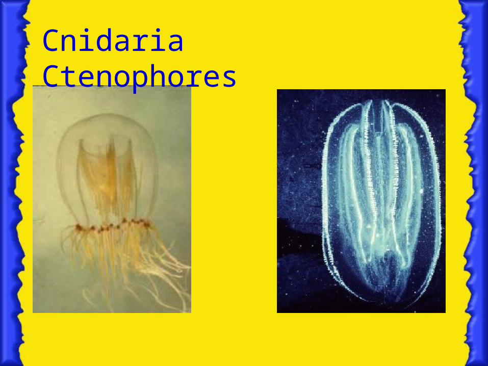
Ctenophores
Phylum Cteno • phora
(Greek: comb bearing) • teen´-ō-for´-ah
Defining Characteristics:
1.) Plates of Fused cilia arranged in rows.
2.)Adhesive prey capturing cells (colloblasts).


General Description- Ctenophores are major predators upon larval fish and
zooplankton. They may prey on small crustaceans (phylum Arthropoda) and the larval stage of various fish and jellyfish species, and oysters. Some are carnivorous on other ctenophores or gelatinous animals in other groups. They play a role in estuarine ecology.
- Most are nearly transparent- The 100 or so described species are exclusively marine
and most are planktonic. A member of most species are weak swimmers, carried about by ocean current

- The ctenophore’s tentacles are solid rather than hollow- Ctenophores have left a poor fossil record- There is an evolutionary relationship between
Ctenophora and Cnidaria.- Nearly all ctenophores are bioluminescent. They give
off excess energy in the form of light instead of heat.The functional significance of bioluminescence includes mate location and species recognition, luring of prey, and the startling of would-be predators.
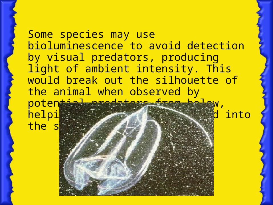
Some species may use bioluminescence to avoid detection by visual predators, producing light of ambient intensity. This would break out the silhouette of the animal when observed by potential predators from below, helping the lighted form blend into the surroundings.

Body Architecture

A. Body Covering
• Its body covering is somewhat reminiscent of Cnidarian medusae.
• The body consists of an outer epidermis, an inner gastrodermis, a thick, middle mesoglea layer.
• It has a basic radial symmetry, with oral and aboral surfaces.


B. Digestive System
• The mouth leads into a pharynx called a stomodeum, which serves as a site for extracellular digestion.
• It goes through a stomach into a series of gastrovascular canals, where digestion is completed intracellularly.

C. Excretory and Respiratory System
• There are no functional Excretory and Respiratory organs found in Ctenophores.
D. Nervous System
• It takes the form of Subepidermal nerve network.
E. Development Stages
• At lest one ctenophore species has a planula larval stage in its life history.
• During embryogenesis, Ctenophores form no distinct mesodermal layer. Thus, they are termed as dipoblastic.
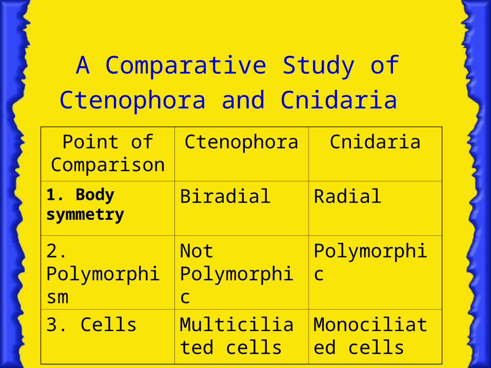
A Comparative Study of Ctenophora and Cnidaria Point of
ComparisonCtenophora Cnidaria
1. Body symmetry
Biradial Radial
2. Polymorphism
Not Polymorphic
Polymorphic
3. Cells Multiciliated cells
Monociliated cells

Ctenophora Cnidaria
4. Type of musculature
Genuine Smooth muscle
Myoephitelial cells
5. Swimming Mechanism
Multiciliated cells
Monociliated cells
6. Mechanism and mode of food capture
Tentacles are studded with colloblasts
Tentacles are studded with nematocysts
7. Means of food digestion and waste elimination
4 digestive canals and anal pores
One opening for both mouth and anus
Ctenophora Cnidaria
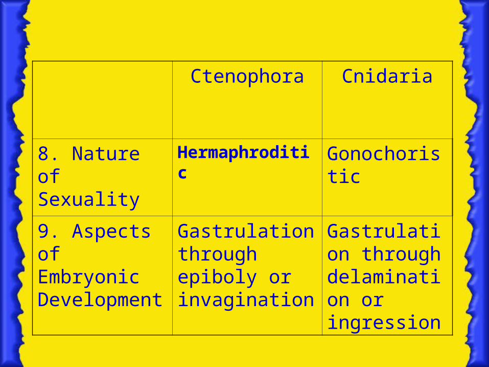
8. Nature of Sexuality
Hermaphroditic Gonochoristic
9. Aspects of Embryonic Development
Gastrulation through epiboly or invagination
Gastrulation through delamination or ingression

• Ctenophores are tripoblastic. Its muscles develop from amoeboid cells found with the mesoglea. Whereas, the muscles of cnidarians are found within the gastrodermis and, to a lesser extent, within the epidermis.
• Ctenophores have genuine smooth muscle tissues and lack the Myoephitelial cells that are found in Cnidarian musculature.
• The Mnemiopsis leidy and Beroë sp. are the first known giant smooth muscle fibers, measuring up to 6 cm long. These are of the Ctenophore species.
Back to Table
Types of Musculature
Cnidaria Ctenophores
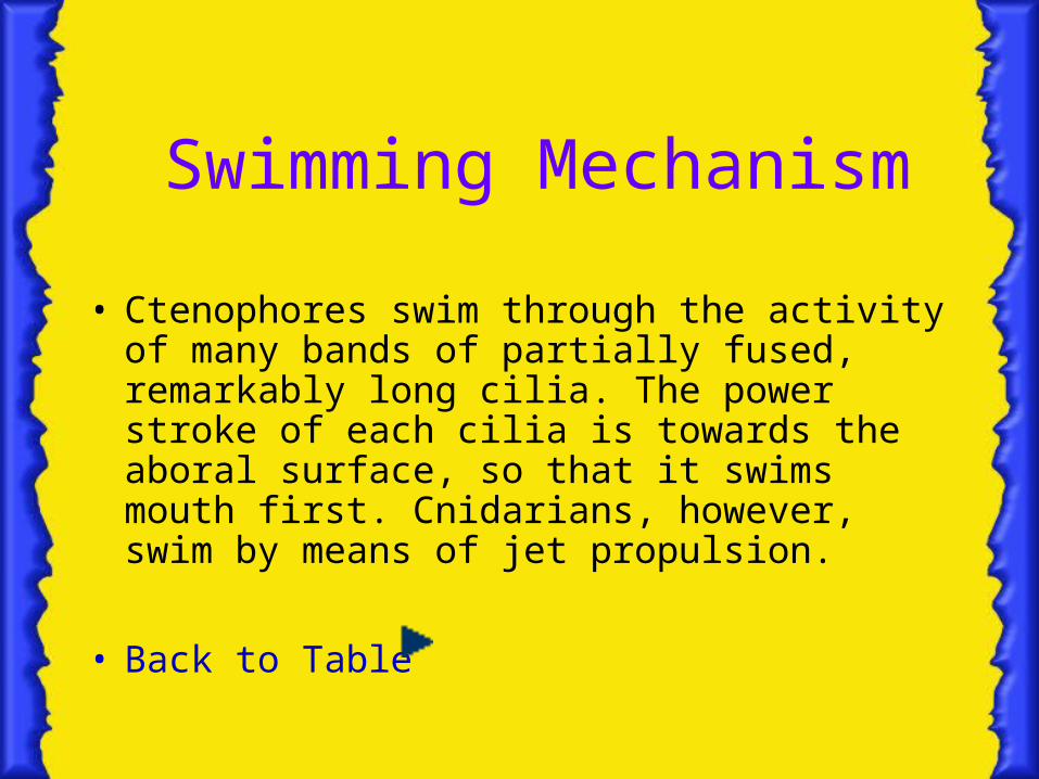
• Ctenophores swim through the activity of many bands of partially fused, remarkably long cilia. The power stroke of each cilia is towards the aboral surface, so that it swims mouth first. Cnidarians, however, swim by means of jet propulsion.
• Back to Table
Swimming Mechanism


• Each band of Cilia is fused to a ctene (Gk. Comb) because of its resemblance to a comb.
• The ctene is organized into 8 distinct rows called costae which extends from the oral to the aboral surface of the animal.
• The intensity of activity in the different comb rows is under the control of an apical sense organ at the aboral end of the ctenophore. Together with an epidermal nerve, It synchronizes the coordination of ciliary beating in different comb rows.
Back
The Ctene

• A single sphere of calcium Carbonate, called statolith, sits atop 4 tufts of fused cilia called balancers, or springs. Each balancer is composed of several hundred cilia.
• A ciliated groove radiates from each balancer and bifurcates to service two other adjacent rows. These serve as agents of nerve impulse conduction from the apicak sense organ to the ctenes of the comb rows.
Back
Mechanism and Mode of Food capture
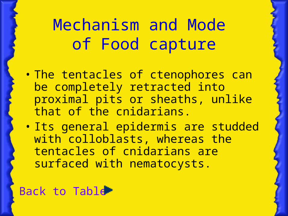
• The tentacles of ctenophores can be completely retracted into proximal pits or sheaths, unlike that of the cnidarians.
• Its general epidermis are studded with colloblasts, whereas the tentacles of cnidarians are surfaced with nematocysts.
Back to Table
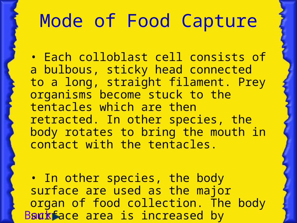
• Each colloblast cell consists of a bulbous, sticky head connected to a long, straight filament. Prey organisms become stuck to the tentacles which are then retracted. In other species, the body rotates to bring the mouth in contact with the tentacles.
• In other species, the body surface are used as the major organ of food collection. The body surface area is increased by lateral compression and is major areas are coated with a sticky mucus and colloblast cells.
Mode of Food Capture
Back

Means of Food Digestion and Waste Elimination
• Ctenophores have 4 digestive canals which lead from the roof of the stomach to the animal’s aboral surface. 2 of these digestive canal terminate as blind sacs, while the other 2 canals open wide outside.Undigested wastes are discharged through anal pores . Cnidarians, on the other hand, has but one opening which serves as both mouth and anus.
Back to Table

Nature of Sexuality• Ctenophores are hermaphrodites; that
is, a single individual has both male and female gonads. In contrast, Cnidarians are gonochoristic, with one sex per individual.
Back to Table
• A few species reproduce asexually through fragmentation and the subsequent development of missing body parts by each fragment.
• Gonads are located on the walls of some or all of the gastrovascular canals, so that gametes are liberated into the digestive tract and are discharged through the mouth.
• Eggs are fertilized externally.
Back

Aspects of Embryonic Development
• Ctenophore cleavage is highly determinate; cell fates are fixed at the first cell division. Cell fates of Cnidarian embryos become fixed later in development.
• Gastrulation,or the formation of distinct inner and outer germs, are achieve by ctenophores either by epiboly or by invagination. Cnidarians gastrulate either through the process of delamination or by ingression
• Ctenophore embryos develop directly into a cydippid, while Cnidarians develop into a ciliated planula larvae
Back to Table

Definition of Terms• Epiboly : A process in which a sheet of micromeres
spreads over what were the adjacent macromeres• Invagination :Process by which groups of cells push
into the blastocoelic space. This may also be exhibited by some Cnidarians
• Delamination :The cells of the blastula divide with the cleavage plane approximately parallel to the surface of the embryo. Thus, the cells divide into the blastocoel, forming an inner and outer cell layer, between which the mesoglea is later secreted
Back
• Ingression :Certain cells become detached from their neighbors and simply move into the blastocoel, creating a second layer of cells.
• Cydippid :A miniature ctenophore which is approximately spherical in shape; is endowed with 8 comb rows, a fully formed apical sense organ, and a pharynx; and usually bears a pair of branched tentacles. It may closely resemble an adult, though in other modern species, the cydippid undergoes metamorphosis before attaining its adult form
Back

Ctenophore Diversity
Class Tentaculata
A. Order Cydippida • Tentaculate species closely resemble the
cydippid larvae except that the functional gonads are present.
• Food is capture through long retractable tentacles and their side branches.
B. Lobata• Body is compressed laterally, only 4 comb rows are fully
developed, and the tentacles are generally much reduced in length.
• Large oral lobes, covered with mucus and colloblasts, constitute the primary food collection surface.
• The muscular cavity of the 2 oral lobes aids locomotion in some species. 2 pairs of ciliated, paddle- or tentacle-like structure,auricles, assist in prey capture

C. Cestida• The body is so compressed laterally that it forms a long
ribbon, with the mouth and apical sense organ on opposite sides of its midpoint.
• Cestids swim through a combination of ctene activity and sinuous, muscular movements of the body; only 4 of the comb rows are well developed in adults.
• Prey are captured by the numerous short tentacles extending along the extensive oral edge of the ctenophore.
D. Platyctenida• The oral and aboral surfaces have moved towards each
other, so the body forms a flattened plate. • The bottom of the plate is formed largely by a pharynx, which
is extensively and permanently everted.• Some species simply float in the water while others creep
slowly over solid substrates through pharyngeal cilia and muscular contractions or by muscular flapping of the lateral lobes of the body..
• The only non-planktatonic ctenophores. • Has 2 long tentacles.

Class NudaBeroida• Has no tentacles and oral lobes.• Has 8 well developed comb rows.• Captures and engulfs prey, including other ctenophores,
through muscular lips surrounding the mouth. The mouth can be widened to accommodate prey substantially larger than the predator.
• Thousands of 9 + 2 axonemes enclosed by a single membrane, Macrocilia, are located just inside the mouth. These Macrocilia are used as teeth to chop large prey into bite-size pieces.