ct intervention tips - de.medical.canon · harter lp, moss aa, golberg hi, gross bh. ct-guided...
TRANSCRIPT
/461
ct intervention tips
geoffrey dick
b.app.sci. (med.rad.) rmit
• deputy chief radiographer angliss hospital, melbourne
• ct supervisor angliss hospital, melbourne
• sessional lecturer monash university department of medical
imaging & radiation sciences
• vsnmt education committee member

/46
objectives
• patient preparation
• ctf
• radiation dose reduction techniques
• step by step set-up/guide
2

/46
patient preparation
3

/46
interventional success
• everyone has a specific job to do!
• patient
• must be cooperative
• radiographer
• nurse
• radiologist
4

/46
patient preparation
• informed consent
• specific instructions
• blood clotting results
• fasting (13)
5

/46
contraindications
• abnormal clotting
• patient on anti-coagulants
• uncontrollable movement
• non-compliant patient
6

/46
nurse preparation
• consent form preparation
• sterile equipment set-up
• drapes
• drugs
• needles etc.
7

/46
radiologist preparation
• get off the phone!
8

/46
radiographer preparation
• previous films/imaging
• correct patient position
• respiration
• inspiration
• expiration
• quiet breathing
9

/46
scan set-up
• localise lesion
• view previous imaging!
• determine most appropriate needle path
• low dose target localisation scan
• must include skin edge!
10

/46
site preparation
• puncture site determined
• triple check location
• check ‘image’ table position
• table position on ‘gantry’
• VERY important
11
/46
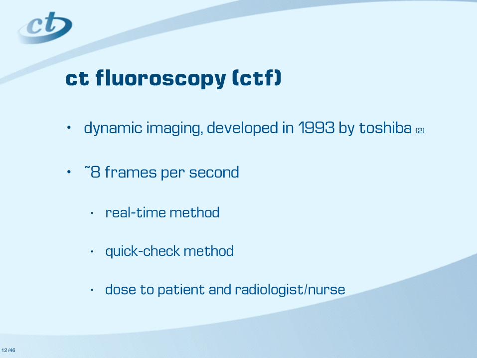
ct fluoroscopy (ctf)
• dynamic imaging, developed in 1993 by toshiba (2)
• ~8 frames per second
• real-time method
• quick-check method
• dose to patient and radiologist/nurse
12

/46
ctf
13

/46
limiting ctf radiation dose
• ↓ kVp
• ↓ examination time
• ↓ mA
• remember, radiation dose is accumulative!
14

/4615

/46
technique example
16

/4617
select correct patient,ORGAN = CTF
select PROTOCOL SELECTION
diamond
select correct body area in GROUP A
perform DUAL scanograms (can have
GRID on patient)
correctly position patient, with table low in the gantry and zero
table
scan correct area (including skin surface) with sticky
BIOPSY GRID in situ using laser lights to position GRID
click FILMING tab current patient will be displayed
hit F4 on the keyboard to bring up patient's image
folderselect image group that
you want displayed
select TOOL2 and INSET (places scanogram in
corner of image)
click on inset image with left mouse button and use
middle mouse to move image and left/middle to
magnify
choose correct level, close INSET application
hit F2 to bring up measurement tool and
select LINE measurement tool
measure needle skin entry point to target, F7 to
screen save with measurement on the
desired image
click SCAN tab and QUIT EXAM
select correct CTF protocol
ensure CTF patient orientation is correct
click and select 3 boxes at top of CTF screen
hit F4 and load screen save from previous step
click and select bottom box on CTF screen
scroll through images to select correct table position that matches screen save
hit F1 on the keyboard to bring up MANUAL
MOVE
enter table position of desired screen save, hit
ENTER
if using "true" CTF radiologist enables hand
controller inside room
press ‘orange’ table move button to move to
CTF location
turn on gantry laser lights and click STOP
SCAN
use SHARPIE to mark up correct needle entry point on patients body,
using grid and laser
enter table position of ~+/-600, hit ‘orange’ table
move button to move table out of gantry
TYPE in desired CTF table location in
MANUAL MOVE, hit ENTER
press ORANGE table move/tilt button on
keyboard to move table to correct position
change SLICE thickness or EXPOSURE if
requiredperform interventional
procedure
to move patient out UNLOCK table using
hand controller or gantry controls and PUSH table/patient out of
gantry
re-LOCK tableTRANSFER image with
needle position/tip to PACS
PRINT image with needle position/tip for
referrer
set-up CTF hand controller and for
switch
click CONFIRM and move patient to scan
1 2
QUIT CTF, QUIT manual move table window, QUIT EXAM, NEXT
PATIENT
CTF CHEAT SHEET CTF CHEAT SHEET

/4618
select correct patient,ORGAN = CTF
select PROTOCOL SELECTION
diamond
select correct body area in GROUP A
perform DUAL scanograms (can have
GRID on patient)
correctly position patient, with table low in the gantry and zero
table
scan correct area (including skin surface) with sticky
BIOPSY GRID in situ using laser lights to position GRID
click FILMING tab current patient will be displayed
hit F4 on the keyboard to bring up patient's image
folderselect image group that
you want displayed
select TOOL2 and INSET (places scanogram in
corner of image)
click on inset image with left mouse button and use
middle mouse to move image and left/middle to
magnify
choose correct level, close INSET application
hit F2 to bring up measurement tool and
select LINE measurement tool
measure needle skin entry point to target, F7 to
screen save with measurement on the
desired image
click SCAN tab and QUIT EXAM
select correct CTF protocol
ensure CTF patient orientation is correct
click and select 3 boxes at top of CTF screen
hit F4 and load screen save from previous step
click and select bottom box on CTF screen
scroll through images to select correct table position that matches screen save
hit F1 on the keyboard to bring up MANUAL
MOVE
enter table position of desired screen save, hit
ENTER
if using "true" CTF radiologist enables hand
controller inside room
press ‘orange’ table move button to move to
CTF location
turn on gantry laser lights and click STOP
SCAN
use SHARPIE to mark up correct needle entry point on patients body,
using grid and laser
enter table position of ~+/-600, hit ‘orange’ table
move button to move table out of gantry
TYPE in desired CTF table location in
MANUAL MOVE, hit ENTER
press ORANGE table move/tilt button on
keyboard to move table to correct position
change SLICE thickness or EXPOSURE if
requiredperform interventional
procedure
to move patient out UNLOCK table using
hand controller or gantry controls and PUSH table/patient out of
gantry
re-LOCK tableTRANSFER image with
needle position/tip to PACS
PRINT image with needle position/tip for
referrer
set-up CTF hand controller and for
switch
click CONFIRM and move patient to scan
1 2
QUIT CTF, QUIT manual move table window, QUIT EXAM, NEXT
PATIENT
CTF CHEAT SHEET CTF CHEAT SHEET

/46
ctf operator dose
19

/4620

/4621

/4622

/46
ctf operator dose
23

/4624

/4625

/4626
pre icrp 60 - 0.6 mSv icrp 103 - 0.4 mSv
icrp 60 - 0.8 mSv icrp 103 - 0.5 mSv
ctf icrp 60 - 0.2 mSv icrp 103 - 0.1 mSv

/46
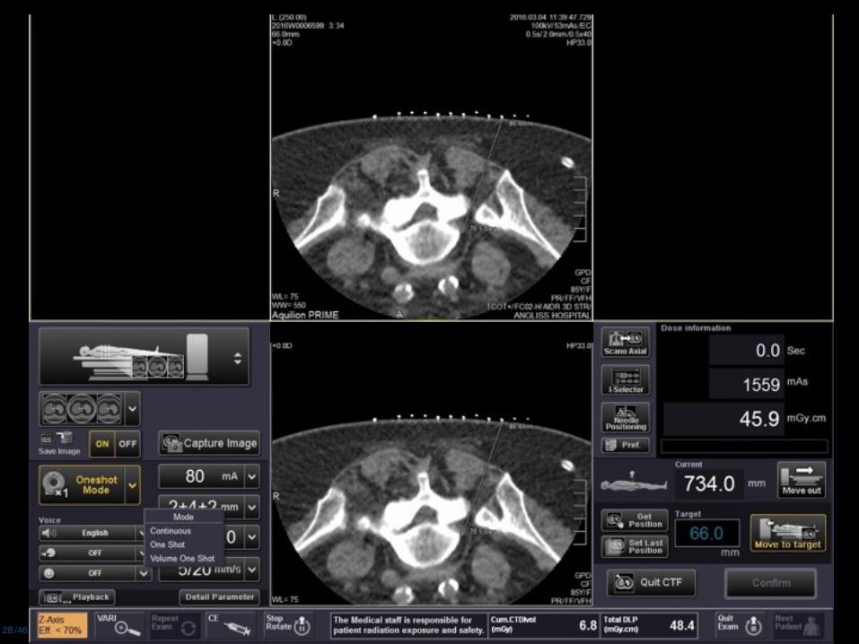
volume one shot
27
/4628

/4629

/4630
/4631

/46
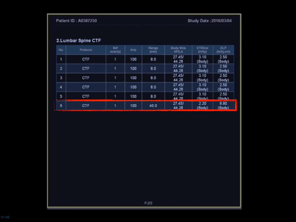
pre-scan dose
• radiation dose reduction for ctf-guided ESI is
best achieved by minimising the dose from the
preliminary planning lumbar spine ct scan (25)
32

/46
pre-scan dose
• ↓ kVp
• ↓ mA
• ↓ scan range
• change detector configuration?
• not a diagnostic scan, just a map
33

/4634

/4635

/4636

/4637
pre-scan
icrp 60 - 0.8 mSv icrp 103 - 1.1 mSv
pre-scan
icrp 60 - 0.3 mSv icrp 103 - 0.5 mSv

/46
previous case
• ↓ kVp (for pre-scan & ctf)
• 120 → 100
• ↑ noise/sd
• 10.5 → 18.0
• 0.5 x 80 mm → 0.5 x 40 mm
• ↓ scan range
38

/4639
Artner et. al. 2012
Conclusion: Radiation dose of CT-guided SI joint injections can be decreased to levels of pulsed fluoroscopy with a precise intra- articular needle placement using the low-dose protocol. The technique is simple to perform, fast, and reproducible.

/46
take home messages
• planning, planning, planning
• dose reduction
• concentrate
• understand
40

/46
thanks
• andrew wilson
• alison stubbs (cook medical)
41

/46
references
42
1. Letourneau, Janis G. Percutaneous Biopsy of Abdominal Masses. Interventional Radiology. Williams and Wilkins. Baltimore, 1988. pp793-813.
2. Maher, Kieran. CT Fluoroscopy. RMIT Computed Tomography Short Course Notes, July 1999.
3. VanSonnenberg E, Lin AS, Deutsch AL, Mattrey RF. Percutaneous Biopsy of Difficult Mediastinal, Hilar, and Pulmonary Lesions by Competed Tomographic Guidance and a Modified Coaxial Technique. Radiology 1983 (148): 300-302.
4. Klein JS, Salomon G, Stewart EA. Transthoracic Needle Biopsy with a Coaxially Placed 20-gauge Automated Cutting Needle: Results in 122 Patients. Radiology 1996 (198): 715-720.
5. Nashed Z, Klein JS, Zarka MA. Special Techniques in CT-Guided Transthoracic Needle Biopsy. AJR 1998 (171): 1665-1668.
6. Sheiman RG, Fey C, McNicholas M, Raptopoulos F. Possible Cause of Inconclusive Results on CT-Guided Thoracic and Abdominal Core Biopsies. AJR 1998 (170): 1603-1607.
7. Silverman SG, Tuncali K, Adams DF, Nawfel RD, Zou KH, Judy PF. CT Fluoroscopy-guided Abdominal Interventions: Techniques, Results, and Radiation Exposure. Radiology 1999 (212): 673-681.

/46
references
43
8. Kazerooni EA, Lim FT, Mikhail A, Martinez FJ. Risk of Pneumothorax in CT-guided Transthoracic Needle Aspiration Biopsy of the Lung. Radiology 1996 (198): 371-375.
9. Westcott JL, Rao N, Colley DP. Transthoracic Needle Biopsy of Small Pulmonary Nodules. Radiology 1997 (202): 97-103.
10. Sheafor DH, Paulson EK, Kliewer MA, DeLong DM, Nelson RC. Comparison of Sonographic and CT Guidance Techniques. AJR 2000 (174): 939-942.
11. Gobien RP, Stanley JH, Vujic I, Gobien BS. Thoracic Biopsy: CT Guidance of Thin-Needle Aspiration. AJR 1984 (142): 827-830.
12. Khouri NF, Stitik FP, Erozan YS, Gupta PK, Kim WS, Scott WW, Hamper UM, Mann RB, Eggleston JC, Baker RR. Transthoracic Needle Aspiration Biopsy of Benign and Malignant Lung Lesions. AJR 1985 (144): 281-288.
13. Tarver RD, Kreipke DL, Conces DJ, Becker GJ. Interventional Radiology of the Chest. Interventional Radiology. Williams and Wilkins. Baltimore, 1988. 770-778.

/46
references
44
14. Silverman SG, Bloom DA, Seltzer SE, Tempany CMC, Adams DF. Needle-Tip Localization During CT-Guided Abdominal Biopsy: Comparison of Conventional and Spiral CT. AJR 1992 (159): 1095-1097.
15. Nawfel RD, Judy PF, Silverman SG, Hooton S, Tuncali K, Adams DF. Patient and Personnel Exposure during CT Fluoroscopy-guided Interventional Procedures. Radiology 2000 (216): 180-184.
16. Cox JE, Chiles C, McManus CH, Aquino SL, Choplin RH. Transthoracic Needle Aspiration Biopsy: Variables That Affect Risk of Pneumothorax. Radiology 1999 (212): 165-168.
17. Daly B & Templeton PA. Real-time CT Fluoroscopy: Evolution of an Interventional Tool. Radiology 1999 (211): 309-315.
18. Swerdlow DR & Thaete FL. CT-Guided Core Biopsy of Difficult Lesions: A Modified Coaxial Approach. AJR 1994 (163): 195-196.
19. Daly B, Krebs TL, Wong-You-Cheong JJ, Wang SS. Percutaneous Abdominal and Pelvic Interventional Procedures Using CT Fluoroscopy Guidance. AJR 1999 (173): 637-644.
20. Harter LP, Moss AA, Golberg HI, Gross BH. CT-Guided Fine-Needle Aspirations for Diagnosis of Benign and Malignant Disease. AJR 1983 (140): 363-367.

/46
references
45
21. Alfidi RJ, Haaga JR, Meaney TF et al. Computed Tomography of the Thorax and Abdomen. Radiology 1975 (117): 257-264.
22. Westcott JL. Direct Percutaneous Needle Aspiration of Localized Pulmonary Lesions: Results in 422 Patients. Radiology 1980 (137): 31-35
23. Chapman S & Nakielny R. A Guide To Radiological Procedures. Bailliere Tindal, London, 1993.
24. Solomon, S. B., et al. (2002). "Robotically driven interventions: a method of using CT fluoroscopy without radiation exposure to the physician." Radiology 225(1): 277-282.
25. Hoang, J. K., et al. (2011). "Radiation dose exposure for lumbar spine epidural steroid injections: a comparison of conventional fluoroscopy data and CT fluoroscopy techniques." AJR Am J Roentgenol 197(4): 778-782.
26. Artner, J., F. Lattig, H. Reichel and B. Cakir (2012). "Effective radiation dose reduction in computed tomography-guided spinal injections: a prospective, comparative study with technical considerations." Orthop Rev (Pavia) 4(2): e24.
27. Hayot M, Michaud A, Koechlin C, Caron MA, Leblanc P, Prefaut C, et al. Skeletal muscle microbiopsy: a validation study of a minimally invasive technique. The European respiratory journal. 2005;25(3):431-40.

/46
references
46
27. Hayot M, Michaud A, Koechlin C, Caron MA, Leblanc P, Prefaut C, et al. Skeletal muscle microbiopsy: a validation study of a minimally invasive technique. The European respiratory journal. 2005;25(3):431-40.