csf shunts: a primer tamara simon, m.d. july 2004
TRANSCRIPT
CSF Shunts:A Primer
Tamara Simon, M.D.July 2004

Purpose• CSF flow:
– produced in choroid plexus of ventricles– Flows through lateral ventricles, through foramen of Monro, to
third ventricle– Flows through aqueduct of Sylvius to fourth ventricle– Flows through foramina of Lushka and Magendie to
subarachnoid space– Reabsorbed by arachnoid villi and arachnoid granulations into
the venous sinuses
• Hydrocephalus develops when there is an increase in CSF production, decrease in CSF absorption, or (most commonly) obstruction to flow

Procedure
• Allows for relief of hydrocephalus • Allows for prevention of increased ICP
– Tumors– Congenital anomalies– Posttraumatic hemorrhage– Intraventricular hemorrhage– Postinfectious obstruction– Other causes

Types of Shunts
• Named for position of proximal and distal catheters
• Most commonly, ventriculoperitoneal shunts are placed
• Proximal catheters are in lateral, third, or fourth ventricles or in intracranial cyst
• Distal catheters can be in peritoneal space, right atrium, pleural space, gallbladder, ureter, urinary bladder, bone marrow, mastoid, thoracic duct, fallopian tube, and other locations

Anatomy of a CSF shunt• Proximal catheter
– Placed in ventricle
– Exits skull through burr hole
• One way valve system– Allows one-way drainage of CSF at
predetermined pressure differential
– May be integrated into distal catheter or separate
– On exterior of skull

Anatomy of a CSF shunt (cont)• Distal catheter
– Tunneled under skin to final destination
• Other components:– On-off valves
• Used for intermittent shunting• Can be used to assess shunt function
– Antisiphon devices • Prevents overdrainage of CSF
– Reservoirs (single or double chamber)• Allows withdrawal of CSF or drug infusion• On exterior of skull proximal to one way valve

Sample CSF Shunts
• Rickham reservoir with antisiphon device (left)
• Single chamber reservoir (middle)
• Rickham reservoir with double chamber reservoir/valve system (right)

Temporary Shunts
• In patients who have rapidly progressive ventriculomegaly or progressive or symptomatic ventriculomegaly, patients
• can be quite small • have persistently proteinaceous and cellular CSF• be at high risk for shunt obstruction and infection
• Some preterm infants will not evolve to permanent, shunt-dependent hydrocephalus by term, so a temporary technique is at times expeditious
• Several studies cite 25% of patients with posthemorrhagic hydrocephalus ultimately recover

Temporary Shunts (continued)
• Surgeons believe that wound dehiscence and skin breakdown over shunt hardware are less frequent in larger infants as well
• Clinical endpoint:• infant reaches term and
• weight of 2 kg.

Temporary Shunt: EVD
• External ventricular drainage• Drain placed into CSF space and drained directly
externally.• Pros: Attain temporary drainage• Cons:
– drain interferes with nursing care– drain is easily dislodged– drain obstruction is frequent because of low CSF flow
volumes– formidable risk of infection with prolonged drainage
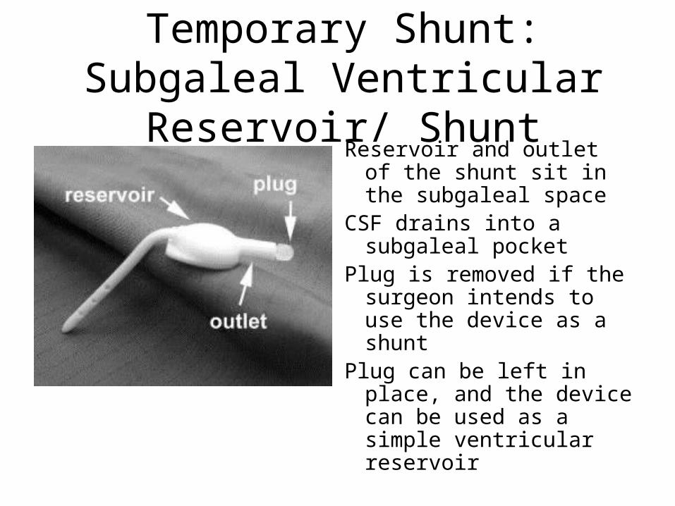
Temporary Shunt: Subgaleal Ventricular Reservoir/ Shunt
Reservoir and outlet of the shunt sit in the subgaleal space
CSF drains into a subgaleal pocket
Plug is removed if the surgeon intends to use the device as a shunt
Plug can be left in place, and the device can be used as a simple ventricular reservoir

Temporary Shunt: Subgaleal Ventricular Reservoir
• Also called ventricular access device • Small, flat-bottomed reservoir attached to a
ventricular catheter– reservoir sits on the surface of the skull under the galea of
the scalp– percutaneous puncture of the reservoir with aspiration of
CSF on a daily or every-other-day schedule serves to keep the ventricular system decompressed.
• Clinical endpoints are arrest of ventricular dilatation, control of head growth, and elimination of symptoms and signs of elevated ICP.

Temporary Shunt: Subgaleal Shunt
• Ventricular access device with an outlet • Reservoir with outlet is placed in a large subgaleal
pocket on the surface (hemicranium) of the skull• CSF decompression occurs by draining through the
reservoir, out the outlet, into the subgaleal pocket – subgaleal space probably has some absorptive capacity– pocket also serves a simple mechanical function as a high-
compliance receptacle for ventricular CSF– over weeks, scarring of scalp to periosteum obliterates the pocket,
and periodic needle aspiration of the shunt reservoir can be initiated
• Some believe that subgaleal shunts control ventricular volume more consistently

Complication: Shunt Malfunction• Most common complication, seen in 30-40% of
shunt procedures and 67% of patients with shunts• Usually caused by simple obstruction
– Debris, fibrosis, choroid plexus, or parenchymal occlusion of proximal catheter (first 2 years after placement in general)
– Kinking, knotting, breaking, obstruction, migration of distal catheter (after 2 years after placement)
• Also caused by infection, disconnection of shunt components, catheter migration, inadequate drainage, overdrainage

Complication: Shunt Malfunction• Varied signs and symptoms
– Swelling or erythema around shunt tract - Lethargy– Bulging or full fontanel - Ataxia– Increased head circumference - Neck pain– Headache - Back pain– Irritability - Blurred vision– Increased seizures - Sun setting eyes– Vomiting - Behavioral changes– Papilledema - Not acting right
• Most predictive?– Vomiting, lack of fever, parental suspicion
Complication: Infection
• Second most common complication, seen in 2-30% of shunt procedures
• Increased risk in children under 1 year of age, a short duration from shunt procedure
• Most common organisms:– Coagulase-negative Staph species– Staph epidermidis– Staph aureus– Gram negative rods (6-20%)– Pathogens that cause meningitis (more remote)
Complication: Infection• Vague signs and symptoms
– Swelling, erythema, cellulitis, or wound infection around shunt tract– Fever - Shunt malfunction– Nausea - Vomiting– Lethargy - Irritability– Headache -Change in sensorium– Feeding problems
• When VP shunt is present, additionally:– Abdominal pain – Diarrhea – Peritonitis

Complication: Slit Ventricle Syndrome
• Found in 50-60% of patients, only symptomatic in 11-37%, require treatment in 6-7%
• Overdrainage of CSF leads to collapse of ventricles, blocking fenestrations in proximal catheter, leading to increased ICP
• Symptoms similar to shunt malfunction until ICP rises and ventricles re-expand
• Some patients are position-sensitive and lying down increases ICP
• Diagnosed when head CT shows small- to normal-sized ventricles

Complication: Proximal Catheter Obstruction
• Medical signs of increased ICP– Hyperventilation– Diuretics (acetazolamide, mannitol)– Elevate head of bed– Ventricular puncture through burr hole or open
fontanel

Complication: VP Shunts• Inguinal hernia
– Increased abdominal fluid increases intra-abdominal pressure, converting potential in clinical hernia
• Perforation of hollow viscus– Bladder, stomach, small intestine, colon, gallbladder, vagina, anus, and mouth have
been reported– Bowel perforation can present with peritonitis, meningitis, ventriculitis; with signs of
shunt infection
• Abdominal pseudocyst (0.8-10%)– Decreased appetite, abdominal pain, tenderness, distention, mass, and guarding;
increased ICP and shunt malfunction– Foreign body reaction with chronic granulomatous inflammation
• Migration of distal catheter tip– Through abdominal incision, through neck incision, into mediastinum, throacic
cavity, umbilicus

Complications: Other Rare Ones
• Intussusception
• Intractable hiccup
• Omental cyst torsion
• Volvulus around catheter

Radiographic studies• Shunt series
– Plain radiographs of skull, neck, chest, abdomen– Used to detect disconnections, kinks, and migration of catheters– Proximal and distal catheters are radiopaque, reservoirs are radiolucent
• Head CT– Demonstrates location of proximal catheter tip and size of ventricles– Comparison to prior study is critical
• Ultrasound– For children with open fontanel
• Radionucleotide clearance study– Radionucleotide is injected into shunt reservoir and observed as it flows
proximally and distally

Further Diagnostic Studies
• Pumping the shunt reservoir– Assesses proximal and distal shunt function– Pitfall abound, experience is needed- consult Neurosurgery
• Tapping the shunt– Assess shunt function and diagnoses shunt infection– Consult Neurosurgery– Reservoir is cleaned, 23 gauge butterfly needle +/- manometer
is inserted into reservoir– Opening pressure, rate of flow, closing pressure, and CSF
sample is obtained– CSF should be sent for culture, Gram stain, protein, glucose,
and cell count

References
• Teoh DL. Tricks of the Trade: Assessment of High-Tech Gear in Special Needs Children. Clinical Pediatric Emergency Medicine. 3(1), March 2002.
• Baddour LM, Flynn PM, Fekete T. Infection of central nervous system shunts and other devices. Up To Date. April 30, 2004.
• Garton HJ, Piatt JH. Hydrocephalus. Pediatric Clinics of North America, 51(2), April 2004.