cryptosporidiosis in cattle - moredun group 6.1.pdf · news sheet vol 6 no 1 3 introduction...
TRANSCRIPT

Cryptosporidiosis in Cattle
The Moredun Foundation
News Sheet Vol. 6, No. 1, February 2014
Beth Wells BSc, PhD
Sarah Thomson BSc, MRes
Moredun Research Institute

News sheet Vol 6 No 1 2
55
Key points
• Cryptosporidiosis is the disease caused by infection with the protozoan parasite Cryptosporidium.
• Only one species, Cryptosporidium parvum, causes disease in cattle and generally only in neonatal calves.
• Clinical signs can range from mild scouring to calf death depending on the parasite burden, susceptibility and health status of the calves.
• Cryptosporidium transmission to young calves can come from many sources including other calves, their dams, animal handlers, other animals and the
environment.
• Cryptosporidium infection can cause serious problems on some farms and current statistics indicate this parasite is the commonest cause of scour in young calves in
the UK.
• The life cycle of the parasite allows it to multiply rapidly in the host leading to the rapid spread of the disease within a susceptible group of animals.
• Infected animals can shed millions of infectious parasites into the environment.
• Accurate diagnosis is crucial and is available through your vet.
• Currently there is no vaccine available and treatment options are limited.
• There are effective management solutions which can significantly reduce the parasite burden on farm and thereby the impact of disease.
• The parasite is opportunistic and is therefore diagnosed in many mixed infections where prevention/treatment of the other pathogens will reduce the impact of
cryptosporidiosis.

News sheet Vol 6 No 1 3
Introduction Cryptosporidiosis is the disease caused by the protozoan parasite Cryptosporidium. There
are currently over 26 species of this parasite which have been identified in many
mammals, birds and reptiles, but fortunately not all of them cause disease!
There are 4 species found in cattle but only one of them, Cryptosporidium parvum, causes
clinical disease and this is usually only in young calves of less than 6 weeks old.
Cryptosporidiosis caused by C. parvum is zoonotic which means it can be passed to
humans where it can cause disease. In the majority of the population, the clinical signs of
cryptosporidiosis are generally mild with self limiting diarrhoea, abdominal pain and
dehydration. In some circumstances however, such as in the very young, elderly or
immuno-compromised individuals, cryptosporidiosis can be more serious or even fatal.
The infective part of the parasite‟s life cycle, the oocyst or egg, has a very tough outer
shell and can survive very happily in a range of environmental conditions particularly if it is
mild and humid. They are therefore very well equipped to survive the UK climate, where
they can remain viable for over a year in soils, on pasture and in water.
The oocyst stage of the parasite can also survive many commonly used farm disinfectants
and water chlorination treatment, making it difficult for farmers, vets and water suppliers
to control or inactivate it.
Given these issues, along with the fact that C. parvum can cause serious disease outbreaks
in susceptible calves, leading to significantly reduced farm incomes in severe cases,
Moredun is continuing research into this parasite and currently have work ongoing in
immunity, strain pathogenicity and transmission, oocyst prevalence and parasite survival in
the farm and catchment environment.
A massive waterborne outbreak of cryptosporidiosis was reported in
1993 at Milwaukee, Wisconsin, USA where an estimated 403,000 people
were infected due to faulty filtration of the water from Lake Michigan
which supplied the area. Many of these people suffered hospitalisation
and serious illness, the average duration of which was 8 weeks. Possible
sources of infection were cited as human sewage, slaughterhouses and
cattle grazing the banks of the rivers flowing into the lake.
News sheet Vol 6 No 1 4
55
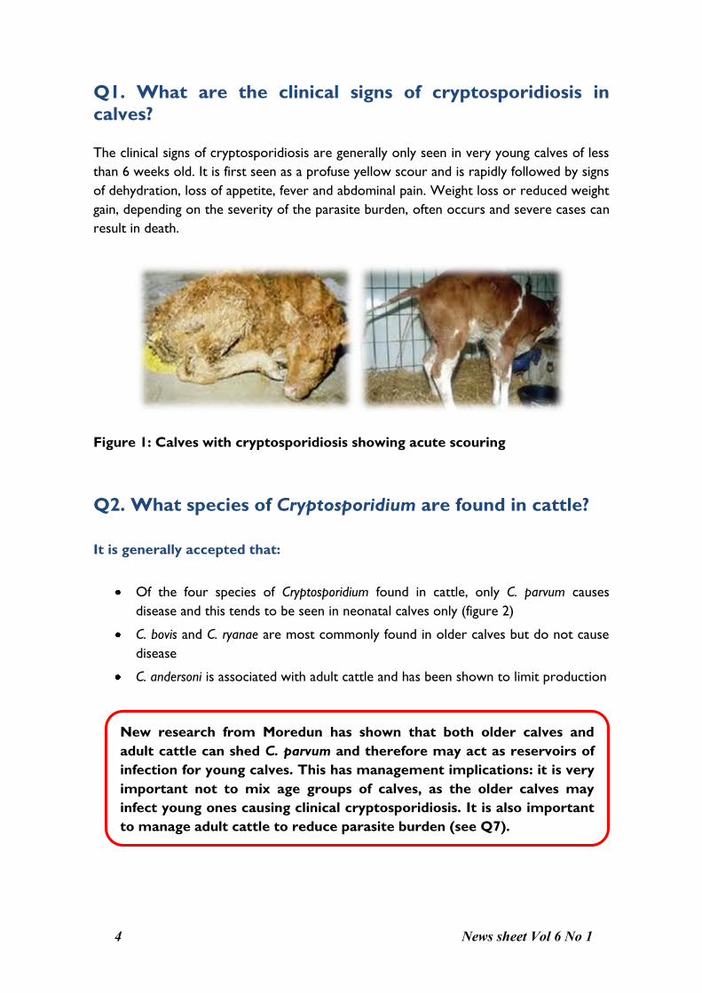
Q1. What are the clinical signs of cryptosporidiosis in
calves? The clinical signs of cryptosporidiosis are generally only seen in very young calves of less
than 6 weeks old. It is first seen as a profuse yellow scour and is rapidly followed by signs
of dehydration, loss of appetite, fever and abdominal pain. Weight loss or reduced weight
gain, depending on the severity of the parasite burden, often occurs and severe cases can
result in death.
Figure 1: Calves with cryptosporidiosis showing acute scouring
Q2. What species of Cryptosporidium are found in cattle?
It is generally accepted that:
Of the four species of Cryptosporidium found in cattle, only C. parvum causes
disease and this tends to be seen in neonatal calves only (figure 2)
C. bovis and C. ryanae are most commonly found in older calves but do not cause
disease
C. andersoni is associated with adult cattle and has been shown to limit production
New research from Moredun has shown that both older calves and
adult cattle can shed C. parvum and therefore may act as reservoirs of
infection for young calves. This has management implications: it is very
important not to mix age groups of calves, as the older calves may
infect young ones causing clinical cryptosporidiosis. It is also important
to manage adult cattle to reduce parasite burden (see Q7).

News sheet Vol 6 No 1 5
Figure 2: Species of Cryptosporidium commonly found in different age groups
of cattle
Q3. Where do calves get Cryptosporidium from? From the time of birth, there are many potential sources of infection of Cryptosporidium
for the calf (figure 3). An understanding of what these are and where they come from will
help achieve a reduction in levels of parasite burden on farm.
The environment is known to be a major source of oocyst infection for calves –
including cattle sheds, bedding, pasture, soil and drinking water.
Other calves, their dams and other animals are also potential reservoirs for the
oocysts. Sheep, and in particular lambs, as well as deer have been shown to shed
C. parvum oocysts.
Animal handlers, vets and any other people accessing the cattle may also act as
sources of infection. Good hygiene procedures using suitable disinfectants for
Cryptosporidium oocysts (see Q7) should be implemented, such as provision of
footbaths and clean clothing.

News sheet Vol 6 No 1 6
55
Figure 3: Potential sources of infection of Cryptosporidium to the calf (main
routes of transmission)
Q4. Is cryptosporidiosis a practical problem on farms? Cryptosporidium infection can cause serious problems on some farms due to the biology of
the parasite oocysts (the infective stage). Problems include:
Environmentally stable oocysts
Resistant to commonly used disinfectants
Resistant to heat (up to 60°C) and cold (down to -20°C)
Can survive up to 12 months in cool, moist environments
Low infectious dose
Infection can spread rapidly through farm/groups of calves
As few as 10 oocysts can cause disease in susceptible calves
One infected animal can shed 1, 000, 000, 000‟s (billions) of oocysts – potentially enough to infect 100, 000, 000 other calves!

News sheet Vol 6 No 1 7
Current figures of calf scour prevalence in the UK demonstrate that cryptosporidiosis
has remained the main cause of enteritis in calves over the past 10 years (Veterinary
Investigation Surveillance Report (VIDA, 2012)) and the cause of 38% of the reported
neonatal calf scours in 2012 (figure 4). It is therefore considered a serious disease
problem to the UK livestock industry.
Figure 4: Main causes of scour in neonatal calves in the UK (VIDA, 2012)
Q5. How do ten oocysts turn into billions?
A feature of Cryptosporidium is its ability to multiply rapidly in the host leading to a
potentially rapid spread of infection through a calf group or farm. The parasite achieves
this as each oocyst hatches in the gut releasing 4 sporozoites which individually invade the
gut wall, multiply and develop into oocysts, completing the life cycle in 2-7 days. The
damage to the gut caused by the invading and multiplying parasites leads to the clinical
signs evident in calves infected with Cryptosporidium.
In addition, during the infection process the parasite produces some thin-walled oocysts
which burst open in the gut causing auto-infection within the host, leading to further
multiplication of the parasite (figure 5).
4%
34%
6%18%
38%
Neonatal Enteritis - UK 2012
E. coli
Rotavirus
Coronavirus
Coccidiosis
Cryptosporidiosis

News sheet Vol 6 No 1 8
55
Figure 5: Cryptosporidium life cycle showing how the parasite can rapidly
increase in number in the host
Q6. How do I know if I have Cryptosporidium on my farm? Highly sensitive and specific diagnostic tests are available to diagnose Cryptosporidium
infections in cattle faecal samples. If you have a scouring calf, the options are:
1. Confirmation of clinical observation by diagnosis using faecal samples
This may be done at your vet‟s surgery either by use of a rapid diagnostic kit for the
detection of C. parvum, E. coli K99 and rotavirus, or by microscopy for the detection
of Cryptosporidium oocysts.
SAC Consultancy (Scotland) and AHVLA (England and Wales) offer calf enteritis
screening packages which can include Cryptosporidium along with choices of
combinations for the main bacterial and viral pathogens of calves (see Q8).
There are also molecular (DNA based) methods available to differentiate between
species of Cryptosporidium which will inform you if the species present in your calves is
pathogenic.

News sheet Vol 6 No 1 9
2. Submission of dead neonatal calves to your Animal Health and
Veterinary Laboratories Agency centre for post-mortem examination
This involves a pathological examination of gut epithelium during post-mortem
examination of a dead calf and subsequent analysis - too late for the calf in question,
but diagnosis of the pathogen(s) present will give you valuable information enabling
you make informed management, treatment or preventative decisions for the other
animals on your farm.
Q7. Cryptosporidium control – what are my options?
There are currently limited treatment options:
There are no vaccines at present to prevent disease and only one licensed product in the
UK for the treatment of calves (Halocur®). It is worth noting that this treatment only
reduces shedding and does not “cure” the disease; it is toxic if overdosed and cannot be
used in dehydrated animals. Also it has to be used for 7 consecutive days from birth
which makes it difficult to administer, particularly in large herds. However, many farmers
report improvements in disease impact when Halocur® is used.
There are effective solutions to reduce Cryptosporidium burden on farm:
Reduce environmental contamination
Steam cleaning of animal pens/calving areas – to kill oocysts
Cleaning calving areas frequently – to reduce oocyst build up
Deep and regular straw bedding – keeps animals clean/away from faeces
Slurry and manure should be well fermented or composted prior to application on
pasture.
Animal management
The three „Q‟s‟ of colostrum – Quickness, Quality and Quantity
Rehydration of sick calves with electrolytes
Keep animals in age groups – do not mix older animals with younger animals
Quarantine scouring animals until 1 week after scouring stops (see figure 6)
Feed healthy animals first before handling sick animals

News sheet Vol 6 No 1 10
55
Figure 6: Typical oocyst output from a calf infected with Cryptosporidium
showing that calves continue to shed parasites when scouring has stopped
Use of effective disinfectants:
Many of the common farm disinfectants such as FAM and Sorgene (if used at the
manufacturer‟s recommended concentration) are NOT effective in killing Cryptosporidium
oocysts. Some effective disinfectants for Cryptosporidium are given below and may be
obtained through your vet or farm supplier. It is important to adhere to safety
precautions provided with each chemical.
Effective disinfectants:
• 2-3% KenoTMCox - kills 99% oocysts after 2 hours contact time
• 2-4% Neopredisan - kills 99% oocysts after 2 hours contact time
• 10% Ox-Virin - reduced oocyst infectivity after 1 hour contact time
• 3% Hydrogen Peroxide - reduced oocyst infectivity after 4 minutes

News sheet Vol 6 No 1 11
Q8. What else causes scour and how do I control mixed
infections in my calves? The other main pathogens which can cause scour in calves include:
1. Bacteria – particularly E. Coli and Salmonella
2. Viruses – rotavirus and coronavirus
3. Another protozoan parasite Eimeria (coccidia)
Frequently calf scour infections are diagnosed as mixed infections and Cryptosporidium is
known as an opportunistic pathogen which can increase rapidly in numbers if calves are
under stress due to environmental factors or other pathogen invasion.
Preventing or treating the factors or pathogens which can be controlled can
significantly reduce the impact of cryptosporidiosis in your herd:
Cryptosporidium mixed infections: the following measures will not prevent
cryptosporidiosis but will reduce other infections and thereby lessen clinical severity.
Vaccination of dams prior to calving with Rotavec™ Corona vaccine can help
control rotavirus, coronavirus and E. Coli if adequate colostrum uptake is achieved
Feeding of adult cattle with Deccox® can help reduce diarrhoea caused by Eimeria
Accurate diagnosis of disease:
It is very important to know what you are dealing with and treat accordingly
Consult your vet who can advise on diagnostic services such as calf enteritis
screening
Important calf management factors promoting high calf health and thereby
reducing disease impact include:
Suitable calf housing facilities - well-ventilated and regularly disinfected
Careful management of calves into similar age groups
Good nutrition and welfare
Effective use of animal health plans for your farm in consultation with your vet

News sheet Vol 6 No 1 12
55
Produced by: The Moredun Foundation
Pentlands Science Park
Bush Loan
Penicuik
EH26 0PZ
Scotland
Phone: +44 (0)131 445 5111
Fax: +44 (0)131 445 6235
E-mail: [email protected]
Website: www.moredun.org.uk
© Moredun 2014. All rights reserved. No part of this publication may be reproduced or transmitted in any
form or in any means, electronic, mechanical, photocopying, recording or otherwise without the prior
permission of the publisher.
Photographs courtesy of: Moredun, Steve Wright, Sarah Thomson, Beth Wells