critical ecosystem studies initiative ......resources provided by nutrients stored in the seeds of...
TRANSCRIPT

CRITICAL ECOSYSTEM STUDIES INITIATIVE EVERGLADES NATIONAL PARK
Broad Agency Agreement (BAA)/Request for Proposal (RFP)
#Q528404CESI Critical Ecosystem Studies Initiative (CESI) - FY 2004 Funds
Final Project Report
For
Hydrologic constraints on establishment of Lygodium microphyllum
By: Principle Investigators:
Thomas E. Philippi
Dept. of Biological Sciences Florida International University
Miami, FL 33199 305-348-1876 (phone) 305-348-1986 (FAX)
Jennifer H. Richards
Dept. of Biological Sciences Florida International University
Miami, FL 33199 305-348-3102 (phone) 305-348-1986 (FAX)
Administrative Contact:
Patricia R. Alvarez Division of Sponsored Research 11200 SW 8th St. – MARC 430
Miami, FL 33199 305-348-2492 (phone) 305-348-4117 (FAX)
Date of Report: May 4, 2007

2
A. Project Abstract
Old World climbing fern, Lygodium microphyllum, is a serious invasive exotic plant in the south Florida ecosystem. L. microphyllum is currently a much greater problem in the northern Everglades than in Everglades National Park (ENP), but it is known to occur in ENP and based on its rapid rate of spread in the northern Everglades it is a serious threat to the Park. Distance is not likely to provide a defense for ENP, as L. microphyllum spores are wind-borne and potentially can travel tens of kilometers.
Successful establishment of new infestations depends on spore dispersal to suitable habitats, germination of spores to produce haploid gametophytes, production of archegonia and antheridia (gametes) by the gametophytes, fertilization by the gametes to produce diploid sporophytes, and survival and growth of the young sporophytes. Little is known about the dynamics of or controls on these processes for L. microphyllum in its adventive range.
We studied spore dispersal, sporeling establishment, and juvenile mortality in response to seasonal variations in temperature and hydrology in south Florida. Seasonal patterns and relative densities of spore dispersal to 6 sites in ENP and 6 sites outside the Park were determined using methods that quantified spore grounding, including by rainfall. We also experimentally determined the effects of environmentally relevant levels of light quantity, temperature and daylength on spore germination and gamete and sporeling production in growth chambers. Finally, we quantified the size-specific tolerance of L. microphyllum juvenile sporphytes to flooding of varying duration.
We found that even sites in the middle of ENP received on the order of one L. microphyllum spore per m2 per day, with higher influx observed in the 2005 wet season but not the 2006 wet season. Loxahatchee National Wildlife Refuge (LNWR) had much higher spore influx rates than ENP and showed peaks of spore rain in both the 2005 and 2006 wet seasons. Like ENP, our other southern sites lacked the increased influx in the 2006 wet season. Spores germinated within a week under a range of temperature and light conditions. Gametophyte growth and gamete production, however, were inhibited at warmer temperatures (30°day:25°night) and longer days (13h light:11h dark). Juvenile L. microphyllum sporophytes were able to survive one week of inundation but showed some mortality at longer periods of submergence. Survival under longer flooding durations was size-dependent, with survival probability increasing with sporophyte height.
These results suggest that new L. microphyllum infestations can become established only where there is sufficient moisture for spore germination, gametophyte growth, and sporophyte production in the cooler temperatures of November - January, but no prolonged inundation before the sporophytes grow sufficiently large. In principle, these conditions can be mapped each year from EDEN water depth data and daily temperature records, providing a more focused target for L. microphyllum early detection and eradication efforts.
B. Introduction and Background Information The invasive exotic fern Lygodium microphyllum is increasing exponentially in natural southern Florida habitats and poses a severe threat to Everglades communities (Pemberton and Ferriter 1998; Volin et al. 2004). While potential biocontrol agents are being tested and appear promising, metapopulation predator-prey theory suggests that because of the patchy distribution of L. microphyllum habitat (such as tree islands) and the long distance dispersal of L. microphyllum spores, stable coexistence of any biocontrol agent and L. microphyllum at low

3
frequencies of L. microphyllum is unlikely. At equilibrium, the fraction of tree island patches not occupied by L. microphyllum is proportional to the ratio of biocontrol colonization of L. microphyllum patches to L. microphyllum colonization of uninfested tree islands. Given the high rate of L. microphyllum spore production and dispersal, biocontrol agents are likely to require targeted releases of the agent, and thus continued detection of incipient infestations, in order to boost their colonization rate sufficiently to provide L. microphyllum suppression.
Although there is debate about what makes a plant a successful invader, dispersal and establishment are critical phases of the life cycle for any invasive species (Lonsdale 1999). As a fern, L. microphyllum produces large numbers of small haploid spores that can disperse long distances: Volin et al. (2004) report over 25000 spores per leaflet. However, the dispersal advantage of large numbers of tiny spores comes at the cost of complicated post-dispersal life stages. The spores must germinate and grow into haploid gametophytic plantlets; those gametophytes must produce antheridia and archegonia; the male gametes must swim to fertilize the female gametes; and the resulting diploid sporophytes must start to grow without the resources provided by nutrients stored in the seeds of seed plants. Because these stages in fern reproduction involve small, susceptible individuals that have strict environmental requirements (especially for moisture), they are likely to act as environmental filters sensu Harper (1977) and control the population dynamics of L. microphyllum.
Because these dispersal and establishment phases are critical for L. microphyllum invasion, understanding when and under what environmental conditions these events occur will allow us to time control efforts more efficiently, to target communities at risk and to predict how altered hydrology will impact L. microphyllum invasion. If sites throughout the Everglades are currently saturated with spores, efforts aimed as controlling spore dispersal are more effectively applied to other types of control. Alternatively, if spores disperse primarily in the wet season, efforts to reduce sporulating plants prior to that season could be especially effective. Similarly, if spores require a moist site to germinate but sporelings need several months without submergence to successfully establish, plants are most likely to be present but vulnerable during the early dry season, and control efforts such as short-duration flooding or applying herbicides could be most effective then. Finally, understanding the necessary timing of hydrology hydrologic conditions for that encourage spore germination and sporeling establishment will allow us to use hydrological models to predict which areas in the Everglades National Park are most likely to have incipient infestations each year, and thus to narrow the focus of detection or biocontrol release efforts.
The factors controlling spore release and dispersal of L. microphyllum in either its native (old World tropics) or adventive range are unknown. When L. microphyllum plants are reproducing sexually, individual leaves produce hundreds of thousands of spores (Volin et al. 2004). Pemberton et al. (2002) state that sporulation appears to occur all year, but Volin et al (2004) report an order of magnitude difference in spore production between September – November 2000 and March – August 2001; quantitative data on how magnitudes of spore production vary throughout the year and across the landscape are limited. The spores are wind dispersed over distances up to tens of kilometers. If spores land in a suitable habitat, they can germinate within a week and produce heart-shaped gametophyes after an additional week (Lott et al. 2003). These gametophytes can self or outcross, although the rate at which they do so and the timing of sporophyte production depends on gametophyte density (Lott et al. 2003). Once a sporophyte is initiated, the juvenile plant produces a series of determinate leaves of increasing size, then initiates indeterminate climbing leaves after 5–6 months (Mueller 1982). Plants with

4
indeterminate leaves are considered to be adult plants capable of sexual reproduction (Mueller 1982).
In order to understand controls on spore dispersal and sporeling establishment of L. microphyllum in south Florida, we studied (1) the seasonal pattern of L. microphyllum spore dispersal; (2) environmental effects on spore germination and young sporophyte growth; and (3) the response of juvenile plants to inundation.
C.1. Materials and Methods
Seasonality of spore dispersal by L. microphyllum In order to quantify seasonal spore dispersal on an area basis, we built spore traps that
captured spores in water inside a Buchner funnel. Each trap consisted of an array of three 17cm diameter funnels connected to a water reservoir that maintained the water level above the filter paper when the traps were deployed, producing 680.9cm2 of water surface for spores to land on. Wet-strengthened filter paper was deployed in the bottom of the funnels and replaced at approximately 2 to 4 week intervals. Filters were replaced by draining water from the funnels, capturing spores and other debris on the filter, and rinsing the sides of the funnels with a wash bottle so that any spores left on the sides by evaporating water were washed onto the filter paper. The filter paper was collected from each drained funnel, sealed individually in a labeled plastic bag, and replaced with fresh filter paper. The samples were taken back to the laboratory and stored in a refrigerator until they could be examined for the presence of L. microphyllum spores. Detailed instructions for spore trap construction, as well as illustrations of the traps, are given in Appendix 1.
In order to count the number of spores on a filter paper, the paper was cut into smaller pieces, stained with 0.01% Toluidine Blue in 0.05 M phosphate buffer, pH 7.2, and examined under a dissecting microscope equipped with a 2x objective lens and fixed magnification stops. Filters were scanned for potential spores at 32x (= 16x * 2) magnification, while positive spore identification was made at 80x (= 40x * 2) magnification. L. microphyllum spores are large (diameter = 68 + 5 μm, N = 51), stain blue in the Toluidine Blue, and have a trilete scar and distinctive corrugated sculpturing on the spore wall. They are either rounded or triangular in outline and could easily be distinguished from the pollen grains and other spores and debris that were also found on the filter papers. A reference slide that had spores collected from a L. microphyllum plant was used to train observers and calibrate their eyes. The numbers of spores per piece of filter paper were added to give the number of spores per filter paper; the sum of the three filter papers from a single trap gave an estimate of number of spores per area accumulated over the duration of that deployment. Protocols for trap sampling, sample processing and spore counting, as well as illustrations of spore structure and what filter paper samples looked like after being in the field are given in Appendix 2.
We put traps out at 12 sites in southern Florida (Figure 1). Six sites were Florida Coastal Everglades Long-term Ecological Monitoring (FCE-LTER) sites in Everglades National Park (ENP) , where they were mounted on existing structures in open marsh habitat (ENP permit nos. EVER-2004-SCI-0070 and EVER-2005-SCI-0081); one additional site was immediately outside the Park at another FCE-LTER site. Three sites were on levees east of the Loxahatchee National Wildlife Refuge (LNWR permit no. 41560-04017). One of the remaining sites was on the roof

5
of a building on the FIU campus and thus in the Miami urban matrix, while the final site was in a small wetland south of Florida City and east of ENP.
Figure 1. Locations of the spore samplers. Everglades National Park is indicated in light green. Sites within or immediately adjacent to ENP are indicated by green triangles; southern sites outside ENP are indicated by red triangles; sites at the Loxahatchee National Wildlife Refuge are indicated by blue triangles.

6
Environmental Effects on Spore Germination and Sporeling Growth Initial attempts to germinate spores and grow gametophytes in the greenhouse failed; we
got poor spore germination and survival, and the resulting gametophytes failed to produce antheridia and archgonia. Therefore, we initiated growth chamber experiments to determine the effects of temperature, daylength and light levels on spore germination, gametophyte growth, and gamete and sporophyte production. Spores collected in September 2005 from a roadside site adjacent to the Loxahatchee National Wildlife Refuge were mixed and soaked for 45 min. in sterile multi-pure water. Half of the spores were surface sterilized for 3 min. with 1% chlorine bleach and half were not. Using sterile technique, spores were sown on nutrient medium (Klekowski 1969) in petri dishes. The experiments followed a factorial design: bleached/unbleached spores, long v. short days, high v. low temperatures, and high v. low light. Experiment 1 had 4 replicate petri dishes per treatment combination for a total of 64 petri dishes; experiment 2 had 6 replicates for a total of 96 dishes. The petri dishes were randomly assigned to one of four EGC growth chambers programmed with different photoperiods (13 hr Light:11 hr Dark v. 11 L:13 D) and day/night temperatures (30°C/25°C v. 25°C/20°C). Photoperiods and temperatures used were selected to reflect annual variations in daylength and temperature in southern Florida. Within each growth chamber, light levels were ~310μmol m-2 sec-1 (roughly 1/4 full sunlight); neutral density 80% shade film produced a lower-light treatment of ~75μmol m-2 sec-1 (roughly 1/20 full sunlight). Light levels measured at the level of the petri dishes from unshaded and shaded parts of the shelves inside the growth chambers are given in Table 1.
Growth Chamber Environment:
Day/Night Light Cycle
(hrs)
Day/Night Temperature
(oC)
Open Light quantity
(μm m-2s-1)
Film Light quantity
(μm m-2s-1)
Open/Film shade ratio
Long days, cooler temperatures 13/11 25/20 300 + 38 73 + 21 4.12
Long days, warmer temperatures 13/11 30/25 328 + 38 81 + 40 4.06
Short days, cooler temperatures 11/13 25/20 321 + 43 85 + 25 3.78
Short days, warmer temperatures 11/13 30/25 302 + 21 62 + 22 4.90
Table 1. Experimental conditions for Lygodium microphyllum spore germination growth chamber experiments. Each combination of environmental conditions had petri dishes with spores that were pre-treated with bleach, as well as petri dishes with spores that were unbleached prior to sowing.
Plates were examined every 2 days initially, then, after germination began, every 7 days. Each dish was scanned under a dissecting microscope and scored for spore germination and for the most advanced and most common developmental stage of the gametophytes (Figure 2a), the presence of antheridia and/or archegonia, and the presence of sporophytes.

7
Juvenile L. microphyllum Survival Under Inundation Over 1400 juvenile sporophytes were generated in the growth chamber experiments.
These sporophytes were transplanted when they had at least one mature leaf (approximately 2-5mm height) into 13.5cm x 9cm x 5cm deep six-well growth packs, with each well 4 x 4 cm. The wells of each six-pack were filled with a 1:1 commercial top soil:sterile potting mix. Transplanted sporophytes were put in the FIU research greenhouse in greenhouse flats flooded with 1-2cm of water, so that sporophytes were grown in saturated but not submerged soil. Pilot experiments growing young sporophytes in the greenhouse indicated that the juvenile plants do not grow well in high light, so both in the greenhouse and in the subsequent outdoor wading pools, all plants were grown under two layers of neutral density screening, which reduced ambient light levels to approximately 20% of full sunlight in the greenhouse and 50% of full sunlight outside. Mortality after transplanting sporophytes from the growth chamber to the greenhouse was high, and growth of sporophytes under our light and nutrient regimes was slow.
After transplanted sporophytes were acclimated and had grown in the greenhouse for at least one month, they were transferred to trays in a 140cm diameter x 30cm deep plastic outdoor wading pool covered with shadecloth, again with 1-2cm of water to keep the soil saturated. As plants reached the desired heights of 1-15cm, the heights and leaflet lengths of each plant in a six-pack were recorded, and the six-pack was transferred to a second drowning pool, with water depth maintained at 20 to 25cm and the shadecloth sloped 45° to the north, allowing for air circulation. Six-packs were left in the pool for 1 - 4 weeks, then removed to the first pool with 1-2cm of water. Survival was recorded immediately upon removal from the drowning pool, then again after 2 weeks of growth in the secondary pool. The 2 week survival obseervation was necessary because, especially with shorter durations, plants would brown or dieback after inundation but then resprout rather than die from the inundation.
Rather than subject all sizes of sporophytes to all durations of inundations, as six-pack of sporophytes became available for flooding they were adaptively assigned to inundation duration treatments to more efficiently parameterize the size – duration threshold for survival. The effects of initial height and inundation duration on sporophyte survival were tested as a generalized linear model with binomial error (SAS PROC GENMOD). Because of this adaptive experimental design, only type 1 tests are appropriate. Because both height and duration are likely to affect survival on a multiplicative scale (e.g., the difference between 10 and 11mm height is equal to the difference between 50 and 55 mm, rather than the difference between 50 and 51 mm), results are reported with log-transformed heights and durations. Because of the strengths of the effects, statistical tests based on linear scales and reversing the order of height and duration terms were all highly significant.
C.2. Results and Discussion
Seasonality of spore dispersal by L. microphyllum Lygodium microphyllum spores were captured at all sites. Over the 18 months of
sampling, the sites in ENP averaged 0.2 to 1.4 spores per m2 per day (Table 2). Even though no L. microphyllum were visible from the samplers, the sites in Loxahatchee NWR were in an area where over 1/4 of the tree islands were infested by L. microphyllum, and thus had daily influx rates 15 to 50 times higher than the sites in ENP. The FIU and Singeltary sites, east of ENP, had slightly higher influx rates than the sites in ENP, but only by a factor of 2. While spores clearly

8
get to every location in the Everglades, the influx rates are currently much lower in ENP than in the heavily infested areas to the north or the areas closer to urban centers.
Site Mean_Influx (m-2 day-1) S.E. of Mean SRS1 1.44 0.84 SRS2 0.39 0.23 SRS4 1.21 0.80 SRS5 0.69 0.39 TS/PH1 0.21 0.12 TS/PH2 0.49 0.20 TS/PH3 0.77 0.33 LOX1 18.25 5.09 LOX2 49.04 10.46 LOX3 40.93 9.56 FIU 2.35 0.90 Singeltary 1.80 0.98
Table 2. Mean L. microphyllum spore influx rates per m2 per day estimated from the entire duration of sampling at each site. Because these are estimated over two wet seasons but only one dry season, they overestimate the annual influx rates.
Because of the large numbers of spores, the seasonal phenology of spore influx is clearest from the Loxahatchee data (Figure 2 top). Influx rates were roughly an order of magnitude higher in the wet seasons than during January-July 2006, but the dry season influx rates were still one to a few spores per m2 per day. The decline in spore influx in December 2005 may be due to the cumulative effects of hurricanes Katrina, Rita, and Wilma rather than a consistent phenological pattern cued by decreasing temperatures or photoperiod or rainfall. However, Volin et al. (2004) sampled spore production over a 16 month period from Sept. 2000 to Feb. 2002 in southern Florida and found a similar sharp decrease in spore production between December and August. Their monitoring, however, coincided with a die-back of L. microphyllum as a result of episodic freezes in December and January. Therefore, whether the seasonal reduction in spore numbers is an annual phenological pattern or the response to specific events remains uncertain, but the combination of our results with Volin’s (2004) data demonstrate that such declines are at least frequent.
The spore influx rates for ENP sites were up to 10 spores per m2 per day during the 2005 wet season, and then declined in January 2006 concordantly with the Loxahatchee sites (Figure 2 middle). The dry season influx rates ranged from 0 to 1 spore per m2 per day, with at least one spore detected at each site during the dry season. In contrast to the Loxahatchee sites, however, spore influx rates did not increase during the 2006 wet season at any of the ENP sites. The Singeltary and FIU sites east of ENP followed the pattern of the ENP sites, albeit at rates 2 – 2.5 times higher (Figure 2, bottom).

Figure 2. Seasonality of L. microphyllum spore influx per m2 per day. The length of each line segment reflects the duration that set of 3 filters was exposed. Note the different scalings of the Y axis for the different panels.

10
Environmental Effects on Spore Germination and Sporeling Growth Spores germinated within 8 days. Chamber conditions affected the timing and early
growth rates of gametophytes (Figure 3). Lower temperatures and shorter days produced earlier spore germination and faster gametophyte development.
Figure 3. Top. Stages of gametophyte development; numbers assigned to each stage were used to score both the most advanced and most common types of gametophytes in populations in petri dishes. Bottom. Most advanced gametophyte developmental stage recorded over time in each petri dish for different photoperiod, temperature, and light treatments from experiment 1.
Gametophyte Developmental Stage
11
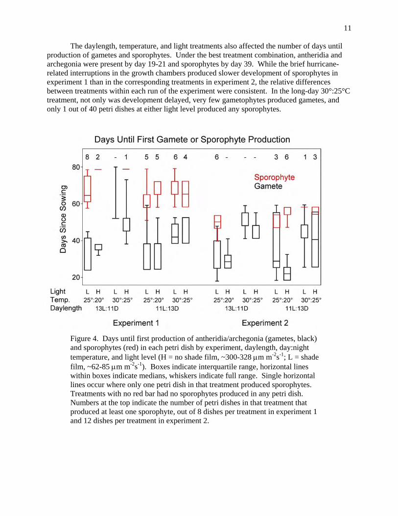
The daylength, temperature, and light treatments also affected the number of days until production of gametes and sporophytes. Under the best treatment combination, antheridia and archegonia were present by day 19-21 and sporophytes by day 39. While the brief hurricane-related interruptions in the growth chambers produced slower development of sporophytes in experiment 1 than in the corresponding treatments in experiment 2, the relative differences between treatments within each run of the experiment were consistent. In the long-day 30°:25°C treatment, not only was development delayed, very few gametophytes produced gametes, and only 1 out of 40 petri dishes at either light level produced any sporophytes.
Figure 4. Days until first production of antheridia/archegonia (gametes, black) and sporophytes (red) in each petri dish by experiment, daylength, day:night temperature, and light level (H = no shade film, ~300-328 μm m-2s-1; L = shade film, ~62-85 μm m-2s-1). Boxes indicate interquartile range, horizontal lines within boxes indicate medians, whiskers indicate full range. Single horizontal lines occur where only one petri dish in that treatment produced sporophytes. Treatments with no red bar had no sporophytes produced in any petri dish. Numbers at the top indicate the number of petri dishes in that treatment that produced at least one sporophyte, out of 8 dishes per treatment in experiment 1 and 12 dishes per treatment in experiment 2.

12
Juvenile L. microphyllum Survival Under Inundation Survival of young L. microphyllum sporophytes following inundation increased with
sporophyte height at the start of drowning and decreased with inundation duration (Table 3). Even the smallest sporophytes were able to survive 7 days of inundation (Figure 5). Inundation for 14 days was sufficient to kill sporophytes less than 2cm in height. Flooding durations of 4 weeks killed more than half of the individuals in that treatment, disproportionately affecting the smaller sporophytes.
Likelihood Ratio Statistics For Type 1 Analysis Predicting Survival of L. microphyllum Sporophytes Source Deviance DF X2 Pr > X2 Intercept 296.34 log(height) 255.29 1 41.06 <.0001 log(duration) 242.20 1 13.08 0.0003
Table 3. Analysis of deviance for generalized linear model of juvenile L. microphyllum sporophytes survival after different periods of inundation.
Figure 5. L. microphyllum sporophyte fate as functions of initial sporophyte height and flooding duration. Each dot represents the fate of an individual sporophyte. The red dots for dead sporophytes are offset slightly to the right for clarity, but they experienced the same durations as the adjacent surviving sporophytes. All sporophytes survived a week of drowning, regardless of size, but smaller sporophytes were susceptible to longer durations of flooding.

13
C.3. Management implications
Our data on spore dispersal shows that spores get everywhere, even to remote sites in Everglades National Park, so special quarantine regulations will not prevent arrival of spores in the Park. Although our study was not designed to test spatial hypotheses, in the contrast between spore influx at LNWR, where sexually reproducing plants were present within a kilometer of the traps and abundant in the surrounding tens of square kilometers, and ENP, where known infestations were more distant and much less abundant on the landscape, the data suggest that proximity to a source of spores is critical to the density of the spore rain and, thus, to the probability of successful colonization. The lack of a seasonal signal in the southern Everglades data, especially in ENP, may reflect eradication efforts in the Park, which would eliminate close spore sources. The seasonal pattern of spore dispersal also indicates that efforts to eradicate adult plants might be especially effective if they occurred in the wet season, prior to the massive release of spores. While spores germinated under all our experimental conditions, gametophyte growth, production of gametes, and production of sporophytes (both requiring wet conditions) were inhibited by higher temperatures and longer days in our growth chamber studies. Again, these data argue that production of new sporophytes is less likely in the warmer wet season and more likely in the cooler dry season. Because the gametophytes need moist humid conditions to grow and reproduce, however, they need either to produce sporophytes before their habitat dries out too much or to colonize a microsite that remains moist. In combination with the pronounced peak in spore influx in the wet season, this suggests that the crucial environmental filter for new infestations occurs at the end of the wet season, in December and January. This is the time when gametes and sporophytes are most likely to be produced. Our data from the inundation experiments shows that this is also the time when changes in the amount and depth of water will have the greatest effect on survival, i.e., when the sporophytes were small, they were more susceptible to inundation. Thus, both water management practices and natural variation in hydroperiod will create conditions that are predictably better or worse for sporophyte survival and establishment; these changes are especially effective in the late wet season/early dry season. Additionally, sites that are most likely to be colonized are transitional habitats. Sites that are either very dry or under deep water are unlikely to have successful colonization by L microphyllum.
C.4. Additional Factors to Consider
Although spores were found at all sites sampled, we do not know whether these spores
were viable. Both spore age and spore exposure could affect viability. The patterns of spore dispersal found in this study, as well as Volin’s (2004) data from sampling spore production on leaves, show a peak of spore production in the fall in south Florida. Year-round spore dispersal could result from this single peak of production and release, with the decrease occurring when most plants have finished sporangia production and spore shedding, and year-round dispersal representing late release of spores produced in the fall. If spores lose viability over time, the spores arriving in ENP during the rainy season would represent even less of a threat than their small numbers suggest. Similarly, if spores lose viability with time of aerial exposure, threats from sporulating plants decrease with distance, and efforts to find and eradicate new infestations can be concentrated closer to sources.

14
Our experience growing both gametophytes and young sporophytes indicates that at these stages of the life cycle, these plants do not grow well in high light environments. Plants grown in the greenhouse yellowed and grew slowly until we put shadecloth over the benches. In addition, we grew sporophytes in nutrient-poor soil in order to provide general Everglades nutrient conditions for the drowning experiments. Young sporophytes were very slow-growing under these conditions, taking up to six months to reach sizes where they could be used in our inundation experiments. This suggests that low-nutrient environments in the Everglades provide longer intervals of susceptibility to drowning for juvenile L. microphyllum.
Literature Cited
Harper, J.L. 1977. Population Biology of Plants. Academic Press, London.
Klekowski E.J. Jr. 1969. Reproductive biology of the pteridophyta. III. A study of the Blechnaceae. Botanical Journal of the Linnean Society 62: 361–377
Lonsdale, W. M. 1999. Global patterns of plant invasions and the concept of invasibility. Ecology 80: 1522-1536.
Lott, M. S., J. C. Volin, R. W. Pemberton, and D. F. Austin. 2003. The reproductive biology of the invasive ferns Lygodium microphyllum and L. japonicum (Schizaeaceae): Implications for invasive potential. American Journal of Botany 90: 1144-1152.
Mueller, R. J. 1982. Shoot morphology of the climbing fern Lygodium (Schizaeaceae): General organography, leaf initiation, and branching. Botanical Gazette 143: 319-330.
Pemberton, R., andAND A. Ferriter. 1998. Old World climbing fern (Lygodium microphyllum), a dangerous invasive weed in Florida. American Fern Journal 88: 165-175.
Pemberton, R., J.A. Goolsby, and T. Wright. 2002. Old world climbing fern. In: Van Driesche, R., S. Lyon, B. Blossey, M. Hoddle, and R. Reardon, editors, Biological Control of Invasive Plants in the Eastern United States, USDA Forest Service Publication FHTET-2002-04, 413 p.
Volin, J. C., M. S. Lott, J. D. Muss, and D. Owen. 2004. Predicting rapid invasion of the Florida Everglades by Old World Climbing Fern (Lygodium microphyllum). Diversity and Distributions 10: in press.
D.1. Names of scientific or other collaborators Dr. Paul Groff, research assistant
Mike Cheek, graduate student
Ana Castellanos, research assistant
Marlene Dow, research assistant
Diana Van Dillewijn, undergraduate research assistant

15
D.2. Publication history
Refereed Articles:
Philippi, T., J.H. Richards, and P. Groff. In preparation. Spore rain and environmental effects on gametophyte growth and early sporophyte survival in the invasive exotic Old World climbing fern, Lygodium microphyllum.
Abstracts, Presentations, and Posters:
Philippi, T. and J.H. Richards. Spore rain and environmental effects on gametophyte growth in the invasive exotic Old World climbing fern, Lygodium microphyllum. Botanical Society of America annual meeting, Chico, CA, July 2006.
Richards, J.H. and T. Philippi. Quantitative analysis of Everglades wetland plant communities and their distribution of across the ecosystem. Greater Everglades Ecosystem Restoration (GEER) meeting, Lake Buena Vista, FL, June 2006.
Philippi, T. and J.H. Richards. Potential hydrologic and environmental constraints on establishment of the invasive exotic Old World climbing fern, Lygodium microphyllum. Ecological Society of America annual meeting, Memphis, TN, August 2006.
Philippi, T. and J.H. Richards. Potential hydrologic and environmental constraints on establishment of the invasive exotic Old World climbing fern, Lygodium microphyllum. Greater Everglades Ecosystem Restoration (GEER) meeting, Lake Buena Vista, FL, June 2006.
Philippi, T. and J.H. Richards. Potential hydrologic and environmental constraints on establishment of the invasive exotic Old World climbing fern, Lygodium microphyllum. Loxahatchee Scientific meeting, Davie, FL, May 1, 2006.
E. Signature of Principle Investigator
_____________________________________ Date: ___________________
Dr. Tom Philippi
Dept. of Biological Sciences
Florida International University,
Miami, FL 33199

16
F. Appendices Appendix 1. Instructions for constructing spore samplers.
Appendix 2. Protocols for spore sampling, filter paper processing and data collection
Appendix 3. List of files included on cd.

17
+ Appendix 1.
Instructions for Construction of Lygodium microphyllum spore traps
By Dr. Tom Philippi
Poster content describing the use of these traps to monitor L. microphyllum spore rain is at:
http://darwin.fiu.edu/lygodium/
Pictures of the spore traps are given in Figures 1-5, below.
The updated directions for making the spore traps are:
The Buchner funnels used are 172 mm 2-piece polypropylene funnels (Fisher cat. No. cat. 10-362G). In the completed traps, the funnels had rounds cut from 185 mm d. Watman 113 wet-strengthened filter paper inserted in the bottom. Although standard Watman or Fisher Scientific filter paper can be used, the wet strengthened filter paper was more resistent to degradation if the traps were left in the field for a month.
For each sampler, cut 2 6cm pieces of 3/4" PVC pipe and 1 11.5cm piece.
Remove the upper baskets from the lower funnels of the Buchner funnels. Cut the Buchner funnels' stems to 5.5cm in length. Test fit the 90deg elbow: it should fit snugly and even snap the last bit if cut right. Try both sides of the elbow-- they tend to be slightly different and one will often fit better than the other. For a set of 3 funnels, make sure that they have the elbow tubes at the same height relative to the tops of the funnels.
Remove the elbows from the funnels, keeping track of which end the funnel goes in. Cover the lab benchtop with cardboard. Put the funnels stem-side up on the cardboard and put on a glove. Use 3M 5200 marine sealant, and do the funnels one at a time. Put a 3-4mm bead of the sealant around the inside of the elbow, then push the elbow onto the stem with a slight twist. Holding the elbow all the way down, drill 3 pilot holes (~1/16" diameter, but check v. your screws) at 120deg spacing through the elbow and stem. Screw #4x3/8" pan head screws into the holes. Before finishing the screw, put a drop of sealant under the head to help prevent leaks, then tighten it down flat. If necessary, use a glove or popsicle stick to smooth the sealant bead on the end of the elbow.
Cut 2 pieces of 3/4" pvc pipe 6cm long, and one piece 12cm long. [Check the particular elbows & crosses you have: the pipe should be 1.3cm + 2 elbow-depths and 7.2cm + 1 elbow and 1 cross.] Wipe a thick coat of PVC cement around the outside 1.5 cm of one end of the pvc, then spin the ball inside the elbow to coat the inside. Excess solvent/cement is good. Push the pipe

18
into the elbow with a slight twisting motion, make sure it goes all the way in. Repeat for the other 2 funnels, using the longer piece for one.
Align the 3 funnels in roughly the right position, upside down on a flat surface, with the cross in the middle. Do a test fitting to make sure that your pipes are the right length, that when pushed in all the way the funnels just touch. [In disassembling, use the end piece to twist the cross relative to the side pipes to partially remove them, then take the end piece off, then finish removing the side pieces.] Working moderately quickly, wipe PVC solvent on the ends of all 3 pipes, then inside 3 sides of the cross. Put the 2 side (short-pipe) pieces on the cross first, twisting slightly and getting the funnels to touch. Add the end piece and twist so that all 3 funnels are again flat on the surface. Wipe any excess solvent into a nice bead.
Cut a piece of 3/4" PVC pipe to the desired length. Cut 1" holes (clearance for the 3/4" pvc pipe) in opposite sides of the bucket, 90deg from the handle attachments. I cut the holes ~.75cm above the inside bottom of the bucket, you could cut them a bit higher. The goal is to make a strong (& nearly waterproof) joint between the bucket and the pipe, using epoxy glue and additional PVC collars for support. Cut 4 ~1cm pieces of 1" pvc pipe for each bucket, and cut a slot lengthwise in each piece so it will slide along the 3/4" pvc. Push the 3/4" pvc pipe in one of the bucket holes, thread on 2 of the 1"pcv collars, then push the 3/4" pvc pipe out the other hole. Adjust the pipe lengths on the outside of the bucket as desired, and put the other 2 collars on the pipe. Mix some epoxy, and apply it to the inside and outside of where the pipe goes through the bucket. Quickly, wipe PVC cement on the pipe just adjacent to the epoxy and slide the collar (rotating it on the pipe to evenly spread the cement) snug against the bucket with the slit in the collar on the top. Do this for all 4 collars. Let dry overnight.
Inspect your joints, and clean off any excess epoxy or PVC cement that flowed out of the joint. Put on a glove, open the tube of 3M 5200 marine sealant, and put it in your calking gun. Start by squeezing a substantial amount against the bucket joint underneath the pipe on the inside of the bucket. Then add a solid covering of the rest of the joint (pipe to collar to bucket) on the inside of the bucket. Use a tongue depressor or similar stick to make sure that you have a solid seal against the bucket side and bottom for the pipe, then work to smooth the rest. Turn the bucket upside down, apply the 5200 all the way around the outside joints, and smooth. Let the 5200 cure for 5-7 days. Then, fill the bucket nearly full and let it sit for a couple of hours to check for leaks. Glue a 3/4" PVC valve to one end of the pipe and a funnel assembly to the other. Drill 3-5 ~1/4" holes in the pipe in the bottom of the bucket. Fill the bucket until the water level has equalized between the bucket and the funnels, and the level in the funnels is 1-2cm below the top. Drill 3-4 small overflow holes in the bucket at that waterline.
You may need to seal the Buchner funnel cylinder to its base to prevent leaks. Use either silicone sealer or petroleum jelly, as you will need to open this joint for cleaning every few months.
19
Figure Legends
Figure A1.1. Spore traps used to monitor Lygodium microphyllum spore rain. The closed bucket has a water reservoir that maintains the water level in the 3 funnels to the right through the PVC pipe, which connects to all three funnels. The funnels trap spores that fall into them in the water. The PVC pipe allows the water to drain when the valve at the end is open. After the traps are out for a given amount of time, the water is drained through filter paper in the bottom of the funnels, the filter paper is collected and examined for L. microphyllum spores.
Figure A1.2. Surface view of a spore trap funnel. Filter paper covers the bottom and is anchored down by a ring of copper wire. The funnel to the lower right does not yet have filter paper in it.
Figure A1.3. A spore trap deployed at a sampling site.
Figure A1.4. The filter papers in traps after 1 month outside at Loxahatchee National Wildlife Refuge. These were as dirty as the filter papers got.
Figure A1.5. Collecting old filter papers and replacing them with new (see “Lygodium Trap Sampling Protocol” in Appendix 2).

20

21
Appendix 2
Spore Sampling, Filter Paper Processing and Data Collection Protocols
Lygodium Trap Sampling Protocol
The funnels of the traps have round pieces of filter paper cut from 185 mm d. Watman 113 wet strengthened filter paper inserted in the bottom. In our experience, the wet strengthened filter paper is more resistent to degradation if the traps were left in the field for a month. Standard Watman filter paper can be used if the filter paper is left out for a shorter amount of time.
Drain the traps by opening the drain valve to lower the water in the reservoir bucket. Wash down the sides of the drained traps with the squeeze bottle. Remove the filter paper, using the flat forceps to begin to get the paper up. Fold the paper into quarters with the up-side in. Place in a whirlpac plastic bag; label with site, filter number (1, 2 or 3), and date.
Put a fresh filter paper in the trap; replace the copper holding-wire. Re-fill the bucket until there are several cm of water in the traps and no air bubbles in the pipe or under the filter paper. Add a capful of bleach to help control algal growth; too much bleach, however, can weaken the filter paper--the right amount will depend on the amount of water in the reservoir. Replace the lid on the bucket. On terrestrial sites, remove nearby weeds, so that the trap doesn’t become overgrown.
Lygodium Spore Counting Protocol: Take the filter paper and use scissors to cut a strip that fits on a 1 x 3 in. microscope slide; alternatively, cut the paper into pieces that fit in a petri dish or some other support that you can move around in a systematic way under the dissecting microscope. Keep the filter paper flat while cutting, so that you don’t let material fall off. After you have cut strips, if you aren’t going to be able to process the entire filter paper, re-fold the filter paper with the up-side in and put it in its plastic bag, so that it doesn’t dry out. We found that cutting the entire paper, sitting it on a piece of glass and staining it all also works well if you are going to be able to process the entire paper at one sitting.
Stain the filter paper strip with 0.01% Toluidine Blue in 0.05 M phosphate buffer, pH 7.2, applied as drops from a pipet. Add enough stain to completely cover the filter paper; let the paper sit for 15 minutes in stain before examining. It is efficient to make a slide and let it sit while you are examining a previously-made slide, or to cut and stain the entire paper, then work through the pieces, as described above. Stained filter papers can sit in a Petri dish sealed with paraplast or in a plastic bag to prevent drying. If filter papers are especially wet, they may need to be stained with a more concentrated solution of Toluidine Blue.
Examine filter paper pieces by scanning them with a good dissecting microscope. We used a Leitz MZ95 or Wild M3C dissecting microscope with set stops at 6.4x, 10x, 16x, 25x and 40x and equipped with a 2x objective, for working magnifications of 12.8x to 80x. Prepare a L. microphyllum reference slide of spores collected from known L. microphyllum plants. Calibrate your eye by looking at the reference slide of stained L. microphyllum spores at 16x and 40x with

22
the 2x objective lens on the dissecting microscope. L. microphyllum spores are large (diameter = 68 + 5 μm, N = 51, plants collected from Wild Turkey Preserve, FL)—larger than the pollen grains that are also found on the filter papers. The spores fit between 2 or 3 lines on the ocular scale at 16x and between 6 and 7 lines at 40 x. They stain blue in the Toluidine blue and have a trilete scar, distinctive corrugated sculpturing, and usually rounded shape with flattened areas at the scar end. Use a white background for this--we have made Petri dishes with a 1 x 1 cm grid on white paper taped to the bottom of the dish; we put the microscope slide with filter paper in the dish and use the Petri dish to move the slide under the microscope.
Examine the strip of filter paper; scan at 16x but check IDs at 40x. Be careful to overlap your scan paths and to keep your place, so that you don’t skip parts of the paper. When you overlap, also be careful that you don’t count spores twice. If grit or other material is obscuring the filter paper, move it or disturb it with a dissecting needle so that you are sure that you have looked through it. After you have finished scanning the paper, look at the liquid on the edges, then lift the paper and look in the liquid under the paper for spores. If liquid ramined in the sammple bag, this should also be examined for spores. Record the number of spores on the data sheet.
When you finish 1 strip, do the next, etc., until the entire filter paper is sampled. A sample data sheet is appended.
Figure Legends
Figure A2.1. Surface view of L. microphyllum spore, viewed with a compound light microscope at 400x. This spore was collected from sporangia of sporulating plants. The surface has corrugated ornamentation on its wall and a trilete mark that reflects the juncture of the 4 products of meiosis from 1 spore mother cell. The surface ornamentation varies from spore to spore but maintains the corrugated pattern.
Figure A2.2. L. microphyllum spore from filter paper from a spore trap left for 1 month at Loxahatchee National Wildlife refuge examined with a compound light microscope at 400x.
Figures A2.3 and A2.4. Samples of filter paper from spore traps examined at 80x in a dissecting light microscope. Arrows point to L. microphyllum spores. Spores are recognized by their size, shape, staining characteristics in buffered Toluidine blue and surface ornamentation, which can be seen by focusing up and down on the specimens.

23

24
Spore Counting Datasheet Site:
Collection
Date:
Name: Analysis
Date:
Filter 1 Filter 2 Filter 3
No. Count No. Count No. Count
1 1 1
2 2 2
3 3 3
4 4 4
5 5 5
6 6 6
7 7 7
8 8 8
9 9 9
10 10 10
11 11 11
12 12 12
13 13 13
14 14 14
15 15 15
16 16 16
17 17 17
18 18 18
19 19 19
20 20 20
21 21 21
22 22 22
23 23 23
24 24 24
25 25 25
26 26 26
27 27 27
28 28 28
29 29 29
30 30 30

25
Appendix 3.
List of Files Included on cd spore_sampler_locations_utmNAD83.xls
The locations of the 11 spore samplers in UTM 17N, NAD83
sporetraps_final.xls Sporetrap data for all 11 sites through 7 January 2007.
site: site name f1, f2, f3: counts on individual filters date: date of collection duration: duration of exposure in days lduration: natural logarithm of the exposure duration spores: average number of spores per filter (averaged across non-missing
filters) w: number of filters for that trap and date (used to weight averages) sporeperday: the corresponding average number of spores per day per m2
sporetrap_analyses.sas SAS code that read the raw files of filter paper sporecounts, combined them, ran PROC GENMOD, generated figure 2, and exported the clean data file sporetraps_final.xls.
Development_exp1.csv
raw data from the first run of the growth chamber spore germination and gametophyte growth experiment.
Development_exp2.csv
raw data from the second run of the growth chamber spore germination and gametophyte growth experiment.
development.sas
SAS code that read the development_exp1.csv and development_exp2.csv files, and produced the boxplots for Figure 4.
drowning_experiment_tp.csv
raw data on individual plants in the drowning experiment. drowning.sas
SAS code that read drowning_experiment_tp.csv, ran PROC GENMOD to test the effetcs of sporophyte height and inundation duration, and produced Figure 5 as well as some experimental graphs.
Q528404CESI_final_report.pdf pdf version of this report.