critical care notes 2e medilibros.com
DESCRIPTION
fTRANSCRIPT

• F. A. Davis Company
Always at your side ...
Critical care 2nd Edition

Contacts • Phone/E-Mail
Name
Ph: e-mail:
Name
Ph: e-mail:
Name
Ph: e-mail:
Name
Ph: e-mail:
Name
Ph: e-mail:
Name
Ph: e-mail:
Name
Ph: e-mail:
Name
Ph: e-mail:
Name
Ph: e-mail:
Name
Ph: e-mail:
Name
Ph: e-mail:
Name
Ph: e-mail:
4223_FM_ii-vi 28/08/14 4:13 PM Page ii

CriticalCare
CriticalCare
NotesNotesClinical Pocket Guide
Janice Jones, PhD, RN, CNSBrenda Fix, MS, RN NP
Purchase additional copies of this book at yourhealth science bookstore or directly from F.A. Davis by shopping online at www.fadavis.com or by calling 800-323-3555 (US) or 800-665-1148 (CAN)
A Davis’s Notes Book
2nd Edition
4223_FM_ii-vi 28/08/14 4:13 PM Page iii

F. A. Davis Company1915 Arch StreetPhiladelphia, PA 19103www.fadavis.com
Copyright © 2009, 2015 by F. A. Davis Company
All rights reserved. This book is protected by copyright. No part of it may be reproduced, storedin a retrieval system, or transmitted in any form or by any means, electronic, mechanical, photo-copying, recording, or otherwise, without written permission from the publisher.
Printed in China by Imago
Last digit indicates print number: 10 9 8 7 6 5 4 3 2 1
Publisher, Nursing: Robert G. MartoneDirector of Content Development: Darlene D. PedersenContent Project Manager: Jacalyn C. ClayIllustration and Design Manager: Carolyn O’Brien
Reviewers: Katrina Allen-Thomas, MSN, CCRN, RN; Marilu Alltop, MSN, RN, CNE; MalloryAntico, MSN, RN; Lisa Ann Behrend, RN, MSNc, CCRN-CSC; Cynthia Berry, RN, DNP, CNE; LauraCarousel, MSN, RN, CCRN; Melanie Cates, RN, HBScN, MSN, ENC(C); Nancy L. Denny, MSN, RN-BC; Abimbola Farinde, PharmD, MS; Sarah Gabua, MSN, RN; Jayme Haynes, MSN, RN;Jeanie Krause-Bachand, MSN, EdD, RN, BC; Janice Garrison Lanham, RN, MSN, CCNS, FNP;Deborah Little, MSN, RN, CCRN, CNRN, APRN, BC; Angela Medina, RN, MSN; Candi Miller-Morris, MSN, RN, CNS, CCRN, CEN, TNS; Michelle Murphy-Rozanski, PhD, MSN, RN, CRNP;Deborah Pool, MS, RN, CCRN; Margaret Sherer, MS, RN, CEN, CNE; Denise A. Tucker, DSN, RN,CCRN; Tamra Weimer, RN, MS, CCRN, CNE; Danette Wood, EdD, MSN, RN, CCRN; ShawnZembles, DNP, RN, CCRN, ACNS-BC
As new scientific information becomes available through basic and clinical research, recom-mended treatments and drug therapies undergo changes. The author(s) and publisher havedone everything possible to make this book accurate, up to date, and in accord with acceptedstandards at the time of publication. The author(s), editors, and publisher are not responsiblefor errors or omissions or for consequences from application of the book, and make no war-ranty, expressed or implied, in regard to the contents of the book. Any practice described in thisbook should be applied by the reader in accordance with professional standards of care usedin regard to the unique circumstances that may apply in each situation. The reader is advisedalways to check product information (package inserts) for changes and new informationregarding dose and contraindications before administering any drug. Caution is especiallyurged when using new or infrequently ordered drugs.
Authorization to photocopy items for internal or personal use, or the internal or personal useof specific clients, is granted by F. A. Davis Company for users registered with the CopyrightClearance Center (CCC) Transactional Reporting Service, provided that the fee of $.25 per copyis paid directly to CCC, 222 Rosewood Drive, Danvers, MA 01923. For those organizations thathave been granted a photocopy license by CCC, a separate system of payment has beenarranged. The fee code for users of the Transactional Reporting Service is: 978-0-8036-4223-3/150 + $.25.
4223_FM_ii-vi 28/08/14 4:13 PM Page iv

TOOLS
Sticky Notes
✓HIPAA Compliant
✓OSHA Compliant
Waterproof and Reusable
Wipe-Free Pages
Write directly onto any page of Critical Care Noteswith a ballpoint pen. Wipe old entries off
with an alcohol pad and reuse.
BASICS CV RESP GU NEURO GI HEMA/ONCO ENDO
MULTISYS CC MEDS
4223_FM_ii-vi 28/08/14 4:13 PM Page v

4223_FM_ii-vi 28/08/14 4:13 PM Page vi
For a complete list of Davis’s Notes and other titles for health care providers,
visit www.fadavis.com.
Look for other davis’s Notes titles!
RN NotesNurse’s Clinical Pocket Guide
Psych NotesClinical Pocket Guide
NCLEX-RN® NotesContent Review & Exam Prep
MedSurg Notes Nurse’s Clinical Pocket Guide

Physical Assessment
Reusable Assessment Form
1
BASICS
Pt. Identifier: Room: Age: Diagnosis:
Surgeries/Past Hx:
Activity: Diet: DNR/DNI:Allergies:Neurological/MS: ICP:Cardiac:
VS/A-line: ECG: Hemodynamics: PAD PAS PCWP CVPIABP:
Respiratory: Ventilator: ABGs/SpO2:
GI:
GU:
Wounds/Incisions:
Drainage tubes:
Treatments:
Special needs:
Other:
4223_Tab01_001-044 29/08/14 10:46 AM Page 1

2
BASICS
Normal Arterial and Venous Blood Gases
Blood Gas
Components Arterial Venous
pH 7.35–7.45 7.31–7.41PO2 80–100 mm Hg 35–40 mm HgPCO2 35–45 mm Hg 41–51 mm HgHCO3- 22–26 mEq/L or mmol/L 22–26 mEq/L or mmol/LBase excess (BE) –2 to +2 mEq/L or mmol/L –2 to +2 mEq/L or mmol/LO2 saturation 95%–100% 68%–77%
Values denoted are at sea level.
Quick Blood Gas Interpretation
Acid-Base Disorder pH PCO2 ↑ HCO3-
Respiratory acidosis ↓ ↑ ↑ if compensatingRespiratory alkalosis ↑ ↓ ↓ if compensatingMetabolic acidosis ↓ and (+) ↓ if ↓
base excess compensatingMetabolic alkalosis ↑ and (–) ↑ if ↑
base excess compensatingMixed respiratory and ↓ ↑ ↓metabolic acidosisMixed respiratory and ↑ ↓ ↑metabolic alkalosis
Full or total compensation: pH will be within normal limits.
4223_Tab01_001-044 29/08/14 10:46 AM Page 2

Compensation:
■ Respiratory problem → the kidneys compensate by conserving or excretingHCO3-
■ Metabolic problem → the lungs compensate by retaining or blowing off CO2
Also look for mixed respiratory and metabolic problems. PaCO2 or HCO3- in adirection opposite its predicted direction or not close to predictive value. Mayresult from cardiac arrest, vomiting with renal failure and COPD as comorbidi-ties, and salicylate toxicity.
3
BASICS
Common Causes of Acid-Base ImbalancesRespiratory acidosis COPD, asthma, head injury, pulmonary edema,
aspiration, pneumonia, ARDS, pneumothorax,cardiac arrest, respiratory depression, CNSdepression, or head injury
Respiratory alkalosis Hyperventilation, anxiety, fear, pain, fever, sepsis,brain tumor, mechanical overventilation
Metabolic acidosis Diabetes mellitus, acute and chronic renal failure,severe diarrhea, alcoholism, starvation, salicylateoverdose, pancreatic fistulas
Metabolic alkalosis Loss of gastric acid (vomiting, gastric suction),long-term diuretic therapy (thiazides, furosemide),excessive NaHCO3 administration, hypercalcemia
Blood Gas Results
Arterial Venous
pHPO2
PCO2
HCO3-Base excess (BE)O2 saturation
4223_Tab01_001-044 29/08/14 10:46 AM Page 3

4
Pulse OximetrySpO2 monitoring may be intermittent or continuous. Indirectly monitors oxygensaturation.
False readings may occur because of anemia, carbon monoxide poisoning,hypothermia, hypovolemia, hypotension, peripheral vasoconstriction, and poorperipheral perfusion caused by disease or medications.
Continuous Monitoring■ Alarms are set for low SpO2, tachycardia, or bradycardia.■ Waveform should be sharp with a clearly identified dicrotic notch.■ The probe may be placed on the finger (preferred), toes, or ear lobe or
pinna.■ Patient must have SBP >80 mm Hg.
Lactic AcidosisLactic acid is a byproduct of anaerobic metabolism. Increased levels indicateinadequate perfusion of vital organs with resultant tissue hypoxia. May resultfrom inadequate perfusion and oxygenation of vital organs; post cardiac or res-piratory arrest; cardiogenic, ischemic, or septic shock; drug overdoses; seizures;cancers; or diabetes mellitus (refer to Multisystem tab).Normal lactate level <2 mmol/L; >5 mmol/L indicates lactic acidosis.
BASICS
SpO2 Level Indication
>95% Normal91%–94% May be acceptable, provide O2 as necessary, encourage
C&DB, or suction prn85%–90% Provide O2 as necessary, encourage C&DB, or suction prn;
may be normal for COPD patient<85% Prepare for possible intubation<70% Unreliable; obtain ABG
Values denoted are at sea level.
4223_Tab01_001-044 29/08/14 10:46 AM Page 4

Respiratory Terms and Calculations■ Functional residual capacity (FRC) is the volume of air in the lungs after
normal expiration. Normal = 2,400 mL.
■ Hypoxemia is the severe reduction of O2 in arterial blood.■ Hypoxia is the severe reduction of O2 at the cellular level.■ Minute ventilation (MV) = respiratory rate (RR) × tidal volume (VT).
To improve MV and ↓ PaCO2 with mechanical ventilation: ↑ VT, and/or ↑ RR;↑ inspiratory pressure, prolong inspiratory time, ↑ pressure support level, ↓ airway resistance, suctioning, use of bronchodilators.
■ P/F (PaO2/FIO2) ratio. The smaller the value, the worse the patient’s gasexchange. Frequently calculated to suggest ARDS and V· /Q· mismatch.Normal = 300–500; impeding or actual respiratory failure = 200–300 (mayneed to intubate); ARDS or V· /Q· mismatch = <200, indicates hypoxemia andneed to intubate. Formula: PaO2 (from ABG in mm Hg) ÷ FIO2 (converted to decimal) = P/Fratio number.Example: PaO2 = 87 mm Hg and patient is on room air (21% = 0.21) = 77 ÷ 0.21 = 366.
■ SaO2 is the saturation of oxygen in hemoglobin in arterial blood =95%–100% normal. Obtained from arterial blood sample.
■ SvO2 is the percentage of O2 bound to hemoglobin in venous blood =60%–80%. Assesses tissue perfusion or oxygenation of tissues. May bemonitored intermittently or continuously using an oximetric pulmonaryartery catheter. ScvO2 is a central venous sample from internal jugular orsubclavian catheters = >70%.↑ SvO2 ( >80%) indicates an↑ in O2 delivery or ↓ O2 extraction by tissues.↓ SvO2 (<60%) indicates a ↓ in O2 delivery or ↑ extraction by tissues → cardiac output not adequate to meet tissue O2 needs; Hgb may be low; O2 consumption > oxygen delivery.
End-Tidal Carbon Dioxide Monitoring (ETCO2)ETCO2 or capnography/capnometry is the measurement, display, and monitor-ing of the concentration or partial pressure of CO2 (ETCO2) in the respiratorygases at the end of expiration. ETCO2 values are usually 2–5 mm Hg lower thanthe PaCO2 value. The capnogram displays the maximum inspiratory and expira-tory CO2 concentrations during a respiratory cycle that indirectly reflect the pro-duction of CO2 by the tissues and the transport and clearance of CO2 to and inthe lungs. Sudden changes in CO2 elimination should be monitored in selectedcardiorespiratory patients and postoperatively after major cardiothoracic surgery.
5
BASICS
4223_Tab01_001-044 29/08/14 10:46 AM Page 5

6
ETCO2 monitoring can also be used to verify ETT position, assess readiness forextubation, and monitor the effectiveness of CPR and predict patient survival.ETCO2 <10 mm Hg after 20 min CPR indicates poor outcome. It is sometimesreferred to as the “ventilation vital sign.”
Normal range of ETCO2 is 35–45 mm Hg. CO2 and ETCO2 should correlate with-in 2–5 mm Hg.
↑ RR (hyperventilation) → ↓ CO2 → ETCO2 < 35 = respiratory alkalosis↓ RR (hypoventilation) → ↑ CO2 → ETCO2 > 45 = respiratory acidosis
Five characteristics of the capnogram should be evaluated: frequency, rhythm,height, baseline, and shape. Also note changes if patient is disconnected from ven-tilator and attempts of spontaneous breaths if patient is receiving paralytic agents.
Normal Capnogram
Phases I, II, and III represent expiration; the bolded lines represent inspiration.Long periods of a flat wave form indicate apnea, dislodged endotracheal tube,esophageal intubation, or patient disconnection from ventilator.
BASICS
Causes of ↑ ETCO2 Causes of ↓ ETCO2
Fever HypothermiaHypertension Hypotension and shockIncreased cardiac output Cardiac perfusion changesRespiratory compromise Decreased cardiac output, heart failureHypoventilation Cardiac arrest and apneaAirway obstruction Hyperventilation Bronchial intubation Airway obstructionHypovolemia Accidental extubationSepsis Pulmonary embolusSeizures Hypervolemia
I
II
III
Expiration Inspiration
mm
Hg
Time
4223_Tab01_001-044 29/08/14 10:46 AM Page 6

Artificial Airways and Mechanical Ventilation
Artificial Airways
Endotracheal Tube■ Adult oral tube sizes: males, 8.0–8.5 internal diameter (I.D.) (mm); females,
7.0–8.0. I.D. (mm).■ Placement is 2–3 cm above the carina. Verify by auscultating for breath
sounds bilaterally, uniform up-and-down chest movement, CXR, and check-ing ETCO2 immediately after intubation.
■ Cuff pressure: 20–25 mm Hg.
Rapid Sequence Induction (RSI): Minimizes time to intubation and secures a
patent airway.
■ Procedure outline:■ Preoxygenate patient with 100% O2.■ Induction drug administered: etomidate, propofol, ketamine, thiopental
or scopolamine.■ Neuromuscular blocking agent administered: succinylcholine.■ Apply cricoid pressure.■ ETT inserted.
■ Nursing concerns:■ Know patient’s K+ level.■ Have routine intubation supplies available.■ Check for workable suction source and provide regular suction catheter
and Yankauer catheter.■ Provide emotional support to patient and notify patient’s family of rapid
induction of ETT.
Cuff pressure can be monitored via a calibrated aneroid manometer device.Connect manometer to cuff. Deflate cuff. Reinflate cuff in 0.5-mL incrementsuntil desired cuff pressure is achieved. Check cuff pressure every 8–12 hr or peragency protocol.
Tracheostomy Tube■ Tracheostomy tubes may be cuffed or uncuffed and have either a reusable
or disposable inner cannula. Both fenestrated and Passy-Muir valves allowthe patient to speak.
■ Size will vary.■ Cuff pressure: 20–25 mm Hg.■ Early replacement of ETT with tracheotomy has not been shown to improve
patient outcomes.■ Other artificial airways include oropharyngeal airway and nasopharyngeal
airway.
7
BASICS
4223_Tab01_001-044 29/08/14 10:46 AM Page 7

8
Oxygen delivery systems include the nasal cannula, face mask, Venturi mask,
partial rebreather mask, nonrebreather mask, tracheostomy collar, and T-piece.
■ Nonrebreather mask allows for 80%–90% FIO2 to treat lowing O2 levels asexhibited by decreasing SpO2 or impending respiratory failure prior to intubation.
■ The T-piece may be connected to either an ETT or a tracheostomy tube.Frequently used in ventilator weaning.
Mechanical Ventilation
Classification of VentilatorsPositive Pressure Ventilation■ Volume-Cycled Ventilator: Delivers a preset constant volume of air and pre-
set O2.■ Pressure-Cycled Ventilator: Produces a flow of gas that inflates the lung
until the preset airway pressure is reached.■ Time-Cycled Ventilator: Programmed to deliver a volume of gas over a spe-
cific time period through adjustments in inspiratory-to-expiratory ratio.Primarily used in neonates.
Negative Pressure VentilationUses the old iron lung principle by exerting negative pressure on the chest wallto cause inspiration. No intubation required. Custom-fitted “cuirass” or “turtle”shell unit fits over the chest wall. May be used at night for patients who requireassistance during sleep.
Modes of Ventilation■ Continuous Mandatory Ventilation (CMV): Machine controls rate of breath-
ing. Delivery of preset volume (TV) and rate regardless of patient’s breath-ing pattern. Sedation or neuromuscular blocking agent usually required.Very restricted use (e.g., SCI).
■ Assist Controlled Ventilation (AC or ACV): Patient controls rate of breath-ing. Inspiratory effort triggers delivery of preset volume.
■ Synchronized Intermittent Mandatory Ventilation (SIMV): A form of pres-sure support ventilation. Administers mandatory ventilator breath at a pre-set level of positive airway pressure. Monitors negative inspiratory effortand augments patient’s spontaneous tidal volume or inspiratory effort.Synchronized with patient’s breathing pattern.
■ Positive End-Expiratory Pressure (PEEP): Increases oxygenation by increas-ing functional residual capacity (FRC). Keeps alveoli inflated after expira-tion. Can use lower O2 concentrations with PEEP; decreases risk of O2 toxicity. Ordered as 5–10 cm H2O.
■ Pressure Support Ventilation (PS or PSV): Patient’s inspiratory effort isassisted by the ventilator to a certain level of pressure. Patient initiates all
BASICS
4223_Tab01_001-044 29/08/14 10:46 AM Page 8

breaths and controls flow rate and tidal volume. Decreases work of breath-ing and promotes weaning.
■ Pressure-Controlled ventilation (PCV): Controls plateau pressures in patients with ARDS and persistent oxygenation problems despite highlevels of PEEP and FIO2.
■ Pressure-Regulated Volume Control (PRVC): Preset rate, FIO2, and pressurelimit. Improves patient-ventilator synchrony and reduces barotrauma. Mayrequire sedation.
■ Volume-Assured Pressure Support (VAPS) or Volume Guaranteed Pressure
Options (VGPO): Combination of pressure with guaranteed volume control.■ High-Frequency Ventilation (HFV): Delivers very high breaths/min with low
tidal volumes. These include high-frequency oscillatory ventilation (HFOV
or HFO), high-frequency jet ventilation (HFJV), and high-frequency positivepressure ventilation (HFPPV).
■ Inverse Ratio Ventilation (IRV): All breaths are pressure limited and timecycled. Inspiratory time usually set shorter than expiratory time. I:E ratio isusually 1:1.3–1.5
Noninvasive Mechanical Ventilation (NIV)■ Continuous Positive Airway Pressure (CPAP): A form of noninvasive
mechanical ventilation (NIMC). Maintains positive pressure throughout therespiratory cycle of a spontaneously breathing patient. Increases theamount of air remaining in the lungs at the end of expiration. Fewer com-plications than PEEP. Ordered as 5–10 cm H2O.
■ Bilevel Positive Airway Pressure (BiPAP): Same as CPAP but settings can beadjusted for both inspiration and expiration.
SIMV, CPAP, BiPAP, and PSV can all be used in the weaning process.
General Nursing Care for Mechanically VentilatedPatients■ General routine head-to-toe assessment to monitor for complications related
to mechanical ventilation.■ Check ventilator settings for accuracy, especially rate, tidal volume, FIO2,
PEEP level, and pressure gauge; monitor ABGs after ventilator settingchanges.
■ Assess for oxygen toxicity. Cellular damage causing capillary leak →pulmonary edema and ARDS. May develop if patient on 100% FIO2 for >12 hr or >50% FIO2 for >24 hr. Monitor for dyspnea, ↑ lung compliance, ↓ A-a gradient, paresthesia in the extremities, and retrosternal pain. KeepO2 at lowest possible concentration. Consider PEEP to ↑ FIO2. If patient isanemic, transfuse RBCs.
■ Administer analgesics, sedation drugs, and neuromuscular blocking agentsas needed.
9
BASICS
4223_Tab01_001-044 29/08/14 10:46 AM Page 9

10
■ Provide communication methods for patients, and communicate freely withfamilies.
■ Prevent infection including catheter-associated urinary tract infection(CAUTI), central catheter-associated bloodstream infection (CLABSI), andventilator-associated pneumonia (VAP) (refer to VAP guidelines inRespiratory tab).
■ Assess readiness to wean.
Weaning
Sample Criteria for Weaning: Readiness■ Alert and cooperative■ FIO2 <40%–50% and PEEP <5–8 cm H2O■ Hemodynamically stable with HR <120 bpm and no significant arrhythmias,
SBP >100 mm Hg■ pH >7.34■ PaO2 >80 mm Hg■ PaCO2 <45 mm Hg■ PaO2/FIO2 ratio >200■ ETCO2 <40 mm Hg■ Vital capacity 15 mL/kg and minute ventilation <10■ Hemoglobin >7–9 g/dL and serum electrolytes within normal limits■ Spontaneous respirations >6 bpm or <35 bpm■ Negative inspiratory pressure –30 cm H2O■ Relatively afebrile with limited respiratory secretions and good cough reflex■ Good pain management■ Inotropes reduced or unchanged within previous 24 hr■ Sedation discontinued■ Spontaneous breathing trials (SBT)
BASICS
Weaning Protocol
Hospital Weaning Protocol Patient’s Readiness for Weaning
Continued
4223_Tab01_001-044 29/08/14 10:46 AM Page 10

Weaning Methods■ T-tube weaning: Place patient on T-tube circuit on same FIO2 as on ventilatory
assistance. Monitor ABGs after 30 min. Provide a brief rest period on theventilator as needed and continue to monitor ABGs until satisfactory.Extubate when patient is rested, has good spontaneous respiratory effort,and ABGs within acceptable parameters.
■ SIMV weaning: Decrease SIMV rate every 1–4 hr or 2 breaths/min. Monitorspontaneous breaths, SpO2 with a goal of >90%, ETCO2, hemodynamics, andECG for dysrhythmias. Obtain ABGs within 30 min of ventilator change.Allows for gradual change from positive pressure ventilation to sponta-neous pressure ventilation. Titrate FIO2.
■ PSV: Use low levels of PSV (5–10 cm H2O). Decrease in 3–6 cm of H2O incre-ments. Useful in retraining respiratory muscles from long-term ventilation.
■ CPAP/BiPAP: Provides expiratory support, maintains positive intrathoracicpressure. BiPAP adds inspiratory support to CPAP. Prevents respiratory musclefatigue.
Nursing Assessment During Weaning■ Vital signs and hemodynamic stability (PAS, PAD, PCWP, CO, CI)■ Dysrhythmias or ECG changes■ Oxygenation/efficiency of gas exchange: SaO2 >90% on <40% FIO2■ CO2 production and elimination■ pH level■ Bedside pulmonary function tests■ Work of breathing including use of accessory muscles■ Adequate clearing of airway though effective coughing■ Level of fatigue■ Patient discomfort■ Adequate nutrition
11
BASICS
Weaning Protocol—cont’d
Hospital Weaning Protocol Patient’s Readiness for Weaning
4223_Tab01_001-044 29/08/14 10:46 AM Page 11

12
Ventilator AlarmsVentilator alarms should never be ignored or turned off. They may be muted orsilenced temporarily until problem is resolved.
Checklist of Common Causes of Ventilator AlarmsPatient causes:
■ Biting down on endotracheal tube■ Patient needing suctioning■ Coughing■ Gagging on endotracheal tube■ Patient “bucking” or not synchronous with the ventilator■ Patient attempting to talk■ Patient experiencing period of apnea >20 sec■ Development of pneumothorax from increasing intrathoracic pressures
Mechanical causes:
■ Kinking of ventilator tubing■ Endotracheal tube cuff may need more air■ Leak in endotracheal tube cuff■ Excess water in ventilator tubing■ Leak or disconnect in the system■ Air leak from chest tube if present■ Malfunctioning of oxygen system■ Loss of power to ventilator
Pathophysiological causes:
■ Increased lung noncompliance, such as in ARDS■ Increased airway resistance, such as in bronchospasm■ Pulmonary edema■ Pneumothorax or hemothorax
Nursing Interventions■ Check ventilator disconnects and tubing.■ Assess breath sounds; suction as needed.■ Remove excess water from ventilator tubing.■ Check endotracheal cuff pressure.■ Insert bite block or oral airway.
If cause of the alarm cannot be found immediately or cause cannot be readilyresolved, remove patient from ventilator and manually ventilate patient using aresuscitation (Ambu) bag.
Call respiratory therapy stat.
Continue to assess patient’s respiratory status until mechanical ventilation isresumed.
BASICS
4223_Tab01_001-044 29/08/14 10:46 AM Page 12

Implementing the ABCDE Bundle at the Bedside From
the American Association of Critical Care NursesThe ABCDE bundle is a group of evidence-based practices that help preventunintended consequences in critically ill patients. Detailed guidelines can befound at: http://www.aacn.org/wd/practice/content/actionpak/withlinks-abcde-toolkit.content?menu=%20practice
The ABCDE bundle consists of:
ABC—Awakening and Breathing Trial Coordination: Awakening trials daily(sedation vacations), with spontaneous Breathing trials to promote earlierextubation as Coordinated with respiratory therapist.
D—Delirium Assessment and Management: Early identification and manage-ment of patients with delirium.
E—Early Exercise and Progressive Mobility: Enable patients to become pro-gressively more active and, possibly, walk while intubated.
Analgesia/Sedation Protocol for Mechanically Ventilated Patients can befound at: http://www.mc.vanderbilt.edu/icudelirium/docs/Sedation_protocol.pdf
■ Sedatives■ Anxiolytics■ Analgesics
Sedation weaning also includes:
■ Use of short-term sedatives■ Daily sedation interruptions using the “Wake Up and Breathe” protocol.
Refer to: http://www.mc.vanderbilt.edu/icudelirium/docs/WakeUpAndBreathe.pdf
■ Treatment of pain■ Use of sedation scales (refer to Basics tab on Sedation)■ Regular assessment of delirium using the CAM-ICU (refer to Basics tab on
Delirium)
13
BASICS
4223_Tab01_001-044 29/08/14 10:46 AM Page 13

14
BASICS
Ventilator Complications
Complication Signs & Symptoms/Interventions
Barotrauma or volutrauma:acute lung injury, may result inpneumothorax or tension pneu-mothorax, pneumomediastinum,pneumoperitoneum, subcuta-neous crepitus
Intubation of right mainstem bronchus
Endotracheal tube out of position or unplanned extubation
Tracheal damage from excessive cuff pressure (>30 cm H2O)
Damage to oral or nasal mucosa
AspirationTracheoesophageal fistulas
Ventilator-associated pneumoniaRespiratory infectionIncreased risk of sinusitis
High peak inspiratory and mean airwaypressures
Diminished breath soundsTracheal shiftSubcutaneous crepitusHypoxemiaInsert chest tube or needle thoracostomy. Absent or diminished breath sounds in
left lungUnilateral chest excursionReposition ETT. Absent or diminished breath soundsNote location of tube at the lip
(21–22 cm).Reposition ETT or reintubate.Restrain only when necessary. Blood in sputum when suctioningFrequent ventilator alarmMonitor ETT cuff pressure every 4–8 hr.Ensure minimal occluding volume.Skin breakdown or necrosis to lips,
nares, or oral mucous membranesReposition tube side-side of mouth
every day.Apply petroleum jelly to nares.Provide oral care with toothbrush every
2 hr. Follow VAP protocol for oral care.Feeding viewed when suctioningKeep head of bed 30–45 degrees.Administer proton pump inhibitors or
histamine H2-receptor antagonists.Blue dye in feeding not recommended.Refer to Respiratory section on VAPAssess color and odor of sputum.Monitor temperature, WBC count, ESR.
Continued
4223_Tab01_001-044 29/08/14 10:46 AM Page 14
15
BASICS
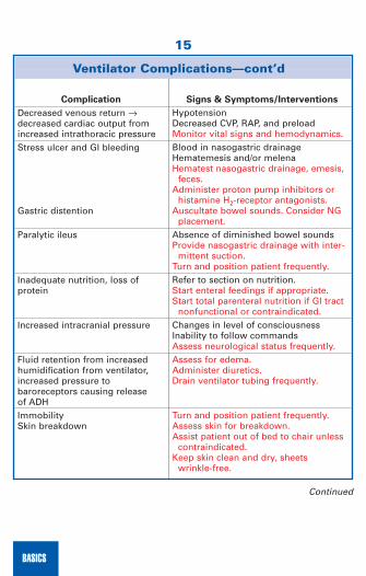
Ventilator Complications—cont’d
Complication Signs & Symptoms/Interventions
Decreased venous return →decreased cardiac output from increased intrathoracic pressureStress ulcer and GI bleeding
Gastric distention
Paralytic ileus
Inadequate nutrition, loss of protein
Increased intracranial pressure
Fluid retention from increased humidification from ventilator, increased pressure to baroreceptors causing release of ADHImmobilitySkin breakdown
HypotensionDecreased CVP, RAP, and preloadMonitor vital signs and hemodynamics.Blood in nasogastric drainageHematemesis and/or melenaHematest nasogastric drainage, emesis,
feces.Administer proton pump inhibitors or
histamine H2-receptor antagonists.Auscultate bowel sounds. Consider NG
placement.Absence of diminished bowel soundsProvide nasogastric drainage with inter-
mittent suction.Turn and position patient frequently. Refer to section on nutrition.Start enteral feedings if appropriate.Start total parenteral nutrition if GI tract
nonfunctional or contraindicated. Changes in level of consciousnessInability to follow commandsAssess neurological status frequently. Assess for edema.Administer diuretics.Drain ventilator tubing frequently.
Turn and position patient frequently.Assess skin for breakdown.Assist patient out of bed to chair unless
contraindicated.Keep skin clean and dry, sheets
wrinkle-free.
Continued
4223_Tab01_001-044 29/08/14 10:46 AM Page 15

16
Neuromuscular Blocking Agents (NMBA)
Purposes■ Facilitate ETT intubation.■ Facilitate mechanical ventilation and improve gas exchange.■ Reduce ICP.■ Control excessive shivering.
Neuromuscular Blocking Agents Used■ Succinylcholine (Quelicin)■ Rocuronium (Zemuron)
BASICS
Ventilator Complications—cont’d
Complication Signs & Symptoms/Interventions
Communication difficulties
Urinary tract infection
Deep vein thrombosis
Psychosocial concerns: fear, loss, powerlessness, pain, anxiety, sleep disturbances, nightmares, loneliness
Keep communication simple.Obtain slate or writing board.Use letter/picture chart.Communicate using sign language. Urine becoming cloudy, concentrated,
odorousChange/remove Foley catheter.Ensure adequate hydration.Administer anti-infectives. Painful, swollen leg; pain may increase
on dorsiflexionAssess for pulmonary embolism. Refer
to Respiratory tab.Administer heparin or enoxaparin. AnxietyDifficulty sleepingPoor pain controlAdminister anxiolytics, sedatives, anal-
gesics.Cluster activities to promote periods of
sleep.Allow patient to make choices when
appropriate.Allow for frequent family visits.Keep patient and family informed.
4223_Tab01_001-044 29/08/14 10:46 AM Page 16

■ Pancuronium (Pavulon)■ Vecuronium
Peripheral Nerve Stimulator ■ Monitors the level of blockade with NMBA use.■ Electrical stimulation is applied to the ulnar nerve, the facial nerve, or the
posterior tibial nerve.■ Train of four (TOF) testing and monitoring should be instituted. The number
of twitches elicited through electrodes along a nerve path is counted. Depthof blockage increases, number of twitches on the TOF decreases. Preventsoverparalyzing the patient and causing prolonged muscular weakness.
Specific Nursing Management■ Patient must be intubated or have tracheostomy in place. Keep airway
patent, and respond to ventilator alarms quickly.■ Monitor and assess response to NMBAs.■ Monitor VS and neurological status, especially pupillary response.■ Monitor ABGs and oxygenation levels.■ Provide eye lubrication and/or taping.■ Provide DVT prophylaxis.■ Initiate pressure sore prevention actions.
Hemodynamic Monitoring
Hemodynamic Parameters
Arteriovenous oxygen difference...................................3.5–5.5 vol% or 4–8 L/minAortic pressure:
■ Systolic.........................................................................................100–140 mm Hg■ Diastolic ...........................................................................................60–80 mm Hg■ Mean ................................................................................................70–90 mm Hg
Cardiac output (CO = HR × SV)..................................................................4–8 L/minCardiac index (CO/BSA) ..........................................................................2.5–4 L/minCentral venous pressure (CVP)...............................................................2–8 mm Hg**Same as right atrial pressure (RAP)Cerebral perfusion pressure (CPP).....................................................70–90 mm HgCoronary artery perfusion pressure (CAPP)......................................60–80 mm HgEjection fraction (Ej Fx or EF).....................................................................60%–75%Left atrial mean pressure ......................................................................4–12 mm HgLeft ventricular systolic pressure ...................................................100–140 mm HgLeft ventricular diastolic pressure..........................................................0–5 mm HgLeft ventricular end-diastolic pressure (LVEDP)..................................5–10 mm HgLeft ventricular end-diastolic volume (LVEDV) .............120–130 mL up to 250 mL
17
BASICS
4223_Tab01_001-044 29/08/14 10:46 AM Page 17

18
Left ventricular stroke work index (LSWI) ...................................30–50 g/beats/m2
Mean arterial pressure (MAP) ..........................................................70–100 mm HgMean arterial pressure used to determine whether BP is sufficient to perfusethe heart, brain, kidneys and other organsOxygen consumption (VO2)............................................................200–250 mL/minOxygen delivery (Do2)...................................................................900–1100 mL/minPulmonary artery pressure (PAP):
■ Systolic.............................................................................................20–30 mm Hg■ Diastolic ...........................................................................................10–20 mm Hg■ Mean ................................................................................................10–15 mm Hg
Pulmonary capillary wedge pressure (PCWP) ....................................4–12 mm HgPulmonary vascular resistance (PVR) .....................................37–250 dyne/sec/cmPulse pressure (SBP-DBP) ........................................................................40 mm HgRight atrial mean pressure .....................................................................2–6 mm HgRight ventricular pressure:
■ Systolic.............................................................................................20–30 mm Hg■ Diastolic ...............................................................................................0–8 mm Hg■ End diastolic ........................................................................................2–6 mm Hg
Right ventricular stroke work index (RSWI) ....................................7–12 g/m2/beatPulmonary vascular resistance (PVR) .................................20–130 dynes/sec/cm-5
Pulmonary vascular resistance index (PVRI) ...............200–400 dynes/sec/cm5/m2
Pulmonary ventricular stroke index .................................................5–10 g/beat/m2
Right atrial pressure (RAP).....................................................................2–6 mm HgStroke index (SI) .........................................................................30–650 mL/beat/m2
Stroke volume (SV = CO/HR)...........................................................60–100 mL/beatSystemic vascular resistance (SVR) ...............................900–1,600 dynes/sec/cm-5
Systemic vascular resistance index.......................1,360–2,200 dynes/sec/cm-5/m2
Systemic venous oxygen saturation (SvO2) .............................................60%–80%
Cardiac Output Components
BASICS
Preload Contractility Afterload
Pao2 Sao2 Hemoglobin (Hgb)Right atrial pressure Stroke volume Pulmonary vascular resistanceCentral venous pressure Cardiac output Systemic vascular resistance Left ventricular end- Tissue perfusion Blood pressure diastolic pressure
4223_Tab01_001-044 29/08/14 10:46 AM Page 18

Pulmonary Artery CatheterThe purpose of the pulmonary artery catheter, also known as the Swan-Ganzcatheter, is to assess and monitor left ventricular function and can determinepreload, assess contractility, and approximate afterload.
PCWP approximates left atrial pressure and left ventricular end-diastolic pressure.Increases in PCWP, LAP, or LVEDP indicate heart failure, hypervolemia, shock,
mitral valve insufficiency, or stenosis. Decreases in PCWP, LAP, or LVEDP indi-cate hypovolemia.
PA Catheter WaveformsThe pulmonary artery catheter is threaded through the right atrium and rightventricle and into the pulmonary artery. Insertion is done via fluoroscopy ormonitoring waveform changes.
19
BASICS
Catheter advanced to right atrium, balloon is inflated. Pressure is low, usually 2–5 mm Hg.
Catheter is floated to right ventricle with the balloon inflated. Waveforms indicate a systolic pressure of 25–30 mm Hg and a diastolic pressure of 0–5 mm Hg.
mm
Hg
Time
Rightatrium
10
0
20
30
40
Ballooncatheter
mm
Hg
Time
10
0
20
30
40
Rightventricle
Ballooncatheter
Continued
4223_Tab01_001-044 29/08/14 10:46 AM Page 19

20
BASICS
As the catheter moves into the pulmonary artery, the systolic pressure remains the same, but the diastolic pressure elevates to 10–15 mm Hg.
The catheter is moved until it can be wedged in a smaller vessel. When the balloon is inflated, the pressure recorded is that pressure in front of the catheter. It is an approximate measure of the left ventricular end-diastolic pressure.
mm
Hg
Time
10
0
20
30
40
Pulmonaryartery
Balloon catheter
Pulmonaryartery wedge
mm
Hg
Time
10
0
20
30
40
Ballooncatheter
Problems With Pulmonary Artery Catheters
Problem Check For/Action
No waveform
Overdamping (smaller waveform with slow rise, diminished or absent dicrotic notch)
• Loose connections• Tubing kinked or compressed• Air in transducer• Loose/cracked transducer• Stopcock mispositioned• Occlusion by clot: Aspirate as per policy• Air bubble or clot in the system• Catheter position: Reposition patient or
have patient cough• Kinks or knotting• Clot: Aspirate as per policy
Continued
4223_Tab01_001-044 29/08/14 10:46 AM Page 20
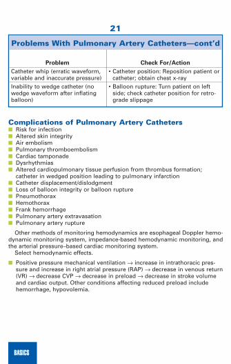
Complications of Pulmonary Artery Catheters■ Risk for infection■ Altered skin integrity■ Air embolism■ Pulmonary thromboembolism■ Cardiac tamponade■ Dysrhythmias■ Altered cardiopulmonary tissue perfusion from thrombus formation;
catheter in wedged position leading to pulmonary infarction■ Catheter displacement/dislodgment■ Loss of balloon integrity or balloon rupture■ Pneumothorax■ Hemothorax■ Frank hemorrhage■ Pulmonary artery extravasation■ Pulmonary artery rupture
Other methods of monitoring hemodynamics are esophageal Doppler hemo-dynamic monitoring system, impedance-based hemodynamic monitoring, andthe arterial pressure–based cardiac monitoring system.
Select hemodynamic effects.
■ Positive pressure mechanical ventilation → increase in intrathoracic pres-sure and increase in right atrial pressure (RAP) → decrease in venous return(VR) → decrease CVP → decrease in preload → decrease in stroke volumeand cardiac output. Other conditions affecting reduced preload includehemorrhage, hypovolemia.
21
BASICS
Problems With Pulmonary Artery Catheters—cont’d
Problem Check For/Action
Catheter whip (erratic waveform, variable and inaccurate pressure)Inability to wedge catheter (no wedge waveform after inflating balloon)
• Catheter position: Reposition patient orcatheter; obtain chest x-ray
• Balloon rupture: Turn patient on leftside; check catheter position for retro-grade slippage
4223_Tab01_001-044 29/08/14 10:46 AM Page 21

22
Intra-Arterial Monitoring
An arterial line (A-line) is used if frequent blood pressure and arterial blood gasdeterminations are needed. It is especially useful
■ After surgery.■ For patients with unstable vital signs.■ For patients experiencing hypoxemia.
Perform Allen’s test prior to insertion. Elevate the patient’s hand with his orher fists clenched. Release pressure over only the ulnar artery. Color returns tothe hand within 6 sec if the ulnar artery is patent and adequate collateral bloodflow present.
BASICS
Compressing the radialand ulnar arteries
Observing for pallor Releasing pressure andobserving for return ofnormal color
4223_Tab01_001-044 29/08/14 10:46 AM Page 22

Intra-Arterial Waveform
Components of Waveform■ Systolic peak: Ventricular ejection and stroke volume. Sharp rise and
rounded top.■ Dicrotic notch: Aortic valve closure, end ventricular systole, start ventricular
diastole. Should be one-third or greater of height of systolic peak. If lower→ suspect ↓ CO.
Tapering of down stroke following dicrotic notch.Important assessments: changes in capillary refill/blanching, sensation,
motion, or color that may indicate lack of perfusion to the extremity.
MAP = systolic BP + (diastolic BP × 2) = 70–100 mm Hg3
Decreased tissue perfusion—decreasing urine output, elevation in BUN:creatinine ratio, altered mental status with decreasing level of consciousness,restlessness, dyspnea, cyanosis, dysrhythmias, abnormal ABGs, weak or absentperipheral pulses, increased capillary refill time (>3 sec), diminished arterial pulsations, bruits.
Potential Complications of Intra-Arterial Monitoring■ Hemorrhage■ Air emboli■ Equipment malfunction/inaccurate pressure■ Dysrhythmias■ Infection■ Altered skin integrity■ Impaired circulation to extremities
23
BASICS
mm
Hg
Time
100
0
4223_Tab01_001-044 29/08/14 10:46 AM Page 23
24
Nutrition Issues in Critical Care
Primary Concerns
■ Malnutrition, starvation, and catabolism■ Stress hypermetabolism■ Fluid volume deficit■ Fluid volume excess
Stress and Nutrition
Prolonged or continual stress depletes glycogen stores → hypermetabolicstate.
Metabolic rate increases with the release of catecholamines + glucagon + cortisol → hyperglycemia and “stress diabetes.”
Protein is lost via gluconeogenesis → decrease in serum protein (albumin).Lipolysis → increase in free fatty acids.Nitrogen excretion increases.Body weight decreases.1 kg body weight = 1 liter of fluid retained or lost.Impaired immune function.
Body Mass Index
BMI is a simple means of classifying sedentary (physically inactive) individualsof average body composition and may indicate obesity. It is calculated by thefollowing: Body mass index (BMI) = weight (kg) ÷ height (meters)2
1 kg = 2.2 lb Normal BMI = 20–25 kg/m2
A BMI >30 kg/m2 indicates obesity; >40 kg/m2 indicates morbid obesity. Anincrease in BMI has been associated with heart disease and diabetes.
A BMI <18.5 kg/m2 suggests a person is underweight. A BMI <17.5 may indi-cate the person has anorexia or a related disorder.
BMI does not take into account factors such as frame size and muscularity.
Signs and Symptoms of Fluid Volume Deficit:Hypovolemia
■ Dry mucous membranes; dry, coated, cracked or fissured tongue■ Thirst; thick, scant saliva■ Poor skin turgor■ Sunken eyeballs
BASICS
4223_Tab01_001-044 29/08/14 10:46 AM Page 24

■ Decreased or orthostatic blood pressure; narrow pulse pressure■ Weak, rapid heart rate and increased respiratory rate■ Decreased capillary refill■ Urine output decreased (<30 mL/hr)■ Increased specific gravity of urine (<1.030)■ Decreased central venous pressure■ Increased hemoglobin and hematocrit■ Increased BUN and serum osmolarity■ Increased BUN:creatinine ratio■ Lethargy, mental confusion
Signs and Symptoms of Fluid Volume Excess:Hypervolemia
■ Crackles in lungs; dyspnea, shortness of breath → pulmonary congestion orpleural effusion
■ Moist mucous membranes■ Full, bounding pulse; tachycardia■ Increased BP, CVP, and PAP■ Distended neck veins and jugular venous pressure■ Edema and decreased serum osmolarity; weight gain■ Decreased hemoglobin and hematocrit■ Decreased specific gravity of urine■ Mental confusion, restlessness
Management
■ Intravenous fluids and volume expanders■ Crystalloids
■ Hypertonic: pulls fluid from the cell: D5/0.25% NaCl, D5/0.45% NaCl,D5/0.9% NaCl, 3% NaCl, D5/LR, D10/W, D10/LR, D50W
■ Hypotonic: moves fluid into the cell: 0.45% NaCl, 2.5%D, D5W■ Isotonic: improves hydration, postop patients: D5W, 0.9% NaCl, LR
■ Colloids■ Plasma or volume expanders: albumin (5% or 25%), or plasma protein,
plasma protein fraction (Plasmanate) may have limited benefit.
Enteral Tube Feedings
Early enteral nutrition has been shown to improve patient outcomes in criticalcare units and reduces ICU length of stay. Safe for patients after successful fluidresuscitation and correction of electrolyte imbalances. Separate IV infusions of
25
BASICS
4223_Tab01_001-044 29/08/14 10:46 AM Page 25

26
the trace elements chromium, copper, manganese, selenium, and zinc havebeen shown to improve patient outcomes.
Types of Tube Feedings
■ Intermittent or bolus feedings: A set volume of formula is delivered at spec -ified times.
■ Continuous feedings: A set rate of formula is delivered over a period oftime.
■ Cyclic feedings: Similar to a continuous feeding, but the infusion is stoppedfor a specified time within a 24-hr period, usually 6–10 hr.
Gastric Access■ Nasogastric tube (NGT)■ Oral■ Percutaneous endoscopic gastrostomy (PEG)■ Nasoduodenal tube (NDT)■ Low-profile gastrostomy device (LPGD)
Small Bowel Access■ Nasal-jejunal tube (NJT)■ Percutaneous endoscopic jejunostomy (PEJ)
The following are based on the Guidelines for the Provision and Assessmentof Nutrition Support Therapy in the Adult Critically Ill Patient: Society of CriticalCare Medicine (SCCM) and American Society for Parenteral and EnteralNutrition (A.S.P.E.N.), 2009 and the Canadian Clinical Practice Guidelines forNutrition Support in Mechanically Ventilated, Critically Ill Adult Patients 2009Clinical Practice Guidelines:
■ Feedings should be instituted within the first 24–48 hr of ICU admission. ■ In the ICU patient population, neither the presence nor absence of bowel
sounds or evidence of passage of flatus and stool is required for the initia-tion of enteral feeding.
■ In patients requiring significant hemodynamic support including high-dosecatecholamine agents, alone or in combination with large-volume fluid orblood product resuscitation to maintain cellular perfusion, enteral nutritionshould be withheld until the patient is fully resuscitated and/or stable.
■ Either gastric or small bowel feeding is acceptable in the ICU setting.■ Consider administration of metoclopramide (Reglan) at the initiation of
feeding or if the gastric residual volume is >250 mL on two consecutivechecks.
BASICS
4223_Tab01_001-044 29/08/14 10:46 AM Page 26

Nursing Care of the Patient Receiving EnteralFeeding■ Use hand hygiene and gloves when setting up or changing feeding
administration set.■ Label with date, time, and nurse’s name initials on all facets of the
administration set.■ Sterile premixed formula should hang for <8 hr or per policy.■ Reconstituted formula exposed to room temperature should hang for
<4 hr or per policy.■ Check expiration date on formula can.■ Change administration sets every 24 hr or per policy.■ Flush tube with water only; avoid juice and carbonated beverages. Flush
tube with 30 mL water every 4 hr for continuous feedings, before and afterintermittent feedings, and after checking residual volume measurement.Flush with 15 mL water before and after each medication administration.
■ Keep HOB elevated 30°–45°.■ Consider hanging IV fluids at the head of the bed only. Use foot of the bed
to hang enteral feedings.■ Assess and monitor serum glucose levels.■ Assess fluid and electrolyte status. Assess hydration.
Checking Tube Placement
Assessing tube placement continues to be controversial as caregivers try tobalance reliability or accuracy and costs. The following are suggested methodsfor verifying enteral tube placement.
■ Obtain chest x-ray or abdominal x-ray. Gold standard for verifying place-ment. Always obtain, and have someone verify, enteral tube placementafter, initial placement.
■ Aspirate gastric contents and check pH.■ Gastric aspirate pH 1–5 in fasting patients but may be as high as 6 if
patient is taking medication to reduce gastric acid (famotidine, ranitidine,pantoprazole).
■ Commercially prepared formulas have a pH close to 6.6.■ Respiratory secretions have pH >6.■ Small intestine aspirate pH >6.
■ Visually inspect gastric aspirate. May aspirate only feeding tube contents.Color varies.
■ Mark location of exit site and note external tube length upon insertion offeeding tube. Does not necessarily indicate location of tube on subsequentinspections.
■ Inject 20–30 mL of air into the tube while auscultating over the epigastriumbelow the diaphragm. Air in the stomach can be heard via a whooshingsound. Considered unreliable.
27
BASICS
4223_Tab01_001-044 29/08/14 10:46 AM Page 27

28
AACN Verification of Feeding Tube Placement Guidelines 2009: Use a variety(two or more) of bedside methods to check feeding tube location:
■ Observe for a change in length of the external portion of the feeding tube.Monitor tube position at 4-hr intervals.
■ Review routine CXR and abdominal x-ray reports.■ Observe changes in volume and appearance of aspirate.■ Measure pH of feeding tube aspirates.■ Obtain an x-ray to confirm tube position when in doubt about tube’s location.
Checking for Gastric Residual Volumes (GRVs)
1. Assess GRVs every 4 hr during initial 48 hr of feeding, then 4–6 hr for con-tinuous feeding and prior to bolus feeding.
2. Using a 30- to 60-mL syringe, withdraw gastric contents from the feeding tube.Note volume of formula. Flush with 30 mL water. Assess for pain, abdominaldistention, “feeling full or bloated,” nausea, and emesis. The following arebased on the A.S.P.E.N. Enteral Nutrition Practice Recommendations.
BASICS
Volume Indication
<250–300 mL>250 mL for 2 consecutive checks>500 mL
Acceptable residual for ICU patients. Administer metoclopramide (Reglan) and
continue to assess residual volume.Stop feeding and reassess patient. Resume
feeding at slower rate, amount, or frequencyas indicated.
Several algorithms have been proposed forICU patients.
Enteral Tube Feeding Complications
Mechanical Complications Interventions
Nasopharyngeal discomfortEsophageal ulceration or bleeding esophageal varicesClogged tube
• Reposition tube.• Consider PEG or PEJ tube.
• Flush with lukewarm water afterevery feeding.
• Hospital protocol:
Continued
4223_Tab01_001-044 29/08/14 10:46 AM Page 28

Total Parenteral Nutrition (TPN)
TPN is an IV solution of 10%–50% dextrose in water (CHO), amino acids (pro-tein), electrolytes, and additives (vitamins, minerals, trace elements of insulin,vitamin K, zinc, famotidine). Fat emulsions provide fatty acids and calories.Solutions >10% dextrose must be infused via a central line.
■ 1000 mL 5% D/W contains 50 g sugar = <200 calories■ 1,000 mL 25% dextrose contains 250 g sugar = 1,000 calories
29
BASICS
Enteral Tube Feeding Complications—cont’d
Mechanical Complications Interventions
Tube displacementExtubation
Stomal leak or infection
Nonmechanical Complications Interventions
Nausea, vomiting, cramps, bloating, abdominal distention
Diarrhea
Aspiration
Gastric reflux
Dumping syndrome: nausea, vomiting, diarrhea, cramps, pallor, sweating, ↑ HR
• Reposition tube.• Insert new tube.• Consider PEG or PEJ tube.• Keep area around stoma clean
and dry.
• Withhold or decrease amount,rate, and frequency of feedings.
• Change to low-fat formula.• Withhold or decrease amount,
rate, and frequency of feedings.• Change formula.• Administer psyllium fiber
(Metamucil)• Hold feedings. Check residuals.• Keep HOB elevated 30°–45°dur-
ing feedings and 1 hr after bolusfeedings.
• Hold feedings. Check residuals.• Keep HOB elevated 30°–45°.• Withhold or decrease amount,
rate, and frequency of feedings.
4223_Tab01_001-044 29/08/14 10:46 AM Page 29

30
Indications■ Severe malnutrition■ Burns■ Bowel disorders (inflammatory disorders, total bowel obstruction, short
bowel syndrome)■ Severe acute pancreatitis■ Acute renal failure■ Hepatic failure■ Metastatic cancer■ Postoperative major surgery if NPO >5 days
Nursing Care■ Each bag of TPN should be changed at least every 24 hr with tubing change.■ Monitor intake and output and weigh the patient daily.■ Monitor glucose levels, including finger stick blood sugars every 4 to 6 hr.
Cover with regular insulin as necessary. If poor control of serum glucose,consider adding insulin to TPN and continue sliding scale coverage.
■ Monitor serum electrolytes including magnesium, phosphate, triglycerides,prealbumin, transferrin, CBC, PT/PTT, and urine urea nitrogen.
■ Assess IV site for redness, swelling, and drainage.■ Change gauze dressing around IV site every 48 to 72 hr, as per protocol.
Transparent dressings may be changed every 7 days.■ If TPN is temporarily unavailable, hang 10% D/W at the same rate as TPN.
Monitor for hypoglycemia.■ Place TPN on infusion pump. Monitor hourly rate. Never attempt to “catch
up” if infusion not accurate.
ComplicationsComplications from TPN may be catheter related, mechanical, or metabolic.
BASICS
Complications of TPN Signs and Symptoms
Infection, catheter-related sepsis, septicemia, septic shockHypoglycemia blood glucose <70 mg/dL
Hyperglycemia blood glucose >200 mg/dLPrerenal azotemia
Leukocytosis; fever; glucose intolerance;catheter site red, swollen, tender; drainage
Shaking, tachycardia, sweating, anxiety,dizziness, hunger, impaired vision, weak-ness, fatigue, headache, irritabilityExtreme thirst, frequent urination, dry skin,hunger, blurred vision, drowsiness, nausea↑ BUN and serum Na+, signs of dehydration,lethargy, coma
Continued
4223_Tab01_001-044 29/08/14 10:46 AM Page 30

Nosocomial Infections in the ICU
Critical Care Risk Factors
■ Nosocomial or hospital-acquired infections (HAIs) develop during hospital-ization or up to 30 days post hospital discharge. They can prolong length ofICU stay; poor patient outcomes. Predisposing factors include:
■ Invasive lines and devices■ Immunocompromising conditions■ Serious underlying illness■ Prolonged stay in critical care unit■ Colonization and cross-infection■ Mechanical ventilation■ Overuse of antibiotics■ Elderly status
31
BASICS
Complications of TPN Signs and Symptoms
Hepatic dysfunction
Pneumothorax, hydrothorax Subclavian/carotid artery puncture
Air embolus
Dysrhythmias
Hypo-/hypernatremia
Hypo-/hyperkalemia
Hypo-/hyperphosphatemia
Hypo-/hypermagnesemia
Hypo-/hypercalcemia
↑ serum liver function tests (SGOT, SGPT,alkaline phosphatase)SOB, restlessness, dyspnea, signs of hypoxia,chest pain radiating to back, arterial blood insyringe, tachycardia, pulsatile blood flow,bleeding from catheter siteRespiratory distress, dyspnea, SOB, tachy-cardia, ↓ BP, neurological deficits, cardiacarrestAtrial, junctional, and ventricular arrhyth-mias; ↓ C.O., ↓ BP, loss of consciousnessNormal values: 135–145 mEq/L or 135–145 mmol/LNormal values: 3.5–5.0 mEq/L or 3.5–5.0 mmol/L Normal values: 3.0–4.5 mg/100 mL or1.0–1.5 mmol/LNormal values: 1.5–2.0 mEq/L or 0.8–1.3 mmol/LNormal values: 8.5–10.5 mg/100 mL or2.1–2.6 mmol/L
4223_Tab01_001-044 29/08/14 10:46 AM Page 31

32
Methicillin-Resistant Staphylococcus Aureus (MRSA)
EtiologyTransmitted by close contact with infected person. Health-care worker may becolonized with MRSA strain with absence of symptoms. The Staphylococcusaureus bacterium is resistant to methicillin, amoxicillin, penicillin, oxacillin, andother antibiotics.
Signs and Symptoms■ Skin infection: Boil or abscess■ Surgical wound: Swollen, red, painful, exudate (pus)■ Bloodstream: Fever, chills■ Lung infection/pneumonia: Shortness of breath, fever, chills■ Urinary tract: Cloudy urine, strong odor■ Infective carditis
Diagnosis■ Culture of infected area
Treatment■ Vancomycin (Vanocin, Vancoled); trough vancomycin levels the most accu-
rate; monitoring of peak vancomycin levels not recommended■ Linezolid (Zyvox)■ Daptomycin (Cubicin)
Clostridium Difficile (C-diff)
2013 Guidelines for Diagnosis, Treatment, and Prevention of Clostridium diffi-cile Infections: http://gi.org/guideline/diagnosis-and-management-of-c-difficile-associated-diarrhea-and-colitis/
EtiologyC. difficile (C-diff) is a common cause of antibiotic-associated diarrhea (AAD)and is transmitted through the feces or any surface, device, or material that hasbecome contaminated with feces.
Signs and Symptoms■ Watery diarrhea (at least 3 BMs/day for 2 or more days), rarely bloody. May
be greenish, mucoid and foul-smelling.■ Fever.■ Loss of appetite.■ Nausea.■ Crampy abdominal pain and lower abdominal tenderness.
BASICS
4223_Tab01_001-044 29/08/14 10:46 AM Page 32

Diagnosis■ Stool culture.■ Glutamine dehydrogenase enzyme immunoassay (EIA).■ Abdominal x-rays, CT, and colonoscopy may also be indicated.
Treatment■ Discontinue current antibiotics. May give metronidazole (Flagyl) or oral or
rectal (retention enema) or vancomycin to treat diarrhea depending onseverity. Consider fidaxomicin (Dificid), which has also been shown to bemore effective for cancer patients with C-diff instead of vancomycin andthose resistant to vancomycin.
■ Contact precautions: Isolation in private room, gloves and gowns for personnel and visitors.
■ Monitor fluid balance, electrolytes, albumin, and CBC.■ Alcohol-based rubs not effective. Soap-and-water hand hygiene
recommended.■ Data do not support the use of probiotics or antiperistaltic agents.■ Opioids and loperamide may increase the risk of toxic megacolon.■ Proton-pump inhibitors (PPIs) may increase the incidence of C-diff–
associated diarrhea.■ Opioids and loperamide may increase the risk of toxic megacolon.■ Consider adding cholestyramine (Questran) to drug regimen.■ Consider fecal microbiota transplantation (FMT). Also referred to as fecal
bacteriotherapy or fecal transplant. Hospital must have a procedure andprotocol in place for implementation.■ Healthy donor who meets select criteria.■ Administer vancomycin preprocedure or per protocol.■ Administer PPI the evening before and morning of procedure to reduce
gastric acid or per protocol.■ Donor stool is prepared into a fecal slurry. ■ Administered via colonoscopy, retention enema, or NG tube.
• Average size adult: 50–200 mL via NG tube or 250–500 mL viacolonoscopy.
• To administer via NGT: Draw up slurry into 60-mL syringe and inject 50 mL/2–3 min.
• Follow with 50 mL flush of NS.■ Keep HOB elevated 30° or more for at least 2 hr post procedure.■ Document diarrhea post procedure. Procedure may be repeated
after 5 days.■ Subtotal colectomy with preservation of the rectum may be indicated for
severely ill patients with grossly elevated WBC and serum lactate levels.
33
BASICS
4223_Tab01_001-044 29/08/14 10:46 AM Page 33

34
Other Hospital-Acquired Infections of Concern in the ICU:
■ Catheter-associated urinary tract infections (CAUTI)■ Central line–associated bloodstream infections (CLABSI)■ Hospital-acquired pneumonia (non-VAP)■ Surgical site infections (SSI)■ Ventilator-associated pneumonia (VAP) (refer to Respiratory tab)
Psychosocial Issues in Critical Care
Environmental, Sensory Overload, and Sleep Deprivation
■ Sensory Overload: A condition in which sensory stimuli are received at arate and intensity beyond the level that the patient can accommodate.
■ Sensory Deprivation: A condition in which the patient experiences a lack ofvariety and/or intensity of sensory stimuli.
Types of Sensory Stimuli■ Visual■ Auditory■ Kinesthetic■ Gustatory■ Tactile■ Olfactory
Signs and Symptoms of Sensory Problems■ Confusion■ Hallucinations■ Lethargy■ Behavioral changes (combativeness)■ Increased startle response■ Disorientation■ Anxiety■ Restlessness■ Panic■ Withdrawal■ Mood swings
Some of these same symptoms may be caused by select medications, espe-cially in elderly patients. Refer to the Updated 2012 Beers Criteria for PotentiallyInappropriate Medication Use in Older Adults from the American Geriatrics
BASICS
4223_Tab01_001-044 29/08/14 10:46 AM Page 34

Society at http://www.americangeriatrics.org/files/documents/beers/2012BeersCriteria_JAGS.pdf
■ Other environmental and psychological concerns include noise, lights andcolor, sleep disturbances, lack of control, helplessness, hopelessness, spiri-tual distress, stress, anxiety, and fear.
■ Nurses may suffer from alarm fatigue and moral distress.■ Psychiatric emergencies in the ICU include agitated delirium and psychosis,
neuroleptic syndrome, serotonin syndrome, and psychiatric medicationoverdose.
■ Palliative care protocols for the ICU are being developed.
Near-Death Experience
The experience of patients that they have glimpsed the afterlife when comingclose to death. These perceptions may include:
■ Seeing an intense light■ Seeing angels or departed loved ones■ Traveling through a tunnel
Out-of-Body Experience
The experience of being away from and overlooking one’s body. The patientfeels that the mind has separated from the body.
Family Needs of the Critical Care Patient
■ Relief of anxiety■ Assurance of competent care■ Timely access to the patient■ Accurate and timely information about the patient’s condition and progno-
sis in easily understandable terms■ Early notification of changes in the patient’s condition■ Explanations regarding the environment, machinery, and monitoring
equipment■ Honest answers to questions■ Emotional support■ Regard for the spiritual needs of the family and patient
35
BASICS
4223_Tab01_001-044 29/08/14 10:46 AM Page 35

36
Organ Donation
Transplantable organs include:
■ Kidneys■ Heart■ Lungs■ Liver■ Pancreas■ Intestines
Corneas, the middle ear, skin, heart valves, bone, veins, cartilage, tendons,and ligaments can be stored in tissue banks and used to restore sight, coverburns, repair hearts, replace veins, and mend damaged connective tissue andcartilage in recipients.
Healthy adults between the ages of 18 and 60 yr can donate blood stem cells:marrow, peripheral blood stem cells, and cord blood stem cells.
Nursing Role in Organ Donation■ Provide accurate information regarding donation.■ Identify possible donors early.■ Work closely with the organ procurement organization and members of the
health team to elicit donations.■ Provide emotional support to families considering donation, and make sure
to respect their cultural and religious beliefs.■ Become a donor advocate among colleagues and for patients and their
families.
General Criteria for Brain Death
■ Absence of purposive movement■ Flaccid tone and absence of spontaneous or induced movements■ Persistent deep coma■ No response to pain■ Absence of spontaneous respiration■ Absence of brainstem reflexes:
■ Midposition or pupils fixed and dilated■ No corneal, gag, or cough reflexes■ Absence of spontaneous oculocephalic (doll’s eye phenomenon) reflex■ No vestibular response to caloric stimulation
■ Isoelectric or flat electroencephalogram (EEG)■ Absent cerebral blood flow
These criteria vary from state to state.
BASICS
4223_Tab01_001-044 29/08/14 10:46 AM Page 36

Hemodynamic Management of Potential Brain-DeadOrgan DonorsEnsure adequate intravascular volume and adequate cardiac output to ensureconsistent perfusion to vital organs.
■ MAP >60 mm Hg■ Urine output >1.0 mL/kg/hr■ Left ventricular ejection fraction >45%
Nursing Care■ Fluid management—fluids or diuretics■ Inotropic agents to correct low cardiac output■ Vasopressors to correct vasodilatation■ Thyroid hormone■ Corticosteroids to reduce inflammation■ Vasopressin to support renal function■ Insulin to control glucose levels■ Regulate ventilator settings including use of PEEP■ Suction frequently to promote adequate oxygenation
Specific organ donation protocols:
Anxiety, Agitation, and SedationPurpose of sedation is to minimize use of neuromuscular paralysis agents. Short-term use will ↓ ventilatory time, ↓ length of stay in ICU, ↓ costs, lead to fewer tracheostomies, and provide early intervention of neurological deterioration.
37
BASICS
4223_Tab01_001-044 29/08/14 10:46 AM Page 37

38
Long-term use has the opposite effects. Contributing factors: stress, ICU envi-ronment, pain, sleep deprivation, surgery, and anesthesia. Anxiety and agita-tion can lead to delirium and changes in LOC with poorer patient outcomes.
Sedatives should be titrated without impairing neurological assessment.Analgesics should be titrated to keep pain level <3 on 0–10 scale.
Assessment
Prior to sedation, exclude and treat possible causes of agitation and confusion:
■ Cerebral hypoperfusion■ Cardiac ischemia■ Hypotension■ Hypoxemia or hypercarbia (elevated blood CO2)■ Fluid and electrolyte imbalance: acidosis, hyponatremia, hypoglycemia,
hypercalcemia, hepatic or renal insufficiency■ Infection■ Drug-induced agitation or confusion, especially in elderly patients■ Furosemide (Lasix) (can contribute to agitation)
Use nonpharmacological therapies such as repositioning, massage, distrac-tion, minimizing of noise, and family support at the bedside. Cluster activities toallow for uninterrupted periods of sleep or “sleep hours.”
Assess pain on 0–10 scale or faces scale and look for nonverbal cues. SeeAgitation, Sedation, and Pain Assessment Scales.
Medications for Agitation, Sedation, and Pain
Benzodiazepines■ Diazepam (Valium).■ Lorazepam (Ativan).■ Midazolam (Versed).■ Frequently combined with opioids. Use caution in elderly patients.■ Keep flumazenil (Romazicon) available (a benzodiazepine antagonist/
antidote).
Opioids■ Morphine sulfate.■ Fentanyl.■ Hydromorphone (Dilaudid).■ Consider oxycodone (OxyContin) as needed.■ Consider remifentanil (Ultiva) as needed.
BASICS
4223_Tab01_001-044 29/08/14 10:46 AM Page 38

Nonopioid Analgesics■ Ketamine.
Alpha-Adrenergic Receptor Agonists■ Dexmedetomidine (Precedex).
Nonbarbiturate Sedatives■ Propofol (Diprivan).■ Etomidate (Amidate).
Physiological Responses to Pain and Anxiety
■ Tachycardia■ Diaphoresis■ Sleep disturbance■ Hypertension■ Tachypnea■ Nausea
Signs of Sedative or Analgesic Withdrawal■ Nausea, vomiting, diarrhea■ Cramps, muscle aches■ Increased sensitivity to pain■ Tachypnea, ↑ HR, ↑ BP■ Delirium, tremors, seizures, agitation
Medication Management
■ Administer analgesics as scheduled doses or continuous infusions; avoidprn analgesics.
■ Other routes include oral, rectal, transdermal, subcutaneous, and spinal.■ Opioids preferred: fentanyl, hydromorphone, and morphine.■ Have naloxone (Narcan), an opioid antagonist, available.■ Monitor body and limb movements, facial expression, posturing, muscle
tension for signs of pain.■ Monitor for acute changes or fluctuations in mental status, LOC, disorienta-
tion, hallucinations, delusions.■ Evaluate arousability.■ Monitor neurological status including pupillary response, response to
verbal commands and pain.■ Monitor respiratory rate and respiratory effort, respiratory depression,
BP, HR.
39
BASICS
4223_Tab01_001-044 29/08/14 10:46 AM Page 39

40
Agitation, Sedation, and Pain Assessment Scales
BASICS
Richmond Agitation Sedation Scale (RASS)
Score Term Description
+4 Combative Overtly combative, violent, immediate dangerto staff
+3 Very agitated Pulls or removes tube(s) or catheter(s); aggressive
+2 Agitated Frequent nonpurposeful movement, fights ventilator
+1 Restless Anxious but movements not aggressive vigorous0 Alert and calm–1 Drowsy Not fully alert, but has sustained awakening to
verbal stimuli (eye-opening/eye contact) tovoice/verbal stimuli (>10 sec)
–2 Light sedation Briefly awakens with eye contact to voice/verbalstimuli (<10 sec)
–3 Moderate Movement or eye opening to voice/verbal sedation stimuli (but no eye contact)
–4 Deep sedation No response to voice, but movement or eyeopening to physical stimulation
–5 Unarousable No response to voice or physical stimulation
Procedure for RASS Assessment
Procedure Patient Assessment Score
1. Observe patient. a. Patient is alert, restless, or agitated. 0 to +42. If not alert, state b. Patient awakens with sustained –1
patient’s name eye opening and eye contact.and say to open c. Patient awakens with eye opening –2eyes and look at and eye contact, but not sustained.speaker. d. Patient has any movement in –3
response to voice but no eye contact.
Continued
4223_Tab01_001-044 29/08/14 10:46 AM Page 40

41
BASICS
Procedure for RASS Assessment—cont’d
Procedure Patient Assessment Score
3. When no e. Patient has any movement to –4response to physical stimulation.verbal f. Patient has no response to –5stimulation, any stimulation.physically stimulate patient by shaking shoulder and/or rubbing sternum.
Reprinted with permission from Vanderbilt University. http://www.icudelirium.org/docs/RASS.pdf.
Sedation-Agitation Scale (SAS)
Score Level of Sedation-Agitation Response
7 Dangerous agitation Pulling at endotracheal tube, thrash-ing, climbing over bed rails
6 Very agitated Does not calm, requires restraints,bites endotracheal tube
5 Agitated Attempts to sit up but calms to verbal instructions
4 Calm and cooperative Obeys commands3 Sedated Difficult to rouse, obeys simple
commands2 Very sedated Rouses to stimuli; does not obey
commands1 Unarousable Minimal or no response to noxious
stimuli
Reprinted with permission from Riker, R, Fraser, GL, and Cox, PM. Continuous infusion ofhaloperidol controls agitation in critically ill patients. Crit Care Med 22(3):433–440, 1994.
4223_Tab01_001-044 29/08/14 10:46 AM Page 41

42
Pain Assessment Scales
Pain Visual Analog Scale (VAS)
no pain 0 worst pain 10no anxiety 5 severe anxiety
■ Nonverbal Adult Pain Assessment Scale (NVPS)■ Behavioral Pain Scale (BPS)■ Critical Care Pain Observation Tool (CPOT) for nonverbal adults■ Faces Pain Scale
Pain Management■ Continuous IV narcotic■ Patient controlled analgesia■ Epidural analgesia
Delirium
Delirium has been associated with poor patient outcomes. Patients with deliri-um have higher ICU and hospital stays along with a higher risk of death. Usuallyreversible. More common in elderly patients, patients with compromised men-tal status, and mechanically ventilated patients. AACN recommends the THINKmnemonic in determining the cause of delirium in ICU patients:
■ Toxic situations ■ HF, shock, dehydration■ Deliriogenic meds (tight titration of sedatives)■ New organ failure (e.g., liver, kidney)
■ Hypoxemia■ Infection/sepsis (nosocomial)■ Immobilization■ Nonpharmacological interventions (Are these being neglected?)
■ Hearing aids, glasses, sleep protocols, music, noise control, ambulation■ K+ or electrolyte problems
May also be precipitated by hypertension, head trauma, and metabolic distur-bances.
Delirium is characterized by an acute onset of mental status changes thatdevelop over a short period of time, usually hours to days. It may fluctuate overthe course of a day. It may be combined with inattention and disorganized think-ing or altered level of consciousness. The DSM-IV TR describes three clinicalsubtypes: hyperactive, hypoactive, and mixed. Hyperactive delirium may beconfused with anxiety and agitation.
BASICS
4223_Tab01_001-044 29/08/14 10:46 AM Page 42

Other signs and symptoms include:
■ Difficulty maintaining or shifting attention with low attention span.■ Variation in levels of confusion; disorientation; fluctuations between lucidity
and confusion.■ Illusions, paranoia, or hallucinations.■ Fluctuating LOC with clouding of consciousness.■ Dysphasia; dysarthria.■ Tremor; motor abnormalities; asterixis if hepatic encephalopathy suspected.■ Benzodiazepines such as lorazepam (Ativan) may cause or worsen delirium
and should be avoided. Opioid analgesics, metoclopramide, antidepres-sants, H2 antagonists and corticosteroids have also been identified as riskfactors for delirium development.
■ Newer neuroleptics preferred. Consider risperidone (Risperdal), olanzapine(Zyprexa), and quetiapine (Seroquel).
■ Haloperidol (Haldol) previously was the drug of choice to treat delirium inthe ICU patient but has shown to have adverse neurological effects. If used,monitor for QT prolongation.
Delirium Assessment ToolsThe Confusion Assessment Method for the Intensive Care Unit (CAM-ICU) scoreis widely used to screen for delirium in the ICU population.
Confusion Assessment Method for the ICU (CAM-ICU) Flowsheet. Refer to:http://www.mc.vanderbilt.edu/icudelirium/docs/CAM_ICU_flowsheet.pdf
Confusion Assessment Method for the ICU (CAM-ICU) Worksheet. Refer to:http://www.mc.vanderbilt.edu/icudelirium/docs/CAM_ICU_worksheet.pdf
Assessment tools to predict delirium and severity of delirium in hospitalizedpatients.
■ Mini-Cog■ Intensive Care Delirium Severity Checklist (ICDSC)■ Delirium Detection Scale (DDS)
Complications of Sedation, Agitation, and DeliriumTherapy
■ Hypotension■ Patient unresponsiveness■ Respiratory depression■ Delayed weaning from mechanical ventilator
Complications associated with immobility: pressure ulcers, thromboem-bolism, gastric ileus, hospital-acquired pneumonia
43
BASICS
4223_Tab01_001-044 29/08/14 10:46 AM Page 43

44
Management of the Patient With Agitation or Delirium
■ Institute delirium protocol. Refer to: http://www.mc.vanderbilt.edu/icudelirium/docs/Delirium_Protocol_2001_30_07.pdf
■ Monitor for delirium onset and resolution of symptoms.Systematic use of standardized assessment tools should be used. Example:All elderly patients should be screened with the CAM-ICU on ICU admission.
■ Provide a quiet and stable environment. Promote rest and control pain.■ Provide reality orientation in any patient encounter.■ Institute safety precautions. Physical restraints should be a last option.■ Monitor VS and pulse oximetry. P/F ratio should improve in ARDS patients.■ Monitor ECG.■ Ensure adequate nutrition.■ Provide progressive mobility exercise.■ Monitor CBC, ESR, electrolytes, glucose levels, renal, liver and thyroid func-
tion tests, urinalysis, vitamin B12 and thiamine levels, HIV status, urine andblood toxicology, and ETOH levels.
■ CT, MRI, ECG, EEG, or CXR may be ordered to rule out physical courses ofdelirium.
■ S-100 calcium-binding protein B (S100B) are seen in higher levels inpatients with delirium.
■ AACN recommends the ABCDE bundle (refer to Basics, MechanicalVentilation).
Medically Induced ComaAlso referred to as barbiturate coma.
Causes a temporary coma or a deep state of unconsciousness to protect thebrain from swelling and allow the brain to rest to help prevent brain damage.Goal is to reduce ICP.
Medications used: propofol, pentobarbital, thiopental.
BASICS
__________________________________________________________________________
__________________________________________________________________________
__________________________________________________________________________
__________________________________________________________________________
__________________________________________________________________________
__________________________________________________________________________
4223_Tab01_001-044 29/08/14 10:46 AM Page 44

45
CV
Acute Coronary Syndrome (ACS)ACS is the term used to denote any one of three clinical manifestations of coro-nary artery disease:
■ Unstable angina■ Non–ST-elevation MI (NSTEMI)■ ST-elevation MI (STEMI)
Pathophysiology
Unstable angina represents the progression of stable coronary artery diseaseto unstable disease. Rupture of atherosclerotic plaque causes thrombus forma-tion and partial occlusion in coronary arteries that precipitate a myocardialinfarction.
Clinical Presentation
ACS manifests with chest pain, diaphoresis, SOB, nausea and vomiting, dysp-nea, weakness, and fatigue. Symptoms of MI are midsternal chest paindescribed as pressure, squeezing, fullness, or pain. May radiate to jaw, neck,arms, or back and usually lasts more than 15 min.
Assessment for chest pain and associated symptoms of ACS include use ofPQRST method when assessing pain, physical exam, vital signs, auscultationfor S3 or S4 gallop, auscultation of lungs for crackles, and assessment of periph-eral vessels for pulse deficits or bruits.
Diagnostic Tests
■ ECG■ Echocardiogram■ Cycle cardiac markers (troponin I, CK, CK-MB, myoglobin, C-reactive
protein)■ Chest x-ray■ Nuclear scan■ CT angiography■ Coronary angiogram■ Fibrinogen level■ Homocystine lipoprotein levels■ Total cholesterol and triglyceride levels■ Brain natriuretic peptide (BNP)■ PT/PTT
4223_Tab02_045-106 29/08/14 10:00 AM Page 45

46
CV
Management
■ Administer oxygen to maintain SaO2 >93%.■ Establish IV access.■ Perform continuous cardiac monitoring.■ Administer SL nitroglycerin tablets or oral spray, every 5 min × 3 doses. If
pain persists, IV nitroglycerin may be started.■ Monitor for hypotension and headaches from vasodilatation.■ Administer non-enteric coated aspirin (162–325 mg) and have patient chew
it, if not already on daily dose.■ Administer IV morphine, 4–8 mg initially with increments of 2–8 mg
repeated at 5–15 min intervals until pain is controlled. Use lower dose in elderly patients.
■ Monitor for hypotension and respiratory depression.■ Unless contraindicated, administer a beta blocker.■ Administer angiotensin converting enzyme inhibitors (ACE-IS) and
angiotensin receptor blockers (ARBs) if patient had moderate to severe MIwith reduced heart’s pumping capacity.
■ Administer calcium channel blockers if symptoms persist after NTG andbeta blockers given.
■ Monitor and immediately treat arrhythmias; pay attention to electrolyte disturbances (especially potassium and magnesium), hypoxemia, drugs, oracidosis.
■ Administer clopidogrel 300–600 mg loading dose (600 mg preferred dose).■ High-risk patients with NSTEMI ACS should also receive unfractionated
heparin or low-molecular-weight heparin (LMWH) and IV platelet glycopro-tein IIb/IIIa complex blockers (tirofiban, eptifibatide), as well as ASA, clopido-grel, and beta blockers.
Unstable AnginaUnstable angina is the sudden onset of chest pain, pressure, or tightness result-ing from insufficient blood flow through coronary arteries.
Pathophysiology
Atherosclerosis → obstruction of coronary arteries → decrease blood flowthrough coronary arteries → decrease oxygen supply to myocardial demand forO2 during exertion or emotional stress → angina.
4223_Tab02_045-106 29/08/14 10:00 AM Page 46

Clinical Presentation
Chest pain manifests as follows: substernal pain, tightness, dullness, fullness,heaviness, or pressure; dyspnea; syncope; and pain radiating to arms, epigas-trium, shoulder, neck, or jaw. Women may experience more atypical symptomssuch as back pain and GI symptoms (e.g., indigestion, nausea and vomiting, and abdominal fullness), whereas men may experience typical symptoms suchas midsternal chest pain radiating to the left arm.
Diagnostic Tests
■ 12-lead ECG■ Lab work: cardiac markers: creatine kinase (CK), creatine kinase–myocardial
band (CK-MB), troponin I (TnI), and myoglobin■ Exercise or pharmacological stress test■ Echocardiogram■ Nuclear scan: single-photon emission computed tomography (SPECT), MRI
with gadolinium enhancement (MUGA)■ Cardiac catheterization and coronary artery angiography■ Percutaneous coronary intervention (PCI)■ Chest x-ray■ MRI/MRA■ Hgb, CMP, lipid panel, BNP
Management
■ Calculate TIMI Risk Score for patients with unstable angina (UA) and non–ST-elevation Ml (NSTEMI). Estimates mortality for patients with unstable anginaand NSTEMI. Refer to: http://www.mdcalc.com/timi-risk-score-for-uanstemi/
■ Ensure bedrest.■ Obtain ECG and lab work.■ Assess chest pain for frequency, duration, cause that triggered pain, radia-
tion of pain, and intensity based on pain scale from 0 to 10, with 0 being nopain and 10 being worst pain.
■ Supply O2.■ Provide continuous cardiac monitoring.■ Pharmacological treatment:
■ Early conservative, for low-risk patient: anti-ischemic, antiplatelet, andantithrombotic drug therapy; stress and treadmill tests.
■ Early invasive: same drug therapy as early conservative but followed bydiagnostic catheterization and revascularization.
■ Administer nitroglycerin (NTG): 0.4 mg (SL or spray) every 5 min for atotal of three doses → IV infusion start at 5–10 mcg/min, titrate for pain
47
CV
4223_Tab02_045-106 29/08/14 10:00 AM Page 47

48
according to hospital policy. Dose should not exceed 400 mcg/min; checkfor contraindications such as hypotension, or if taking the followingmeds: Viagra, Cialis, or Levitra.
■ Administer morphine sulfate IV if symptoms persist after receiving NTGor in patients who have pulmonary congestion or severe agitation.
■ Administer beta blocker: metoprolol (Lopressor).■ Administer ACE-IS in patients with LV dysfunction or HF with HTN; not
recommended in patients with renal failure.■ Administer calcium channel blockers: verapamil (Calan, Isoptin) or dilti-
azem (Cardizem) if patient not responding to beta blocker or nitrates.
*Use severe caution when combining blocking agents*
■ Administer antiplatelet: aspirin 162–325 mg, chewed.■ Administer GP IIb/IIIa inhibitor: eptifibatide (Integrilin) or tirofiban
(Aggrastat) if no contraindications (i.e., bleeding, stroke in past month,severe HTN, renal dialysis, major surgery within the past 6 wk, or plateletcount <100,000 mm3 ).
■ Administer antithrombotic: heparin.■ Administer anticoagulant: enoxaparin (Lovenox).■ Administer clopidogrel (Plavix).■ Administer direct thrombin inhibitor (hirudin, bivalirudin).
Acute Myocardial Infarction (AMI)AMI is the acute death of myocardial cells resulting from lack of oxygenatedblood flow in the coronary arteries. It is also known as a heart attack.
Pathophysiology
Injury to the artery’s endothelium → increases platelet adhesion → inflammatoryresponse causing monocytes and T lymphocytes to migrate in the intima →macrophages and smooth muscle distend with lipids, to form fatty streaks and afibrous cap → thinning of cap increases susceptibility to rupture or hemorrhage →rupture triggers thrombus formation and vasoconstriction → result: thrombuswith narrowing artery. If occlusion lasts more than 20 min, can lead to AMI.
Clinical Presentation
AMI manifests with chest pain or discomfort lasting 20 min or longer. Pain canbe described as pressure, tightness, heaviness, burning, or a squeezing orcrushing sensation, located typically in the central chest or epigastrium; it mayradiate to the arms, shoulders, neck, jaw, or back.
CV
4223_Tab02_045-106 29/08/14 10:00 AM Page 48

Discomfort may be accompanied by weakness, dyspnea, diaphoresis, or anx-iety; not relieved by NTG. Women may experience atypical discomfort, SOB, orfatigue. Diabetic patients may not display classic signs and symptoms of AMI.Elderly patients may experience SOB, pulmonary edema, dizziness, alteredmental status.
ST-segment elevation MI: Look for tall positive T waves and ST-segment ele-vation of 1 mm or more above baseline.
Non–ST-segment elevation MI: May include ST-segment depression and T-wave inversion.
Diagnostic Tests
■ ECG findings.■ Cardiac markers (CK, myoglobin, and troponins).■ Ischemia modified albumin (IMA): measures changes in serum albumin
when in contact with ischemic tissue. IMA rises faster than other cardiacenzymes.
■ Serum cardiac biomarkers; timeline varies according to reference.
49
■ Echocardiogram.■ PCI.
Management
Management is based on the 2013 ACCF/AHA Guideline for the Management ofST-Elevation Myocardial Infarction.
■ Calculate TIMI Risk Score for ST Elevation MI (STEMI). Estimates mortality inpatients with STEMI. Refer to: http://www.mdcalc.com/timi-risk-score-for-stemi/
Levels Levels RemainBiomarker Increase Peak Elevated for
CK 3–6 hr 24–36 hr 3–5 daysCK-MB 3–8 hr after chest 12–24 hr 48–72 hr
pain onsetMyoglobin 30 min –4 hr after 6–12 hr 24–36 hr
chest pain onsetTroponin Within 3–12 hr after MI 14–48 hr 5–14 daysTroponin I Within 3–12 hr 14–24 hr 5–14 daysTroponin T Within 3–12 hr 12–48 hr 5–14 daysIMA Within 2 min of MI 6 hr 12 hr
CV
4223_Tab02_045-106 29/08/14 10:00 AM Page 49

50
■ Calculate the HEART Score, a tool for predicting and managing the risk ofheart attack and stroke. Refer to: http://www.escardio.org/communities/EACPR/toolbox/health-professionals/Pages/SCORE-Risk-Charts.aspxCalculate in-hospital and 6-mo mortality rate for patients with ACS includ-ing those with ST elevation or ST depression using the Global Registry of Acute Coronary Events GRACE) risk model. Refer to: http://www.outcomes-umassmed.org/grace/acs_risk/acs_risk_content.html
■ Focus on pain radiation, SOB, and diaphoresis.■ Obtain a 12-lead ECG and lab draw for cardiac markers.■ MONA: morphine, O2, NTG, and 162–325 mg non-enteric coated aspirin
po or chewed. If allergic to aspirin, give ticlopidine (Ticlid) or clopidogrel(Plavix).
■ Administer supplemental O2 to maintain SpO2 >90%.■ Administer sublingual NTG tablets or spray.■ Administer IV morphine 4–8 mg initially and then 2-8 mg every 5-15 min until
pain is controlled. (Monitor for hypotension and respiratory depression.)■ Administer ACE-IS or ARBs.■ Administer beta blocker.■ Administer statin.■ Administer unfractionated heparin, low-molecular-weight heparin.■ Administer glycoprotein IIb/IIIa antagonists (abciximab, eptifibatide,
tirofiban).■ Coronary arterial bypass graft (CABG) is warranted in setting of failed PCI
with instability.
ST-Segment Monitoring■ Continuous ST-segment monitoring is used to detect silent ischemia in
asymptomatic select patients. Monitoring ST changes in a 12-lead ECG ismost accurate. If continuous 12-lead ECG not available, use leads III and V3.Be sure to select patient’s most sensitive monitoring leads (ST fingerprint).
■ Evaluate ST segment with patient in supine position (change in body posi-tion can alter ST segment and mimic ischemia). If ST alarm sounds withpatient in side-lying position, return patient to supine. If deviation persistsin supine, may indicate ischemia.
■ Measure the ST-segment changes 60 msec beyond the J point (the junctionof the QRS complex with the ST segment).
■ Alarm parameters: ■ Patients at high risk for ischemia: Set ST-segment alarm parameters 1 mm
above and below baseline ST segment.■ Stable patients: Set segment alarm parameters 2 mm above and below
baseline ST segment.■ Document actual millimeters of ST-segment depression or elevation.
CV
4223_Tab02_045-106 29/08/14 10:00 AM Page 50

■ Cause for concern: ST depression or elevation of 1–2 mm that lasts for at least 1 min can be clinically significant and warrants further patientassessments.
■ Refer to AACN Practice Alert at http://www.aacn.org/WD/Practice/Docs/PracticeAlerts/ST_Segment_Monitoring_05-2009.pdf.
Hypertensive CrisisHypertensive crisis is defined as a severe elevation in blood pressure (systolicBP >179 mm Hg, diastolic BP >109 mm Hg), which may or may not lead to organdamage. There are two types of hypertensive crisis:
■ Hypertensive emergency: Rapid (hours to days) marked elevation in BP(SBP >180 mm Hg or DBP >120 mm Hg) → acute organ tissue damage.
■ Hypertensive urgency: Slow (days to weeks) elevation in BP (SBP >180 mm Hgor DBP >110 mm Hg) usually does not lead to organ tissue damage.
Pathophysiology
Any disorder or cause (essential hypertension, renal parenchymal disease, re -novascular disease, pregnancy, endocrine drugs, autonomic hyperreactivity,CNS disorder) → BP → vessel becomes inflamed → leak fluid or blood to thebrain → CVA → long-term disability. Intense vasoconstriction may also causeencephalopathy, myocardial ischemia or infarction, acute pulmonary edema,aortic dissection, and acute renal failure, among other conditions.
Clinical Presentation
Hypertensive crisis manifests with:
■ Chest pain■ Dyspnea, orthopnea, paroxysmal nocturnal dyspnea■ Neurological deficits■ Severe, throbbing headache■ Visual disturbances■ Nausea and vomiting■ Dysphagia■ Back pain■ Severe anxiety■ Irritability, confusion■ Possible seizures■ Oliguria if kidneys affected
↓
51
CV
4223_Tab02_045-106 29/08/14 10:00 AM Page 51

52
Diagnostic Tests
■ CT or MRI of chest, abdomen, and brain■ 2-D echocardiogram or transesophageal echocardiogram■ ECG■ CBC■ Cardiac biomarkers if appropriate■ Serum BUN, creatinine, BMP■ Urinalysis especially specific gravity, urine toxicology■ Renal ultrasound if kidney involvement■ Chest x-ray (if dyspnea or chest pain present)
Management
■ Administer O2 to maintain PaO2 >92%. Monitor ABGs.■ Obtain VS: orthostatic BP every 5 min, then longer intervals. Arterial line
recommended.■ Check BP in both arms.■ Palpate pulses in all extremities.■ Provide continuous ECG monitoring and treatment of arrhythmias.■ Assess cardiac, respiratory, and neurological status.■ Administer analgesics for pain or headache.■ First-line medical therapy: labetalol (Trandate) or esmolol (Brevibloc),
adrenergic-receptor blockers with both selective alpha-adrenergic and nonselective beta-adrenergic receptor blocking actions.
■ Administer vasodilators: nitroprusside (Nipride) and nitroglycerin (NTG).Monitor thiocyanate levels with Nipride administration.
■ Consider fenoldopam (Corlopam), clevidipine (Cleviprex), or enalapril(Vasotec).
■ Avoid nitroprusside and hydralazine if hypertensive encephalopathy sus-pected. Labetalol, nicardipine, or esmolol preferred.
■ Propranolol (Inderal) is not recommended for hypertensive crisis.■ Hypertensive emergency: IV route is preferred; reduce mean arterial pres-
sure (MAP) by no more than 25% in the first hour; if stable, ↓ diastolic BP to 100–110 mm Hg over the next 2–6 hr.
■ If neurological complication develops, primary goal → maintain adequatecerebral perfusion, control HTN, minimize cerebral edema; ↓ BP by 10% butno more than 20%–30% from initial reading.
■ Hypertensive urgency: Give po meds; ↓ BP in 24–48 hr; use short-actingagents: captopril (Capoten) or clonidine (Catapres).
■ Provide a quiet environment with low lighting. Cluster activities to providerest and sleep.
CV
4223_Tab02_045-106 29/08/14 10:00 AM Page 52

Heart Failure (HF)HF, which is sometimes referred to as “pump failure,” is a general term for theinadequacy of the heart to pump blood throughout the body. This deficit causesinsufficient perfusion to body tissues with vital nutrients and oxygen.
Pathophysiology
HF failure can be classified into left-sided failure and right-sided failure. Bothmay be acute or chronic and mild to severe, caused by HTN, CAD, or valvulardisease, involving the mitral or aortic valve. HF can also be divided into twosubtypes: systolic heart failure and diastolic heart failure.
■ Systolic heart failure results when the heart is unable to contract forcefullyenough, during systole, to eject adequate amounts of blood into the circula-tion. Preload increases with decreased contractility, and afterload increasesas a result of increased peripheral resistance.
■ Diastolic heart failure occurs when the left ventricle is unable to relax ade-quately during diastole. Inadequate relaxation prevents the ventricle fromfilling with sufficient blood to ensure adequate cardiac output.
Clinical Presentation
Left-sided HF manifests as:
■ Dyspnea, SOB, orthopnea, paroxysmal nocturnal dyspnea■ Nonproductive cough■ Diaphoresis■ Chest pain, pressure or palpitations■ Tachycardia, which may be weak and thready■ S3 gallop■ Tachypnea■ Pulmonary crackles or wheezes■ JVD■ Frothy, pink-tinged sputum; hemoptysis; pulmonary edema■ Central or peripheral cyanosis or pallor■ Fatigue, weakness■ Confusion, restlessness
Right-sided HF manifests as:
■ Right upper quadrant pain■ Peripheral edema, initially dependent then pitting. Weight gain.■ JVD■ Hepatomegaly, hepatojugular reflux
53
CV
4223_Tab02_045-106 29/08/14 10:00 AM Page 53

54
■ Ascites or anasarca■ Nocturia■ HTN■ Anorexia, nausea
Diagnostic Tests
■ B-type natriuretic peptide (BNP) level■ CBC■ BMP, lipid profile, iron studies, renal and liver function tests■ TSH levels■ Urinalysis■ Chest x-ray■ ABGs or pulse oximetry. Pulmonary function tests.■ ECG■ 2-D echocardiogram■ MUGA scan■ Hemodynamic studies using a PAP catheter■ Stress test (noninvasive) if possible
Management
The following serves only as a guide to the treatment of heart failure. For a full discussion and criteria for the administration of medications, refer to the2013 American College of Cardiology Foundation/American Heart AssociationGuideline for the Management of Heart Failure at http://circ.ahajournals.org/content/early/2013/06/03/CIR.0b013e31829e8776
■ Primary goal in managing heart failure is to maintain cardiac output.■ Secondary goal is to decrease venous (capillary) pressure to limit edema.■ Monitor cardiac, respiratory, renal, and neurological status.■ Place patient in Fowler’s position■ Administer O2 via cannula or mask. Intubate if necessary. Consider
noninvasive positive pressure ventilation first.■ Monitor O2 saturation.■ Provide continuous ECG monitoring.■ Monitor hemodynamic status.■ Weigh daily. Assess edema.■ Provide sodium and fluid restrictions.■ Assess fluid and electrolyte balance.■ Provide maximum rest. Cluster activities.■ Administer diuretics to control edema and fluid retention: furosemide
(Lasix), bumetanide, torsemide (Demadex).
CV
4223_Tab02_045-106 29/08/14 10:00 AM Page 54

■ In patients not responsive to loop diuretics, administer hydrochlorothiazide(Microzide) indapamide, chlorthalidone (Thalitone), chlorothiazide (Diuril),metolazone (Zaroxolyn), spironolactone (Aldactone), amiloride (Midamor),or triamterene (Dyrenium).
■ Consider low-dose dopamine to support renal function.■ Administer beta blockers to reduce cardiac workload, improve LV ejection
fraction, and prevent arrhythmias: metoprolol (Lopressor, Toprol XL),carvedilol (Coreg), or bisoprolol (Zebeta).
■ Administer nitrates to enhance myocardial contractility and decrease LV filling pressure and SVR: NTG, isosorbide, nitroprusside.
■ Administer inotropes to enhance myocardial contractility and increaseorgan perfusion: dopamine, dobutamine, milrinone, and digoxin(Lanoxin).
■ Administer ACE-IS to reduce peripheral resistance, afterload, preload, andheart size: captopril enalapril (Vasotec), lisinopril (Prinivil), ramipril (Altace)or quinapril (Accupril).
■ Administer angiotensin II receptor blockers to reduce afterload: losartan(Cozaar), valsartan (Diovan), candesartan (Atacand), irbesartan (Avapro),azilsartan (Edarbi).
■ Administer vasodilators if adequate BP to decrease preload and/or afterloadand SVR: nitroprusside (Nitropress) or hydralazine.
■ Administer nesiritide (Natrecor) to reduce PCWP and improve dyspnea.■ Administer alpha-beta-adrenergic agonists to improve cardiac output and
organ perfusion: epinephrine or norepinephrine (Levophed).■ Consider aldosterone antagonists in select cases: eplerenone (Inspra).■ Administer anticoagulants to prevent thromboembolism: warfarin
(Coumadin), dabigatran (Pradaxa).■ Consider sildenafil (Viagra) in select instances.■ Administer morphine for pain.■ Avoid NSAIDs and calcium channel blockers as they can exacerbate HF.
Many antiarrhythmics are also contraindicated in HF.■ Prepare patient for biventricular pacemaker or AICD.■ Other procedures that may be necessary include:
■ Ultrafiltration for patients who have significant volume overload unresponsive to diuretics
■ Coronary artery bypass graft (CABG) or percutaneous coronary intervention (PCI)
■ Valvular surgery on the aortic or mitral valve■ Cardiac resynchronization therapy (CRT)■ Ventricular restoration procedures■ Extracorporeal membrane oxygenator (ECMO)■ Ventricular assist device (VAD)■ Heart transplant
55
CV
4223_Tab02_045-106 29/08/14 10:00 AM Page 55
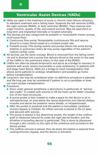
56
Ventricular Assist Devices (VADs)■ VADs are used in the treatment of acute or chronic heart failure refractory
to standard treatment and a failing heart. Supports the left ventricle (LVAD),the right ventricle (RVAD), or both ventricles (BIVAD). They improve end-organ function, quality of life, and extend survival. May be used short orlong term and implanted internally or located externally.
■ The devices are also categorized as pulsatile or nonpulsatile (newer pumps,continuous flow).■ Continuous flow pump: Nonpulsatile; continuously fills and returns
blood to a major blood vessel at a constant preset rate.■ Pulsatile pump: Fills during systole and pumps blood into aorta during
diastole or pulmonary artery or may pump regardless of the patient’s(native) cardiac cycle.
■ All devices use the same concept. Blood is removed from the failing ventri-cle and is diverted into a pump that delivers blood to the aorta (in the caseof the LVAD) or the pulmonary artery (in the case of the RVAD).
■ LVADs can often be placed temporarily and serve as a bridge to recovery inpatients with acute, severe myocarditis or post cardiotomy. In patients withend-stage heart failure, VADs are a bridge to heart transplantation thatallows some patients to undergo rehabilitation and possibly go homebefore transplantation.
■ Long-term use may be considered when no definitive procedure is planned,and life-long use may be considered if patient is not a candidate or doesnot want cardiac transplantation.
■ Procedure:■ Done under general anesthesia; a sternotomy is performed. A “pericar-
dial cradle” is created with sutures to lift the heart up for better visualiza-tion of the heart structures.
■ A pump pocket is then created to make a space for the device to sit in(extraperitoneal, an area below the rectus abdominis and internal obliquemuscles and above the posterior rectus sheath, or intraperitoneal).
■ After the pocket is prepared and the patient is cannulated, cardiopul-monary bypass is initiated. An inflow cannula is placed in the LV apexand secured with sutures.
■ The pump is placed in the abdominal pocket, the length of the outflowgraft is measured (should lie under the right sternal border), and the driveline (a tunneling device) is performed. This is done by placementthrough a skin incision to the right of the umbilicus and tunneled to thedevice pocket.
■ The outflow cannula is placed, then de-aired; the patient is weaned fromcardiopulmonary bypass, and the device is activated.
CV
4223_Tab02_045-106 29/08/14 10:00 AM Page 56

Management
Routine postoperative care with special attention to the following:
■ Assess lung sounds, respirations and O2 saturation and ABGs. Pulse oxime-try unreliable.
■ Keep intubated until anesthesia cleared from system.■ Assess cardiac sounds, rate, rhythm. Continuous ECG monitoring. Pacing
with epicardial wires may be warranted.■ A VAD-generated pulse and patient’s intrinsic or native electrical activity
will both appear on the monitoring system. The VAD-generated pulse isordered in a fixed-rate mode or volume or automatic mode.
■ If VF or asystole occurs, patient will still have a palpable pulse resultingfrom ongoing pump ejection of the VAD.
■ Assess neurological status to ensure cerebral perfusion.■ Monitor VS. Administer vasodilators as needed for MAP 65–80/90 mm Hg.
Automated BP cuffs unreliable. Use arterial line. BP measurement viaDoppler recommended if no A-line.
■ Monitor hemodynamics: cardiac index, cardiac output, CVP, PAP, PCWP.■ Monitor fluid and electrolyte balance including urine output. Note signs of
fluid overload and decreased urinary output.■ Administer anticoagulants (unfractionated heparin, warfarin, aspirin).■ Assess incisions and dressings for drainage, bleeding, erythema.■ Obtain and monitor labs: CMP, CBC, PT/PTT, with close attention to K+
and Mg.■ Medicate for pain as needed.■ Antibiotics may be ordered prophylactically.■ The patient is also monitored via chest x-rays and echocardiograms.■ Address psychosocial issues and refer as needed.
Complications
■ Device malfunction resulting from mechanical problems or thrombosis■ Infection■ Low flow or output of the VAD■ Pulmonary embolism or stroke■ Cardiac arrhythmias■ RV dysfunction in left VAD implantation■ Hemorrhage, cardiac tamponade■ Secondary organ dysfunction including kidneys, liver, lungs, and brain
57
CV
4223_Tab02_045-106 29/08/14 10:00 AM Page 57

58
Abdominal Aortic Aneurysm (AAA)AAA is a localized, chronic abnormal dilation of an artery located between therenal and iliac arteries with a diameter at least 1.5 times that of the expecteddiameter and a natural history toward enlargement and rupture.
Pathophysiology
Atherosclerosis and destruction of elastin and collagen fibers in the vessel wallscontribute to its development.
Pathophysiology for AtherosclerosisFatty streaks deposited in arterial intima → stimulates inflammatory responsethat causes proliferation → proliferation causes blood vessel to form fibrouscapillaries → deposits build up as atheromas or plaques → plaques pile up,obstructing the blood flow → outpouching of abdominal aneurysm.
Pathophysiology Involvement with Elastin and CollagenMedia thins → decrease in elastin fibers in vessel walls → collagen weakens →leads to aneurysm growth → outpouching of abdominal aorta.
Clinical Presentation
AAA may manifest as asymptomatic or symptomatic. When asymptomatic,look for a pulsatile, periumbilical mass with or without a bruit. When sympto-matic, symptoms include:
■ Sudden, severe constant pain in the abdomen, flank, groin, or back■ Early satiety, gastric or abdominal fullness, nausea and vomiting, GI bleeding■ Syncope■ Unequal carotid pulses and different BP in right and left arms■ ↓ peripheral circulation, venous thrombosis, lower extremity ischemia■ Symptoms of shock with rupture
AAA can also mimic:
■ Urinary tract infection■ Renal calculus or obstruction■ Ruptured disc■ Diverticulitis■ Pancreatitis■ Incarcerated hernia■ Abdominal neoplasm■ Peptic ulcer perforation
CV
4223_Tab02_045-106 29/08/14 10:00 AM Page 58

Diagnostic Tests
■ Abdominal ultrasound (first line of diagnostic testing)■ CT scan of the abdomen■ Abdominal x-ray■ Echocardiogram may be useful■ Aortogram■ MRI■ Preoperative labs and type and crossmatch for surgery
Management
■ Administer beta blocker to lower arterial pressure to the lowest SBP (120 mmHg or less). Use alpha-beta blocker labetalol (Trandate) or metoprolol(Lopressor) in place of nitroprusside and a beta blocker; do not give directvasodilators such as hydralazine.
■ If aneurysm ruptures:■ Administer IV propranolol (Inderal) to ↓ myocardial contractility and keep
HR 60–80 bpm.■ Administer nitroprusside to maintain MAP 70–80 mm Hg.■ Provide fluid resuscitation.■ Administer morphine for pain.■ Prepare patient for emergency surgery.
Postoperative Management■ Goal of postoperative care is to reduce afterload and pressure at the
repair site.■ Administer IV nitroprusside with esmolol (Brevibloc) or labetalol (Trandate)
and titrate the dosage to keep systolic BP below 120 mm Hg as ordered.■ Starting immediately after surgery, continuously monitor the patient’s neu-
rological status, cardiac rhythm, RR, hemodynamics, urine output, corebody temperature, fluid and electrolyte imbalance.
■ Provide analgesia. Morphine recommended.■ Monitor for acute renal failure, ischemic colitis, spinal cord ischemia, and
aortoenteric fistula.■ Assess patient’s gastrointestinal function.■ Report urine output less than 0.5 mL/kg/hr, which indicates dehydration,
volume deficit, or decreased renal function.■ Anticoagulants may be needed to prevent thrombi formation.■ Assess for poor peripheral perfusion.
59
CV
4223_Tab02_045-106 29/08/14 10:00 AM Page 59

60
Aortic DissectionAortic dissection is a tear (without hematoma or an intramural hematoma) inthe aortic wall that causes a longitudinal separation between the intima andadventitia layers resulting in diversion of blood flow from its normal arterialpathway. Aortic dissection requires emergency surgery.
Pathophysiology
Tear in aorta intima → blood flows into subintimal region → pulsatile pressurecreates a false channel between intimal and medial layers of aorta → intimaland medial layer is separated → circulatory volume decreases → channelexpands and creates either an expanding mass or a hematoma (from bloodcoagulating) → lumen narrows and obstructs blood flow, cardiac outputdecreases → results in end-organ failure, also diverted blood can pool aroundheart, resulting in cardiac tamponade.
Clinical Presentation
Consider acute phase if diagnosed within first 2 wk of onset of pain.
Presenting Symptoms■ Mimics inferior wall MI■ Standard type A aortic dissection: severe chest pain, sometimes sharp■ Standard type B dissection: sudden, severe chest pain radiating to the back;
described as “ripping or tearing” pain■ Pain can shift to the abdomen■ Increasing restlessness (sign of extending dissection)■ Decrease in urine output■ Anxiety: “feeling of doom”■ Fainting or dizziness■ Clammy skin■ Nausea and vomiting■ Pallor■ Tachycardia, shortness of breath, orthopnea■ Altered mental status and CVA-type symptoms
Diagnostic Tests
■ Chest x-ray: shows widening mediastinum■ ECG■ Transthoracic echocardiogram
CV
4223_Tab02_045-106 29/08/14 10:00 AM Page 60

■ Transesophageal echocardiogram■ CT scan■ Aortic angiography■ MRI■ Doppler imaging■ Cardiac biomarkers if coronary ischemia suspected■ Routine preoperative blood work if surgery anticipated
Management
■ The goals of therapy are to control BP and manage pain.■ Measure BP in both arms. Monitor HR, RR, and pain level.■ Perform frequent peripheral pulse checks, ankle brachial index measure-
ments, and neurological assessments.■ Administer beta blockers as first line of treatment; if hypertensive → give
nitroprusside IV.■ Administer analgesics for pain as appropriate.■ Plan for emergency surgery.
Pericardial EffusionPericardial effusion is the abnormal accumulation of more than 50 mL of fluid(normal: 15–50 mL to serve as lubricant for the visceral and parietal layers ofpericardium) in the pericardial sac that may lead to noncompression of theheart, which interferes with heart function.
Pathophysiology
Causes: chest trauma, accidents, stab wounds, gunshot wounds, rupture oftumors, obstruction of lymphatic or venous flow, pericarditis, viral infections, can-cer, MI, uremia, autoimmune diseases (lupus, rheumatoid arthritis, and others),bacterial infections, or idiopathic → accumulate blood in the pericardial sac →pressure → compresses or does not compress the heart.
Clinical Manifestations
Pericardial effusion can be asymptomatic with up to 2 L accumulated fluid in thepericardial sac. Pericardial effusions caused by pericarditis manifest with itsmain symptom of chest pain; chest pain may be worsened by deep breathing
↓
61
CV
4223_Tab02_045-106 29/08/14 10:00 AM Page 61

62
and lessened by leaning forward. If pericarditis is not the cause, there areoften no symptoms.
■ Complaints of dull, constant pain, pressure, ache, or discomfort in the leftside of the chest with symptoms of cardiac compression
■ Fever■ Fatigue■ Muscle aches■ Cough, SOB, dyspnea, hoarseness, hiccups■ Nausea, vomiting, and/or diarrhea■ Tachycardia, palpitations■ Lightheadedness, syncope■ Cool, clammy skin■ Weakened peripheral pulses, edema, cyanosis■ Anxiety, confusion■ Muffled heart sounds■ May or may not manifest with pericardial friction■ Dullness of percussion of the left lung over the angle of scapula
(Ewart’s sign)■ ECG showing ↓ voltage of complexes, diffuse ST elevation with PR depression
Diagnostic Tests
■ Echocardiogram■ ECG■ Chest x-ray■ CBC with differential, ESR, CRP■ Serum electrolytes■ Blood cultures and cardiac biomarkers as appropriate
Management
■ Manage pain.■ Pericardiocentesis or pericardectomy (pericardial window) is performed by
a physician.■ Position changes decrease SOB.■ Provide wound care after pericardiocentesis, pericardectomy, or other
pericardial surgical procedures, care of pericardial catheters and chesttubes.
■ Assess VS, pulses, LOC, respiratory status, skin and temperature changes,intake and output frequently.
■ Monitor and record any drainage from pericardial catheters and chesttubes. Refer to Respiratory Tab.
■ Depending on etiology of effusion, administer NSAIDs or antibiotics.
CV
4223_Tab02_045-106 29/08/14 10:00 AM Page 62

Cardiac Surgeries
Coronary Artery Bypass Graft (CABG)
CABG is an open-heart surgical procedure in which a blood vessel from anoth-er part of the body, usually the saphenous vein from the leg, is grafted belowthe occluded coronary artery so that blood can bypass the blockage. CABG maybe done with the use of a bypass machine (on pump) or without the use of themachine (off-pump CABG).
PathophysiologySurgery is performed on patients with coronary artery disease that causesblockage to the coronary arteries. Fatty streaks deposited in arterial intima stim-ulate an inflammatory response that causes proliferation. Proliferation causesblood vessels to form fibrous caps, and deposits build up as atheromas orplaques. Plaques pile up, obstructing the blood flow.
Clinical ManifestationsIschemia: If ischemic episode lasts long enough can cause death to myocardialcells → MI, angina, chest pain, somatic and visceral pain and discomfort. Atrialfibrillation a common complication of cardiac surgery.
Diagnostic Tests■ Health history■ Routine preoperative lab work including lipid profile, clotting profiles, liver
function tests■ Type and crossmatch for blood■ Chest x-ray■ ECG■ Exercise treadmill testing■ Gated SPECT imaging■ Coronary angiography■ Echocardiography■ Electron beam computed tomography (EBCT)
Postoperative Care■ Common postop care includes maintaining airway patency and monitoring
the patient’s pulmonary status, vital signs, and intake and output.■ Perform peripheral and neurovascular assessments hourly for first 8 hr.■ Monitor neurological status.■ Monitor cardiac status. Provide continuous ECG monitoring. Treat arrhythmias.■ Titrate drugs: vasopressor and inotropes to optimize cardiac function and BP.■ Monitor respiratory status including O2 saturation and ABGs. Patients are
mechanically ventilated on arrival at the ICU.■ Monitor chest tube drainage and record amount.
63
CV
4223_Tab02_045-106 29/08/14 10:00 AM Page 63

64
■ Monitor glucose levels and administer insulin as needed.■ Provide DVT and stress ulcer prophylaxis.■ Watch for signs of bleeding, and monitor hemoglobin and hematocrit
every 4 hr.■ Monitor patient’s pain, and medicate as needed.
Complications■ Complications are associated with anesthesia, cardiopulmonary bypass,
sternotomy, and the operation itself. These complications may include thefollowing: ■ Myocardial dysfunction including hemodynamic instability■ Aortic dissection■ Cardiac tamponade■ Cerebrovascular complications■ Acute renal failure■ Respiratory tract infections
Variations of On-Pump CABG■ Off-pump CABG■ Totally endoscopic coronary artery bypass graft■ Minimally invasive direct coronary artery bypass (MIDCAB): small incision
made in left chest; internal mammary gland artery sewn to left anteriordescending artery
Coronary Stenting/Percutaneous Coronary Intervention (PCI)
PCI is a common intervention for angina, obstructive CAD, myocardial ischemia,prevention of myocardial necrosis, and a nonsurgical alternative to CABG. In acatheterization lab, a catheter equipped with an inflatable balloon tip is insertedinto the appropriate coronary artery. When the lesion is located, the catheter is passed through the lesion, the balloon is inflated, and the atheroscleroticplaque is compressed, resulting in vessel dilatation. Intracoronary stents areusually inserted during PCI. Stents are used to treat abrupt or threatened abruptclosure or restenosis following PCI.
ProcedureStents are expandable meshlike structures designed to maintain vessel paten-cy by compressing the arterial walls and resisting vasoconstriction. Stents arecarefully placed over the angioplasty site to hold the vessel open.
Clinical Presentation Prior to PCI■ Atypical or typical chest pain■ SOB■ Dyspnea■ Symptoms of angina or CAD
CV
4223_Tab02_045-106 29/08/14 10:00 AM Page 64

Diagnostic Tests Prior to PCI■ ECG■ Echocardiogram■ Chest x-ray■ Lab tests: cardiac markers, serum electrolytes, coagulation studies, BUN,
creatinine, CBC
Management Post PCI■ Administer antiplatelet agents (aspirin, ticlopidine, clopidogrel).■ Administer IV infusion of glycoprotein IIb/IIIa inhibitors (eptifibatide).■ Monitor for signs and symptoms of bleeding at catheter site.■ Maintain pressure dressing to insertion site for 12 hr after procedure.■ Keep the involved leg straight.■ Set HOB to 45° or less.■ Monitor for cardiac arrhythmias.■ Monitor for chest pain and report immediately to MD. Give O2, NTG, isosor-
bide (Imdur), or nifedipine (Procardia); obtain 12-lead ECG.
Complications■ Abrupt closure of dilated segment or stent thrombosis■ Angina, MI, vasospasm, coronary artery dissection
Other interventional cardiology techniques include:
■ Laser angioplasty■ Atherectomy, directional coronary atherectomy■ Rotational ablation■ Brachytherapy
Stent Insertion
65
CV
4223_Tab02_045-106 29/08/14 10:00 AM Page 65

66
Cardiac Valve Replacement
Clinical conditions that require a surgical procedure to replace the valve witheither a mechanical valve or a porcine valve include:
■ Acquired valvular dysfunction■ Mitral valve stenosis■ Mitral valve regurgitation■ Mitral valve prolapse■ Aortic stenosis■ Aortic regurgitation■ Pulmonary stenosis (less common)
In some cases of damaged aortic valves, the patients’ own pulmonary valvecan be used to replace the aortic valve, and then the pulmonary valve isreplaced with an artificial valve (the Roth procedure). This is ideal for peoplewho cannot or will not take blood thinners for the rest of their life. The surgicalprocedure is the same as open-heart surgery except the heart is not bypassed,only the valve is replaced, or it can be minimally invasive; techniques includelaparoscopic or endoscopic robot-assisted and percutaneous surgery.
CV
Stent Inflation and Expansion
Balloon Removal and Stent Implantation
4223_Tab02_045-106 29/08/14 10:00 AM Page 66

Pathophysiology■ Mitral stenosis: Usually results from rheumatic fever (which can cause
valve thickening), atrial myxoma (tumor), calcium accumulation, or throm-bus formation → valve becomes stiff. The valve opening narrows → pre-vention of normal blood flow from left atrium to left ventricle → pulmonarycongestion → right-sided heart failure.
■ Mitral valve regurgitation: Fibrotic and calcific changes prevent mitral valve from closing completely during systole → incomplete closure of the valve → backflow of blood into left atrium when left ventricle contracts→ increased volume ejection with next systole → increased pressure → leftventricular hypertrophy.
■ Mitral valve prolapse: The valvular leaflets enlarge and prolapse into leftatrium during systole → usually benign but may lead to mitral valve regur-gitation.
■ Aortic stenosis: Aortic valve orifice narrows and obstructs left ventricle outflow during systole → increased resistance to ejection or afterload → ventricular hypertrophy.
■ Aortic regurgitation: Aortic valve leaflets do not close properly → regurgita-tion of aortic blood back into ventricle during diastole → left ventricularhypertrophy.
Clinical Presentation■ Mitral stenosis: Fatigue, dyspnea on exertion, heart palpitations, heavy
coughing, frequent respiratory infections, orthopnea, paroxysmal nocturnaldyspnea, hemoptysis, hepatomegaly, JVD, pitting edema, atrial fibrillation,apical diastolic murmur
■ Mitral valve regurgitation: Fatigue, dyspnea on exertion, orthopnea, palpi-tations, atrial fibrillation, JVD, pitting edema, high-pitched holosystolicmurmur
■ Mitral valve prolapse: Atypical chest pain, dizziness, syncope, palpitations,atrial tachycardia, ventricular tachycardia, systolic click
■ Aortic stenosis: Dyspnea on exertion, angina, syncope on exertion, fatigue,orthopnea, paroxysmal nocturnal dyspnea, harsh systolic crescendo-decrescendo murmur
■ Aortic insufficiency: Palpitations, dyspnea, orthopnea, paroxysmal nocturnaldyspnea, fatigue, angina, sinus tachycardia, blowing decrescendo diastolicmurmur
Diagnostic Tests■ Echocardiogram■ ECG■ Cardiac angiogram■ Exercise tolerance test■ Chest x-ray
67
CV
4223_Tab02_045-106 29/08/14 10:00 AM Page 67

68
Management■ Routine postoperative care: Maintain airway patency; monitor pulmonary
status.■ Monitor vital signs and intake and output.■ Monitor dressing and incision for drainage, erythema.■ Perform peripheral and neurovascular assessments hourly for first 8 hr
after surgery.■ Monitor neurological status for first 8 hr postoperatively.■ Titrate medications, vasopressors, and inotropes to optimize cardiac
function and BP.■ Monitor chest tube drainage.■ Watch for signs of bleeding by monitoring Hgb and Hct every 4 hr.■ Monitor for pain.■ Start on anticoagulation therapy when approved by cardiac surgeon.
Transcatheter Aortic Valve Replacement (TAVR)
■ TAVR was developed for the treatment of severe, symptomatic aortic stenosis,for patients at very high surgical risk for traditional valve replacement surgery,or for patients with technical issues with surgery (i.e., porcelain aorta or signif-icant mediastinal radiation).
■ Bilateral femoral arteries are prepped, the femoral artery is cannulatedwith a sheath, and the vessel is dilated. The sheath is replaced by a largersheath, which is used to accommodate the stent-valve delivery system.An arterial catheter is placed in the left femoral artery for hemodynamicmonitoring.
■ A guide wire and valvuloplasty balloon catheter are used to steer anddeploy the stent-valve. Aortic balloon valvuloplasty is performed to sepa-rate the leaflets. Contrast is injected, and fluoroscopy is used to visualizethe stent placement.
■ When the position of the stent-valve is confirmed, the device is thendeployed. Rapid right ventricular pacing reduces cardiac wall motion andprevents the stent-valve from slipping from the correct position during bal-loon inflation. A temporary pacing lead is placed in the right ventricle viathe femoral vein, and temporary pacing is initiated. This is followed byrapid inflation and deflation of the stent-deployment valvuloplasty balloon,and termination of pacing and a return of a normal rhythm.
■ Once the catheter is pulled back, the stent-valve will cover the native aorticleaflets and fit tightly in the aortic annulus. Angiography of the right andleft coronary arteries is performed to assess blood flow. The femoral arteryis repaired, and the sheath is removed.
PathophysiologyAortic stenosis. See under cardiac valve replacement
CV
4223_Tab02_045-106 29/08/14 10:00 AM Page 68

Management■ Postoperative care: Monitor vital signs and intake and output.■ Monitor central venous pressures.■ Check femoral sites, dressings for drainage, hematoma, erythema.■ Assess bilateral lower extremities for warmth, color, sensation, and movement
every 15 min for first hour, then hourly × 4 hr (or per hospital protocol).■ Keep affected limb straight for 8 hr.■ Head of bed should be elevated no more than 30°.■ Maintain bedrest for 12–24 hr post procedure; then the patient must ambulate.■ Assess neurological status frequently.■ Medicate for pain as needed.■ Administer IV fluids as ordered for 24 hr postop.■ Monitor CBC and BMP labs.■ Provide continuous ECG monitoring.■ Administer clopidogrel (Plavix).
Endoscopic (Minimally Invasive) Coronary Artery Bypass Graft
■ This less invasive cardiac surgical technique employs smaller incisions andtotally “robotic” surgery using a computer-enhanced telemanipulation sys-tem. Saphenous vein harvesting can also be accomplished through smallincisions using video-based surgical techniques, decreasing the morbidityassociated with leg incision (pain, infection) and permitting more rapidrecovery.
■ The term minimally invasive CABG is applied to procedures that use alter-natives to standard median sternotomy and off-pump CABG or on-pumpCABG when a sternotomy is utilized. Two alternative techniques are used:beating heart CABG surgery and MID CABG off pump.
■ Less invasive minimal access CABG surgeries emphasize the use of a limit-ed thoracotomy incision as an alternative to a sternotomy, with either directthoracoscopic or robotic-assisted left and right internal mammary arteryharvesting and direct LIMA to coronary artery anastomotic techniques formyocardial stabilization.
■ Beating heart CABG surgery is performed with specialized platforms thatenable myocardial surface stabilization and limit myocardial motion and byuse of temporary endovascular shunts to limit ischemia.
■ MID CABG off pump: The pericardium is opened, and cardiac motion islimited by the creation of accurate technical anastomosis and designed to minimize disruption of the adjacent beating heart. Cardiac motion canfurther be limited by use of pharmacological interventions that temporarilydecrease the heart rate or cause transient cardiac asystole for several seconds. Refer to CABG surgery for the detailed description of the actualbypass procedure.
69
CV
4223_Tab02_045-106 29/08/14 10:00 AM Page 69

70
ManagementPostop care is generally the same as traditional CABG surgery.
Cardiac and Cardiopulmonary Transplant
Cardiac transplant is a surgical procedure to remove a diseased heart andreplace it with a healthy donor heart. The same is true for lung transplant.Frequently both are done together for end-stage disease of the heart and lungswhen no other therapies are viable.
End-stage heart disease includes congenital heart disease (single ventricle physiol-ogy, coronary sinusoids), cardiomyopathy (primary: idiopathic, familial, secondarypregnancy, drug-induced), and acquired heart disease (valvular disease).
Candidates for Heart Transplant■ HF■ CAD with intractable angina symptoms■ Cardiomyopathy■ Pulmonary hypertension with irreversible right heart failure■ Sarcoidosis of the heart and lungs■ Ventricular dysrhythmias unresponsive to medical or surgery therapy■ Primary cardiac tumors with no evidence of spread to other body systems
Candidates for Lung Transplant■ COPD (chronic obstructive pulmonary disease)■ Cystic fibrosis■ Idiopathic pulmonary fibrosis■ Interstitial lung diseases■ Primary pulmonary hypertension
Diagnostic Tests Prior to TransplantationA battery of tests are done prior to transplantation. These may include but arenot limited to the following:
■ Echocardiogram■ Right heart catheterization■ Pulmonary function tests■ Exercise treadmill test■ Abdominal ultrasound■ Chest x-rays■ CT■ Coronary angiogram■ Cardiac biopsy■ Chromosome testing■ Updating of immunizations■ Low PSA and negative Pap smear
CV
4223_Tab02_045-106 29/08/14 10:00 AM Page 70

■ Lab tests: serum chemistries, CBC, human leukocyte antigen (HLA) antibodyscreening, viral antibody screening (HIV, cytomegalovirus, herpes virus, vari-cella, Epstein-Barr), platelet count, urinalysis
■ Psychosocial history obtained
Postoperative Care■ Admit to cardiothoracic ICU; 24–48 hr on ventilator until anesthesia cleared
from system.■ Insert Foley catheter to gravity, monitor output closely.■ Obtain daily chest x-ray.■ Monitor chest tube sites and drainage (generally 2–3 chest tubes in place).■ Perform pulmonary toilet measures hourly, once extubated.■ Perform and document complete nursing assessments frequently during
first 12–24 hr after surgery.■ Watch for signs and symptoms of bleeding.■ Treat dysrhythmias.■ Prevent right-sided heart failure.■ Watch for early signs of rejection, infection, immunosuppressive issues.■ Monitor for signs of drug toxicity.■ Provide ICU care for about 3–5 days postop.■ Prevent rejection: cyclosporine (Neoral), azathioprine (Imuran), prednisone,
methylprednisolone (Solu-Cortef), tacrolimus (Prograf), mycophenolatemofetil (CellCept), sirolimus (Rapamycin, Rapamune), monoclonal antibod-ies or polyclonal antibodies.
■ Highest risk for infection: 1 wk postop.
Signs and Symptoms of RejectionHyperacute rejection can occur immediately after transplantation in the OR.Acute rejection occurs within the first 3 mo after transplantation. Chronic rejec-tion can occur any time after 3 mo of transplant.
■ Low-grade fever with elevated WBC■ Fatigue■ SOB■ Pulmonary crackles with/without pulmonary edema■ Pericardial friction rub■ Arrhythmias especially atrial flutter or fibrillation.■ Decreased ECG voltage■ Increased JVD■ Signs of decreased cardiac output including hypotension with tachycardia■ Cardiac enlargement on x-ray■ Vascular degeneration■ Palpitations■ Nausea and vomiting
71
CV
4223_Tab02_045-106 29/08/14 10:00 AM Page 71

72
Other Complications■ Infection■ CAD■ GI complications related to medications
Carotid Endarterectomy (CE)
CE is a surgical procedure to remove plaque material from inside the carotidartery that improves the carotid luminal diameter, allows adequate blood flow,and therefore prevents stroke. The procedure is indicated in symptomaticpatients with carotid-territory TIA or minor strokes who have carotid arterystenosis of 70%–99%.
Pathophysiology of the Disease Process Leading to CESurgery is performed on patients with symptomatic carotid artery stenosis.Fatty streaks are deposited in arterial intima → stimulates inflammatoryresponse that causes proliferation → blood vessels forming fibrous caps →deposit builds up as atheromas or plaques → plaques build up, causing bloodflow obstruction.
Clinical Presentation■ Signs and symptoms of TIA or stroke. Refer to Neuro Tab.■ Sudden dysphagia.■ Dizziness.■ Lightheadedness.
Diagnostic Tests■ Ultrasound of carotid artery■ Computed tomographic angiography (CTA)■ Magnetic resonance angiography (MRA)■ Contrast-enhanced MRA (CEMRA)
Management of Clinical Condition■ Assess surgical site for bleeding.■ Perform frequent neurological checks.■ Administer antihypertensive medications. Maintain SBP between 120 and
170 mm Hg.■ Administer statins.■ Administer antiplatelet agents.■ Administer aspirin (81–325 mg) before and after surgery.■ Monitor for difficulty speaking, swallowing, or breathing. Report immediately.
CV
4223_Tab02_045-106 29/08/14 10:00 AM Page 72

Infections of the Heart
Endocarditis
Endocarditis is the inflammation of the innermost layer of the heart. It can includethe heart valves, chordae tendineae, cardiac septum, or lining of the cham-bers. It is caused typically by bacterial infections (Streptococcus viridans orStaphylococcus aureus).
PathophysiologyMicrobe invades the valve leaflets → infection occurs, causing valve leaflets tobe deformed.
Clinical PresentationEndocarditis manifests as:
■ Low-grade and intermittent fever■ Chills, night sweats■ Anorexia, weight loss■ Myalgia, arthralgia■ Extreme fatigue and malaise■ Nausea and vomiting■ Headache■ SOB■ Chest pain■ Abdominal pain■ Confusion, delirium■ Pain in the muscles, joints, and back■ Influenza-like symptoms■ Loud regurgitant murmur; pericardial rub, or friction rub■ Possible embolic stroke with focal neurological deficits
Be suspicious if petechiae appear on the conjunctiva, neck, anterior chest,abdomen, or oral mucosa. Look for Janeway lesions (nontender macule) onpatient’s palms and soles, Osler’s nodes (tender, erythematous, raised nodules)on fingers and toe pads, and splinter hemorrhages under fingernails. Roth’sspots are white spots that can be seen on the retina.
Diagnostic Tests■ Transthoracic echocardiogram■ ECG■ Chest x-ray■ Abdominal CT or MRI■ Positive blood cultures■ Refer to the Duke Criteria for Infective Endocarditis found at: http://www.
medcalc.com/endocarditis.html
73
CV
4223_Tab02_045-106 29/08/14 10:00 AM Page 73

74
Management■ Administer antimicrobial therapy.■ Priorities include supporting cardiac function, eradicating the infection, and
preventing complications, such as systemic embolization and heart failure.■ Do not give anticoagulants because of risk of intracerebral hemorrhage.
Pericarditis
Pericarditis is inflammation of pericardium that can cause fluid to accumulatein the pericardial space resulting from idiopathic causes, infection (viral, bacterial or fungal), MI, autoimmune reactions, certain drugs, cancer thera-pies, or trauma. Dressler’s syndrome is a type of pericarditis following damage to heart tissue or the pericardium, such as a heart attack, surgery, ortraumatic injury.
PathophysiologyPrimary illness of medical or surgical disorder can be the etiology → pericardi-um becomes inflamed → can lead to excess fluid accumulation or increasedpressure on the heart leading to tamponade.
Clinical PresentationSharp, constant chest pain that is located in the midchest (retrosternal) is themost common symptom. A hallmark sign of pericarditis is if the patient leansforward while sitting to relieve chest pain. Pain may radiate to the neck, shoul-ders, and back; radiation to the ridge of the left trapezius muscle is specific forpericarditis. ECG changes are new widespread ST elevation or PR depression,and pericardial effusion may be present.
Depending on the cause, patient may also have fever, malaise, tachypnea,dyspnea, JVD, and tachycardia. Pericardial friction rub, heard in the lower ster-nal border, is the most important physical sign.
Diagnostic Tests■ ECG■ Echocardiogram■ Chest x-ray■ Lab work: cardiac markers, erythrocyte sedimentation rate (ESR), serum
C-reactive protein ■ Serum electrolytes, BUN, creatinine■ Complete blood count■ Urinalysis■ Blood cultures■ Additional lab work may include tuberculin skin test, antinuclear antibody
titer (ANA), and/or HIV serology■ Transesophageal echocardiogram (TEE)
CV
4223_Tab02_045-106 29/08/14 10:00 AM Page 74
Management■ NSAIDs may be used for up to 2 wk.■ Add colchicine (Colcrys) for recurrent symptoms or continued symptoms
>14 days.■ Monitor for cardiac tamponade.■ More severe pain can be controlled with morphine.■ If cause is infectious, antibiotics or antifungal drugs may be administered.■ Treatment consists of antibiotics specific to the pathogen for at least 6 wk.■ If a pericardiectomy is performed, continue assessments of VS, lab work,
and the appearance of wounds and insertion sites of invasive lines.■ Other surgical procedures include pericardiocentesis, pericardial window,
pericardiotomy.■ Monitor temperature and cardiac rhythm; assess for heart murmurs.■ Perform neurological assessments, inspect skin surfaces, and monitor drug
peaks and troughs and urine output.■ Disease may progress to cardiac tamponade and chronic constrictive pericarditis.
Pacemakers/AICDPacemakers generate an electrical impulse that stimulates the myocardium todepolarize, thereby initiating a cardiac contraction. Aim is to keep the ventriclesbeating to sustain adequate BP to perfuse all organs sufficiently. Consists of apulse generator and 1–3 leads with electrodes.
Pacemakers can also stimulate only selected areas of the heart. Codes havebeen established for general widespread use: Refer to the 2002 North AmericanSociety for Pacing and Electrophysiology Position Statement: The RevisedNASPE/BPEG Generic Code for Antibradycardia, Adaptive Rate, and MultisitePacing at http://www.hrsonline.org/Practice-Guidance/Clinical-Guidelines-Documents/2002-The-Revised-NASPE-BPEG-Generic-Code-for-Antibradycardia-AdaptiveRate-and-Multisite-Pacing#axzz2aSv6txwb
75
CV
4223_Tab02_045-106 29/08/14 10:00 AM Page 75

76
CV
Po
sition
III
IIIIV
VC
ham
ber(s)
Ch
amb
er(s)R
espo
nse to
Rate
Mu
ltisiteC
atego
ryp
acedsen
sedsen
din
gm
od
ulatio
np
acing
Manufactures’
designationonly
O = n
on
eA
= atrium
V = ven
tricleD
= du
al (A+V
)
O = n
on
eA
= atrium
V = ven
tricleD
= du
al (A+V
)
O = n
on
eT
= trigg
eredI = in
hib
itedD
= du
al (T+I)
O = n
on
eR
= rate m
od
ulatio
n
O = n
on
eA
= atrium
V = ven
tricleD
= du
al (A+V
)
S = single (A
or V)
S = single (A
or V)
4223_Tab02_045-106 29/08/14 10:00 AM Page 76

■ VVI mode: The ventricle is paced, sensed, and inhibited. The pacemakerfires if no QRS complex is sensed. The patient’s rate is greater than the pro-grammed rate and inhibits the pacemaker from firing. Most popular mode.
■ DVI or AV sequential mode: The atrium is paced at the programmed rate →no ventricular activity during a certain interval → ventricle is paced.
■ DDD or fully automated mode: Senses and paces both the atrium and ven-tricle and can inhibit either the atrium or ventricle if a patient’s native orintrinsic beat is sensed.
■ Position I: Heart chamber paced.■ Position II: Heart chamber where intrinsic electrical activity sensed.■ Position II: Pacemaker response to what it senses.■ Position IV: Presence (R) or absence (O) of rate modulation or rate respon-
siveness. Presence means that the pacemaker will alter its pacing rate tomeet metabolic demands.
■ Position V: Multisite pacing settings indicate the ability to stimulate morethan 1 site in a chamber.
■ Fixed rate pacemakers (asynchronous) discharge at a preset rate (usually70–80 bpm) regardless of the patient’s own electrical activity.
■ Demand (synchronous) pacemakers discharge only when the patient’s HR < thepacemaker’s preset rate.
■ Pacemakers may also be classified as temporary or permanent. Temporarypacemakers are used in emergency situations and may serve as a bridge toa permanent pacemaker. The pulse generator is located externally. Thepulse generator is implanted internally in permanent pacemakers.
■ Temporary pacemakers include transvenous, epicardial, transcutaneous,and transthoracic.
■ Transvenous pacing: A lead wire is passed through the brachial, internal orexternal jugular, subclavian, or femoral vein to the myocardium.
■ Epicardial pacing is used with cardiac surgical patients. Epicardial wires areattached to the epicardium during surgery.
■ Transthoracic pacing is used as a last resort, with limited success rates. Apacing needle is inserted directly into the anterior wall of the heart.
Transcutaneous Pacemakers
Transcutaneous pacemakers are used for noninvasive temporary pacing usingtwo large external electrodes. The electrodes are attached to an external pulsegenerator, which operates on alternating current or battery. The generator emitselectrical pulses, which are transmitted through the electrodes and then trans -cutaneously to stimulate ventricular depolarization when the patient’s heart rateis slower than the rate set on the pacemaker. Used as an emergency measure,a transcutaneous pacemaker should be used for 48–72 hr only. Electrodesshould be changed every 24 hr minimally.
77
CV
4223_Tab02_045-106 29/08/14 10:00 AM Page 77

78
Permanent Pacemakers
Permanent pacing is performed for the resolution of nontemporary conductiondisorders, including complete heart block and sick sinus syndrome. Permanentpacemakers are usually powered by a lithium battery and have an average lifespan of 10 yr.
Indications■ Class I: The procedure should be performed.■ Class IIa: It is reasonable to perform the procedure, but additional studies
with focused objectives are needed.■ Class IIb: The procedure may be considered, but additional studies with
broad objectives are needed.■ Class III: The procedure should not be performed; it is not helpful and may
be harmful.
In 2008 the American College of Cardiology (ACC), the American HeartAssociation (AHA), and the Heart Rhythm Society (HRS), jointly published guide-lines for pacemaker insertion indications:
■ Class I indications■ Sinus node dysfunction■ Acquired atrioventricular block in adults■ Chronic bifascicular block■ After acute MI■ Hypersensitive carotid sinus syndrome and neurocardiogenic syncope■ After cardiac transplantation■ Pacing to prevent tachycardia■ Congenital heart disease
■ Class IIa indications■ Sinus node dysfunction■ Acquired atrioventricular block■ Hypersensitive carotid sinus syndrome and neurocardiogenic syncope■ Congenital heart disease■ Pacing to prevent tachycardia■ Permanent pacemakers that automatically detect and pace to terminate
tachycardia■ Class IIb indications
■ Sinus node dysfunction■ Acquired atrioventricular block in adults■ Chronic bifascicular block■ After acute MI■ Hypersensitive carotid sinus syndrome and neurocardiogenic syncope■ After cardiac transplant
CV
4223_Tab02_045-106 29/08/14 10:00 AM Page 78

■ Pacing to prevent tachycardia■ Congenital heart disease
Contraindications■ Local infection at implantation site■ Active systemic infection with bacteremia■ Severe bleeding tendencies (relative contraindication)■ Active anticoagulation therapy (relative contraindication)■ Severe lung disease and positive end-expiratory pressure ventilation
(relative contraindication for internal jugular and subclavian access.
Procedure■ Performed in an electrophysiology (EP) lab or in a cardiac catheterization
lab. Usually done using local anesthesia. ■ Small incision is made at the insertion site (usually right or left subclavian); a
sheath or introducer is inserted into the subclavian vein; a lead wire is thread-ed through the introducer into the vessel; and the lead wire is then advancedinto the atrium, ventricle, or both (depending on the type of pacemaker).
■ Once the lead wire is in the proper position, it is tested to verify properlocation and to make sure it is functioning properly. The number of leadsplaced (1, 2, or 3) depends on the type of pacemaker.
■ Once the lead wires are tested, an incision is made under the clavicle. Thepacemaker generator is slipped under the skin through the incision after thelead wire is attached to the generator. An ECG is obtained to verify thepacer is working correctly.
■ The incision is then closed with sutures, adhesive strips, or surgical glue; adry sterile dressing is then applied.
Postoperative CarePatients are not always admitted or returned to the ICU post procedure; atelemetry floor may be appropriate.
■ Continuous cardiac monitoring. A pacemaker-generated impulse shouldappear as a spike on the ECG. The spike indicates the pacemaker has fired.Pacemaker rhythm:■ Rate: Varies according to the preset pacemaker rate and patient’s native
heart rate.■ Rhythm: Irregular for demand pacemaker unless patient is 100% based
with no native beats.■ P wave: None for ventricular pacemaker. P waves may be seen but unre-
lated to QRS. Atrial or dual chambered pacemakers should have P waveafter each atrial spike.
■ PR interval: None for ventricular pacing. Atrial or dual chambered pacingshould produce constant PR intervals.
■ QRS: Wide (>0.10 sec.) after each ventricular spike in a paced beat.Patient’s native QRS will look different from paced QRS. Atrial pacingonly, QRS may be normal.
79
CV
4223_Tab02_045-106 29/08/14 10:00 AM Page 79

80
■ Obtain a 12-lead ECG. ■ Assess VS per hospital protocol. Assess peripheral pulses.■ Assess LOC.■ Administer antibiotics (cefazolin, vancomycin).■ Assess dressing for bleeding, drainage, or signs of infection.■ If epicardial wires present, clean site and cover wires per hospital policy.■ Monitor CBC, BMP.■ Administer oral pain medication as needed.■ Patients with pacemakers may be defibrillated. Avoid placing the defibrilla-
tor paddles or pads closer than 5 inches from the pacemaker battery packor pulse generator.
■ Patient education is crucial: activity level, signs of pacemaker malfunction,signs of infection, electrical safety precautions, medication usage. Providepatient with identification card related to pacemaker. Consider telemonitor-ing of these patients for compliance, pacemaker function, and avoidance ofcomplications.
Complications■ Pacemaker malfunction:
■ Failure to discharge: Pacemaker fails to fire. Seen as patient’s HR programmed rate
■ Failure to capture: Pacing stimulus not followed by depolarization of atrium and/or ventricle
■ Oversensing: Pacemaker detects noncardiac electrical events (electro-magnetic interference, large T waves) as depolarization
■ Undersensing: Pacemaker cannot sense patient’s intrinsic beats, withresulting inappropriately placed pacemaker artifacts
■ Pneumothorax■ Ventricular irritability■ Perforation of ventricular wall or septum■ Catheter or lead dislodgment■ Infection■ Hematoma■ Abdominal twitching or hiccups■ Pocket erosion
Automatic Implantable Cardioverter-Defibrillator Device (AICD)
■ The AICD continuously monitors the patient’s heart rhythm, diagnoses anyrhythm changes, and treats life-threatening ventricular arrhythmias.
■ Main indications for use of an AICD relate to secondary prevention inpatients with prior sustained ventricular tachycardia (VT), ventricular fibril-lation (VF), or resuscitated sudden cardiac death (SCD) thought to be
CV
4223_Tab02_045-106 29/08/14 10:00 AM Page 80

caused by VT/VF as well as primary prevention in patients at risk of life-threatening VT/VF.
■ The 2008 American College of Cardiology/American Heart Association/HeartRhythm Society (ACC/AHA/HRS) established guidelines for device-basedtherapy of cardiac rhythm abnormalities include guidelines for ICD therapy.
Indications for ICD TherapySecondary prevention: Implantation of an ICD is recommended for the second-ary prevention of death resulting from VT/VF in the following settings:
■ Patients with prior episode of resuscitated VT/VF or sustained hemodynam-ically unstable VT in whom a completely reversible cause cannot be identi-fied. This includes patients with a variety of underlying heart diseases andthose with idiopathic VT/VF and congenital long QT syndrome, but notpatients who have VT/VF limited to the first 48 hr after an AMI.
■ Patients with episodes of spontaneous sustained VT in the presence of heartdisease (valvular, ischemic, hypertrophic, dilated, or infiltrative cardiomy-opathies) and other settings (i.e., channelopathies).
Primary prevention: Implantation of an ICD is recommended for the primary pre-vention of life-threatening VT/VF in patients at risk of SCD from VT/VF who have optimal medical management (including use of beta blockers and ACE-IS), including:
■ Patients with prior myocardial infarction (at least 40 days ago) and left ven-tricular ejection fraction ≤30%.
■ Patients with cardiomyopathy, New York Heart Association functional class IIor III, and left ventricular ejection fraction <35%. Patients with nonischemiccardiomyopathy generally require optimal medical therapy for 3 mo withdocumentation of persistent left ventricular ejection fraction <35% at thattime. Recommended patients be evaluated at least 3 mo after revasculariza-tion (CABG or stent placement).
ProcedureRefer to pacemaker insertion.
ManagementPostop care: Refer to Pacemakers.
Cardiac TamponadeCardiac tamponade is the acute compression of the heart caused by excessivefluid or blood in the pericardial space. This results in accumulated pressure inthe pericardial sac and affects the heart’s function, especially cardiac output.Normal pericardial fluid volume = 10–30 mL, but a rapid accumulation of 50–100 mLof fluid can be fatal. It is a medical emergency that can result in pulmonaryedema, shock, and death.
81
CV
4223_Tab02_045-106 29/08/14 10:00 AM Page 81

82
Pathophysiology
Causes: accidents, stab wounds, gunshot wounds, rupture of tumors, obstruc-tion of lymphatic or venous flow, pericardial effusion → accumulate blood inthe pericardial sac → ↑ venous pressure → compress all four chambers of theheart → RA and RV are compressed → ↓ RA filling during diastole → ↓ venousblood returns to RA → ↑ venous pressure → JVD, edema, hepatomegaly, ↑ DBP→ continued compression of the heart → ↓ diastolic filling of ventricles → ↓ SV,↓ CO, ↓ tissue perfusion → body attempts to increase blood volume, increaseSV ↑ → workload of the heart → body compensation → tachycardia → all thesecomplications last for a limited amount of time → shock, cardiac arrest, or deathif not immediately corrected.
Clinical Manifestations
First Signs■ Anxiety and restlessness, syncope■ SOB, dyspnea on exertion, orthopnea, cough■ Unwillingness to lie flat■ Cool, diaphoretic skin■ Sense of impending doom
Classic Signs■ Beck’s triad: muffled heart sounds, increased JVD, and hypotension■ Narrow pulse pressure (↓ SBP and ↑ DBP)■ Hemodynamics: ↑ CVP >20 mm Hg, ↑ PCWP, ↓ CO■ Tachycardia■ Weak, thready pulse
Late Signs■ Pulsus paradoxus (↓ SBP of more than 10 mm Hg on inspiration)■ Electrical alternans (alternating levels of voltage in P waves and QRS
complexes in all leads and possibly in T waves)
Diagnostic Tests
■ Chest x-ray■ Echocardiogram■ ECG■ CT, MRI
CV
4223_Tab02_045-106 29/08/14 10:00 AM Page 82

Management
■ If acute onset, call MD stat, obtain stat 2-D echocardiogram and stat chest x-ray.
■ Obtain stat lab work.■ MD will place pulmonary artery catheter.■ Place patient in supine position with HOB elevated 30°–60°.■ Assess O2 saturation levels and administer O2 by cannula, mask or intubate.■ If on mechanical ventilation, no positive pressure used.■ Give sedatives; administer morphine for chest pain.■ Monitor cardiovascular, respiratory, neurological, and renal function
status.■ Assess hemodynamics.■ Obtain 12-lead ECG, and provide continuous ECG monitoring. Notify MD of
increasing dyspnea and new-onset arrhythmias.■ Administer inotropic drugs (dobutamine) for low BP.■ Consider volume expansion with blood, plasma, dextran, or isotonic
sodium chloride solution, as necessary, to maintain adequate intravascu-lar volume.
■ Prepare patient for surgery or procedure to remove pericardial fluid: pericardiotomy, pericardiocentesis, surgical creation of pericardial win-dow. Other procedures may be considered for recurrent cardiac tampon-ade or pericardial effusion. Removal of as little as 10–20 mL of fluid mayrelieve symptoms.
ACLS Algorithms: Cardiac/Respiratory ArrestBased on the Adult Basic Life Support: 2010 American Heart Association Guidelinesfor Cardiopulmonary Resuscitation and Emergency Cardiovascular Care. Refer to:http://circ.ahajournals.org/content/122/18_suppl_3/S685.full.pdf+html for full report.
Ventricular Fibrillation (VF) or Pulseless VentricularTachycardia (VT)
■ Shock: Biphasic: 120 J; monophasic: 200 J. Reassess rhythm.■ CPR: Immediately perform 5 cycles of CPR (should last about 2 min).■ Epinephrine: 1 mg IV or IO (2–2.5 mg, endotracheal tube) every 3–5 min or
vasopressin: 40 units IV or IO, one time only. May use to replace 1st or 2nddose of epinephrine (given without interrupting CPR).
■ Shock: Biphasic: 150 J then 200 J.; monophasic: 300 J then 360 J. Reassessrhythm.
83
CV
4223_Tab02_045-106 29/08/14 10:00 AM Page 83

84
■ Consider antiarrhythmics (given without interrupting CPR):■ Amiodarone: 300 mg IV or IO, may repeat 150 mg in 3–5 min.■ Lidocaine: 1.0–1.5 mg/kg IV or IO, may repeat 0.5–0.75 mg/kg q5–10 min,
max 3 mg/kg.■ Magnesium: 1–2 g IV or IO for torsades de pointes.
■ A 2011 study published in Circulation showed that patients with a shockablerhythm (VF or pulseless VT) had better survival rates if CPR is paused foronly 10 sec or less before delivery of electric shock to the heart (preshockpause) or a total of 20 sec or less pause in chest compressions both beforeand after defibrillatory shock (perishock pause).
■ 76% of patients show signs of ongoing instability for several hours prior tocardiac arrest.
■ 70% of patients show signs of respiratory distress within 8 hr of arrest.■ 66% of patients show abnoral signs and symptoms within 6 hr of arrest.
Asystole or Pulseless Electrical Activity (PEA)
■ Resume CPR for 5 cycles (should last about 2 min).■ Epinephrine: 1 mg IV or IO (2 to 2.5 mg ET) every 3–5 min or vasopressin:
40 units IV or IO, one time only. May use to replace 1st or 2nd dose of epi-nephrine (given without interrupting CPR).
Intra-aortic Balloon Pump Counterpulsation (IABP)
IABP counterpulsation provides temporary mechanical circulatory support.Goals of IABP counterpulsation: increase O2 to myocardium and coronary arteries,decrease LV workload by decreasing impedance to ejection (afterload), andincrease cardiac output. An IABP device consists of a 30-cm polyurethane balloonattached to one end of a large-bore catheter. The device is inserted into the femoralartery at the groin, either percutaneously or via arteriotomy, with the balloonwrapped tightly around the catheter. Once inserted, the catheter is advanced upthe aorta until the tip lies just beyond the origin of the left subclavian artery. Whenin place, the balloon wrapping is released to allow periodic balloon inflations.
Effects
The intra-aortic balloon is inflated with helium at the onset of each diastolicperiod (middle of T wave), when the aortic valve closes. The balloon is deflatedat the onset of ventricular systole, just before aortic valve opens (prior to QRScomplex). An arterial waveform can be used as an alternative to ECG triggering.Inflation of the balloon increases the peak diastolic pressure and displaces
CV
4223_Tab02_045-106 29/08/14 10:00 AM Page 84

blood toward the periphery. Deflation of the balloon decreases the end-diastolicpressure, which reduces impedance to flow when aortic valve opens at onset ofsystole. This decreases ventricular afterload and promotes stroke volume, thusincreasing coronary artery perfusion and blood flow during diastole.
85
Diastole
Aorta
Heart
Systole
Descendingaorta
Ballooncatheter
Renalartery
mm
Hg
Time
Systole Diastolic augmentation
IABP Waveforms
CV
4223_Tab02_045-106 29/08/14 10:00 AM Page 85

86
Indications
■ Complement to cardiopulmonary bypass and low cardiac output after cardiacsurgery
■ Bridge to VAD, CABG, or cardiac transplant■ AMI with cardiogenic shock■ Decompensated heart failure■ Acute mitral valve insufficiency or regurgitation■ Ventricular septal defect or papillary muscle ruptures■ Severe unstable or intractable angina■ Septic shock
Timing
■ Balloon should inflate at or slightly above the dicrotic notch.■ A crisp V shape should appear on inflation.■ Diastolic augmentation peak > preceding systolic peak.■ The following systolic peak (assisted systole) < preceding systole (unassisted
systole).■ The IABP should be set on a 1:2 augmentation to assess timing and then
1:1 or 1: 3 augmentation after timing has been optimized.■ Avoid early or late inflation and deflation.
Management
■ Routine critical care nursing practices are followed.■ Instruct patient not to sit up, bend knee, or flex hip >45°. Keep HOB <45°.■ Provide continuous ECG monitoring.■ Arterial line and pulmonary artery catheter are in place.■ Assess insertion site for bleeding and infection.
Complications
■ Balloon leakage, rupture, or entrapment. The catheter can migrate andocclude subclavian artery.
■ Lower limb ischemia and compartment syndrome of cannulated extremity.■ Arterial occlusion → mesenteric infarction.■ Catheter-associated infection.
CV
4223_Tab02_045-106 29/08/14 10:00 AM Page 86

Contraindications
■ Aortic insufficiency and regurgitation■ Aortic aneurysm and dissection■ Recently placed (within 12 mo) prosthetic graft in thoracic aorta
Induced or Therapeutic Hypothermia Also called targeted temperature management. To reduce the damage to braincells through reduction of the brain’s metabolic activity, hypothermia is intention-ally induced by bringing the core body temperature down to 32°–34°C(89.6°–93.2°F). It is recommended that therapeutic hypothermia be used for up to 24 hr, although little evidence supports this time frame. Cerebral metabolism ↓ 6%–10% for every 1° decrease in body temperature. As cerebral metabolism ↓ → brain O2 requirements ↓. Goals: ↓ ICP, ↓ HR, ↓ brain’s O2 demand to obtain goodneurological outcome. Indications for medically induced hypothermia include:
■ Ischemic cerebral or spinal injury, including stroke recovery■ Cerebral edema and increased intracranial pressure■ Heart surgery■ Patients with asystole, pulseless electrical activity, or cardiac arrest result-
ing from ventricular fibrillation (VF) or ventricular tachycardia (VT); this ismost effective within 1 hr of cardiac arrest and no later than 10 hr afterreturn of spontaneous circulation.
The three phases in instituting induced hypothermia are induction, maintenance,and rewarming.
Hypothermia may be accomplished by several methods:
■ Rapid infusion of ice-cold IV fluids; use 0.9% NS or LR at 30 mL/kg■ NG lavage with ice water■ Evaporative cooling of the external body surface■ External cooling with ice packs (axilla, groin, around head) or special cool-
ing blankets, iced wet sheets, fans, gel pads■ Invasive cooling catheters
The patient may be rewarmed through the use of:
■ Cardiopulmonary bypass■ Warm IV fluid administration■ Warm humidified O2 administration via ventilator■ Warm peritoneal lavage■ Warming blankets and over-the-bed heaters
Caution should be taken with active core rewarming because VF can occur asthe patient’s temperature increases.
87
CV
4223_Tab02_045-106 29/08/14 10:00 AM Page 87

88
■ Monitor for hypotension.■ Rewarm slowly at 0.3° C–0.5° C/hr.■ If no signs of neurological improvement 72 hr after rewarming and discon-
tinuation of neuromuscular blockers, determine brain death and seek fullneurological evaluation.
Management
■ Monitor BP. Arterial line is preferred.■ Provide continuous ECG monitoring and monitor for sinus bradycardia, pro-
longed PR interval, widened QRS, prolonged QT interval, atrial fibrillation,VT, and VF. Avoid atropine.
■ Consider pacemaker or AICD.■ Monitor hemodynamics. Pulmonary artery catheter is preferred. IABP may
be indicated.■ Administer vasopressors as needed.■ Monitor for seizures.■ Monitor fluids and electrolytes, especially K+ and glucose levels. Induction
causes mild diuresis. Provide fluid replacement. Hyperglycemia can occurduring maintenance phase, and hypoglycemia can occur during rewarming.
■ Consider CRRT.■ Prevent skin breakdown resulting from vasoconstriction from cold. Consider
prone position.■ Ileus is common during hypothermia.■ Hypothermia suppresses inflammatory response → infection. Hand hygiene
and infection control practices are imperative.■ Assess for bleeding. For every 1°C drop in temperature, coagulation-factor
function ↓ by 10% and affects the coagulation cascade.■ Obtain oral or axillary temperatures only or use temperature probe,
esophageal bladder, pulmonary artery catheter, or other methods.■ Prevent shivering through the use of sedation and neuromuscular blockade.
Administer chlorpromazine (Thorazine) or diazepam (Valium). ■ Hypothermia ↓ body’s ability to respond to stimulation, making assessment
of sedation level difficult. Use bispectral index monitors or continuous EEGmonitoring.
■ Assessing level of neuromuscular blockade using train-of-four monitoringnot always reliable. Refer to Tab 1, Basics.
■ Hypothermia affects drug metabolism. Lower doses of drugs may be needed.
■ Prepare patient for immediate coronary angiography and/or PCI.
CV
4223_Tab02_045-106 29/08/14 10:00 AM Page 88

ECGs
12-Lead ECG
89
I aVR V1 V4
II aVL V2 V5
III aVF V3 V6
Lead Placement
Midclavicularline
V1V2
V3V4
V5
V6
Anterioraxillary line
Midaxillaryline
CV
4223_Tab02_045-106 29/08/14 10:00 AM Page 89
90
Hemodynamics of Dysrhythmias
Atrial Dysrhythmias
Atrial dysrhythmias are caused by increased automaticity in the atria. Thepatient may complain of palpitations or “heart racing.” Loss of atrial contraction→ shortens diastole → loss of atrial kick (25%–30% of CO) → ↓ CO → ↓ coro-nary perfusion → ischemic myocardial changes. Atrial flutter or fibrillation canlead to a pulmonary embolus, stroke, or MI. These patients should be takinganticoagulants. Calculation for Atrial Fibrillation Stroke Risk found at: http://www.mdcalc.com/cha2ds2-vasc-score-for-atrial-fibrillation-stroke-risk/
Surgical ablation via a Cox-Maze procedure during mitral valve repair orreplacement has been shown to be an effective treatment for atrial fibrillation.
Causes■ Amphetamines■ Cocaine■ Decongestants■ Hypokalemia■ Hyperthyroidism■ COPD■ Pericarditis■ Digoxin toxicity■ Hypothermia■ Alcohol intoxication■ Pulmonary edema
Ventricular Dysrhythmias
Ventricular dysrhythmias are caused by increased automaticity in the ventricles.PVCs can be manifested as complaints of “heart skipping a beat.” This dysrhyth-mia can lead to bradycardia → ↓ CO → ↓ BP and eventually VT, VF, and death.
BradycardiaBradycardia is defined as a heart rate less than 60 bpm. ↓ HR → ↓ CO → ↓ BP→ ↓ perfusion to brain, heart, kidneys, lung, and skin.
Causes■ Vomiting■ Gagging■ Valsalva maneuver■ ETT suctioning
CV
4223_Tab02_045-106 29/08/14 10:00 AM Page 90

Symptoms■ Chest pain■ SOB■ Altered mental status■ Signs of ↓ CO: dizziness, weakness, low BP
Treatment Considerations■ Atropine■ Epinephrine■ Isoproterenol (Isuprel)■ Pacemaker■ Dopamine if hypotensive
Tachycardia
Tachycardia is defined as a heart rate greater than 100 bpm. ↑ ↑ HR can com-promise cardiac output (CO) by ↓ ventricular filling → ↓ SV → ↓ CO → ↑ work-load of the heart → ↑ O2 consumption.
Causes■ Bronchodilators■ Caffeine■ Nicotine■ Pain■ Fever■ Pulmonary embolism■ Hypoxia■ Heart failure■ Stress■ Anxiety
Symptoms■ Altered LOC■ Chest pain or discomfort■ Palpitations■ SOB■ Diaphoresis■ Hypotension■ Jugular venous distention
Treatment Considerations■ Carotid massage■ Valsalva maneuver■ Cardiovert at 100–360 J
91
CV
4223_Tab02_045-106 29/08/14 10:00 AM Page 91

92
■ Radiofrequency ablation■ Pacemaker■ If arrhythmia converts to pulseless VT or VF → defibrillate■ Implantable cardioverter defibrillator (ICD), if indicated
QTc Prolongation■ Acquired drug-induced cardiac conduction abnormality causing delayed
repolarization of ventricular myocardium. ■ Normal QT interval is <440 msec. QT intervals 440–460 msec considered
borderline for men and 440–470 msec for women. Greater values for eachgender are considered a prolonged QT interval.
■ QT interval shortens with tachycardia and lengthens with bradycardia. Arate corrected (QTc) is then calculated. No one best method to calculate.QTc interval >510 msec is clinically significant. Quick calculation method:preceding RR interval ÷ 2. A calculator for corrected QT interval can befound at http://www.mdcalc.com/corrected-qt-interval-qtc/ or http://www.medical-calculator.nl/calculator/QTc/.
■ Criteria for QT monitoring (at least one must be met)■ Use of a QT-prolonging medication■ Presence of cardiac arrhythmias causing severe bradycardia or long
pauses■ Patients with hypomagnesemia or hypokalemia
Causes■ Select antimicrobials: macrolides, fluoroquinolones, azole antifungals■ Select antiarrhythmics: classes Ia, Ic, III■ Select antipsychotics: haloperidol, risperidone, and others■ Select antidepressants■ Select amphetamines and anticholinergics■ Select sympathomimetics and vasodilators■ Methadone and oxycodone■ Phenothiazines and protease inhibitors■ Benadryl and nonsedating antihistamines; select decongestants■ Select diuretics■ Select antiemetics: ondansetron (Zofran), cisapride, prochlorperazine, and
metoclopramide (Reglan)■ Hypokalemia and hypomagnesemia■ Bradycardia, complete heart block, and long sinus pauses■ SAH and stroke■ HF
CV
4223_Tab02_045-106 29/08/14 10:00 AM Page 92

Complications of Prolonged QTTorsades de pointes (TdP): a fatal ventricular arrhythmia causing syncope, palpi-tations, dizziness, seizures, VT, or no symptoms. ECG findings: QTc >0.50 msec,frequent polymorphic PVCs and couplets, T-wave alternans, prominent U waves(>1 mm) that may be fused with T wave, nonsustained polymorphic VT, rate200–250 bpm, frequently preceded by a long RR interval followed by a short RR interval and a PVC, more organized than VF, duration may be short and mayself-terminate.
93
Torsade de Pointes “twisting of points”
Rhythm: Irregular P: None PRI: None QRS: >0.10 (wide)
Start300 150 100 80 75 60 50 43
Management■ Print a long rhythm strip to determine the average QT interval.■ Obtain a 12-lead ECG with a lead of well-defined T waves. Use same lead
for QTc monitoring.■ Administer bolus of 50% magnesium sulfate 2 g IV over 1–2 min followed
by another such bolus if required; a continuous infusion of 2–4 mg/minregardless of serum magnesium level may be required in accordance withhospital policy.
■ Administer beta blockers if QTc > 460 ms in women or > 440 ms in men.■ Perform overdrive transvenous cardiac pacing.■ Correct electrolyte abnormalities.■ Consider isoproterenol and atropine in resistant cases or prior to pacing.■ Review medication profile and discontinue offending drug.■ Perform defibrillation if prolonged. Consider implantable cardiac defibrillator.
CV
4223_Tab02_045-106 29/08/14 10:00 AM Page 93

94
Determining Rate and Measurement
CV
To figure out rate (regular rhythms only), you can do one
of the following:
Count the number of QRS complexes(regular rhythms only) in a 6-sec strip
and multiply by 10.
Divide the number of large boxesbetween two R waves into 300.
Remember the number sequence below and find anR wave that falls on a heavy line. Starting from thenext heavy line, count 300, 150, 100, and so forth,and whatever line the next R wave falls on is the
heart rate (see below for example).
300 150 100 60 75 50 43
Irregular
rhythms
should becounted foran entireminute.
One small boxrepresents
0.04 sec andis 1 mm2.
One big box represents0.20 sec and is 5 mm2.
1st R wave
Inherent rates of different cardiac regions:
SA Node......................60–100 bpmAV Node......................40–60 bpmVentricles....................20–40 bpm
Next R wave herewould be 60 bpm.
Next R wave herewould be 150 bpm.
4223_Tab02_045-106 29/08/14 10:00 AM Page 94

Normal Cardiac Cycle and Measurements
95
0.04 sec
0.20 sec
P QRS T
P
P-R interval
R
SQ
Normal Rate → 60 to 100 bpmNormal PR → 0.12–0.20 secNormal QRS → 0.08–0.12 secP wave → atrial depolarization; QRS → ventricular depolarization; T wave →ventricular repolarization
Quick Guide to Analyzing the ECG
■ Determine the overall rate: <60 bpm → bradycardia, >100 bpm → tachycardia.■ Determine the regularity: regular or irregular.
■ If irregular, is there any pattern?■ Examine the P waves.
■ Is there a P wave before each QRS? Is there more than 1 P wave?■ Are P waves absent?■ What is the configuration of the P wave? Round? Saw toothed?■ Do they look the same?■ Do any occur earlier or later than expected?■ Are any P waves located within the QRS or T wave?
■ Determine the PR interval.■ Is it normal, prolonged, or shortened?■ Can it be measured?■ Is it the same for each beat? Any pattern?
■ Examine the QRS complex.■ Is there a QRS after each P wave?■ Do they look the same?■ Do any occur earlier than expected?■ Is there a pattern to QRS complexes occurring early?
CV
4223_Tab02_045-106 29/08/14 10:00 AM Page 95

96
Normal Cardiac Rhythm Parameters
NSR • 60–100 bpmBradycardia • <60 bpm; consider sinus bradycardia, AV blockTachycardia • >100 bpm; consider atrial fibrillation, atrial flutter,
supraventricular tachycardia, ventricular tachycardiaPR interval • 0.12–0.20 sec
• >0.20 sec; consider AV block• <0.12 sec; consider junctional rhythm• Unable to determine; consider atrial arrhythmia, junc-
tional arrhythmia; examine QRS to determine whether ventricular arrhythmia
P wave • Generally round• Saw toothed → consider atrial flutter• Spiked, nonrounded; consider atrial fibrillation or PACs
QRS • 0.06–0.10 sec• Wide, bizarre; consider PVC, VT
CV
If baseline is grossly irregular with no discernible P waves, consider VT. Flatbaseline, asystole; begin CPR for either dysrhythmia.
4223_Tab02_045-106 29/08/14 10:00 AM Page 96

Su
praven
tricular Tach
ycardia (S
VT
)
97
P w
ave b
urie
d in
T w
ave
■R
ate:150–250 bp
m■
Rh
ythm
: Usu
ally regu
lar■
PR
interval: U
nab
le to d
etermin
e■
P w
aves: Usu
ally hid
den
in p
recedin
g T
wave
■Q
RS
: 0.06–0.10 sec, >0.10 sec if con
du
cted th
rou
gh
the ven
tricles■
Cau
ses: Nico
tine, stress, an
xiety, caffeine
■M
anag
emen
t: Vagal m
aneu
ver, aden
osin
e (Ad
eno
card, A
den
oscan
), amio
daro
ne (C
ord
aron
e, Pacero
ne),
diltiazem
(Card
izem), p
rop
afeno
ne (R
ythm
ol), flecain
ide (Tam
bo
cor); card
ioversio
n o
r overd
rive pacin
g if
dru
gs in
effective
CV
4223_Tab02_045-106 29/08/14 10:00 AM Page 97

98
Atrial Flu
tterCV
Flu
tter w
aves
AV
no
de co
nd
ucts im
pu
lses to th
e ventricle w
ith varyin
g d
egrees o
f blo
ck (2:1 →2 flu
tter waves:1 Q
RS
; 4:1 →
4 flutter w
aves: 1 QR
S); lo
ss of atrial kick. S
een in
coro
nary artery d
isease and
valvular d
isease.
■A
trial rate:250–400 bp
m.
■P
Rin
terval:Un
able to
determ
ine.
■R
hyth
m:R
egu
lar or irreg
ular d
epen
din
g if co
mb
inatio
n o
f deg
rees of b
lock (e.g
., 2:1 + 4:1).■
P w
aves:Saw
-too
thed
flutter w
aves.■
Ven
tricular rate:S
low
or fast d
epen
din
g o
n d
egree o
f blo
ck.■
QR
S:N
orm
al or n
arrow
.■
Man
agem
ent:D
iltiazem; p
rop
afeno
ne (rh
ythm
ol), flecain
ide (tam
bo
cor), aceb
uto
lol, esm
olo
l, meto
pro
lol,
ateno
lol, p
ind
olo
l, , dig
oxin
, amio
daro
ne, electrical card
ioversio
n if ven
tricular rate >150 b
pm
, radio
fre-q
uen
cy ablatio
n; an
ticoag
ulate. C
on
sider Tiko
syn (d
ofetilid
e) if refractory to
cardio
version
. Assess fo
r and
treat OS
A.
4223_Tab02_045-106 29/08/14 10:00 AM Page 98

99
Atrial Fib
rillation
Atriu
m q
uivers in
stead o
f con
tracting
; loss o
f atrial kick. Mu
ral thro
mb
i can lead
to p
ulm
on
ary emb
olism
o
r stroke. S
ymp
tom
s inclu
de p
alpitatio
ns, fatig
ue, m
alaise, pu
lse deficit ↑
risk of m
yocard
ial ischem
ia.
■A
trial rate:400–600 bp
m.
■P
R in
terval:Un
able to
determ
ine.
■R
hyth
m:Irreg
ularly irreg
ular.
■P
waves:N
on
e; fibrillato
ry waves.
■V
entricu
lar rate:No
rmal o
r fast.■
QR
S:U
sually n
arrow
.■
Man
agem
ent:S
ame as atrial flu
tter; ibu
tilide (C
orvert) after card
ioversio
n; an
ticoag
ulate. C
on
sider Tiko
syn(d
ofetilid
e) if refractory to
cardio
version
. Assess fo
r and
treat OS
A. C
on
sider C
ox-M
aze pro
cedu
re du
ring
mitral valve rep
air or rep
lacemen
t.
Irreg
ula
r R-R
inte
rvals
CV
4223_Tab02_045-106 29/08/14 10:00 AM Page 99

100
CVP
rematu
re Ven
tricular C
on
traction
(PV
C)
PV
C
May b
e un
iform
(on
e ectop
ic focu
s or u
nifo
cal) or d
ifferent fo
ci (mu
ltifocal). P
atient m
ay com
plain
of lig
hth
ead-
edn
ess, palp
itation
s, heart skip
pin
g a b
eat.
■P
wave:A
bsen
t befo
re PV
C.
■R
hyth
m:Irreg
ular w
here P
VC
occu
rs.■
QR
S:W
ide, b
izarre, >0.10 sec; may b
e follo
wed
by co
mp
ensato
ry pau
se.■
Cau
ses:Health
y perso
ns, caffein
e, nico
tine, stress, card
iac ischem
ia or in
farction
, dig
oxin
toxicity, elec-
trolyte im
balan
ces, hyp
ovo
lemia, fever, h
ypo
kalemia, h
ypo
xia, hyp
ermag
nesem
ia, acid-b
ase imb
alance.
■M
anag
emen
t:Co
rrect the cau
se; amio
daro
ne, lid
ocain
e.
4223_Tab02_045-106 29/08/14 10:00 AM Page 100

101
Ven
tricular Tach
ycardia (V
T)
Th
ree or m
ore P
VC
s tog
ether w
ith th
e same sh
ape an
d am
plitu
de.
Un
stable rh
ythm
. Easily p
rog
resses to V
F if VT
sustain
ed an
d u
ntreated
. Patien
t may o
r may n
ot h
ave ap
ulse, n
o B
P.
■A
trial rate:Un
able to
determ
ine; n
o P
waves; n
o P
R in
terval.■
Ven
tricular rate:100–250 b
pm
.■
Rh
ythm
:Usu
ally regu
lar.■
QR
S:W
ide &
bizarre, >0.10 sec.
■M
anag
emen
t:Ep
inep
hrin
e or vaso
pressin
, amio
daro
ne, p
rocain
amid
e, lido
caine, so
talol, im
med
iate syn
chro
nized
cardio
version
; pu
lseless VT
is treated th
e same as V
F. Co
nsid
er AIC
D.
CV
4223_Tab02_045-106 29/08/14 10:00 AM Page 101

102
CVV
entricu
lar Fibrillatio
n (V
F)
Ch
aotic p
attern. N
o effective ven
tricular co
ntractio
n. N
o C
O, n
o p
ulse, n
o B
P. Brain
death
occu
rs with
in 4–6 m
inif u
ntreated
.
■A
trial rate:Un
able to
determ
ine; n
o P
waves; n
o P
R in
terval.■
Ven
tricular rate:Fib
rillatory w
aves with
no
pattern
.■
Rh
ythm
:Irregu
lar.■
Managem
ent:Am
iod
aron
e, pro
cainam
ide, lid
ocain
e, mag
nesiu
m su
lfate, imm
ediate d
efibrillatio
n at
200–360 J; CP
R w
ith ep
inep
hrin
e, vasop
ressin, an
d so
diu
m b
icarbo
nate; in
tub
ate, IV access if n
on
e presen
t,in
du
ce mild
hyp
oth
ermia 32°C
–34°C (89.6°F–93.2°F). C
on
sider A
ICD
.
4223_Tab02_045-106 29/08/14 10:00 AM Page 102

103
First-Deg
ree AV
Blo
ck
Pro
blem
in th
e con
du
ction
system. M
ay pro
gress to
mo
re severe blo
ck. Patien
t usu
ally has n
o sym
pto
ms an
dn
o h
emo
dyn
amic ch
ang
es.
■P
wave:P
resent, b
efore each
QR
S.
■R
hyth
m:R
egu
lar.■
PR
interval:>0.20 sec.
■Q
RS
:No
rmal.
■M
anag
emen
t:Co
rrect the cau
se, mo
nito
r closely; u
sually b
enig
n. E
valuate m
edicatio
n u
sage fo
r cause.
CV
4223_Tab02_045-106 29/08/14 10:00 AM Page 103

104
CVS
econ
d-D
egree A
V B
lock—
Mo
bitz I o
r Wen
ckebach
Ph
eno
men
on
Blo
cked
beat
X
Alm
ost alw
ays temp
orary. If b
radycard
ia → ↓
CO
. Reso
lves wh
en u
nd
erlying
con
ditio
n co
rrected (M
I, CA
D,
dru
g in
du
ced: b
eta blo
ckers, calcium
chan
nel b
lockers).
■P
wave:P
resent u
ntil o
ne P
wave is b
locked
with
no
resultan
t QR
S.
■R
hyth
m:Irreg
ular.
■P
R in
terval:Beco
mes p
rog
ressively lon
ger u
ntil a Q
RS
is dro
pp
ed.
■Q
RS
:No
rmal.
■M
anagement:C
orrect the underlying cause; atropine, temporary pacem
aker; discontinue digoxin.
4223_Tab02_045-106 29/08/14 10:00 AM Page 104

105
Seco
nd
-Deg
ree AV
Blo
ck—M
ob
itz II
Pro
blem
with
bu
nd
le of H
is or b
un
dle b
ranch
es. Brad
ycardia →
↓C
O →
↓B
P. Patien
t symp
tom
atic. May
pro
gress to
mo
re seriou
s blo
ck.
■P
wave:P
resent b
ut atrial rate > ven
tricular rate.
■C
on
du
ction
of P
waves: Q
RS
com
plexes in
2:1, 3:1, or 4:1 m
ann
er.■
Rh
ythm
:Reg
ular.
■P
R in
terval:No
rmal if P
wave fo
llow
ed b
y QR
S.
■Q
RS
:No
rmal b
ut Q
RS
perio
dically m
issing
, som
etimes w
ide.
■M
anag
emen
t:Atro
pin
e for b
radycard
ia, isop
roteren
ol if very slo
w rate, p
acemaker. C
on
sider ep
inep
hrin
eo
r do
pam
ine as n
eeded
.
CV
4223_Tab02_045-106 29/08/14 10:00 AM Page 105

106
CVT
hird
-Deg
ree AV
Blo
ck—C
om
plete H
eart Blo
ck
Loss o
f synch
ron
y betw
een atrial an
d ven
tricular co
ntractio
ns. P
oten
tially life-threaten
ing
. Brad
ycardia →
↓↓C
O →
↓B
P. Patien
t symp
tom
atic. Dig
oxin
toxicity a freq
uen
t cause.
■P
wave:P
resent b
ut atrial rate > ven
tricular rate
■C
on
du
ction
of P
waves in
no
relation
to Q
RS
com
plexes
■R
hyth
m:R
egu
lar atrial rate and
ventricu
lar rate■
PR
interval:N
o relatio
n o
f P w
aves to Q
RS
com
plexes
■Q
RS
:Usu
ally wid
e■
Man
agem
ent:A
trop
ine fo
r brad
ycardia, ep
inep
hrin
e, isop
roteren
ol, p
acemaker
4223_Tab02_045-106 29/08/14 10:00 AM Page 106

RESP
Respiratory Disorders
Respiratory Failure
Respiratory failure occurs when there is either insufficient oxygenationand/or inadequate CO2 elimination. Broadly defined as PaO2 <50 mm Hg orPaCO2 >50 mm Hg and arterial pH <7.35 when baseline ABGs are considerednormal. The PaCO2/FIO2 ratio is generally <200. Respiratory failure generallyclassified as either hypoxemic (type I, PaO2 <60 mm Hg or low PaCO2) orhypercapnic (type II, PaCO2 >50 mm Hg). May also be classified as acute orchronic.
Clinical Presentation■ Tachypnea, dyspnea■ Diminished breath sounds, wheezing and rhonchi, crackles■ Use of accessory muscles to breathe■ Tachycardia and cardiac arrhythmias■ Cold, clammy skin, diaphoresis■ Ashen skin■ Peripheral cyanosis of skin, oral mucosa, lips, and nailbeds■ Sitting bolt upright or slightly hunched over■ Asterixis if severe hypercapnia■ Agitation, anxiety■ Restlessness, lethargy, altered mental status (confused, disoriented),
somnolence■ Seizures, coma■ Can lead to sepsis and ventilator-associated pneumonia after intubation
Diagnostic Tests■ ABGs■ CXR and sputum cultures■ Pulmonary function tests■ CBC especially WBC, Hgb, and Hct■ ECG, echocardiogram may be helpful■ CT scan■ V·/Q· scan■ Angiography■ Toxicology screen■ Serum chemistry tests
Management■ Treat underlying cause.■ Monitor VS, heart rhythm, fluid and electrolyte balance, intake and output.■ Assess cardiac, respiratory, and neurological status.
107
4223_Tab03_107-130 29/08/14 8:28 AM Page 107

108
■ Administer O2 via mask or mechanical ventilation.■ Assess oxygenation: ABGs, pulse oximetry (SpO2), capnography. Consider
bicarbonate if acidosis present.■ Administer bronchodilators: terbutaline (Brethaire, Bricanyl), albuterol
(Proventil), ipratropium bromide (Atrovent HFA).■ Administer corticosteroids: methylprednisolone (Solu-Medrol).■ Obtain blood cultures.■ Administer antibiotics if infection suspected.■ Administer analgesics for pain as indicated.■ Administer nitrates: nitroglycerin or nitroprusside (Nitropress), inotropes
(dopamine, dobutamine, norepinephrine), and vasopressors as indicated toprovide hemodynamic support.
■ Provide enteral or parenteral nutritional support.■ Administer diuretics: furosemide (Lasix) or metolazone (Zaroxolyn) if
refractory to Lasix.■ Consider extracorporeal membrane oxygenation (ECMO).■ Assess for complications related to mechanical ventilation (refer to
Basics tab).
Acute Respiratory Distress Syndrome (ARDS)
ARDS is defined as noncardiogenic pulmonary edema characterized by severerefractory hypoxemic respiratory failure and decreased pulmonary compliancequickly leading to acute respiratory failure.
Pathophysiology↑ capillary/alveolar membrane permeability → interstitial and alveolar leak →right-to-left intrapulmonary shunting → severe and refractory hypoxemia, meta-bolic acidosis. Inactivation of surfactant → alveolar atelectasis and collapse, ↑ alveolar dead space, ↓ lung compliance, → hypoventilation and hypercapniawith severe hypoxemia.
The intrapulmonary shunt is a type of V·/Q· mismatch because a percentage of cardiac output is not oxygenated as a result of collapsed or fluid-filled andnonventilated alveoli (physiological shunt), there is an absence of blood flow toalready ventilated alveoli (alveoli dead space), or combination of both (refer tosection on V·/Q· Mismatch). In ARDS, this shunt is ≥15%.
Clinical PresentationSymptoms of ARDS occur within 24 to 48 hr of cause and include:
■ Increased respiratory rate, increased work of breathing, dyspnea, cyanosis■ Crackles, rhonchi or wheezes, dry cough■ Intercostal and suprasternal retraction, retrosternal discomfort■ Agitation, restlessness, apprehension, anxiety, confusion■ Diaphoresis■ Abdominal paradox
RESP
4223_Tab03_107-130 29/08/14 8:28 AM Page 108

■ Increased pressure to ventilate■ Hypoxemia refractory to increased fractional concentration of oxygen in
inspired gas (FIO2)■ Increased peak inspiratory pressure■ Decreased lung volume, decreased functional residual capacity, low V·/Q· ratio■ PCWP <18 mm Hg and/or no evidence of HF or left atrial hypertension■ Acute respiratory alkalosis initially, which may progress to respiratory acidosis■ Worsening ABGs with increased FIO2, leading to marked hypoxemia,
increased crackles■ Worsening PaO2/FIO2 ratio (P/F ratio)■ Increase in A-a gradient (difference between alveolar and arterial oxygen
tension = normal value of <15 mm Hg)■ Diffuse bilateral pulmonary infiltrates on CXR indicating “whiteout”■ Fluid and electrolyte problems■ Tachycardia and arrhythmias, especially PVCs■ Labile blood pressure, hypotension■ Decreased gut motility■ Generalized edema with poor skin integrity and skin breakdown■ Symptoms of impaired coagulation■ Can lead to sepsis and ventilator-associated pneumonia after intubation
Diagnostic Tests■ ABGs and venous blood gases■ Mixed venous oxygen saturation■ Continuous oxygenation monitoring via pulse oximetry■ Pulmonary function tests■ Intrapulmonary shunt fraction is the ratio; PaO2 to FIO2 ratio (P/F ratio) of
<200 mm Hg indicative of ARDS■ P/F ratio >300 considered normal■ P/F ratio >200 indicative of a 15%–20% intrapulmonary shunt■ P/F ratio >100 indicative of an intrapulmonary shunt >20%
■ Pulmonary artery catheter■ Serial CXRs■ Chest CT■ ECG and echocardiogram■ CBC, metabolic panel, serum lactate (lactic acid)■ Plasma brain (B-type) natriuretic peptide (BNP) levels
Management■ Treat underlying cause.■ Administer antibiotics if infection suspected.■ Assess respiratory, cardiac, and neurological status frequently.■ Administer O2 mask or mechanical ventilation with positive end-expiratory
pressure (PEEP) and high FIO2. Consider high-frequency oscillation ventilation(HFOV)—used when difficulty oxygenating a patient on conventional settingbecause of poor lung compliance (required neuromuscular blockade).
109
RESP
4223_Tab03_107-130 29/08/14 8:28 AM Page 109

110
■ Assess serial ABGs and pulse oximetry monitoring.■ Use ECG monitoring; assess for and treat arrhythmias.■ Perform bedside pulmonary function testing.■ Monitor airway pressures: mean airway pressure, peak inspiratory pressure,
plateau pressure.■ Provide continuous arteriovenous hemofiltration (CAVH).■ Maintain hemodynamic stability. Administer vasopressors and inotropes as
indicated.■ Administer glucocorticosteroids.■ Administer bronchodilators and mucolytics.■ Administer surfactant therapy.■ Place patient in prone position. If unable, use semi-Fowler’s or high
Fowler’s position.■ Administer diuretic; fluid management. Administer IV fluids cautiously.■ Evaluate electrolytes, intake and output; weigh daily.■ Provide sedation or therapeutic paralysis if necessary.■ Provide pain control.■ Provide nutritional support. Consider enteral or parental feedings. Use
small-bore feeding tube.■ Cluster activities to decrease fatigue.■ Institute VAP and sepsis bundles of nursing care. ■ Other management therapies for ARDS include inhaled nitrous oxide, ECMO,
administration of prostaglandins, statins, albuterol, and recombinant humanactivated protein C.
■ Treatment-induced complications include:■ Cardiac arrhythmias■ GI bleeding, DIC■ Malnutrition and paralytic ileus■ Pneumothorax, pulmonary fibrosis, tracheal stenosis■ Infections■ Renal failure, multiple organ dysfunction
Extracorporeal Membrane Oxygenation (ECMO)ECMO is a modified form of cardiorespiratory bypass. It provides oxygenationand pulmonary support for patients in severe respiratory failure, particularlyARDS. Its purpose is to avoid high oxygen concentrations and high peak inspi-ratory pressures, PEEP, and tidal volume, while allowing the lung to rest andheal. It can also serve as a bridge to lung transplantation. Specific inclusion andexclusion criteria are related to its use.
Venovenous (VV) ECMOThe right internal jugular and right common femoral veins are cannulated. Thepatient’s blood is circulated through a membrane oxygenator in which O2 isinfused and CO2 removed. Blood is then returned to the venous circulation viathe right common femoral vein. ECMO can compensate for approximately 70%
RESP
4223_Tab03_107-130 29/08/14 8:28 AM Page 110

of the patient’s gas exchange requirements. Provides respiratory support with-out hemodynamic support.
Functional oxygen saturation (SpO2) and CO2 are monitored continuously tomaintain values of 50%–80% and 35–45 mm Hg, respectively.
Complications include infections and sepsis, bleeding, DIC, intracranial bleed-ing, air emboli, renal failure, pressure ulcers, and heparin-induced thrombocytope-nia. Nursing care focuses on maintenance of the ECMO system and prevention ofcomplications.
Venoarterial (VA) ECMOBlood from a large central vein is circulated through a membrane oxygenatorand returned to the arterial system. VA ECMO provides both respiratory andhemodynamic support.
Ventilation/Perfusion (V./Q
.) Mismatch
Both lung capillary perfusion and alveolar ventilation affected by body positionand gravity.
Perfusion and ventilation adequate and adequate gas exchange → V· /Q·
match exists. Normal V· /Q· ratio 4:5 = 0.8. If ventilation or lung capillary perfu-sion, or both, is not adequate → V· /Q· mismatch. There are several types of V· /Q·
imbalances:Shunting or physiological shunt (anatomical shunt; or right-to-left shunt) is
defined as the flow of blood from the right side of the heart → lungs → left sideof the heart without taking part in alveolar and capillary diffusion → pulmonaryblood perfuses completely unventilated alveoli.
■ Occurs in severe ARDS, pneumonia, tumor, mucous plug, and pulmonary edema. ■ A low V· /Q· ratio exists when capillary perfusion adequate but gas exchange
ineffective in the alveoli. Therefore, perfusion exceeds ventilation.■ Dead-space V· /Q· mismatch occurs when alveoli ventilation normal but
inadequate or absent perfusion, so that adequate gas exchange is unableto occur.
■ Results from pulmonary embolism, pulmonary infarction, or cardiogenic shock. ■ A high V·/Q· ratio exists when ventilation is normal but perfusion inadequate.
Therefore, a high V·/Q· ratio exists.■ An absolute shunt (true shunt, silent unit) occurs with combination of
shunting and dead-space V· /Q· . ■ Little to no perfusion and ventilation are present. This V· /Q· mismatch is
generally refractory to oxygen therapy. ■ It is primarily caused by pneumothorax and severe ARDS.
Diagnostic Tests■ V·/Q· scan■ ABGs
111
RESP
4223_Tab03_107-130 29/08/14 8:28 AM Page 111

112
■ Alveolar-arterial (A-a) gradient (PAO2/ PaO2)■ PAO2 represents the partial pressure of alveolar O2 (mm Hg).■ PaO2 represents the partial pressure of arterial O2 (mm Hg).■ Value is used to calculate the percentage of the estimated shunt.■ Value represents the difference between the alveolar and arterial oxygen
tension.■ Normal A -a gradient value <15 mm Hg.■ Value is increased in atrial or ventricular septal defects, pulmonary
edema, ARDS, pneumothorax, and V· /Q· mismatch.■ a/A ratio (PaO2/PAO2):
■ If ratio <0.60, shunt is worsening.■ Estimation of shunt using PaO2/FIO2 (P/F) ratio:
■ P/F ratio 500 indicates a 5% shunt.■ P/F ratio 300 indicates a 15% shunt.■ P/F ratio 200 indicates a 20% shunt.
Management■ Treatment of underlying cause and symptoms■ Continuous positive airway pressure (CPAP), PEEP, or bilevel positive airway
pressure (BiPAP)
Ventilator-Associated Pneumonia (VAP)
VAP is an airway infection that develops more than 48 hr after the patient isintubated. It is associated with increased mortality, prolonged time spent on aventilator, and increased length of ICU/hospital stay.
PathophysiologyVAP is usually caused by gram-negative bacilli or Staphylococcus aureus viamicroaspiration of bacteria that colonize the oropharynx and upper airways orbacteria that form a biofilm on or within an endotracheal tube (ETT). The pres-ence of an ETT also impairs cough and mucociliary clearance. Suctioning alsocontributes to VAP.
Clinical PresentationVAP manifests with:
■ Increased RR, HR, and temperature (>38.3°C or 101°F)■ Increased WBC (>10,000/mm3)■ Increased purulent tracheal secretions■ Crackles■ Worsening oxygenation, hypoxemia, PaO2/FIO2 changes
Diagnostic Tests■ CXR showing new or persistent infiltrates■ Tracheal aspirate and blood cultures
RESP
4223_Tab03_107-130 29/08/14 8:28 AM Page 112

■ Clinical Pulmonary Infection Score (CPIS) >6■ Bronchoscopy or bronchoalveolar lavage
Management■ Early tracheostomy has not reduced VAP incidence.■ Data are inconclusive regarding early postpyloric feeding. Feeding tubes
should be placed beyond the pylorus of the stomach. ■ Probiotics may be considered a preventive measure.■ Monitor for signs and symptoms of respiratory infection.■ Monitor CXR and amount and color of tracheal secretions.■ Give IV antibacterials to which the known causative bacteria are sensitive,
but avoid unnecessary antibiotics. Consider:■ Piperacillin/tazobactam (Zosyn)■ Gentamicin (Garamycin)■ Tobramycin (Nebcin)■ Vancomycin (Vancocin)■ Ceftazidime (Fortaz, Ceptaz)■ Levofloxacin (Levaquin)■ Imipenem/cilastatin (Primaxin)■ Linezolid (Zyvox)■ Ticarcillin (Ticar)■ Daptomycin (Cubicin)■ Ticarcillin (Ticar)■ Ciprofloxacin (Cipro)■ Amikacin (Amikin)■ Aztreonam (Azactam)
Evidence-Based Practice Guidelines to Prevent VAP(Ventilator Bundle)■ Elevate head of bed 45° at all times. Also consider lateral horizontal position.■ Consider continuous lateral rotation therapy (CLRT) or mechanical rotation
of patients with 40° turns.■ Provide stress ulcer disease prophylaxis with H2-receptor inhibitors.■ Provide DVT prophylaxis.■ Use meticulous hand hygiene, and use gloves appropriately. Implement
and monitor strict infection control procedures before and after patient contact, contact with respiratory equipment, items in the patient’s room,and contact with respiratory secretions.
■ Use meticulous sterile technique when appropriate.■ Provide oral care every 12 hr, including brushing the teeth with a soft-bristle
toothbrush, tap water, and toothpaste for 1–2 min; brushing the tongue;and applying lip balm and moisturizing swabs. Follow with 0.5 oz of 0.12%chlorhexidine gluconate rinse to tooth enamel, gums, and posteriororopharynx. Continue for 24 hr after extubation.
113
RESP
4223_Tab03_107-130 29/08/14 8:28 AM Page 113

114
■ Use silver- or antimicrobial-coated ETTs if patient is mechanically ventilated>48 hr.
■ Use a subglottic secretion drainage system (CASS). Intermittent aspirationpreferred over continuous aspiration of subglottic secretions.
■ Consider Mucus Shaver or Mucus Slurper to clean inner lumen of ETT ofsecretions and biofilm.
■ Maintain ETT cuff pressures at ≥20 mm Hg. Check cuff pressures every 8 hr. Consider a pneumatic device that will maintain ETT cuff inflating pressure.
■ Eliminate routine saline bronchial lavage during ETT suctioning.■ Drain condensation in ventilator tubing down and away from patient.■ The practice of routine suctioning every 4 hr is being questioned. Do suction
as needed. Replace all suction equipment every 24 hr.■ Provide “sedation vacations” at least once every 24 hr. Slowly decrease
amount and frequency of sedation.■ Assess readiness to extubate, and extubate as soon as possible.■ Establish spontaneous breathing trials and protocols.■ Discontinue mechanical ventilation as soon as possible. Consider
noninvasive ventilation (NIV) such as BiPAP and CPAP.
RESP
Continued
Hospital-Acquired Pneumonia Risk Index
Factor Points Patient Patient A B
Temperature (°C) >36.5 and <38.4 0>38.5 and <8.9 1>9 and <36 2
Blood leukocytes, μL >4,000 and <11,000 0<4,000 or >11,000 1
Band forms >50% 1Tracheal secretions None 0
Nonpurulent 1Purulent 2
Oxygenation: PaO2/FIO2, mm Hg >240 or ARDS 0
<240 and no ARDS 2
4223_Tab03_107-130 29/08/14 8:28 AM Page 114

Community-Acquired Pneumonia (CAP)
CAP is pneumonia or inflammation of the lung parenchyma that develops inthe community setting or within 48 hr of hospitalization. There are two typesof CAP:
■ Typical or bacterial: Infection by bacteria in the alveoli that cause inflammation
115
RESP
PaO2/FIO2 = ratio of arterial O2 pressure to fraction of inspired O2; ARDS = acute respiratory distresssyndrome.
*Criteria applicable 72 hr after initial diagnosis.Score >6 suggests hospital-acquired pneumonia.Score <6 suggests alternative process.Adapted from Singh, N, Rogers, P, Atwood, CW, et al: Short-course empiric antibiotic therapy
for patients with pulmonary infiltrates in the intensive care unit. Am J Respir Crit Care Med162:505–511, 2000. In Beers, MH, Porter RS, Jones TV (eds): The Merck Manual, ed. 18. Merck,Rahway, NJ, 2006.
Hospital-Acquired Pneumonia Risk Index—cont’d
Factor Points Patient Patient A B
Pulmonary No infiltrate 0radiography:
Diffuse (or patchy) 1infiltrateLocalized infiltrate 2
Progression of None 0infiltrate*:Progression (heart 2failure and ARDS excluded)Growth of pathogenic No, rare, or lightbacteria on tracheal growthaspirate culture*: 0
Moderate or heavy 1growthSame bacteria as on 1Gram stain
Total
4223_Tab03_107-130 29/08/14 8:28 AM Page 115

116
■ Atypical or nonbacterial: “Patchy” inflammatory change in the alveolarsputum and the interstitium of the lungs with less severe symptoms thantypical pneumonia
PathophysiologyBacteria (Streptococcus pneumoniae, Haemophilus influenzae, Staphylococcusaureus) → aspirated to lung → trapped by mucus-producing cells → alveolarmacrophages fail → activation of inflammatory mediators, cellular inflation,immune activation → damage bronchial mucous membrane and alveolocapil-lary membrane → fill acini and terminal bronchioles with infectious debris andexudates → edema and impaired gas exchange.
Clinical PresentationCAP manifests with:
■ Rapidly rising temperature (101°F–105°F or 38.5°C–40.5°C)■ Chest tightness or discomfort; pleuritic chest pain■ Diaphoresis, chills, general malaise or weakness■ Tachycardia■ Tachypnea (25–45 breaths/min), shortness of breath (SOB), dyspnea■ Cough with or without sputum (creamy yellow, green, or rust colored)■ Inspiratory and expiratory crackles■ Diminished breath sounds■ Hypoxia
Diagnostic Tests■ CXR■ Sputum and blood cultures■ CBC, ESR■ ABGs or SaO2 (<95%)■ Bronchoscopy or transtracheal aspiration for cultures■ Pneumonia Severity Index (PSI/PORT) for adult CAP interactive scoring
system can be found at http://www.mdcalc.com/psi-port-score-pneumonia-severity-index-adult-cap/
Management■ Monitor CXR and amount and color of tracheal secretions.■ Provide oxygenation and ventilation: O2 by cannula/mask, mechanical
ventilation. Assess oxygenation status by ABGs or pulse oximetry.■ Provide adequate hydration and nutrition.■ Administer mucolytics and encourage effective coughing and deep breathing.
Provide chest physiotherapy.■ Administer bronchodilators.■ Change patient’s position frequently to enhance clearance of secretions and
improve ventilation. Place in semi-Fowler’s position.
RESP
4223_Tab03_107-130 29/08/14 8:28 AM Page 116

■ Give antibacterials to which the known causative bacteria are sensitive. Consider:■ Uncomplicated CAP: clarithromycin (Biaxin), azithromycin (Zithromax),
erythromycin, doxycycline (Vibramycin).■ Complicated CAP: clarithromycin (Biaxin), azithromycin (Zithromax),
erythromycin, moxifloxacin (Avelox), levofloxacin (Levaquin), gemifloxacin(Factive), gatifloxacin (Tequin).
■ Amoxicillin, cefotaxime, ceftriaxone, and fluoroquinolone may also beconsidered.
Pulmonary Edema
Pulmonary edema is defined as abnormal accumulation of fluid in the alveoli,lung tissues, or airway. “Flash” pulmonary edema (FPE) is a medical emergencyin which there is a sudden accumulation of fluid in the lungs. May occur follow-ing pneumonectomy, AMI, mitral or aortic regurgitation, or HF.
PathophysiologyInadequate LV function → blood backs up into the pulmonary venous system→ ↑ pressure in the pulmonary vasculature, ↑ COP → forces intravascularfluid into alveoli and interstitial spaces of lungs → impaired gas exchange → respiratory distress.
Risk factors include:
■ Excess fluid in pulmonary capillaries (e.g., HF)■ Cocaine-induced pulmonary vasoconstriction■ Leakage of pulmonary capillary membrane (e.g., ARDS, pneumonia)
Clinical Presentation■ Pink, foamy, and frothy sputum■ Cough■ Decreased cardiac output■ Tachycardia■ Weak peripheral pulses■ Capillary refill >3 seconds
Symptoms of Vasoconstriction or ImpendingRespiratory Failure■ Peripheral cyanosis■ Pallor, diaphoresis■ Arrhythmias■ Respiratory distress: SOB, decreased respiratory rate, crackles at lung base■ Tachypnea■ Decreased SpO2 or PaO2 with dyspnea■ Decreased urine output■ Lethargy, fatigue■ Anxiety, agitation■ Change in mental status
117
RESP
4223_Tab03_107-130 29/08/14 8:28 AM Page 117

118
Diagnostic Tests■ CXR■ ABGs and/or pulse oximetry■ ECG■ Plasma BNP level
■ Normal level: 34–42 pg/mL (11.0–13.6 pmol/L)■ Serum cardiac markers■ Two-dimensional transthoracic echocardiogram■ Transesophageal echocardiogram
Management■ Maintain sitting position if BP reading permits.■ Start IV and obtain ABGs.■ Administer O2 of 5–6 L/min by simple face mask or 1–15 L/min by nonre-
breather mask with reservoir. Consider BiPaP or CPAP. Keep SpO2 >90%.■ Increase O2 concentration if needed. If unable to resolve respiratory distress,
persistent hypoxemia, and acidosis, intubation or mechanical ventilation isneeded. Consider PEEP.
■ Monitor patient with cardiac monitor and pulse oximetry.■ If systolic BP >100, administer nitroglycerin.■ Administer diuretics: IV furosemide (Lasix) 0.5–1 mg/kg. Restrict fluids.■ Administer morphine slowly if BP is stable. Questionable use to decrease
preload.■ Administer vasodilators and/or inotropes (dopamine, dobutamine, norepi-
nephrine, milrinone), preload (NTG, diuretics, morphine, nesiritide) or after-load agents (ACE inhibitors, nitroprusside), and contractility medications asindicated.
■ Consider intra-aortic balloon pumping.■ Consider ultrafiltration if evidence of renal dysfunction or diuretic resistance.■ Treat the underlying cause.
Pulmonary Arterial Hypertension (PAH) Cor Pulmonale
PAH is defined as mean pulmonary artery pressure (PAPm) ≥25 mm Hg andPCWP ≤15 mm Hg as measured by cardiac catheterization, with a resultantincreased pulmonary vascular resistance.
Cor pulmonale is RV hypertrophy or failure resulting from pulmonary hyperten-sion, massive pulmonary embolism, and other pulmonary/pulmonary vascularconditions. Living at high altitudes can also cause this condition.
PathophysiologyPAH is seen in preexisting pulmonary or cardiac disease, LVF, familial pulmonaryor cardiac disease, COPD, obesity, alveolar hypoventilation, smoke inhalation, highaltitude, collagen vascular disease, vasoconstriction of pulmonary bed resulting
RESP
4223_Tab03_107-130 29/08/14 8:28 AM Page 118

from hypoxemia or acidosis, and congenital heart disease. Idiopathic causes → pri-mary PAH.
Hypoxemia → hypertrophy of smooth muscle in pulmonary arteries → ↑lumen vessel size → vasoconstriction → narrow of artery vessels → resistanceto blood flow → RV pumps harder to move blood across the resistance → ↑ pul-monary vascular resistance → ↑ RV workload → smooth muscle proliferated →vascular obliteration → luminal obstruction → ↑ pulmonary artery pressure andPVR → RV hypertrophy, right heart dilation → RV cardiac function → RV failure.Acute PAH → cor pulmonale and may be the result of a massive pulmonaryembolism.
Clinical Presentation■ Increased mean right atrial and RV pressure, decreased cardiac index,
increased PAPm■ ECG: Increased P-wave amplitude (lead II), incomplete right bundle branch
block (RBBB), tall right precordial R waves, right axis deviation, and RV strain■ Hypoxemia, central cyanosis■ Labored and painful breathing, crackles, wheezing; possible pleural effusion■ JVD, liver engorgement and hepatomegaly, ascites■ Atrial gallop, splitting of S2 or increased S2 intensity, S3 or S4, ejection click■ Tachycardia, weak pulse, heart palpitations, angina-like chest pain■ LVF: SOB, DOE, hypoventilation, tachypnea, coughing, fatigue, syncope,
hypotension, decreased urinary output, decreased cardiac output, shock■ RVF: Peripheral and dependent edema, weight gain, tricuspid regurgitation,
JVD, prominent heave over RV palpated■ Hoarseness if pressure on left recurrent laryngeal nerve■ Anorexia; right upper quadrant pain, epigastric distress■ Fatigue, weakness, drowsiness, restlessness, agitation, confusion
Diagnostic Tests■ Electrocardiogram showing RV hypertrophy, right axis deviation■ Two-dimensional echocardiogram with Doppler flow■ CXR or CT■ Polysomnography for PAH sleep-disordered breathing■ V·/Q· scan—Contraindicated in patients with primary pulmonary hypertension■ Pulmonary angiography with right-sided heart catheterization■ Pulmonary function tests■ ABGs, CBC■ Autoantibody tests, HIV, liver function tests
Management■ Therapy depends on the stage of the disease and precipitating cause of PAH.■ There are no clear guidelines for the management of patients with PAH and
progressive right HF.■ Patients with PAH need to be in a center with specific pulmonologist expert-
ise in managing the disease.
119
RESP
4223_Tab03_107-130 29/08/14 8:28 AM Page 119

120
■ The aims are to decrease pulmonary pressure, remove excessive fluid, anddecrease the risk of clotting.
■ Monitor respiratory, cardiac, and neurological status.■ Institute ECG and hemodynamic monitoring. Administer antiarrhythmics as
indicated. Cardioversion or radiofrequency ablation may be needed to treatatrial fibrillation/flutter. A pulmonary artery catheter (Swan Ganz) is generallyindicated.
■ Provide O2 therapy: cannula, mask, ventilator. Monitor ABGs. Avoid intubationif possible.
■ Administer bronchodilators as indicated.■ Consider standby therapeutic phlebotomy if Hct >60%.■ Maintain Hgb to minimum 10 g/dL.■ Provide low-sodium diet and fluid restrictions. Correct fluid and electrolyte
and acid-base disturbances. Administer diuretics as needed.■ Complex management is needed to maintain adequate preload but avoid
overfilling RV → compress LV → ↓ cardiac output.■ Treat hypotension and low cardiac output as needed.■ The use of calcium channel blockers, steroids, anticoagulation, antiplatelet
drugs, and endothelin receptor antagonists have been found to be moresuited for non–critically ill patients with PAH but may be considered for critically ill patients as needed. Calcium channel blockers and beta blockersshould be avoided in the critically ill because they reduce cardiac function.
■ Dobutamine preferred if inotropes necessary. Also use of norepinephrinepreferred.
■ Administer digoxin (Lanoxin) as necessary. ■ Administer prostanoids: treprostinil (Remodulin, Tyvaso), epoprostenol
(Flolan), iloprost (Ventavis).■ Administer endothelin-receptor antagonists: bosentan (Tracleer), sitaxsentan
(Thelin), ambrisentan (Letairis).■ Administer phosphodiesterase type 5 (PDE5) inhibitors: sildenafil (Viagra),
tadalafil (Cialis), vardenafil (Levitra).■ Treat pulmonary embolism with thrombolytics or anticoagulants as
appropriate.■ Surgery (optional): atrial septostomy, pulmonary thromboendarterectomy.■ Consider ECMO as a bridge to transplantation.■ Consider lung or heart-lung transplantation because biventricular HF may
develop.
Pulmonary Embolism
Pulmonary embolism is defined as an obstruction of the pulmonary artery orits branch (pulmonary vasculature) by a thrombus or thrombi (blood clot) thatoriginates in the venous circulatory system or the right side of the heart. A V· /Q·
mismatch → hypoxemia and intrapulmonary shunt.
RESP
4223_Tab03_107-130 29/08/14 8:28 AM Page 120

PathophysiologyUsually the result of DVT in the legs. Also femoral, popiteal, and iliac veins.Other types: air, fat especially due to long bone fractures, amniotic fluid,tumors, bone marrow, septic thrombi, vegetation on heart valves.
Risk factors include:
■ Venous stasis■ Surgery (gynecological, abdominal, thoracic, orthopedic)■ Pregnancy■ Estrogen therapy (BCP, HRT)■ Obesity■ Advanced age■ Carcinomas■ Immobilization■ Trauma■ Heart failure■ Stroke■ Sepsis
Thrombus obstructs the pulmonary artery or branch → ↓ blood flow to lungs → impaired/absent gas exchange → V·/Q· mismatch (dead space ventilation) → platelets accumulate around thrombus → release of endotoxins → constrictregional blood vessels + bronchioles → ↑ pulmonary vascular resistance →↑ pulmonary arterial pressure → ↓ RV work to maintain pulmonary blood flow→ RVF → ↓ cardiac output → ↓ systemic blood pressure → shock. A pulmonaryembolism may occur with or without pulmonary infarction; small, large or multiple thrombi may exist.
Clinical PresentationSymptoms, which depend on size of the thrombus and areas of the occlusion andmay vary from progressive dyspnea to hemodynamic collapse, may include:
■ SOB, dyspnea, tachypnea, crackles, wheezing, cyanosis■ Hypoxemia with PaO2 <80 mm Hg and SaO2 <95%■ Chest pain (sudden, pleuritic, sharp), angina pectoris, MI■ Cardiac arrhythmias, especially new onset of atrial fibrillation, palpitations
or tachycardia, hypotension, S3 or S4 gallop, cardiac murmur■ Acute cor pulmonale■ Fever (>37.8°C), diaphoresis, chills
Clinical signs and symptoms of thrombophlebitis include:
■ Lower extremity edema■ Leg cramps■ Nausea and vomiting■ Abdominal of flank pain■ Hemoptysis, productive cough■ Mental confusion, decreased level of consciousness, delirium in elderly patients
121
RESP
4223_Tab03_107-130 29/08/14 8:28 AM Page 121

122
■ Restlessness, anxiety, apprehension■ Seizures, syncope■ Major obstruction of the pulmonary artery → severe dyspnea, sudden
substernal pain and signs of shock, with sudden death possible within 1 hr
Diagnostic TestsSome diagnostic tests are used to rule out other respiratory problems ratherthan confirm a diagnosis of pulmonary embolism.
■ CXR, CT, MRI■ ECG (tall, peaked P wave; tachycardia; atrial fibrillation; RBBB)■ Echocardiogram■ ESR, WBC■ BNP■ ABGs (low PaO2)■ D-dimer assay: controversial diagnostic use■ Venous ultrasonography and impedance plethysmography■ V·/Q· scan■ Pulmonary angiography
Diagnostic and Prognostic Clinical Scoring Systemsor Calculators■ Pulmonary Embolism Severity Index (PESI simplified) interactive scoring
system can be found at http://www.mdcalc.com/simplified-pesi-pulmonary-embolism-severity-index/
■ Well’s Criteria for Pulmonary Embolism (PE) interactive scoring system can befound at http://www.mdcalc.com/wells-criteria-for-pulmonary-embolism-pe/
■ The PERC Rule for Pulmonary Embolism interactive scoring system can befound at http://www.mdcalc.com/perc-rule-for-pulmonary-embolism/
Management■ Provide oxygen by cannula, mask, or ventilator as indicated.■ Administer heparin bolus and start heparin infusion per policy.■ Administer sodium bicarbonate if acidotic.■ Monitor PT, PTT, INR.■ Administer pain medication if needed.■ Elevate head of bed; elevate lower extremities if DVT present.■ Assess vital signs, cardiac, respiratory and neurological status frequently.■ Assess for cardiac arrhythmias.■ Administer digoxin, diuretics, and antiarrhythmics as indicated.■ Treat shock symptoms as needed with dopamine (Intropin) or dobutamine
(Dobutrex).■ Prepare for embolectomy or vena cava filter.■ Administer thrombolytic drug therapy: alteplase (Activase), Reteplase
(Retavase), streptokinase (Streptase), or urokinase (Kinlytic), Alteplase ispreferred, followed by reteplase.
RESP
4223_Tab03_107-130 29/08/14 8:28 AM Page 122

■ Administer morphine to manage pain and anxiety.■ Administer inotropic agents if heart failure present.■ Prevention:
■ Enoxaparin (Lovenox) 30–40 mg daily■ Dalteparin (Fragmin) 2,500–5,000 units preoperatively and postoperatively■ Heparin 5,000 units every 8 hr■ Fondaparinux (Arixtra)■ Tinzaparin (Innohep)■ Progression to warfarin (Coumadin) once patient is well anticoagulated■ Leg exercises: dorsiflexion of the feet■ Frequent position changes, ambulation■ Intermittent pneumonic leg compression devices and antiembolism
stockings
Carbon Monoxide (CO) Toxicity
CO poisoning or toxicity is defined as an abnormal level of CO in the blood-stream. Normal carboxyhemoglobin (COHgb) level for nonsmokers is <2%; forsmokers it is 5% but may be as high as 13%.
COHgb level >60% → cardiac toxicity, neurotoxicity, systemic acidosis, respi-ratory arrest, death.
PathophysiologyCO affinity for Hgb is 200–300 times that of O2. CO binds with Hgb → ↓ O2 bind-ing sites → more CO binds → carboxyhemoglobin forms → CO changes struc-ture of Hgb molecules → more difficult for O2 to bind → ↓ O2 to tissues → tissueischemia and hypoxemia → acute respiratory failure, ARDS, cerebral hypoxia,end-organ dysfunction and death.
CO as an inflammatory mediator → tissue damage with ↑ capillary leakageand edema → tracheal and bronchial constriction.
CO → ↓ activity of nitric oxide → (1) peripheral vasodilation → ↓ cerebral bloodflow and systemic hypotension; (2) formation of free radicals → endothelial dam-age and oxidative damage to brain; myocardial depression and arrhythmias →↓ cardiac output → impaired tissue perfusion.
Clinical PresentationEarly signs: patient may appear intoxicated:
■ COHgb saturation of ≤10% possible with no symptoms■ Headache, nausea, and vomiting with COHgb of ≥20%■ Diarrhea■ General fatigue, weakness■ Difficulty staying focused, dizziness■ Flu-like symptoms
123
RESP
4223_Tab03_107-130 29/08/14 8:28 AM Page 123
124
Later signs:
■ Chest pain, palpitations■ Dysrhythmias■ MI■ Pulmonary edema■ Throbbing headache, weakness, fatigue, dizziness, memory loss, ataxia,
gait disturbances, confusion, inability to concentrate■ Skin pale to reddish purple (not a reliable sign; “When you’re red, you’re
dead.”)■ Blurred vision, retinal hemorrhages■ Tachypnea, dyspnea, respiratory alkalosis■ Nausea, vomiting, lactic acidosis, rhabdomyolysis■ Fecal and urinary incontinence■ Upper airway obstruction: hoarseness, dry cough, DOE or labored breathing,
stridor, difficulty swallowing■ Brassy cough with carbonaceous (soot or carbon) sputum■ Wheezing, bronchospasm■ Hallucinations, seizures, syncope, coma when COHgb ≥40%■ Death possible if COHgb ≥60%
Diagnostic TestsDiagnostic testing is done not only to confirm the diagnosis of CO toxicity butalso to determine complications.
■ COHgb and myoglobin concentration, methemoglobin■ ABG (pulse oximetry SpO2 inaccurate)■ CO oximetry of an ABG sample or bedside pulse CO oximetry■ ECG, CXR ■ CT to rule out cerebral edema and focal lesions■ Troponin, CK, CK-MB, myoglobin■ CBC and serum chemistry tests including electrolytes, glucose, BUN,
creatinine, and liver function tests■ Urinalysis■ Toxicology screen, ethanol level, and cyanide levels possibly ordered■ Fiberoptic bronchoscopy and V· /Q· scan
Management■ Assess respiratory and neurological status.■ Monitor COHgb levels until <10%.■ Administer 100% O2 via rebreather mask or ETT (mechanical ventilation) to
increase PaO2 levels and decrease PaCO2 levels. Pulse oximetry not valid.■ Assess LOC using Glasgow Coma Scale.■ Monitor pH level if lactic acidosis present.■ Monitor cardiac status. Myocardial injury is a common consequence of
moderate to severe CO poisoning → higher risk of death. Monitor forarrhythmias.
RESP
4223_Tab03_107-130 29/08/14 8:28 AM Page 124

■ Administer hyperbaric O2 therapy within 2–6 hr after exposure if symptomsare severe or if COHgb levels ≥25% (controversial).
■ Provide supportive care.
Pneumothorax
Pneumothorax is defined as air entering the pleural space and interrupting thenegative pressure, with resulting partial or total lung collapse.
Types of pneumothorax include:
■ Spontaneous pneumothorax: Rupture of subpleural bleb with unknowncause that may be related to smoking and connective tissue disorder.Patients with chronic lung disease (COPD) have a higher incidence.
■ Traumatic pneumothorax: Caused by blunt chest trauma, penetratinginjury, pulmonary contusion → rib fracture or puncture directly to the lung→ penetrates parietal and visceral pleura → punctures the lung parenchyma→ lung air pressure from positive to negative pressure environment (insidethe lung) → pneumothorax. If pneumothorax remains confined → ↑ air inpleural space on inspiration → air cannot exit on expiration → pressure ↑.
■ Tension pneumothorax: Resulting from increased pressure in the pleuralspace that causes the lung to collapse. The increase in pressure may impaircirculation by compressing the heart and vena cava.
Pneumothorax can also be categorized by size:
■ Small pneumothorax (<15%)■ Moderate pneumothorax (15%–60%)■ Large pneumothorax (>60%)
Clinical PresentationPneumothorax manifests with:
■ Respiratory distress, including shortness of breath, dyspnea, or air hunger■ Use of accessory muscles■ Anxiety■ Sharp pleuritic chest pain that increases with deep inspiration or chest
movement and cough on the ipsilateral side, with pain possibly radiating tothe shoulder, neck, or epigastrium
■ Decreased or absent breath sounds, hyperresonance to percussion, absenttactile fremitus on the affected side, asymmetrical chest wall movement
■ Hypoxemia, decreased oxygenation levels■ Cool, clammy skin or central cyanosis if hypoxemia severe■ Tachycardia, hypotension■ Subcutaneous emphysema → swelling in affected area with crepitus upon
auscultation
125
RESP
4223_Tab03_107-130 29/08/14 8:28 AM Page 125

126
Can develop into tension pneumothorax → severe respiratory distress,cyanosis, absent breath sounds on the affected side, sinus tachycardia >140 bpm, JVD → tracheal deviation → midline shift, hypotension, changesin mental status.
Diagnostic Tests■ CXR■ ABGs■ CT scan■ ECG
Management■ Assess vital signs, skin color, breathing pattern, breath sounds, pain level,
and oxygenation.■ Assess for arrhythmias.■ Keep patient upright.■ Administer O2 as needed by nasal cannula or mask, and monitor O2
saturation.■ Administer analgesics as needed.■ Insert chest tube (refer to section on Chest Tubes).■ In the case of a small pneumothorax (usually <30%, no symptoms, and
uncomplicated), observe and monitor the patient for pneumothorax resolution at 1.25% every day.
Thoracic Procedures
Thoracic Surgery
Segmental resection is the removal of the bronchus, a portion of the pulmonaryartery and vein, and tissue of the involved lung segment.
Lobectomy is the removal of an entire lobe of the lung.Pneumonectomy is the removal of the entire lung, generally for treatment of
lung cancer, bronchiectasis, tuberculosis, or lung abscess. Removal of the rightlung is more dangerous because of its larger vascular bed.
ManagementPneumonectomy (Postoperative; First 24–48 Hours)■ Routine postoperative care includes frequent assessments of cardiopulmonary
and hemodynamic status.■ Patient should lie on back or operative side only. This prevents leaking of
bronchial stump, prevents fluid from draining into operative site, andallows full expansion of remaining lung.
■ Perform CXR to check for deviation of trachea from midline indicatingmediastinal shift.
RESP
4223_Tab03_107-130 29/08/14 8:28 AM Page 126

■ Patient may need chest tube or thoracostomy needle aspiration.■ Provide routine nursing care related to chest tubes (refer to section on
Chest Tubes). Avoid connecting chest tube to wall suction. Chest tube isconnected to gravity drainage only.
■ Assess for signs and symptoms: neck vein distention, ↑ HR, ↑ RR, dyspnea,trachea displaced to one side.
■ Note that remaining lung needs 2–4 days to adjust to increased blood flow.■ Monitor fluid and electrolyte balance to prevent fluid overload (e.g., crack-
les, increased HR, increased BP, dyspnea). Administer IV fluids with cautionto prevent fluid overload and pulmonary edema.
■ Provide O2 therapy. Mechanical ventilation may be needed; monitor level ofoxygenation. Administer pulmonary function tests, such as forced expiratoryvolume (volume of air patient can forcibly exhale after a full inspiration).Monitor respiratory and oxygenation status.
■ Encourage coughing, deep breathing, and splinting.■ Elevate head of bed 30°–45°.■ Administer analgesia as needed.■ Monitor ECG to detect cardiac arrhythmias, especially atrial arrhythmias.■ Monitor vital signs to detect hypotension.■ Monitor for pulmonary edema and subcutaneous emphysema.
Complications of Pneumonectomy■ Atelectasis, pneumonia, tension pneumothorax, empyema, bronchopleural
fistula (increased temperature, cough, increased WBC, anorexia, purulentsputum)
■ Excessive blood loss, hemorrhage■ Respiratory distress and pulmonary edema■ Cardiac dysrhythmias and hypotension
Chest Tubes
A chest tube is inserted into the pleural space to reestablish negative intrapleuralpressure or to remove air, fluid, or blood. It is inserted after cardiac surgery, ifneeded, and to treat pneumothorax, hemothorax, empyema, or pleural effusion.It may also be used for intrapleural administration of chemotherapeutic agents ora mechanical agent (talc slurry).
A mediastinal tube is inserted into the mediastinal space to provide fluid andbloody drainage after cardiac surgery to prevent tamponade. It is managed thesame as a chest tube.
A three-chamber water system or dry suction (waterless) is generally used.
■ Three-chamber system has water seal, drainage, and suction chambers.■ Dry suction has only water seal and drainage chambers. Can provide higher
levels of suction, easier to set up and manage, quiet, and absence of waterevaporation.
127
RESP
4223_Tab03_107-130 29/08/14 8:28 AM Page 127

128
■ Heimlich valves (one-way flutter valves) are used in the outpatient and EDsetting or by emergency medical providers in the field.
Management■ Perform CXR immediately after insertion and every day thereafter.■ Apply sterile occlusive gauze dressing to the chest tube site. Change
dressing as per institutional policy.■ Attach chest tube to water seal drainage; use wall suction or gravity as
indicated. ■ Suction control chamber should be set to 20 cm H2O level. Fluid level
regulates amount of suction → low → add sterile water to chamber.Bubbling should be constant but gentle → “slow boil.”
■ Wall suction is contraindicated after pneumonectomy.■ Monitor vital signs every 15 min until stable, then every 4 hr.■ Monitor color, amount, and consistency of drainage every 2 hr. Notify
physician if drainage >100–200 mL/hr or if sudden change in drainage characteristics. Progress to every 8 hr.
■ Administer O2 via nasal cannula or mask; monitor oxygenation levels.■ Medicate for pain as needed. ■ Reposition patient every 2 hr.■ Make sure all connections are tight.■ Avoid dependent loops, kinks, or pressure in tubing.■ Keep drainage system below the level of the chest.■ Palpate for subcutaneous emphysema or crepitus around insertion site and
chest wall. Assess for air leak around insertion site and within chestdrainage system.
■ Signs of air leak may include bubbles in water seal chamber during inspiration,coughing, and large area of subcutaneous emphysema. Locate source of airleak by gently clamping chest tube near insertion site. If bubbling stops → leakis at the insertion site or inside the patient. If bubbling persists → leak in thesystem → replace chest tube, retape connections or replace drainage system.
■ Auscultate breath sounds; assess respirations.■ Observe color and consistency of drainage; mark fluid level of drainage.■ Check water seal level; add sterile water if needed.■ Avoid clamping the chest tube; can lead to tension pneumothorax.■ Never clamp the chest tube to transport the patient.■ Change drainage system if significant air leak in the system or if drainage
chambers full. Double clamp chest tube close to the insertion site with twoclamps facing in opposite directions. Never leave chest tube clamped formore than 1 min.
Complications:
■ Rapid and shallow breathing■ Cyanosis■ Hemorrhage
RESP
4223_Tab03_107-130 29/08/14 8:28 AM Page 128

■ Significant changes in vital signs■ Increased subcutaneous emphysema
Chest tube removal (when there is no air leak for 24 hr, drainage <100 mL/day,and CXR shows almost complete lung expansion):
■ Premedicate with analgesics 1⁄2 hr before removal.■ Place patient in semi-Fowler’s position.■ Use Vaseline gauze on a sterile dressing and hold over chest tube site.
Have patient take a deep breath and perform Valsalva maneuver duringexpiration → chest tube is removed while rapidly covering the chest tubeinsertion site with the gauzed dressing.
■ Perform follow-up CXR.
129
RESP
Chest tube placement between ribs and pleural space of lung.
RibIntercostalmuscle
Parietal pleura
Visceral pleura
Lung
4223_Tab03_107-130 29/08/14 8:28 AM Page 129

130
RESP
Three-chamber flow in a chest tube drainage system.
From patient
To suction
Atmospheric vent
__________________________________________________________________________
__________________________________________________________________________
__________________________________________________________________________
__________________________________________________________________________
__________________________________________________________________________
__________________________________________________________________________
__________________________________________________________________________
__________________________________________________________________________
__________________________________________________________________________
__________________________________________________________________________
__________________________________________________________________________
__________________________________________________________________________
__________________________________________________________________________
4223_Tab03_107-130 29/08/14 8:28 AM Page 130

131
GU
Acute Kidney Injury (AKI) ■ Acute kidney injury (AKI) was previously referred to as acute renal failure
(ARF).■ AKI is a clinical syndrome characterized by rapid decline in renal function
(within 48 hr) → progressive azotemia and ↑ creatinine. It is associated witholiguria, which can progress over hours or days with ↑ in BUN, creatinine,and K+ with or without oliguria. It is usually reversible.
■ Chronic kidney disease develops slowly over months to years and necessi-tates the initiation of dialysis or transplantation. Chronic kidney disease isnot a critical care issue. Although it is seen regularly in an ICU setting, it isgenerally not the reason for admission to the ICU.
Pathophysiology
The three primary causes of AKI are:
■ Prerenal failure: Caused by renal hypoperfusion conditions such as hem-orrhage, myocardial infarction, heart failure, cardiogenic shock, sepsis, andanaphylaxis → impaired blood flow to kidneys → hypoperfusion of kidneys→ retention of excessive amount of nitrogenous compounds → intensevasoconstriction → ↓ GFR. Patient can recover if fluid is replaced. Mostcommon cause of AKI.
■ Intrarenal failure: Caused by burns, crush injuries, infections, glomeru-lonephritis, lupus erythematosus, diabetes mellitus, malignant HTN,nephroseptic agents → acute tubular necrosis → afferent arteriole vasocon-striction → hypoperfusion of the glomerular apparatus → ↓ GFR → obstruc-tion of tubular lumen by debris and casts, interstitial edema, or release ofintrarenal vasoactive substances. Nonrecovery is common.
■ Postrenal failure: Caused by any obstruction to urine flow such as blad-der tumors, renal calculi, enlarged prostate, or blocked catheter betweenthe kidneys and urethral meatus → ↑ pressure in kidney tubules → ↓ GFR.
4223_Tab04_131-140 29/08/14 8:28 AM Page 131

132
GU
Causes of AKI
Prerenal Intrarenal Postrenal
Renal perfusion Parenchymal damage
Nephron destruction
Decreased glomerular filtration
Acute kidney injury
Obstruction
Risk Injury Failure Loss End-Stage (RIFLE)Classification System for AKI Consists of Three
Stages and Two Outcome Measures
GFR or Serum Stage Creatinine Criteria Urine Output Criteria
RiskStage 1
InjuryStage 2
FailureStage 3
Loss
End-stage kidney disease (ESKD)
GFR decrease >25% baseline orSerum creatinine increased
× 1.5 baselineGFR decrease >50% baseline orSerum creatinine increased
× 2 baselineGFR decrease >75% baseline orSerum creatinine increased × 3
baseline or creatinine >4 mg/dL;acute rise >0.5 mg/dL
Complete loss of renal function>4 wk
Complete loss of renal function>3 mo
Urine output <0.5 mL/kg/hrfor 6 hr
Urine output <0.5 mL/kg/hrfor 12 hr
Urine output <0.3 mL/kg/hrfor 24 hr or anuria for 12 hr
Patients can be classified by GFR criteria or urine output criteria, but criteria that support themost severe classification should be used.
4223_Tab04_131-140 29/08/14 8:28 AM Page 132

Clinical Presentation
AKI manifests as:
■ Critical illness■ Lethargy■ Persistent nausea and vomiting■ Diarrhea■ Dry skin and mucous membranes from dehydration■ Drowsiness■ Headache■ Muscle twitching■ Seizures■ Heart failure■ Liver disease
Signs of AKI include:
■ Urine < 400 mL/24 hr; hematuria may or may not be present■ ↑ serum urea and creatinine■ Peripheral and systemic edema, JVD, weight gain, ascites■ ↑ BP → fluid overload → pulmonary and peripheral edema■ ↓ BP → dehydration and dry mucous membranes, flat JVD■ Tachycardia, abnormal and irregular pulse → cardiac arrhythmia■ Kussmaul’s respirations → metabolic acidosis■ ↑ temperature → infection; sepsis■ ↓ LOC, lethargy, coma, seizures■ Electrolyte imbalance (increased serum BUN, creatinine, K+, Na+, phos-
phate; decreased serum calcium)■ High bladder pressures: patients with bladder pressures >25 mm Hg
suspected of having AKI from abdominal compartment syndrome
Diagnostic Tests
■ Serum BUN, creatinine, BUN/Cr ratio, electrolytes, CBC, coagulation studies(PT/PTT), serum osmolarity, chemistry panel, total protein, albumin, uric acid
■ Fractional excretion of Na (FENa) and urea (FEUrea)■ Urinalysis with microscopic examination for protein and casts■ Urine culture and sensitivity■ Urine electrolytes, urine creatinine, specific gravity, and urine osmolarity■ 24-hr urine for creatinine clearance■ ECG■ Renal ultrasound scanning■ ABGs and CXR■ Renal biopsy
133
GU
4223_Tab04_131-140 29/08/14 8:28 AM Page 133

134
■ GFR rate■ Kidney-ureters-bladder (KUB) x-ray, intravenous pyelogram (IVP), excretory
urography, nephrotomography■ CT scan or MRI of kidneys■ Renal angiography
Management
■ Monitor and correct fluid and electrolyte imbalances. ■ Assess and correct acid-base imbalances.■ Assess respiratory status and monitor oxygenation. Administer O2 as
indicated.■ Institute cardiac monitor, and observe for arrhythmias.■ Restrict fluid intake, and measure intake and output strictly. Assess for
edema.■ Assess color and clarity of urine output. Check specific gravity.■ Institute renal diet with adequate protein and low K+, Na+, and phosphorus.
Protein may be restricted if BUN and creatinine greatly elevated. Dietaryconsult.
■ Treat anorexia, nausea, and vomiting.■ Monitor daily weight.■ Insert a large-bore central line.■ Administer medications, including antihypertensives, calcium channel
blockers, beta blockers, and diuretics such as bumetanide (Bumex) andfurosemide (Lasix). Administer meds cautiously in patients with impairedrenal function.
■ Administer iron supplement or recombinant human erythropoietin(Epogen).
■ Monitor hemoglobin and hematocrit levels for anemia and O2-carryingcapacity of hemoglobin. Administer blood products as needed.
■ Maintain meticulous skin care to prevent skin breakdown.■ Ensure prevention of secondary infections.■ Assess for gastrointestinal and cutaneous bleeding.■ Assess neurological status for changes in LOC and confusion.■ Administer dialysis (hemodialysis, peritoneal dialysis, or continuous renal
replacement therapy [CRRT]).■ Provide patient and family support.
GU
4223_Tab04_131-140 29/08/14 8:28 AM Page 134

Treatment of Renal Disorders
Renal Replacement Therapy
Renal replacement therapy (RRT) is a general term used to describe the varioussubstitution treatments available for severe, acute, and end-stage chronic renalfailure (ESCRF), including dialysis (hemodialysis and peritoneal dialysis),hemofiltration, and renal transplant. Peritoneal dialysis is generally not recom-mended in critically ill patients. Research has shown that CRRT and hemodialy-sis improve patient outcomes better than peritoneal dialysis in this population.
HemodialysisHemodialysis is one of several RRTs used in the treatment of renal failure toremove excess fluids and waste products → restores chemical and electrolyteimbalances.
PathophysiologyHemodialysis involves passing the patient’s blood through an artificial semiper-meable membrane to perform filtering and excretion functions that the kidneycan no longer do effectively.
ProcedureDialysis works by using passive transfer of toxins by diffusion (movement ofmolecules from an area of higher concentration to an area of lower concentra-tion). Blood and dialysate (dialyzing solution) containing electrolytes and H2O(closely resembling plasma) flow in opposite directions through the semiper-meable membrane. The patient’s blood contains excess H2O and excess elec-trolyte and metabolic waste. During dialysis, the waste products and excessH2O move from blood → dialysate because of the differences in concentrations.Electrolytes can move in or out of blood or dialysate. This circulating patterntakes place over a preset length of time, generally 3–4 hr.
ComponentsThe components of the hemodialysis system include:
■ Dialyzer■ Dialysate■ Vascular access: subclavian vein, arteriovenous fistula or graft, femoral vein■ Hemodialysis machine
Heparin is used to prevent blood clots from forming in the dialyzer or in theblood tubing. The heparin dose is adjusted to the patient’s needs.
Hemodialysis Nursing Care■ Obtain baseline VS and Spo2 via pulse oximetry.■ Obtain baseline weight. Assess for edema and JVD.
135
GU
4223_Tab04_131-140 29/08/14 8:28 AM Page 135

136
■ Assess breath and heart sounds.■ Monitor ECG for arrhythmias.■ Assess for disequilibrium syndrome: restlessness, ↓ LOC, headache, muscle
twitching, and seizures, and stop dialysis if necessary.■ Many drugs are dialyzable. Medications may be held during dialysis.■ Vasoactive drugs can cause hypotension → may be held until after dialysis.■ Antibiotics may be given after dialysis and administered on days patients
receive dialysis.
Postdialysis Care■ Monitor vital signs every hour × 4 hr, then every 4 hr (hypotension may
occur secondary to hypovolemia thus requiring IV fluids; ↑ temperaturemay occur after dialysis secondary to blood warming mechanism of thehemodialysis machine).
■ Weigh after dialysis.■ Monitor vascular access site for bleeding.■ Assess fistula or graft for bruit and thrill (“hear the bruit and feel the thrill”).■ Avoid all invasive procedures for 4–6 hr after dialysis if anticoagulation used.
Continuous Renal Replacement Therapy (CRRT)
CRRT represents a family of modalities that provide continuous support ofseverely ill patients in AKI. It is used when hemodialysis is not feasible. CRRTworks more slowly than hemodialysis and requires continuous monitoring. It isindicated for patients who are no longer responding to diuretic therapy, are influid overload, and/or are hemodynamically unstable.
ProcedureCRRT requires placement of a continuous arteriovenous hemofiltration (CAVH)catheter or continuous venovenous hemofiltration (CVVH) catheter and a meanarterial pressure of 60 mm Hg.
Other types of CRRT include:
■ Continuous arteriovenous hemodialysis (CAVHD)■ Continuous venovenous hemodialysis (CVVHD)■ Slow continuous ultrafiltration (SCUF)■ Continuous arteriovenous hemodiafiltration (CAVHDF)■ Continuous venovenous hemodiafiltration (CVVHDF)
Because it is difficult to obtain and maintain arterial access, CVVH or venousaccess is preferred.
CRRT provides for the removal of fluid, electrolytes, and solutes.CRRT differs from hemodialysis in the following ways:
■ It is continuous rather than intermittent, and large fluid volumes can beremoved over days instead of hours.
GU
4223_Tab04_131-140 29/08/14 8:28 AM Page 136

■ Solute removal can occur by convection (no dialysate required), in additionto osmosis and diffusion.
■ It causes less hemodynamic instability.■ It requires a trained ICU RN to care for patient but does not require con-
stant monitoring by a specialized hemodialysis nurse.■ It does not require hemodialysis equipment, but a modified blood pump is
required.■ It is the ideal treatment for someone who needs fluid and solute control but
cannot tolerate rapid fluid removal.■ It can be administered continuously, for as long as 30–40 days. The
hemofilter is changed every 24–48 hr or per policy.
Nursing Care■ Monitor fluid and electrolyte balance including intake and output.■ Weigh daily.■ Monitor vital signs every hour. A fever may be masked by the continuous
cool fluids circulating. Assess Spo2 by pulse oximetry as needed.■ Perform ECG monitoring.■ Assess and provide care of vascular access site every shift.
Complications■ Electrolyte and pH imbalances■ Increased blood glucose levels■ Hypotension■ Hypothermia■ Clogged filters
Renal Transplant
A renal transplant is the surgical placement of a cadaveric kidney or live donorkidney (including all arterial and venous vessels and long piece of ureter) into apatient with end-stage kidney disease (ESKD).
Operative ProcedureThe surgical procedure takes 4–5 hr. The transplanted kidney is usually placedin the right iliac fossa to allow for easier access to the renal artery, vein, andureter attachment. The patient’s nonfunctioning kidney usually stays in placeunless concern exists about chronic infection in one or both kidneys.
Postoperative Care■ Monitor vital signs frequently as per policy. Note signs and symptoms of
infection.■ Monitor hourly urine output, and assess urine color. Strict intake and out-
put. Provide meticulous Foley catheter care.
137
GU
4223_Tab04_131-140 29/08/14 8:28 AM Page 137

138
■ Obtain daily urinalysis, urine electrolytes, urine for acetones, and urine culture and sensitivity.
■ Administer immunosuppressive drug therapy (↑ risk of infection). Thesemay include glucocorticosteroids, cyclosporine, tacrolimus.
■ Monitor daily weight.■ Administer IV fluids cautiously. ■ Administer diuretics as needed.■ Obtain daily basic metabolic panel (BMP). Assess electrolytes, BUN, creati-
nine, osmolarity, CBC.
Institution-Specific Care:
■
■
■
■
■
Complications■ Rejection (most common and serious complication): A reaction between the
antigens in the transplanted kidney and the antibodies in the recipient’sblood → tissue destruction → kidney necrosis. Rejection can occur at anytime from immediately to many years later.
■ Thrombosis to the major renal artery may occur up to 2–3 days postop → may be indicated by sudden ↓ in urine output → emergency surgery isrequired to prevent ischemia to the kidney.
■ Renal artery stenosis → HTN is the manifestation of this complication → abruit over the graft site or ↓ in renal function may be other indicators → maybe repaired surgically or by balloon angioplasty.
■ Vascular leakage or thrombosis → requires emergency nephrectomy surgery.■ Wound complications: hematomas, abscesses → ↑ risk of infection → exer-
tion on new kidney. Infection is major cause of death in transplant recipient.These patients are on immunosuppressive therapy → signs and symptomsof infection may not manifest in the usual way. Watch for low-grade fevers,mental status changes, and vague complaints of discomfort.
Radical Nephrectomy
Radical nephrectomy is the removal of the kidney; the ipsilateral adrenal gland;surrounding tissue; and, at times, surrounding lymph nodes. Because of theincreased risk of recurrence in the ureteral stump, ureterectomy may be per-formed as well.
GU
4223_Tab04_131-140 29/08/14 8:28 AM Page 138

PathophysiologyPrimary indication is for treatment of renal cell carcinoma (adenocarcinoma ofthe kidney), in which the healthy tissue of the kidney is destroyed and replacedby cancer cells.
Secondary indication is for treatment of renal trauma → penetrating woundsor blunt injuries to back, flank, or abdomen injuring the kidney; injury or lacer-ation to the renal artery → hemorrhage.
Clinical Presentation■ Flank pain (dull, aching)■ Gross hematuria■ Palpable renal mass■ Abdominal discomfort or pain (present in 5%–10% of cases)■ Hematuria (late sign)■ Muscle wasting, weakness, poor nutritional status, weight loss (late signs)
Diagnostic Tests■ Urinalysis (may show RBCs)■ CBC■ Complete metabolic panel (CMP)■ ESR (sed rate)■ Human chorionic gonadotropin (hcG) level■ Cortisol level■ Adrenocorticotropic hormone level■ Renin level■ Parathyroid hormone level■ Cystoscopy■ IV urogram■ Renal angiogram or nephrotomogram■ Ultrasound, CT of abdomen/pelvis with contrast■ MRI, MRA
Postoperative Management■ Monitor vital signs frequently.■ Provide pain management.■ Encourage patient to cough and deep breathe and to use incentive spirom-
eter every hour.■ Encourage early mobilization.■ Monitor intake and output strictly.■ Assess for bleeding.■ Administer IV fluids.■ May require blood transfusion.■ Obtain CBC every 6 hr × 24 hr, then every 12 hr for 24 hr early postop.■ Monitor daily weight.■ Monitor for adrenal insufficiency.■ If drain in place, monitor and record color and amount of drainage.
139
GU
4223_Tab04_131-140 29/08/14 8:28 AM Page 139

140
Cystectomy
Radical cystectomy is the removal of the bladder, prostate, and seminal vesiclesin men and the bladder, ureters, cervix, urethra, and ovaries in women. Theureters are diverted into collection reservoirs → urinary diversion (ileal conduit,continent pouch, bladder reconstruction [neobladder], ureterosigmoidostomy).
PathophysiologyPrimary indication is for treatment of carcinoma of the bladder (transitional cell,squamous cell, or adenocarcinoma). Once the cancer spreads beyond the tran-sitional cell layer, the risk of metastasis ↑ greatly.
Secondary indication is as part of pelvic exenteration for sarcomas or tumorsof the GI tract or gynecological system.
Clinical Presentation■ Gross painless hematuria (chronic or intermittent)■ Bladder irritability with dysuria, urgency, and frequency■ Urine cytology positive for neoplastic or atypical cells■ Urine tests positive for bladder tumor antigens■ Incidental or symptomatic obstructive hydroureteronephrosis
Diagnostic Tests■ Urine cytology■ Urine for bladder tumor antigens■ IV pyelogram or retrograde ureteropyelography of bladder■ Ultrasound of bladder, kidneys, and ureters■ CT of abdomen and pelvis■ MRI of abdomen and pelvis■ Cystoscopy and biopsy (confirmation of bladder carcinoma)■ Bladder barbotage (bladder washings) for cytology
Postoperative Management■ Provide routine postoperative care.■ Monitor vital signs per hospital policy immediately postoperatively.■ Encourage patient to cough and deep breathe and to use incentive spirom-
eter every hour.■ Monitor and record amount of bleeding from incision and in urine.■ Monitor and record intake and output.■ If patient has a cutaneous urinary diversion, assess stoma for warmth and
color every 8 hr in early postop period (ostomy appliance will collect urine).■ Collaborate with enteral stoma nurse regarding stoma, skin, and urinary drainage.■ If Penrose drain or plastic catheters in place, monitor and record drainage.■ Monitor hemoglobin and hematocrit levels.■ Provide pain management.■ Encourage early ambulation.■ Provide patient and family support.
GU
4223_Tab04_131-140 29/08/14 8:28 AM Page 140

141
NEURO
Increased Intracranial Pressure (ICP)Increased ICP is an increase in pressure on the brain within the cranium or skull caused by an increase in cerebrospinal fluid (CSF) pressure. Normal ICP is1–10 mm Hg with an upper limit of 15 mm Hg. Sustained ICP >60 mm Hg is usually fatal.
Cerebral perfusion pressure (CPP) is a function of the mean arterial pressure(MAP) and ICP. If the CPP drops to <80 mm Hg, ischemia may occur. A minimumof 50 mm Hg is needed for brain perfusion; brain death occurs if perfusion pres-sure <30 mm Hg.
CPP should be maintained at 70–90 or 80–100 mm Hg and the ICP at <15 mm Hg.CPP = MAP – ICP.MAP = systolic blood pressure (SBP) + 2 (diastolic blood pressure [DBP]) ÷ 3.
Pathophysiology
■ Risk factor → ↑ intracranial volume of CSF → ↑ ICP → ↑ cerebral perfusion,↑ brain swelling, cerebral edema → a shift in brain tissue through the dura→ herniation → death. Increased ICP also leads to brain tissue ischemia/infarction and brain death.
■ Herniation results in a downward shifting of brain tissue from an area ofhigh pressure to low pressure, usually into the brainstem → coma anddeath.
Clinical Presentation
■ Slow, bounding pulse and irregular respirations. Widening pulse pressure,especially with bradycardia, is highly indicative of elevated ICP.
■ Decreased level of consciousness (LOC) with increasing ICP: headache andchanges in LOC, slow speech, restlessness, anxiety, confusion, ↑ drowsi-ness, inability to be aroused.
■ Stupor, coma, decortication, decerebration, and flaccidity.■ Sluggishly reactive pupils → fixed and dilated (“blown pupils”). May be
unequal.■ Blurred vision, photophobia.■ Vomiting.■ Respiratory impairment (Cheyne-Stokes), irregular or absence of breathing
→ death.■ Cushing’s triad or response/reflex: ↓↓ cerebral blood flow → cerebral
ischemia → ↑ arterial pressure and ↑ SBP, bradypnea, widening pulse pres-sure and reflex bradycardia (late sign). Changes in VS are late signs.
4223_Tab05_141-174 29/08/14 8:28 AM Page 141

142
NEURO
Complications
■ Brainstem herniation, brain anoxia, death■ Diabetes insipidus■ SIADH
Diagnostic Tests
■ Serum electrolytes and serum osmolarity■ Cerebral angiography, CT scan, MRI, PET to rule out physiological cause■ Transcranial doppler (TCD) studies■ Avoid lumbar puncture; can lead to brain herniation■ ICP monitoring devices: ventricular drainage, intracranial bolts, intra-
parenchymal fiberoptic catheter
Management
■ Treatment is based on trends and sustained elevations of ICP and low CPP.Goal is to maintain MAP >90 mm Hg and CPP >70 mm Hg.
■ Administer osmotic diuretic (mannitol [Osmitrol] 0.25–1 g/kg). Refer to hospital policy for administration procedures. Restrict fluids if necessary.
Decerebrate Position
Decorticate Position
4223_Tab05_141-174 29/08/14 8:28 AM Page 142

■ Administer diuretics such as furosemide (Lasix).■ Insert Foley catheter to monitor output. Monitor fluid intake.■ Administer IV hypertonic saline (>0.9% NaCl). Administer fluid boluses only
with hypertonic or isotonic fluids. Avoid hypotonic dextrose solutions.■ Institute mechanical ventilation according to ABGs.■ Maintain patent airway, suction cautiously (can ↑ ICP), oxygenate, and
avoid PEEP (can ↑ ICP).■ Assess respiratory status, and monitor ABGs and oxygenation.■ Perform hyperventilation maneuvers or aggressive “bagging” to ↓ PaCO2
and ↓ ICP.■ Consider neuromuscular blocking agents: pancuronium, atracurium,
cisatracurium.■ Assess neurological and mental status by Glasgow Coma Scale or FOUR
Score coma scale, including reflexes, pupils, motor and sensory function,and cranial nerve function (extraocular movements, peripheral facial droop,tongue deviation, gag reflex, corneal reflex, cough reflex, doll’s eyes).
■ Assess for meningeal signs such as headache, nuchal (neck) rigidity, photo-phobia.
■ Assess response to verbal and painful stimuli.■ Monitor vital signs, CPP, and ICP, and control fever with antipyretics. Avoid
shivering, which ↑ ICP. For every 1ºC rise in body temperature → 5%–6% ↑ cerebral blood flow.
■ Consider therapeutic hypothermia to ↓ cerebral edema. Hypothermia use iscontroversial.
■ Keep head in midline position (HOB 30°–60°).■ Avoid extreme rotation of neck and neck flexion.■ Avoid extreme hip flexion.■ Monitor serum glucose and administer insulin as indicated as metabolic
changes may occur.■ Administer stool softeners to avoid straining at stool and prevent Valsalva
maneuver.■ Maintain cardiac output using inotropes such as dobutamine (Dobutrex)
and norepinephrine (Levophed).■ Administer IV sedation cautiously. Consider midazolam (Versed), diazepam
(Valium), lorazepam (Ativan), propofol (Diprivan), and dexmedetomidine(Precedex). Consider haloperidol (Haldol) if delirium present. Refer to tab 1,Basics.
■ Administer analgesics as needed. Consider morphine, fentanyl, and hydromorphone.
■ Administer high doses of barbiturates to ↓ ICP and ↓ metabolic demands.Consider pentobarbital (Nembutal), phenobarbital, and thiopental(Pentothal)).
■ Institute seizure precautions; administer anticonvulsants as necessary.Consider phenytoin (Dilantin) or carbamazepine (Tegretol). Monitor therapeutic drug levels.
143
NEURO
4223_Tab05_141-174 29/08/14 8:28 AM Page 143

144
■ Provide DVT and stress ulcer prophylaxis.■ Prepare patient and family for possible decompression craniectomy.■ The following can ↑ ICP and should be avoided: hypercapnia, hypoxemia,
Valsalva maneuver, isometric muscle contractions, REM sleep, and noxiousstimuli.
NEURO
Glasgow Coma Scale
Response Patient Response Score Patient A Patient BEye opening Spontaneous 4response To voice 3
To pain 2None 1
Best verbal Oriented 5response Confused 4
Inappropriate words 3Incomprehensible 2soundsNone 1
Best motor Obeys command 6response Localizes pain 5
Withdraws 4Flexion 3Extension 2None 1
Total 3–15
A score of 8 or less indicates severe head injury.Reprinted from Teasdale, G., and Jennett, B.: Assessment of coma and impaired consciousness:
A practical scale. Lancet 304:4, 1994. Copyright 1994, with permission from Elsevier.
Full Outline of UnResponsiveness Score (FOUR Score)
Eye Response Findings ScoreEyelids open and tracking, or 4blinking on commandEyelids open but not tracking 3
Continued
4223_Tab05_141-174 29/08/14 8:28 AM Page 144

145
NEURO
Full Outline of UnResponsiveness Score (FOUR Score)—cont’d
Eye Response Findings ScoreEyelids closed but open to 2loud voiceEyelids closed but open to pain 1Eyelids closed with pain 0
Motor Response Findings ScoreMakes sign (thumbs-up, fist, 4other)Localizing to pain 3Flexion response to pain 2Extension response to pain 1No response to pain 0Generalized myoclonus status 0
Respiratory Intubation Breathing Score
Not intubated Regular 4Not intubated Cheyne-Stokes 3Not intubated Irregular 2Not intubated Apnea 0Intubated Above ventilator rate 1Intubated Breathes at ventilator rate 0
Pupil Reflexes Corneal Reflexes Cough Brainstem ScorePresent Present Present 4One pupil wide Present Present 3and fixedAbsent Present NA 2Present Absent NA 2Absent Absent Present 1Absent Absent Absent 0
The total score = SUM (points for all 4 parameters). Minimum score = 0; maximum score = 16.The lower the score, the greater the coma.
From Hopkins, T.: MedSurg Notes, ed. 3. F.A. Davis, Philadelphia, 2011, pp 71–73.
4223_Tab05_141-174 29/08/14 8:28 AM Page 145

146
NEURO
Mini Mental Status Examination
Task Instructions Scoring ScoreDate orientation
Place orientation
Register three objects
Serial 7s
Recall three objects
Naming
Repeating a phraseVerbal commands
Written commands
“Tell me the date.” Ask foromitted items.
“Where are you?” Ask foromitted items.
Name three objects slowlyand clearly. Ask patient torepeat them.Ask patient to count back-ward from 100 by 7. Stopafter 5 answers (or askpatient to spell “world”backwards).Ask patient to recall the objects mentioned previously.Point to your watch andask, “What is this?” Repeatwith a pencil.Ask patient to say “No ifs,ands, or buts.”Give patient a plain pieceof paper and say, “Takethis paper in your righthand, fold it in half, andput it on the floor.”Show patient a piece ofpaper with “Close youreyes” printed on it.
1 point each foryear, season, date,day of week, andmonth1 point each forstate, county, town,building, and flooror room.1 point each foreach item repeatedcorrectly1 point for each correct answer (orletter)
1 point for eachitem rememberedcorrectly1 point for each correct answer
1 point if successfulon first try1 point for each correct answer
1 point if patientcloses eyes
Continued
4223_Tab05_141-174 29/08/14 8:28 AM Page 146

ICP Measurement and MonitoringCatheters or devices placed inside the head that sense the pressure inside thebrain cavity and send measurement to a recording device include:
■ Intraventricular catheter (ventriculostomy), which allows CSF to drain andallows for intraventricular administration of medications
■ Intraventricular fiberoptic devices■ Subarachnoid bolt or screw■ Epidural or subdural catheter or sensor/monitor
Risks■ Infection and bleeding■ Obstruction■ Damage to the brain tissue → neurological effects■ Inability to accurately place catheter
Troubleshooting ICP Monitoring Problems■ Check all connections. ■ Catheter may need to be repositioned.■ Check for air in the system.■ Check monitor cable.■ Recalibrate or reposition transducer.■ Increase gain or range on monitor.■ Flush catheter with 0.25 mL NS as ordered.
ICP Catheter Waveforms■ A (plateau) waves → cerebral ischemia■ B waves → intracranial HTN and change in vascular volume■ C waves → variations in systemic arterial pressure and respirations
147
NEURO
Mini Mental Status Examination—cont’d
Task Instructions Scoring ScoreWriting
Drawing
Total possible score = 30. Score of 24 or above is considered normal.
Ask patient to write a sentence.
Ask patient to copy a pairof intersecting pentagonsonto a piece of paper.
1 point if sentencehas a subject and averb and makessense1 point if the figurehas 10 corners and2 intersecting lines
4223_Tab05_141-174 29/08/14 8:28 AM Page 147

148
Brain Monitoring or Intraparenchymal MonitoringCerebral or jugular venous oxygen saturation (SjvO2) (virtually all blood fromthe brain drains into internal jugular veins):
■ 60%–80% is normal.■ <50% indicates cerebral hypoxia.■ Brain tissue O2 monitoring (partial pressure of brain tissue O2 [PbtO2] using
LICOX catheter): >25 mm Hg is normal; <20 mm Hg needs to be treated.■ Brain temperature monitoring: 0.5°–1.0° C > core body temperature is normal.■ Bispectral index (BIS): EEG of critically ill patients with a decreased LOC is
continually analyzed.
Management of CSF Drainage System■ Calibrate transducer per hospital policy. Recalibrate with any changes in
patient’s position. Place venting port of transducer at the level of the fora-men of Monro (either tragus of ear or outer canthus of eye but must beconsistent). Every 2.54 cm (1 in.) the transducer is below the pressuresource → –2 mm Hg error.
■ Observe ICP readings and waveforms.■ Assess neurological status and VS.■ Calculate CPP hourly; CPP = MAP – ICP.
■ Monitor and measure CSF output every hour. Note color, clarity, blood,sediment.
■ Excessive drainage may cause headache, tachycardia, nausea, ordiaphoresis.
NEURO
mm
Hg
Time (minutes)
10
20
30
40
50
60
70
80
90
600 120 180 240 3000
A-waves
B-waves
C
N
4223_Tab05_141-174 29/08/14 8:28 AM Page 148

■ Rapid drainage is a neurosurgical emergency.■ Stoppage of drainage may indicate a clot.■ Check dressing for drainage or signs of infection.■ Check tubing patency.■ Keep patient on bedrest with HOB 30º–45º.
Traumatic Brain Injury (TBI)TBI refers to trauma to the scalp and skull that may or may not include injury tothe brain. There are several types of acute head injuries:
■ Closed head injury: The skull is not broken.■ Penetrating head injury: Object pierces the skull and breaches the dura
mater.■ May also be diffuse or focal.
Pathophysiology
Trauma → intracranial hemorrhage and hematoma → brain swelling → ↑ intracra-nial volume and ↑ ICP → displacement or herniation of the brain. Pressure on cere-bral blood vessels → ↓ blood flow to brain → ↓ O2 to brain → cerebral hypoxia → cerebral ischemia, infarction, and irreversible brain damage → brain death.
Clinical Presentation
■ Persistent, localized pain; headache■ Loss of consciousness, confusion, drowsiness, lethargy, personality change,
irritability, restlessness, agitation, anterograde or retrograde amnesia■ Sudden onset of neurological deficits including hemiparesis, “dolls’ eyes”■ Decorticate or decerebrate posturing■ Bruising over mastoid (Battle’s sign)■ Nausea and vomiting, dizziness■ CSF otorrhea (ears) or rhinorrhea (nose)■ Halo sign: blood stain surrounded by a yellowish stain on bed linens or
head dressing that may indicate CSF leak■ Abnormal pupillary response; pupils may be unequal; diplopia possible■ Altered or absent gag or cough reflex■ Absent corneal reflex■ Change in vital signs: altered respiratory pattern, widened pulse pressure,
bradycardia, or tachycardia; labored breathing that may be shallow andirregular with periods of apnea
■ Seizures
149
NEURO
4223_Tab05_141-174 29/08/14 8:28 AM Page 149

150
Complicating Factors
■ Skull fracture, scalp lacerations■ Cerebral contusion, concussion■ Subarachnoid hemorrhage (SAH)■ Subdural, extradural, epidural hematoma■ Cerebral edema■ ↑ ICP, ↓ cerebral perfusion, cerebral ischemia■ Seizures■ Impaired oxygenation/ventilation → respiratory depression and failure■ Infection■ Herniation, coma, persistent vegetative state, or death
Diagnostic Tests
■ Check for CSF leak■ X-ray, CT of the head, MRI, or PET to assess hematoma, swelling, and
injury■ TCD ultrasound■ EEG■ Cerebral angiography or CTA■ CBC, chemistry panel, and blood coagulation studies■ Global cerebral oxygenation as measured by jugular venous bulb oximetry
(SjO2). Measures mixed venous O2 saturation of blood leaving the brain:50%–75% normal; <50% indicates an ↑ rate O2 extraction from cerebralischemia.
■ Urinalysis for specific gravity
Management
■ Management is similar to that for increased ICP.■ Stabilize cardiac and respiratory function to ensure adequate cerebral per-
fusion. Maintain optimum ABGs or O2 saturation. Assess oxygenation andrespiratory status. Mechanical ventilation may be needed.
■ Assess and monitor neurological status and ICP; calculate CPP to maintain>70 mm Hg.
■ Perform frequent neurological checks, including Glasgow Coma Scale. ■ Provide light sedation as necessary to ↓ agitation. Administer analgesics for
pain. Induce barbiturate coma if necessary.■ Administer hypertonic saline and osmotic diuretics (Mannitol) as needed.■ Institute ICP monitoring and control for elevations in ICP.■ Induce therapeutic hypothermia.
NEURO
4223_Tab05_141-174 29/08/14 8:28 AM Page 150

■ Prepare patient for craniotomy or evacuation of hematoma to lessen thepressure in the brain if necessary.
■ Assess for vision and hearing impairment and sensory function.■ Assess for hypothermia and hyperthermia. Control fever.■ Institute seizure precautions. Administer anticonvulsants. Minimize stimuli
and excessive suctioning.■ Monitor ECG for cardiac arrhythmias. ■ Institute DVT precautions.■ Assess fluid and electrolyte balance. Control hemorrhage and hypovolemia.■ Administer stool softeners to prevent Valsalva maneuver.■ Administer corticosteroids.■ Consider prophylactic antibiotics.■ Refer to Increased ICP Management for list of drugs for sedation.■ Keep head and neck in neutral alignment; no twisting or flexing of neck.■ Keep HOB elevated.■ Maintain adequate nutrition orally or enterally. ■ Assess and maintain skin integrity.■ Institute aspiration precautions as necessary.■ Provide DVT and stress ulcer prophylaxis.■ Patient may be OOB when ICP controlled.
Subarachnoid Hemorrhage (SAH) orHemorrhagic Stroke and AneurysmSubarachnoid Hemorrhage (aSAH)
SAH is bleeding into the subarachnoid space between the arachnoid membraneand the pia mater of the brain primarily. SAH is a medical emergency.
Pathophysiology
■ SAH is caused by cerebral aneurysm (usually in the area of circle of Willis),cerebral/head trauma, HTN, or arteriovenous malformation (AVM). AVM is atangle of blood vessels without a capillary system. Aneurysms are causedby a weakening and thinning of the arterial wall → balloon or distendedblood vessel and can rupture over time. Generally, aneurysms are morelikely to bleed or rupture if >7 mm. Approximately 50% of aneurysmsrebleed within 6 hr of initial bleeding.
■ Blood rapidly passes into the subarachnoid space and then spreads overthe brain and to the spinal cord leading to ↑ ICP → coma → death. Bleedingcan also occur intracerebrally and subdurally depending on the cause of thebleeding.
151
NEURO
4223_Tab05_141-174 29/08/14 8:28 AM Page 151

152
Clinical Presentation
■ Sudden, severe “thunderclap” headache developing over seconds to minutes.Patient complains of it being “the worst headache in their life” (WHOL).
■ Lethargy, irritability, ↓ LOC; confusion and agitation. May progress to coma.■ Pain in neck or back.■ Nuchal rigidity (stiff neck).■ Nausea and vomiting.■ Photophobia, diplopia, orbital pain, visual loss, blurred vision, and oculo-
motor nerve abnormalities (affected eye looking downward and outward,pupil widened and less responsive to light) involving optic, oculomotor, ortrigeminal cranial nerves.
■ Paralysis; positive Brudzinski’s sign and Kernig’s sign.■ Tinnitus, dizziness, vertigo, neurological deficits including hemiparesis.■ Fatigue, fever, and HTN.■ Cardiac arrhythmias (can progress to cardiac arrest).■ Seizures may occur early on or later.
Diagnostic Tests
■ Calculate the severity of SAH using the Hunt and Hess Classification of SAH:http://www.mdcalc.com/hunt-and-hess-classification-of-subarachnoid-hemorrhage-sah/
■ Hunt and Hess scale is used to classify the severity of nontraumatic SAH.
NEURO
Hunt and Hess Classification of SAH
GradeI Asymptomatic, mild headache, slight nuchal rigidityII Moderate to severe headache, nuchal rigidity, no neurological
deficit other than cranial nerve palsyIII Drowsiness/confusion, mild focal neurological deficitIV Stupor, moderate to severe hemiparesisV Coma, decerebrate posturing
From Hunt, W.E., Hess, R.M.: Surgical risk as related to time of intervention in the repair ofintracranial aneurysms. J Neurosurg 28(1):14–20, 1968.
4223_Tab05_141-174 29/08/14 8:28 AM Page 152

■ Serum chemistry panel■ CBC■ PT, PTT■ Cardiac enzymes■ ABGs■ Baseline CXR■ ECG■ CT scan of brain or MRI of brain■ TCD studies■ Single proton emission computed tomography (SPECT)■ PET scan■ ECG (changes in ST segment and T wave, prominent U wave)■ Lumbar puncture and CSF analysis if CT inconclusive and no ↑ ICP present■ CSF clear and colorless, with no organisms present; normally tests positive
for protein and glucose■ Cerebral angiography: cerebral digital subtraction angiography (DSA),
multidetector computed tomography angiography (CTA)
Management
■ For patients with an unavoidable delay in obliteration of aneurysm, a signif-icant risk of rebleeding, and no compelling medical contraindications,short-term (72-hr) therapy with tranexamic acid or aminocaproic acid is reasonable to reduce the risk of early aneurysm rebleeding.
■ Neurological assessment: LOC, papillary reaction, motor and sensory function, cranial nerve deficits, and speech and visual disturbances.
■ Assess for headache and nuchal rigidity.■ Provide intubation and mechanical ventilation as needed; assess ABGs and
pulse oximetry.■ Monitor end-tidal CO2 if indicated.■ Assess BP, HR, RR, and Glasgow Coma Scale frequently.■ Monitor and control fever.■ Initiate cardiac monitoring.■ Control BP with a titratable antihypertensive. Administer nicardipine
(Cardene) or clevidipine (Cleviprex), preferred over nitroprusside andlabetalol. Keep SBP <160 mm Hg. Balance risk or rebleeding, stroke, andCPP levels. Keep BP <160 mm Hg.
■ Avoid nitrates such as NTG and vasodilators such as nitroprusside (can elevate ICP).
■ Monitor and control ICP. Monitor CSF drainage systems for cloudy (infection) or bloody (rebleeding) drainage.
■ Calculate and monitor CPP = MAP – ICP.■ Administer osmotic agents (mannitol), loop diuretics (Lasix).■ Consider IV steroids (controversial).
153
NEURO
4223_Tab05_141-174 29/08/14 8:28 AM Page 153

154
■ Monitor fluids and electrolytes.■ Insert Foley catheter if necessary and monitor urine output.■ Consider packed RBC transfusions to treat anemia.■ Monitor and control blood glucose levels.■ Institute seizure precautions and start antiepileptic drug (AED) therapy.■ Institute aneurysm precautions: bedrest; dark, quiet room with minimal stim-
ulation and nonstressful environment; pain control (consider fentanyl); ↑ HOB15°–30°; stool softeners and bowel regimen (avoid enemas). Restrict visitors.
■ Avoid Valsalva maneuver, straining, forceful sneezing, and acute flexion ofhead and neck. Eliminate caffeine from diet.
■ Administer analgesics for pain control; use nonsedating agents. Controlanxiety. Consider midazolam.
■ Administer nimodipine (Nymalize) for cerebral vasodilation. Therapy shouldstart within 96 hr of SAH.
■ Provide DVT and stress ulcer prophylaxis.■ Monitor and treat heparin-induced thrombocytopenia.
Triple-H Therapy to Prevent Vasospasms■ Hypovolemia is treated with colloids and crystalloids to keep CVP
10–12 mm Hg and PCWP 14–20 mm Hg.■ Hemodilution is used to keep hematocrit level at 33%–38%.■ Hypertensive therapy is given to keep SBP 110–160 mm Hg.■ Also monitor levels of oxygenation.■ Administer oral nimodipine.
Prepare patient for surgery:■ Surgical aneurysm repair: surgical clipping. Complete obliteration of
aneurysm recommended.■ Endovascular (aneurysm) coiling: obstruction of aneurysm site with coil.
Coiling preferred over clipping.■ After aneurysm repair, immediate cerebrovascular imaging is recommended
to identify remnants or recurrence of aneurysm.■ Stenting of a ruptured aneurysm is not recommended.
Complications
Additional management is aimed at preventing the following complications:
■ Increased ICP■ Coma and brainstem herniation■ Rebleeding: greatest risk within first 24 hr of rupture. Assess for ↑ ICP and
fresh bloody CSF, sudden and severe headache, altered LOC, new neuro-logical deficits
■ Cerebral vasospasm, delayed cerebral ischemia, cerebral infarction,changes in LOC, headache, new neurological deficits, seizures
■ Seizures
NEURO
4223_Tab05_141-174 29/08/14 8:28 AM Page 154

■ Hyponatremia from SIADH or cerebral salt-wasting syndrome■ Heparin-induced thrombocytopenia■ Cardiac arrhythmias and myocardial damage■ Acute hydrocephalus: managed by external ventricular drainage or lumbar
drainage■ Pneumonia, DVT, pulmonary embolus, and respiratory failure■ Neurogenic cardiac stunning (reduction of function of heart contraction)
and pulmonary edema
Management of Cerebral Vasospasm and DelayedCerebral Ischemia After aSAH
■ Oral nimodipine should be administered to all patients with aSAH. (Thisagent has been shown to improve neurological outcomes but not cerebralvasospasm. The value of other calcium antagonists, oral or IV, remainsuncertain.)
■ Maintenance of euvolemia and normal circulating blood volume is recom-mended to prevent delayed cerebral ischemia.
■ Prophylactic hypervolemia or balloon angioplasty before the developmentof angiographic spasm is not recommended.
■ TCD is reasonable to monitor for the development of arterial vasospasm. ■ Perfusion imaging with CT or MRI can be useful to identify regions of
potential brain ischemia. ■ Induction of HTN is recommended for patients with delayed cerebral
ischemia unless BP is elevated at baseline or cardiac status precludes it.Cerebral angioplasty or selective intra-arterial vasodilator therapy is reason-able in patients with symptomatic cerebral vasospasm, particularly thosewho are not rapidly responding to hypertensive therapy.
Cerebral Vascular Accident (CVA): Ischemic Stroke
CVA is a sudden disruption of blood flow to a part of the brain resulting in braintissue damage and neurological deficits. CVA is also called brain attack orstroke.
Pathophysiology
■ Causes of CVA include thrombosis, embolism, systemic hypoperfusion.Cocaine use doubles the risk of CVA.
155
NEURO
4223_Tab05_141-174 29/08/14 8:28 AM Page 155

156
■ In CVA, disruption of blood flow to the brain → ↓ O2 and glucose to thebrain → ischemic cascade → neurons unable to maintain aerobic respira-tion → switch to anaerobic respiration → build-up of lactic acid → change inblood pH → influx of intracellular calcium and increase in glutamate →destroys cell membrane → cell membrane and proteins break down, forming free radicals → cell injury and death → neurological dysfunction.
■ Low cerebral blood flow → ↓ O2 to the brain → ↑ extraction of O2 by the brain.
■ Ischemia penumbra (zone of ischemic area) forms around an infarct instroke lesions. This penumbra may be reversible.
Clinical Presentation
■ Sudden neurological deficits such as muscle weakness (hemiplegia) of face,arm, or leg (especially if confined to one side of body); confusion or troublespeaking or understanding speech; trouble seeing in one or both eyes; trou-ble walking; dizziness; loss of balance or coordination; and severeheadache with no known cause
■ Other neurological assessments: sudden ↓ LOC, ptosis (drooping of eye-lids), weakness of ocular muscles, ↓ gag and swallow reflex, slow papillaryreactivity to light, visual field defects, memory defects, confusion, andhypersexual gestures
■ Numbness; ↓ sensory or vibratory sensation; altered sense of smell, taste, hearing; ↓ sensation and muscle weakness to face (facial paresis);nystagmus; and dysphagia
■ Altered breathing and heart rate■ Inability to turn head to one side (weak sternocleidomastoid muscle)
NEURO
Benign oligemia
Perfusion abnormality
Diffusion abnormality
Core infarct zone
Penumbra
Brain
4223_Tab05_141-174 29/08/14 8:28 AM Page 156

■ Inability to protrude tongue and/or move it from side to side■ Aphasia (difficult to speak or understand language)■ Apraxia (altered voluntary movements)■ Vertigo and disequilibrium, with difficulty walking, altered movement
coordination, and arm drift■ Urinary and fecal incontinence
Complications
■ Physical: pressure sores, incontinence, pneumonia, seizures, coma, anddeath
■ Emotional: anxiety, panic attacks, flat affect, depression, withdrawal, sleepdisturbances, lethargy, irritability, and emotional lability
Diagnostic Tests
■ The timing and use of diagnostics highly depends on the time of admissionand on whether the patient is a candidate for rtPA or is post-procedure rtPA.
■ Calculate NIH Stroke Score for quantifying stroke severity:■ http://reference.medscape.com/calculator/nih-stroke-score■ http://www.mdcalc.com/nih-stroke-scale-score-nihss/
■ CBC with platelet count■ Serum chemistry including electrolytes, glucose and renal function tests;
consider hepatic function tests■ Coagulation studies■ Cardiac biomarkers, especially troponin■ Toxicology screen and blood ETOH level if indicated because stroke
symptoms may mimic substance abuse■ Pregnancy testing ■ Noncontrast brain CT or brain MRI■ CXR if lung disease suspected■ CT/MRI perfusions and diffusion imaging■ Carotid artery vascular ultrasound or carotid Doppler ultrasound■ ECG, especially if atrial fibrillation suspected■ Consider EEG, especially if seizure activity present; consider lumbar
puncture if SAH or CNS infection suspected■ O2 saturation and ABGs if suspected hypoxia■ Vascular imaging: CTA, intracranial MRA, TCD ultrasonography, DSA,
conventional angiography
157
NEURO
4223_Tab05_141-174 29/08/14 8:28 AM Page 157

158
Management
Based on the American Heart Association/American Stroke Association 2012Guidelines for the Early Management of Patients with Acute Ischemic Stroke(http://stroke.ahajournals.org/content/44/3/870.full.pdf+html):
■ Administer recombinant tissue plasminogen activator (rtPA) within 3–4.5 hrof onset of symptoms if eligibility criteria are met.
■ Alteplase is recommended. Streptokinase is not recommended.■ The greatest benefits are seen in patients treated within 90 min of symptom
onset.■ Door to needle time within 60 min of hospital arrival if patient eligible for IV
fibrinolysis. ■ Antiplatelet agents and anticoagulants are contraindicated during the first
24 hr after IV rtPA treatment.■ Administer 325 mg aspirin within 24–48 hr after stroke onset.■ Use of clopidogrel (Plavix), tirofiban (Aggrastat), and eptifibatide (Integrilin)
is not well established. ■ Use of argatroban (Acova, Novastan) is not well established. ■ Initiation of anticoagulant therapy within 24 hr of treatment with intra-
venous rtPA is not recommended. ■ Assess for bleeding related to therapy, including intracranial hemorrhage.■ Use of induced hypothermia is not well established. Consider use in select
patients. ■ Assess and monitor neurological status for increasing neurological deficits. ■ Assess and monitor respiratory function because hypoxia frequently
occurs. Administer O2 only as necessary by cannula, mask, BiPAP, or CPAP,or intubate and place on mechanical ventilation as needed. Keep O2 saturation at >94%.
■ Monitor and manage ↑ ICP and cerebral edema. Administer mannitol orfurosemide (Lasix). Corticosteroids are not recommended.
■ Administer IV fluids cautiously in patients with cardiac or renal disease.Correct hypovolemia with IV 0.9% normal saline. Avoid IV dextrose solutions, 0.45% NS, and albumin.
■ Monitor and control BP cautiously. Initially lower SBP by 15% while moni-toring for neurological worsening. BP must be <185/100 mm Hg before IVrtPA. After rtPA, maintain BP <185/110 mm Hg for at least 24 hr after start-ing therapy. Administer labetalol 10–20 mg IV over 1–2 min, repeat 1 time;or nicardipine 5 mg/hr IV, titrate up by 2.5 mg/hr every 5–15 min, maximum 15 mg/hr. Consider hydralazine and enalapril. If BP not controlled or DBP >140 mm Hg, consider IV nitroprusside. Vasodilators not recommend-ed. Use vasopressors if patient is hypotensive.
■ Provide DVT and stress ulcer disease prophylaxis if patient is immobilized.
NEURO
4223_Tab05_141-174 29/08/14 8:28 AM Page 158

■ Maintain glycemic control. Administer IV insulin cautiously to preventhypoglycemia. Target serum glucose levels >60 and <140–180 mg/dL.
■ Provide either continuous ECG monitoring or Holter monitoring to detectbradycardia → ↓ cardiac output and ↓ CPP and arrhythmias especially atrialfibrillation.
■ Treat and control fever. Administer antibiotics for pneumonia and UTIs.Prophylactic antibiotics not recommended.
■ Avoid use of Foley catheter if possible. Monitor intake and output closely.■ Institute seizure precautions, and administer anticonvulsants if necessary.■ Provide enteral or PEG tube feedings, following aspiration precautions.
Elevate HOB 30°. Consider swallowing assessment.
Endovascular Interventions■ Intra-arterial fibrinolysis■ A combination of IV and intra-arterial fibrinolysis■ Mechanical thrombectomy to remove offending thrombus with select
thrombus retrievers ■ Extra-intracranial bypass (EC-IC) not recommended; no benefit shown over
medical therapy■ Use of intracranial angioplasty and/or stenting not well established
Herniation of the BrainA brain herniation occurs when brain tissue, CSF, and blood vessels are movedaway from their usual position inside the skull. May result from brain swellingcaused by a head injury, stroke, or brain tumor. Other causes include abscess,hemorrhage, hydrocephalus, and swelling after radiation therapy.
Brain herniation can cause a massive stroke and can quickly lead to braindeath or death.
Signs and symptoms:
■ Cardiac arrest■ Cushing’s triad of impending herniation: an increase in pulse pressure;
bradycardia; and slow, irregular respiratory rate■ Headache■ Lethargy, difficulty concentrating, drowsy or agitated → stupor and coma■ Loss of brainstem reflexes such as blinking, gagging■ Changes in pupillary reaction, sluggish → fixed and dilated pupils■ Hemiparesis → decortication, decerebration, flaccidity■ Increased BP, irregular and slow HR■ Increased BP, hyperventilation → irregular breathing → Cheyne-Stokes →
respiratory arrest
159
NEURO
4223_Tab05_141-174 29/08/14 8:28 AM Page 159

160
Management
■ Brain herniation is a medical emergency.■ Refer to ICP section.■ Insert catheter to remove CSF.■ Administer corticosteroids (dexamethasone).■ Administer mannitol.■ Intubate and put patient on mechanical ventilation to reduce CO2 levels.■ Remove blood or blood clots if causing herniation.■ Remove part of the skull.
Spinal Cord Injury (SCI)SCI may be classified as complete (loss of conscious sensory and motor func-tion below the level of SCI as a result of transection of the spinal cord) or incom-plete (preservation of some sensory and motor function below the level of SCIas a result of partial spinal cord transection). The most common sites of SCI areC4–C7, T12, and L1.
Causes of SCI include:
■ Motor vehicle accidents■ Diving accidents■ Falls■ Blunt force trauma■ Penetrating force trauma■ Spinal abscesses and tumors, especially lymphoma and multiple myeloma
Pathophysiology
■ ↓ blood flow to gray matter of spinal cord with 8-hr delay of ↓ blood flow towhite matter → thrombi form furthering ↓ blood flow to spinal cord.
■ ↑ interstitial pressure related to edema → ↓ blood flow to spinal cord.■ Inflammatory process → edema of injured area → ↓ blood flow to spinal
cord. Edema moves up and down the spinal cord rather than laterally.■ Release of norepinephrine, histamine, and prostaglandins → vasoconstric-
tion → ↓ cellular perfusion.■ ↑ extracellular fluid concentrations of Na+ and K+ → ↑ osmotic pressure in
area of injury → edema.■ Ischemia, hypoxia, and edema → tissue necrosis and cell membrane dam-
age → destruction of myelin and axons → neuronal death.
NEURO
4223_Tab05_141-174 29/08/14 8:28 AM Page 160

Clinical Presentation
■ Spinal shock initially: flaccid paralysis with ↓ or absent reflex activity■ Partial or total loss of motor function below the level of SCI (includes volun-
tary movement and movement against gravity or resistance)■ Partial or total loss of sensory function below the level of SCI (includes
touch, temperature, pain, proprioception [e.g., position])■ ↑ HR initially → bradycardia; ↑ BP initially → ↓ BP■ Acute pain in back or neck that may radiate along nerve■ Abnormal deep tendon reflex and perianal reflex activity■ Loss of sweating and vagomotor tone■ Loss of sensory, motor, and deep tendon reflexes below the level of injury■ Retention of lung secretions, ↓ vital capacity, ↑ PaCO2, ↓ O2 → respiratory
failure and pulmonary edema■ Bladder and bowel incontinence with urine retention and bladder distention■ Paralytic ileus causing constipation and/or bowel impaction■ Loss of temperature control → hyperthermia■ Sweating above level of lesion■ Priapism in male patients■ Associated injuries resulting from trauma: broken teeth, swollen tongue,
neurological and orthopedic injuries, hemothorax, pneumothorax, flailchest, intrathoracic injuries, abdominal and retroperitoneal injuries
Diagnostic Tests
■ Lateral, anteroposterior (cervical, thoracic, lumbar, sacral), and odontoidfilms
■ CXR and ECG■ CT scan, MRI■ Myelography■ Standard Neurological Classification of Spinal Cord Injury categorizes
motor and sensory impairments in SCI indicating the lowest spinal levelsdemonstrating “unimpaired function.” The following worksheet may bedownloaded at: http://www.asia-spinalinjury.org/elearning/ ISNCSCI_Exam_Sheet_r4.pdf
■ American Spinal Injury Association (ASIA) Impairment Scale■ A = Complete: No motor or sensory function is preserved in the sacral
segments S4–S5.■ B = Incomplete: Sensory but no motor function is preserved below the
neurological level and includes the sacral segments S4–S5.■ C = Incomplete: Motor function is preserved below the neurological level,
and more than half of key muscles below the neurological level have amuscle grade less than 3.
161
NEURO
4223_Tab05_141-174 29/08/14 8:28 AM Page 161

162
■ D = Incomplete: Motor function is preserved below the neurologicallevel, and at least half of key muscles below the neurological level have amuscle grade of 3 or more.
■ E = Normal: Motor and sensory function are normal
Note: Loss of motor function means a person has no voluntary control of hisor her muscles. Loss of sensory function means a person has no sense of touchand cannot feel hot or cold, pain, or pressure. The person also has no sense ofwhere in space his or her limbs are.
■ Routine blood tests: CBC, serum chemistry panel, toxicology if suspectedETOH or substance abuse, coagulation studies, ABGs, or pulse oximetry
Management
■ Stabilize and support the spine through various devices and specializedbeds/frames.
■ Assess motor and sensory function, including deep tendon reflexes.■ Assess neurological status, including LOC and papillary action.■ Assess for closed head injury.■ Maintain spinal and proper body alignment.■ Assess respiratory status. Monitor ABGs or pulse oximetry. Administer O2
by nasal cannula or mask. Provide mechanical ventilation as determined by ABGs.
■ Monitor pulmonary function: tidal volume, minute volume, inspiratoryforce.
■ Suction cautiously → stimulate vagus nerve → bradycardia → cardiacarrest.
■ Monitor ECG for cardiac dysrhythmias, especially bradycardia (may needatropine or pacemaker). Monitor BP for hypotension. Administer vasopres-sors (dopamine, dobutamine, phenylephrine) as needed.
■ Provide intermittent bladder catheterization or temporary Foley catheter.■ Provide DVT and stress ulcer prophylaxis.■ Insert nasogastric tube initially to prevent vomiting and aspiration.■ Assess bowel sounds and abdomen for paralytic ileus and/or distention.■ Stabilize body temperature resulting from problems with thermoregulation.■ Administer analgesics as necessary.■ Control skeletal muscle spasms by administering dantrolene (Dantrium),
baclofen (Lioresal), or tizanidine (Zanaflex).■ Start oral or enteral feedings. Consider TPN as necessary.■ Institute safety precautions.■ Follow skin care protocol to prevent pressure ulcers.■ Insert Foley catheter as necessary and monitor urine output. Avoid and
treat bladder spasms.
NEURO
4223_Tab05_141-174 29/08/14 8:28 AM Page 162

■ Avoid bowel impaction by administering stool softeners and establishbowel control.
■ Prevent autonomic dysreflexia.■ Provide IV fluids cautiously because they can precipitate heart failure as a
result of poor heart rate response to ↓ circulating blood volume. Administercrystalloids and blood products as indicated.
■ Administration of methylprednisolone Na succinate within 8 hr of injury (30 mg/kg IV over 15 min; infusion of 5.4 mg/kg/hr for 24–48 hr) remainscontroversial and is associated with severe pneumonia and sepsis.
■ Administer vasodilators as needed: nitroglycerine, nifedipine (Procardia),phenoxybenzamine (Dibenzyline), hydralazine or nitroprusside (Nipride).
■ Prevent sepsis and infections (respiratory, urinary tract, and wound).■ Provide emotional support for patient and family.■ Prepare patient for surgical management to reduce spinal fracture or
dislocation and decompression of the spinal cord:■ Skeletal fracture reduction and traction with skeletal tongs or calipers,
skeletal traction device, and halo device
Autonomic DysreflexiaAutonomic dysreflexia is also known as hyperreflexia. It occurs in people withSCI at or above the level of T5–T7 and rarely as low as T8. It is considered amedical emergency because it precipitates life-threatening HTN.
A stimulus causes ↑ sympathetic nervous system response below the level ofSCI and systemic vasoconstriction of blood vessels → bradycardia, sudden ↑ inSBP and DBP, facial and neck flushing (reddening above the level of SCI) asso-ciated with pale, cold skin on the trunk and extremities (below the SCI), sweat-ing, anxiety, pounding headache, nausea, metallic taste, nasal congestion,“goose bumps,” blurred vision, difficulty breathing, increased spasticity, andchest pain. It may lead to CVA, renal failure, atrial fibrillation, seizures, and acutepulmonary edema.
Causes
■ Numerous causes including fractures, surgery, diagnostic procedures, anda variety of medical conditions
■ Bladder distention or spasm (most common cause); UTI■ Bowel distention or impaction■ Stimulation of anal reflex (stimulation of skin around the anus produces
contraction of the anal sphincter)■ Gastric irritation including gastric ulcers or gastritis■ Menstruation, vaginitis, sexual intercourse, ejaculation■ Labor and delivery in women
163
NEURO
4223_Tab05_141-174 29/08/14 8:28 AM Page 163

164
■ Temperature change■ Acute pain■ Spasticity■ DVT and pulmonary embolus■ Pressure ulcers■ Burns, blisters, sunburn, insect bites■ Tight, constrictive clothes or shoes■ Ingrown toenails
Management
■ Place patient in sitting position and monitor vital signs every 5 min.■ Loosen constrictive clothing or devices.■ If no indwelling catheter, palpate bladder for distention → insert Foley
catheter.■ If indwelling catheter, check for kinks and obstruction and irrigate
if necessary.■ Check for fecal impaction and administer laxative as needed. Use 2% lidocaine
jelly 10–15 min before removing impaction.■ Assess skin for pressure or irritation.■ If SBP >150 mm Hg, administer an antihypertensive such as nifedipine
(Procardia) sublingually and nitrates (nitroglycerine paste) as first line oftreatment. Consider hydralazine (Apresoline), mecamylamine (less com-monly used), phenoxybenzamine (Dibenzyline), diazoxide (Hyperstat).■ The sweating will become less profuse or stop.■ There will usually be an immediate lowering of the BP, although it may
take about 1 hr for BP to decrease if BP is very high.■ Fatal complications include pneumonia, renal failure, pulmonary embolism,
and septicemia.
Neurogenic ShockNeurogenic shock generally occurs in SCIs above T6. It may develop in days ormonths after the injury.
Neurogenic shock is caused by vasoconstriction and venous pooling (veinsdilated and filled with blood) → ↓ BP, ↓ systemic vascular resistance, ↓ HR, ↓ CO,↓ respiration rate → ↓ blood flow to vital organs → organ damage and ischemia.The patient may also have the inability to sweat below the level of injury → hypothermia.
Treatment involves the administration of vasopressors, atropine, and treat-ment of hypothermia.
NEURO
4223_Tab05_141-174 29/08/14 8:28 AM Page 164

Myasthenia Gravis (MG)MG is a neuromuscular autoimmune disease causing muscle weakness andfatigability of skeletal muscles.
Pathophysiology
Circulating antibodies block acetylcholine receptors at the postsynaptic neuro-muscular junction → inhibits acetylcholine → inhibits depolarization of muscles→ ↑ nerve impulse transmission → diminishes muscle contraction, includingdiaphragmatic muscle → ↓ vital capacity and respiratory failure.
Causes include thymic hyperplasia and tumor of the thymus gland.
Clinical Presentation
■ Abnormal muscle weakness that increases during activity and improvesafter rest; eye muscle weakness; possible ptosis, diplopia, and inability tomaintain upward gaze
■ Weakness of limb, axial, bulbar, and/or respiratory muscles, especiallythose related to chewing, talking, swallowing (dysphagia), breathing, andneck and limb movements; inability to close mouth and inability to raisechin off chest
■ Slurred speech, neck muscle weakness with head bobbing■ Diaphragmatic and intercostal weakness → dyspnea, difficulty coughing■ Crises that may be triggered by fever, infection, trauma, extreme tempera-
tures, adverse reaction to medication or emotional stress → respiratory dis-tress, dysphagia, dysarthria (difficulty speaking), eyelid ptosis, diplopia,prominent muscle weakness, neurological changes, absence of sweating,hyperthermia → respiratory failure
■ Myasthenia crisis manifested by respiratory paralysis
Diagnostic Tests
■ Muscle fatigability test■ Antibodies against acetylcholine receptor (AChR Ab) ≤0.03 nmol/L or
negative■ Edrophonium chloride (Tensilon) test: Available as 10 mg/mL. Administer
2 mg IV over 15–20 sec; resolution of facial muscle weakness and ptosisand improved muscle strength should be seen in 30 sec; if no response in45 sec, inject the remaining 8 mg. May repeat the test after 30 min; haveatropine, ECG monitoring, and advanced life-saving equipment available.
165
NEURO
4223_Tab05_141-174 29/08/14 8:28 AM Page 165

166
■ Ice pack test: Placing ice over an eyelid if ptosis is present; clear resolutionof the ptosis is a positive test result; ptosis occurs in approximately 80% ofpatients with ocular myasthenia
■ Single-fiber electromyography and repetitive nerve stimulation■ Blood test to identify antibodies against the acetylcholine receptor (AChR Abs)■ Thyroid function and PFTs■ CXR, CT scan, or MRI to detect thymomas
Management
■ Assess and monitor respiratory status and oxygenation (PFTs, ABGs).■ Provide mechanical ventilation if paralysis of respiratory muscles is pres-
ent. Consider NIPPV.■ Monitor for and treat bronchospasm: albuterol (Proventil, Ventolin),
ipratropium (Atrovent), glycopyrrolate (Robinul).■ Monitor risk for aspiration and pneumonia. Initiate enteral feedings if
dysphagic.■ Avoid sedatives and tranquilizers.■ Initiate plasmapheresis to treat exacerbations.■ Administer intervenous immune globulin (IVIG).■ Administer corticosteroids: prednisone, methylprednisolone (Solu-Medrol).■ Administer immunomodulators: azathioprine (Imuran, Azasan), cyclosporine
(Neoral. Sandimmune), cyclophosphamide, mycophenolate mofetil (CellCept),rituximab (Rituxan), tacrolimus (Prograf).
■ Administer acetylcholine esterase (AChE) inhibitors: neostigmine(Prostigmin), pyridostigmine (Mestinon).
■ Provide DVT and stress ulcer prophylaxis.■ Prepare patient for surgical thymectomy.
Guillain-Barré Syndrome (GBS)GBS is an autoimmune acute inflammatory disease causing demyelination ofthe lower motor neurons of the peripheral nervous system. It is also sometimesreferred to as acute idiopathic polyneuritis of infectious polyneuritis.
Pathophysiology
■ Immune-mediated response → destruction of myelin sheath → interfereswith nerve signal transmission → slowing of nerve signals → weakness oflimbs → ascending paralysis → total paralysis.
■ Paralysis of the diaphragm → respiratory failure and respiratory arrest.■ If mild, remyelination can occur.
NEURO
4223_Tab05_141-174 29/08/14 8:28 AM Page 166

Clinical Presentation
■ Infection of the respiratory or GI tract 7–14 days before onset of neurologi-cal symptoms
■ Symmetrical muscle weakness → flaccidity of muscle → symmetricalascending paralysis from the legs (hours or days) leading to upper limbsand face with/without numbness or tingling
■ Loss of deep tendon reflexes (areflexia)■ Difficulty with eye movements; double vision■ Difficulty with swallowing; drooling; facial droop■ Loss of pain and temperature sensation■ Loss of proprioception (position sense)■ Sinus tachycardia or bradycardia and cardiac dysrhythmias■ Orthostatic hypotension; HTN■ Absence of fever; excessive diaphoresis■ Urinary retention common leading to UTI■ Seizures■ Bowel and bladder retention or incontinence■ Decrease in vital capacity and negative inspiratory force → respiratory
failure/arrest■ ↑ pulmonary secretions■ ↑ protein in CSF (100–1,000 mg/dL)■ SIADH■ Residual damage possibly occurring after the acute phase
Diagnostic Tests
■ Lumbar puncture and CSF analysis■ EMG and nerve conduction velocity studies■ CBC, PFTs, and ABGs■ Serum immunoglobulin
Management
■ Assess respiratory status and ABGs.■ Provide early respiratory support, including mechanical ventilation with or
without tracheostomy.■ Assess neurological function especially sensory and motor loss; start with
lower extremities. Assess LOC.■ ECG and BP monitoring.■ Administer propranolol (Inderal) to treat tachycardia and HTN and atropine
if bradycardia.
167
NEURO
4223_Tab05_141-174 29/08/14 8:28 AM Page 167

168
■ Administer antihypertensive or vasopressors to maintain BP within normallimits. Consider IV fluid resuscitation as indicated.
■ Monitor for and provide preventive care for infections caused by complica-tions: pneumonia, UTIs, septicemia.
■ Insert indwelling Foley catheter if incontinent.■ Provide enteral feedings and nutritional support.■ Active and passive range of motion and physical or occupational therapy
are provided.■ Corticosteroids such as prednisone may be tried but generally are not
effective.■ Administer IVIG at 400 mg/kg for 5 consecutive days.■ Plasmapheresis (therapeutic plasma exchange): two sessions for patients
with mild disease, 4 exchanges for moderate disease, up to 6 exchanges for severe disease. Side effects increase with number of exchanges.
■ Provide DVT and stress ulcer prophylaxis.■ Institute safety precautions if changes in mental status occur.■ Provide short- and long-term rehabilitation, physical therapy, and occupa-
tional therapy consultations.■ Complications include:
■ Loss of bowel and bladder control■ Pressure ulcers, contractures and muscle wasting■ Thrombophlebitis■ Respiratory tract infections, respiratory failure and sepsis
Bacterial MeningitisBacterial meningitis is an inflammation that involves the arachnoid and piamater of the brain, the subarachnoid space, and the CSF.
Pathophysiology
■ Bacteria enter the CNS through the bloodstream and cross the blood-brainbarrier or directly enter the bloodstream via penetrating trauma, invasiveprocedures, cancer, certain drugs, or ruptured cerebral abscess. An upperrespiratory infection → bacteria in nasopharynx → bloodstream → CSF subarachnoid space and pia-arachnoid membrane → meninges.
■ Purulent exudate → clings to meningeal layers → clogs CSF → tissue and vascular congestion and obstruction → cranial nerve dysfunction,hyperemia of meningeal blood vessels, brain tissue edema, ↑ CSF, ↑ ICP, ↑ WBC in subarachnoid space → acute hydrocephalus and seizures.
■ Abnormal stimulation of hypothalamic area → inappropriate ADH produc-tion → water retention.
NEURO
4223_Tab05_141-174 29/08/14 8:28 AM Page 168

Clinical Presentation
■ Symptoms of upper respiratory infection possibly preceding meningeal irritation → severe and unrelenting headache, nausea, vomiting, fever andchills, nuchal rigidity (stiff neck), irritability, malaise, restlessness, myalgia,and tachycardia
■ Photophobia and signs of ↑ ICP■ Problems with memory; ↓ LOC; disorientation to person, place, and year;
sleepiness, confusion, irritability, abnormal eye movements → coma, delirium, and seizures
■ Kernig’s sign: inability to extend the leg at the knee when the thigh is flexed■ Brudzinski’s sign: flexion of the hip and knee when the patient’s neck is
flexed
Complications
■ Septic emboli and septic shock with vascular dysfunction, or DIC■ Pericardial effusion■ Fluid and electrolyte imbalances■ Seizures and hemiparesis■ Cranial nerve (CN) dysfunction: CN III, IV, VI, VII, VIII■ Cerebral infarction, brain parenchymal damage■ Hydrocephalus and cerebral edema■ ↑ ICP → herniation of the brain
Diagnostic Tests
■ Lumbar puncture, culture, and assessment of CSF and pressure → ↑ CSFpressure (CSF pressure >180 mm Hg is indicative of bacterial meningitis);CSF fluid cloudy with ↑ protein, ↓ glucose, ↑ WBCs and neutrophils
■ CBC, especially ↑ WBC■ Blood cultures and cultures from nasopharynx, respiratory secretions, urine■ Serum electrolytes, especially Na (dilutional hyponatremia), BUN, glucose■ CXR to determine concomitant pneumonia■ CT scan or MRI if ↑ ICP, or brain abscess or hydrocephalus
Management
■ Maintain respiratory isolation until pathogen is not cultured in nasopharynx(usually 24 hr after antibiotic treatment).
■ Monitor neurological status, cranial nerve function, and vital signs every1–2 hr. Check pupils, LOC, and motor activity.
169
NEURO
4223_Tab05_141-174 29/08/14 8:28 AM Page 169

170
■ Assess vascular function for signs of septic emboli.■ Ensure ↓ environmental stimuli, quiet environment, and ↓ exposure
to lights.■ Administer corticosteroids to decrease inflammation: dexamethasone.■ Administer anticonvulsants for seizures: phenytoin, phenobarbital,
lorazepam.■ Administer antipyretics for fever.■ Administer analgesia for headache.■ Administer hyperosmolar agents for cerebral edema: mannitol and diuretics:
Lasix.■ Insert surgical shunt if hydrocephalus is present and adjust antibiotic therapy
per the causative agent and antibiotic sensitivity.■ Consider the following antibiotic therapy:
■ Cefotaxime (Claforan)■ Ceftazidime (Ceptaz, Fortaz)■ Ceftriaxone (Rocephin)■ Vancomycin■ Meropenem (Merrem)
Assess CSF analysis, Gram stain, and cultures for antibiotic sensitivity.
Seizure DisorderA seizure disorder is a temporary, abnormal, sudden, excessive, uncontrolledelectrical discharge of neurons of the cerebral cortex. Status epilepticus (SE),which denotes continuous seizure activity (lasting >5 min or two or moresequential seizures), is a medical emergency.
Pathophysiology
Repetitive depolarization of hyperactive hypersensitive brain cells → abnormalelectrical activity in the brain.
Risk factors for seizure disorder include:
■ Epilepsy■ Drug or alcohol abuse■ Drug toxicity (aminophylline)■ Recent head injury■ Infection■ Headache■ Acute metabolic disturbances (hypoglycemia, hyponatremia, hypocalcemia,
renal failure)■ CVA■ CNS infection (meningitis, encephalitis)
NEURO
4223_Tab05_141-174 29/08/14 8:28 AM Page 170

■ CNS trauma or tumor■ Hypoxemia■ Fever (children)■ HTN■ Allergic reaction■ Eclampsia related to pregnancy
Clinical Presentation
■ From simple staring to prolonged convulsions■ Brief loss of memory, sparkling or flashes, and sensing of an unpleasant odor■ Classified as motor, sensory, autonomic, emotional, or cognitive■ Aura occurring before the seizure, along with tachycardia■ Alteration in mental state; confusion or dazed state■ Automatisms such as lip smacking or chewing■ Tonic or clonic movements■ Loss of consciousness or inability to respond to external stimuli■ No memory of recent events■ Déjà vu or jamais vu (any familiar situation that is not recognized by the
observer)
The clinical presentation of seizure disorder depends on the type of seizure.
Complications
■ Pulmonary edema■ Pulmonary aspiration■ Cardiac dysrhythmias■ HTN or hypotension■ Hyperthermia■ Hyperglycemia/hypoglycemia■ Hypoxia■ Dehydration and metabolic imbalances■ Myoglobinuria■ Oral or musculoskeletal injuries■ Brain death
Diagnostic Tests
■ EEG■ CT scan, MRI, or PET to rule out cerebral lesions■ Serum drug screen to rule out drug or alcohol intoxication
171
NEURO
4223_Tab05_141-174 29/08/14 8:28 AM Page 171

172
■ Serum electrolytes, BUN, calcium, magnesium, glucose■ CBC■ ECG to detect cardiac arrhythmias■ ABGs or pulse oximetry
Management
■ The primary aim of seizure management is to protect the airway and prevent injury to the patient. Refer to Guidelines for the Evaluation andManagement of Status Epilepticus (2012): https://www.neurocriticalcare.org/sites/default/files/pdfs/SE%20Guidelines%20NCS%200412.pdf
■ Administer fast-acting anticonvulsants:■ Lorazepam (Ativan): 0.1 mg/kg up to 4 mg per dose IV. Administer no
faster than 2 mg/min. May repeat in 5–10 min. Treatment of choice.■ Midazolam: 0.2 mg/kg IM up to 10 mg maximum.■ Diazepam (Valium): 0.15 mg/kg IV up to 10 mg per dose. Administer no
faster than 5 mg/min. May repeat in 5 min.■ Administer long-acting anticonvulsants:
■ Phenytoin (Dilantin): 20 mg/kg IV. Administer no faster than 50 mg/min.May give an additional 5–10 mg/kg 10 min after loading infusion.
■ Phenobarbital (Luminal): 20 mg/kg IV. Administer no faster than 50–100 mg/min. May give an additional 5–10 mg/kg 10 min after loading infusion.
■ Fosphenytoin (Cerebyx): 20 mg/kg IV. Administer no faster than 150 mg/min. May give an additional 5 mg/kg 10 min after loading infusion.
■ Propofol (Diprivan): dosage per anesthesiologist.■ Midazolam (Versed): dosage per anesthesiologist.
■ Other anticonvulsants for seizure control maintenance are given orally andare not for acute seizure activity:■ Carbamazepine (Tegretol)■ Gabapentin (Neurontin)■ Levetiracetam (Keppra)■ Valproate (Depakote)
■ Identify precipitating factors and preceding aura.■ What was the patient doing at the time of the seizure?■ Where there any unusual symptoms or behaviors before the seizure
(e.g., movements, sensations, sounds, tastes, smells)?■ Did the patient know he or she was going to have a seizure?
■ During seizure:■ Observe seizure type, point of origin, and spread of seizure activity.■ Note length of time of the seizure.■ Note automatisms, such as lip smacking and repeated swallowing.■ Assess LOC, bowel and bladder incontinence, and tongue biting.
NEURO
4223_Tab05_141-174 29/08/14 8:28 AM Page 172

■ Avoid restraining the patient.■ Avoid forcing airway into patient’s mouth when jaws clenched.■ Avoid use of tongue blade.■ Maintain patent airway during seizure.
■ During postictal state (after seizure):■ Assess vital signs closely; provide ECG monitoring.■ Monitor oxygenation and respiratory status (ABGs, SpO2, breath
sounds).■ Turn patient to side-lying position; administer O2 therapy; suction prn.■ Check level of orientation and ability to speak (patient usually sleeps
afterward).■ Note headache and signs of increased ICP.■ Check pupil size, eye deviations, and response to auditory and tactile
stimuli.■ Note paralysis or weakness of arms or legs.
■ Keep oral or nasal airway or ETT at bedside.■ Ensure patient’s safety (pad side rails, bed at lowest position).■ Prevent Wernicke-Korsakoff syndrome; administer thiamine 100 mg IV
and 50 mL of 50% glucose if chronic alcohol ingestion or hypoglycemia is present.
■ Surgery may be indicated in select cases.
NeurosurgeryIndications include tumors, AVMs and aneurysms. Craniotomy is the most com-mon procedure. Others include stereotactic biopsy and neuroendoscopy.
Complications
■ Neurological deficits: some may be expected, others not■ Cerebral edema, increased ICP, hydrocephalus, CSF leak■ Vasogenic edema■ Cerebral infarction■ Seizures■ Pneumocephalus■ Infections: wound, meningitis, abscesses■ Hemorrhage■ Hyponatremia and electrolyte imbalances such as SIADH, cerebral salt
wasting■ DVT and pulmonary embolism
173
NEURO
4223_Tab05_141-174 29/08/14 8:28 AM Page 173

174
Postoperative Care
■ Provide routine critical care postop nursing interventions with focus onneurological assessment.
■ Control BP.■ Consider corticosteroids.■ Administer anticonvulsants.■ Perform DVT and stress ulcer prophylaxis.■ Avoid excessive suction with drainage devices.■ Note a swallow evaluation may be indicated before starting oral fluids.■ Avoid IV fluids with dextrose. Use hypertonic solutions.
NEURO
C3 C3
C2
C4
T1
T12
T2T3T4T5T6T7T8T9T10T11
S2L1
L2L2
L3L3
L4L5 L5
S1S1
S1 S1S2 S2
L4
C6
C7C8 C8
L4
L5S1
L4
S3S4S5
L5L1
L2
L3
S2S1
T12L1L2L3L4L5
C7
C6
C5
4223_Tab05_141-174 29/08/14 8:28 AM Page 174

175
GI
Acute Gastrointestinal BleedingCauses of upper GI (UGI) bleeding include:
■ Gastric or duodenal ulcers including stress-related ulceration; nonsteroidalanti-inflammatory drugs (NSAIDs)–related peptic ulcer disease, erosive orhemorrhagic gastritis, or esophagitis
■ Esophagogastric varices■ Mallory-Weiss tear from ↑ abdominal pressure (coughing, vomiting) →
esophageal wall rupture ■ Neoplasms■ Liver disorders
Causes of lower GI bleeding include:
■ Diverticulitis■ Infectious colitis, Crohn’s disease■ Bowel disease or trauma, ischemic bowel■ Eroding aortic aneurysm■ Neoplasms and polyps■ Hemorrhoids or anorectal disorders
Pathophysiology
■ Constriction of peripheral arteries → ↓ blood flow to skin and kidneys → renal failure; ↓ blood flow to GI tract → mesenteric insufficiency → bowelinfarction and liver necrosis; ↓ blood flow to coronary arteries → myocardialinfarction (MI), pulmonary edema, heart failure, and dysrhythmias; ↓ blood flow to brain → confusion, anxiety, restlessness, stupor, and coma.
■ Acute massive GI bleeding → ↓ blood volume → ↓ venous return, ↓ cardiacoutput → ↓ BP, ↑ HR → hypovolemic shock and multiple organ dysfunction.
■ Metabolic acidosis and lactic acid accumulation → anoxia and respiratoryfailure.
Clinical Presentation
■ Hematemesis: Bright red or brown, coffee-ground emesis or NG tubedrainage
■ Melena: black, tarry stools■ Hematochezia: Maroon stools or bright red blood from rectum■ Hypotension: May be orthostatic, lightheadedness, fainting. Orthostatic
changes: ↓ BP >10 mm Hg with ↑ HR 20 bpm sitting or standing indicatingvolume depletion of 15% or more
4223_Tab06_175-198 29/08/14 8:27 AM Page 175

176
■ Tachycardia, ↓ pulse pressure, MAP <60 mm Hg, capillary refill sluggish■ Cardiac dysrhythmias■ Tachypnea, shortness of breath, chest pain■ Pallor, apprehension, confusion, lethargy, weakness■ ↓ urine output, ↑ urine concentration■ ↑ bowel sounds, diarrhea■ Cool, clammy skin and diaphoresis■ Stupor and coma if large blood loss■ Multiple organ dysfunction if severe blood loss (>30% blood volume) and
hypovolemic shock
Diagnostic Tests
■ CBC, platelets, and coagulation studies■ Serum chemistries, liver function tests, and BUN/creatinine ratio
(if >36 → GI bleeding if no renal insufficiency)■ ECG to detect arrhythmias■ ABGs or pulse oximetry■ UGI series■ Abdominal x-ray or CT of abdomen■ Barium enema■ GI bleeding scan■ Endoscopy including test for Helicobacter pylori■ Colonoscopy or sigmoidoscopy■ Mesenteric angiography
Management
■ Monitor vital signs and hemodynamics (central venous pressure (CVP), pul-monary artery pressure [PAP]). Arterial line recommended. Note ↓ BP, ↑ HR,↓ CVP and cardiac output and other signs of shock. Treat shock symptoms.
■ Use ECG monitoring and assess for cardiac dysrhythmias.■ Assess respiratory status and ABGs or pulse oximetry. Administer O2 via
cannula, mask, or mechanical ventilation. Assess for signs of hypoxia.■ Insert large-bore NG tube and set at low intermittent suction. Lavage as
necessary with tap water or saline. Avoid iced lavage. Assess color andamount of drainage. Note bright red to coffee-ground drainage. Keeppatient NPO if actively bleeding. Start clear liquids when bleeding stops.Keep head of bed elevated.
■ Place patient in left lateral decubitus position to ↓ risk of aspiration fromacute bleeding.
■ Assess bowel sounds; assess abdomen for distention and palpate for pain.■ Fluid resuscitation with two large-bore IV catheters or central access. Administer
IV fluids (LR or NS), colloids, crystalloids, blood, and blood products.
GI
4223_Tab06_175-198 29/08/14 8:27 AM Page 176

■ Note amount and color of feces. Hematest stool prn. Monitor CBC, PT, PTT,and blood chemistries.
■ Insert Foley catheter. Monitor intake and output. Assess fluid and elec-trolyte balance.
■ Administer high-dose proton pump inhibitors to maintain gastric pH >6.0. Histamine antagonists are not recommended. Consider misoprostol(prostaglandin analog), anticholinergics, or mucosal protective agents.
■ Administer IV or intra-arterial vasopressin (Pitressin) with caution. Consideroctreotide (Sandostatin), terlipressin, ornipressin, or somatostatin, especial-ly if varices suspected.
■ If coagulopathy is present (↑ PTT), administer vitamin K 10 mg IV and freshfrozen plasma.
■ Administer tranexamic acid (Cyklokapron) if excessive bleeding anddecreased fibrinolysis.
■ A specific protocol of medications is ordered if patient is H. pylori positive.■ Provide emotional support to patient and family. Relieve anxiety and pain.■ Prepare patient for possible endoscopic or surgical procedures:
■ Laser phototherapy■ Endoscopic thermal or injection therapy■ Intra-arterial embolization■ Vagotomy, pyloroplasty, or total or partial gastrectomy
Complications
■ Gastric perforation → sudden and severe generalized abdominal pain withrebound tenderness and board-like abdominal rigidity
■ Reduced cardiac output, including hypovolemic shock■ Nausea, vomiting, and diarrhea; pulmonary aspiration■ Altered nutritional status with nutritional deficits; aspiration■ Infection; fever, ↑ WBC and ↑ HR; sepsis
Esophageal VaricesEsophageal varices are dilated, distended, tortuous veins in the esophagus.They may also occur in the proximal stomach. These varices most commonlyresult from portal hypertension (>10 mm Hg) secondary to hepatic cirrhosiscaused by the consumption of large amounts of alcohol. Severe liver disease →blood coagulation abnormalities → bleeding.
177
GI
4223_Tab06_175-198 29/08/14 8:27 AM Page 177

178
Pathophysiology
Impaired liver structure and function → ↑ resistance to portal blood flow at portalvena and inferior vena cava and ↑ pressure in the liver → ↑ portal venous pres-sure (portal hypertension) → collateral circulation from the liver to the veins of theesophagus, spleen, intestines, and stomach → engorged and dilated blood ves-sels → esophagogastric varices → rupture → massive hemorrhage → death.
Clinical Presentation
■ Vomiting of blood (hematemesis) or massive bleeding (hematochezia)■ Tachycardia; ↓ BP; cool, clammy skin; decreased urine output■ Bright red to black stools, indicating blood in feces■ Abdominal pain and weakness■ Other signs of UGI bleeding
GI
Rockall Scoring System to Predict Mortality in Acute UGI Bleeding
Score Score Score Score Variable of 0 of 1 of 2 of 3
Age (yr) <60 60–79 ≥80Shock No shock
HR <100 bpm HR >100 bpm HR >100 bpmSBP >100 mm SBP <100 mm SBP <100 mm Hg Hg Hg
Comorbidity No major Heart failure, Renal comorbidity ischemic heart failure,
disease, any liver major failure, comorbidity metastatic
cancerDiagnosis Mallory- All other Malignancy of
Weiss tear, diagnoses: e.g., UGI tractno pathology esophagitis,
gastritis, pepticulcer disease, varices
Continued
4223_Tab06_175-198 29/08/14 8:27 AM Page 178

Diagnostic Tests
■ Refer to Acute Gastrointestinal Bleeding for appropriate diagnostic tests.■ CBC, serum chemistries, and liver function tests■ Platelet count, PT/PTT, and fibrinogen■ Type and crossmatch for possible blood administration■ Endoscopy, ultrasound, CT■ Liver biopsy■ Splenoportography, hepatoportography, or celiac angiography
Management
■ Administer antibiotics to prevent/control infection.■ Provide nutritional supplementation. ■ Administer lactulose (Cephulac).■ Administer octreotide (Sandostatin) infusion to ↓ portal pressure. Usually
given as a 50-mcg IV bolus followed with a drip at 25 mcg/hr. Consideradministration of somatostatin, which has a shorter half-life and differenthemodynamic effects than Sandostatin. Usually given as a 250 mcg IV bolusfollowed with a drip of 250 mcg/hr.
■ Initiate arterial or central line infusion of vasopressin (Pitressin). Use withcaution → myocardial or mesenteric ischemia and infarction because ofvasoconstriction.
■ Insert esophagogastric balloon tamponade.■ Prepare patient for endoscopic injection therapy (sclerotherapy).
179
GI
Rockall Scoring System to Predict Mortality in Acute UGI Bleeding—cont’d
Score Score Score Score Variable of 0 of 1 of 2 of 3
Endoscopic None or dark Blood in UGI evidence of spot only tract, adherent bleeding or clot, visible or recent spurting vesselhemorrhage
Interpretation: Score <3 indicates good prognosis. Total score >8 indicates a high risk of mortality.
4223_Tab06_175-198 29/08/14 8:27 AM Page 179

180
■ Prepare patient for endoscopic variceal ligation/banding or application ofhemoclips.
■ Prepare patient for transjugular intrahepatic portosystemic shunt (TIPS) orendoscopic injection. Percutaneous TIPS is an interventional procedure todecrease portal hypertension and reduce complications of high hepaticpressures. A catheter is placed in a hepatic vein, and a stent is placed in theliver parenchyma. After the procedure: Observe for bleeding from hepaticor portal vein puncture, puncture of the biliary tree, bile duct trauma, andstent migration or thrombosis.
■ Prepare patient for esophageal variceal band ligation, esophageal transec-tion, or surgical bypass procedure: Portacaval shunt, mesocaval shunt, orsplenorenal shunt.
■ Refer to assessment and management discussions in Acute GastrointestinalBleeding.
Esophagogastric Balloon Tamponade■ Esophagogastric balloon tamponade is used to control esophageal variceal
bleeding through use of the Sengstaken-Blakemore tube or Minnesota tube.A Linton-Nachlas tube is used for isolated gastric hemorrhage, such as with gastric varices. The balloons apply direct pressure to the varices →↓ blood flow and stop variceal bleeding.
■ The Sengstaken-Blakemore tube has three lumens: gastric aspiration,esophageal balloon inflation, and gastric balloon inflation. The Minnesotatube has a fourth lumen for esophageal aspiration. The inflation of the balloons is as follows or per policy:■ The esophageal balloon is inflated to 25–35/40 mm Hg pressure for a
maximum of 24 hr. Note pressure on manometer.■ The gastric balloon is inflated in 100-mL increments to 25–500 mL of air
or as specified by manufacturer. Note pressure on manometer.■ Never inflate the esophageal balloon before the gastric balloon.■ 1–3 lb of pressure is used for tension or traction on the balloons by using
a pulley system with a 500-mL bag of IV fluid, a football helmet, or afoam rubber cuff.
■ One port is connected to intermittent suction.
Nursing Management of Esophageal BalloonTamponade■ Confirm placement by chest x-ray.■ Assess airway patency and signs of respiratory distress. Sudden rupture of
balloon → airway obstruction and pulmonary aspiration of gastric contentsand asphyxiation.
■ Scissors should be placed at the bedside for cutting the balloons if airwayis obstructed.
■ Position patient in high-Fowler’s position or on left side.■ Provide frequent oral and nares care and oral suction.
GI
4223_Tab06_175-198 29/08/14 8:27 AM Page 180

■ Monitor lumen pressures, cardiac rhythm, VS, and respiratory status.■ Consider sedation or restraints.■ An optional NG tube may be inserted along the course of the Sengstaken-
Blakemore tube with the tip of the NG tube 3–4 cm proximal to theesophageal balloon.
■ Monitor gastric and esophageal output. Maintain suction on ports. The bal-loons may be deflated for about 30 min every 8–12 hr or per policy todecompress the esophagus and stomach. Assess for bleeding.
■ The esophageal balloon must be deflated before the gastric balloon to pre-vent upward migration of the esophageal balloon → airway occlusion.
■ To discontinue tamponade therapy, gradually decrease esophageal balloonpressure. Observe for bleeding. If no further bleeding, then deflate the gas-tric balloon. If no further bleeding within the following 4 hr, the tube maybe removed. Continue to monitor for bleeding.
Complications
■ Esophageal erosion and rupture → bleeding and shock■ Pulmonary aspiration■ Balloon migration resulting in asphyxiation■ Nasal necrosis and mucosal ulceration■ Hiccups
Institution-specific care:■
■
■
■
■
Hepatic FailureHepatic failure occurs when 60% of hepatocytes are lost. It may be chronic oracute and can lead to hepatic encephalopathy or hepatic coma. Causes ofhepatic failure include:
■ Cirrhosis of the liver■ Hepatitis A, hepatitis B, hepatitis C, Epstein-Barr virus, and other viral
infections■ IV drug use, cocaine use, and acetaminophen toxicity
181
GI
4223_Tab06_175-198 29/08/14 8:27 AM Page 181

182
■ Repeated environmental and hepatotoxin exposure■ Malignancy■ Hypoperfusion of the liver■ Metabolic disorders: Reye’s syndrome, Wilson’s disease■ Autoimmune disorders■ Malnutrition, diabetes mellitus, chronic cholestatic disease, and
hypertriglyceridemia■ Postoperatively: jejunoileal bypass, partial hepatectomy, liver transplant failure
Pathophysiology
■ Severe liver impairment such as necrosis or ↓ blood supply to liver → toxicsubstances accumulating in the blood.
■ Impaired bilirubin conjugation, ↓ clotting factors, ↓ glucose synthesis, ↓ lactate clearance → jaundice, coagulopathies, hypoglycemia, and metabolic acidosis.
■ Decreased macrophages in liver → ↑ risk of infection and spleen enlargement.■ Hypoalbuminemia, fluid and electrolyte imbalances, acute portal hyperten-
sion → development of ascites.■ Ineffective fat metabolism → ↓ bile salt production.■ Cirrhosis: Fibrotic tissue replaces healthy liver tissue.■ Fatty liver disease: Fatty cells replace healthy liver tissue.■ Hepatic failure may progress to hepatic encephalopathy.
Clinical Presentation
■ Jaundice and pruritus, ascites, edema, JVD■ Abdominal pain■ Malnutrition, nausea, vomiting, and anorexia■ Weakness, fatigue, and confusion■ Hyperventilation, respiratory alkalosis, dyspnea, pleural effusion, crackles,
and hypoxemia■ Hypokalemia and hyponatremia or hypernatremia■ Palmar erythema, spider nevi, spider angiomas, and bruising; patient prone
to bleeding■ Asterixis: Liver flap (patient extends arms → wrist dorsiflexes downward
involuntarily)■ Metabolic acidosis, hypoglycemia, hypokalemia, and hyponatremia■ Gallstones, malnutrition, light or clay-colored stools, and dark urine■ Diarrhea and steatorrhea (fatty, greasy, foul-smelling stools)■ Hepatic encephalopathy: Drowsiness, confusion, delirium or coma, inappro-
priate behavior, fetor hepaticus (sweet, slightly fecal breath odor), and day-night reversal
GI
4223_Tab06_175-198 29/08/14 8:27 AM Page 182

Diagnostic Tests
■ CT scan or ultrasound■ Serum chemistries, bilirubin, total protein, and albumin ■ AST, APT, ALT, and cholesterol■ Ammonia levels■ CBC and platelets■ ABGs or pulse oximetry■ ECG to detect arrhythmias■ PT, PTT, INR, plasmin, plasminogen, fibrin, and fibrin-split products■ Urinalysis, urine bilirubin, and urine urobilinogen■ EEG to assess brain function if indicated
Management
■ Administer lactulose (Cephulac) orally or rectally to ↓ ammonia levels.■ Administer neomycin orally or rectally if not contraindicated.■ Administer diuretics such as furosemide (Lasix) if ascites present. Monitor
intake and output.■ Prepare patient for paracentesis.■ Measure abdominal girth; weigh daily. Assess intake and output.■ Monitor for cardiac dysrhythmias.■ Provide stress ulcer prophylaxis. Elevate head of bed 20°–30°. Assess for
signs of GI bleeding. Gastric suction as necessary.■ Administer vitamin K and platelets. Use bleeding precautions. Avoid
frequent venipunctures.■ Administer thiamine, riboflavin, pyridoxine, folic acid.■ Treat fever and control BP.■ Correct fluid and electrolyte imbalances. Prevent and correct hypokalemia,
which increases renal ammonia production → ammonia across the blood-brain barrier. Avoid lactated Ringer’s solution.
■ Prevent infection. Administer prophylactic antibiotics. Consider rifaximin(Xifaxan) or metronidazole (Flagyl).
■ Assess neurological status including mental status, level of consciousness,Glasgow Coma Scale score, and response to verbal and noxious stimuli.
■ Assess for signs of ICP. Administer mannitol if indicated.■ Consider sorbitol-induced catharsis to prevent osmotic diarrhea.■ Assess respiratory status, and monitor ABGs or pulse oximetry. Correct
hypercapnia and hypoxemia via O2 administration or mechanical ventilation.■ Provide CRRT if renal failure present.■ Avoid benzodiazepines and other sedatives that may mask symptoms.
Consider oxazepam (Serax), diazepam (Valium), or lorazepam (Ativan) ifsedation is required.
183
GI
4223_Tab06_175-198 29/08/14 8:27 AM Page 183

184
■ Administer flumazenil (Romazicon), a benzodiazepine antagonist, if necessary.
■ Use physical restraints as necessary. Provide reality orientation. Institutemeasures for patient safety.
■ Administer medications with caution. Adjust dosage per liver function tests.■ Provide a low-protein, low-sodium diet. Restrict fluids as necessary.
Consider enteral feeding or TPN if oral intake insufficient. Assess for hypo-glycemia. Monitor serum albumin, electrolytes, and liver function tests. IV glucose may minimize protein breakdown. Monitor glucose levels.
■ Prevent intravascular volume depletion through IV fluids, colloids, and crystalloids. Avoid lactated Ringer’s solution.
■ Avoid hazards of immobility. Provide meticulous skin care.■ Monitor ammonia levels (80–110 mcg/dL or 47–65 mcmol/L [SI units] is
normal).■ Provide comfort measures and emotional support.■ Prepare patient for TIPS to ↓ portal hypertension, prevent rebleeding from
varices, and ↓ formation of ascites or shunt surgery if indicated. Refer tomanagement under Esophageal Varices.
■ Prepare patient for liver transplantation if necessary. Calculate a MELDscore (Model for End-Stage Liver Disease) for transplant patient selection.Refer to http://optn.transplant.hrsa.gov/resources/MeldPeldCalculator.asp?index=98
Complications
■ Cerebral edema and increased ICP, and low cerebral perfusion pressure■ Cardiac dysrhythmias and coagulopathy■ Respiratory depression, acute respiratory failure, and respiratory arrest■ Sepsis and circulatory failure■ Acute renal failure■ Hypoxemia, metabolic acidosis, and electrolyte imbalances■ Hypoglycemia■ GI bleeding■ Hepatic failure may progress to hepatic encephalopathy → death.
Hepatic encephalopathy is divided into the following types:■ Type A: Hepatic encephalopathy associated with acute liver failure■ Type B: Hepatic encephalopathy caused by portosystemic shunting
without associated intrinsic liver disease■ Type C: Hepatic encephalopathy associated with cirrhosis
■ The severity of hepatic encephalopathy is evaluated according to the following grades:■ Grade 1: Euphoria or anxiety, shortened attention span■ Grade 2: Lethargy, apathy, subtle personality change, inappropriate
behavior, minimal disorientation to time or place
GI
4223_Tab06_175-198 29/08/14 8:27 AM Page 184

■ Grade 3: Somnolence to semistupor, response to verbal stimuli, confusion
■ Grade 4: Coma, lack of response to stimuli
Acute PancreatitisPancreatitis is an inflammation of the pancreas that can be categorized as ede-matous interstitial pancreatitis or acute necrotizing pancreatitis; 10%–20% ofcases of pancreatitis are idiopathic and have no etiologic factor. Causes of pan-creatitis include:
■ Alcohol abuse■ Gallstones, biliary tract disease, hypercholesteremia, hypertriglyceridemia■ Pancreatic cysts, abscess, necrosis, or tumors■ Infection (e.g., mumps, ischemia)■ Blunt abdominal trauma and surgical trauma■ Hyperparathyroidism, hypercalcemia, and hyperthyroidism■ Systemic lupus erythematosus and vasculitis■ Medications such as glucocorticoids, sulfonamides, tetracyclines, NSAIDs,
furosemide, procainamide, hydrochlorothiazide, ACE inhibitors, valproicacid, and estrogen
Pathophysiology
■ Trypsinogen is converted to trypsin (pancreatic enzymes) → destruction of ductal tissue and pancreatic cells → autodigestion and fibrosis of thepancreas.
■ An increase in capillary permeability → leakage of fluid into the interstitium →edema, hypovolemia, hemorrhage, pancreatic, and adipose tissue necrosis →third spacing of fluids → systemic inflammatory response syndrome (SIRS).
■ Obstruction of the pancreatic duct → reflux of bile into the pancreas →enzyme reaction.
■ Drugs and toxins → autodigestion and inflammation.
Clinical Presentation
■ Severe knife-like midepigastric or midabdominal pain that may radiate tothe back; onset of pain frequently 24–48 hr after a heavy meal or alcoholingestion; pain possibly also diffuse and difficult to localize
185
GI
4223_Tab06_175-198 29/08/14 8:27 AM Page 185

186
■ Nausea and vomiting■ Fever, diaphoresis, and weakness■ Tachypnea, ↓ BP, ↑ HR, and other symptoms of hypovolemic shock or
respiratory distress■ Hypoactive or absent bowel sounds, and abdominal tenderness and
distention■ Ascites and jaundice if illness severe■ Steatorrhea: Fatty, foul-smelling stools■ Pancreatic hemorrhage → Grey Turner’s sign (gray-blue
discoloration of the flank) or Cullen’s sign (discoloration of the umbilicalregion)
■ Palpable abdominal mass if pseudocyst or abscess present■ Hypocalcemia causing twitching and seizures; hyperlipidemia■ Restlessness, mental confusion, agitation
■ Score of <3 predicted mortality 1%■ Score of 3–4 predicted mortality 15%■ Score of 5–6 predicted mortality 40%■ Score of >6 predicted mortality 100%
Diagnostic Tests
■ Serum amylase (30–220 Units/L [SI units] normal) and/or lipase (0–160 Units/L[SI units] normal) more than three times the upper limit of normal
■ Serum lactate
GI
Ranson’s Criteria for Predicting Mortality in Pancreatitis
Assessment completed on admission and again in 48 hr.
On Admission 48 Hours After AdmissionAge >55 y.o. 1 point WBC >16,000 cells/mm2 1 pointSerum glucose >200 mg/dL 1 pointSerum LDH >350 IUnits/L 1 pointSerum AST (SGOT) 1 point
>250 Units/mL
Hct decrease >10% 1 pointBUN >5 mg/dL after IV 1 point
hydrationSerum calcium <8 mg/dL 1 pointArterial PO2 <60 mm Hg 1 pointBase deficit >4 mEq/L 1 pointFluid sequestration/ 1 point
needs >6 L
4223_Tab06_175-198 29/08/14 8:27 AM Page 186
■ Abdominal flat plate radiograph or ultrasound of abdomen, CT, MRI, andendoscopic cholangiopancreatography
■ Chest x-ray to detect pleural effusions■ Serum chemistries, including ↓ calcium, ↓ magnesium, ↑ bilirubin, ↑ glu-
cose, ↓ potassium, ↑ liver enzymes, ↓ albumin, and ↑ triglycerides■ Urinalysis and ↑ urinary amylase (6.5–48.1 Units/hr [SI units] normal)■ CBC (↑ WBC, hematocrit and hemoglobin may be ↑ or ↓), PT/PTT,
↑ C-reactive protein■ ABGs to assess for hypoxemia and metabolic acidosis■ ECG – ST-segment depression and T-wave inversion
Management
■ Administer analgesics; position patient in knee-chest position.■ Consider prophylactic antibiotics. For necrotizing pancreatitis, administer
imipenem-cilastatin (Primaxin) for its high concentration of the drug in thepancreas.
■ Assess fluid and electrolyte balance. Note hypokalemia, hypomagnesemia,or hypocalcemia. Administer IV fluids, crystalloids, and colloids. Monitorintake and output.
■ Assess nutritional status. Keep patient NPO initially. Consider TPN or gastric or jejunal enteral feedings. Stress ulcer prophylaxis.
■ Insert NG tube if vomiting, obstruction, or gastric distention is present.Provide frequent oral care.
■ Assess for metabolic acidosis.■ Assess neurological status for confusion and lethargy.■ Assess respiratory status and monitor ABGs or venous oxygen saturation.
Administer O2 as needed. Prevent ARDS from loss of surfactant.■ Administer insulin if elevated blood glucose levels exist.■ Assess abdomen for distention, rigidity, ascites, and increasing pain or rebound
tenderness; auscultate bowel sounds and measure abdominal girth.■ Administer anticholinergics, pantoprazole (Protonix) or famotidine (Pepcid)
as indicated.■ Treat fever and monitor WBC count.■ Assess vital signs. Monitor for cardiac arrhythmias.■ Prepare patient for surgical débridement or pancreatic resection for necro-
tizing pancreatitis or drainage of pancreatic pseudocyst or abscess orWhipple’s procedure.
Complications
■ Pancreatic abscess, necrosis or pseudocyst formation, and bowel infarction■ Acute lung injury (ALI), ARDS, pleural effusion, atelectasis, pneumonia,
pneumonitis, hypoxemia, and respiratory failure
187
GI
4223_Tab06_175-198 29/08/14 8:27 AM Page 187
188
■ Hypotension, pericardial effusion, myocardial depression, heart failure, cardiac dysrhythmias, and DIC
■ Acute renal failure, acute tubular necrosis, and azotemia■ Hepatic dysfunction, obstructive jaundice, and paralytic ileus■ Long-term insulin dependence and frequent exacerbations■ Stress ulcers and esophageal varices → GI hemorrhage■ SIRS■ Severe hemorrhage and shock■ Multiorgan failure, sepsis, and death
PeritonitisPeritonitis is inflammation of the peritoneum, the serous membrane lining theabdominal cavity and covering the viscera. It may be localized or generalized.Peritonitis is an example of acute abdomen.
Pathophysiology
Inflammation, bacterial infection, ischemia, tumor, and trauma → leakage ofcontents from the abdominal organs into the abdominal cavity → proliferationof bacteria → tissue edema → fluid in peritoneal cavity.
Clinical Presentation
■ Abdominal pain that increases with movement such as coughing and flex-ing the hips; rebound tenderness, guarding, and abdominal rigidity (wash-board abdomen); Blumberg’s sign: Pressing a hand on the abdomen elicitspain, but pain increases when releasing the hand as the peritoneum movesback into place
■ Air and fluid in the bowel■ Abdominal distention, hyperactive → hypoactive bowel sounds →
paralytic ileus■ Nausea and vomiting, anorexia■ Fever, chills and ↑ HR■ Cloudy effluent if patient undergoing peritoneal dialysis
Diagnostic Tests
■ CBC: Assess for leukocytosis, and ↓ hemoglobin and hematocrit■ Serum chemistries
GI
4223_Tab06_175-198 29/08/14 8:27 AM Page 188

■ Abdominal x-ray, CT abdomen, MRI■ Peritoneal lavage or peritoneal aspiration and C&S studies of peritoneal
fluid or peritoneal effluent
Complications
■ Fluid and electrolyte imbalance, ↓ CVP, hypovolemia → shock → acute renalfailure
■ Intestinal obstruction from bowel adhesions■ Peritoneal abscess■ Sepsis
Management
■ Administer antibiotics. Obtain blood cultures to assess for sepsis.■ Obtain peritoneal effluent cultures.■ Provide fluid and electrolyte replacement. Administer colloids. Monitor
intake and output.■ Use gastric or intestinal tubes to suction as indicated.■ Administer analgesics and antiemetics. Place patient on side with knees
flexed.■ Monitor vital signs. Assess for ↓ BP and ↑ HR. Provide cardiac monitoring.■ Assess respiratory status. Administer O2 or ventilator assistance as indicat-
ed by ABGs or pulse oximetry.■ Assess abdomen for pain and distention. Auscultate bowel sounds.■ Prepare patient for surgery as indicated to remove infected material and
correct the cause.■ Complications include wound evisceration and abscess formation.
Crohn’s DiseaseCrohn’s disease is an inflammatory bowel disease that may occur anywherealong the GI tract. The terminal ileum and proximal large intestine are usuallyinvolved.
Pathophysiology
Chronic inflammation → edema and thickening of intestinal mucosa → ulcersforming in the intestines → fistulas, fissures, and abscesses → thickening ofbowel wall → narrowing of intestinal lumen → scar tissue and granulomas →weepy edematous intestines.
189
GI
4223_Tab06_175-198 29/08/14 8:27 AM Page 189

190
Clinical Presentation
■ Lower right quadrant crampy abdominal pain that usually occurs after meals■ Abdominal tenderness and spasm■ Chronic diarrhea and steatorrhea (excessive fat in stool)■ Weight loss, anorexia, malnutrition, anemia, and nutritional deficiencies
Diagnostic Tests
■ Sigmoidoscopy, colonoscopy, intestinal biopsies, and testing forClostridium difficile
■ Stool analysis for occult blood and steatorrhea and stool C&S■ UGI series or endoscopy and barium enema/series■ Abdominal x-rays and CT, MRI, or ultrasound of the abdomen■ CBC, ESR, and C-reactive protein■ Serum chemistries, including albumin, protein, and calcium, and liver
function tests
Management
■ Administer aminosalicylates:■ Sulfasalazine (Azulfidine)■ Mesalamine or mesalazine (5-ASA, Asacol, Pentasa)■ Balsalazide (Colazal)■ Olsalazine (Dipentum)
■ Administer corticosteroids:■ Prednisone or hydrocortisone■ Prednisolone or methylprednisolone■ Beclomethasone or budesonide
■ Consider administration of antibiotics.■ Consider: natalizumab (Tysabri), infliximab (Remicade), azathioprine
(Imuran), mercaptopurine (6-MP), methotrexate (MTX), cyclosporine(Neoral).
■ Administer analgesics for pain.■ Assess vital signs for ↑ HR and fever, and assess for pallor.■ Assess bowel sounds, and examine abdomen for distention and
tenderness.■ Assess number and frequency of stools, and test stool for occult blood and
parasites.■ Administer IV fluids to correct fluid and electrolyte imbalance.■ Maintain NPO with TPN, or provide diet high in protein and calories with
vitamins and iron.
GI
4223_Tab06_175-198 29/08/14 8:27 AM Page 190

■ Administer bulk hydrophilic agents.■ Prepare patient for surgery as needed (partial or complete colectomy with
ileostomy of anastomosis.
Complications
■ Small bowel obstruction■ Right-sided hydronephrosis■ Cholelithiasis■ Arthritis and erythema nodosum
Ulcerative ColitisUlcerative colitis is an inflammatory autoimmune disease of the bowel. It ischaracterized by ulcers or open sores in the colon that affect the mucosal layer.The patient experiences remissions and exacerbations and has an increasedrisk of colorectal cancer.
Pathophysiology
Multiple ulcerations of the colon → bleeding → mucosa becomes edematousand inflamed → abscesses → narrowing, shortening and thickening of the bowel.
Clinical Presentation
■ Diarrhea mixed with blood and mucus (as many as 10–20 liquid stools/day)and an urgent need to defecate
■ Crampy abdominal pain in the left lower quadrant and rebound tendernessin the right lower quadrant
■ Intermittent tenesmus: Constant feeling of the need to defecate with little orno fecal output
■ Rectal bleeding■ Pallor, anemia, and fatigue■ Anorexia, weight loss, vomiting, and dehydration■ Fever and tachycardia
Diagnostic Tests
■ Sigmoidoscopy, colonoscopy, intestinal biopsies, and testing for C. difficile■ Stool analysis for occult blood and steatorrhea and stool C&S
191
GI
4223_Tab06_175-198 29/08/14 8:27 AM Page 191

192
■ UGI series or endoscopy and barium enema■ Abdominal x-rays and CT, MRI, or ultrasound of the abdomen■ CBC, ESR, and C-reactive protein■ Serum chemistries, including albumin, protein, and calcium, and liver func-
tion tests
Management
■ Assess vital signs for ↑ HR, ↓ BP, ↑ RR, and fever, and assess for pallor.■ Assess skin in the perianal area for redness and skin breakdown.■ Assess bowel sounds, and examine abdomen for distention and tenderness.■ Assess number and frequency of stools, and test stool for occult blood and
parasites.■ Administer IV fluids to correct fluid and electrolyte imbalance.■ Maintain NPO with TPN, or provide diet high in protein and calories with
vitamins and iron.■ Administer bulk hydrophilic agents.■ Administer antibiotics, such as metronidazole (Flagyl).■ Administer aminosalicylates:
■ Sulfasalazine (Azulfidine)■ Mesalamine or mesalazine (5-ASA, Asacol, Pentasa)■ Balsalazide (Colazal)■ Olsalazine (Dipentum)
■ Administer corticosteroids:■ Prednisone or hydrocortisone■ Prednisolone or methylprednisolone■ Beclomethasone or budesonide
■ Administer GI anti-inflammatory drugs/monoclonal antibodies:■ Infliximab (Remicade)■ Visilizumab (Nuvion)
■ Administer immunosuppressants:■ Mercaptopurine (6-MP)■ Azathioprine (Imuran, Azasan)■ Methotrexate (Amethopterin)■ Tacrolimus (Prograf)
■ Administer analgesics, sedatives, and antidiarrheals as needed.■ Prepare patient for surgery as needed (total colectomy with ileostomy,
continent ileostomy, or bowel resection).
Complications
■ Toxic megacolon → colonic distention → fever, abdominal pain and disten-tion, vomiting, and fatigue (does not respond to medical management within 24–72 hr); total colectomy possibly indicated
GI
4223_Tab06_175-198 29/08/14 8:27 AM Page 192

■ Intestinal perforation and bleeding■ Pyelonephritis and nephrolithiasis■ Malignant neoplasms
Small Bowel Obstruction (SBO)SBO is a mechanical or functional obstruction of the small intestines. The nor-mal transit of the products of digestion through the intestines is blocked.
Causes
■ Adhesions, hernias, volvulus, intussusception, strictures, abscess, paralytic ileus
■ Crohn’s disease■ Benign or malignant tumors■ Foreign bodies
Pathophysiology
■ Intestinal contents, gas, and fluid accumulate above the obstruction →abdominal distention and fluid retention → ↓ venous and arteriolar capil-lary pressure → edema, congestion of the intestine → rupture or perfora-tion of the intestinal wall → peritonitis. ↓ blood supply to obstruction → ischemia → necrosis and gangrene of the intestine. Obstruction may be partial or complete and may also lead to intestinal strangulation.
■ Reflex vomiting also occurs → ↓ H+ and K+ → metabolic acidosis, and ↓ H2Oand ↓ Na+ → dehydration → hypovolemic shock.
■ Obstruction may resolve spontaneously.
Clinical Presentation
■ Crampy, colicky, wave-like central or midabdominal pain■ No bowel movement and absence of flatus → abdominal distention →
bowel ischemia or perforation■ Nausea and vomiting → dehydration (drowsiness, malaise, parched
tongue and mucous membranes, intense thirst) and electrolyte imbalances → hypovolemic shock
■ Possible aspiration of vomitus (vomitus may be fecal in nature)
193
GI
4223_Tab06_175-198 29/08/14 8:27 AM Page 193

194
Diagnostic Tests
■ Abdominal x-ray, CT scan, and ultrasound of the abdomen■ Contrast enema or small bowel series■ Colonoscopy and laparoscopy■ CBC and serum chemistries
Management
■ Insert NG tube and connect to low intermittent suction; assess color andamount of drainage.
■ Administer IV fluids, and assess fluid and electrolyte balance.■ Monitor nutritional status. Monitor intake and output.■ Assess abdomen for bowel sounds, pain, and distention.■ Administer analgesics for pain.■ Prepare patient for surgery as indicated to relieve the obstruction. Acute
complete SBO is a surgical emergency.
Bowel Infarction or Acute Mesenteric Ischemia
Pathophysiology
Decreased blood supply to bowel and mesenteric circulation → ischemia →gangrene of bowel wall → bowel infarction.
Clinical Presentation
■ Severe acute abdominal pain■ Bloody stools with diarrhea■ Fever■ Nausea and vomiting, abdominal distention with guarding and tenderness
Diagnostic Tests
■ CBC with elevated WBC■ Barium enema, colonoscopy■ Abdominal x-ray, CT of abdomen, ultrasound of abdomen
GI
4223_Tab06_175-198 29/08/14 8:27 AM Page 194

Management
■ Surgical treatment may include colostomy or ileostomy.■ Complications include peritonitis and sepsis.
Intra-Abdominal Hypertension (IAH) orAbdominal Compartment Syndrome (ACS)
IAH is characterized by an increase in abdominal pressure primarily caused byabdominal trauma, major burns, ruptured aortic aneurysm, mechanical intestin-al obstruction, abdominal operations with tight closures, or intraperitoneal dis-eases such as severe pancreatitis, intra-abdominal infection, ascites, abdominaltumors, or sepsis. It may lead to the development of abdominal compartmentsyndrome, shock, multiple organ failure, and death.
Pathophysiology
Compression of intestinal tract → thrombosis and bowel wall edema → fluidaccumulation and ↑ intra-abdominal pressure (IAP) → ↓ arterial blood flow toabdominal organs → ischemia and anaerobic metabolism.
Normal IAP = 0–5 mm Hg. Mean IAP in critically ill adults = 5–7 mm Hg.IAH is sustained/repeated ↑ of IAP >12 mm Hg.Abdominal perfusion pressure (APP) = MAP – IAP. Maintain at >60 mm Hg.An increase IAP to 20 mm Hg → ↓ in mesenteric perfusion by 40%. IAP 40 mm Hg→ ↓ mesenteric perfusion by 70%.
Clinical Presentation
■ Wheezes, crackles, tachypnea, respiratory distress■ Increase in abdominal girth■ Syncope■ Decreased urine output■ Nausea, vomiting, melena■ Signs and symptoms of multiple organ failure
Diagnostic Tests
■ Primarily by abdominal CT or abdominal ultrasound■ Abdominal x-rays of little value
195
GI
4223_Tab06_175-198 29/08/14 8:27 AM Page 195

196
Management
■ IAP monitoring (Refer to World Society of the Abdominal CompartmentSyndrome at http://www.wsacs.org)
■ Fluid resuscitation■ Paracentesis■ Diuretics: furosemide (Lasix), spironolactone (Aldactone), amiloride
(Midamor)■ Laparoscopic decompression■ Surgery possibly indicated to correct the underlying problem
Complications
■ Renal failure■ Respiratory failure■ Bowel ischemia■ Shock■ Multisystem organ failure and death
Morbid Obesity and Bariatric SurgeryMorbid obesity is defined as a body mass index (BMI) >30–40 kg/m2, a bodyweight twice the person’s ideal body weight, or a body weight more than 100 lbgreater than the ideal body weight.
Persons who are morbidly obese are at a higher risk for:
■ Diabetes mellitus■ Cardiovascular disease, including stroke and hypertension■ Pickwickian syndrome and hypertrophic cardiomyopathy■ Hyperlipidemia■ Gallbladder disease■ Osteoarthritis■ Obstructive sleep apnea■ Obesity hypoventilation syndrome■ Polycystic ovarian syndrome■ Hypothyroidism, Cushing’s disease, or adrenal hyperplasia■ Certain cancers (uterine, breast, colorectal, kidney, and gallbladder)
Psychosocial problems may also coexist:
■ Low self-esteem■ Impaired body image■ Depression■ Social anxiety/isolation
GI
4223_Tab06_175-198 29/08/14 8:27 AM Page 196

Management
Surgical ManagementBariatric surgical techniques include those based on gastric restriction andthose combining gastric restriction and malabsorption. They include gastricbypass (Roux-en-Y), gastric banding, horizontal or vertical-banded gastroplasty(Mason procedure or stomach stapling), sleeve surgery/gastrectomy, duodenal-switch procedures, and biliopancreatic diversion (BPD)/bypass.
Postoperative ManagementStandard postoperative care should be provided, with the following specialattention:
■ Administer analgesics for pain.■ Vigilantly assess respiratory status. Patient may need long-term ventilatory
support with use of tracheostomy.■ Elevate head of bed 30° to reduce weight of adipose tissue on the
diaphragm.■ Encourage early ambulation; turn and position frequently with use of tra-
peze on the bed.■ Assess for skin breakdown, especially within skin folds.■ Slowly advance diet and fluids appropriate to surgical procedure.■ Provide DVT prophylaxis.
Complications may include:
■ Bleeding from surgical site or internally■ Thromboembolism and pulmonary embolism■ Atelectasis and pneumonia■ Bowel obstruction, incisional or ventral hernias, wound dehiscence, and
slow wound healing■ Infection: Respiratory, urinary, wound, or sepsis■ Anastomosis leak → peritonitis■ ACS■ Nausea, vomiting, gastric dumping syndrome (↑ HR, nausea, tremor, dizzi-
ness, fatigue, abdominal cramps, and diarrhea), and diarrhea or constipation■ Fluid and electrolyte imbalances; dehydration■ Gallstones, nutritional deficiencies, hyperparathyroidism, electrolyte imbal-
ance, anemia, seizures, and weight gain (long-term complications)
Gastrointestinal SurgeryEsophagectomy is the removal of the entire esophagus and part of the stomachand lymph nodes in the surrounding area.
197
GI
4223_Tab06_175-198 29/08/14 8:27 AM Page 197

198
Whipple’s procedure or pancreaticoduodenectomy is the removal of the headof the pancreas, duodenum, part of the jejunum, common bile duct, gallbladder,and part of the stomach.
Complications
■ Atelectasis, pneumonia, and respiratory failure■ DVT and pulmonary embolism■ UGI bleeding■ Gastritis, esophagitis, dumping syndrome, peritonitis■ Anastomotic leak: Tachycardia, tachypnea, fever, abdominal pain, anxiety
and restlessness, subcutaneous emphysema (crepitus), and sepsis■ Hepatorenal failure
Management
■ Provide standard postoperative care, including administration of analgesicsand provision of pulmonary care.
■ Assess for GI bleeding.■ Provide DVT prophylaxis. Encourage early ambulation.
GI
__________________________________________________________________________
__________________________________________________________________________
__________________________________________________________________________
__________________________________________________________________________
__________________________________________________________________________
__________________________________________________________________________
__________________________________________________________________________
__________________________________________________________________________
__________________________________________________________________________
__________________________________________________________________________
__________________________________________________________________________
__________________________________________________________________________
__________________________________________________________________________
4223_Tab06_175-198 29/08/14 8:27 AM Page 198

199
HEMA/ONCO
Hematological and Oncological Disorders
Disseminated Intravascular Coagulation (DIC)
DIC is a disorder characterized by massive systemic intravascular activation ofthe coagulation cascade caused by a variety of clinical conditions, includingsepsis (gram-positive and gram-negative infections), severe trauma or burns,solid or hematological cancers, organ destruction (i.e., pancreatitis), severetransfusion reactions, and vascular abnormalities. It can also be caused bysome obstetrical conditions, such as placental abruption, amniotic fluidembolism, placenta previa, and retained dead fetus syndrome.
Pathophysiology■ Activation of thrombus → fibrinogen fibrin formation and deposition of
fibrin in the microvasculature → an ↑ in platelet aggregation or adhesions →formation of fibrin clots to form → diffuse obstruction of the smaller vessels → progressive organ dysfunction (i.e., renal insufficiency, ARDS,hypotension, circulatory failure, skin necrosis).
■ Concurrent with these events, platelets, prothrombin, and fibrinogen aredepleted → a deficiency of these factors compromising coagulation andpredisposing to bleeding.
■ The excessive clotting at the microvasculature level activates the fibrinolyticsystem → production of fibrin degradation products (FDPs) (i.e., fibrin splitproducts) → an anticoagulation effect of FDP with fibrinogen and thrombin →interference with the formation of fibrin clot and decreased platelet function →bleeding → hemorrhagic bleeding.
Clinical Presentation■ Bleeding (purpura, petechiae, ecchymosis)■ GI bleeding (hematemesis, melena, tarry stools)■ GU/GYN bleeding (hematuria, menorrhagia in women) and oliguria■ Wound bleeding■ Bleeding and oozing from puncture sites and around invasive catheters
and lines■ Hematoma formation■ Dyspnea■ Pulmonary hemorrhage and hypoxia■ Large foci of skin necrosis (resulting from tissue injury and necrosis
associated with compromised circulation)■ Acrocyanosis (cyanosis of hands and feet)■ Acute multiorgan dysfunction (characterized by hypotension, tachycardia,
oliguria, dyspnea, confusion, convulsions, coma, abdominal pain, diarrhea,and other GI symptoms)
4223_Tab07_199-215 03/09/14 4:25 PM Page 199

200
■ Palpitations■ Angina■ Malaise, fatigue, and weakness■ Acidosis■ Headache■ Nausea and vomiting■ Severe pain in abdomen, back, muscles, joints, and bones■ Sudden vision changes■ Vertigo■ Confusion and anxiety or irritability■ Convulsions■ Coma
Diagnostic Tests■ CBC (trend platelet counts)■ BUN and creatinine, bilirubin■ ABGs■ PT, PTT, INR■ Fibrinogen level■ Fibrin degradation/split products■ D-dimer assay■ Thrombin time■ Antithrombin III (AT III)■ Blood smear (schistocytes present)■ Factor V and factor VII■ Protein C and protein S level
Management■ Treat underlying cause, if it can be treated.■ Be aware of early signs of impaired tissue perfusion in patients at high risk
for DIC (subtle mental status change, hypotension [especially orthostatic]),dyspnea, tachypnea, syncope, decreased urine output.
■ Monitor cardiac, respiratory, and neurological status.■ Assess for abnormal bleeding, and apply pressure to injection sites for at
least 15 min.■ Monitor O2 saturation and ABGs. Administer O2 or mechanical ventilation
as indicated.■ Administer IV fluids as needed.■ Monitor intake and output and signs of fluid overload.■ Consider heparin infusion.■ Institute bleeding precautions.■ Replace deficient clotting factors.■ Administer vitamin K and folate.■ Consider platelet infusions.■ Administer fresh frozen plasma (FFP) infusion.
HEMA/ONCO
4223_Tab07_199-215 03/09/14 4:25 PM Page 200

■ Consider cryoprecipitate infusion.■ Provide blood transfusion and monitor for reactions.■ Provide support to patient and family.■ Assess for thrombotic and hemorrhagic conditions. Monitor for complica-
tions such as pulmonary embolism (PE), airway obstruction, acute tubularnecrosis, increased ICP, or multiple organ failure and shock.
Heparin-Induced Thrombocytopenia (HIT)
HIT is a transient disorder in which thrombocytopenia (>50% ↓ in platelet count)appears 7–10 days after exposure to heparin. There is a strong association withvenous and arterial blood clot formation. HIT is a thrombotic, not a bleeding,process.
PathophysiologyAfter heparin is administered, an immune complex can form between heparinand specific blood factor (platelet factor 4 [PF4]) that is released by platelets →body viewing this “heparin-PF4” as a foreign body → formation of antibodiesagainst the heparin PF4 complex → antibodies binding to the complex →platelet destruction → disruption of platelets → formation of new blood clot →deep vein thrombosis (DVT) or arterial occlusion, PE, myocardial infarction (MI),or cerebrovascular accident (CVA).
Clinical Presentation■ Signs and symptoms of DVT (pain or tenderness, sudden swelling, discol-
oration of visible leg veins) or manifestation of venous thrombus■ Signs and symptoms of PE (shortness of breath, change in HR, sharp chest
pain, dizziness, anxiety, excessive sweating)■ Acute limb ischemia (from peripheral arterial occlusion)■ Venous limb gangrene (distal ischemic necrosis following DVT)■ Cerebral sinus thrombosis■ Manifestation of arterial thrombosis (less common)■ Stroke■ MI■ Organ infarction (kidney, mesentery)■ Fall in platelet count of >50% 5–10 days after initiation of heparin therapy■ Severe indicators:
■ Skin changes (bruising or blackening around injection site as well as onfingers, toes, and nipples), ↑ gangrene
Diagnostic Tests■ Serotonin release assay (SRA): remains gold standard among diagnostic
tests for HIT■ PT/PTT■ PF4 assay
201
HEMA/ONCO
4223_Tab07_199-215 03/09/14 4:25 PM Page 201

202
■ Heparin-induced platelet aggregation assay■ Platelet activation assay (C-SRA; heparin-induced platelet activation assay)
Management■ Discontinue all heparin products.■ Administer IV direct thrombin inhibitor for anticoagulation:
■ Lepirudin (Refludan)■ Hirudin■ Fondaparinux ( Arixtra)■ Argatroban (Acova)■ Bivalirudin (Angiomax)■ Danaparoid (not available in the United States)
■ CBC: monitor platelet count.■ Platelet transfusions may be needed if platelet count is low or spontaneous
bleeding occurs.■ Once platelet count has increased to at least 150,000/mcL AND the patient
has been stably anticoagulated with a thrombin-specific inhibitor, initiateCoumadin therapy.
■ Provide a complete skin and neurovascular assessment.■ Review patient’s medication list to avoid aspirin-containing products,
NSAIDs, and antiplatelet agents.■ Provide support to patient and family.■ If a diagnosis of HIT has been made, heparin “allergy” should be included
in patient’s record, and a sign should be posted at bedside.
Neutropenia
Neutropenia is an abnormally low absolute neutrophil count.
Pathophysiology■ Neutropenia is caused by problems with neutrophil production and/or prob-
lems with neutrophil distribution as a result of infection, treatment, or drugs:■ Decreased production of neutrophils caused by aplastic anemia, medica-
tions or toxins, metastatic cancer, lymphoma or leukemia, myelodysplas-tic syndrome, chemotherapy, or radiation.
■ Increased destruction of neutrophils (medication induced), resulting fromimmunological disease (e.g., systemic lupus erythematosus), viral dis-ease (e.g., infectious hepatitis, mononucleosis), or bacterial infection.
■ Interruption of neutrophil production or neutrophil distribution → decreasein neutrophil count.
HEMA/ONCO
4223_Tab07_199-215 03/09/14 4:25 PM Page 202

Clinical PresentationWhen a patient is neutropenic, the following usual signs of infection may not bepresent because of the lack of sufficient number of neutrophils needed to pro-duce common infectious signs:
■ Local warmth■ Local swelling■ Shaking chills
Common presenting symptoms of neutropenia include the following:
■ Sore throat, mouth■ Low-grade fever■ Gingival pain and swelling■ Skin abscess■ Recurrent sinusitis, otitis■ Cough■ Shortness of breath■ Nasal congestion■ Diarrhea or loose stools■ Burning during urination■ Perirectal pain■ Unusual redness
Diagnostic Tests■ Blood cultures■ CBC with manual differential■ Antinuclear antibody (ANA)■ Rheumatoid factor (RF)■ Vitamin B12 and folate levels■ Basic metabolic panel (BMP)■ Kidney and liver function tests■ Urinalysis and urine culture■ Site-specific cultures such as stool, skin, and vascular access devices■ Skin biopsy, if new erythematous tender skin lesions are present■ HIV testing if risk factors are present■ Chest x-ray
Management■ Begin treatment with broad-spectrum antibiotics within 1 hr of obtaining
cultures until an organism is identified.■ Check temperature every 4 hr; avoid rectal temps.■ Monitor for signs of infection.■ Assess and monitor CBC with differential.■ Discontinue any medications that could be the cause.■ Use good skin care for wound and abrasions.
203
HEMA/ONCO
4223_Tab07_199-215 03/09/14 4:25 PM Page 203

204
■ Educate patient’s family members to avoid visiting if they have cold or flu-like symptoms.
■ Maintain good hand washing procedures.
Coagulopathy
Abnormalities in blood coagulation may comprise a large number of disorders,including deficiency (or single-factor) abnormalities and acquired forms associ-ated with multiple coagulation abnormalities. The disorders are discussed here.
Vitamin K DeficiencyVitamin K deficiency occurs when stores of this vitamin are deficient or abnor-mal, causing inhibition of normal coagulation.
Pathophysiology■ Prothrombin factors VII, IX, and X (FVII, FIX, and FX) and proteins C and S
are synthesized by the liver through a process that depends on vitamin K.■ Vitamin K deficiency → synthesized hypofunctional by-products → inhibition
of normal coagulation. These byproducts do not bind to cellular phospho-lipid surfaces and therefore do not participate in cell-associated coagulationreactions. Coumadin produces a similar coagulation abnormality that antag-onizes the action of vitamin K.
■ Because vitamin K is fat soluble, the absorption from the GI tract isdecreased in biliary obstruction and in fat malabsorption syndromes.
■ Antibiotics that inhibit gut flora decrease the amount of vitamin K ordinarilysupplied by these organisms.
Clinical Presentation■ Epistaxis and/or bleeding from puncture sites or invasive lines, wounds■ Prolonged PT and elevated INR
Diagnostic Tests■ PT (most sensitive early indicator)
Management■ Administer FFP (treatment of choice for acute hemorrhage or to reverse for
a procedure).■ Administer vitamin K (1–10 mg × 3 days).■ Continue to monitor PT.■ Assess for bleeding.■ Provide emotional support to patient and family.
Liver DiseaseCoagulation disorder caused by liver disease is multifactorial and involves ↓synthesis of coagulation proteins, ↑ clearance of FDPs, and ↑ fibrinolysis.
HEMA/ONCO
4223_Tab07_199-215 03/09/14 4:25 PM Page 204

Pathophysiology■ In cirrhosis, FVII and protein C are the first to fall → a low FVII level, resulting
in a prolonged PT. The remaining vitamin K–dependent factors decrease →prolonged aPTT. The fibrinogen level is usually maintained until end-stagedisease. Because of impaired synthetic function → factors and fibrinogenmay be functionally abnormal.
■ In acute toxic or infectious hepatitis, impairment of coagulation correlateswith the severity of cell damage.
Unlike the situation with other types of coagulopathy → coagulation factorsand fibrinogen may be dysfunctional → because of abnormal hepatic synthet-ic function platelets may be dysfunctional by circulating FDPs that the liver failsto clear.
Clinical Presentation■ Bleeding■ Prolonged PT, activated PTT (aPTT)■ Elevated FDPs■ Low platelet count
Diagnostic Tests■ PT/PTT■ D-dimer assay■ Fibrin degradation/split products■ CBC■ Liver function tests
Management■ Administer FFP.■ Administer platelets.■ Administer vitamin K.■ Administer desmopressin acetate (DDAVP) 2-4 mcg/d IV in two divided
doses IV by direct injection or SC (may improve platelet function).
Massive TransfusionCoagulopathy can be caused by massive transfusion when the replacement of1 or more blood volumes occurs in a 24-hr period (1 blood volume in a 70-kgadult is about a 5-L blood loss or transfusion volume of 10 units of packed redblood cells [PRBCs]). Common complications of massive transfusion are dilu-tional coagulopathy, DIC and fibrinolysis, hypothermia, citrate toxicity,hypokalemia, hyperkalemia, and infection.
PathophysiologyDilutional thrombocytopenia is the most common cause of bleeding after mas-sive transfusion. If ongoing blood loss is replaced with only PRBCs → ↓ in plateletcount → splenic pool mobilizing and counteracting loss during hemorrhage. Ifpatient’s count was high prior to transfusion → remainder may be adequate to
205
HEMA/ONCO
4223_Tab07_199-215 03/09/14 4:25 PM Page 205

206
prevent bleeding. If pretransfusion count was low or normal → the count may be50,000–100,000/mcL → bleeding. Repletion of platelets → enhanced function ofthe coagulation factors and platelet plug formation. Platelets provide the surfaceon which many of the factors are activated and fibrin strands are formed.
Clinical Presentation■ Bleeding from areas other than the area of hemorrhage■ Low platelet count■ Prolonged PT, aPTT, and thrombin time■ Decreased fibrinogen
Diagnostic Tests■ PT/PTT■ CBC■ D-dimer assay■ Fibrin degradation/split products
Management■ Administer platelets.■ Administer cryoprecipitate.■ Replace electrolytes as needed.■ Provide support to patient and family members.
Refer to previous sections on DIC and HIT.
Oncological EmergenciesOncological emergencies are complications or conditions of cancer and/or itstreatments that require urgent or emergency interventions to avoid life-threat-ening situations.
Sepsis
Sepsis is a condition in which organisms enter into the bloodstream and causeactivation of the host inflammation defense mechanism → release of cytokinesand the activation of plasma protein cascade systems → septic shock → multi-system organ failure. Patients with cancer are at an ↑ risk for sepsis because of ↓WBC and poor immune systems.
Clinical Presentation■ ↑ WBC■ Fever■ Hypotension■ Tachycardia
HEMA/ONCO
4223_Tab07_199-215 03/09/14 4:25 PM Page 206

■ Lethargy■ Agitation and confusion
Diagnostic Tests■ Blood cultures■ Urine cultures and urinalysis■ CBC with differential■ PT/PTT
Management■ Administer broad-spectrum antibiotics until organism identified.■ Support BP with vasopressors, such as vasopressin, Levophed, and
dopamine.■ Administer IV fluids.■ Provide emotional support to patient and family members.■ Also refer to Multisystem Tab.
Disseminated Intravascular Coagulation (DIC)
See DIC as previously discussed.
Syndrome of Inappropriate Antidiuretic Hormone (SIADH)
SIADH is caused by malignant tumors, usually small cell lung cancer, tumors ofthe head and neck, tumors of the pancreas, and urological and gastric malignan-cies, that produce or secrete ADH or tumors that stimulate the brain to make andsecrete ADH. SIADH can also be caused by medications frequently used bypatients with cancer (e.g., morphine, cyclophosphamide) and by chemotherapyagents such as cisplatin, cyclophosphamide, imatinib, vinorelbine, alemtuzumab,ifosfamide, vincristine, and morphine. In SIADH, an excessive amount of wateris reabsorbed by the kidney → ↑ in excessive fluid in circulation → hyponatremiaand fluid retention.
Thoracic or mediastinal tumors can cause compression of major blood vessels → ↓ → fluid volume stimulation of the brain to secrete ADH, which can leadto ↓ urinary output.
The clinical presentation, diagnostic tests, and management of SIADH arediscussed in the Endocrine Tab.
Management■ Radiation or chemotherapy may be given to ↓ tumor progression and cause
SIADH to subside and a return to normal ADH production.
207
HEMA/ONCO
4223_Tab07_199-215 03/09/14 4:25 PM Page 207

208
Malignant Spinal Cord Compression
Compression of the spinal cord is caused by a tumor that directly enters thespinal cord or by vertebrae collapsing in response to deterioration of the bonesecondary to a tumor. The compression site can be from a primary tumor, butcompression usually results from metastases from the lung, prostate, breast,colon, thoracic spine, or pelvic lumbosacral spine.
Clinical Presentation■ Back pain■ Numbness■ Tingling■ Loss of urethral, vaginal, and rectal sensation■ Muscle weakness (neurological deficits are later signs)■ Paralysis (usually permanent)
Diagnostic Tests■ CT scan of torso■ MRI of spine
Management■ Provide early recognition and treatment.■ Perform comprehensive neurological examination.■ Administer high-dose corticosteroids to reduce swelling and relieve
symptoms.■ Administer high-dose radiation to reduce tumor size and relieve symptoms.■ Be aware surgery may be indicated to remove the tumor.■ Apply external neck or back braces.
Malignant Hypercalcemia
Cancer in the bone → bone releasing Ca2+ into bloodstream → ↑ serum Ca2+ lev-els. Cancer in other parts of the body (especially squamous cell tumors of thelung, head and neck, kidney, breast, or lymph nodes) → secretion of parathyroidhormone by the tumor → release of Ca2+ by the bone → ↑ serum Ca2+ levels.Decreased mobility and dehydration worsen hypercalcemia. Hypercalcemic cri-sis is usually defined as serum Ca2+ >14 mg/dL (8.7–10.4 mg/dL normal) withacute signs and symptoms.
Clinical Presentation The mnemonic associated with this diagnosis: “Bones, stones, moans, andgroans”
■ Fatigue■ Loss of appetite
HEMA/ONCO
4223_Tab07_199-215 03/09/14 4:25 PM Page 208

■ Nausea and vomiting■ Paralytic ileus■ Constipation■ Polyuria (early sign)■ Kidney stones■ Severe muscle weakness■ Loss of deep-tendon reflexes■ More severe changes: dehydration, ECG changes (shortened QT interval)■ CNS changes■ Seizures■ Coma
Diagnostic Tests■ Parathyroid hormone levels■ BMP every 6 hr■ Ca2+ levels
Management■ Provide oral hydration.■ Provide IV hydration with normal saline.■ Administer medications to decrease Ca2+ levels temporarily.■ Administer glucocorticoids: prednisone or hydrocortisone.■ Administer calcitonin (Miacalcin, Osteocalcin).■ Administer bisphosphonates: pamidronate (Aredia), etidronate (Didronel),
alendronate (Fosamax).■ Administer gallium nitrate (Ganite).■ Consider IV phosphate and cinacalcet (Sensipar).■ Dialysis may be indicated to decrease serum Ca2+ levels in life-threatening
situations or in patients with renal impairment.
Superior Vena Cava Syndrome (SVCS)
SVCS occurs when the SVC is compressed or obstructed by tumor growth →painful life-threatening emergency, most often seen in patients with breast cancer,lymphoma, prostate cancer, and lung cancer, especially mediastinal malignan-cies. SVCS results in blockage of blood flow in the venous system of the chest,neck, and upper trunk. SVCS is also common in patients with intravasculardevices and in hypercoagulability syndromes.
Clinical PresentationEarly Symptoms■ Edema of the face, especially around the eyes, when patient arises from
night’s sleep■ Dysphagia, cough, and hoarseness■ Tightness of shirt or collar (Stokes’ sign) as compression worsens
209
HEMA/ONCO
4223_Tab07_199-215 03/09/14 4:25 PM Page 209

210
■ New onset of back pain, worsening at night with lying down■ Epistaxis
Late Symptoms■ Decreased cardiac output → hypotension■ Hemorrhage■ Edema in arms and hands■ Venous distention of neck veins■ Dyspnea■ Pleural effusions■ Erythema of upper body■ Cyanosis■ Mental status changes■ Death (if compression not relieved)
Diagnostic Tests■ Spiral CT with contrast or MRI of chest■ Selective venography■ ECG
Management■ Monitor cardiac, respiratory, and neurological status.■ Keep head of bed elevated.■ Maintain patent airway. Provide O2 therapy as indicated.■ Administer diuretics and corticosteroids as needed.■ Provide high-dose radiation to the mediastinal area (provides temporary
relief).■ Avoid chest and neck central venous catheters.■ Interventional radiology may place a metal stent in the vena cava to relieve
swelling.■ Be aware thrombolytic therapy or follow-up angioplasty may be needed to
keep the stent open longer.■ Surgery rarely is performed. The tumor may have caused a large increase
in intrathoracic pressure, so closing the chest postoperatively would beimpossible. Surgery may be indicated in patients who have a life expectancyof >3 mo and are at risk for paraplegia.
■ Know best treatment results occur in the early stages of SVCS.■ Monitor for complications such as right-sided heart failure, blood vessel
rupture, and radiation pneumonitis.
Tumor Lysis Syndrome (TLS)
Tumor lysis syndrome (TLS) occurs when large numbers of tumor cells aredestroyed rapidly → the release of intracellular contents (K+ and purines) intothe bloodstream faster than the body can eliminate them → tissue damage and
HEMA/ONCO
4223_Tab07_199-215 03/09/14 4:25 PM Page 210

death if severe or untreated → ↑ K+ levels → severe hyperkalemia → severe car-diac dysfunction. An ↑ in purines (converted in the liver to uric acid) releasedinto bloodstream → hyperuricemia → precipitation of these crystals in the kidney → sludge in the tubules → blockage → acute renal failure. TLS is mostoften seen in patients receiving chemotherapy or radiation for cancers highlyresponsive to this treatment, including leukemia (acute lymphocytic leukemia,acute myelogenous leukemia), lymphoma (non-Hodgkin’s lymphoma, Burkitt’slymphoma), small cell lung carcinoma, germ cell tumors, inflammatory breastcancer, melanoma, and multiple myeloma. This oncological emergency is a positive sign the treatment is working.
Clinical PresentationTLS is diagnosed if one or more of these three conditions arise:
■ Cardiac arrhythmias■ Arthralgias from gout flare-up■ Signs and symptoms of increased phosphorus, K+, Ca2+, and uric acid
Diagnostic Tests■ CMP including uric acid level■ CBC■ Urinary uric acid/creatinine ratio■ ECG■ Ultrasound scan of kidneys
Management■ Monitor fluid and electrolyte balance including intake and output.■ Provide IV hydration, which ↓ serum K+ level and ↑ kidney filtration rate.■ Instruct patient to drink at least 3–5 L of fluid the day before, the day of,
and 3 days after treatment (especially in patients with tumors highly sensi-tive to treatment, as mentioned earlier).
■ Ensure that some fluids are alkaline (Na+).■ Health teach importance of consistent fluid intake over 24 hr (help patient
draw up a schedule).■ Health teach importance of taking antiemetics after treatment to prevent
nausea and vomiting, which would hinder fluid intake.■ Administer diuretics, especially osmotic types, to increase urine flow. Use
with caution because diuretics may cause dehydration.■ Administer medications that increase secretion of purines: allopurinol
(Zyloprim), rasburicase (Elitek).■ Administer medications to decrease hyperkalemia: sodium polystyrene
sulfonate (Kayexalate), either orally or rectally by enema.■ Administer IV infusion containing dextrose and insulin if hyperglycemic.■ Initiate hemodialysis or CRRT as needed.
211
HEMA/ONCO
4223_Tab07_199-215 03/09/14 4:25 PM Page 211

212
Leukemia
Leukemia is the uncontrolled neoplastic reproduction of WBCs. Causes include sig-nificant bone marrow damage that can result from radiation, drugs, or chemicals.Genetic, viral, and environmental factors also play a role in developing leukemia.
PathophysiologyMyeloid Leukemia■ Acute: Malignant alterations in hematopoietic stem cells. Risk factors
include advanced age, therapeutic radiation, receipt of supportive care,smoking, and exposure to chemicals. It is the most common form ofmyeloid leukemia among adults.
■ Chronic: Uncontrolled mutation of myeloid cells. This disease is rare inchildren, and risk increases with age. The incidence of this type of leukemiais increased with radiation exposure.
Lymphocytic Leukemia■ Acute: Large numbers of bone marrow stem cells develop into lympho-
cytes. Most prevalent in children 3–7 yr old (most common childhood acuteleukemia).
■ Chronic: Only leukemia not related to radiation or chemicals. B lympho-cytes do not go through apoptosis → excessive cells in bone marrow andblood → enlarged nodes → hepatomegaly and splenomegaly → anemiaand thrombocytopenia. This type of leukemia has a slight genetic predisposition.
Clinical PresentationAcute Myeloid Leukemia■ Fatigue■ Shortness of breath■ Bruising or bleeding■ Petechiae rash■ Weight loss or anorexia■ Fever■ Infection■ Pain from enlargement of liver and spleen■ Gum hyperplasia■ Bone pain
Chronic Myeloid Leukemia■ Many patients asymptomatic for long periods of time■ Malaise■ Loss of appetite■ Weight loss■ Spleen tenderness and enlargement
HEMA/ONCO
4223_Tab07_199-215 03/09/14 4:25 PM Page 212

Acute Lymphocytic Leukemia■ Reduced leukocytes, erythrocytes, and platelets■ Bone and joint pain■ Easy bruising and bleeding (gums, skin, nose, abnormal periods)■ Feeling weak or tired■ Fever■ Loss of appetite and weight loss■ Paleness■ Pain or feeling of fullness below ribs■ Abdominal swelling or left upper quadrant fullness; feeling full after eating
small amounts■ Petechiae rash■ Lymphadenopathy (neck, axilla, groin)■ Night sweats■ Pain from enlarged liver, or spleen■ Headache■ Vomiting
Chronic Lymphocytic Leukemia■ Asymptomatic in many cases■ Elevated WBCs■ Enlarged lymph nodes and spleen■ Possible development of B-symptoms: fever, night sweats, weight loss,
bacterial infections, and viral infections
Diagnostic Tests■ CBC with differential■ Peripheral blood smear■ Bone marrow aspiration and biopsy■ Lumbar puncture (to check for leukemia cells in spinal fluid)■ Immunophenotyping■ CT
Management■ Perform daily assessment of body systems. ■ Monitor for anemia and infections.■ Administer chemotherapy and assess for side effects.■ Assess for side effects of radiation therapy.■ Monitor CBC closely.■ Maintain bleeding precautions.■ Provide nutritional support.■ Provide meticulous oral hygiene.■ Provide perirectal hygiene.■ Apply mask to patient when out of room.■ Assess patient’s anxiety level.■ Monitor intake and output, and assess hydration status.
213
HEMA/ONCO
4223_Tab07_199-215 03/09/14 4:25 PM Page 213

214
■ Administer analgesic and antipyretics; provide comfort measures as needed.■ Provide emotional support to patient and family members.
Bone Marrow Transplantation
Bone marrow transplantation is the aspiration of marrow from the posterioriliac crest of a marrow donor with the use of regional or general anesthesia andthe IV transfusion of the marrow into a donor-matched recipient. The threekinds of bone marrow transplants are autologous bone marrow transplant(patient’s own), allogeneic bone marrow transplant (from a donor), and umbilicalcord blood transplant (stem cells from a newborn baby’s umbilical cord imme-diately after birth).
ProcedureBefore being ready to receive the transplant, the patient must:
■ Undergo high-dose chemotherapy, radiation, or both (myeloablative treat-ment) or lower doses of chemotherapy and radiation (nonmyeloablative) inan effort to treat the underlying disease and overcome rejection.
■ Be treated with immunosuppressive drugs to decrease the risk of rejection.T factors influencing the outcome of bone marrow transplantation include:■ Disease status at transplantation■ Type of donor■ Recipient’s age■ Comorbid medical conditions
Early-Stage ComplicationsThe time of greatest risk is between 0 and 100 days.
■ Rejection■ Mucositis, pain issues secondary to oral ulcerations and reactive herpes
virus; oral nutritional deficits secondary to oral pain■ Hemorrhage, caused by chronic thrombocytopenia and tissue injury; can be
life threatening, but rare■ Common minor bleeding, such as petechiae, epistaxis, or GI or GU bleeding
(not life-threatening, but worrisome to patients)■ Infections
■ Bacterial; usually gram-positive, but can be gram-negative■ Fungal■ Viral; can be life-threatening to these patients
■ Acute graft-versus-host disease: One of the most serious and challengingcomplications; caused by immunologically competent donor-derived T cellsthat react with recipient tissue antigens
■ Veno-occlusive disease of the liver: One of the most feared complications;signs and symptoms include unexplained weight gain, jaundice, abdominalpain, and ascites
HEMA/ONCO
4223_Tab07_199-215 03/09/14 4:25 PM Page 214

Lab Conventional SI Units
Activated coagulation time (ACT)
Activated partial thromboplastin
time (aPTT) (>70 sec)
Bleeding time (>14 min)
Fibrinogen (<80 mg/dL)
International normalized ratio
(INR) (>5)
Plasminogen
Plateelets (<20,000; >1,000,000)
Prothrombin time (PT) (>27 sec)
Thrombin time
■ Pulmonary complications: A common problem; causes of lung injury orpneumonitis possibly infection, chemical, bleeding, or idiopathic
Management■ Provide emotional support to patient and family.■ Perform good hand washing and aseptic technique.■ Use reverse isolation procedures.■ Monitor CBC and BMP laboratory tests frequently.■ Provide frequent oral care.■ Assess for signs and symptoms of bleeding.■ Monitor vital signs every 4 hr or more frequently if needed.■ Administer immunosuppressive drugs as ordered.■ Consider IV or enteral nutritional support.
Coagulation
215
HEMA/ONCO
90–130 sec25–39 sec
2–7 min200–400 mg/dLNormal: <2Target therapeutic: 2–380%–120% of normal
150,000–450,000/mm3
10–13 sec11–15 sec
90–130 sec25–39 sec
2–7 min2–4 g/L<22–380%–120% of normal150–450 ’109/L10–13 sec11–15 sec
T: Tumor Size N: Nodes Involved M: Metastasis
T1 ........................SmallT2 or T3 ..........MediumT4.........................Large
N0 ......No involvementN1–N3...........ModerateN4 .................Extensive
M0........................NoneM1...............Metastasis
TNM Staging of Cancer
4223_Tab07_199-215 03/09/14 4:25 PM Page 215

216
ENDO
Glucose Control in the Critically Ill PatientHyperglycemia in hospitalized patients has been linked to poorer outcomes:
■ Increased length of stay and mortality■ Development of DKA, HHS, and ketosis■ Osmotic diuresis causing electrolyte abnormalities, volume depletion, and
dehydration■ Impaired leukocyte function leading to impaired wound healing and
increased risk of infection; greater antibiotic usage
Conditions that trigger hyperglycemia include:
■ Pancreatitis and other pancreatic and endocrine disorders■ Sepsis■ Pregnancy■ Head injury■ Glucocorticoid therapy■ Immunosuppressants (e.g., tacrolimus, cyclosporine)■ Sympathomimetic agents (e.g., norepinephrine, dopamine, dobutamine,
isoproterenol)■ Enteral and parenteral nutrition■ Liver or renal failure■ Stress-induced insulin resistance or a hypermetabolic stress state; surgery■ Dextrose containing IV infusions
Management
■ The American Diabetes Association (ADA) and the American Association ofClinical Endocrinologists (AACE) now recommend the following serum glu-cose targets for critically ill patients:■ Serum glucose of 140–180 mg/dL is recommended.■ Serum glucose of 110–140 mg/dL is acceptable in selected patients but
increases the risk of hypoglycemia.■ For non–critically ill patients, the preprandial serum glucose target is
<140 mg/dL and the random serum glucose target is <180 mg/dL. In olderpatients, the serum glucose target is 140–180 mg/dL. These may be consid-ered target levels for transfer out of an ICU.
■ Insulin should not be started unless the patient exhibits persistent hyper-glycemia (serum glucose >180 mg/dL).
■ Scheduled basal and meal insulin is recommended to improve glycemiccontrol compared with sliding-scale insulin coverage alone. Subcutaneousinsulin may be administered every 6 hr.
■ IV infusion of insulin is preferred to subcutaneous insulin administration.
4223_Tab08_216-229 29/08/14 8:26 AM Page 216

CAUTION: Hypoglycemia among critically ill patients increases the risk ofdeath. There should be a well-devised plan for transitioning the patient from anIV insulin infusion to subcutaneous insulin.
■ IV insulin infusion protocols and algorithms differ by hospitals. Examplesinclude:■ Atlanta Medical Center protocol:
• http://www.hospitalmedicine.org/ResourceRoomRedesign/pdf/Atlanta.pdf■ Yale University protocol:
• http://www.hospitalmedicine.org/AM/Template.cfm?Section=Home&Template=/CM/ContentDisplay.cfm&ContentID=11492
■ University of Washington algorithm: • http://www.uthsc.edu/endocrinology/documents/Protocols/UWMC_
Insulin_Infusion_Protocol.pdf■ Computer-based systems: Glucommander, GlucoStabilizer, proportional
integral derivative algorithm
Institution-specific care:
Diabetic Ketoacidosis (DKA)DKA is a life-threatening metabolic complication caused by an absence or inad-equate amount of insulin. Affecting those mostly with type 1 diabetes but notuncommon in type 2 diabetes, it is marked by three concurrent abnormalities:severe hyperglycemia (300–1,000 dL), dehydration, and electrolyte loss(ketonemia, ketonuria, and metabolic acidosis [bicarbonate level <15 mEq/Land pH <7.30]).
Pathophysiology
DKA can be initiated by trauma or conditions such as new-onset diabetes, heartfailure, pancreatitis, infection, illness, surgery, or stress. The most commoncauses are infections including urinary tract infections and pneumonia. Thebody under stress → ↓ in the amount of insulin → ↓ of glucose entering cells and↑ glucose production by the liver → hyperglycemia → liver attempting to removeexcess glucose by excreting glucose with water, Na+, and K+ → polyuria → dehydration.
217
ENDO
4223_Tab08_216-229 29/08/14 8:26 AM Page 217

218
DKA can also be caused by a lack of insulin → ↑ breakdown of fat → ↑ fattyacid and glycerol → fatty acids converted into ketones → metabolic acidosis → ↑respiratory rate and abdominal pain, and acetone breath. Can lead to hyper-kalemia, hypoxemia, coma, and death. For every 0.1 change in pH → change in K+.
Clinical Presentation
■ Severe hyperglycemia■ Polyuria, polydipsia■ Dry mucous membranes, thirst, loss of skin turgor, dehydration■ Weakness, malaise, fatigability■ Headache■ Ketonic (acetone) or fruity breath■ Anorexia, nausea and vomiting, diarrhea, weight loss■ Abdominal pain or cramps, usually generalized or epigastric■ Rigid abdomen, irregular bowel sounds■ Tachypnea or Kussmaul’s respirations■ Hypothermia■ Hypotension and tachycardia; shock■ Glycosuria■ Ketones in blood and urine■ Metabolic acidosis: pH <7.3, bicarbonate <15 mmol/L, blood glucose
>14 mmol/L, ketonuria■ Lethargy, coma, stupor
Diagnostic Tests
■ ECG■ Chest x-ray■ Urinalysis (note presence of ketones)■ CBC■ Serum electrolytes and chemistries, glucose and ketone levels, BUN, and
creatinine■ Urine, sputum, and wound and blood cultures if infection suspected■ ABGs and ↑ anion gap (8–16 mEq/L or 8–16 mmol/L normal)■ Serum osmolarity (↑)■ Cardiac enzymes■ Amylase and lipase levels
NOTE: Serum and urine should be negative for ketones. High serum glucoselevels → osmotic diuresis → dilutional hyponatremia and hypokalemia anddehydration.
ENDO
4223_Tab08_216-229 29/08/14 8:26 AM Page 218

Management
■ Administer O2. Provide airway support. Monitor ABGs or Svo2.■ Monitor respiratory rate and rhythm and blood pH. Sodium bicarbonate
administration is controversial.■ Monitor vital signs. Administer vasopressors as indicated. Assess for
arrhythmias.■ Assess for changes in mental status.■ Assess for signs of hypokalemia. Replace K+ as needed.■ Monitor serum glucose and ketone levels every 1–2 hr until patient stable →
every 4–6 hr.■ Monitor serum or capillary beta-hydroxybutyrate levels to assess effective-
ness of DKA treatment.■ Provide insulin replacement (insulin drip) as per policy. Use only short-
acting insulin to correct hyperglycemia with an optimum glucose decline of 100 mg/dL/hr.
■ Provide electrolyte replacement.■ Provide aggressive fluid resuscitation and monitor intake and output. Most
patients require rapid infusion of 500 mL 0.9% NS or LR over 10-15 min. if systolic BP less than 90 mm Hg or 1000 mL over 60 min. for systolic BP greater than 90 mm Hg. Continue with 1000 ml/hr during second hour,1000 mL during the following 2 hr and then 1,000 mL every 4 hrs depend-ent on degree of dehydration and hemodynamic parameters. Administer 5%–10% D/0.45% NS when blood sugar decreases to less than 180 mg/dL.After initial stabilization, administer 0.45% NS at 200–1000 mL/hr to matchlosses due to osmotic diuresis.
■ Monitor for Somogyi effect (rebound hyperglycemia after an episode ofhypoglycemia).
■ Provide seizure and safety precautions.
Hyperosmolar Hyperglycemic State (HHS)Previously called hyperosmolar hyperglycemic nonketotic coma (HHNC), HHS isan acute hyperglycemic crisis that usually occurs in patients with type 2 diabetesmellitus who develop a concomitant illness leading to reduced fluid intake.
Pathophysiology
Insulin deficiency → hyperosmolarity → cell dehydration and hyperglycemia →osmotic diuresis. May progress to coma.
219
ENDO
4223_Tab08_216-229 29/08/14 8:26 AM Page 219

220
Clinical Presentation
■ Extreme hyperglycemia with serum glucose ≥600 mg/dL■ Serum hyperosmolarity of ≥320 mOsm/kg■ Severe dehydration■ Poor skin turgor, sunken eyes, dry mouth■ Tachycardia, hypotension■ Visual changes or disturbances, sensory deficits■ Polyuria, polydipsia■ No significant ketoacidosis■ Hypotension, tachycardia, tachypnea■ Hypothermia a poor prognostic factor in HHS■ Fatigue, drowsiness, lethargy, stupor, delirium■ Neurological changes suggestive of CVA or other neurological conditions■ Seizures and coma
Diagnostic Tests
■ Serum electrolytes and chemistries including BUN and creatinine■ CBC■ Serum and urine osmolarity and ketones■ Urinalysis■ Urine, sputum, and blood cultures if infection suspected■ ECG■ ABGs to assess acidosis■ Chest x-ray■ If neurological symptoms present, head CT
Management
■ Correct fluid and electrolyte imbalances. Administer 500 mL bolus of iso-tonic (0.9%) NS IV followed by 1–2 L of either 0.9% or 0.45% NS in the first2 hours. Use 0.45% NS once BP and urine output adequate. Administer allIV fluids cautiously or at a slower rate if cardiac or renal disease presentand to prevent cerebral edema.
■ Monitor blood glucose levels carefully■ Insulin infusion as per policy. Blood glucose levels sensitive to insulin
because the patient may still be secreting some insulin.■ Assess VS, hemodynamic parameters, intake and output.■ Assess hydration status frequently.
ENDO
4223_Tab08_216-229 29/08/14 8:26 AM Page 220

■ Assess respiratory and neurological status. Hypoglycemia and decompen-sated hyperglycemia may manifest as mental changes.
■ Provide ventilatory support as indicated.■ Provide seizure and safety precautions.
Diabetes Insipidus (DI)DI has been defined as the excretion of a large volume (>3L/24 hr) of dilute urine(<300m Osm/kg) caused by a decrease in secretion of vasopressin (ADH) orresistance of kidneys to vasopressin.
Pathophysiology
Decrease ADH → decreased ability of kidneys to reabsorb water → increasein urine output or excessive urination (polyuria and nocturia) → excretion oflarge amounts of dilute urine → hyponatremia → excessive thirst (polydip-sia). Can also lead to dehydration and decreased cardiac output → decreasedBP → hypovolemic shock.
Types of DI include:
■ Central DI, which includes neurogenic■ No ADH or inadequate synthesis or secretion of ADH■ Cause can be congenital or idiopathic:
• CNS tumors• Cerebrovascular disease, aneurysms, thrombosis, or trauma• Infection• Granulomas• Pregnancy• Brain death
■ Nephrogenic DI: rare genetic disorder■ Secretion of ADH but no stimulation to the nephron’s collecting tubules■ Cause can be congenital or idiopathic:
• Obstruction that hinders normal urine excretion• Chronic tubulointerstitial disease, pyelonephritis, polycystic disease• Medications• Electrolyte imbalance
■ Dipsogenic DI■ Caused by a defect or damage to the thirst mechanism in the hypothalamus■ Results in an abnormal increase in thirst with an increased fluid intake
that suppresses ADH secretion and increases urine output■ Gestational DI
■ Occurs only during pregnancy
221
ENDO
4223_Tab08_216-229 29/08/14 8:26 AM Page 221
222
Clinical Presentation
■ Symptoms may be transient, temporary, partial, or permanent■ Polyuria or large volume of very diluted urine with a low specific gravity
(1.001–1.005) (volume does not decrease even with restricted fluids)■ Polydipsia or extreme thirst, especially for cold water and sometimes ice or
ice water■ Dehydration■ Weight loss, dizziness, weakness and fatigue, sleep disturbances■ Drowsiness, confusion, and headache may indicate water intoxication■ Symptoms of hypovolemic shock: changes in level of consciousness (LOC),
tachycardia, tachypnea, and hypotension
Diagnostic Tests
■ Water deprivation test (Miller-Moses test)■ Desmopressin stimulation■ Plasma ADH ■ Plasma and urine osmolarity■ Serum chemistries and electrolytes■ Urinalysis including specific gravity■ CT scan of head to detect cranial lesions
Management
■ Management of DI is highly dependent on the type.■ Administer desmopressin (DDAVP, Stimate) for central DI; ineffective in
nephrogenic DI. ■ Administer synthetic vasopressin (Pitressin).■ Administer chlorpropamide (Novo-Propamide).■ Administer hydrochlorothiazide (Microzide) or indomethacin (Indocin) for
nephrogenic DI.■ Monitor vital signs frequently.■ Assess neurological status for changes in mentation.■ Monitor and treat fluid and electrolyte balance.■ Assess intake and output. Weigh daily.■ Administer hypotonic IV fluids to match urine output.■ Assess for water intoxication: drowsiness, lightheadedness, headache →
seizures and coma.■ If surgery is indicated, provide emotional support to patient and family.
ENDO
4223_Tab08_216-229 29/08/14 8:26 AM Page 222

Adrenal CrisisAcute adrenal crisis, also known as acute adrenal insufficiency, is a seriouscomplication of a dysfunctional adrenal gland causing difficulties in producingaldosterone and cortisol hormones.
Pathophysiology
Destruction of adrenal cortex → hindered secretion of aldosterone and cortisol →Addison’s disease → hypoglycemia and hypovolemic shock → coma and death.
Risk factors for adrenal crisis include:
■ Recently discontinued long-term corticosteroid therapy■ Injury to or infection of the adrenal gland■ Chronic adrenal insufficiency■ Bilateral adrenalectomy■ Medications that suppress adrenal hormones■ Medications that enhance steroid metabolism■ Sepsis
Clinical Presentation
■ Serious muscle weakness, fatigue■ Hyperpigmentation■ Hypoglycemia■ Fever■ Headache■ Nausea, vomiting, poor appetite or anorexia■ Dehydration and electrolyte imbalances■ Abdominal or flank pain■ Diarrhea■ Altered mental status, lethargy, or confusion■ Hypotension, circulatory shock■ Tachycardia, dysrhythmias■ Lack of response to vasopressors
Diagnostic Tests
■ Cosyntropin (ACTH) stimulation test■ Plasma cortisol■ Metyrapone (Metopirone) test for diagnosis
223
ENDO
4223_Tab08_216-229 29/08/14 8:26 AM Page 223

224
■ CBC, serum electrolytes and chemistries, BUN and creatinine■ Serum thyroid levels■ Urine, sputum, and blood cultures if infection suspected■ CT scan or ultrasound of the adrenal glands
Management
■ Assess vital signs and hemodynamic parameters as indicated. Considervasopressors.
■ Assess respiratory status and oxygenation.■ Assess for arrhythmias.■ Weigh daily.■ Strictly monitor intake and output and renal function.■ Monitor serum electrolytes and glucose levels frequently.■ Administer cortisol replacement medications (hydrocortisone IV).■ Insert nasogastric tube if vomiting.■ Reorient and minimize stress.■ Provide small, frequent meals and nutritional supplements.■ Administer aggressive fluid and electrolyte therapy.
CAUTION: Patients with Addison’s disease should not receive insulin becauseof resultant hypoglycemia.
Thyroid StormThyroid storm is a rare, life-threatening complication of a severe form of hyper-thyroidism that is characterized by high fever, extreme tachycardia, and alteredmental status. It is precipitated by stress and usually has an abrupt onset. Alsocalled thyrotoxic crisis or thyrotoxicosis.
Pathophysiology
Thyrotropin-releasing hormone (TRH) is released from the hypothalamus afterexposure to stress → the pituitary gland releasing thyroid-stimulating hormone(TSH) → causes the release of thyroid hormone (T3 and T4) from the thyroidgland → T3 active form of thyroid hormone → increased levels of thyroid hor-mone leading to hyperthyroidism or thyrotoxicosis → if precipitated by stress(surgery, infection, trauma, DKA, heart failure, pulmonary embolism, toxemia ofpregnancy, thyroid hormone ingestion, radioiodine therapy, discontinuation ofantithyroid), further increase in serum thyroid hormone → thyroid storm. Canlead to vascular collapse, hypotension, coma, and death.
ENDO
4223_Tab08_216-229 29/08/14 8:26 AM Page 224

Clinical Presentation
■ High fever, hyperthermia■ Heat intolerance, excessive sweating■ Severe tachycardia (>200 bpm), hypertension with heart failure and shock■ Cardiovascular dysfunction and arrhythmias possibly primary symptoms in
elderly patients■ Goiter■ Vision disturbances, exophthalmos■ Nausea, vomiting■ Restlessness, agitation, irritability■ Tremors, muscle weakness■ Severe agitation, confusion, delirium, psychosis, stupor, weakness,
somnolence■ Seizures, coma
Exaggerated symptoms of hyperthyroidism with disturbances of major systems:
■ GI:■ Weight loss■ Diarrhea■ Abdominal pain■ Jaundice
■ CV:■ Edema■ Chest pain■ Shortness of breath, dyspnea, cough ■ Palpitations■ Cardiac arrhythmias, especially atrial fibrillation, supraventricular tachy-
cardia, sinus bradycardia, heart block, ventricular dysrhythmias
Diagnostic Tests
■ Serum thyroid panel■ Liver function tests■ CBC, serum electrolytes, chemistries■ ABGs, ECG■ CT, MRI
225
ENDO
4223_Tab08_216-229 29/08/14 8:26 AM Page 225

226
Management
“Triangle of treatment”:
■ Decrease sympathetic outflow (Administer beta blockers: esmolol the drugof choice. Calcium channel blockers may be administered if beta blockerscontraindicated).
■ Decrease production of thyroid hormone (Administer propylthiouracil[PTU], methimazole [Tapazole], or radioactive iodine [Lugol’s iodine orpotassium iodide]).
■ Decrease peripheral conversion of T4 to T3 (PTU, beta blockers, and gluco-corticoids such as hydrocortisone [Solu-Cortef]).
■ Assess neurological, respiratory, and cardiovascular status.■ Monitor vital signs frequently.■ Provide cooling measures for fever. Give acetaminophen; salicylates are
contraindicated.■ Monitor for cardiac arrhythmias and signs of heart failure and cardiac
collapse.■ Monitor ABGs and provide continuous SvO2 monitoring. Administer O2 or
ventilatory support as indicated.■ Monitor serum glucose levels closely.■ Administer IV fluids containing dextrose to replace liver glycogen.■ Correct electrolyte imbalances.■ Monitor intake and output.■ Maintain a quiet environment.
Syndrome of Inappropriate AntidiureticHormone (SIADH)
SIADH is the continuous secretion of ADH from the pituitary gland despite lowosmolarity. It is frequently manifested by hyponatremia and water intoxication.Oat cell lung cancer is the most common cause of SIADH.
Pathophysiology
Malignant tumors, pulmonary and CNS disorders, and select medications → ↑secretion of ADH → hyponatremia → ↑ water retention → stimulation of renin-angiotensin system → ↑ excretion of sodium in urine and excess of water.
ENDO
4223_Tab08_216-229 29/08/14 8:26 AM Page 226

Clinical Presentation
■ Concentrated urine and decreased urine output■ Water retention and weight gain, edema despite low urine output■ Lethargy, weakness, fatigue, malaise■ Dilutional hyponatremia (serum Na+ 115–120 mEq/L)■ Poor skin turgor, dry mucosa, thirst■ Headache■ Decreased saliva■ Orthostatic hypotension, tachycardia■ Loss of appetite, nausea, vomiting■ Abdominal or muscle cramps■ Extreme muscle weakness■ Irritability, confusion, disorientation, delirium, hallucinations■ Emotional and behavioral changes■ Seizures, coma, and death if serum Na+ <110 mEq/L
Diagnostic Tests
■ Comprehensive metabolic panel including BUN and creatinine. Serum uric acid■ CBC■ Serum ADH and serum cortisol■ Urinalysis and specific gravity■ Urine Na+ and electrolytes■ Plasma and urine osmolarity■ Ultrasound of kidneys■ Chest x-ray and CT or MRI of head
Management
■ Assess cardiac and respiratory status for heart failure.■ Monitor vital signs frequently, O2 saturation, and arrhythmias.■ Assess for edema of extremities. Administer loop diuretics (e.g., furosemide
[Lasix]) or osmotic agents (e.g., mannitol [Osmitrol]).■ Institute fluid restrictions to 500–1000 mL/day.■ Monitor intake and output and fluid balance.■ Weigh daily.■ Increase Na+ intake.■ Administer 3% hypertonic saline cautiously if low serum Na+ levels.■ Closely monitor electrolytes, especially Na+ levels.■ Monitor for CNS changes.
227
ENDO
4223_Tab08_216-229 29/08/14 8:26 AM Page 227

228
■ Administer demeclocycline (Declomycin, Declostatin, Ledermycin) to treathyponatremia by inhibiting the action of ADH.
■ Administer vasopressin-2 receptor antagonists such as conivaptan (Vaprisol)or tolvaptan (Samsca) to treat hyponatremia.
■ Institute seizure and safety precautions■ Radiation or chemotherapy may be administered to reduce tumor progres-
sion, with resulting normal ADH production.
Myxedema Coma or CrisisMyxedema coma is a form of severe hypothyroidism. It is usually precipitated bya secondary condition such as infection, a systemic disease, or select medications.
Pathophysiology
Hypothyroidism → ↓ metabolic rate, ↓ O2 consumption → peripheral vasocon-striction → diastolic hypertension and ↓ blood volume.
Clinical Presentation
■ Fatigue■ Hypothermia and cold intolerance; absence of shivering■ Absence of fever in the presence of infection■ Abdominal distention, decreased bowel sounds, constipation■ Dry skin, cold extremities■ Periorbital edema; sacral or peripheral edema■ Normal systolic BP, elevated diastolic BP, slow heart rate, depressed
respirations■ Hypotension, shock■ Lethargy, confusion, paranoia, stupor, delirium■ Seizures, coma
Diagnostic Tests
■ Serum thyroid tests■ Serum cortisol level■ Serum electrolytes and chemistries including BUN and creatinine■ Serum osmolarity■ CBC■ Creatine kinase (CK) levels to rule out MI■ ABGs
ENDO
4223_Tab08_216-229 29/08/14 8:26 AM Page 228

■ Electrocardiogram■ Chest x-ray■ CT or MRI
Management
■ Provide airway support. Monitor ABGs.■ Monitor cardiac and respiratory status.■ Monitor vital signs. Assess for arrhythmias.■ Assess for changes in mental status.■ Provide passive rewarming of patient with blankets and warm room.
External warming is contraindicated.■ Correct fluid and electrolyte imbalances.■ Administer levothyroxine (Synthroid, Levoxyl) IV.■ Consider steroids (Hydrocortisone IV) after a cortisol level is obtained.■ Provide seizure and safety precautions.
229
ENDO
__________________________________________________________________________
__________________________________________________________________________
__________________________________________________________________________
__________________________________________________________________________
__________________________________________________________________________
__________________________________________________________________________
__________________________________________________________________________
__________________________________________________________________________
__________________________________________________________________________
__________________________________________________________________________
__________________________________________________________________________
__________________________________________________________________________
__________________________________________________________________________
__________________________________________________________________________
__________________________________________________________________________
__________________________________________________________________________
4223_Tab08_216-229 29/08/14 8:26 AM Page 229

230
Systemic Inflammatory Response Syndrome (SIRS)
SIRS is a widespread systemic inflammation that may be caused by either aninfectious or a noninfectious process. It may progress to acute respiratory failure,acute renal failure, disseminated intravascular coagulopathy (DIC), sepsis, septicshock, multiple organ dysfunction syndrome (MODS), and eventually death.
SIRS is diagnosed if two or more of the following signs are present:
■ Temp >38°C (100.4°F) or <36°C (96.8°F)■ HR >90 bpm■ Tachypnea with RR >20 bpm or PaCO2 <32 mm Hg■ WBC count >12,000 cells/mm3 or <4,000 cells/mm3, or >10% immature
bands
Very young and very old patients may not present with typical symptoms.Tachycardia may not be evident if patient is taking beta blockers or calciumchannel blockers. Signs, symptoms, and management can be similar to thosefor sepsis, severe sepsis, and septic shock.
Sepsis, Severe Sepsis, and Septic ShockIn sepsis, microorganisms invade the body. The result is SIRS that may lead tosevere sepsis, ARDS, septic shock, MODS, and eventually death. Causes aregram-positive and gram-negative aerobes, anaerobes, fungi, and viruses.
Pathophysiology
Sepsis is a condition in which organisms enter into the bloodstream and causesystemic activation of the host inflammation defense mechanism → release ofcytokines and the activation of plasma protein cascade systems → septic shock →hypoperfusion of organs → multisystem organ failure. Massive vasodilationoccurs in septic shock → ↓ peripheral vascular resistance → relative hypo -volemia → ↓ venous return → ↓ stroke volume and cardiac output → insufficientorgan perfusion → multisystem organ failure. A coagulation cascade is alsoactivated causing platelets to adhere to the vascular endothelium →microthrombi → impede blood flow → microvascular hypoperfusion → inhibitO2 to tissues, inhibit gas exchange → hypoxia and ischemia to major organs.
Patients with cancer in particular are at an increased risk of sepsis because of↓ WBC, poor immune systems, and other contributing factors.
MULTISYS
4223_Tab09_230-248 29/08/14 8:26 AM Page 230

Clinical Presentation
■ Temp >38°C (100.4°F) or <36°C (96.8°F)■ HR >90 bpm■ RR >20 bpm or Paco2 <32 mm Hg■ WBC >12,000/mm3, <4,000/mm3, or >10% immature (band) forms■ Fever and chills■ Fatigue and malaise■ Warm and pink skin, progressing to cold, clammy, and mottled skin■ Significant edema or positive fluid balance (>20 mL/kg over 24 hr)■ Hypotension or normal BP. SBP <90 mm Hg or SBP decrease >40 mm Hg,
MAP <70 mm Hg■ Widening pulse pressure■ ↓ right atrial pressure (RAP) and left ventricular stroke work index (LVSWI)■ Pao2/FIO2 ratio <300■ ↑ lactate levels and lactic acidosis■ Decreased urine output progressing to oliguria with ↑ serum creatinine levels■ Acute changes in mental status, such as anxiety, apprehension, delirium,
disorientation, confusion, combativeness, agitation, lethargy, or coma■ Increased RR, SOB, crackles, hypoxemia progressing to pulmonary edema,
acute lung injury, hypoxemia, and respiratory failure■ Nausea, vomiting, jaundice, ↓ GI motility, and ileus■ Changes in carbohydrate, fat, and glucose metabolism. Hyperglycemia
>140 mg/dL in the absence of diabetes■ Signs of thrombocytopenia and coagulopathies (possibly progressing to DIC)■ Possible development of signs of septic shock
Diagnostic Tests
■ CBC with differential (↑ or ↓ WBC)■ Platelet count■ Basic metabolic panel, bilirubin (↑), serum lactate (↑), liver function tests
(abnormal), amylase, and protein C (↓)■ Urinalysis■ 1,3-beta-D-glucan assay■ Anti-mannan antibody assays■ Plasma procalcitonin (PCT)■ Insulin resistance with elevated blood glucose■ ABGs (hypoxemia, lactic acidosis)■ ECG■ Urine, sputum, wound, and blood cultures■ Gram-Positive Blood Culture Nucleic Test (BC-GP)■ Coagulation studies including aPTT (↑), INR (↑), and D-dimer (↑)■ Imaging studies to confirm source of infection
231
MULTISYS
4223_Tab09_230-248 29/08/14 8:26 AM Page 231

232
Management
Management depends on the degree of sepsis and whether or not septic shockis present.
Surviving Sepsis Campaign Care Bundles Based onthe Surviving Sepsis Campaign Guidelines 2012Within 3 hr of diagnosis of severe sepsis:
■ Measure serum lactate level.■ Obtain at least two sets of blood cultures (both aerobic and anaerobic bot-
tles) before antibiotic administration, one drawn percutaneously and onedrawn through each vascular access device.
■ Obtain BC-GP test if available.■ Administer broad-spectrum IV antibiotics within 1 hr of recognition of
severe sepsis with or without shock. Reassess daily for effectiveness.Administer antivirals as soon as indicated.
■ If hypotension is present and/or serum lactate is >4 mmol/L, administer 30 mL/kg of crystalloid. Albumin recommended. Consider fluid challenge ifhypovolemia suspected.
Within 6 hr of presentation with severe sepsis or septic shock:
■ Insert arterial catheter for monitoring of BP.■ Administer vasopressors for hypotension not responding to fluid resuscita-
tion to maintain MAP at ≥65 mm Hg. Norepinephrine (NE) is recommendedas the first drug of choice. Dopamine tends to trigger arrhythmias.
■ Add vasopressin 0.03 units/min to raise MAP or decrease NE dosage ifneeded as adjunct therapy to achieve MAP; not recommended as single initial vasopressor.
■ Epinephrine may be added or substituted for NE if adjunct therapy is needed.
■ Phenylephrine is recommended only if arrhythmias persist with other vasopressors or inotropes.
■ Dopamine is not recommended for renal protection.■ Sodium bicarbonate therapy is not recommended for lactic acidemia with
pH ≥7.15.■ Placement of arterial catheter (A-line) is highly recommended. Pulmonary
artery catheter is not recommended.■ If arterial hypotension persists despite volume resuscitation or initial lactate
≥4 mmol/L (36 mg/dL), measure CVP and central venous oxygen saturation(Scvo2).
■ Remeasure lactate level if initial level was elevated.■ Target guidelines include:
■ CVP 8–12 mm Hg■ Scvo2 >70%
MULTISYS
4223_Tab09_230-248 29/08/14 8:26 AM Page 232

■ MAP >65% mm Hg■ Svo2 saturation 65%■ Urine output >0.5 mL/kg/hr■ Lactate level within normal limits
■ Administer hydrocortisone IV only if hemodynamic instability persists.Administer 200 mg/day by continuous flow. Taper drug when vasopressorsare no longer required. Avoid ACTH stimulation test.
■ Transfuse patient with RBCs only when Hgb ≤7.0 g/dL. Target Hgb level is7.0–9.0 g/dL in adults. Erythropoietin and fresh frozen plasma are not recommended.
■ Administer platelets if platelet count is <10,000/mm3.■ Antithrombin administration is not recommended.■ Selenium IV and IVIGs are not recommended.■ Start glucose control protocol if two consecutive blood glucose levels are
>180 mg/dL. Target glucose level is <180 mg/dL.■ Provide low-calorie oral or enteral feeding recommended and advance as
tolerated. Consider IV glucose and enteral nutrition rather than solely TPNin the first 7 days after diagnosis.
■ Remove intravascular devices if deemed a source of infection.■ Administer O2 via nasal cannula, mask, or mechanical ventilation. Use
minimum positive end-respiratory pressure to achieve tidal volume andend-inspiratory plateau pressure goals.
■ Follow protocols for mechanically ventilated patients; prevent ventilator-associated pneumonia (VAP). Refer to Basics and Respiratory Tabs.
■ Maintain a median inspiratory plateau pressure of ≤30 cm H2O for mechani-cally ventilated patients.
■ Use PEEP as needed.■ Avoid sodium bicarbonate for hypoperfusion-induced lactic academia
pH ≥7.15.■ Institute stress ulcer and DVT prophylaxis.■ Sedate patient as necessary, but limit or avoid sedation when possible.
Consider train-of-four monitoring.■ Consider CRRT and hemodialysis as needed.■ Discuss goals of care and treatment with patient and family within 72 hr of
ICU admission.■ Use palliative care protocols where appropriate.■ Refer to the reference section in the Tools Tab for complete guide.
Other Nursing Care ■ Assess vital signs and monitor mean arterial pressure.■ Assess hemodynamic status. Refer to the previously noted target
guidelines. ■ Assess cardiac, respiratory, neurological, and GI status.■ Monitor ABGs and note ↓ pH, ↓ PaO2, and ↑ PaCO2. Monitor for ARDS.
Refer to the Respiratory tab.
233
MULTISYS
4223_Tab09_230-248 29/08/14 8:26 AM Page 233

234
■ Assess and monitor anion gap and lactate if metabolic acidosis is present.■ Position patient to promote optimal O2 exchange. Reposition every 2 hr.■ Keep head of bed (HOB) elevated 45°. Use prone position if possible.■ Institute VAP precautions if patient is on mechanical ventilation.■ Treat fever if present.■ Insert Foley catheter and monitor intake and output. Maintain urine output
at ≥0.5 mL/kg/hr.■ Assess nutritional and fluid and electrolyte balance.■ Prevent nosocomial infections.■ Institute skin care protocols.
ShockThe four types of shock are:
■ Hypovolemic shock: Resulting from decreased circulating or intravascularvolume. Causes include hemorrhage, burns, acute pancreatitis, ascites,dehydration.
■ Cardiogenic shock: Resulting from the inability of the heart to pump effec-tively. Causes include MI, cardiomyopathy, left-sided heart failure, blunt cardiac injury, acute mitral or aortic insufficiency.
■ Obstructive shock: Resulting from a physical obstruction to blood circula-tion and inadequate blood oxygenation. Causes include pulmonaryembolism, pneumothorax, aortic stenosis.
■ Distributive shock: Resulting from maldistribution of circulating blood volume. Examples include:■ Septic shock (caused by an infectious process)■ Anaphylactic shock (a hypersensitivity reaction)■ Neurogenic shock (from alterations in vascular smooth muscle tone)
Pathophysiology
The pathophysiology of shock is complex and not fully understood. It is charac-terized by hypoperfusion, hypercoagulability, and activation of the inflammatoryresponse.
■ Blood volume displaced in the vasculature → ↑ cardiac output (CO) → ↑ tis-sue perfusion → ↑ HR and contractility with shunting of blood to the vitalorgans (brain).
■ Renin-angiotensin response → angiotensin II (vasoconstriction) → release ofaldosterone and antidiuretic hormone → Na+ and H2O retention → ↑ preload.
■ Stimulation of the anterior pituitary gland → secretion of adrenocorticotrop-ic hormone → stimulation of adrenal cortex → release of glucocorticoids →↑ blood glucose.
MULTISYS
4223_Tab09_230-248 29/08/14 8:26 AM Page 234

■ Stimulation of the adrenal medulla → release of epinephrine and norepi-nephrine → cell metabolism changes from aerobic to anaerobic → lactic acidosis.
■ Cardiac hypoperfusion → ventricular failure.■ Cerebral hypoperfusion → failure of sympathetic nervous system, cardiac
and respiratory depression, and thermoregulatory failure.■ Respiratory depression → ↑ pulmonary capillary membrane permeability →
pulmonary vasoconstriction → acute respiratory failure and ARDS.■ Renal hypoperfusion → acute tubular necrosis → acute renal failure.■ GI hypoperfusion → failure of GI organs.■ MODS is failure of two or more body systems → death.
Clinical Presentation
■ SBP <90 mm Hg. MAP <70 mm Hg and cardiac index <2.2 L/min/m2, pulmonary wedge pressure >18 mm Hg, and systemic vascular resistance>1,400 dynes/sec/cm-5 in cardiogenic shock. Hypotension indicates >30% blood volume loss.
■ ↑ HR, weak and thready pulse, narrowing of pulse pressure, dysrhythmias,absent peripheral pulses, dysrhythmias, chest pain.
■ Delayed capillary refill and flat jugular veins. Distended neck veins are seenin cardiogenic shock.
■ Altered mental status, confusion, altered level of consciousness (LOC) →lethargy and unconsciousness or unresponsiveness.
■ Hypoxemia and respiratory alkalosis initially from RR and ↑ depth of respi-rations → respiratory distress → respiratory failure → respiratory and meta-bolic acidosis. Crackles or wheezing may also be present.
■ Oliguria progressing to anuria, and ↑ urine osmolarity and specific gravity.Urine is dark and concentrated.
■ ↑ BUN and creatinine.■ Pale and cool skin progressing to ashen, cold-clammy to cyanotic, mottled,
and diaphoretic skin.
235
MULTISYS
Hemodynamic Effects
Type of Shock Preload Afterload Cardiac OutputCardiogenic ↑ ↑ ↓Hypovolemic ↓ ↑ ↓Distributive ↓ ↑↓ ↓↑
4223_Tab09_230-248 29/08/14 8:26 AM Page 235

236
Diagnostic Tests
Diagnosis and treatment of shock must be tailored to the cause.
■ Serum chemistries, including electrolytes, BUN, and creatinine■ CBC and coagulation profile■ ABGs or pulse oximetry■ Cardiac output studies → ↓ CI, ↓ CO, ↓ preload, ↓ right atrial pressure (RAP),
↑ afterload, and ↑ systemic vascular resistance■ Cardiac markers may be useful: MB-CPK, troponin, BNP■ Serum lactate■ Urinalysis with specific gravity, osmolarity, and urine electrolytes■ ECG■ Echocardiogram and cardiac angiography if cardiac origin■ CXR possibly helpful
Management
■ Monitor vital signs and hemodynamics via arterial line and pulmonaryartery catheter.
■ Institute cardiac monitoring and treatment of dysrhythmias.■ Assess respiratory status and ABGs or pulse oximetry.■ Administer O2 via cannula, mask, or mechanical ventilation. Assess for
signs of hypoxia.■ Note skin color and temperature. Control fever.■ Assess neurological status and LOC.■ Administer IV fluids such as 0.9% NS and LR. Consider colloids and other
crystalloids cautiously to prevent heart failure. ■ Consider albumin and blood transfusions.■ Insert Foley catheter. Monitor intake and output. ■ Assess fluid and electrolyte balance.■ Administer IV vasopressors as indicated by hemodynamic parameters.■ Administer IV vasodilators and diuretics to ↓ preload or afterload.■ Administer sympathomimetics and digoxin to ↑ contractility.■ Administer antiarrhythmics if cardiac dysrhythmias are present. Consider
cardioversion or a pacemaker if appropriate.■ Provide nutritional support, either enterally or parenterally.■ Institute intra-aortic balloon pump counterpulsation for cardiogenic shock
or ventricular assist device. Consider percutaneous coronary intervention(PCI) or coronary artery bypass graft (CABG) to decrease myocardial work-load and improve end-organ perfusion.
■ Monitor serum lactic acid level. Administer sodium bicarbonate (not recom-mended in the treatment of shock-related lactic acidosis).
MULTISYS
4223_Tab09_230-248 29/08/14 8:26 AM Page 236

■ Provide stress ulcer and DVT prophylaxis per institution policy and protocols.
■ Provide analgesics for pain. Sedate as necessary.■ Provide emotional support to patient and family. Relieve anxiety.
MedicationsSympathomimetics are administered to improve contractility, ↑ SV, and ↑ CO:
■ Dobutamine (Dobutrex)■ Dopamine (Intropin)■ Inamrinone (Amrinone)■ Milrinone (Primacor)■ Epinephrine (Adrenalin)■ Milrinone (Primacor)■ Digoxin (Lanoxin)
Vasodilators are administered to ↓ preload and afterload and to ↓ O2 demandon the heart:
■ Nitroglycerine (Tridil)■ Nitroprusside (Nipride)■ ACE inhibitors
Vasoconstrictors are administered to ↑ BP:
■ Norepinephrine (Levophed)■ Phenylephrine (Neo-Synephrine)
Caution must be used in titrating medications to the patient’s hemodynamicresponse.
Specific considerations for anaphylactic shock include:
■ Administer an antihistamine or epinephrine.■ Administer corticosteroids or bronchodilators as indicated.■ Infuse crystalloid fluids over 1–3 min if patient is severely hypotensive or
unresponsive to treatment.
Multiple Organ Dysfunction Syndrome (MODS)MODS is defined as the physiological failure of two or more separate organ sys-tems. The lungs are the most common organ to fail, followed by the kidneysand heart. With MODS, homeostasis cannot be maintained without specificinterventions because of the body’s inability to activate its own defense mech-anisms sufficiently.
Those at high risk for developing MODS include patients with:
■ Multiple trauma■ Massive infection or sepsis
237
MULTISYS
4223_Tab09_230-248 29/08/14 8:26 AM Page 237

238
■ Hemorrhage or shock■ Surgical complications■ Acute pancreatitis■ Burns, extensive tissue damage, and/or necrotic tissue■ Aspiration■ Multiple blood transfusions■ Inadequate fluid resuscitation
Signs, symptoms, and management are similar to those for sepsis, severesepsis, and septic shock. Initial organ failure includes the cardiovascular, pul-monary and renal systems and can progress rapidly to the neurological, hema-tological, hepatic, metabolic, and GI systems.
■ Monitor for decreasing SBP, MAP, O2 saturation, urine output, platelets,bilirubin, alkaline phosphatase, aspartate aminotransferase, alanine amino-transferase, serum lactate, and a Glasgow Coma Scale <15 with changes inmental status.
■ Monitor for increasing respiratory rate, coagulation times, and creatininelevel.
■ There is also an increased risk for DIC.■ The need for dialysis is an early warning sign of MODS. ■ The Sequential Organ Failure Assessment score (SOFA score) can be
calculated online at http://www.mdcalc.com/sequential-organ-failure-assessment-sofa-score/
Trauma
Pathophysiology
■ Blunt trauma: Resulting from motor vehicle crashes (MVCs), falls, blows,explosions, contact sports, and blunt force injuries (e.g., from a baseball bat).■ Estimating the amount of force a person sustains in an MVC = person’s
weight × miles per hour of speed.■ During an MVC, the body stops but the tissues and organs continue to
move forward and then backward (acceleration-deceleration force).■ Penetrating trauma: Resulting from gunshot wounds, stabbings, and
firearms or implement (missile, shrapnel) injuries.■ There is direct damage to internal structures, with damage occurring
along the path of penetration.■ Penetrating trauma usually requires surgery.
■ Traumatic brain injury (TBI): Resulting from a skull fracture, concussion,contusion, cerebral hematoma, and diffuse axonal injury.
MULTISYS
4223_Tab09_230-248 29/08/14 8:26 AM Page 238

■ Chest or thoracic injuries: Resulting from either blunt trauma or a penetrat-ing injury.■ Common injuries include rib fractures, flail chest, ruptured diaphragm,
aortic disruption, pulmonary contusion, tension or open pneumothorax,hemothorax, penetrating or blunt cardiac injuries, and cardiac tamponade.
■ Abdominal injuries: Caused by blunt trauma or penetrating injury.■ Common injuries include liver and spleen damage, renal trauma, bladder
trauma, and pelvic fractures.■ Musculoskeletal injuries: Include spinal cord injury, fracture, dislocation,
amputation, and tissue trauma.■ Fat embolism may occur secondary to fractures of the long bones.
Diagnostic Tests
■ CBC■ Serum chemistry panel, including electrolytes, glucose, BUN, and creatinine■ Liver function tests■ Serum amylase if pancreatic injury suspected or GI perforation is present■ Serum lactate level■ PT and PTT■ Urinalysis■ ABGs or pulse oximetry■ ECG■ Type and crossmatch for possible blood transfusion■ Drug and alcohol toxicology screens■ Pregnancy test for female patients of childbearing age■ X-rays specific to injury (e.g., chest, abdomen and pelvis, extremity)■ CT scan of the abdomen (ultrasound if indicated)■ Diagnostic peritoneal lavage if internal abdominal bleeding suspected■ Rectal or vaginal examination if indicated
Management
Management is highly dependent on the type of trauma.
■ Maintain patent airway. Assess respiratory status for signs of trauma,tachypnea, accessory muscle use, tracheal shift, stridor, hyperresonance,dullness to percussion, rate depth, and symmetry.
■ Monitor ABGs or pulse oximetry.■ Observe for respiratory distress and rising peak inspiratory pressure.■ Assess chest wall integrity for flail chest or pneumothorax.■ Administer O2 via nasal cannula, mask, or mechanical ventilation. Use
oropharyngeal or nasopharyngeal airway or endotracheal tube.■ Insert chest tube if pneumothorax is present.
239
MULTISYS
4223_Tab09_230-248 29/08/14 8:26 AM Page 239

240
■ Assess for signs of bleeding. Control hemorrhage. Transfuse as needed.Consider autotransfusion of shed blood, autologous blood, unmatchedtype-specific blood, or type O (universal donor) blood.
■ Consider pneumatic antishock garment to control hemorrhage.■ Replace each milliliter of blood loss with 3 mL of crystalloid (3:1 rule).■ Monitor vital signs frequently, and assess for signs of hypovolemic shock.
Maintain BP within acceptable parameters.■ Provide continuous cardiac monitoring.■ Provide peripheral IV access or insert a central venous catheter for IV fluids.
Use rapid infuser devices as needed.■ Assess neurological status for confusion and disorientation. Use Glasgow
Coma Scale.■ Immobilize the spine and cervical area until assessment is made of spinal
cord injury and head and neck injuries. If no injury, elevate HOB or turnpatient to side to prevent aspiration.
■ Prevent hypothermia through the use of blankets, warming blankets, orwarming lights.
■ Insert Foley catheter. Monitor intake and output. Assess fluid and elec-trolyte balance. Assess urine for bleeding.
■ Assess abdomen. Note bowel sounds, guarding, bruising, tenderness, pain,rigidity, and rebound tenderness.
■ Obtain focused abdominal sonography for trauma (FAST) followed by helical CT.
■ Diagnostic peritoneal lavage (DPL) may be done if CT is not available ordetects minimal abdominal fluid, if patient is unstable and needs immedi-ate diagnosis of possible intra-abdominal hemorrhage, or if peritonitis issuspected. Most sensitive test for mesenteric and hollow viscus injuries. If >10 mL blood is aspirated, procedure stops and peritoneal cavity islavaged with NS or LR; effluent sample is sent to the laboratory for analysis.
■ Insert NG tube to prevent gastric distention, decrease risk of aspiration, andassess for GI bleeding.
■ Provide nutritional support orally, enterally (gastric, duodenal, or jejunalroute), or parenterally (TPN and lipids).
■ Note skin color, pallor, bruising, distended neck veins, and edema.■ Inspect for soft tissue injury, deformities, wounds, ecchymosis, and tender-
ness. Palpate for crepitus and subcutaneous emphysema.■ All wounds should be thoroughly cleansed to the degree possible.■ Administer broad-spectrum antibiotics to prevent and treat infection.
Observe for sepsis. Avoid nosocomial infections.■ Provide analgesics for pain. Sedate as necessary.■ Provide DVT and stress ulcer prophylaxis.■ Administer tetanus prophylaxis.■ Calculate and monitor a Trauma Score: http://www.mdcalc.com/revised-
trauma-score/
MULTISYS
4223_Tab09_230-248 29/08/14 8:26 AM Page 240

■ Calculate and monitor a Trauma Associated Severe Hemorrhage Score(TASH Score) for Massive Transfusion: http://www.mdcalc.com/tash-score-trauma-associated-severe-hemorrhage-for-massive-transfusion/
Complications
■ Hypermetabolism; occurs 24 to 48 hr after traumatic injury■ Infection and sepsis; SIRS■ Acute respiratory failure or ARDS■ DVT; pulmonary or fat embolism■ Acute renal failure■ Compartment syndrome■ Dilutional coagulopathy■ MODS
BurnsBurns may be thermal, electrical, or chemical.
241
MULTISYS
Type of Burn Extent of Burn Injury Description of Burn InjuryFirst-degree burn or • Epidermis destroyed • Pinkish-red or red, drysuperficial partial- • Usually not blisteredthickness burn • Blanching with pressure
• Little or no edema• Tingling• Painful• Soothed by cooling
Second-degree burn: • Epidermis and upper • Very painful superficial partial- layer of dermis • Red or pinkthickness destroyed • Blistered, edema
• Fluid exudate, “wet”• Hair follicles intact• Blanching with pressure• Sensitive to cold air
Second-degree burn: • Injury to deeper deep partial-thickness portions of dermis
4223_Tab09_230-248 29/08/14 8:26 AM Page 241

242
A cold burn may occur when the skin is in contact with cold bodies (e.g.,snow or cold air, as in cases of frostbite) or is exposed to dry ice or canned air.The treatment is the same for this type of burn.
Pathophysiology
Burns of <25% total body surface area (TBSA) → local response to injury.Burns of >25% TBSA → local and systemic response to injury. Hypovolemia candevelop if the burns involve >15%–20% of TBSA.
There are three zones of thermal injury:
■ Zone of coagulation (inner zone), where tissue necrosis is irreversible■ Zone of stasis (middle zone) surrounding the zone of coagulation
■ There is ↓ blood flow to the area and vascular damage.■ Tissue damage may potentially be reversed with adequate care and
treatment.■ Zone of hyperemia (outer zone) surrounding the zone of stasis
■ There is minimal injury to the area and evidence of early recovery.
Burn StagesThe two burn stages are the resuscitative phase and the acute phase.
MULTISYS
Type of Burn Extent of Burn Injury Description of Burn InjuryThird-degree or full- • Epidermis and • Pale white, red, mottledthickness burn dermis destroyed • Broken skin with fat
• May involve exposedconnective tissue, • Edema or drymuscle, and bone • No to slow blanching
• Underlying tissue with pressuremay be destroyed • Painless
• Hair follicles and sweatglands destroyed
Fourth-degree or • Skin, fascia, muscle, • Hard, dry, black, charred, full-thickness burn and bone irreversibly cracked leather-like eschar
destroyed • No pain or sensation• No blanching• Charred bones
4223_Tab09_230-248 29/08/14 8:26 AM Page 242

Emergency or Resuscitative Phase ■ The resuscitative phase begins at the time of injury and continues during
the first 48–72 hr, until fluid and protein shifts are stabilized.■ Burn tissue injury occurs → loss of capillary integrity → ↑ permeability of
capillary membrane → fluid shifts from the vascular to the interstitialspaces → ↓ cardiac output, ↓ BP, and ↑ HR and peripheral vasoconstriction→ ↓ renal blood flow → ↓ renal function → acute renal failure.
■ Fluid loss, ↓ renal blood flow, Na+ and water retention → ↓ urine output.Na+ and fluid pass through the burn areas → blisters, local edema, and fluidexudate → compartment syndrome.
■ K+ is released from the tissue injury, and there is ↓ renal excretion of K+ →hyperkalemia.
■ Lysis of RBCs occurs → hematuria, myoglobin in the urine, and anemia (↓ Hgb).
■ Hemoconcentration → increase in Hct.■ Coagulation abnormalities occur → prolonged clotting and prothrombin
times.■ Metabolic acidosis occurs.■ General tissue hypoperfusion results from ↓ circulating blood volume →
burn shock → ↓BP and tachycardia.■ Hypothermia results from loss of skin barrier → thermoregulation problems.
Intermediate or Acute Phase■ The acute phase of burn injury is characterized by the onset of diuresis. It
generally begins approximately 48–72 hr after the burn injury and contin-ues through near completion of wound closure. The last phase is the reha-bilitation phase.
■ Capillary membrane integrity returns → fluid shifts from interstitial tointravascular space → ↑ in blood volume → diuresis if good renal functionexists.
■ Fluid overload can occur.■ Hemodilution causes a ↓ in serum electrolytes and hematocrit.■ Na+ deficit may continue.■ K+ moves back into the cells → hypokalemia.■ Protein continues to be lost from the wound.
243
MULTISYS
4223_Tab09_230-248 29/08/14 8:26 AM Page 243

244
Diagnostic Tests
■ Calculation of TBSA injured according to the rule of nines:
You can also calculate the TBSA of a burn with the Lund-Browder chart. Refer tohttp://www.tg.org.au/etg_demo/phone/etg-lund-and-browder.pdf orhttp://medical-dictionary.thefreedictionary.com/Lund-Browder+classification
■ CBC■ Serum chemistry panel, including electrolytes, glucose, BUN, and creatinine■ Serum total protein and albumin■ Liver function tests■ Serum lactate level
MULTISYS
4223_Tab09_230-248 29/08/14 8:26 AM Page 244

■ Carboxyhemoglobin level within 1 hr after injury■ CK and myoglobin levels possibly helpful■ PT and PTT■ Urinalysis (especially specific gravity, pH, glucose, acetone, protein, and
myoglobin)■ ABGs or pulse oximetry and carboxyhemoglobin (COHgb)■ ECG■ Type and crossmatch for blood■ Drug and alcohol toxicology screens■ Pregnancy test for women of childbearing age■ X-rays specific to injury (e.g., chest, abdomen and pelvis, extremity)■ CT scan of abdomen (ultrasound if indicated)■ Bronchoscopy if inhalation injury is present
Management
Management of burns depends on the location of the burn and the extent of theburn injury.
■ Maintain patent airway. Intubate or perform tracheostomy as needed.Administer 100% humidified O2 by nasal cannula, mask, or mechanical ventilation/CPAP.
■ Assess respiratory status. Encourage the use of incentive spirometer,coughing and deep breathing, suctioning, or bronchodilators. Note respiratory distress.
■ Monitor ABGs or pulse oximetry for hypoxemia and metabolic acidosis.Monitor COHgb levels.
■ Monitor for and prevent pneumonia and other nosocomial infections.■ Immobilize the spine until assessment can be made of injury.■ Irrigate chemical burns immediately.■ Assess for inhalation injury.■ Control active bleeding if associated injuries.■ Assess TBSA burned and depth of burn injuries. Provide IV analgesics
(opioids) for pain.■ Maintain good pain control. Consider ketamine and benzodiazepines before
procedures.■ Initiate aggressive but cautious IV fluid resuscitation to replace fluid and
electrolytes:■ Consensus Formula (Parkland Formula) recommendation is to give 4 mL/kg
per percentage TBSA of lactated Ringer’s solution in first 24 hr after burninjury.
■ One half of the calculated total is given over the first 8 hr after burninjury, and the other half is given over the next 16 hr.
245
MULTISYS
4223_Tab09_230-248 29/08/14 8:26 AM Page 245

246
■ Adjust fluids based on patient needs. Greatest volume fluid leak is in thefirst 24–36 hr after burn injury and peaks in 6–8 hr. Administer IV fluidscautiously if pulmonary or cerebral edema suspected.
■ Use of colloids controversial.■ Provide IV fluid boluses if ↓ BP, low urine output. Consider hemodynamic
parameters and Hgb in view of patient’s condition and comorbidities.■ Consider insertion of pulmonary artery catheter to monitor hemodynamics.■ Insert arterial line to monitor BP and obtain blood specimens. BP by cuff is
inaccurate.■ Palpate peripheral pulses. Use Doppler if necessary.■ Provide continuous ECG monitoring.■ Insert NG tube if burn is >20% of the TBSA. Note GI bleeding.■ Assess GI status, noting bowel sounds, abdominal distention, and nausea.
Administer antiemetics if nausea and vomiting present.■ Keep patient NPO initially. Assess nutritional status and provide feedings
orally, enterally, or parenterally. Maintain aspiration precautions.■ Insert Foley catheter. Monitor intake and output with goal of 30–50 mL
of urine/hr. Note hematuria. Burgundy-colored urine is composed ofhemochromogen and myoglobin (hemoglobinuria and myoglobinuria).
■ Consider bladder pressure monitoring if TBSA burn >30%.■ Weigh patient daily. Note height on admission to unit.■ Monitor electrolyte balance.■ Monitor serum glucose levels and treat per protocol.■ Assess neurological status for restlessness, confusion, difficulty concentrat-
ing, and changes in LOC.■ Assess warmth, capillary refill time, sensation, and movement of extremities.■ Keep HOB elevated 30°–45°. Elevate burned extremities.■ Monitor for infection and sepsis. Prevent nosocomial infection. Provide
aseptic management of burn areas and invasive lines. Administer antibi-otics as indicated.
■ Provide DVT and stress ulcer prophylaxis.■ Administer tetanus vaccine prophylactically.■ Provide active and passive range-of-motion exercises.■ Treat anemia. Consider blood transfusions. Monitor coagulation factors.■ Provide warm environment through the use of clean sheets and blankets,
or warm IV fluids.■ Monitor temperature, and prevent chills and shivering.■ Consider high-dose ascorbic acid in first 24 hr after burn.■ Provide psychosocial support to patient and family. Be alert for signs of
depression. Provide antianxiety medications.■ In elderly patients, balance the risk of hypovolemia and fluid overload.
MULTISYS
4223_Tab09_230-248 29/08/14 8:26 AM Page 246

■ In circumferential burns (burn completely surrounds body part), assessneed for escharotomy, in which an incision is made through a full-thicknesschest wound to decrease constriction, relieve pressure, and restore ventila-tion (chest) and/or improve blood flow and tissue perfusion.
■ Refer to management of carbon monoxide toxicity in the Respiratory Tab.
Wound Care
■ Cleanse wound as per protocol.■ Prepare patient for wound débridement (natural, mechanical, chemical, or
surgical).■ Initiate topical antibacterial therapy:
■ Silver sulfadiazine (Silvadene) 1%■ Silver nitrate aqueous solution (AgNO3) 0.5%■ Mafenide acetate (Sulfamylon) 5%–10%■ Acticoat (silver-coated dressing)■ Aquacel Ag (silver-coated dressing)■ Silverlon (silver-coated dressing)■ Mupirocin (antimicrobial)■ Clotrimazole cream or nystatin (Mycostatin) if fungal infection is present■ Diluted Dakin’s solution■ Petroleum-based ointments (bacitracin, gentamicin, neomycin)
■ A vacuum-assisted closure (VAC) device may be used for wound healingalong with a variety of dressings.
Patient/hospital-specific wound and dressing protocol
247
MULTISYS
4223_Tab09_230-248 29/08/14 8:26 AM Page 247

248
Wound Grafting and Wound Care MaterialsTemporary impervious dressing materials: TransCyte. A variety of biologicaland synthetic skin grafts may be used for wound grafting:
■ Homografts or allografts: Skin from another human, such as a cadaver■ Heterografts or xenografts: Skin from another animal, such as pigskin■ Autografts: Skin from oneself that is transferred from one part of the body
to another■ Skin substitutes: Biobrane, Dermagraft, Integra, Apligraft, Matriderm, Orcel,
Hyalomatrix, Renoskin, Kollagen, Matriderm, and AlloDerm
Complications
■ Hypovolemia■ Decreased renal function, possibly leading to acute renal failure■ Infection and sepsis■ Paralytic ileus (decreased bowel sounds, distention, nausea, and vomiting)■ Curling’s ulcer (stress ulcer or duodenal erosion)■ Metabolic acidosis■ Neurovascular compromise secondary to decreased blood flow to areas
distal to the burn. It can result in tissue necrosis and possible amputation of extremity
■ Hypothermia■ Acute respiratory failure and ARDS■ VAP■ Heart failure, pulmonary edema■ Sepsis■ Scarring■ Compromised immunity■ Changes in functional status, appearance, and body image, with associated
depression■ Extremity compartment syndrome resulting from increased pressure in the
fascial compartments of an extremity → compression and occlusion ofblood vessels and nerves to the extremity. Symptoms include delayed cap-illary refill, tense skin, progressively diminishing or absent pulse to theextremity, intense pain, paresthesia, and paralysis of the extremity →ischemia and necrosis → loss of limb. Escharotomy is the treatment ofchoice. Eye orbital edema may require lateral canthotomy to reduceintraocular pressure.
■ Abdominal compartment syndrome with symptoms including an increasein intra-abdominal pressure, tense abdomen, decreased urine output,hypercapnia, and difficulty with ventilation. This condition is treated bylaparotomy, trunk escharotomies, and diuretics. Refer to GI tab.
MULTISYS
4223_Tab09_230-248 29/08/14 8:26 AM Page 248

249
Commonly Used Critical Care MedicationsThis list is for reference use only and is not meant to replace any hospital policyor protocol. ALWAYS administer medications with caution in those patientswith actual or suspected renal or liver impairment.
abciximab, Reopro: Antiplatelet agent, glycoprotein IIb/IIIa inhibitor. Uses: Prevention of cardiac ischemic complications in patients undergoing PCI;prevention of cardiac ischemic complications in patients with unstable angina/NSTEMI unresponsive to conventional therapy when PCI is scheduled with-in 24 hr. Dosages: Adult PCI: IV bolus 0.25 mg/kg, administer 10–60 minbefore start of PCI by an infusion of 0.125 mcg/kg/min (max 10 mcg/min) for 12 hr; Unstable angina/NSTEMI unresponsive to conventional medicaltherapy with planned PCI within 24 hr: IV bolus 0.25 mg/kg followed by an18–24 hr infusion of 10 mcg/min, concluding 1 hr after PCI.
adenosine, Adenocard: Antidysrhythmic. Uses: SVT, as a diagnostic aid toassess myocardial perfusion defects in CAD. Usual dosages: IV bolus 6 mgover 1–3 sec followed by rapid 20 mL NS flush. If conversion to NSR doesnot occur within 1–2 min, give 12 mg by rapid IV bolus; may repeat 12-mgdose again in 1–2 min. Administer via closest proximal port. A rapid 20-mLNS flush (as fast as possible) should be administered after each dose.Patient may be placed in mild reverse Trendelenburg position before givingdrug and a rhythm strip recorded during administration
alteplase, Activase: Thrombolytic enzyme. Uses: Lysis of obstructing thrombiassociated with AMI, ischemic conditions requiring thrombolysis (i.e., PE,DVT, unclotting arteriovenous shunts, acute ischemic CVA). Dosages: >65 kgIV a total of 100 mg; 6–10 mg given IV bolus over 1–2 min, 60 mg givenover first hr, 20 mg given over second hr, 20 mg given over third hr, 1.25 mg/kg given over 3 hr for patients <65 kg.
amiodarone, Cordarone: Antidysrhythmic. Uses: Severe VT, SVT, atrial fibrilla-tion, VF not controlled by first-line agents, cardiac arrest. Dosages: PO load-ing dose 800–1,600 mg/day for 1–3 wk; then 600–800 mg/day for 1 mo;maintenance 400 mg/day; IV loading dose (first rapid) 150 mg over the first10 min, then slow 360 mg over the next 6 hr; maintenance 540 mg givenover the remaining 18 hr, decrease rate of the slow infusion to 0.5 mg/min.
argatroban, Argatroban: Anticoagulant. Uses: Thrombosis, prophylaxis or treat-ment; PCI, anticoagulation prevention/treatment of thrombosis in heparin-induced thrombocytopenia. Dosages: Heparin-induced thrombocytopenia/thrombosis syndrome (HIT or HITTS): IV: 2 mcg/kg/min (1 mg/mL) give at 6 mL/hr for 50 kg, at 8 mL/hr for 70 kg, at 11 mL/hr for 90 kg, at 13 mL/hr for 110 kg, at 16 mL/hr for 130 kg. Hepatic dose: Continue infusion 0.5 mcg/kg/min, adjust rate based on aPTT. PCI in HIT: IV infusion25 mcg/kg/min and a bolus of 350 mcg/kg given over 3–5 min, check ACT 5–10 min after bolus is completed; proceed if ACT >300 sec.
CC MEDS
4223_Tab10_249-258 29/08/14 10:03 AM Page 249

250
atenolol, Tenormin: Antianginal; beta blocker, beta-1 selective. Uses: Treatmentof HTN, alone or in combination with other agents; management of anginapectoris; secondary prevention of MI. Dosages: HTN: PO 25–50 mg oncedaily, may increase to 100 mg/day: doses >100 mg are unlikely to produceany further benefit. Angina pectoris: PO 50 mg once daily; may increase to100 mg/day. Some patients may require 200 mg/day. Post MI: PO 100 mg/dayor 50 mg twice a day for 6–9 days.
atracurium, Tracrium: Neuromuscular blocker. Uses: Facilitation of trachealintubation, skeletal muscle relaxation during mechanical ventilation, surgery,or general anesthesia. Dosages: IV bolus 0.3–0.5 mg/kg, then 0.8–0.10 mg/kg20–45 min after first dose if needed for prolonged procedures.
atropine: Antidysrhythmic, anticholinergic, antimuscarinic. Parasympatholyticuses: Bradycardia, 40–50 bpm, bradydysrhythmia, reversal of anticholinesteraseagents, insecticide poisoning, blocking cardiac vagal reflexes, decreasing secre-tions before surgery, antispasmodic with GU, biliary surgery, bronchodilator.Dosages: Bradycardia/bradydysrhythmias: IV bolus 0.5–1 mg given every 3–5 min, not to exceed 2 mg. Organophosphate poisoning: IM/IV 2 mg every hruntil muscarinic symptoms disappear, may need 6 mg every hr. PresurgerySC/IM/IV 0.4–0.6 mg before anesthesia.
bivalirudin, Angiomax: Anticoagulant, thrombin inhibitor. Uses: Anticoagulationused in conjunction with aspirin for patients with unstable angina undergoingPTCA or PCI with provisional glycoprotein IIb/IIIa inhibitor; anticoagulant usedin conjunction with aspirin for patient undergoing PCI with (or even at risk)for HIT/thrombosis syndrome (HITTS). Dosages: IV bolus initially 0.75 mg/kgimmediately before procedure, followed by 1.75 mg/kg/hr for the duration of procedure and up to 4 hr post procedure if needed; may determine ACT 5 min after bolus dose: may administer additional bolus of 0.3 mg/kg if necessary. If continuous anticoagulation is needed after the initial 4-hr post-procedure infusion, the infusion may be continued at 0.2 mg/kg/hr for up toan additional 20 hr (U.S. labeling) or 0.25 mg/kg/hr for 4–12 hr post procedure(Canadian labeling). Please note: There are many off-label uses for this drugin the United States.
cisatracurium, Nimbex: Neuromuscular blocker agent, nondepolarizing. Uses:Adjunct to general anesthesia to facilitate endotracheal intubation and torelax skeletal muscles during surgery; to facilitate mechanical ventilation inICU patients; does not relieve pain or produce sedation. Dosages: Operatingroom administration: intubating dose IV 0.15-0.2 mg/kg as components ofpropofol/nitrous oxide/oxygen induction-intubation technique. (Note: Mayproduce generally good or excellent conditions for tracheal intubation in1.5–2 min with clinically effective duration of action during propofol anesthe-sia of 55–61 min) Initial dose after succinylcholine for intubation 0.1 mg/kg.Maintenance dose IV 0.03 mg/kg 40–60 min after initial dose, then at ~20-minintervals based on clinical criteria. Continuous infusion: After initial bolus adiluted solution can be given by continuous infusion for maintenance of
CC MEDS
4223_Tab10_249-258 29/08/14 10:03 AM Page 250

neuromuscular blockade during extended surgery; adjust the rate of adminis-tration according to the patient’s response as determined by peripheral nervestimulation. An initial infusion rate of 3 mcg/g/min may be required to rapidlycounteract the spontaneous recovery of neuromuscular function; thereafter arate of 1–2 mcg/kg/min should be adequate to maintain continuous neuro-muscular block in the 89%–99% range in most patients. Spontaneous recov-ery from neuromuscular blockade following discontinuation of infusion ofcisatracurium may be expected to proceed at a rate comparable to that fol-lowing single bolus administration. ICU administration: Follow the principlesfor infusion in the operating room. At initial signs of recovery from bolusdose, begin the infusion at a dose of 3 mcg/kg/min and adjust rates accord-ingly. If patient is allowed to recover from neuromuscular blockade, readmin-istration of a bolus dose may be necessary to re-establish neuromuscularblock quickly before reinstituting the infusion.
cosyntropin, Cortrosyn: Pituitary hormone. Uses: Testing adrenal cortical function. Dosage: IM/IV 0.25–1 mg between blood sampling.
dexmedetomidine, Precedex: Sedative, alpha-2-adrenoceptor agonist. Uses:Sedation in mechanically ventilated, intubated patients in the ICU. Dosages:IV loading dose of 1 mcg/kg over 10 min, then 0.2–0.7 mcg/kg/hr; do not usefor more than 24 hr.
diltiazem, Cardizem: Calcium channel blocker. Uses: IV: Atrial fibrillation, flut-ter, paroxysmal supraventricular tachycardia. Dosages: IV bolus 0.25 mg/kgover 2 min initially, then 0.35 mg/kg may be given after 15 min; if noresponse, may give continuous infusion 5–15 mg/hr for up to 24 hr.
dobutamine, Dobutrex: Adrenergic direct-acting beta-1-agonist, cardiac stimulant. Uses: Cardiac decompensation from organic heart disease or cardiac surgery. Dosages: IV infusion 2.5–10 mcg/kg/min; may increase to 40 mcg/kg/min if needed.
dopamine, Intropin: Adrenergic. Uses: Shock, increased perfusion, hypotension.Dosages: Shock: IV infusion 2–5 mcg/kg/min, not to exceed 50 mcg/kg/min,titrate to patient’s response.
epinephrine: Bronchodilator nonselective adrenergic agonist, vasopressor. Uses:Acute asthmatic attacks, hemostasis, bronchospasm, anaphylaxis, allergicreactions, cardiac arrest, adjunction in anesthesia, shock. Dosages: Asthma:Inhaler 1–2 puffs of 1:100 or 2.25% racemic every 15 min. Bronchodilator:SC/IM 0.1–0.5 mg (1:1,000 sol) every 10–15 min–4 hr, max 1 mg/dose.Anaphylactic reaction/asthma: SC/IM 0.1–0.5 mg, repeat every 10–15 min,max 1 mg/dose; epinephrine suspension 0.5 mg SC, may repeat 0.5–1.5 mgevery 6 hr. Cardiac arrest (ACLS): IV 1 mg every 3–5 min; endotracheal 2–2.5 mg; intracardiac (IC) 0.3–0.5 mg.
eptifibatide, Integrilin: Antiplatelet agent. Uses: Acute coronary syndromeincluding those undergoing PCI. Dosages: Acute coronary syndrome: IVbolus 180 mcg/kg as soon as diagnosed, then IV Continuous 2 mcg/kg/minuntil discharge or CABG up to 72 hr. PCI in patients without acute coronarysyndrome: IV bolus 180 mcg/kg given immediately before PCI; then
251
CC MEDS
4223_Tab10_249-258 29/08/14 10:03 AM Page 251

252
2 mcg/kg/min for 18 hr and a second 180 mcg/kg bolus, 10 min after firstbolus; continue infusion for up to 18–24 hr.
esmolol, Brevibloc: Beta-adrenergic blocker (antidysrhythmic II). Uses: SVT, non-compensatory sinus tachycardia, hypertensive crisis, intraoperative and post-operative tachycardia and HTN. Dosages: IV loading dose 500 mcg/kg/minover 1 min; maintenance 50 mcg/kg/min for 4 min; if no response in 5 min,give second loading dose; then increase infusion to 100 mcg/kg/min for 4 min; if no response, repeat loading dose, then increase maintenance infu-sion by 50 mcg/kg/min (max 200 mcg/kg/min), titrate to patient response.
etomidate, Amidate: General anesthetic. Use: Induction of general anesthesia.Dosages: IV 0.2–0.6 mg/kg over 1⁄2–1 min.
fenoldopam, Corlopam: Antihypertensive, vasodilator. Uses: Hypertensive crisis, malignant HTN. Dosages: IV 0.01–1.6 mcg/kg/min.
fentanyl, Fentanyl: Opioid analgesic. Uses: Preoperatively, postoperatively;adjunct to general anesthetic, adjunct to regional anesthesia; FentanylOralet: Anesthesia as premedication, conscious sedation. Dosages:Anesthetic: IV 25–100 mcg (0.7–2 mcg/kg) every 2–3 min prn. Anesthesiasupplement: IV 2–20 mcg/kg; IV infusion 0.025–0.25 mcg/kg/min. Inductionand maintenance: IV bolus 5–40 mcg/kg. Preoperatively: IM 0.05–0.1 mgevery 30–60 min before surgery. Postoperatively: IM 0.05–0.1 mg every 1–2 hr prn.
haloperidol, Haldol: Antipsychotic agent, typical. Uses: labeled indications:Management of schizophrenia; control of tics and vocal utterances ofTourette’s disorder in children and adults; severe behavioral problems in children. Unlabeled uses: Treatment of non-schizophrenia psychosis; may be used for the emergency sedation of severely-agitated or deliriouspatients; treatment of ICU delirium; adjunctive treatment of ethanol dependence; postoperative nausea and vomiting (alternative therapy); psychosis/agitation related to Alzheimer’s dementia. Dosages: Psychosis: PO0.5–5 mg 2–3 times/day; usual max 30 mg/day. IM (as lactate): 2–5 mg every4–8 hr as needed. IM (as decanoate): Initial 10–20 times the daily oral doseadministered at 4-wk intervals. Maintenance dose: 10–15 times initial oraldose; used to stabilize psychiatric symptoms. Delirium in the ICU, treatment(unlabeled use, unlabeled route): IV initial dose 0.5–10 mg depending ondegree of agitation; if inadequate response, may repeat bolus dose (withsequential doubling of initial bolus dose) every 15–30 min until calm isachieved, then administer 25% of the last bolus dose every 6 hr; monitorECG and QTc interval. After the patient is controlled, haloperidol therapyshould be tapered over several days. Delirium in the ICU (patients at highrisk of delirium), prevention (unlabeled use, unlabeled route): IV 0.5 mg fol-lowed by a continuous infusion of 0.1 mg/hr for 12 hr or 0.5 mg every 8 hr.Rapid tranquilization of severely-agitated patient (unlabeled use; administerevery 30–60 min): PO 5–10 mg. IM (as lactate) 5 mg. Average total dose (oralor IM) for tranquilization: 10–20 mg. Postoperative nausea and vomiting(unlabeled use): IM, IV 0.5–2 mg.
CC MEDS
4223_Tab10_249-258 29/08/14 10:03 AM Page 252

heparin, Heparin: Anticoagulant. Uses: labeled indications: Prophylaxis andtreatment of thromboembolic disorders; as an anticoagulant for extracorpo-real and dialysis procedures. Unlabeled uses: STEMI as an adjunct to throm-bolysis; unstable angina/NSTEMI; anticoagulant used during PCI. Dosages:Acute coronary syndromes: IV infusion (weight based dosing per institution-al nomogram recommended: STEMI, as adjunct to fibrinolysis: Initial bolusof 60 units/kg (max: 4,000 units) then 12 units/kg/hr as continuous infusion.Check aPTT every 4–6 hr: adjust to target of 1.5–2 times the upper limit ofcontrol (50–60 sec). Duration of heparin therapy depends on concurrent therapy and the specific patient risks for systemic or venous thromboem-bolism. Unstable angina: IV bolus initially 60 units/kg, followed by an initialinfusion of 12 units/kg/hr. Check aPTT every 4–6 hr; adjust to target rate of1.5–2 times the upper limit of control (50–70 sec). Continue for 48 hr in low-risk patients managed with a conservative strategy. PCI: No prior anticoagu-lation therapy: if no GPIIb/IIIa inhibitor use planned: IV bolus 70–100 units, kg or if planning GPIIB/IIIa inhibitor use: IV bolus of 50–70 units/kg Prior anticoag-ulant therapy: If no GPIIb/IIIa inhibitor use planned: additional heparin asneeded or if planning GPIIb/IIIa inhibitor use: additional heparin as needed.Thromboprophylaxis (low-dose heparin): SC 5,000 units every 8–12 hr.Treatment of venous thromboembolism: Start warfarin on the first or second treatment day and continue heparin until INR is >2 for at least 24 hr (usually 5–7 days).
hetastarch, Hespan: Plasma expander. Uses: Plasma volume expander, hypovolemia. Dosages: IV infusion 500–1,000 mL (30–60 g), total dose not to exceed 1,500 mL/day, not to exceed 20 mL/kg/hr (hemorrhagic shock).
ibutilide, Covert: Antiarrhythmic agent, class III. Uses: Acute termination ofatrial fibrillation or flutter of recent onset. Dosages: Atrial fibrillation/flutter: IV < 60 kg: 0.01 mg/kg over 10 min. > 60 kg: 1 mg over 10 min. Note:Discontinue infusion if arrhythmia terminates, if sustained or nonsustainedVT occurs, or if marked prolongation of QT/QTc occurs. If the arrhythmiadoes not terminate within 10 min after the end of the initial infusion, a second infusion of equal strength may be infused over a 10-min period. isoproterenol, Isuprel: Beta-adrenergic agonist. Uses: Bronchospasm, asthma, heart block, ventricular dysrhythmias, shock. Dosages: Asthma,bronchospasms: SL 10–20 mg every 6–8 hr, max 60 mg/day; INH 1 puff, mayrepeat in 2–5 min, maintenance 1–2 puffs 4–6 times/day; IV 10–20 mcg dur-ing anesthesia. Shock: IV infusion 0.5–5 mcg/min. 1 mg/500 mL D5W, titrateto BP, CVP, hourly urine output.
labetalol, Normodyne: Antihypertensive, antianginal. Uses: Mild to moderateHTN; treatment of severe HTN. Dosages: HTN: PO 100 mg twice a day; maybe given with a diuretic; may increase to 200 mg twice a day after 2 days;may continue to increase every 1–3 days; max 2,400 mg/day in divided doses.Hypertensive crisis: IV infusion 200 mg/160 mL D5W, infuse at 2 mL/min; stopinfusion at desired response, repeat every 6–8 hr as needed; IV bolus 20 mgover 2 min, may repeat 40–80 mg every 10 min, not to exceed 300 mg.
253
CC MEDS
4223_Tab10_249-258 29/08/14 10:03 AM Page 253

254
lidocaine: Antidysrhythmic (class Ib). Uses: Ventricular tachycardia, ventriculardysrhythmias during cardiac surgery, MI, digitalis toxicity, cardiac catheteri-zation. Dosages: IV bolus 50–100 mg (1 mg/kg) over 2–3 min, repeat every3–5 min, not to exceed 300 mg in 1 hr; begin IV infusion. IV infusion20–50 mcg/kg/min; IM 200–300 mg (4.3 mg/kg) in deltoid muscle, mayrepeat in 1–11⁄2 hr if needed.
lorazepam, Ativan: Benzodiazepine. Uses: labeled indications: PO manage-ment of anxiety disorders or short-term (<4 mo) relief of symptoms of anxiety, anxiety associated with depressive symptoms, or insomnia relatedto anxiety or transient stress. IV status epilepticus, amnesia, sedation.Unlabeled indications: Ethanol detoxification; psychogenic catatonia; partialcomplex seizures; agitation; antiemetic for chemotherapy; rapid tranquiliza-tion of the agitated patient. Dosages: Antiemetic: PO, IV- 0.5–2 mg every 4–6 hr as needed. Anxiety, sedation, and procedural amnesia: PO 1–10 mg/dayin 2–3 divided doses; usual dose 2–6 mg/day in divided doses or 1–2 mg 1 hr before procedure. IM 0.05 mg/kg administered 2 hr before surgery (max dose 4 mg). IV 0.044 mg/kg 15–20 min before surgery (usual dose 2mg; max dose 4 mg). Insomnia: PO 2–4 mg at bedtime. Status epilepticus:IV 4 mg/dose slowly (max rate 2 mg; max dose 4 mg). Rapid tranquilizationof agitated patient (unlabeled use): PO, IM 1–2 mg administered every30–60 min. Agitation in the ICU patient (unlabeled use): IV loading dose:0.02–0.04 mg/kg (max single dose 2 mg); maintenance 0.02 mg–0.06 mg/kgevery 2–6 hr as needed or .01–0.1 mg/kg/hr. Alcohol withdrawal syndrome(unlabeled use): PO 2 mg every 6 hr for 4 doses, then 1 mg every 6 hr for 8 additional dosages. Alcohol withdrawal delirium (unlabeled use): IV1–4 mg every 5–15 min until calm, then every hr as needed to maintain light somnolence. IM 1–4 mg every 30–60 min until calm, then every hr asneeded to maintain light somnolence.
magnesium sulfate: Anticonvulsant, miscellaneous, electrolyte supplement,parenteral; magnesium salt. Uses: Treatment and prevention of hypomag-nesemia; prevention and treatment of seizures in severe pre-eclampsia oreclampsia, torsade de pointes; treatment of cardiac arrhythmias (VT/VF)caused by hypomagnesemia; soaking aid. Dosages: Dose represented asmagnesium sulfate unless stated otherwise. Hypomagnesemia—Mild magnesium deficiency: IM 1 g every 6 hr for 4 doses, or as indicated byserum magnesium concentrations. Severe magnesium deficiency: IM Up to 250 mg/kg within a 4-hr period. Severe non–life-threatening deficiency: IV 1–2 g/hr for 3–6 hr then 0.5 g/hr as needed to correct deficiency.Symptomatic magnesium deficiency: IV 1–2 g over 50–60 min: maintenanceinfusion may be required to correct deficiency (0.5–1 g/hr). Polymorphic VT(including torsades de pointes): IV push 1–2 g. Seizures IV 2 g over 10 min.Eclampsia: IV 4–5 g infusion: followed by a 1–2 g/hr continuous infusion ormay follow with IM 4–5 g in each buttock every 4 hr. Pre-eclampsia (severe):IV infusion 4–5 g; followed by a 1–2 g/hr continuous infusion; or may followwith IM doses of 4–5 g in each buttock every 4 hr; max dose 40 g/24 hr.
CC MEDS
4223_Tab10_249-258 29/08/14 10:03 AM Page 254

Torsades de pointes or VF/pulseless VT associated with torsades depointes: IV, IO: 1–2 g over 15 min or faster with cardiac arrest. Parenteralnutrition supplementation: IV 8–24 mEq elemental magnesium/day.
metoprolol, Lopressor: Antianginal agent; beta blocker, beta-1 selective. Uses:Labeled indications: Treatment of angina pectoris, HTN, or hemodynamicallystable AMI. Unlabeled indications: Treatment of ventricular arrhythmias,atrial ectopy, migraine prophylaxis, essential tremor, prevention of reinfarc-tion and sudden death after MI; prevention and treatment of atrial fibrilla-tion and atrial flutter; multifocal atrial tachycardia; symptomatic treatmentof hypertrophic obstructive cardiomyopathy; management of thyrotoxicosis.Dosages: Angina: PO immediate release: initial: 50 mg twice/day, max dose400 mg/day; increase dose at weekly intervals to desired effect. Extendedrelease: Initial: 100 mg/day (max 400 mg/day). Atrial fibrillation/flutter (ventricular rate control), SVT: IV 2.5–5 mg every 2–5 min (max total dose: 15 mg over a 10–15 min period). Maintenance PO immediate release25–100 mg twice daily. Heart failure: PO extended release: initial: 25 mgonce daily; may double dosage every 2 wk as tolerated. HTN PO immediaterelease: initial: 50 mg twice daily, increase dose at weekly intervals todesired effect. Max dose 450 mg/daily. Extended release: initial 25–100 mgonce daily; increase doses at weekly intervals to desired effect. Max dose400 mg/daily. HTN/ventricular rate control: IV initial dose 1.25–5 mg every 6–12 hr; titrate initial dose to response. AMI: IV 5 mg every 2 min for3 doses in early treatment of MI, thereafter give 50 mg PO every 6 hr begin-ning 15 min after last IV dose and continue for 48 hr, then a maintenancedose of 100 mg twice daily. Thyrotoxicosis: PO immediate release 25–50 mgevery 6 hr.
midazolam, Versed: Sedative, hypnotic, antianxiety. Uses: Preoperative seda-tion, general anesthesia induction, sedation for diagnostic endoscopic pro-cedures, intubation. Dosages: Preoperative sedation: IM 0.07–0.08 mg/kg1/2–1 hr before general anesthesia. Induction of general anesthesia: IV(unpremedicated patients) 0.3–0.35 mg/kg over 30 sec, wait 2 min, followwith 25% of initial dose if needed; (premedicated patients) 0.15–0.35 mg/kgover 20–30 sec, allow 2 min for effect. Continuous infusion for intubation(critical care): IV 0.01–0.05 mg/kg over several min; repeat at 10–15 minintervals, until adequate sedation; then 0.02–0.10 mg/kg/hr by continuousinfusion; adjust as needed.
milrinone, Primacor: Inotropic/vasodilator agent with phosphodiesterase activ-ity. Uses: Short-term management of advanced CHF that has not respondedto other medication; can be used with digitalis. Dosages: IV bolus 50 mcg/kggiven over 10 min; start infusion of 0.375–0.75 mcg/kg/min; reduce dose inrenal impairment.
naloxone, Narcan: opioid-agonist, antidote. Uses: Respiratory depressioninduced by opioids, pentazocine, propoxyphene; refractory circulatoryshock, asphyxia neonatorum, coma, hypotension. Dosages: Opioid-inducedrespiratory depression: IV/SC/IM 0.4–2 mg; repeat every 2–3 min if needed.
255
CC MEDS
4223_Tab10_249-258 29/08/14 10:03 AM Page 255

256
Postoperative opioid-induced respiratory depression: IV 0.1–0.2 mg every2–3 min prn. Opioid overdose: IV/SC/IM 0.4 mg (10 mcg/kg) (not opioiddependent), may repeat every 2–3 min (opioid dependent).
nesiritide, Natrecor: Vasodilator. Uses: Acutely decompensated CHF. Dosages:IV bolus 2 mcg/kg, then IV infusion 0.01 mcg/kg/min nitroglycerine: Coronaryvasodilator, antianginal. Uses: Chronic stable angina pectoris prophylaxis ofangina pain, CHF associated with AMI, controlled hypotension in surgicalprocedures. Dosages: SL, transdermal, topical doses available. IV infusionuses listed only IV 5 mcg/min, then increase by 5 mcg/min every 3–5 min; ifno response after 20 mcg/min, increase by 10–20 mcg/min until desiredresponse
nicardipine, Cardene: Antianginal agent; calcium channel blocker, dihydropyri-dine. Uses: Chronic stable angina (immediate release product only); manage-ment of HTN (immediate and sustained release products); IV only for short-term use only when oral treatment not feasible. Dosing: Angina: Immediaterelease, PO 20 mg 3 times day; usual range 60–120 mg/day; increase dose at3-day intervals. HTN: PO immediate release initial dose 20 mg 3 times a day;usual dose 20–40 mg 3 times a day (allow 3 days between dose increases).Sustained release: Initial 30 mg twice a day. Acute HTN: IV initial dose 5 mg/hrincreased by 2.5 mg/hr every 5 min (for rapid titration) to every 15 min (forgradual titration) up to max of 15 mg/hr; rapidly titrated patients, considerreduction to 3 mg/hr after response is achieved.
nitroglycerin: Antianginal agent, vasodilator. Uses: Labeled indications:Treatment or prevention of angina pectoris. Unlabeled indications: Short-term management of pulmonary HTN, esophageal spastic disorders, uterine relaxation. Dosages: Angina/coronary artery disease: PO 2.5–6.5 mg3–4 times/daily. IV 5 mcg/min, increase by 5 mcg/min every 3–5 min to 20 mcg/min. If no response at 20 mcg/min, may increase by 10–20 mcg/minevery 3–5 min. Topical ointment 1/2 inch–2 inches every 6 hr; patch, transdermal 0.2–0.4 mg/hr initially and titrate to doses of 0.4–0.8 mg/hr (tolerance is minimized by using patch 12–14 hr/day and off 10–12 hr/day SL 0.3–0.6 mg every 5 min for max of 3 doses in 15 min, may also use prophylactically 5–10 min before activities that may provoke an attack.Translingual 1–2 sprays onto or under tongue every 3–5 min for max of 3 doses in 15 min, may also be used prophylactically 5–10 min before activi-ties that may provoke an angina attack. Esophageal spastic disorders: SL0.3–0.5 mg. Uterine relaxation: IV bolus 100–200 mcg; may repeat doseevery 2 min as necessary.
nitroprusside, Nitropress: Antihypertensive, vasodilator. Uses: Hypertensivecrisis, to decrease bleeding by creating hypotension during surgery, acuteheart failure. Dosages: IV infusion dissolve 50 mg in 2–3 mL of D5W, thendilute in 250–1,000 mL of D5W; infuse at 0.5–8 mcg/min.
norepinephrine, Levophed: Adrenergic. Uses: Acute hypotension, shock.Dosages: IV infusion 8–12 mcg/min titrated to BP.
CC MEDS
4223_Tab10_249-258 29/08/14 10:03 AM Page 256

pancuronium, Pavulon: Neuromuscular blockade. Uses: Facilitation of endotra-cheal intubation, skeletal muscle relaxation during mechanical intubation,surgery or general anesthesia. Dosages: IV 0.04–0.1 mg/kg, then 0.01 mg/kgevery 1⁄2–1hr.
phenylephrine, Neo-Synephrine: adrenergic, direct acting. Uses: Hypotension,paroxysmal SVT, shock, maintain BP for spinal anesthesia. Dosages:Hypotension: SC/IM 2–5 mg, may repeat every 10–15 min if needed, do notexceed initial dose; IV 50–100 mcg, may repeat every 10–15 min if needed, donot exceed initial dose. SVT: IV bolus 0.5–1 mg given rapidly, not to exceedprior dose by >0.1 mg, total dose �1 mg. Shock: IV infusion 10 mg/500 mLD5W given 100–180 mcg/min, then maintenance of 40–60 mcg/min.
procainamide, Pronestyl: Antidysrhythmic. Uses: Life-threatening ventriculardysrhythmias. Dosages: Atrial fibrillation/PAT: PO 1–1.25 g; may give another750 mg if needed; if no response, 500 mg–1 g every 2 hr until desiredresponse; maintenance 50 mg/kg in divided doses every 6 hr. Ventriculartachycardia: PO 1 g; maintenance 50 mg/kg/day given in 3-hr intervals; sustained release 500 mg–1.25 g every 6 hr. Other dysrhythmias: IV bolus 100 mg every 5 min, given 25–50 mg/min, not to exceed 500 mg; or 17 mg/kg total, then IV infusion 2–6 mg/min.
propofol, Diprivan: General anesthetic. Uses: Induction or maintenance ofanesthesia as part of balanced anesthetic technique; sedation in mechani-cally ventilated patients. Dosages: Induction IV 2–2.5 mg/kg, approximately40 mg every 10 sec until induction onset. Maintenance IV 0.1–0.2 mg/kg/min(6–12 mg/kg/hr). ICU sedation: IV 5 mcg/kg/min over 5 min; may give 5–10 mcg/kg/min over 5–10 min until desired response.
rocuronium, Zemuron: Neuromuscular blocker (nondepolarizing). Uses:Facilitation of endotracheal intubation, skeletal muscle relaxation duringmechanical ventilation, surgery or general anesthesia. Dosages: Intubation:IV 0.6 mg/kg.
streptokinase, Streptase: Thrombolytic enzyme. Uses: DVT, PE, arterial throm-bosis, arteriovenous cannula occlusion, lysis of coronary artery thrombiafter MI, acute evolving transmural MI. Dosages: Lysis of coronary arterythrombi: IC 20,000 units, then 2,000 international units/min over 1 hr as IV infusion. Arteriovenous cannula occlusion: IV infusion 250,000 interna-tional units/2 mL solution into occluded limb of cannula run over 1⁄2 hr;clamp for 2 hr, aspirate contents; flush with NaCl solution and reconnect.Thrombosis/embolism/DVT/pulmonary embolism: IV infusion 250,000international units over 1/2 hr, then 100,000 international units/hr for 72 hrfor deep vein thrombosis; 100,000 international units/hr over 24–72 hr forpulmonary embolism; 100,000 international units/hr for 24–72 hr for arterialthrombosis or embolism. Acute evolving transmural MI: IV infusion1,500,000 international units diluted to a volume of 45 mL; give within 1 hr;intracoronary infusion 20,000 international units by bolus, then 2,000 inter-national units/min for 1 hr, total dose 140,000 international units.
257
CC MEDS
4223_Tab10_249-258 29/08/14 10:03 AM Page 257

258
succinylcholine, Anectine: Neuromuscular blocker (depolarizing–ultrashort).Uses: Facilitation of endotracheal intubation, skeletal muscle relaxation dur-ing orthopedic manipulations. Dosages: IV 0.6 mg/kg, then 2.5 mg/min asneeded; IM 2.5 mg/kg, not to exceed 150 mg.
tenecteplase, TNKase: Thrombolytic enzyme. Use: AMI. Dosages: Adult <60 kg:IV bolus 30 mg, give over 5 sec. Adult <70 kg: IV bolus 35 mg, give over 5 sec.Adult ≥70–<80 kg: IV bolus 40 mg, give over 5 sec. Adult ≥80–<90 kg: IV bolus45 mg, over 5 sec. Adult ≥90 kg: IV bolus 50 mg, give over 5 sec.
tirofiban, Aggrastat: Antiplatelet. Use: Acute coronary syndrome in combina-tion with heparin. Dosages: IV 0.4 mcg/kg/min × 30 min, then 0.1 mcg/kg/minfor 12–24 hr after angioplasty or atherectomy.
tubocurarine, Tubarine: Neuromuscular blocker. Uses: Facilitation of endotra-cheal intubation, skeletal muscle relaxation during mechanical ventilation,surgery or general anesthesia. Dosages: IV bolus 0.4–0.5 mg/kg, then0.8–0.10 mg/kg 20–45 min after first dose if needed for long procedures.
urokinase, Abbokinase: Thrombolytic enzyme. Uses: Venous thrombosis, PE,arterial thrombosis, arterial embolism, arteriovenous cannula occlusion,lysis of coronary artery thrombi after MI. Dosages: Lysis of pulmonaryemboli: IV 4,400 international units/kg/hr for 12–24 hr, not to exceed 200 mL;then IV heparin, then anticoagulants. Coronary artery thrombosis: Instill6,000 international units/min into occluded artery for 1–2 hr after giving IV bolusof heparin 2,500–10,000 units. May also give as IV infusion 2 million–3 million units over 45–90 min. Venous catheter occlusion: Instill 5,000international units into line, wait 5 min, then aspirate; repeat aspirationattempts every 5 min for 1/2 hr; if occlusion has not been removed, cap lineand wait 1/2–1 hr, then aspirate; may need second dose if still occluded.
vasopressin, Pitressin: Pituitary hormone, vasoconstrictor. Uses: Diabetesinsipidus (non-nephrogenic/nonpsychogenic), abdominal distention postop-eratively, bleeding esophageal varices, sepsis. Dosages: Diabetes insipidus:IM/SC 5–10 units 2–4 times a day as needed; IM/SC 2.5–10 units every 2–3 days for long-term therapy. Abdominal distention: IM 5 units, thenevery 3–4 hr, increasing to 10 units if needed. Sepsis IV infusion 0.03 units/hr,may titrate up to 0.04 units/hr for BP.
verapamil, Calan: Antianginal agent, antiarrhythmic agent (class IV), calcium channel blocker, calcium channel blocker-nondihydropyridine. Uses: Labeledindications: Treatment of HTN, angina pectoris, SVT. Unlabeled indications:Hypertrophic cardiomyopathy, bipolar disorder. Dosages: Angina PO immediaterelease initial 80–120 mg 3 times/day. Extended release initial 180 mg once dailyat bedtime; if inadequate response, may increase dose at weekly intervals to240 mg once daily, then 360 mg once daily, then 480 mg once daily. Chronicatrial fibrillation (rate-control), PSVT prophylaxis: PO immediate release240–480 mg/day in 3–4 divided doses. HTN: PO immediate release 80 mg 3 times/day. Sustained release 120–140 mg/day in 1–2 divided doses. Extendedrelease usual dose range 120–360 mg once daily. SVT: IV 2.5–5 mg over 2 min;second dose of 5–10 mg may be given 15–30 min after the initial dose.
CC MEDS
4223_Tab10_249-258 29/08/14 10:03 AM Page 258

TOOLS
Symbols and Abbreviations
259
↑ ..........................................increase(d)↑↑..............................greatly increased↓ .........................................decrease(d)↓↓.............................greatly decreased→.................................leads to, causes≤ ..........................less than or equal to≥ .....................greater than or equal to/................................per, or, divided by% ...............................................percent(–).............................................negative(+)..............................................positive° .............................................degree(s)mc ...............................................microA-a ...............................alveolar-arterialAAA.........abdominal aortic aneurysmAAD.....................antibiotic-associated
diarrheaAb............................................antibodyABG..........................arterial blood gasACE.................angiotensin-converting
enzymeACE-I...............angiotensin-converting
enzyme inhibitorAChr .................acetylcholine receptorACS ............acute coronary syndrome
or abdominal compartmentsyndrome
ACT...............activated coagulation timeACTH ....................adrenocorticotropic
hormoneACV .........assist controlled ventilationADH....................antidiuretic hormoneAgNO3 .............silver nitrate aqueous
solutionAICD................automatic implantable
cardioverter defibrillatordevice
AIS.....................acute ischemic strokeAKI.............................acute kidney injuryALI............................ acute lung injuryA-line ..................................arterial lineAMI..........acute myocardial infarction
ANA......................antinuclear antibodyAPP.....................abdominal perfusion
pressureaPTT ...........................activated partial
thromboplastintime
ARB ....................angiotensin receptorblocker
ARDS...........adult respiratory distress syndrome
ASA ............................................aspirinAV...............................atrial-ventricularAVM............arteriovenous malformationBCP ...........................birth control pillsBE ......................................base excessBiPAP ...........................bilevel positive
airway pressureBIVAD...........right and left ventricular
assist deviceBM............................bowel movementBMI ...........................body mass indexBMP.....................basic metabolic panelBNP.............B-type natriuretic peptideBP .................................blood pressureBPD .............billopancreatic diversion/
bypassbpm ........breaths or beats per minuteBPS ...................Behavioral Pain ScaleBSA .........................body surface areaBSI ..............................bispectral index
monitoringBUN/CR...............blood urea nitrogen/
creatinineC....................Celsius or cervical spineCa2+.................................... .......calciumCABG..............coronary artery bypass
graftCAD .............................coronary artery
diseaseCAM-ICU ........Confusion Assessment
Method for the IntensiveCare Unit
4223_Tab11_259-274 29/08/14 8:25 AM Page 259

260
TOOLS
CAP.....................community acquired pneumonia
CAPP ..........coronary artery perfusion pressure
CAUTI......catheter-associated urinary tract infection
CASS..........continuous aspiration ofsubglottic secretions
CAVH..........continuous arteriovenous hemofiltration
CAVHDF........continuous arteriovenous hemodifiltration
CBC...................complete blood countC&DB............cough and deep breatheCE...................carotid endarterectomyCHO ...............................carbohydratesCI .....................................cardiac indexCK .................................creatine kinaseCK-MB......creatine kinase myocardial
bandCLABSI........central catheter-associated
bloodstream infectionCLRT .........continuous lateral rotation
therapycm ........................................centimeterCMP ...........complete metabolic panelCMV ................continuous mandatory
ventilationCN ....................................cranial nerveCNS ................central nervous systemCO....................cardiac output, carbon
monoxideCO2 ..............................carbon dioxideCOHgb .................carboxyhemoglobinCOP ..............colloid osmotic pressureCOPD.....................chronic obstructive
pulmonary diseaseCPAP.....................continuous positive
airway pressureCPIS........................clinical pulmonary
infection scoreCPOT.........................Critical Care Pain
Observation ToolCPP .........................cerebral perfusion
pressure
CPR..........................cardio-pulmonary resuscitation
CRP...........................c-reactive proteinCRRT .........................continuous renal
replacementtherapy
CRT.............cardiac resynchronization therapy
C&S....................culture and sensitivityCSF.........................cerebrospinal fluidCT(A) ..............computed tomography
(angiograph)CVA..............cerebrovascular accidentCVP ...............central venous pressureCVVH.............continuous venovenous
hemofiltrationCVVHDF ...................continuous ven-
ovenous hemodi-afiltration
CXR .....................................chest x-rayDBP ...............diastolic blood pressureDDS .............Delirium Detection ScaleDI .............................diabetes insipidusDIC ...........disseminated intravascular
coagulationDKA ....................diabetic ketoacidosisdL..............................................deciliterDNI...............................do not intubateDNR .........................do not resuscitateDO2..............................oxygen deliveryDOE.....................dyspnea on exertionDPL ........diagnostic peritoneal lavageDSA.........................digital subtraction
angiographyDVT ...................deep vein thrombosisD/W ..........................dextrose in waterD5W.....................5% dextrose in waterEBCT ...........electron beam computed
tomographyECG.........................electrocardiogramEC-IC ........... extra-intracranial bypassECMO ........extracorporeal membrane
oxygenatorED...................emergency departmentEEG ..................electroencephalogram
4223_Tab11_259-274 29/08/14 8:25 AM Page 260

e.g. ....................................for exampleEIA ...................enzyme immunoassayEjFx or EF...................ejection fractionEMG........................electromyelogramEP.............................electrophysiologyESCRF............end-stage chronic renal
failureESKD...........end-stage kidney diseaseESR ...........erythrocyte sedimentation
rateETCO2............end-tidal carbon dioxideETOH .........................................alcoholETT..........................endotracheal tubeF ...........................................FahrenheitFAST............Focused abdominal sonog -
raphy for traumaFIO2 ...........fraction of inspired oxygenFDP.................fibrin degradation productFENa........fractional excretion sodiumFEUrea .........fractional excretion ureaFFP........................fresh frozen plasmaFRC..........functional residual capacityg ....................................................gramGBS............. Guillain-Barré syndromeGFR ..............glomerular filtration rateGI ................................ gastrointestinalGRV(s)........gastric residual volume(s)GU...................................genitourinaryGYN..............gynecology, gynecologicalH+ ....................................hydrogen ionHAIs................hospital-acquired (noso-
comial) infectionshCG ...........................human chorionic
gonadotropinHCO3
_..........................bicarbonate ion
Hct .......................................hematocritHF ......................................heart failureHFJV .......................high-frequency jet
ventilationHFOV...........high-frequency oscillatory
ventilationHFPPV............high-frequency positive
pressure ventilationHgb....................................hemoglobin
HIT..........................heparin-inducedthrombocytopenia
HLA .............human leukocyte antigenH2O ...............................................waterHOB ...................................head of bedHR..........................................heart ratehr................................................hour(s)HRT ..................hormone replacement
therapyHSS ......hyperosmolar hyperglycemic
stateHTN .................................hypertensionHx ...............................................historyIABP............intra-aortic balloon pumpIAH.......intra-abdominal hypertensionIAP...............intra-abdominal pressureIC ........................intracardiac (route of
administration)ICDSC.............Intensive Care Delirium
Severity ChecklistICP.......................intracranial pressureICU..........................intensive care unitI.D..............................internal diameterIM ..............................intramuscular(ly)IMA ...........................ischemia modified
albuminINR.......international normalized ratioIO......................................intraosseousIRV .................inverse ratio ventilationIV ..................................intravenous(ly)IVIG......intravenous immune globulinIVP ..................intervenous pyelogramJ ...................................................joulesJVD.............jugular venous distentionK+..........................................potassiumkg ............................................kilogramKUB .................kidney-ureters-bladderL...........................liter or lumbar spineLAP..........................left atrial pressureLMWH...............low molecular weight
heparinLOC..................level of consciousnessLPGD.............low-profile gastrostomy
device
261
TOOLS
4223_Tab11_259-274 29/08/14 8:25 AM Page 261

262
LR................lactated Ringer’s solutionLV ......................................left ventricleLVAD.......................left ventricle assist
deviceLVEDP.....left ventricular end-diastolic
pressureLVF.....................left ventricular failureLVSWI ................left ventricular stroke
work indexm ..................................................metermax.......................................maximumMAP.................mean arterial pressuremcg .....................................microgrammEq ..............................milliequivalentmg .........................................milligramMG..........................myasthenia gravisMI.......................myocardial infarctionMIDCAB ...........minimally invasive direct
coronary artery bypassgraft
min.............................................minutemL ............................................millilitermm .......................................millimetermm Hg............millimeters of mercurymmol .....................................millimoleMODS ......multiple organ dysfunction
syndromeMRA ....................magnetic resonance
angiographyMRI ......................magnetic resonance
imagingMS ..............................musculoskeletalmsec...................................millisecondMUGA................MRI with gadolinium
enhancementMV...........................minute ventilationMVC .....................motor vehicle crashNa+ ............................................sodiumNaCl............................sodium chlorideNaHCO3 ...............sodium bicarbonateNDT.......................nasoduodenal tubeNE ................................norepinephrineNGT ...........................nasogastric tubeNIMC.............noninvasive mechanical
ventilation
NJT ............................nasojejunal tubeNMBA...........neuromuscular blocking
agentnmol .....................................nanomoleNPO.........................nothing by mouthNS...................................normal salineNSAIDs ....................nonsteroidal anti-
inflammatorydrugs
NSR........................normal sinus rhythmNSTEMI ....non–ST-segment elevation
myocardial infarctionNTG ..................................nitroglycerinNVPS....................Nonverbal Adult Pain
Assessment ScaleO2...............................................oxygenOOB......................................out of bedOSA....................obstructive sleep apneaPAC.........premature atrial contractionPaco2..........partial pressure of carbon
dioxide in arterial blood.......................dioxide in arterial bloodPAD ...........pulmonary artery diastolic
pressurePAH ........................pulmonary arterial
hypertensionPaO2 ........................partial pressure of
oxygen in arterialblood
PAO2..........partial pressure of alveolar oxygen
PAP ...........pulmonary artery pressurePAPm..............mean pulmonary artery
pressurePAS.............pulmonary artery systolic
pressurePASG ..................pneumatic antishock
garmentPAT ...........................paroxysmal atrial
tachycardiaPbtO2 .............partial pressure of brain
tissue oxygenPCI ..................percutaneous coronary
intervention
TOOLS
4223_Tab11_259-274 29/08/14 8:25 AM Page 262

PCO2 ...........partial pressure of carbon dioxide
PCV .......................pressure controlled ventilation
PCWP...................pulmonary capillary wedge pressure
PE ......................pulmonary embolismPEA ...........pulseless electrical activityPEEP ...............positive end-expiratory
pressurePEG ............percutaneous endoscopic
gastrostomyPEJ .............percutaneous endoscopic
jejunostomyPET ..........................positron emission
tomographyPFT ..............pulmonary function testsP/F ratio..................................PAO2/FIO2pH......................potential of hydrogenPO ..............................by mouth, orallyPO2.............partial pressure of oxygenPPI ....................proton pump inhibitorPPV ...............................positive pressure
ventilationPQRST.................palliative/provoking,
quality, radiation,severity, timing
PRBC................packed red blood cellsprn ........................................as neededPSV/PS......................pressure support
ventilationPTCA ........percutaneous transluminal
coronary angioplastyPT/PTT .........prothrombin time/partial
thromboplastin timePVC ...................premature ventricular
contractionPVR ......................pulmonary vascular
resistancePVRI .....................pulmonary vascular
resistance indexRA ......................................right atriumRAP.......................right atrial pressureRASS ...................Richmond Agitation
Sedation Scale
RBBB..........right bundle branch blockRBCs .............................red blood cellsREM...................... rapid eye movementRF ............................rheumatoid factorRR ................................respiratory rateRRT............renal replacement therapyRSI ..............rapid sequence inductionRSWI ...............right ventricular stroke
work indexrtPA .......................recombinant tissue
plasminogen activator
RV ...................................right ventricleRVAD....right ventricular assist deviceRVF..................right ventricular failureRVSW ....right ventricular stroke workSAH...........subarachnoid hemorrhageSaO2 ........................oxygen saturationSAS..............Sedation Agitation ScaleSBO...............small bowel obstructionSBP.................systolic blood pressureSC ..............................subcutaneous(ly)SCI............................spinal cord injurySCUF..........................slow continuous
ultrafiltrationScvO2...............central venous oxygen
saturationSE .............................status epilepticussec(s) .....................................second(s)SGOT ......serum glutamic oxaloacetic
transaminaseSGPT .............serum glutamic pyruvic
transaminaseSI .......................................stroke indexSIADH ......syndrome of inappropriate
antidiuretic hormoneSIMV ...........synchronous intermittent
mandatory ventilationSIRS ................systemic inflammatory
response syndromeSjvO2 .........cerebral or jugular venous
oxygen saturationSL .........................................sublingualSOB.......................shortness of breathsol.............................................solution
263
TOOLS
4223_Tab11_259-274 29/08/14 8:25 AM Page 263

264
TOOLS
SPECT............single-photon emission computed tomography
SpO2...............saturation of peripheral oxygen via pulse oximetry
SRA................serotonin release assaySTAT .................................immediatelySTEMI ...............ST-segment elevation
myocardial infarctionSV ..................................stroke volumeSVCS .....................superior vena cava
syndromeSvO2 .............systemic venous oxygen
saturationSVR........systemic vascular resistanceSVT........supraventricular tachycardiaT .....................................thoracic spineVAPS...........volume assured pressure
supportTAVR .....................transcatheter aortic
valve replacementTBI.....................traumatic brain injuryTBSA...............total body surface areaTCD.......................transcranial DopplerTEE.............................transesophageal
echocardiogram
Temp .................................temperatureTIA ...............transient ischemic attackTnI .........................................troponin ITOF ....................................train of fourTPN ..............total parenteral nutritionTSH.......thyroid-stimulating hormoneUA..........................................urinalysisUGI...................... upper gastrointestinalUTI.....................urinary tract infectionVAC ..............vacuum-assisted closureVAD..................ventricular assist deviceVAP .....................ventilator-associated
pneumoniaVAS ......................Visual Analog ScaleVF ......................ventricular fibrillationVO2 .....................oxygen consumptionvol .............................................volumeV/Q ......................ventilation:perfusionVR...................................venous returnVS .........................................vital signsVT....................ventricular tachycardiaV–V...............................venous-venousWBC .................white blood cell count
Troubleshooting ECG Problems■ Place leads in the correct position. Incorrect placement can give false
readings.■ Avoid placing leads over bony areas.■ In patients with large breasts, place the electrodes under the breast.
Accurate tracings are obtained through the least amount of fat tissue.■ Apply tincture of benzoin to the electrode sites if the patient is diaphoretic.
The electrodes will adhere to the skin better.■ Shave hair at the electrode site if it interferes with contact between the
electrode and skin.■ Discard old electrodes and use new ones if the gel on the back of the
electrode dries.
4223_Tab11_259-274 29/08/14 8:25 AM Page 264

265
TOOLS
Cable ConnectionsIt is important to know whether you are using a U.S. or European cable for ECGmonitoring. The colors of the wires differ, see table.
Monitoring Cable Connections
United States Connect To EuropeWhite Right arm RedBlack Left arm YellowRed Left leg GreenGreen Right leg BlackBrown Chest White
Patient CableMonitoring cables contain varying numbers of wires.
■ 3- and 4-wire cables: Allow a choice of limb and augmented leads.■ 5-wire cable: Allows a choice of limb and augmented leads plus a
chest lead.■ 10-wire cable: Records a 12-lead ECG.
4223_Tab11_259-274 29/08/14 8:25 AM Page 265

266
TOOLS
Patient ECG RecordPatient name:____________________________Sex F MHeart rate: __________ bpm
■ Normal (60–100 bpm) Y N■ Bradycardia (<60 bpm) Y N■ Tachycardia (>100 bpm) Y N
Rhythm
■ Regular Y N■ Irregular Y N■ P waves Y N
P waves (form)
■ Normal (upright and uniform) Y N■ Inverted Y N
P wave associated with QRS Y NPR interval normal (0.12–0.20 sec) Y NP waves and QRS complexes associated with one another Y N
QRS interval
■ Normal (0.6–0.10 sec) Y N■ Wide (>0.10 sec) Y N
Are the QRS complexes grouped or not grouped?Are there any dropped beats?Is there a compensatory or noncompensatory pause?QT interval: Interpretation:
4223_Tab11_259-274 29/08/14 8:25 AM Page 266

267
TOOLS
Frequently Used Phone Numbers
Overhead Code: 99/Blue:Rapid Response Team:Fire:Security: Emergency ext:Other emergency:Admitting:Behavioral Health:Blood Bank:Burn Unit:Cardiothoracic or Cardiovascular ICU:Coronary Care Unit (CCU):Medical ICU (MICU):Neuro ICU:Respiratory ICU (RICU):Surgical ICU (SICU):Trauma ICU:Other ICUs:Chaplain-Pastor-Pastoral Care:Computer Help (IS, IT):CT (Computed Tomography): Dietary—Dietician:ECG—12-Lead:Housekeeping:Infection Control:Interventional Radiology:Laboratory:Maintenance—Engineering:Med-Surg Units/Telemetry:MRI (Magnetic Resonance Imaging):Nutrition—Food Services:
Continued
4223_Tab11_259-274 29/08/14 8:25 AM Page 267

268
TOOLS
Community Resources
OT (Occupational Therapy): PACU (Recovery):Pediatrics:Pharmacy (Rx):Poison Control: USA – 1-800-222-1222PT (Physical Therapy):Respiratory (RT):Social Services:Speech Language Pathology (SLP):Supervisor—Manager:Surgery—Inpatient (OR):Surgery—Day/Outpatient:X-ray:
Abuse/Assault—Physical/Sexual• Children• Women• Rape/sexual• Men• ElderlyAbuse—Substance• Alcohol• DrugCommunicable Disease Programs• AIDS• Hepatitis
Continued
4223_Tab11_259-274 29/08/14 8:25 AM Page 268

TOOLS
Mental Health• SuicideMedical/Hospitals• State program• Dept. of Health• Free clinicsOther
Basic English-to-Spanish Translation
English Phrase Pronunciation Spanish Phrase
Introductions—GreetingsHello oh-lah HolaGood morning bweh-nohs dee-ahs Buenos díasGood afternoon bweh-nohs tahr-dehs Buenos tardesGood evening bweh-nahs noh-chehs Buenas nochesMy name is meh yah-moh Me llamoI am a nurse soy lah oon en-fehr-meh-ra Soy la enfermeraWhat is your name? koh-moh seh yah-mah ¿ Cómo se llama usted?
oo-sted?How are you? koh-moh eh-stah oo-stehd? ¿Como esta usted?Very well mwee b’ yehn Muy bienThank you grah-s’yahs GraciasYes, no see, noh Sí, noPlease pohr fah-vohr Por favorYou’re welcome deh nah-dah De nada
Continued
269
4223_Tab11_259-274 29/08/14 8:25 AM Page 269

270
TOOLS
English Phrase Pronunciation Spanish Phrase
Assessment—Areas of the BodyHead kah-beh-sah CabezaEye oh-hoh OjoEar oh-ee-doh OídoNose nah-reez NarizThroat gahr-gahn-tah GargantaNeck kweh-yoh CuelloChest, heart peh-choh, kah-rah-sohn Pecho, corazónBack eh-spahl-dah EspaldaAbdomen ahb-doh-mehn AbdomenStomach eh-stoh-mah-goh EstómagoRectum rehk-toh RectoPenis peh-neh PeneVagina vah-hee-nah VaginaArm, hand brah-soh, mah-noh Brazo, manoLeg, foot p’yehr-nah, p’yeh Pierna, pie
Assessment—HistoryDo you have... T’yeh-neh oo-stehd... ¿Tiene usted...Difficulty breathing? di-fi-kul-thad ¿Dificultad para
par-rehspee-rahr respirar?Chest pain? doh-lorh en el peh-chow ¿Dolor en el pecho?Abdominal pain? doh-lorh ab-doh-mee-nahl ¿Dolor abdominal?Diabetes? dee-ah-beh-tehs ¿Diabetes?Are you... ehs-tah ¿Esta...Dizzy? mar-eh-a-dho(dha) ¿Mareado(a)?Nauseated? kahn now-say-as ¿Con nauseas?Pregnant? ¿ehm-bah-rah-sah-dah? ¿Embarazada?Allergic to any ¿ehs ah-lehr-hee-koh ah ¿Es alergico a alguna medications? ahl-goo-nah meh-dee- medicina?
see-nah?
Continued
4223_Tab11_259-274 29/08/14 8:25 AM Page 270

TOOLS
Online Translation Programs
■ Google Translate: https://translate.google.com/?hl=en&tab=wT ■ BabelFish: http://altavista.babelfish.com/?ptype=ask-a-question■ PRPMT-online: http://translation2.paralink.com/
English Phrase Pronunciation Spanish Phrase
Assessment—PainDo you have pain? T’yeh-neh oo-stehd ¿Tiene usted dolor?
doh-lorh?Where does it hurt? dohn-deh leh dweh-leh? ¿Donde le duele?Is the pain... es oon doh-lor... ¿Es un dolor...Dull? Leh-veh ¿Leve?Aching? kans-tan-teh ¿constante?Crushing? ah-plahs-than-teh? ¿Aplastante?Sharp? ah-goo-doh? ¿Agudo?Stabbing? ah-poo-nya-lawn-teh ¿Apuñalante?Burning? Ahr-d’yen-the? ¿Ardiente?Does it hurt when Leh dweh-leh kwahn-doh ¿Le duele cuando le I press here? ah-pree-eh-toh ah-kee? aprieto aqui?Does it hurt to S’yen-teh oo-sted doh-lor ¿Siente usted dolor breathe deeply? kwahn-doh reh-spee-rah cuando respira
pro-foon-dah-men-teh? profundamente?Does it move to Lh doh-lor zeh moo-eh-veh ¿El dolor se mueve a another area? a oh-tra ah-ri-ah otra area?Is the pain S’yen tey al-goo-nah ¿Siente alguna mejoría?better now? me-horr-ee-ah
271
4223_Tab11_259-274 29/08/14 8:25 AM Page 271

272
TOOLS
Communication with a Nonverbal Patient
Web Resources for Evidence-Based Practices in Critical Care
Agency for Healthcare Research and Quality..................................www.ahrq.govAlgorithms for the Medical ICU ...............http://www.clevelandclinicmeded.com/American Association of Critical-Care Nurses ................................www.aacn.orgAmerican Association of Neuroscience Nurses .................. http://www.aann.org/American College of Cardiology ....................... http://www.cardiosource.org/accAmerican Heart Association ..................................................http://www.heart.org
Pain 1 2 3 4 5 6 7 8 9 10
Yes No Thank YouCold Hot Sick
Thirsty HungryPlease Bring: Empty:■ Blanket ■ Bed Pan■ Eyeglasses ■ Urinal■ Dentures ■ Trash■ Hearing Aids Raise – Lower:
■ Head■ Legs
Oral Care Bath Shower
TV Lights On Off
4223_Tab11_259-274 29/08/14 8:25 AM Page 272

273
TOOLS
American Society for Parenteral and Enteral Nutrition ....................................................http://www.nutritioncare.org/
American Stroke Association............................ http://www.strokeassociation.orgAmerican Thoracic Society .........................................................www.thoracic.orgCenters for Disease Control and Prevention .................................... www.cdc.govCritical Care Nutrition .................................. http://www.criticalcarenutrition.com/Emergency Nurses Association .........................................................www.ena.orgICU Delirium.........................................http://www.mc.vanderbilt.edu/icudelirium/ Institute for Healthcare Improvement ..................................................www.ihi.orgInternet Stroke Center ..........................................................www.strokecenter.orgMDCalc................................................................................http://www.mdcalc.com/MEDLINE ...........................................http://search.medscape.com/medline-searchMedscape .................................................................................www.medscape.comMedscape Reference: emedicine .......................http://emedicine.medscape.com/National Guideline Clearinghouse ......................................... www.guideline.govNational Heart, Lung and Blood Institute .................................www.nhlbi.nih.govNational Spinal Cord Injury Association ................................www.spinalcord.orgOncology Nursing Society EBP
Resource Area ...........................................http://www.ons.org/Research/EBPRA Registered Nurses’ Association of Ontario:
Best Practice Guidelines..................................................................http://rnao.ca/Society of Critical Care Medicine .............................................................SCCM.orgSurviving Sepsis Campaign ............................................www.survivingsepsis.org
Selected References
Alexander E, Susla GM. QTc prolongation in the intensive care unit: a review ofoffending agents. AACN Adv Crit Care 22(4):289–295, 2011.
Balas MC, Rice M, Chaperon C, Smith H, Disbot M, Fuchs B. Management of delirium in critically ill older adults. Crit Care Nur. 32(4):15–25, 2012.
Barr J, et al. Clinical practice guidelines for the management of pain, agitation,and delirium in adult patients in the intensive care unit. Crit Care Med 41(1):263–306, 2013.
Brady V. Management of hyperglycemia in the intensive care unit. Crit Care NursClin North Am 25(1):7–13, 2013.
Bridges E. Using functional hemodynamic indicators to guide fluid therapy. Am J Nurs 113(5):42–50, 2013.
Browning AM. Life-support technology and the dying experience: implications forcritical-care nursing practice. Dimens Crit Care Nurs 29(5):230–237, 2010.
Davies H, Leslie, G. Acute kidney injury and the critically ill patient. Dimens CritCare Nurs 31(3):135–152, 2012.
Deckard ME, Ebright, PR. Therapeutic hypothermia after cardiac arrest: what,why, who, and how. Am Nurse Today 6(7):23–27, 2011.
4223_Tab11_259-274 29/08/14 8:25 AM Page 273

274
TOOLS
Gay SE, Ankney N, Cochran J, Highland KB. Critical care challenges in the adultECMO patient. Dimens Crit Care Nurs 24(4):157–162, 2005.
Harrington J. Thyroid gland emergencies. AACN Adv Crit Care 24(3):325–332, 2013.Lauck S, Mackay M, Galte C, Wilson M: A new option for the treatment of aortic
stenosis: percutaneous valve replacement. Crit Care Nurs 28(3):40–51, 2008.Lee RK. Intra-abdominal hypertension and abdominal compartment syndrome: a
comprehensive overview. Crit Care Nurs 32(1):19–31, 2012.Martin A. Intravenous insulin infusions: what nurses need to know. Crit Care Nurs
Clin North Am 25(1):15–20, 2013.Munro N. Hematologic complications of critical illness. AACN Adv Crit Care
20(2):145–154, 2009.O’Shea G. Ventricular assist devices. AACN Adv Crit Care 23(1):69–83, 2012.Schall M, Orr J, Metheny N, Pierce J. Gastreoesophageal reflux in critically ill
patients. Dimens Crit Care Nurs 32(2):69–77, 2013.Scott SA. Life-support interventions at the end-of-life: unintended consequences.
Am J Nurs 110(1):32–39, 2010.Shulaw RK. Transcatheter aortic valve replacement. Dimens Crit Care Nurs
31(6):311–317, 2012.Stites M. Observational pain scales in critically ill adults. Crit Care Nurs
33(3):68–78, 2013.Timmerman RA. A mobility protocol for critically ill adults. Dimens Crit Care Nurs
26(5):175–178, 2007.Valentine G, Phillips A. Blood glucose control within critical care: a review of the
literature and practice. J Diabetes Nursing 6(8):332–337, 2012.
Illustration Credits
Pages 65 and 74: From Jones, J: ECG Notes, ed. 2. F.A. Davis, Philadelphia, 2010.
Pages 68, 69, and 75: From Hopkins, T: MedSurg Notes, ed. 3. F.A. Davis,Philadelphia, 2011.
Pages 71–74, and 76–80: From Armstrong Medical Industries, Inc. Lincolnshire, IL.
Page 86: Adapted from Singh, N, Rogers, P, Atwood, CW, et al: Short-courseempiric antibiotic therapy for patients with pulmonary infiltrates in the inten-sive care unit. Am J Respir Crit Care Med 162:505–511, 2000. In Beers, MH,Porter RS, Jones TV (eds): The Merck Manual, ed. 18th. Rahway, NJ, MerckPublishing, 2006.
Pages 112 and 198: From Myers E: RNotes, ed. 3. F.A. Davis, Philadelphia, 2011.
Page 114: From Teasdale G, Jennett B: Assessment of coma and impaired consciousness: a practical scale. Lancet 304:4, 1994. Copyright 1994, with permission from Elsevier.
4223_Tab11_259-274 29/08/14 8:25 AM Page 274

275
A
A-a gradient, 109, 112Abbreviations and symbols, 259–263ABCDE bundle, 13Abciximab, 249Abdominal aortic aneurysm (AAA),
58–59Abdominal compartment syndrome
(ACS), 195–196Abdominal trauma, 239Acid–base imbalances, 3–4Acidosis, 3–4ACLS algorithms, 83–84Acute coronary syndrome (ACS),
45–51Acute gastrointestinal bleeding,
174–181Acute kidney injury (AKI), 131–134Acute lymphocytic leukemia, 213Acute mesenteric ischemia, 194–195Acute myeloid leukemia, 212Acute myocardial infarction (AMI),
48–50Acute pancreatitis, 185–188Acute renal failure, 131Acute respiratory distress syndrome
(ARDS), 108–111Adenosine, 249Adrenal crisis, 223–224Afterload, 118Agitation, 37–44Agitation assessment scales, 40–41Airways, artificial, 7–8Alarms, ventilator, 12–13Alkalosis, 3Allen’s test, 22
Alpha-adrenergic receptor agonists, 39Alteplase, 249Amiodarone, 249Analgesics, 38–39Analgesic withdrawal, 39Anaphylactic shock, 234Aneurysm, abdominal aortic (AAA),
58–59Aneurysm subarachnoid hemorrhage
(aSAH), 151–155Angina, unstable (UA), 46–48Antianxiety medications, 38–39Antidiuretic hormone, inappropriate
secretion of, 207, 226–228Anxiety, 37–41Anxiety response, 39Aortic aneurysm, abdominal (AAA),
58–59Aortic dissection, 60–61Aortic pressure, 17Aortic valve replacement, transcatheter
(TAVR), 68–69Argatroban, 249Arterial blood gases, 2Arterial line, 22Artificial airways, 7–8Assist controlled ventilation (AC or
ACV), 8Asystole, 84Atenolol, 250Atracurium, 250Atrial dysrhythmias, 90Atrial fibrillation, 99Atrial flutter, 98Atrioventricular (AV) block, 103–106Atropine, 250
Index
INDEX
4223_Index_275-283 28/08/14 4:11 PM Page 275

276
INDEX
Automatic implantable cardioverter-defibrillator device (AICD), 80–81
Autonomic dysreflexia, 163–164AV block, 103–106
B
Bacterial endocarditis, 73–74Bacterial meningitis, 168–170Barbiturate coma, 44Bariatric surgery, 196–197Benzodiazepines, 38Bilevel positive airway pressure
(BiPAP), 9, 11Bispectral index monitoring, 88, 148Bivalirudin, 250Blood gases, 2Blood transfusion-related
coagulopathy, 204Blunt trauma, 238Body mass index (BMI), 24, 196Body surface area, in burn injuries,
242, 244Bolus feedings, 26Bone marrow transplantation, 214–215Bowel infarction, 194–195Brain death, 36–37Brain herniation, 159–160Brain injury, traumatic (TBI),
149–151, 238Brain monitoring, 148Burns, 241–248
C
Cable connections, in ECG, 265Cancer-related emergencies,
206–215Capnography/capnometry, 5–6Carbon dioxide monitoring,
end-tidal, 5–6Carbon monoxide (CO) toxicity,
123–125
Cardiac cycle, normal, 95Cardiac dysrhythmias, 90–106Cardiac infections, 73–75Cardiac output, 17, 18Cardiac/respiratory arrest, 83–84Cardiac surgeries, 63–72Cardiac tamponade, 81–83Cardiac and cardiopulmonary
transplant, 70–72Cardiac valve replacement, 66–69Cardiogenic shock, 234Carotid endarterectomy (CE), 72Catheter-associated urinary tract
infections (CAUTI), 34Central line-associated bloodstream
infections (CLABSI), 34Cerebral ischemia, delayed, 155Cerebral perfusion pressure (CPP), 17Cerebral vascular accident (CVA),
155–159Cerebral vasospasm, 155Cerebrospinal fluid, 141–149Chest trauma, 239Chest tubes, 127–130Chronic lymphocytic leukemia,
213Chronic myeloid leukemia, 212Cirrhosis, 181Cisatracurium, 250–251Clostridium difficile (C-diff), 32–33Clotting disorders, 204–206Coagulopathy, 204–206Colitis, ulcerative, 191–193Coma
medically induced, 44myxedema, 228–229
Coma scale, 144Communication
English-to-Spanish translation for,269–271
4223_Index_275-283 28/08/14 4:11 PM Page 276

277
INDEX
with nonverbal patient, 272online translation programs for, 269
Community-acquired pneumonia(CAP), 115–117
Community resources, 268–269Compartment syndrome, abdominal
(ACS), 195–196Complete heart block, 106Confusion Assessment Method for the
Intensive Care Unit (CAM-ICU), 43Consciousness, assessment of,
144–145Continuous feedings, 26Continuous mandatory ventilation
(CMV), 8Continuous positive airway pressure
(CPAP), 9, 11Continuous renal replacement
therapy (CRRT), 136–137Contractility, 18Coronary artery bypass graft
(CABG), 63–64Coronary artery bypass graft,
endoscopic, 69–70Coronary heart disease, 45–51Coronary stenting, 64–66Cor pulmonale, 118–120Cosyntropin, 251Craniotomy, 173Critical care medications, 249–258Crohn’s disease, 189–191Cyclic feedings, 26Cystectomy, 140
D
Death, brain, 36–37Delayed cerebral ischemia, 155Delirium, 42–44Delirium assessment tools, 43Delirium Detection Scale, 43Dexmedetomidine, 251Diabetes insipidus (DI), 221–222
Diabetic ketoacidosis (DKA), 217–219Dialysis, 135–136Diltiazem, 251Disseminated intravascular
coagulation (DIC), 199–201Distributive shock, 234Dobutamine, 251Dopamine, 251Dressler’s syndrome, 74Dysreflexia, autonomic, 163–164Dysrhythmias, 90–106
atrial, 90ventricular, 90–91
E
ECGs, 89–106analysis of, 95–96cable connections in, 265dysrhythmias in, 90–106lead placement in, 89normal, 95–96patient record of, 266troubleshooting problems in, 26412-lead, 89
Embolism, pulmonary, 120–123Encephalopathy, hepatic, 184–185Endocarditis, 73–74Endoscopic (minimally invasive)
coronary artery bypass graft,69–70
Endotracheal tube, 7End-tidal carbon dioxide monitoring,
5–6English-to-Spanish translation,
268–269Enteral tube feedings, 25–29Environmental overload or
deprivation, 34–35Epinephrine, 251Eptifibatide, 251–252Esmolol, 252Esophageal varices, 177–181
4223_Index_275-283 28/08/14 4:11 PM Page 277

278
INDEX
Esophagectomy, 197–198Esophagogastric balloon tamponade,
180–181Etomidate, 252Evidence-based practices, Web
resources for, 271–272Extracorporeal membrane
oxygenation (ECMO), 110–111
F
Family needs, 35Fecal microbiota transplant, 33Feeding tube placement guidelines,
27–28Fenoldopam, 252Fentanyl, 252First-degree AV block, 103Flash pulmonary edema, 117Fluid volume deficit, 24–25Fluid volume excess, 25Full Outline of UnResponsiveness
Scale (FOUR Score), 144–145Functional residual capacity (FRC), 5
G
Gastric banding, 197Gastric bypass, 197Gastric residual volumes (GRVs), 28Gastrointestinal bleeding, acute,
174–181Gastrointestinal surgery, 197–198Glasgow Coma Scale, 144Glucose control, in critically ill
patient, 216–221Guillain–Barré syndrome (GBS),
166–168
H
Haloperidol, 252Heart and lung transplant, 70–72Heart dysrhythmias, 90–106
Heart failure (HF), 53–55Heart infections, 73–75HEART score, 50Heart surgeries, 63–72Heart transplant, 70–72Heart valve replacement, 66–69Hematological and oncological
disorders, 199–215Hemodialysis, 135–136Hemodynamic monitoring, 17–23Hemodynamic parameters, 17–18Heparin, 253Heparin-induced thrombocytopenia
(HIT), 201–202Hepatic encephalopathy, 184–185Hepatic failure, 181–185Herniation of brain, 159–160High-frequency jet ventilation
(HFJV), 9High-frequency oscillatory ventilation
(HFOV or HFO), 9High-frequency positive pressure
ventilation (HFPPV), 9High-frequency ventilation (HFV), 9Hospital-acquired infections (HAIs),
31–34Hospital-acquired pneumonia
(non-VAP), 34Hospital-acquired pneumonia (VAP),
14, 34, 112–115Hospital-acquired pneumonia risk
index, 114–115Hunt and Hess Scale SAH, 152Hypercalcemia, malignant, 208–209Hyperglycemia, in critically ill
patient, 216–221Hyperosmolar hyperglycemic state
(HHS), 219–221Hyperreflexia, 163–164Hypertension, intra-abdominal (IAH),
195–196
4223_Index_275-283 28/08/14 4:11 PM Page 278

279
INDEX
Hypertension, pulmonary artery,118–120
Hypertensive crisis, 51–52Hyperthyroidism, thyroid storm in,
224–226Hypervolemia, 25Hypothermia, induced or therapeutic,
87–88Hypothyroidism, myxedema coma
or crisis in, 228–229Hypovolemia, 24–25Hypovolemic shock, 234Hypoxemia, 5Hypoxia, 5
I
Ibutilide, 253Induced hypothermia, 87–88Infections
cardiac, 73–75meningitis, 168–170nosocomial, 31–34pneumonia, 14, 34, 112–117sepsis, 206–207, 230–234
Inflammatory bowel disease,189–193
Insulin IV infusion protocols, 217Intermittent or bolus feedings, 26Intra-abdominal hypertension (IAH),
195–196Intra-aortic balloon pump
counterpulsation (IABP), 84–87Intra-arterial monitoring, 22–23Intracranial pressure (ICP),
increased, 141–149Intracranial pressure (ICP) monitoring,
147–149Intraparenchymal monitoring,
148Inverse ratio ventilation (IRV), 9Ischemic stroke, 155–159
K
Ketoacidosis, diabetic, 217–219Kidney disorders, treatment of,
135–139Kidney failure, 131Kidney injury, acute (AKI), 131–134Kidney transplant, 137–138
L
Labetalol, 253Lactic acidosis, 4Lead placement, in ECG, 89Left ventricular assist device (LVAD),
56–57Leukemia, 212–214Lidocaine, 254Liver disease, coagulation disorder
in, 204–205Liver failure, 181–185Lorazepam, 254Low-profile gastrostomy device
(LPGD), 26Lung and heart transplant, 70–72Lymphoid leukemia, 212–213
M
Magnesium sulfate, 254–255Malignant hypercalcemia, 208–209Malignant spinal cord compression,
208Massive transfusion, 205–206Mean arterial pressure (MAP), 18Mechanical ventilation, 8–16Medically induced coma, 44Medications, critical care, 249–258Meningitis, bacterial, 168–170Mental status exam, 146–147Mesenteric ischemia, acute, 194–195Metabolic acidosis, 3Metabolic alkalosis, 3Methicillin-resistant Staphylococcus
aureus (MRSA), 32
4223_Index_275-283 28/08/14 4:11 PM Page 279

280
INDEX
Metoprolol, 255Midazolam, 255Milrinone, 255Mini Mental Status Examination,
146–147Minute ventilation (MV), 5Mobitz I AV block, 104Mobitz II AV block, 105Morbid obesity, 196–197Multiple organ dysfunction syndrome
(MODS), 237–238Musculoskeletal trauma, 239Myasthenia gravis (MG), 165–166Myeloid leukemia, 212Myocardial infarction, acute (AMI),
48–50Myxedema coma or crisis, 228–229
N
Naloxone, 255–256Nasal-jejunal tube (NJT), 26Nasoduodenal tube (NDT), 26Nasogastric tube (NGT), 26Near-death experience, 35Negative pressure ventilation, 8Nephrectomy, radical, 138–139Nesiritide, 256Neuroendoscopy, 173Neurogenic shock, 164, 234Neuromuscular blocking agents
(NMBA), 16–17Neurosurgery, 173–174Neutropenia, 202–204Nicardipine, 256Nines, rule of, 244Nitroglycerin, 256Nitroprusside, 256Nonbarbiturate sedatives, 39Noninvasive mechanical ventilation
(NIV), 9Nonopioid analgesics, 39
Nonverbal patient, communicationwith, 271
Norepinephrine, 256Nosocomial infections, 31–34Nutrition, 24–31
O
Obesity, morbid, 196–197Obstructive shock, 234Oncological and hematological
disorders, 199–215Oncological emergencies,
206–215Opioids, 38Organ donation, 36–37Out-of-body experience, 35Oxygen delivery systems, 8
P
Pacemakers, 75–80Pain assessment scales, 42Pain medications, 38–39Pain response, 39Pain Visual Analog Scale (VAS), 42Pancreaticoduodenectomy, 198Pancreatitis, acute, 185–188Pancuronium, 257Parenteral nutrition, total, 29–31Penetrating trauma, 238PERC rule for pulmonary embolism,
122Percutaneous coronary intervention
(PCI), 64–66Percutaneous endoscopic
gastrostomy (PEG), 26Percutaneous endoscopic
jejunostomy (PEJ), 26Pericardial effusion, 61–62Pericarditis, 74–75Peripheral nerve stimulator, 17Peritoneal dialysis, 135–136
4223_Index_275-283 28/08/14 4:11 PM Page 280

281
INDEX
Peritonitis, 188–189Permanent pacemakers, 77–80P:F ratio, 109Phenylephrine, 257Phone numbers, frequently used,
267–264Physical assessment form, 1Pneumonectomy, 126–127Pneumonia
community-acquired, 115–117hospital acquired (non-VAP), 34ventilator-associated (VAP), 14, 34,
112–115Pneumothorax, 125–126Positive end-expiratory pressure
(PEEP), 8Preload, 120Premature ventricular contraction
(PVC), 100Pressure-controlled ventilation
(PCV), 9Pressure-cycled ventilator, 8Pressure-regulated volume control
(PRVC), 9Pressure support ventilation (PS or
PSV), 8–9, 11Procainamide, 257Propofol, 257Psychosocial issues, 34–37Pulmonary artery catheter, 19–21Pulmonary artery hypertension
(PAH), 118–120Pulmonary edema, 117–118Pulmonary embolism, 120–123Pulmonary Embolism Severity Index
(PESI), 122 Pulseless electrical activity (PEA),
84Pulseless ventricular tachycardia
(VT), 83–84Pulse oximetry, 4
Q
QTc prolongation, 92–93
R
Racial nephrectomy, 138–139Ranson’s criteria, for pancreatitis, 186Rapid sequence induction (RSI), 7Renal disorders, treatment of,
135–139Renal failure, 131–134Renal replacement therapy (RRT),
135–136Renal transplant, 137–138Respiratory acidosis, 2, 3Respiratory alkalosis, 3Respiratory/cardiac arrest, 83–84Respiratory disorders, 107–126Respiratory failure, 107–108Respiratory terms and calculations, 5Richmond Agitation Sedation Scale
(RASS), 40Right ventricular assist device
(RVAD), 56–57Risk Injury Failure Loss End-Stage
(RIFLE) classification, for acutekidney injury, 132
Rockall scoring system, for upper GIbleeding, 178–179
Rocuronium, 257Rule of nines, 244
S
Second-degree AV block, 104–105Sedation, 37–44Sedation-Agitation Scale (SAS), 41Sedation assessment scales, 40–41Sedatives, 38–39Sedative withdrawal, 39Seizure disorder, 170–173Sensory deprivation, 34–35Sensory overload, 34–35Sepsis, 206–207, 230–234
4223_Index_275-283 28/08/14 4:11 PM Page 281

282
INDEX
Septic shock, 230–234Severe sepsis, 230–234Shock, 234–237
anaphylactic, 234cardiogenic, 234distributive, 234hypovolemic, 234neurogenic, 164, 234obstructive, 234septic, 230–234, 234
Sleep deprivation, 34–35Small bowel obstruction (SBO),
193–194Spanish language, translation to,
268–269Spinal cord compression, malignant,
208Spinal cord injury (SCI), 160–163
autonomic dysreflexia in, 163–164neurogenic shock in, 164
Spontaneous pneumothorax, 125Staphylococcus aureus, methicillin-
resistant (MRSA), 32Status epilepticus (SE), 170Stereotactic biopsy, 173Streptokinase, 257Stress and nutrition, 24Stroke
hemorrhagic, 151–155ischemic, 155–159
Stroke, hemorrhagic, 151–155ST-segment monitoring, 50–51Subarachnoid hemorrhage (SAH),
151–155Succinylcholine, 258Superior vena cava syndrome
(SVCS), 209–210Supraventricular tachycardia (SVT), 97Surgical site infections (SSI), 34Surviving Sepsis Campaign, 232–233Swanz–Ganz catheter, 19–21
Symbols and abbreviations, 259–264
Synchronized intermittent mandatoryventilation (SIMV), 8, 9, 11
Syndrome of inappropriate antidiuretic hormone (SIADH),207, 226–228
Systemic inflammatory responsesyndrome (SIRS), 230
T
Tachycardia, 83–84, 91–92, 97, 101Telephone numbers, frequently
used, 266–267Tenecteplase, 258Tension pneumothorax, 125Therapeutic hypothermia, 87–88Third degree AV block, 106Thoracic procedures, 126–130Thoracic surgery, 126–127Thoracic trauma, 239Thrombocytopenia, heparin-induced,
201–202Thyroid disorders, 224–226, 228–229Thyroid storm, 224–226Time-cycled ventilator, 8TIMI Risk Score, 47, 49Tirofiban, 258Torsades de pointes, 93Total body surface area (TBSA), in
burn injuries, 242, 244Total parenteral nutrition (TPN),
29–31Tracheostomy tube, 7–8Transcatheter aortic valve
replacement (TAVR), 68–69Transcutaneous pacemakers, 77Transfusion, massive, 205–206Translation
English-to-Spanish, 269–271online programs for, 271
4223_Index_275-283 28/08/14 4:11 PM Page 282

283
INDEX
Trauma, 238–241Traumatic brain injury (TBI),
149–151, 238Traumatic pneumothorax, 125T-tube weaning, 11Tube feedings, 25–29Tubocurarine, 258Tumor lysis syndrome (TLS),
210–21112-lead EGC, 89
U
Ulcerative colitis, 191–193Unstable angina (UA), 46–48Upper GI (UGI) bleeding, 174–181Urinary tract infections, catheter-
associated (CAUTI), 34Urokinase, 258
V
Valve replacement, cardiac, 66–69Varices, esophageal, 177–181Vasopressin, 258Venoarterial (VA) ECMO, 111Venous blood gases, 2Venovenous (VV) ECMO, 110–111Ventilation/perfusion (V/Q) mismatch,
111–112Ventilators, 8–16
alarms in, 12–13classification of, 8complications of, 14–16
modes of ventilation, 8–9nursing care for patients with, 9–10pneumonia associated with, 14,
34, 112–115weaning from, 10–12
Ventricular assist devices (VADs),56–57
Ventilator associated pneumoniaVAP), 110, 112
Ventricular dysrhythmias, 90–91Ventricular fibrillation (VF), 83–84,
102Ventricular tachycardia (VT), 83–84,
101Verapamil, 258Vitamin K deficiency, 204–205Volume-assisted pressure support
(VAPS), 9Volume-cycled ventilator, 8Volume guaranteed pressure
options (VGPO), 9
W
Weaning from ventilator, 10–12Web resources for evidence-based
practices, 272–273Well’s Criteria for Pulmonary
Embolism, 122Wenckebach phenomenon, 104Whipple’s procedure, 198Withdrawal, sedative or analgesic, 39Wound care in burns, 247–248
4223_Index_275-283 28/08/14 4:11 PM Page 283