cristina clement l08 presentation yi aps 2015 orlando florida
TRANSCRIPT
LYMPH-CARRIED SELF-PEPTIDOME DERIVES FROM A VARIETY OF PROCESSING ENZYMES
AND CONTRIBUTES TO THE DENDRITIC CELLS MHC II (DR1-0101) PEPTIDOME
Cristina C. Clement Aniuska Becerra Scott Shafer Lawrence J. Stern and Laura Santambrogio
American Peptide Symposium Orlando, Florida June 21th 2015
L08, YI

Peptidome Pattern Diagnostic”
healthy vs.cancer/autoimmne diseases
Peptidome/Degradome in Biological Fluids All analyzed biological fluids contain a peptidome/ degradome composed of naturally processed protein fragments and peptides. Under physiological conditions the degradome/peptidome reflects the metabolic and catabolic activities ongoing in the different organs from where the biological fluid is drained During inflammatory or neoplastic conditions additional peptides are observed, which reflect ongoing organ-specific pathological processes.
Peptidome Atlas
RA JRA OA
PROTEOME PROTEASES
and DEGRADOME SUBSTRATES
Activation Inhibition
Degradation
PEPTIDOME
MW>8-10 kDa MW<8-10 kDa
Human Lymph Peptidome nLC-LTQ MS/MS
Improving collision induced dissociation (CID), high energy collision dissociation (HCD), and electron transfer dissociation (ETD) fourier transform MS/MS degradome-peptidome identifications using high accuracy mass information. Shen Y, Tolić N, Purvine SO, Smith RD.
Blood peptidome-degradome profile of breast cancer. Shen Y, Tolić N, Liu T, Zhao R, Petritis BO, Gritsenko MA, Camp DG, Moore RJ, Purvine SO, Esteva FJ, Smith RD. Strategy for degradomic-peptidomic analysis of human blood plasma. Shen Y, Liu T, Tolić N, Petritis BO, Zhao R, Moore RJ, Purvine SO, Camp DG, Smith RD.
Clement CC et al. PLoS One 2010
>6000 peptides in the plasma

Lymph Biological fluid rich in biomarkers
Prenodal lymph is generated from the interstitial fluid that surrounds organs, and thus contains products of organ metabolism and catabolism. New proteomic analyses of lymph have
identified proteins and peptides that are derived from capillary extravasation and tissue-specific proteins.
Proteins and processed peptides are filtered from the lymph by circulating immature dendritic cells (DCs) or non-activated nodal antigen-presenting cells (APCs) (macrophages, B cells and immature DCs.
Hypothesis: Organ-specific self-antigens are displayed to circulating and nodal APCs, thus contributing to the maintenance of peripheral tolerance.
When the tolerance to self-antigens is broken autoimmune diseases
PEPTIDOME OF HUMAN LYMPH
Endogenously Peptides MW<8-10 kDa NEW BIOMARKERS
The lymph as a pool of self-antigens. Clement CC, Rotzschke O, Santambrogio L. Trends Immunol. 2011 Jan;32(1):6-11
DC DC
DC
DC DC Lymph node

CD4+T cell repertoire is restricted to processed and MHC-II presented peptide
biochemical studies have revealed that MHC II–peptide complexes have half-lives of days to weeks
Immunologically relevant
Peptides Epitopes
1. Enough antigen amount (pM-M; ng-mg???) 2. Relevant MHC binding affinity <50 nM strong binding 50-500 nM weak binding 3. TCR repertoire available (clonal selection)
TOLERANCE OR AUTOIMMUNITY?!
THE MHC-II bound peptidome
DC (other APC)
CD4+T cell
Plasma membrane
Plasma membrane
MHC-II are highly polymorphic
DRB1*0101 haplotype: autoimmune diseases: RA< JIA, OA

Lymph-carried self-peptidome 1. Extraction, fractionation and middle-down sequencing by nLC MS/MS on Orbitrap. 2. Analysis of the molecular function and cellular pathways associated with the self-peptidome carried by the human lymph. 3. Degradome analysis: antigen processing pathways. 4. Prediction and functional validation of the immunological significance for the human lymph peptidome by measuring the affinity of lymph-derived endogeneous peptides for the MHC-II DR1 molecules (ELISA competitive displacement)
The human Lymph Peptidome Can be displayed on MHC-II on DC
Dendritic cells
Clement CC et al. L. Santambrogio lab Aniuska Becerra Lawrence J. Stern lab
DRB1*0101 haplotype: autoimmune diseases: RA< JIA, OA
Discovery of neo-peptide epitopes
SAR for MHC-II binding
Novel potential immnomodulators of T cell activity
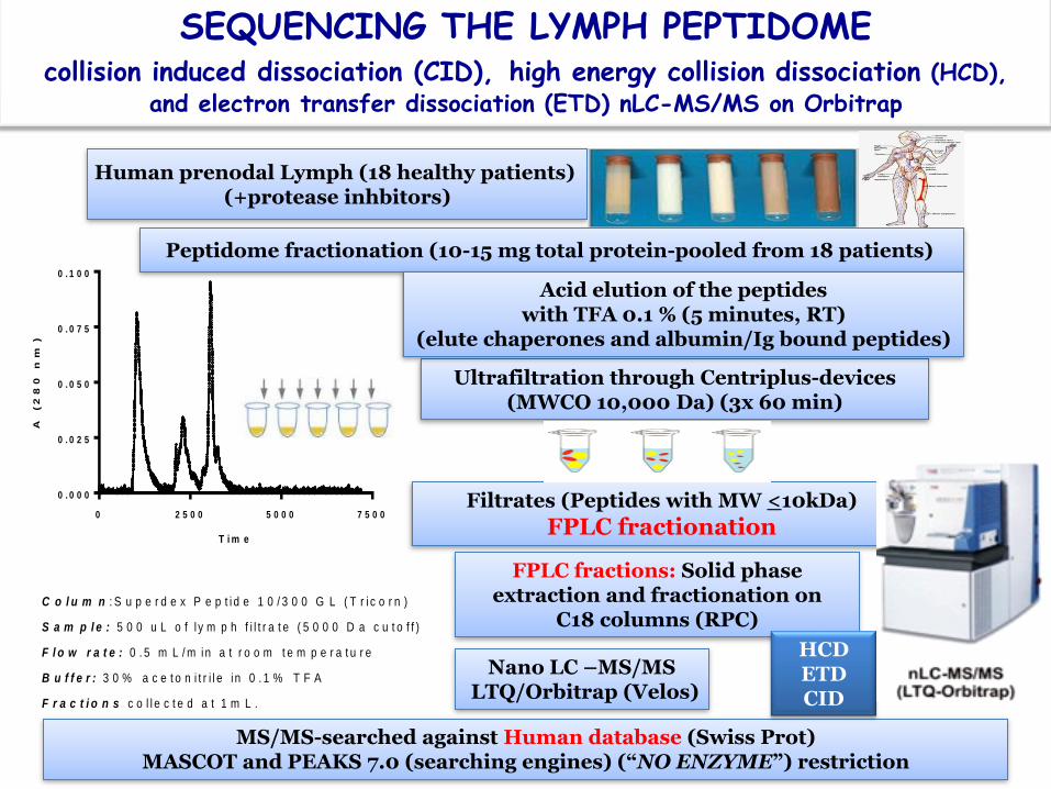
SEQUENCING THE LYMPH PEPTIDOME collision induced dissociation (CID), high energy collision dissociation (HCD),
and electron transfer dissociation (ETD) nLC-MS/MS on Orbitrap
Human prenodal Lymph (18 healthy patients) (+protease inhbitors)
Peptidome fractionation (10-15 mg total protein-pooled from 18 patients)
Ultrafiltration through Centriplus-devices (MWCO 10,000 Da) (3x 60 min)
Filtrates (Peptides with MW <10kDa)
FPLC fractionation
Acid elution of the peptides with TFA 0.1 % (5 minutes, RT)
(elute chaperones and albumin/Ig bound peptides)
FPLC fractions: Solid phase extraction and fractionation on
C18 columns (RPC)
Nano LC –MS/MS LTQ/Orbitrap (Velos)
MS/MS-searched against Human database (Swiss Prot) MASCOT and PEAKS 7.0 (searching engines) (“NO ENZYME”) restriction
HCD ETD CID
0 2 5 0 0 5 0 0 0 7 5 0 0
0 . 0 0 0
0 . 0 2 5
0 . 0 5 0
0 . 0 7 5
0 . 1 0 0
C o l u m n : S u p e r d e x P e p t i d e 1 0 / 3 0 0 G L ( T r i c o r n )
S a m p l e : 5 0 0 u L o f l y m p h f i l t r a t e ( 5 0 0 0 D a c u t o f f )
F l o w r a t e : 0 . 5 m L / m in a t r o o m t e m p e r a t u r e
B u f f e r : 3 0 % a c e t o n i t r i l e i n 0 . 1 % T F A
F r a c t i o n s c o l l e c t e d a t 1 m L .
T i m e
A (2
80
n
m)

FDR <0.5%; p<0.05
Representative PEAKS 7.0 output for a FPLC-lymph peptidome fraction
552 PSM >300 peptides/FPLC fraction

We sequenced over 900 peptides, each assigned with <0.5% false discovery rate, which corresponds to a p-value between 0.01-0.05. This analysis mapped naturally processed peptides deriving from over 480 proteins with different intra and extra-cellular locations.
The human lymph peptidomes sequenced at high resolution on Orbitrap:CID/HCD/ETD-nLC-MS/MS

Oncostatin-M-specific Receptor subunit beta
Glyceraldehyde-3-phosphate dehydrogenase
HCD
Human lymph peptidome-novel epitopes
Elongation factor 1-alpha 1
Chromosomal protein HMG 17

Cytochrome c oxidase subunit 5A Malate dehydrogenase, mitochondrial
HCD
Splicing factor 3A subunit-2 Solute carrier organic anion transporter
family member 6A1

Insulin-like growth factor 1 receptor
Transmembrane glycoprotein NMB
HCD
Collagen alpha-1(XV)
Complement C3

Alpha-1-antitrypsin
Pigment epithelium-derived factor
ETD
Histone H1.4
Histone H2B type 1
ETD ETD
HCD

Polymeric immunoglobulin receptor
Apolipoprotein A1
HCD
Fibrinogen alpha chain
Kininogen 1

Macrophage migration inhibitory factor
Triosephosphate isomerase
HCD
Proteoglycan 4
Collagen alpha-1 (I)

Molecular functions and cellular distribution of the human lymph peptidome
The endogenous peptides from human lymph derived from mitochondria, ER, Golgi enzymes, cytosolic proteins, nuclear proteins and peptides from extracellular matrix proteins; playing important roles as chaperones and in innate immune responses. Several additional naturally-processed peptides derived from plasma membrane proteins and surface receptors, in addition to the proteins found in the filtrate of the plasma (fibrinogen and other acute-phase response proteins).
Lymph-is not only a filtrate of plasma

Empty HLA-DR1 +/- DM helper for peptide
loading)
Analysis of MHC-II-DR1 binding affinity
Competition ELISA displacement assays with recombinant MHC-II molecules
IPMYSIITPNVLRL
DIC50= 0 IC50= 0.03 ± 0.01 M
SSKITHRIHWESASLLR
DIC50= 0.06 M IC50= 0.07 ± 0.01 M
EEFGRFASFEAQGALA
DIC50= 0.07 M IC50= 0.14 ± 0.04 M
ASFEAQGALANIAVDKA
DIC50= 3.40 M IC50= 1.71 ± 0.28 M
Complement Protein C3 MHC-II a chain
Lymph MHC-II-eluted Lymph MHC-II-eluted
Peptide loaded HLA-DR1 +/- DM
(helper for peptide loading)
The immunological significance of the human lymph peptidome In VITRO

Structure-Activity Relationship (SAR) for MHC-II DR1 peptides binders
The immunological significance of the human lymph peptidome In VITRO
Protein Sequence
Peptide
source IC50 (DR1)(nM)
Actin_ cytoplasmic 1 ALDFEQEMATAASSS DC 116 ± 47
Annexin A2 RDALNIETAVKTKG DC 283 ± 119
Cathepsin S TGKLISLSAQNLVD DC 18 ± 1
Complement factor B KGGSFQLLQGGQALE DC 42 ± 8
C-type lectin domain family 12 member A SEKMFLSEESERSTD DC 896 ± 735
Dihydropyrimidinase-related protein 2 DENQFVAVTSTNAAK DC 48 ± 9
Endoplasmic reticulum resident protein 29 GEFIKASSIEARQ DC 158 ± 52
Integrin beta-2 NSNQFQTEVGKQLIS DC 96 ± 21
Ras-related protein Rab-5C SPNIVIALAGNKAD DC 123 ± 34
Syntenin-1 KVIQAQTAYSANPA DC 53 ± 27
Transforming growth factor-beta-induced protein ig-h3 EAFQAMPPEELNK DC 292 ± 38
HLA class II histocompatibility antigen_ DR alpha chain EEFGRFASFEAQGALA DC 50 ± 9
Complement C3 IPMYSIITPNVLRL DC
17 ± 1
HLA class II histocompatibility antigen_ DR alpha chain
ASFEAQGALANIAVDKA Lymph
740 ± 109
Complement C3 SSKITHRIHWESASLLR Lymph 53 ± 4
Collagen II alpha-1 AGFKGEQGPKGEP Lymph 4399 ± 243
Netrin G2 MLHLLALFLH Lymph 7558 ± 3617
Fibrosin PGALLGAPPPLVPAP Lymph 2121 ± 152
Cartilage-oligomeric protein AWLDLEFISTVLGAP Lymph 141 ± 7
Collagen VI alpha-2 RNMTLFSDLVAEKFI Lymph 2542 ± 203
Adlican EDTFAHLTPTPT Lymph 221 ± 9
Collagen alpha-1(XII) KNFASVQGVSLESG Lymph 242 ± 55
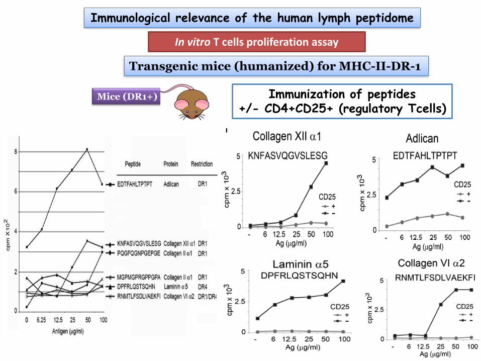
In vitro T cells proliferation assay
Immunological relevance of the human lymph peptidome
Mice (DR1+)
Transgenic mice (humanized) for MHC-II-DR-1
Immunization of peptides +/- CD4+CD25+ (regulatory Tcells)

Acute phase response signaling
Major biochemical pathways: the human lymph peptidome
Lymph-filtrate of plasma

Coagulation system
Major biochemical pathways: the human lymph peptidome
Lymph-filtrate of plasma

Development of a Label Free Quantitation (LFQ) Platform for quantifying peptides epitopes
from human lymph and other biological fluids (Translational Research)
P1 P2 P3 P4 P5 P6 P7 Lymph peptidome: healthy individual patients samples
LFQ (MS1-based area ratios) PEAKS 7.0-analysis of equal amounts of the extracted lymph peptidome (average of 3 replicates/patient).
Healthy Patients

Human Lymph Peptidome ????Degradome
Processing Pathways of lymph peptidome THE DEGRADOME
Experimentally-based databases

cDC MHC-II Eluted Peptidome degradome
Human Lymph Peptidome degradome
Overall a wide variety of proteases were implicated in the generation of the lymph-bound peptidome, with matrix metalloproteases (MMPs) and cathepsins being highly represented, followed by caspases and enzymes involved in the innate immune responses (angiotensin converting enzyme, complement factor I, granzymes) and enzymes of the coagulation cascade (thrombin,/plasmin, kallikreins).
Roughly one third of the eluted peptides from MHC-II were processed by cathepsins at either the N or C terminus; another third were cleaved at the N terminus by a signal peptidase or another S26 homolog; with the remainder cleaved by a metalloprotease, calpain, caspase, or other protease.
Thus, it appears that many proteases besides endosomal cathepsins can contribute to the formation of the MHC II-bound proteome.
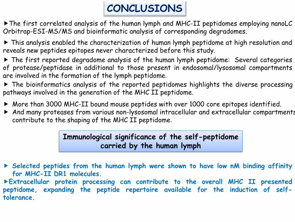
CONCLUSIONS
Selected peptides from the human lymph were shown to have low nM binding affinity for MHC-II DR1 molecules.
Extracellular protein processing can contribute to the overall MHC II presented peptidome, expanding the peptide repertoire available for the induction of self-tolerance.
The first correlated analysis of the human lymph and MHC-II peptidomes employing nanoLC Orbitrap-ESI-MS/MS and bioinformatic analysis of corresponding degradomes.
Immunological significance of the self-peptidome carried by the human lymph
This analysis enabled the characterization of human lymph peptidome at high resolution and reveals new peptides epitopes never characterized before this study.
The bioinformatics analysis of the reported peptidomes highlights the diverse processing pathways involved in the generation of the MHC II peptidome.
The first reported degradome analysis of the human lymph peptidome: Several categories of protease/peptidase in additional to those present in endosomal/lysosomal compartments are involved in the formation of the lymph peptidome.
More than 3000 MHC-II bound mouse peptides with over 1000 core epitopes identified. And many proteases from various non-lysosomal intracellular and extracellular compartments
contribute to the shaping of the MHC II peptidome.

Acknowledgements
Albert Einstein College of Medicine (AECOM) Laura Santambrogio lab Cristina C. Clement Valerio Zolla Lawrence Stern Lab UMASS Aniuska Becerra Scott Shafer
$$$ NIH
Mass spectrometry Collection of data and “Raw files”
Edward Nieves (AECOM) Fa-Yun Che (AECOM)
MS Bioworks