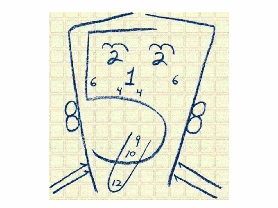
cranial nerves 360°
TRANSCRIPT
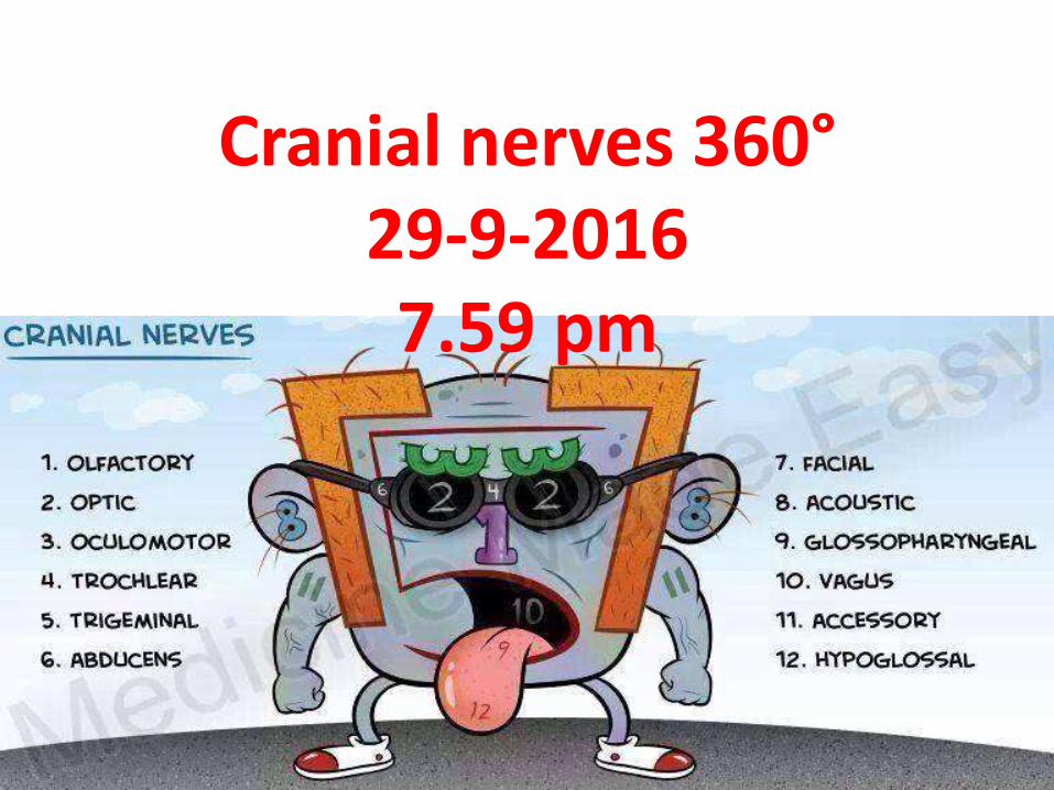
Cranial nerves 360°29-9-20167.59 pm

Great teachers – All this is their work . I am just the reader of their books .
Prof. Paolo castelnuovo
Prof. Aldo Stamm Prof. Mario Sanna
Prof. Magnan

For Other powerpoint presentatioinsof
“ Skull base 360° ”I will update continuosly with date tag at the end as I am
getting more & more information
click
www.skullbase360.in- you have to login to slideshare.net with Facebook
account for downloading.
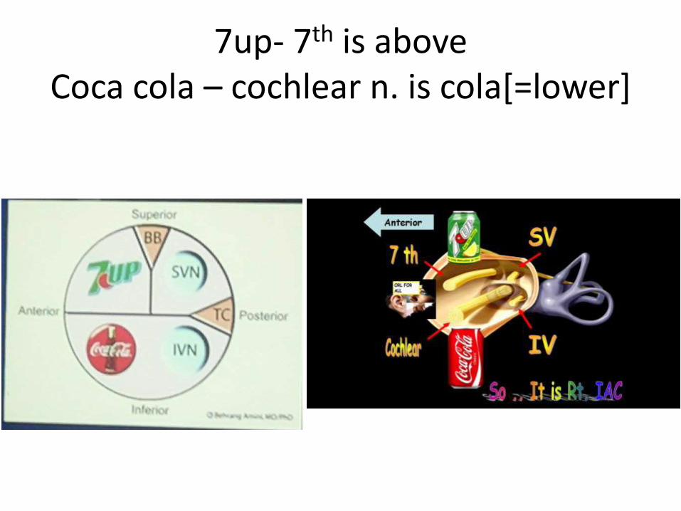
7up- 7th is aboveCoca cola – cochlear n. is cola[=lower]

Crannial nerves 360 video
Click https://www.youtube.com/watch?v=
PSTzJOHpDCM

1st nerve
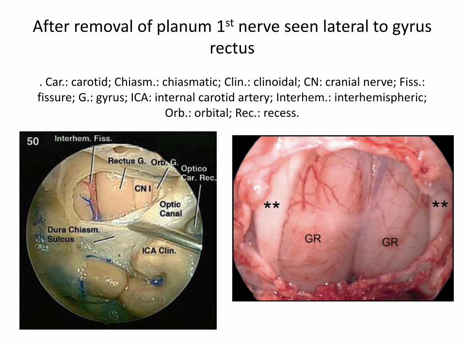
After removal of planum 1st nerve seen lateral to gyrusrectus
. Car.: carotid; Chiasm.: chiasmatic; Clin.: clinoidal; CN: cranial nerve; Fiss.: fissure; G.: gyrus; ICA: internal carotid artery; Interhem.: interhemispheric;
Orb.: orbital; Rec.: recess.
2nd nerve
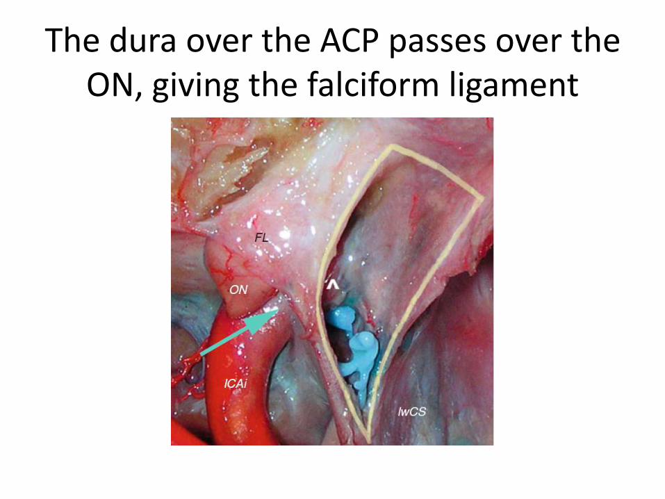
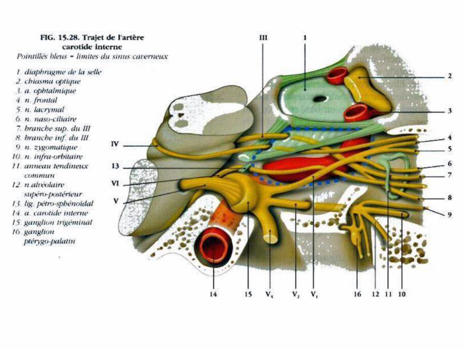
The dura over the ACP passes over the ON, giving the falciform ligament
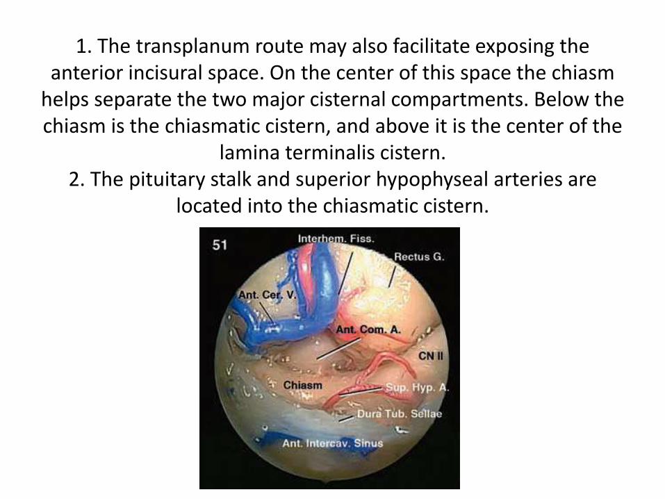
1. The transplanum route may also facilitate exposing the anterior incisural space. On the center of this space the chiasm
helps separate the two major cisternal compartments. Below the chiasm is the chiasmatic cistern, and above it is the center of the
lamina terminalis cistern.2. The pituitary stalk and superior hypophyseal arteries are
located into the chiasmatic cistern.
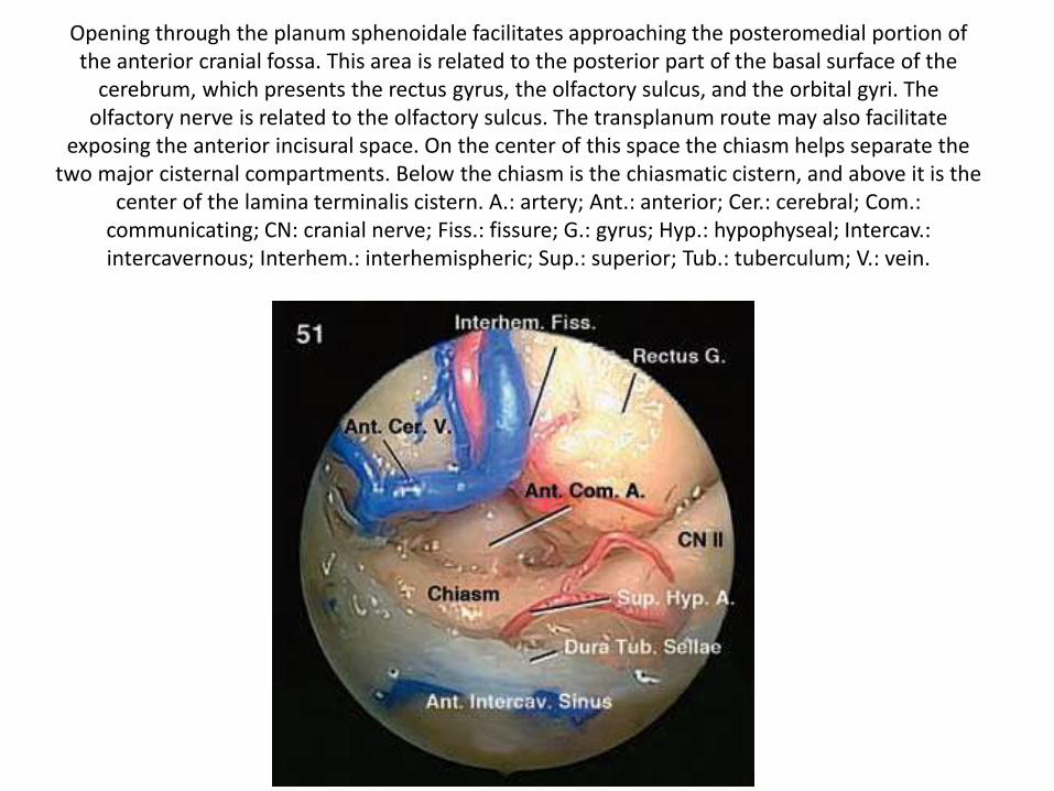
Opening through the planum sphenoidale facilitates approaching the posteromedial portion of the anterior cranial fossa. This area is related to the posterior part of the basal surface of the
cerebrum, which presents the rectus gyrus, the olfactory sulcus, and the orbital gyri. The olfactory nerve is related to the olfactory sulcus. The transplanum route may also facilitate
exposing the anterior incisural space. On the center of this space the chiasm helps separate the two major cisternal compartments. Below the chiasm is the chiasmatic cistern, and above it is the
center of the lamina terminalis cistern. A.: artery; Ant.: anterior; Cer.: cerebral; Com.: communicating; CN: cranial nerve; Fiss.: fissure; G.: gyrus; Hyp.: hypophyseal; Intercav.: intercavernous; Interhem.: interhemispheric; Sup.: superior; Tub.: tuberculum; V.: vein.
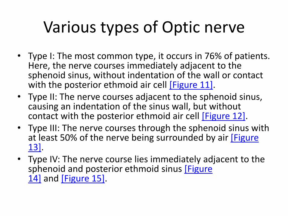
Various types of Optic nerve
• Type I: The most common type, it occurs in 76% of patients. Here, the nerve courses immediately adjacent to the sphenoid sinus, without indentation of the wall or contact with the posterior ethmoid air cell [Figure 11].
• Type II: The nerve courses adjacent to the sphenoid sinus, causing an indentation of the sinus wall, but without contact with the posterior ethmoid air cell [Figure 12].
• Type III: The nerve courses through the sphenoid sinus with at least 50% of the nerve being surrounded by air [Figure 13].
• Type IV: The nerve course lies immediately adjacent to the sphenoid and posterior ethmoid sinus [Figure 14] and [Figure 15].

Figure 11: Coronal CT showing type I optic nerve (arrows) the nerve is seen to course immediately adjacent to the sphenoid sinus, without contact with the posterior
ethmoid air cell

Figure 12: Coronal CT showing type II optic nerve (curved arrows) causing an indentation of the sinus wall, but without contact with the posterior ethmoid air cell
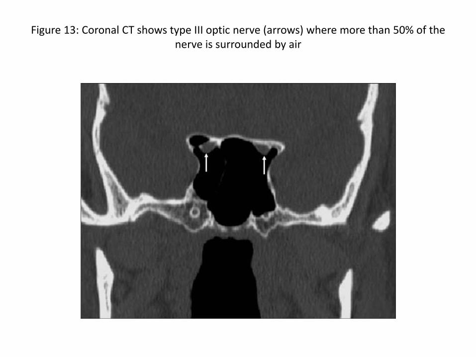
Figure 13: Coronal CT shows type III optic nerve (arrows) where more than 50% of the nerve is surrounded by air
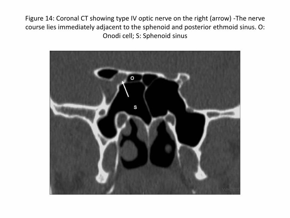
Figure 14: Coronal CT showing type IV optic nerve on the right (arrow) -The nerve course lies immediately adjacent to the sphenoid and posterior ethmoid sinus. O:
Onodi cell; S: Sphenoid sinus
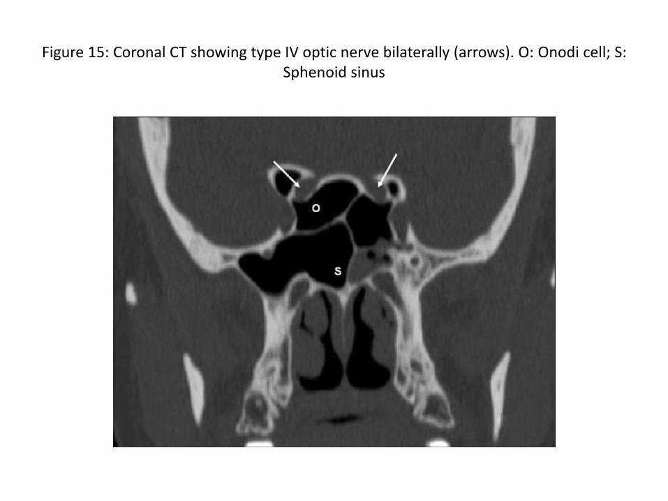
Figure 15: Coronal CT showing type IV optic nerve bilaterally (arrows). O: Onodi cell; S: Sphenoid sinus

Delano, et al., found that 85% of optic nerves associated with a pneumatized anterior clinoid process were of type II or type III configuration, and of these, 77% showed dehiscence [Figure 16], indicating the vulnerability of the optic nerve during FESS.
Figure 16: Coronal CT shows pneumatisation of anterior clinoid process (stars) with type III optic nerve (stars) with bony canal dehiscence bilaterally
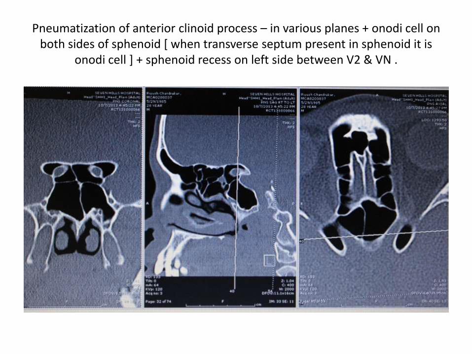
Pneumatization of anterior clinoid process – in various planes + onodi cell on both sides of sphenoid [ when transverse septum present in sphenoid it is
onodi cell ] + sphenoid recess on left side between V2 & VN .
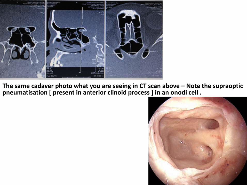
The same cadaver photo what you are seeing in CT scan above – Note the supraopticpneumatisation [ present in anterior clinoid process ] in an onodi cell .
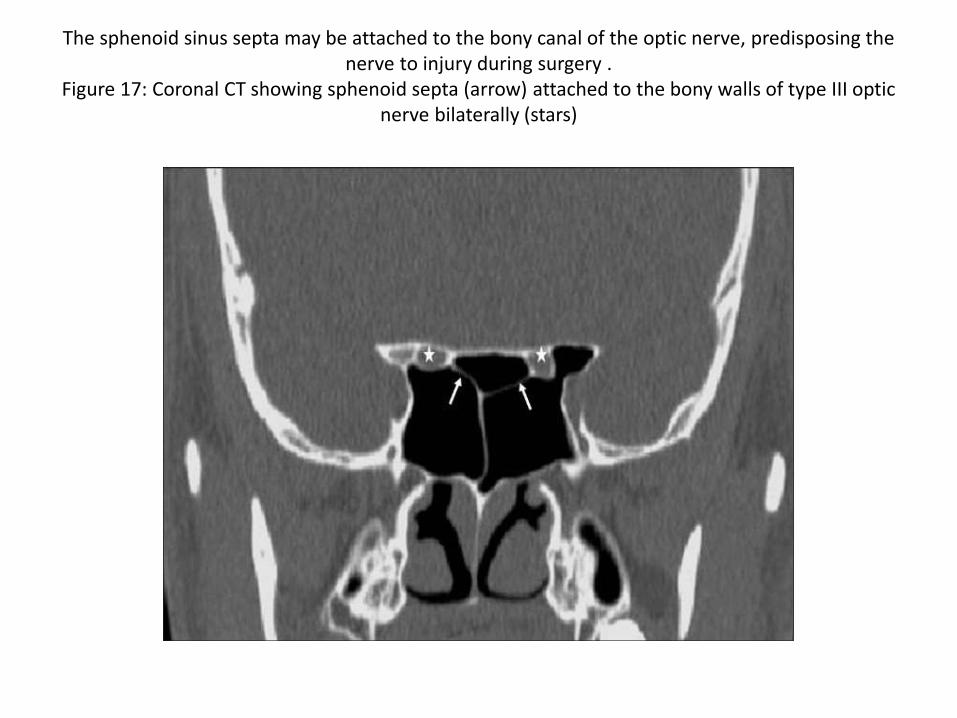
The sphenoid sinus septa may be attached to the bony canal of the optic nerve, predisposing the nerve to injury during surgery .
Figure 17: Coronal CT showing sphenoid septa (arrow) attached to the bony walls of type III optic nerve bilaterally (stars)

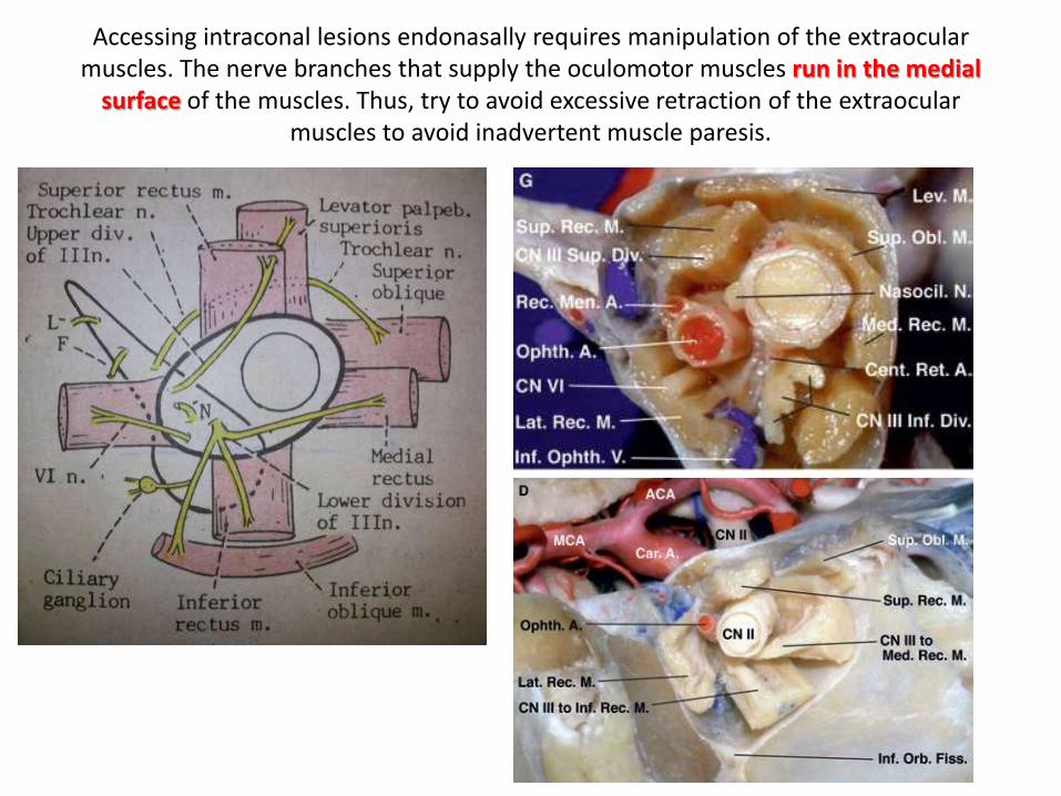
Accessing intraconal lesions endonasally requires manipulation of the extraocularmuscles. The nerve branches that supply the oculomotor muscles run in the medial
surface of the muscles. Thus, try to avoid excessive retraction of the extraocularmuscles to avoid inadvertent muscle paresis.
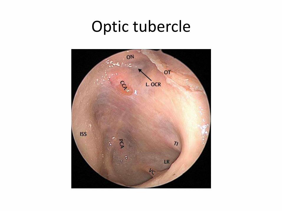
Optic tubercle
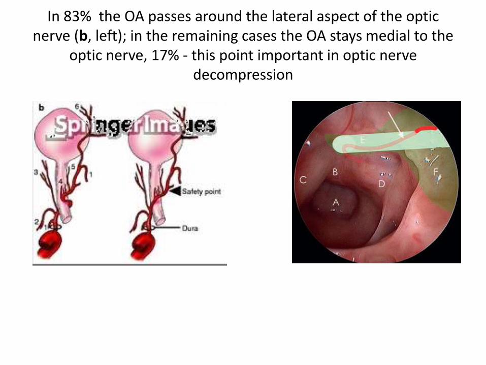
In 83% the OA passes around the lateral aspect of the optic nerve (b, left); in the remaining cases the OA stays medial to the
optic nerve, 17% - this point important in optic nerve decompression
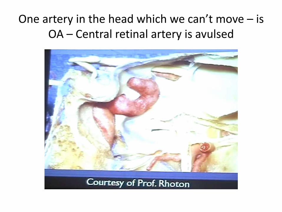
One artery in the head which we can’t move – is OA – Central retinal artery is avulsed
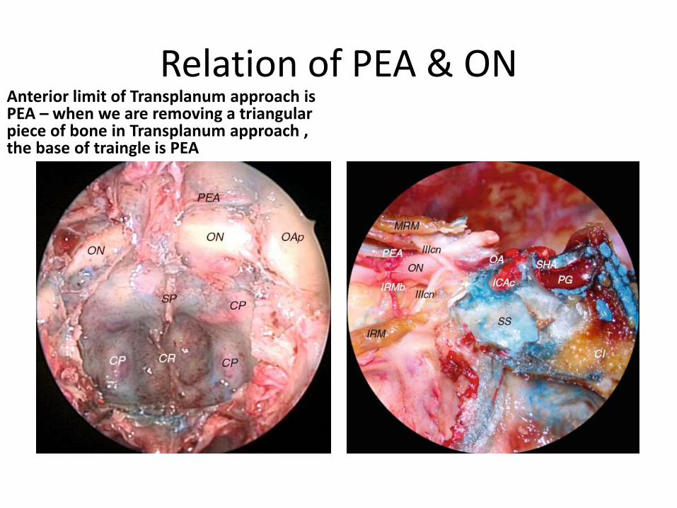
Relation of PEA & ONAnterior limit of Transplanum approach is PEA – when we are removing a triangular piece of bone in Transplanum approach , the base of traingle is PEA
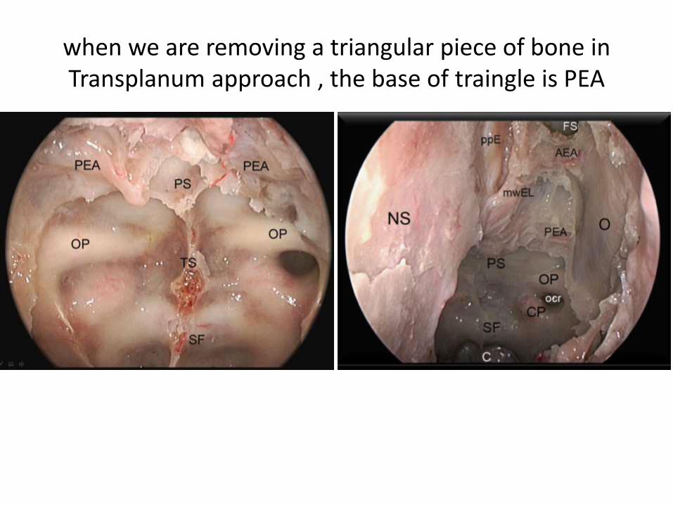
when we are removing a triangular piece of bone in Transplanum approach , the base of traingle is PEA
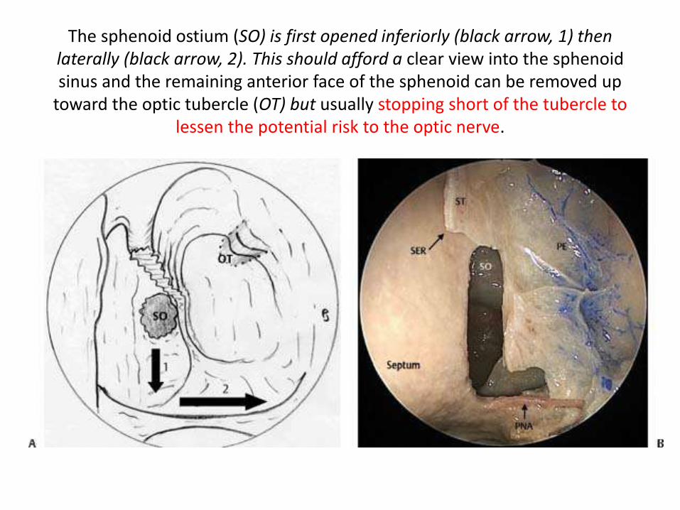
The sphenoid ostium (SO) is first opened inferiorly (black arrow, 1) then laterally (black arrow, 2). This should afford a clear view into the sphenoid sinus and the remaining anterior face of the sphenoid can be removed up
toward the optic tubercle (OT) but usually stopping short of the tubercle to lessen the potential risk to the optic nerve.

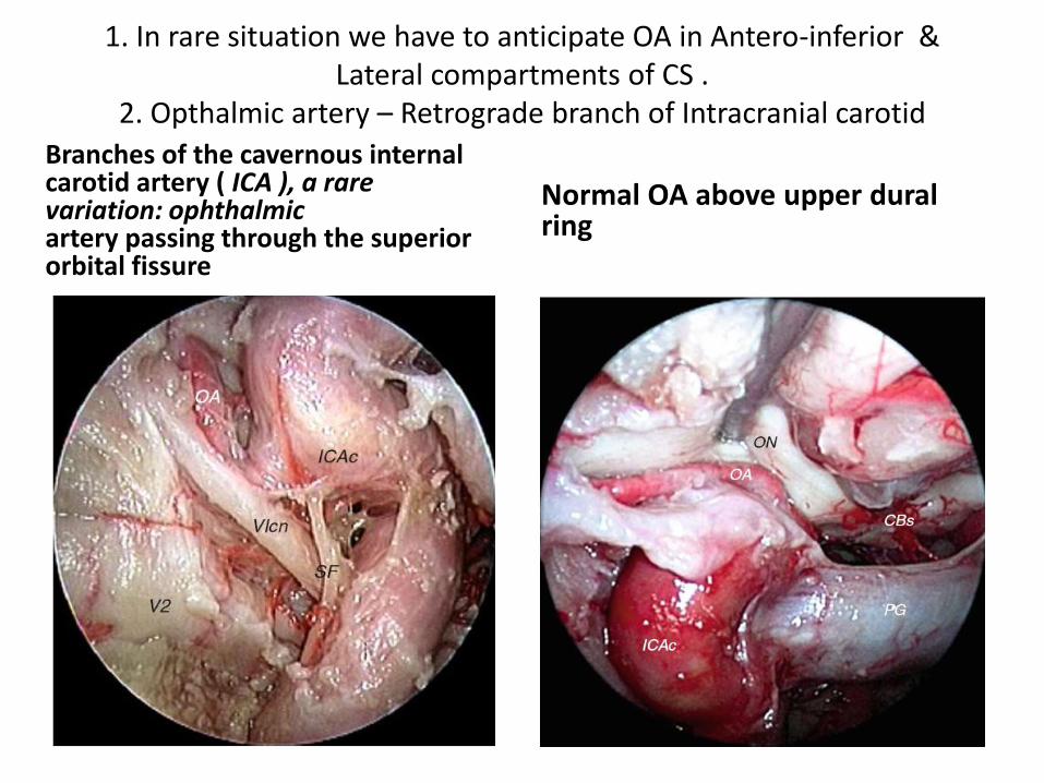
1. In rare situation we have to anticipate OA in Antero-inferior & Lateral compartments of CS .
2. Opthalmic artery – Retrograde branch of Intracranial carotid
Branches of the cavernous internalcarotid artery ( ICA ), a rare variation: ophthalmicartery passing through the superiororbital fissure
Normal OA above upper duralring

classification of the ophthalmic artery typeshttp://www.springerimages.com/Images/MedicineAndPublicHealth/1-
10.1007_s10143-006-0028-6-1a = intradural type,
b = extradural supra-optic strut type [ Optic strut = L-OCR ]c = extradural trans-optic strut type
on optic nerve, pr proximal ring, cdr carotid duralring= upper dural ring , ica internal carotid artery
I think this variation is type c
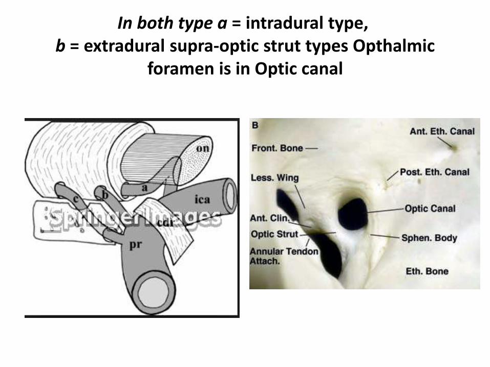
In both type a = intradural type,b = extradural supra-optic strut types Opthalmic
foramen is in Optic canal
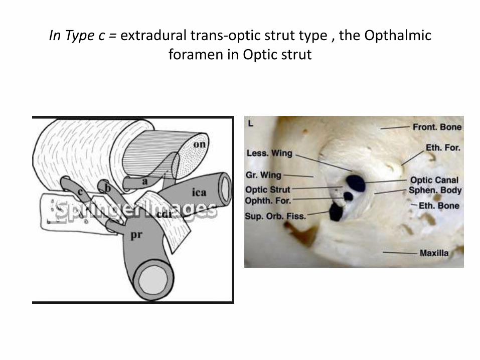
In Type c = extradural trans-optic strut type , the Opthalmicforamen in Optic strut
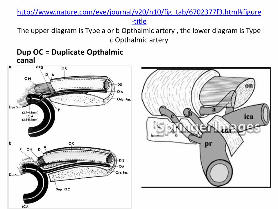
http://www.nature.com/eye/journal/v20/n10/fig_tab/6702377f3.html#figure-title
The upper diagram is Type a or b Opthalmic artery , the lower diagram is Type c Opthalmic artery
Dup OC = Duplicate Opthalmiccanal

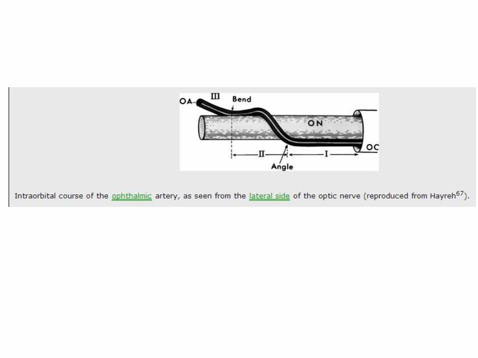
Origin and intracranial and intracanalicular course of the ophthalmic artery and its subdivisions, as seen on opening the optic canal (reproduced from Hayreh67).
Both from one specimen. (a) The extraduralorigin of the right ophthalmic artery, so that no ophthalmic artery is seen even on opening theoptic canal; a thinning of the dural sheath is seen at 'X', indicating the position of the artery. (b) The ophthalmic artery is seen after removing the duralsheath covering it (reproduced from Hayrehand Dass2).
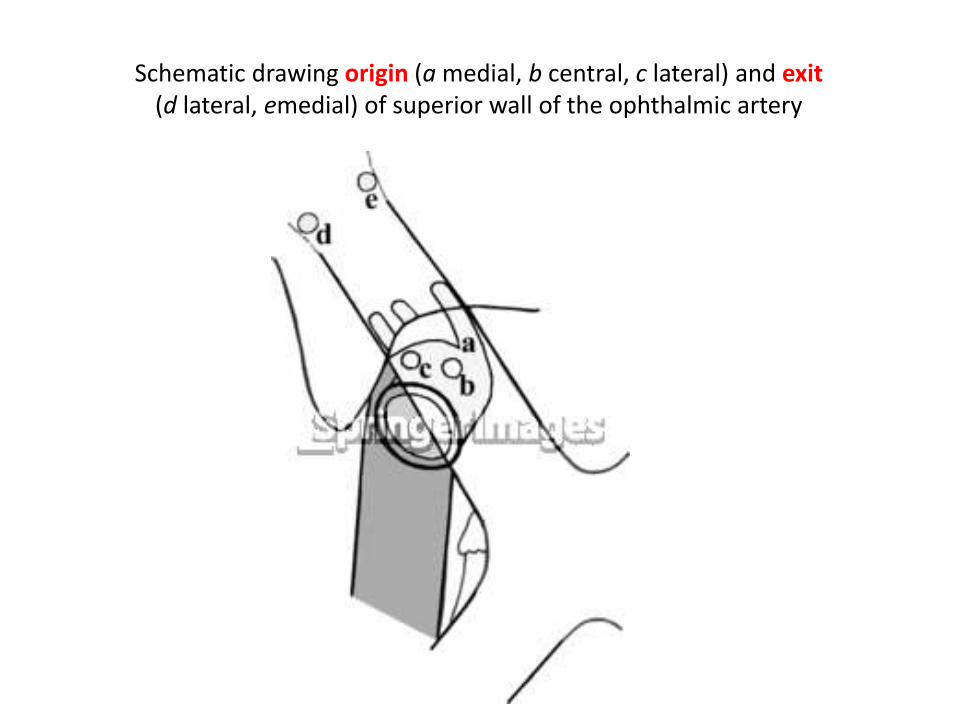
Schematic drawing origin (a medial, b central, c lateral) and exit(d lateral, emedial) of superior wall of the ophthalmic artery
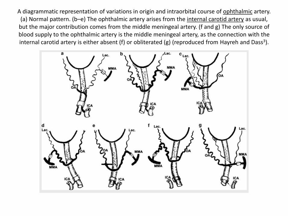
A diagrammatic representation of variations in origin and intraorbital course of ophthalmic artery. (a) Normal pattern. (b–e) The ophthalmic artery arises from the internal carotid artery as usual,
but the major contribution comes from the middle meningeal artery. (f and g) The only source of blood supply to the ophthalmic artery is the middle meningeal artery, as the connection with the internal carotid artery is either absent (f) or obliterated (g) (reproduced from Hayreh and Dass3).

Origin, course, and branches of the ophthalmic artery in two adult specimens. Segment Y disappeared in (a) and segment Z disappeared in (b), resulting in the ophthalmic artery crossing under the optic nerve in both. In (b) an anastomosis is seen in lateral wall of the cavernous sinus between the part of the internal carotid artery lying in proximal part of the cavernous sinus and a branch from the ophthalmic artery passing through the superior orbital fissure (reproduced from
Hayreh67).
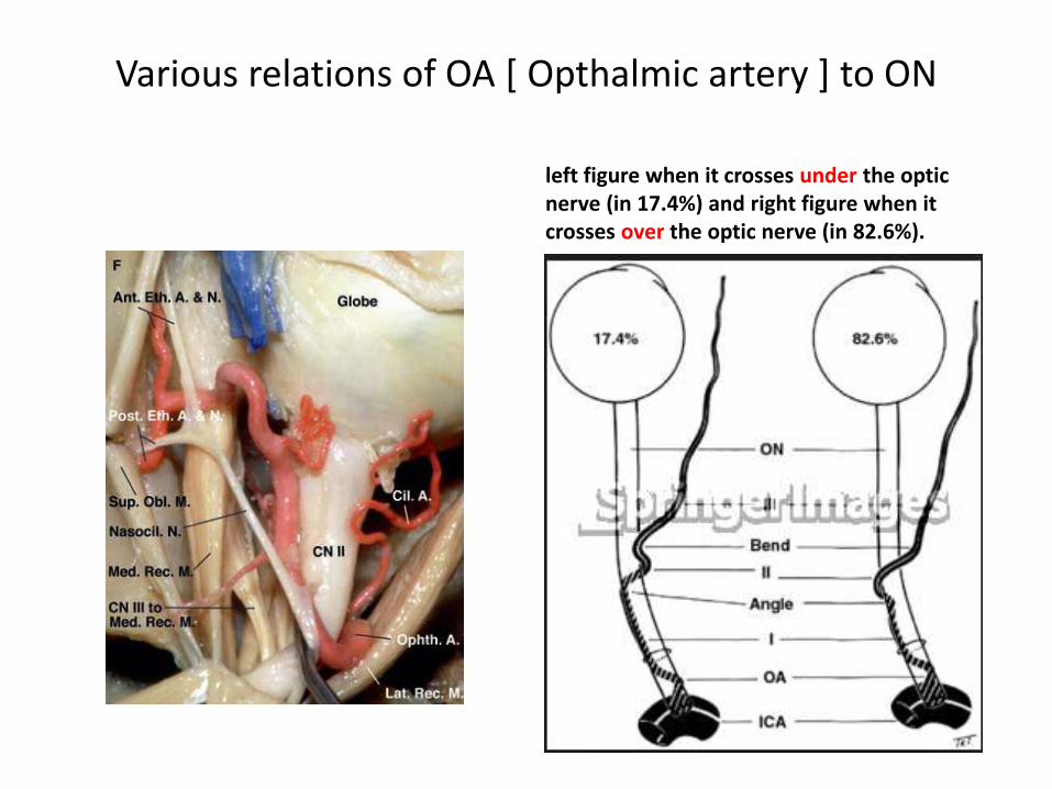
Various relations of OA [ Opthalmic artery ] to ON
left figure when it crosses under the optic nerve (in 17.4%) and right figure when it crosses over the optic nerve (in 82.6%).

Give incision in supero-medial area in optic nerve decompression – add
scott brown information
3rd nerve

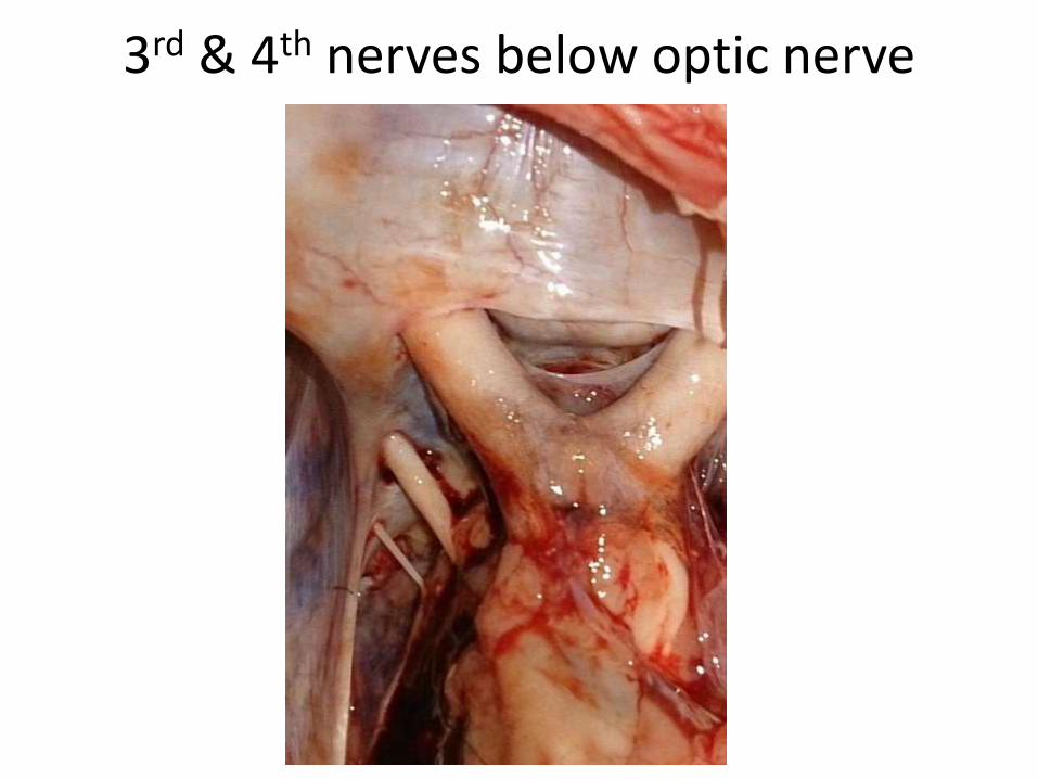
3rd & 4th nerves below optic nerve

Lilliquits membrane present over the basillar artery & 3rd N. origin area

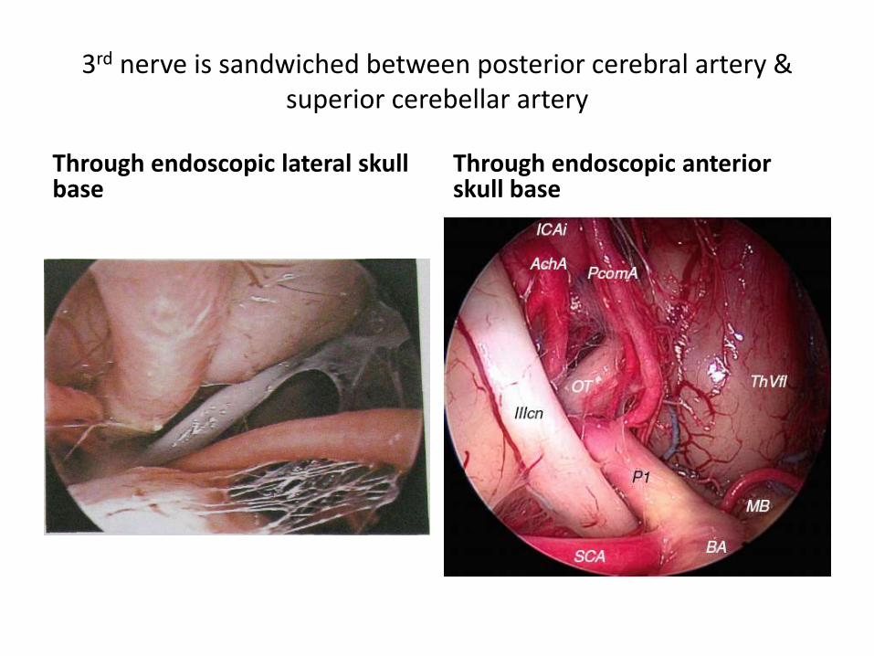
3rd nerve is sandwiched between posterior cerebral artery & superior cerebellar artery
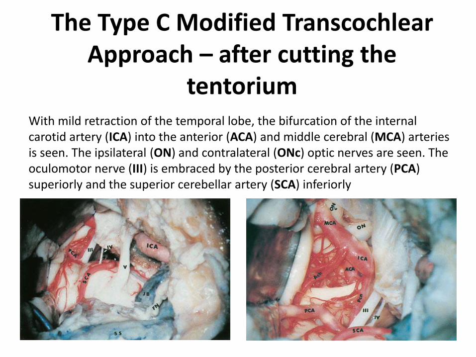
The Type C Modified TranscochlearApproach – after cutting the
tentoriumWith mild retraction of the temporal lobe, the bifurcation of the internal carotid artery (ICA) into the anterior (ACA) and middle cerebral (MCA) arteries is seen. The ipsilateral (ON) and contralateral (ONc) optic nerves are seen. The oculomotor nerve (III) is embraced by the posterior cerebral artery (PCA) superiorly and the superior cerebellar artery (SCA) inferiorly

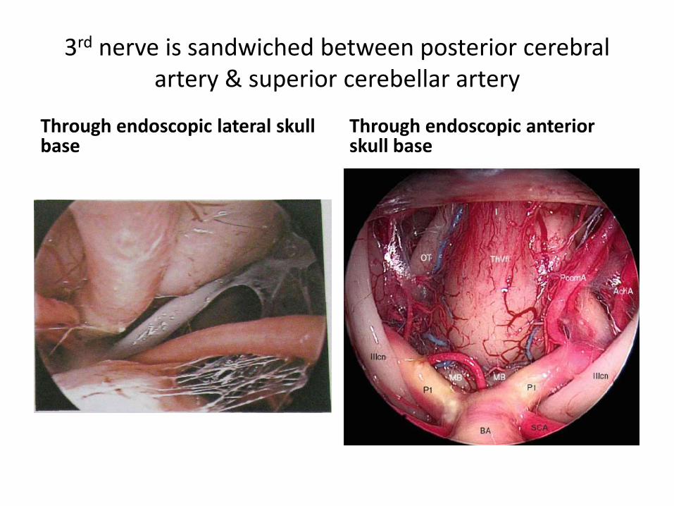
3rd nerve is sandwiched between posterior cerebral artery & superior cerebellar artery
3rd nerve is sandwiched between posterior cerebral artery & superior cerebellar artery
Through endoscopic lateral skull base
Through endoscopic anterior skull base
3rd nerve is sandwiched between posterior cerebral artery & superior cerebellar artery
Through endoscopic lateral skull base
Through endoscopic anterior skull base
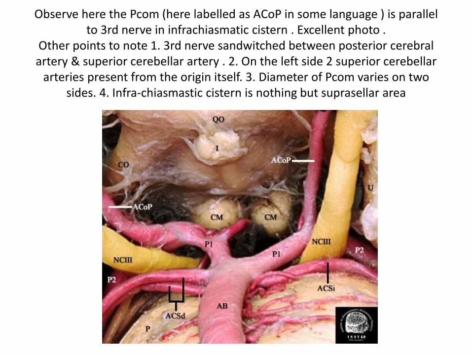
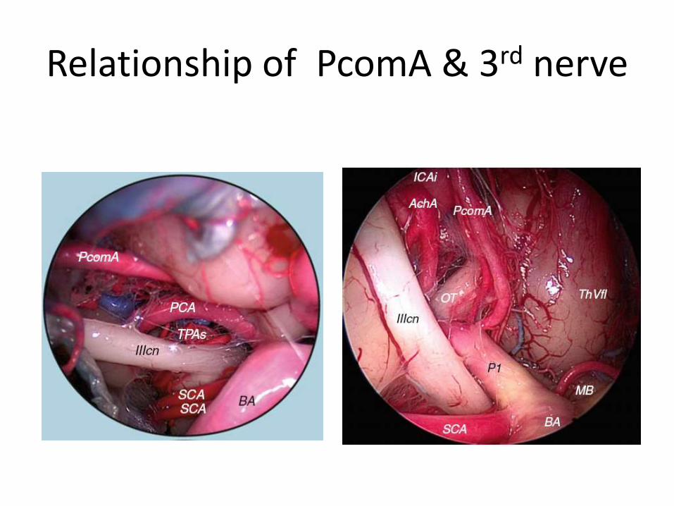
Observe here the Pcom (here labelled as ACoP in some language ) is parallel to 3rd nerve in infrachiasmatic cistern . Excellent photo .
Other points to note 1. 3rd nerve sandwitched between posterior cerebral artery & superior cerebellar artery . 2. On the left side 2 superior cerebellar
arteries present from the origin itself. 3. Diameter of Pcom varies on two sides. 4. Infra-chiasmastic cistern is nothing but suprasellar area
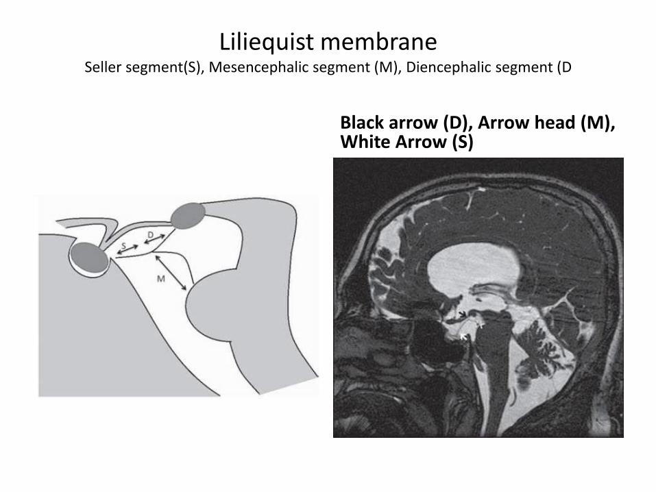
Liliequist membraneSeller segment(S), Mesencephalic segment (M), Diencephalic segment (D
Black arrow (D), Arrow head (M), White Arrow (S)
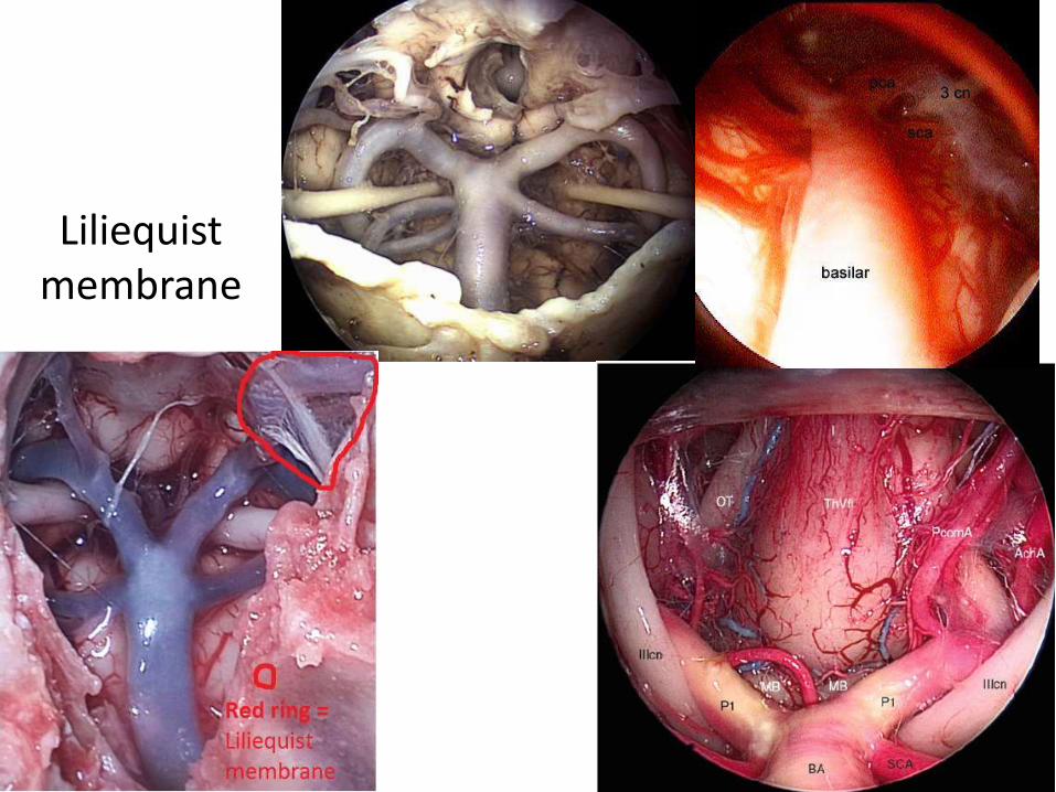
Liliequistmembrane
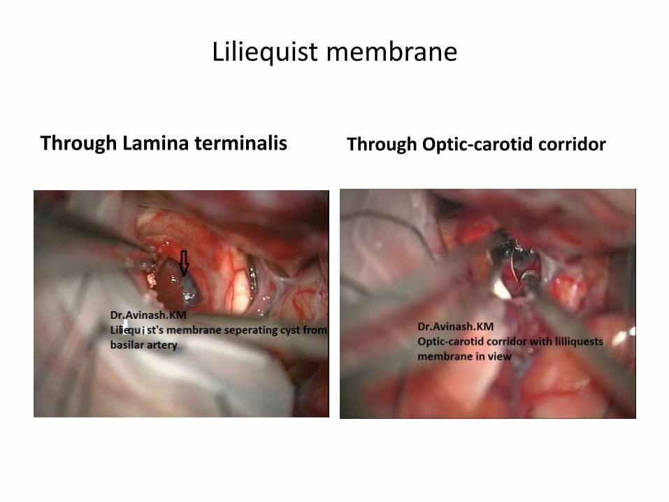
Through Lamina terminalis Through Optic-carotid corridor
Liliequist membrane

P1 in relation to 3rd nerve P2 in relation to 3rd nerve

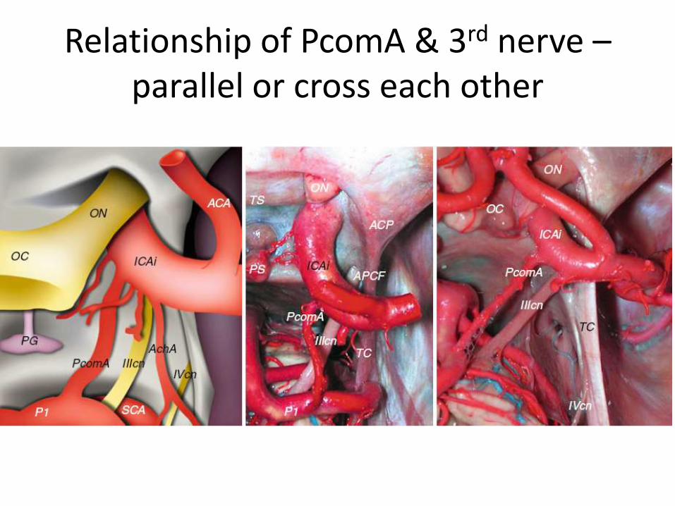
Relationship of PcomA & 3rd nerve –parallel or cross each other
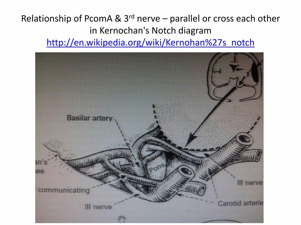
Relationship of PcomA & 3rd nerve – parallel or cross each other in Kernochan's Notch diagram
http://en.wikipedia.org/wiki/Kernohan%27s_notch
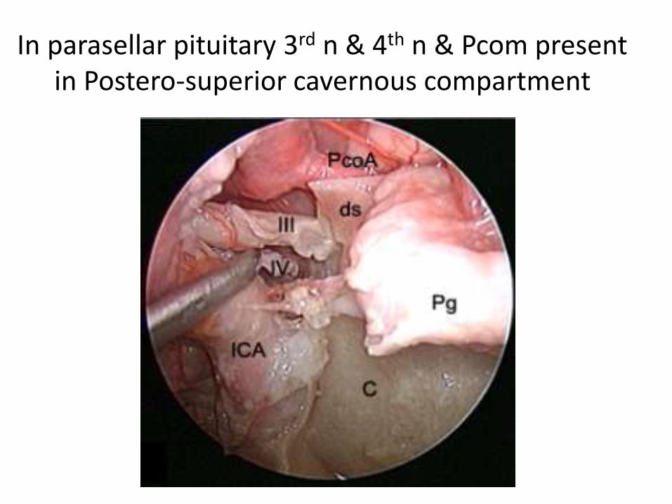
In parasellar pituitary 3rd n & 4th n & Pcom present in Postero-superior cavernous compartment
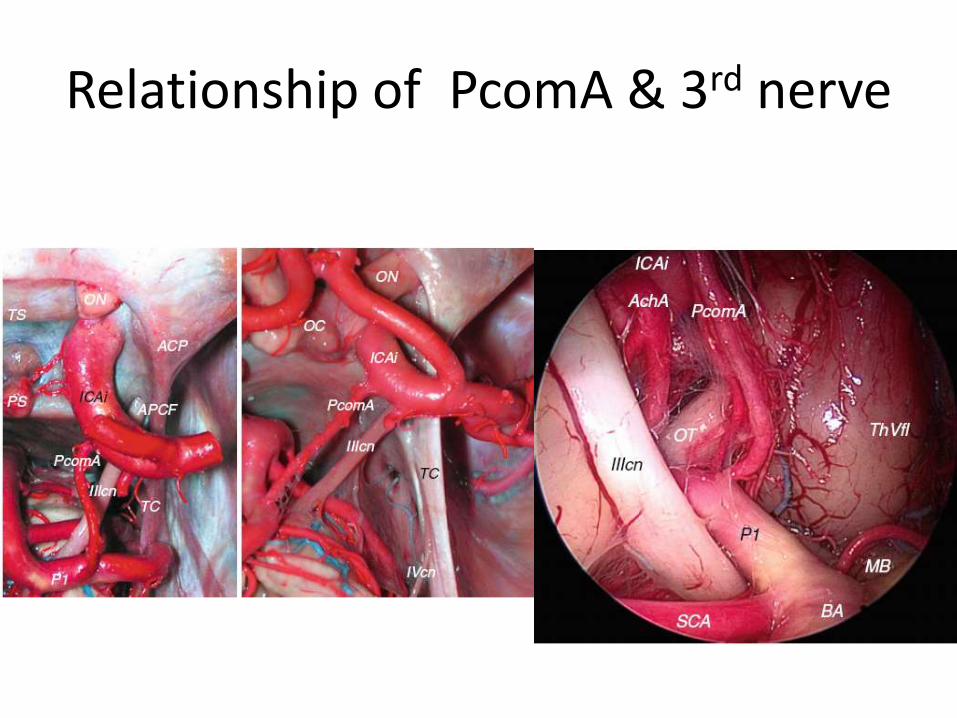
Relationship of PcomA & 3rd nerve
Relationship of PcomA & 3rd nerve
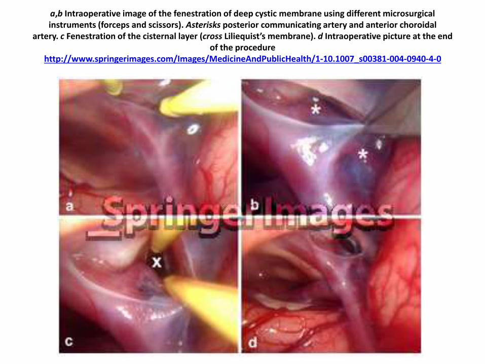
a,b Intraoperative image of the fenestration of deep cystic membrane using different microsurgical instruments (forceps and scissors). Asterisks posterior communicating artery and anterior choroidal
artery. c Fenestration of the cisternal layer (cross Liliequist’s membrane). d Intraoperative picture at the end of the procedure
http://www.springerimages.com/Images/MedicineAndPublicHealth/1-10.1007_s00381-004-0940-4-0
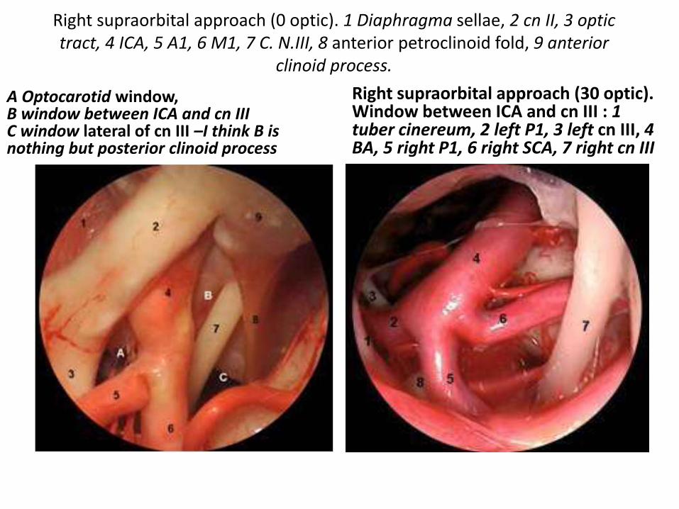
Right supraorbital approach (0 optic). 1 Diaphragma sellae, 2 cn II, 3 optic tract, 4 ICA, 5 A1, 6 M1, 7 C. N.III, 8 anterior petroclinoid fold, 9 anterior
clinoid process.
A Optocarotid window, B window between ICA and cn IIIC window lateral of cn III –I think B is nothing but posterior clinoid process
Right supraorbital approach (30 optic). Window between ICA and cn III : 1 tuber cinereum, 2 left P1, 3 left cn III, 4 BA, 5 right P1, 6 right SCA, 7 right cn III
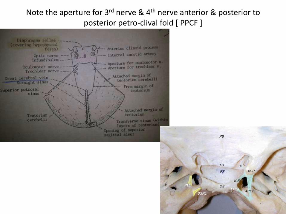
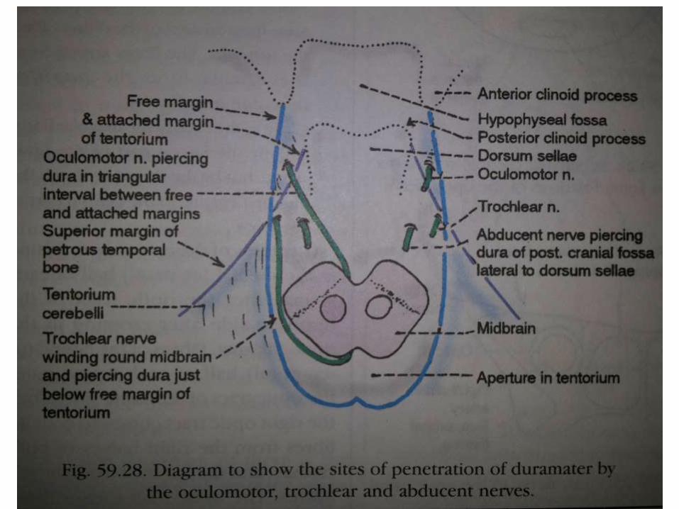
Note the aperture for 3rd nerve & 4th nerve anterior & posterior to posterior petro-clival fold [ PPCF ]
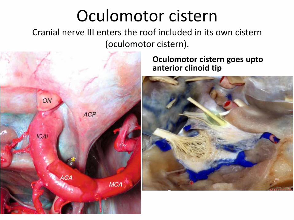
Oculomotor cistern Cranial nerve III enters the roof included in its own cistern
(oculomotor cistern).
Oculomotor cistern goes uptoanterior clinoid tip
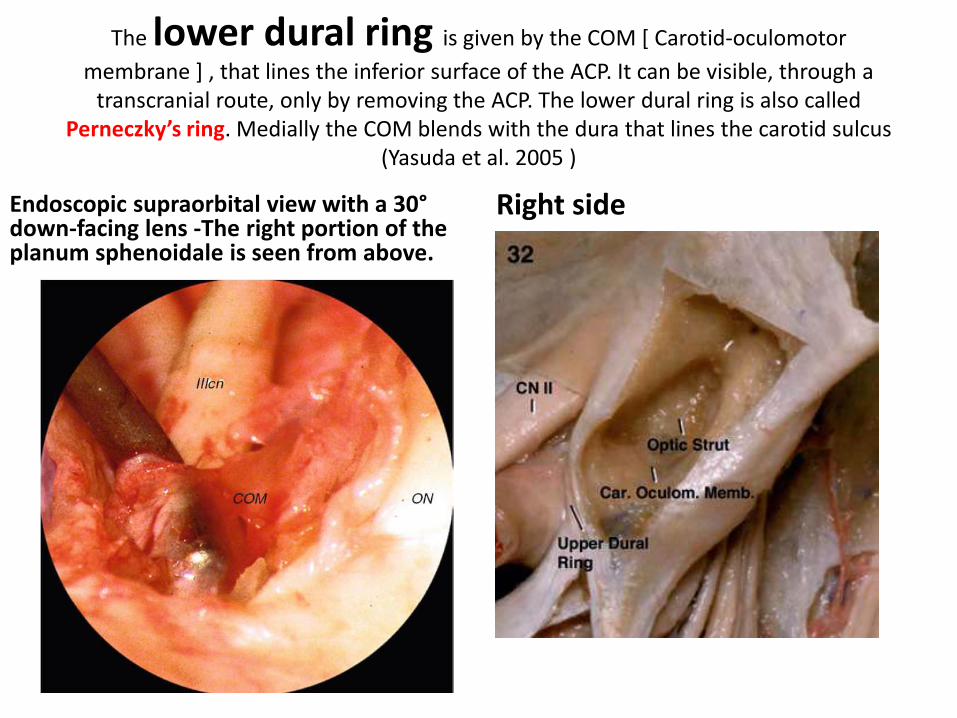
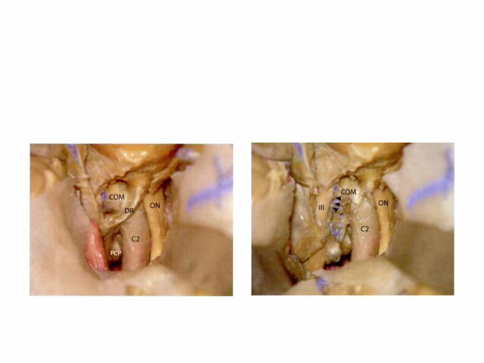
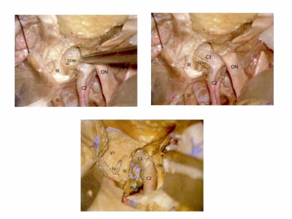
The lower dural ring is given by the COM [ Carotid-oculomotor
membrane ] , that lines the inferior surface of the ACP. It can be visible, through a transcranial route, only by removing the ACP. The lower dural ring is also called
Perneczky’s ring. Medially the COM blends with the dura that lines the carotid sulcus(Yasuda et al. 2005 )
Endoscopic supraorbital view with a 30°down-facing lens -The right portion of the planum sphenoidale is seen from above.
Right side

Fronto-temporal orbitozygomatictranscavernous approach
COM= Caratico-occulomotormembrane , DR = dural ring
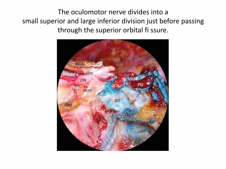
The oculomotor nerve divides into asmall superior and large inferior division just before passing
through the superior orbital fi ssure.
4th nerve
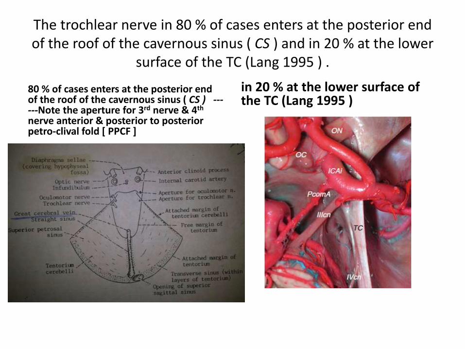
The trochlear nerve in 80 % of cases enters at the posterior end of the roof of the cavernous sinus ( CS ) and in 20 % at the lower
surface of the TC (Lang 1995 ) .
80 % of cases enters at the posterior end of the roof of the cavernous sinus ( CS ) ------Note the aperture for 3rd nerve & 4th
nerve anterior & posterior to posterior petro-clival fold [ PPCF ]
in 20 % at the lower surface of the TC (Lang 1995 )
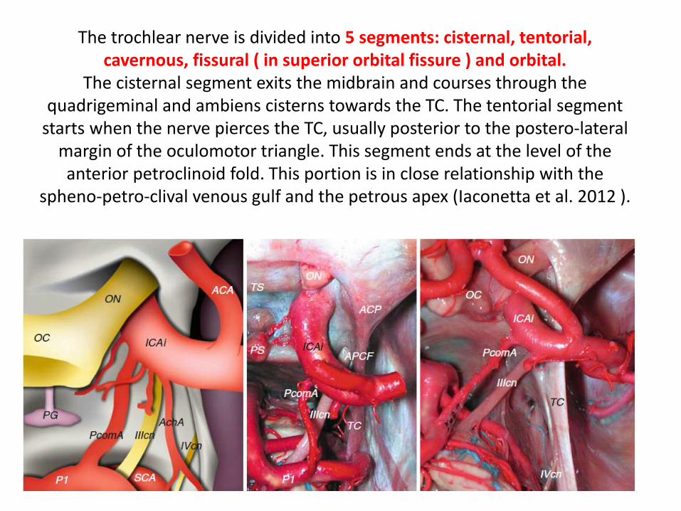
The trochlear nerve is divided into 5 segments: cisternal, tentorial, cavernous, fissural ( in superior orbital fissure ) and orbital.
The cisternal segment exits the midbrain and courses through the quadrigeminal and ambiens cisterns towards the TC. The tentorial segment
starts when the nerve pierces the TC, usually posterior to the postero-lateral margin of the oculomotor triangle. This segment ends at the level of the anterior petroclinoid fold. This portion is in close relationship with the
spheno-petro-clival venous gulf and the petrous apex (Iaconetta et al. 2012 ).
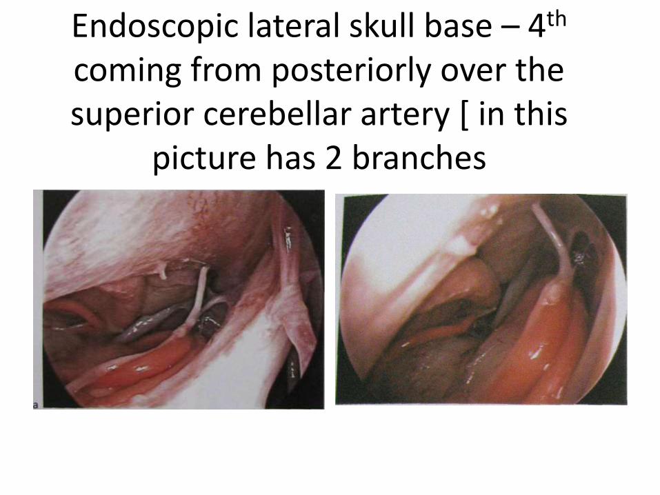
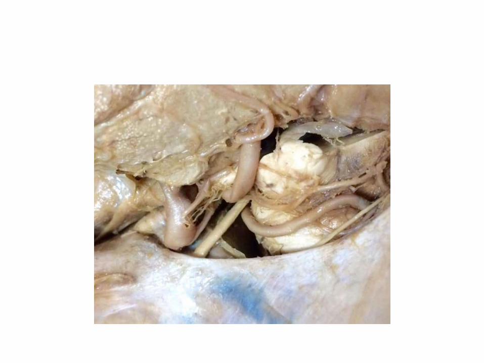
Endoscopic lateral skull base – 4th
coming from posteriorly over the superior cerebellar artery [ in this
picture has 2 branches
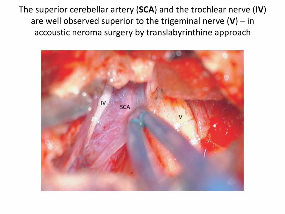
The superior cerebellar artery (SCA) and the trochlear nerve (IV) are well observed superior to the trigeminal nerve (V) – in accoustic neroma surgery by translabyrinthine approach
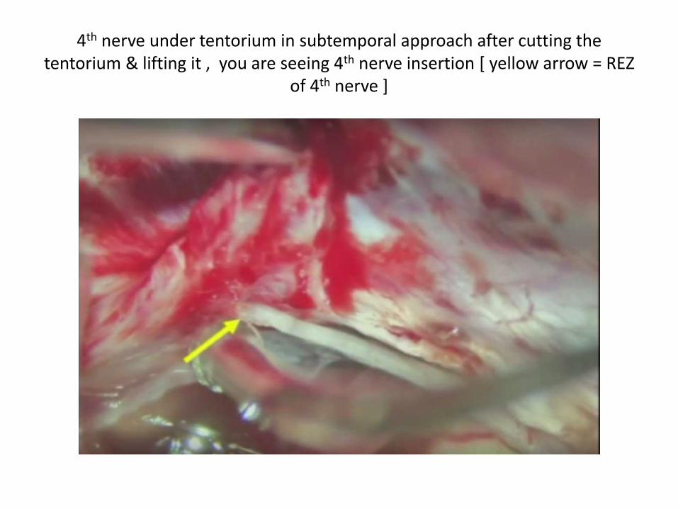
4th nerve under tentorium in subtemporal approach after cutting the tentorium & lifting it , you are seeing 4th nerve insertion [ yellow arrow = REZ
of 4th nerve ]
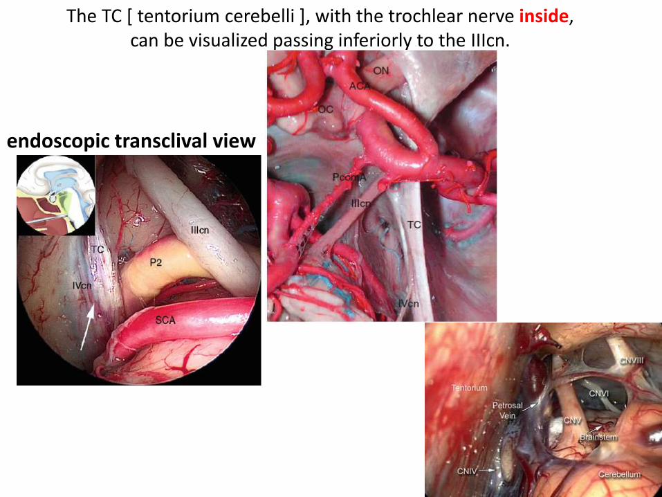
The TC [ tentorium cerebelli ], with the trochlear nerve inside,can be visualized passing inferiorly to the IIIcn.
endoscopic transclival view
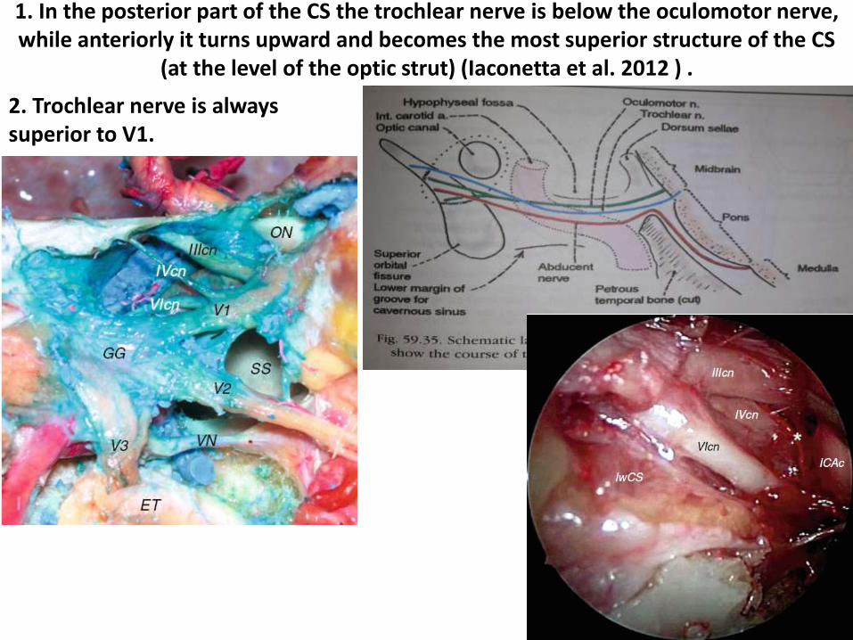
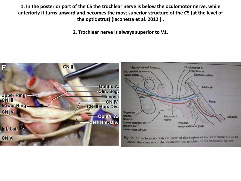
1. In the posterior part of the CS the trochlear nerve is below the oculomotor nerve, while anteriorly it turns upward and becomes the most superior structure of the CS
(at the level of the optic strut) (Iaconetta et al. 2012 ) .
2. Trochlear nerve is always superior to V1.
1. In the posterior part of the CS the trochlear nerve is below the oculomotor nerve, while anteriorly it turns upward and becomes the most superior structure of the CS (at the level of
the optic strut) (Iaconetta et al. 2012 ) .
2. Trochlear nerve is always superior to V1.

Observe 4th nerve in tentorium Cadaveric dissection image taken with a 30-degree endoscope following removal of the superior third of the
clivus, visualizing the small trochlear nerve seen running along the tentorial membrane edge. BA, basilar artery; PCA, posterior cerebral artery; SCA, superior cerebellar artery; CN III, occulomotor nerve; CN IV, trochlear
nerve; CN V, trigeminal nerve; TM, tentorial membrane; PComA, posterior communicating artery; MB, mamillary body.

(A) Intraoperative endoscopic close-up view showing the trigeminal nenre and the related neurovascular anatomy. a Trigeminal nerve (V).
b Superior aspect of cerebellum. c Petrosal veins. d Petrous apex. e Dense araclmoid adhesions (post-Gamma KnifeX2). f Trochlear nerve (IV).
g Brainstem. h Tentorium. i Tentorial incisura.
From Prof.shahanian endoskullbase book pg 127
5th nerve
Trigeminal area at Cerebello Pontine Angle – along with my voice
Clickhttp://www.youtube.com/watch?
v=YBqk4Jdnxic
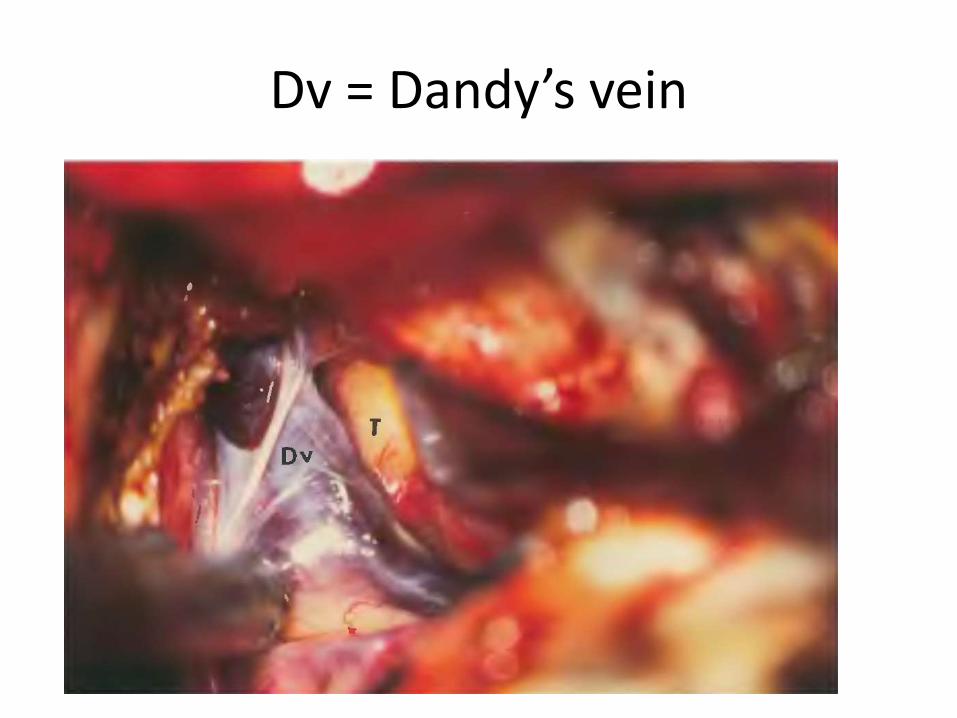
Dv = Dandy’s vein
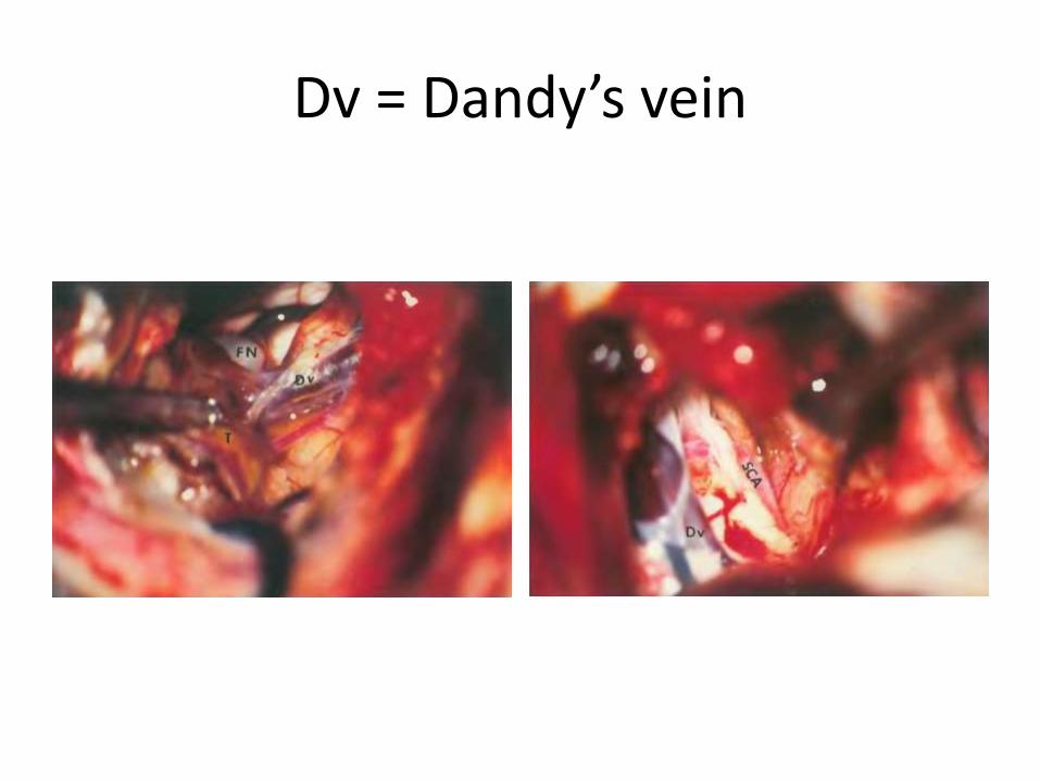
Dv = Dandy’s vein
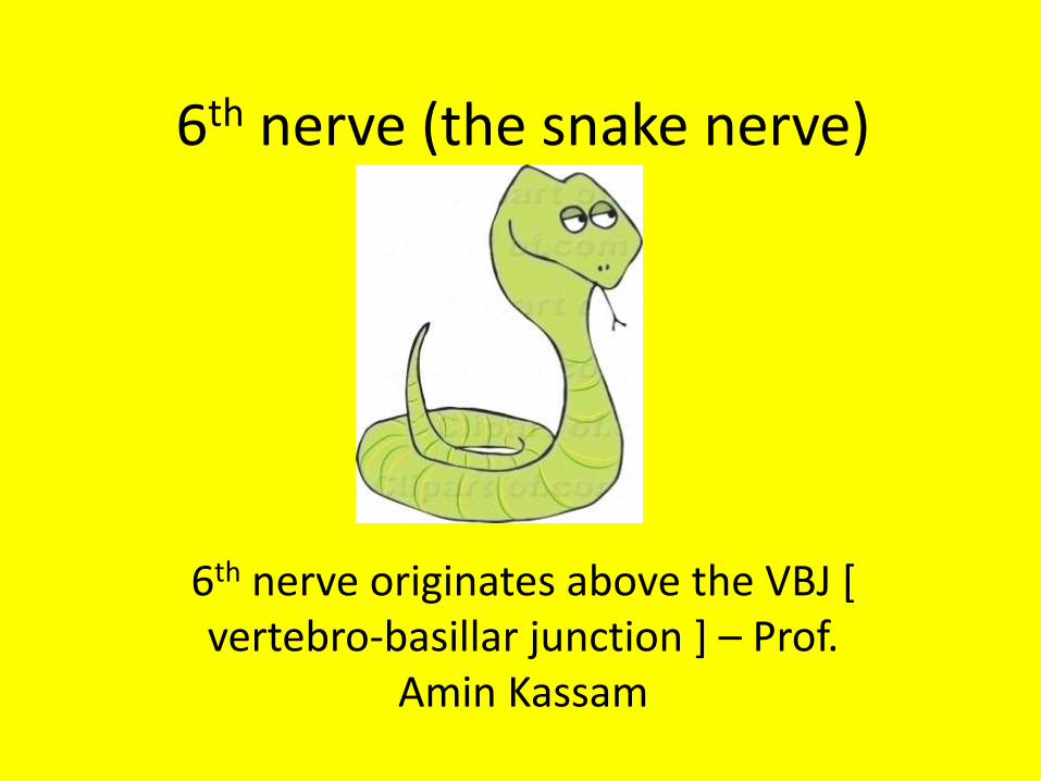
6th nerve (the snake nerve)
6th nerve originates above the VBJ [ vertebro-basillar junction ] – Prof.
Amin Kassam
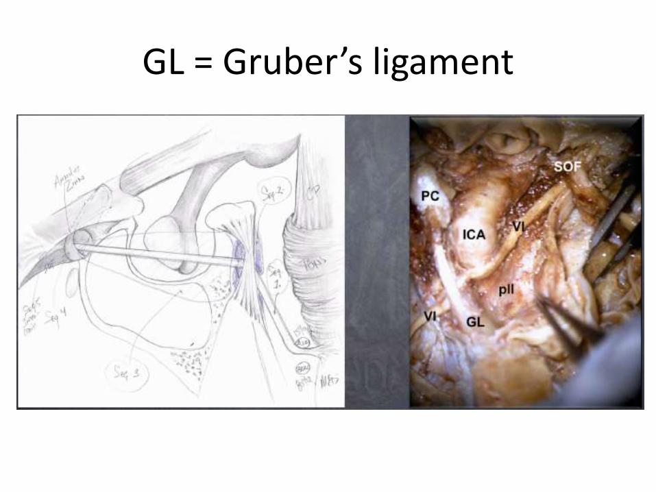
GL = Gruber’s ligament
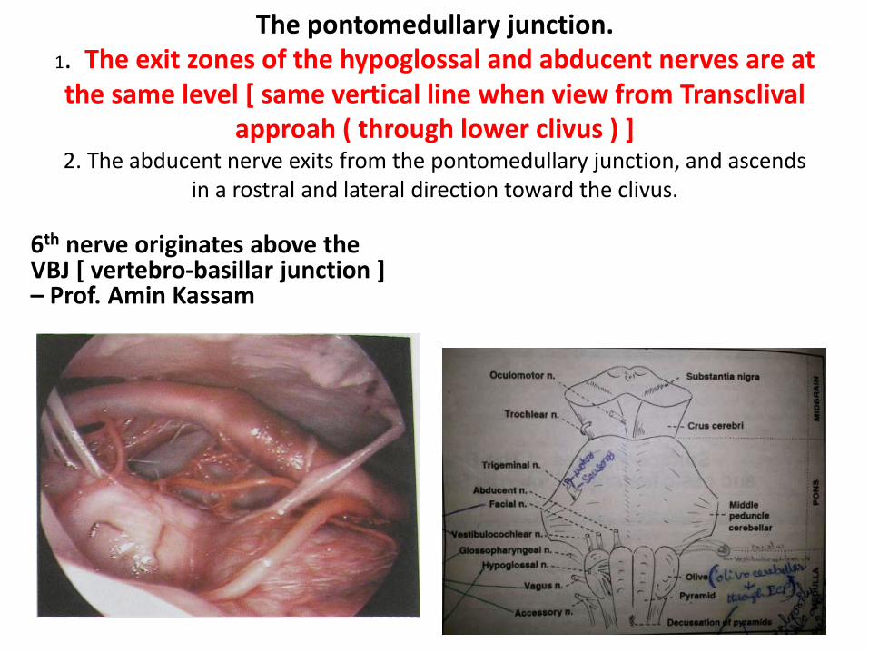
The pontomedullary junction.1. The exit zones of the hypoglossal and abducent nerves are at the same level [ same vertical line when view from Transclival
approah ( through lower clivus ) ] 2. The abducent nerve exits from the pontomedullary junction, and ascends
in a rostral and lateral direction toward the clivus.
6th nerve originates above the VBJ [ vertebro-basillar junction ] – Prof. Amin Kassam

6th nerve origin is above or below AICA or has two rootlets of origin
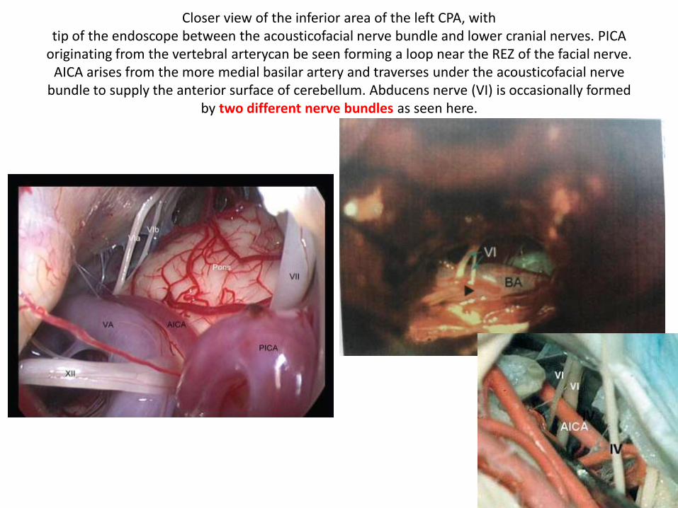
Closer view of the inferior area of the left CPA, withtip of the endoscope between the acousticofacial nerve bundle and lower cranial nerves. PICA
originating from the vertebral arterycan be seen forming a loop near the REZ of the facial nerve. AICA arises from the more medial basilar artery and traverses under the acousticofacial nerve
bundle to supply the anterior surface of cerebellum. Abducens nerve (VI) is occasionally formed by two different nerve bundles as seen here.
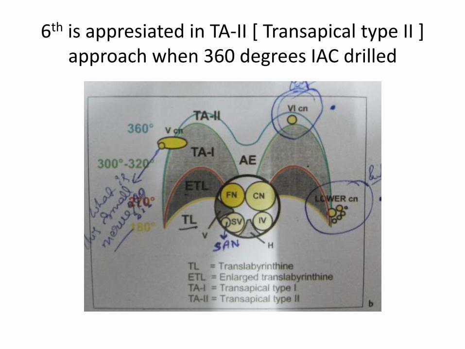
6th is appresiated in TA-II [ Transapical type II ] approach when 360 degrees IAC drilled

6th nerve – enters the dorellos canal –Intradural course
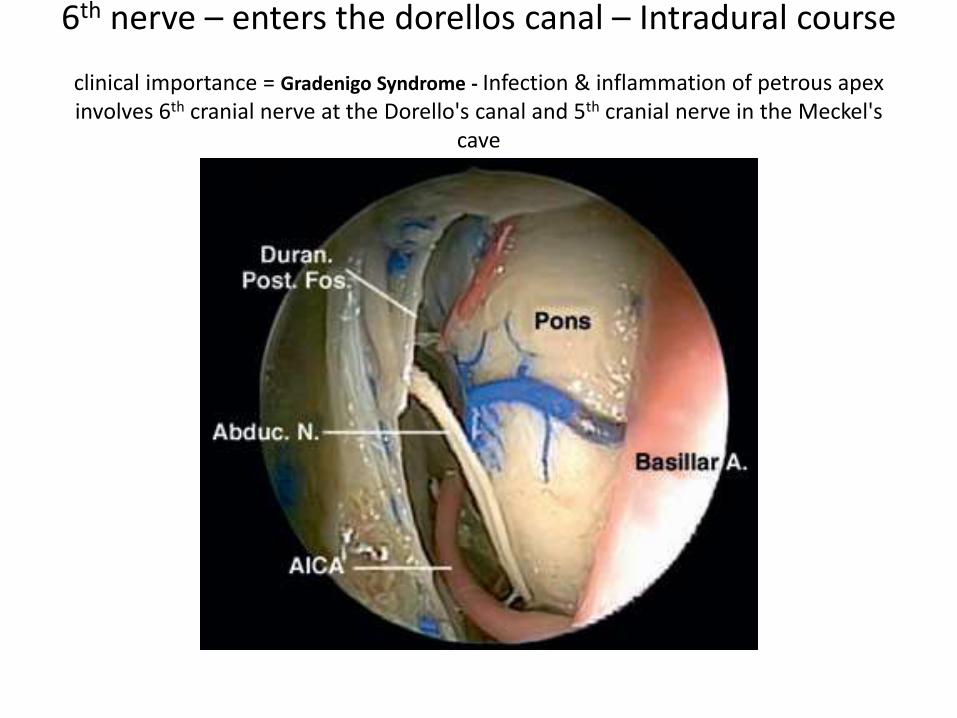
6th nerve – enters the dorellos canal – Intradural course
clinical importance = Gradenigo Syndrome - Infection & inflammation of petrous apex involves 6th cranial nerve at the Dorello's canal and 5th cranial nerve in the Meckel's
cave
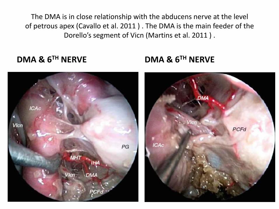
The DMA is in close relationship with the abducens nerve at the levelof petrous apex (Cavallo et al. 2011 ) . The DMA is the main feeder of the
Dorello’s segment of Vicn (Martins et al. 2011 ) .
DMA & 6TH NERVE DMA & 6TH NERVE
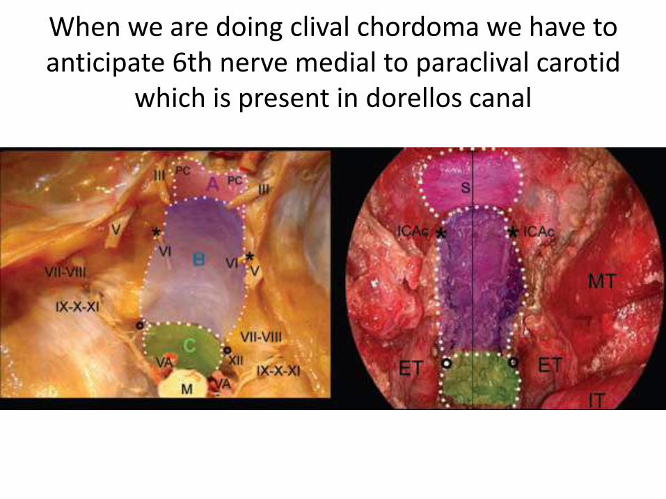
When we are doing clival chordoma we have to anticipate 6th nerve medial to paraclival carotid
which is present in dorellos canal

The basilar artery (BA) can be seenvery tortuous.
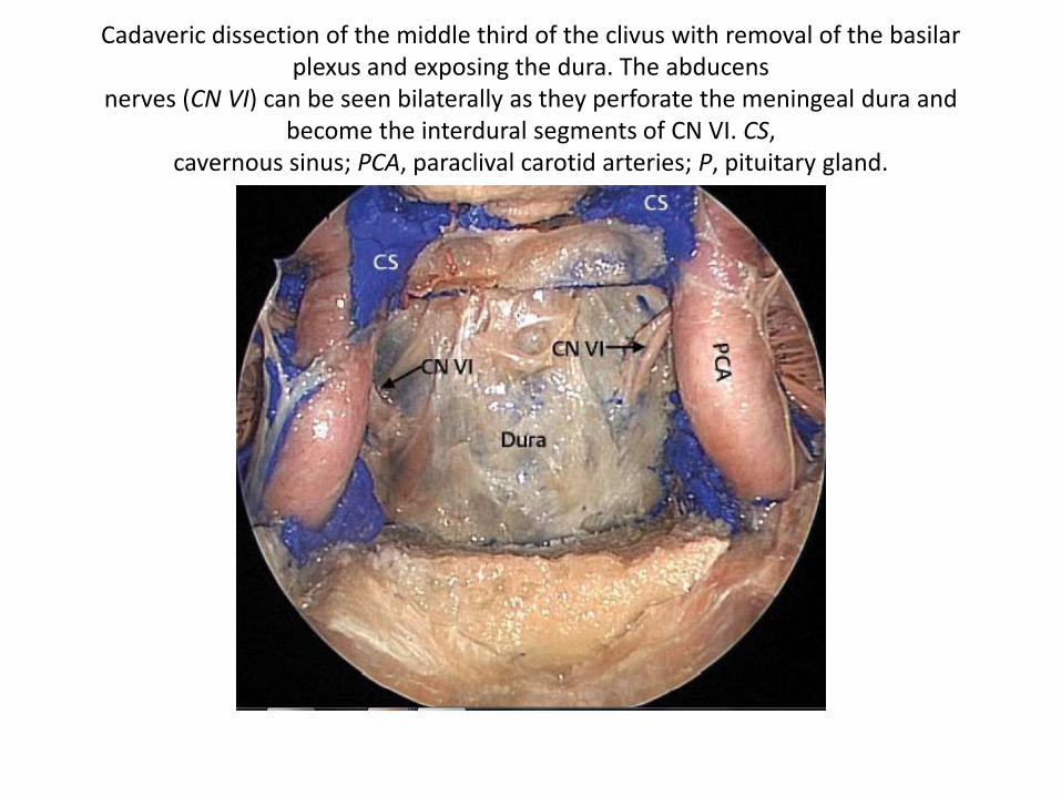
Cadaveric dissection of the middle third of the clivus with removal of the basilar plexus and exposing the dura. The abducens
nerves (CN VI) can be seen bilaterally as they perforate the meningeal dura and become the interdural segments of CN VI. CS,
cavernous sinus; PCA, paraclival carotid arteries; P, pituitary gland.

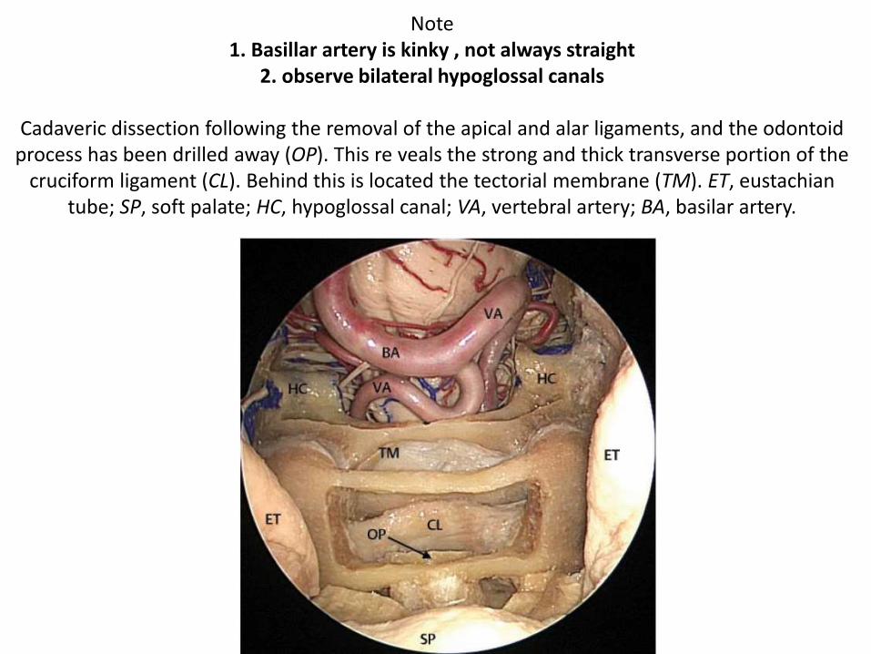
Note 1. Basillar artery is kinky , not always straight
2. observe bilateral hypoglossal canals
Cadaveric dissection following the removal of the apical and alar ligaments, and the odontoid process has been drilled away (OP). This re veals the strong and thick transverse portion of the
cruciform ligament (CL). Behind this is located the tectorial membrane (TM). ET, eustachiantube; SP, soft palate; HC, hypoglossal canal; VA, vertebral artery; BA, basilar artery.

Gulfar segment of 6th nerve (GS in left picture ) ( gVIcn in right picture ) - Thegulfar segment can be identified at the intersection of the sellar floor and the
proximal parasellar internal carotid artery (ICA) (Barges-Coll et al. 2010 ).
6th nerve enters dorello’s canal between the meningeal layer of dura and the periosteal layer of dura (POD).
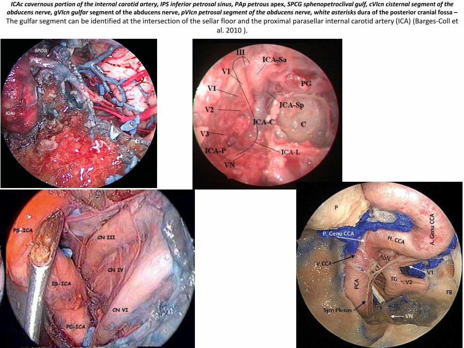
ICAc cavernous portion of the internal carotid artery, IPS inferior petrosal sinus, PAp petrous apex, SPCG sphenopetroclival gulf, cVIcn cisternal segment of the abducens nerve, gVIcn gulfar segment of the abducens nerve, pVIcn petrosal segment of the abducens nerve, white asterisks dura of the posterior cranial fossa –
The gulfar segment can be identified at the intersection of the sellar floor and the proximal parasellar internal carotid artery (ICA) (Barges-Coll et al. 2010 ).
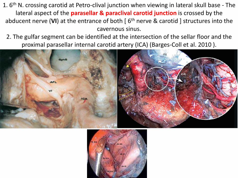
1. 6th N. crossing carotid at Petro-clival junction when viewing in lateral skull base - The lateral aspect of the parasellar & paraclival carotid junction is crossed by the
abducent nerve (VI) at the entrance of both [ 6th nerve & carotid ] structures into the cavernous sinus.
2. The gulfar segment can be identified at the intersection of the sellar floor and the proximal parasellar internal carotid artery (ICA) (Barges-Coll et al. 2010 ).
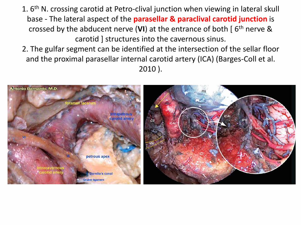
1. 6th N. crossing carotid at Petro-clival junction when viewing in lateral skull base - The lateral aspect of the parasellar & paraclival carotid junction is crossed by the abducent nerve (VI) at the entrance of both [ 6th nerve &
carotid ] structures into the cavernous sinus.2. The gulfar segment can be identified at the intersection of the sellar floor
and the proximal parasellar internal carotid artery (ICA) (Barges-Coll et al. 2010 ).
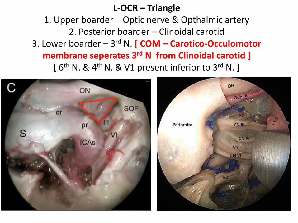
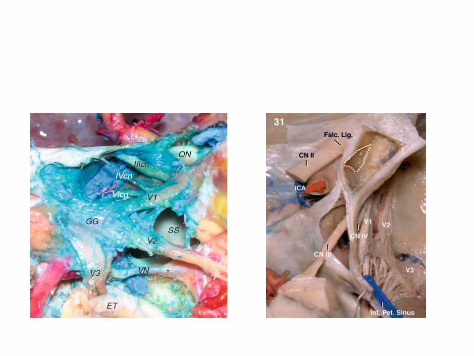
L-OCR – Triangle 1. Upper boarder – Optic nerve & Opthalmic artery
2. Posterior boarder – Clinoidal carotid 3. Lower boarder – 3rd N. [ COM – Carotico-Occulomotor
membrane seperates 3rd N from Clinoidal carotid ] [ 6th N. & 4th N. & V1 present inferior to 3rd N. ]
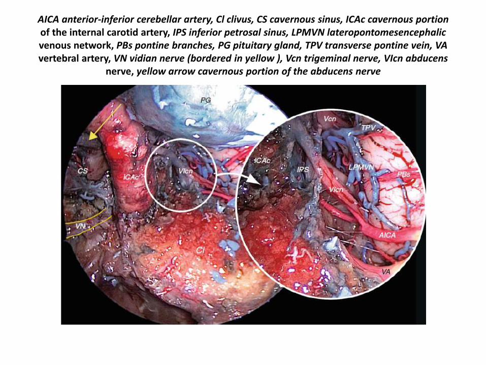
AICA anterior-inferior cerebellar artery, Cl clivus, CS cavernous sinus, ICAc cavernous portionof the internal carotid artery, IPS inferior petrosal sinus, LPMVN lateropontomesencephalicvenous network, PBs pontine branches, PG pituitary gland, TPV transverse pontine vein, VAvertebral artery, VN vidian nerve (bordered in yellow ), Vcn trigeminal nerve, VIcn abducens
nerve, yellow arrow cavernous portion of the abducens nerve
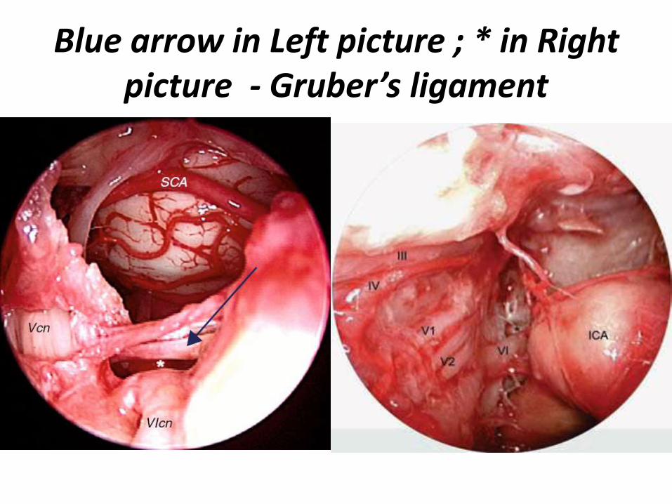
Blue arrow in Left picture ; * in Right picture - Gruber’s ligament

Usually, the IPS passes beneath the superior petro-sphenoidalligament (l. of Gruber) with the abducens nerve.
Anterior skull base Lateral skull base
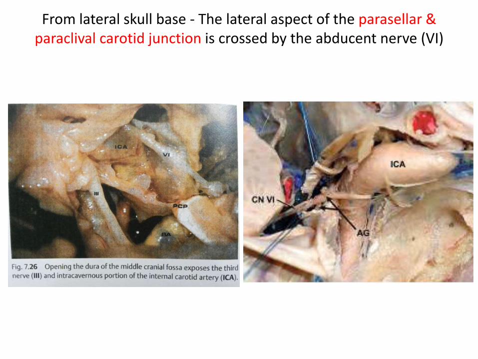
From lateral skull base - The lateral aspect of the parasellar & paraclival carotid junction is crossed by the abducent nerve (VI)
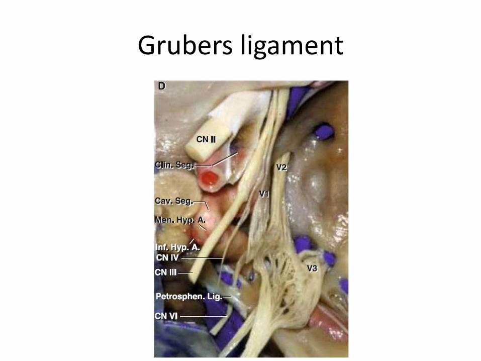
Grubers ligament
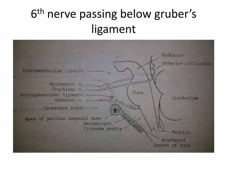
6th nerve passing below gruber’sligament

ACP anterior clinoid process, APCF anterior petroclinoid fold, DS dorsum sellae, ICF interclinoidfold, PF pituitary fossa, PLL petrolingual ligament (inferior sphenopetrosal ligament),
PPCF posterior petroclinoid fold, PS planum sphenoidale, SSPL superior sphenopetrosalligament (Gruber’s ligament), TS tuberculum sellae, black asterisk middle clinoid process
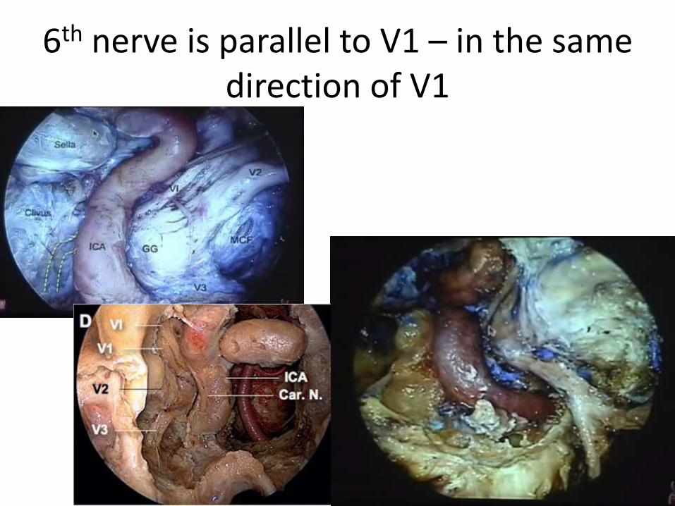
6th nerve is parallel to V1 – in the same direction of V1
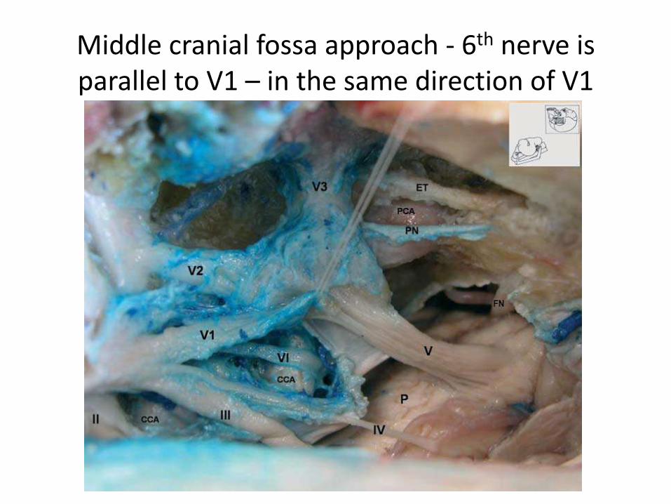
Middle cranial fossa approach - 6th nerve is parallel to V1 – in the same direction of V1
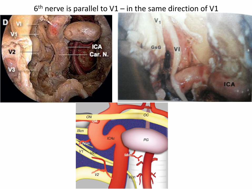
6th nerve is parallel to V1 – in the same direction of V1
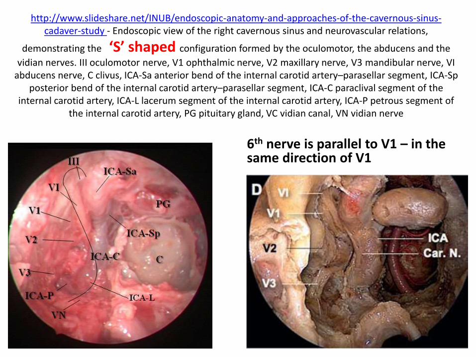
http://www.slideshare.net/INUB/endoscopic-anatomy-and-approaches-of-the-cavernous-sinus-cadaver-study - Endoscopic view of the right cavernous sinus and neurovascular relations,
demonstrating the ‘S’ shaped configuration formed by the oculomotor, the abducens and the
vidian nerves. III oculomotor nerve, V1 ophthalmic nerve, V2 maxillary nerve, V3 mandibular nerve, VI abducens nerve, C clivus, ICA-Sa anterior bend of the internal carotid artery–parasellar segment, ICA-Sp
posterior bend of the internal carotid artery–parasellar segment, ICA-C paraclival segment of the internal carotid artery, ICA-L lacerum segment of the internal carotid artery, ICA-P petrous segment of
the internal carotid artery, PG pituitary gland, VC vidian canal, VN vidian nerve
6th nerve is parallel to V1 – in the same direction of V1

Upper part of S-shaped configuration –3rd & 6th nerves.
6th nerve is freely hanging in the cavernous injury when compared to 3rd & 4th nerve – so postential for injury in
tumor dissection

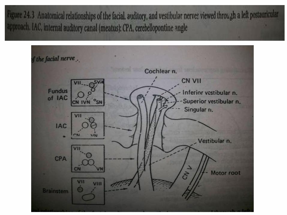
7th nerve
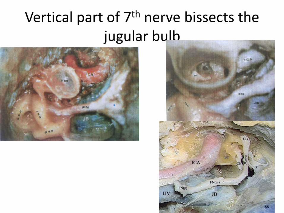
Vertical part of 7th nerve bissects the jugular bulb
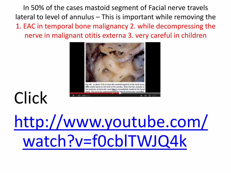
In 50% of the cases mastoid segment of Facial nerve travels lateral to level of annulus – This is important while removing the 1. EAC in temporal bone malignancy 2. while decompressing the
nerve in malignant otitis externa 3. very careful in children
Click
http://www.youtube.com/watch?v=f0cblTWJQ4k
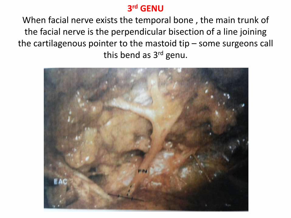
3rd GENU When facial nerve exists the temporal bone , the main trunk of the facial nerve is the perpendicular bisection of a line joining
the cartilagenous pointer to the mastoid tip – some surgeons call this bend as 3rd genu.
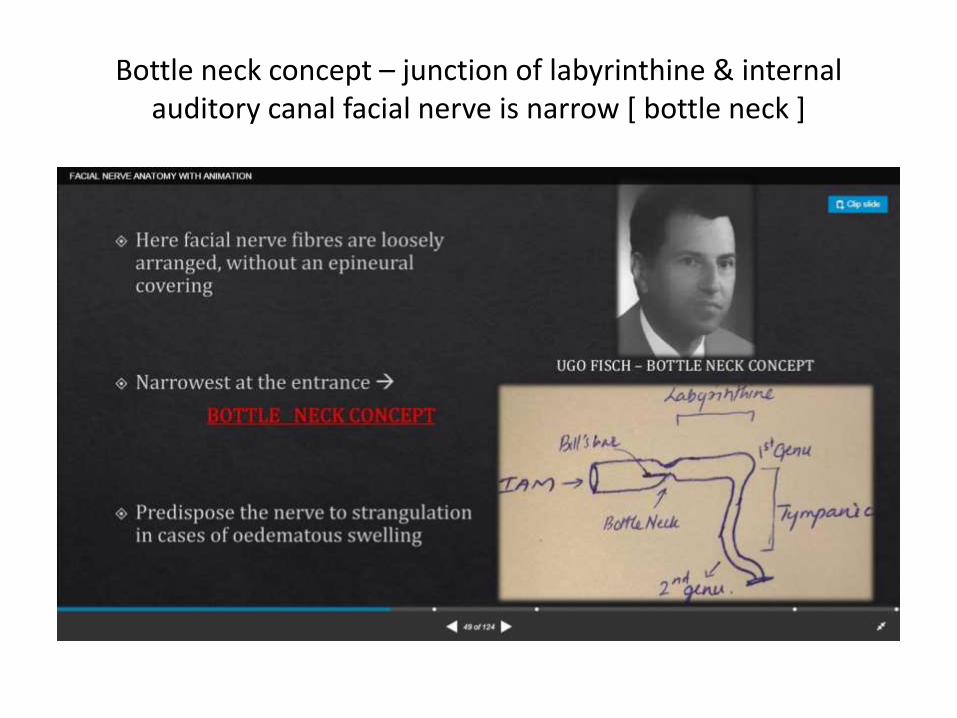
Bottle neck concept – junction of labyrinthine & internal auditory canal facial nerve is narrow [ bottle neck ]

7up- 7th is aboveCoca cola – cochlear n. is cola[=lower]

9th nerve

A closer view of the pars nervosa of the jugularforamen. The glossopharyngeal nerve has its own duralporus, which is situated 0-3 mm upwards from the duralporus of the tenth cranial nerve. The vagus and the accessorynerve exit the posterior fossa together in a sleeve of durathrough the jugular foramen.
Left side. The 30° angled endoscope provides anoverview of the inferior part of the CPA. On the right lies theacousticofacial nerve bundle, with the anterior inferior cerebellarartery; the glossopharyngeal nerve and the vagus nerve,as multiple filaments, form three to five major nerve bundlesand the accessory nerve.

Note the bone (>, <) left to protect the dura from the drill.AC Supralabyrinthine air cells, CA Cochlear aqueduct, FN Facial nerve,SA Ampulla of the superior canal, V Vestibule
Fig. 4.30 The internal auditory canal (IAC) has been identified, but the
overlying bone needs to be thinned further. CA Cochlear aqueduct,
FN Facial nerve, V Vestibule
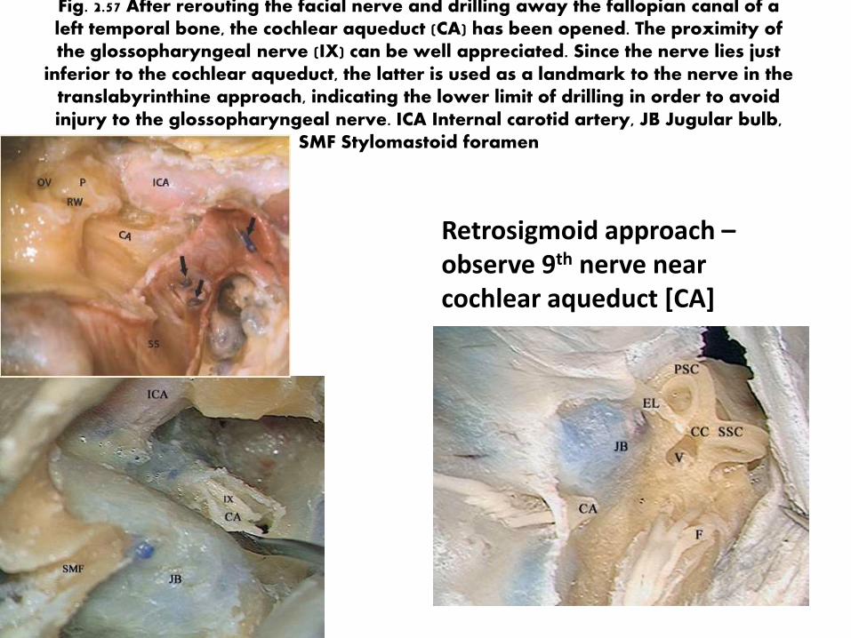
Fig. 2.57 After rerouting the facial nerve and drilling away the fallopian canal of a left temporal bone, the cochlear aqueduct (CA) has been opened. The proximity of the glossopharyngeal nerve (IX) can be well appreciated. Since the nerve lies just
inferior to the cochlear aqueduct, the latter is used as a landmark to the nerve in the translabyrinthine approach, indicating the lower limit of drilling in order to avoid injury to the glossopharyngeal nerve. ICA Internal carotid artery, JB Jugular bulb,
SMF Stylomastoid foramen
Retrosigmoid approach –observe 9th nerve near cochlear aqueduct [CA]
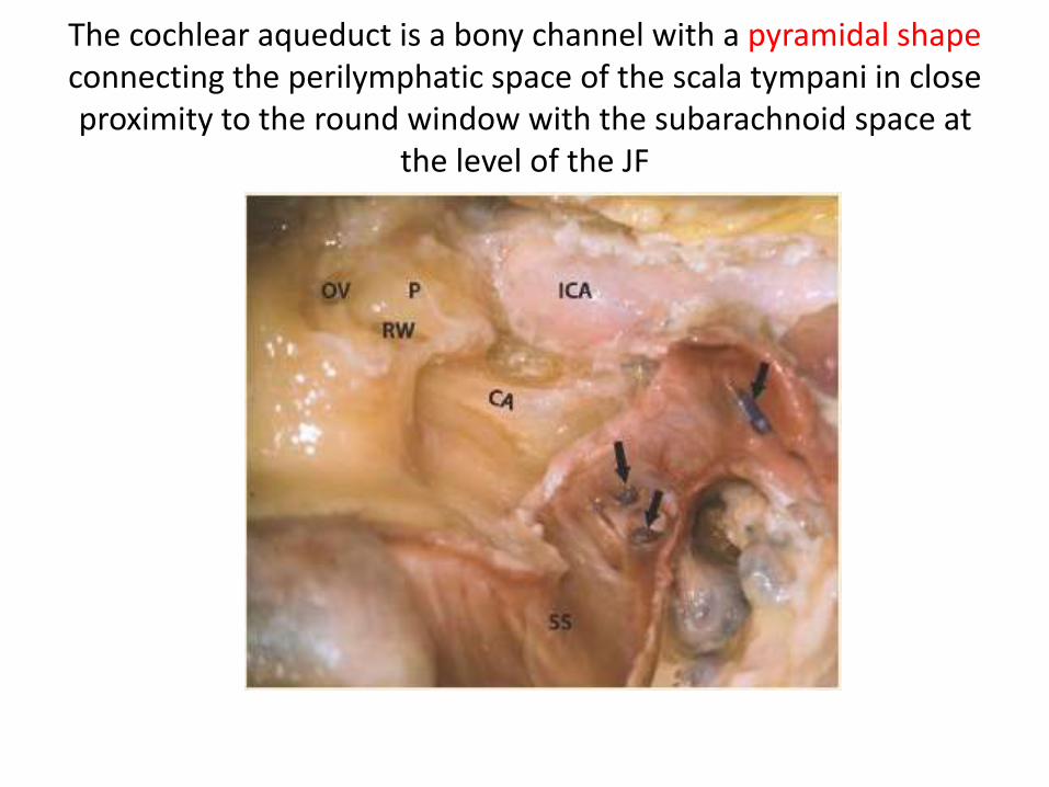
The cochlear aqueduct is a bony channel with a pyramidal shape connecting the perilymphatic space of the scala tympani in close proximity to the round window with the subarachnoid space at
the level of the JF
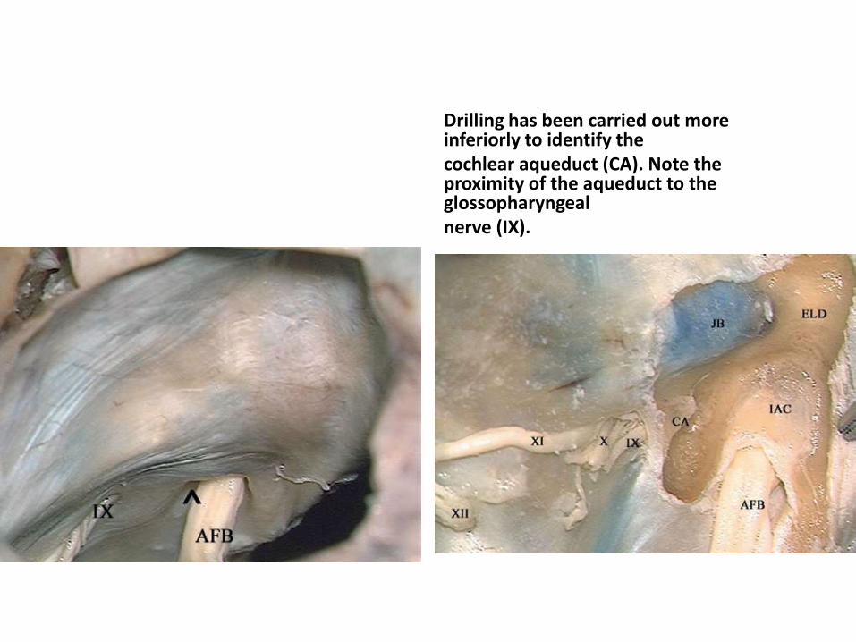
Drilling has been carried out more inferiorly to identify thecochlear aqueduct (CA). Note the proximity of the aqueduct to the glossopharyngealnerve (IX).
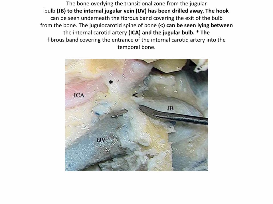
The bone overlying the transitional zone from the jugularbulb (JB) to the internal jugular vein (IJV) has been drilled away. The hook
can be seen underneath the fibrous band covering the exit of the bulbfrom the bone. The jugulocarotid spine of bone (<) can be seen lying between
the internal carotid artery (ICA) and the jugular bulb. * Thefibrous band covering the entrance of the internal carotid artery into the
temporal bone.
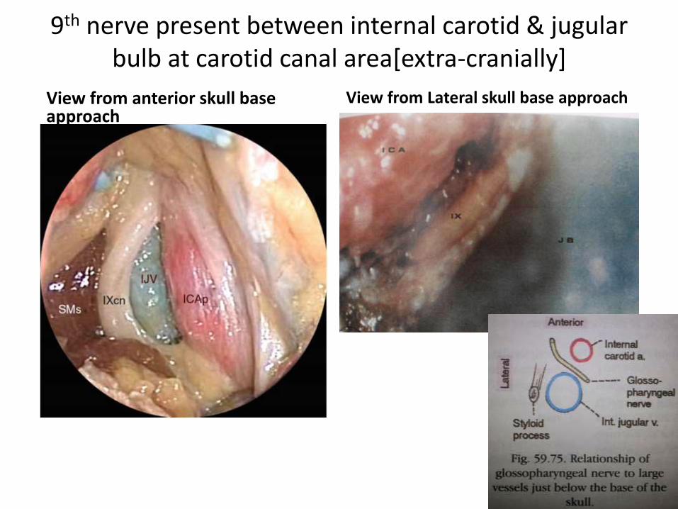
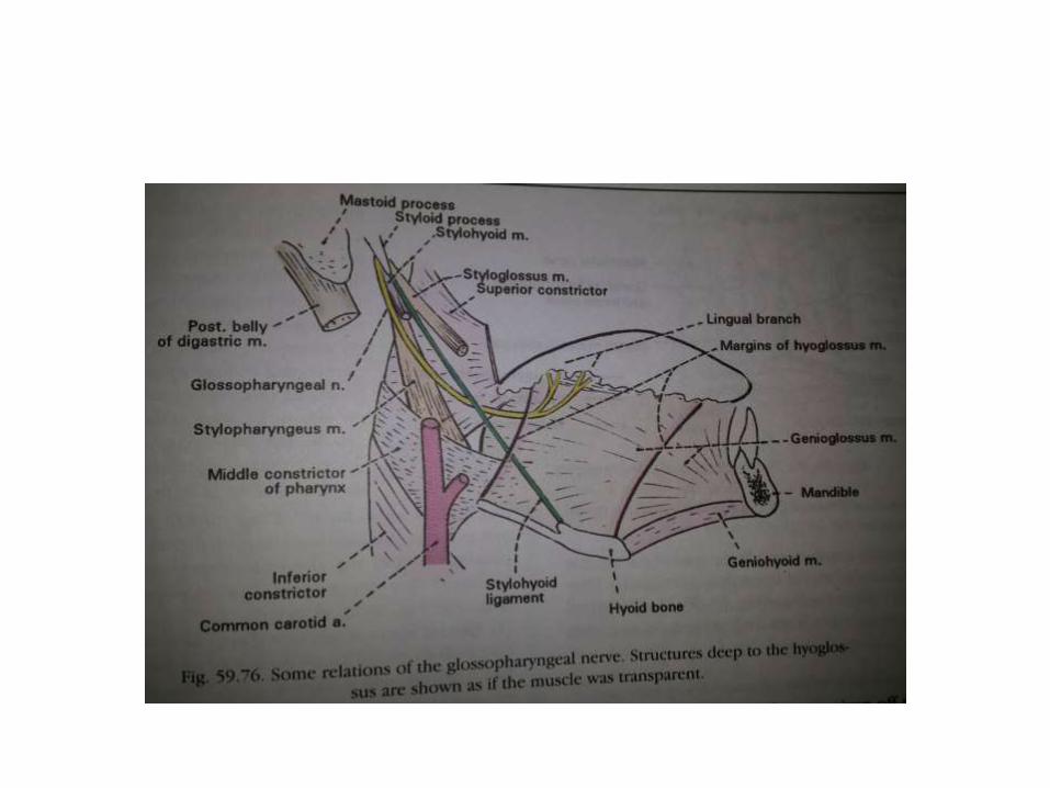
9th nerve present between internal carotid & jugular bulb at carotid canal area[extra-cranially]
View from anterior skull base approach
View from Lateral skull base approach

9th nerve – in cadaver
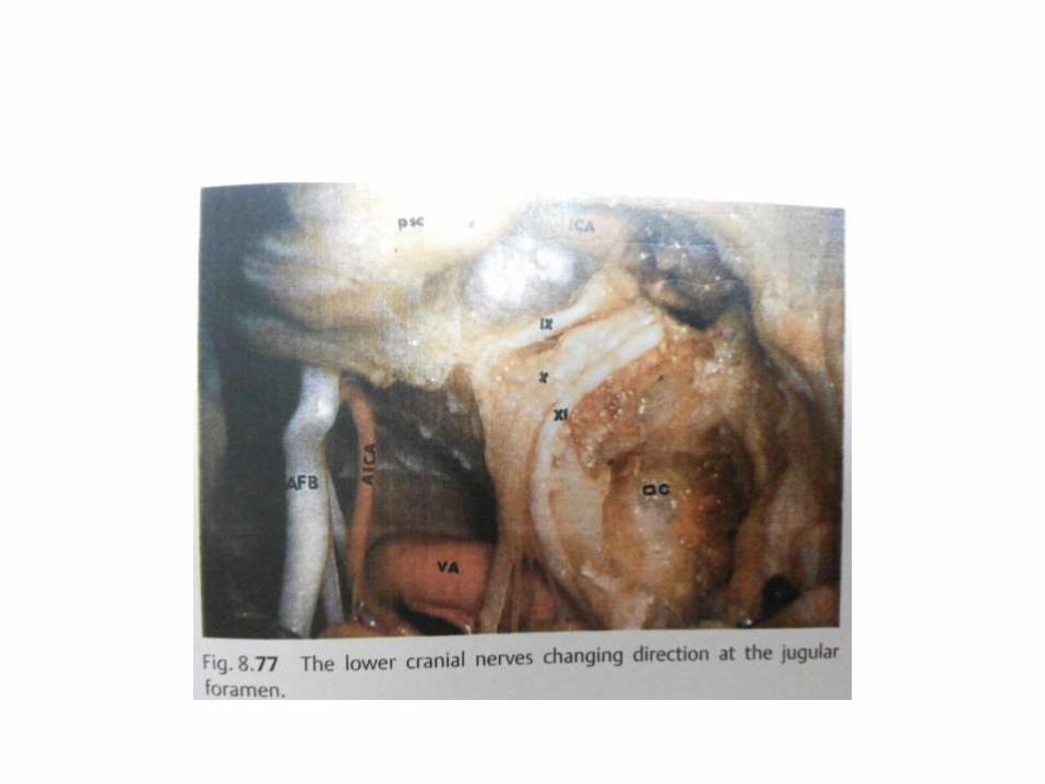
Jugular foramen area [ 9,10,11,12 nerves]
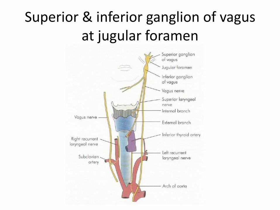
Superior & inferior ganglion of vagusat jugular foramen
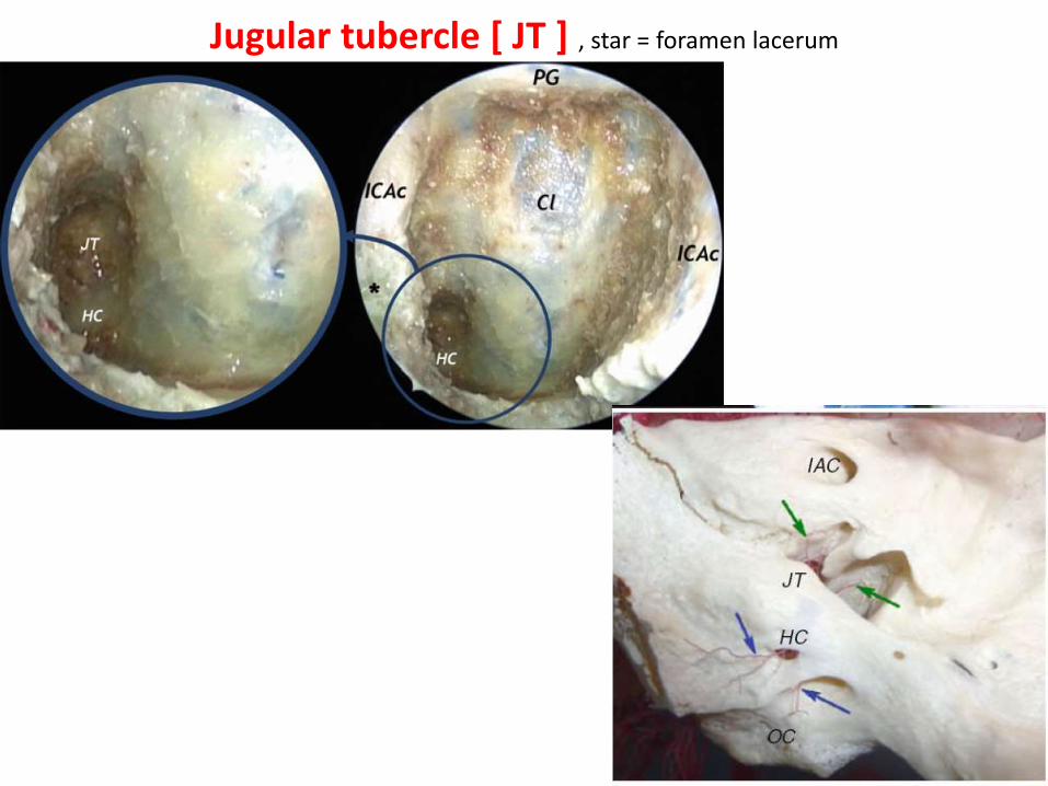
Jugular tubercle [ JT ] , star = foramen lacerum
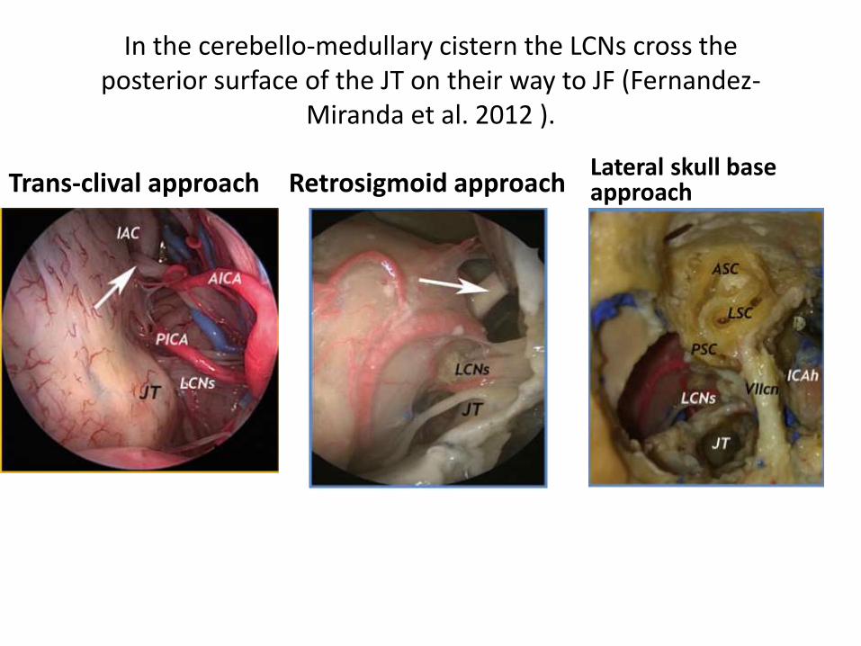
In the cerebello-medullary cistern the LCNs cross theposterior surface of the JT on their way to JF (Fernandez-
Miranda et al. 2012 ).
Trans-clival approach Retrosigmoid approach Lateral skull base approach
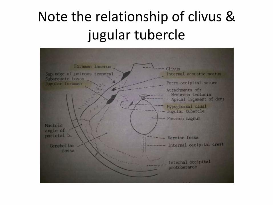
Note the relationship of clivus & jugular tubercle
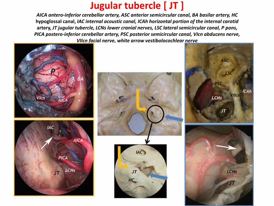
Jugular tubercle [ JT ] AICA antero-inferior cerebellar artery, ASC anterior semicircular canal, BA basilar artery, HChypoglossal canal, IAC internal acoustic canal, ICAh horizontal portion of the internal carotidartery, JT jugular tubercle, LCNs lower cranial nerves, LSC lateral semicircular canal, P pons,
PICA postero-inferior cerebellar artery, PSC posterior semicircular canal, VIcn abducens nerve,VIIcn facial nerve, white arrow vestibolocochlear nerve
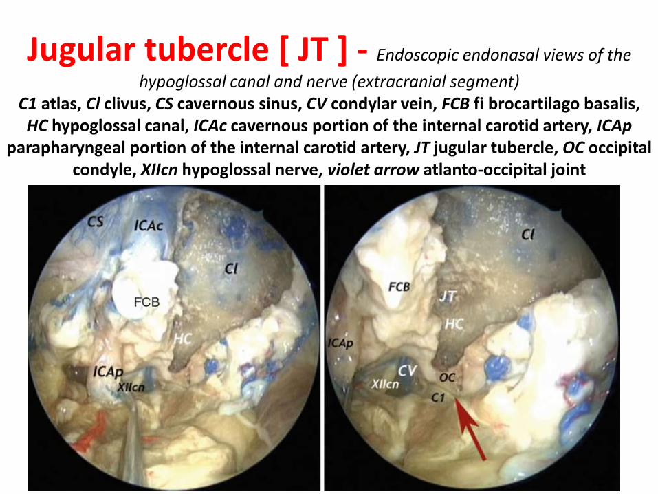
Jugular tubercle [ JT ] - Endoscopic endonasal views of the
hypoglossal canal and nerve (extracranial segment)C1 atlas, Cl clivus, CS cavernous sinus, CV condylar vein, FCB fi brocartilago basalis, HC hypoglossal canal, ICAc cavernous portion of the internal carotid artery, ICAp
parapharyngeal portion of the internal carotid artery, JT jugular tubercle, OC occipital condyle, XIIcn hypoglossal nerve, violet arrow atlanto-occipital joint
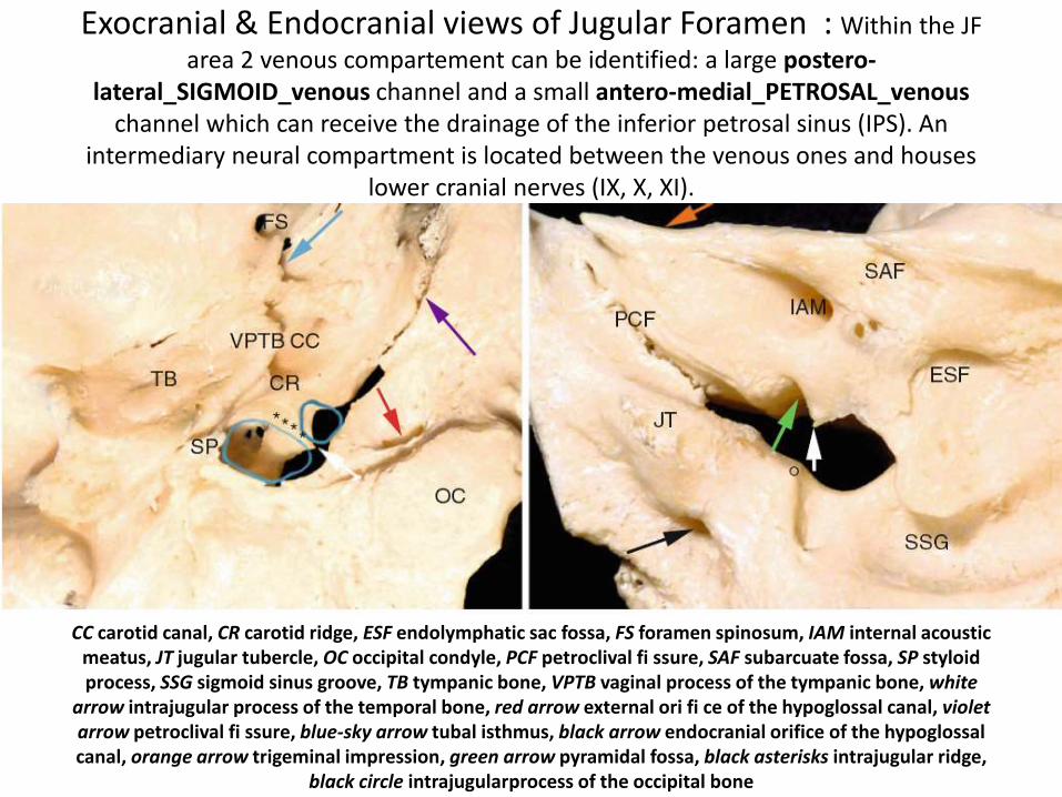
Exocranial & Endocranial views of Jugular Foramen : Within the JF
area 2 venous compartement can be identified: a large postero-lateral_SIGMOID_venous channel and a small antero-medial_PETROSAL_venous
channel which can receive the drainage of the inferior petrosal sinus (IPS). An intermediary neural compartment is located between the venous ones and houses
lower cranial nerves (IX, X, XI).
CC carotid canal, CR carotid ridge, ESF endolymphatic sac fossa, FS foramen spinosum, IAM internal acoustic meatus, JT jugular tubercle, OC occipital condyle, PCF petroclival fi ssure, SAF subarcuate fossa, SP styloidprocess, SSG sigmoid sinus groove, TB tympanic bone, VPTB vaginal process of the tympanic bone, white
arrow intrajugular process of the temporal bone, red arrow external ori fi ce of the hypoglossal canal, violet arrow petroclival fi ssure, blue-sky arrow tubal isthmus, black arrow endocranial orifice of the hypoglossal canal, orange arrow trigeminal impression, green arrow pyramidal fossa, black asterisks intrajugular ridge,
black circle intrajugularprocess of the occipital bone

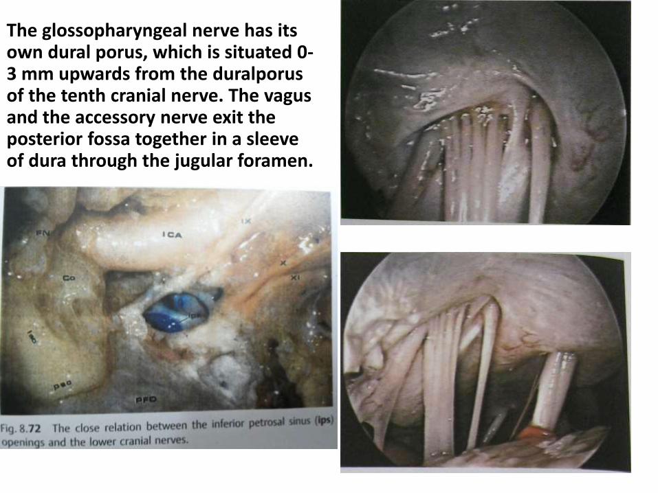
The glossopharyngeal nerve has its own dural porus, which is situated 0-3 mm upwards from the duralporusof the tenth cranial nerve. The vagusand the accessory nerve exit the posterior fossa together in a sleeve of dura through the jugular foramen.
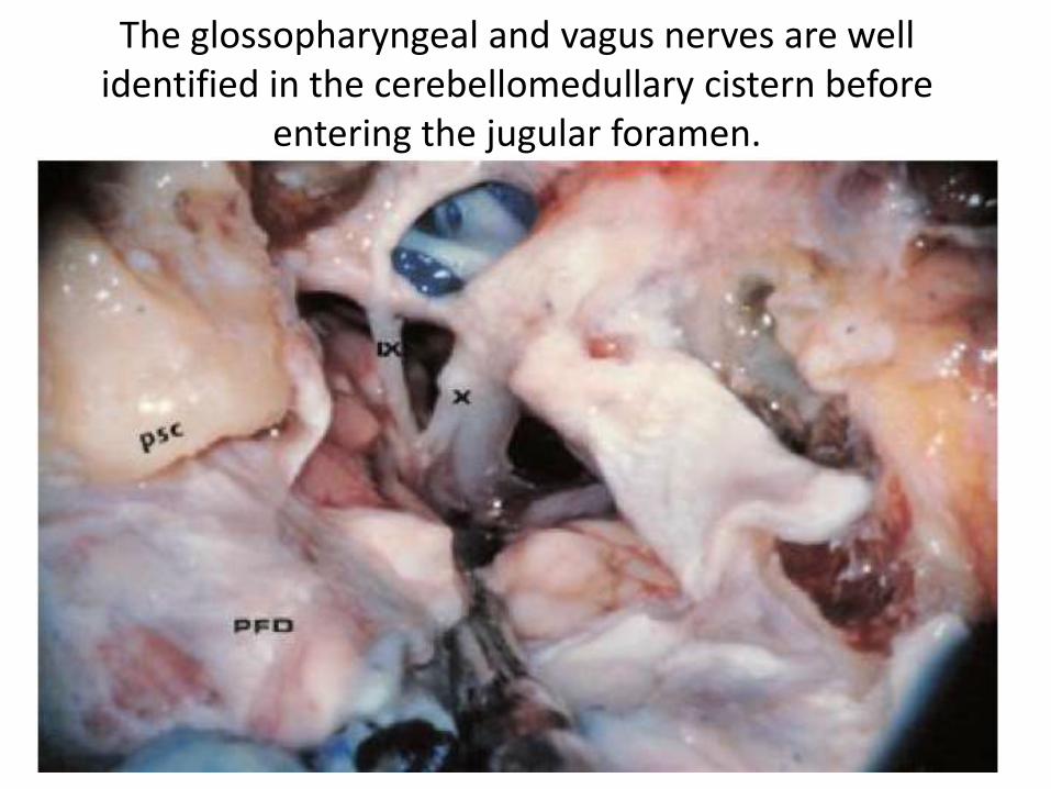
The glossopharyngeal and vagus nerves are well identified in the cerebellomedullary cistern before
entering the jugular foramen.

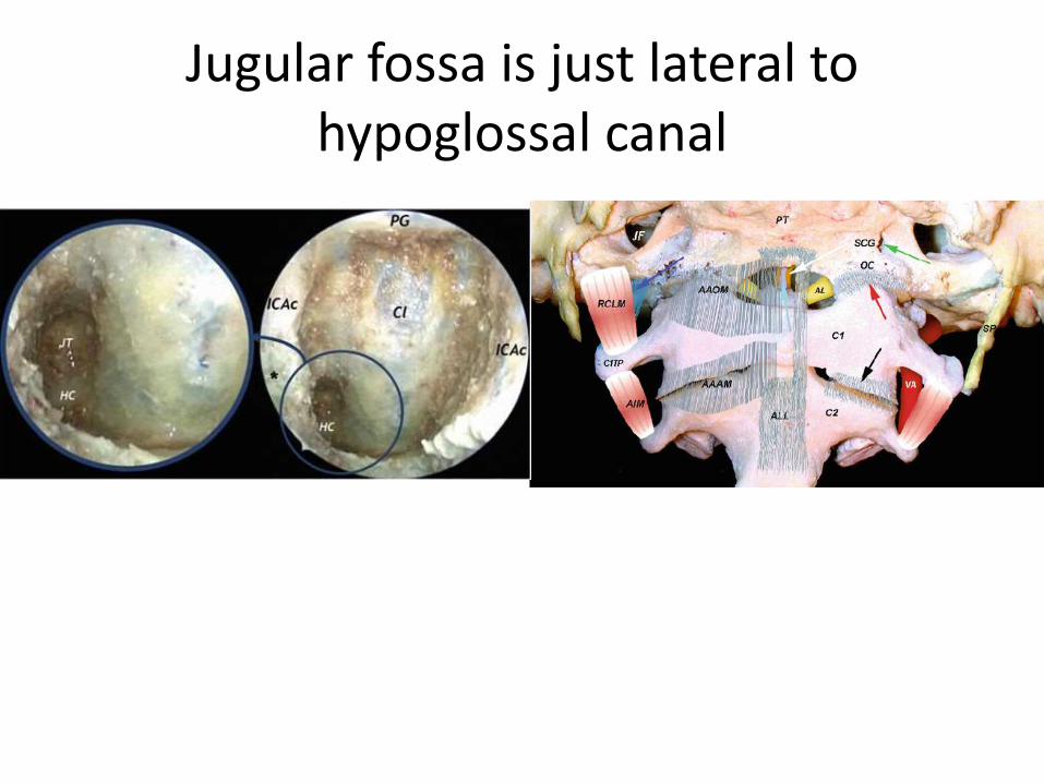
Jugular fossa is just lateral to hypoglossal canal

The jugular bulb lies beneath the fl oor of the middle ear cavity (Roche et al. 2008 ) . It can be of variable shape and size. All the lower cranial nerves ( LCNs ) exit the foramen anteromedially to the jugular bulb, separated from it by connective
tissue. The superior ganglion of the vagus nerve is within the jugular foramen ( JF ). At the level of the intraforaminal
course, there is a strict connection between the LCNs. The vagus nerve exits the JF vertically, behind IXcn and ICAp
(Roche et al. 2008 ) and gives its inferior ganglion on the outer skull base surface. The accessory nerve lies immediately lateral to the vagus nerve.
CR carotid ridge, DM digastric muscle (posterior belly), ICAp parapharyngeal portion of theinternal carotid artery, IJV internal jugular vein, JB jugular bulb, MMA middle meningeal
artery, VIIcn facial nerve, IX glossopharyngeal nerve, X vagus nerve, XI accessory nerve, XIIhypoglossal nerve, black arrow inferior ganglion of vagus nerve
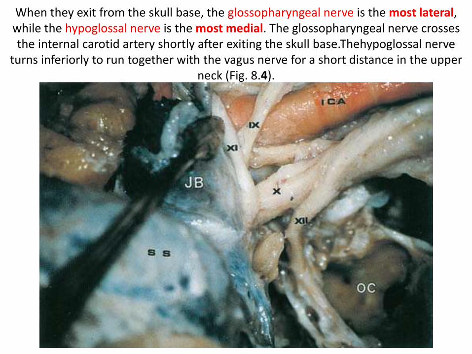
When they exit from the skull base, the glossopharyngeal nerve is the most lateral, while the hypoglossal nerve is the most medial. The glossopharyngeal nerve crosses the internal carotid artery shortly after exiting the skull base.Thehypoglossal nerve
turns inferiorly to run together with the vagus nerve for a short distance in the upper neck (Fig. 8.4).
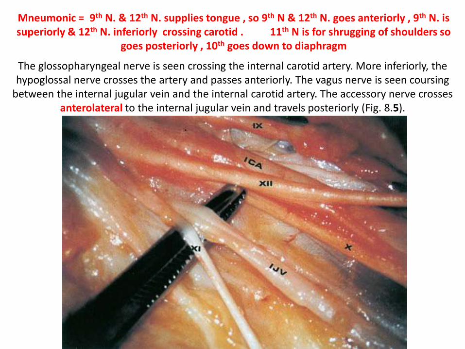
The glossopharyngeal nerve is seen crossing the internal carotid artery. More inferiorly, the hypoglossal nerve crosses the artery and passes anteriorly. The vagus nerve is seen coursing
between the internal jugular vein and the internal carotid artery. The accessory nerve crosses anterolateral to the internal jugular vein and travels posteriorly (Fig. 8.5).
Mneumonic = 9th N. & 12th N. supplies tongue , so 9th N & 12th N. goes anteriorly , 9th N. is superiorly & 12th N. inferiorly crossing carotid . 11th N is for shrugging of shoulders so
goes posteriorly , 10th goes down to diaphragm
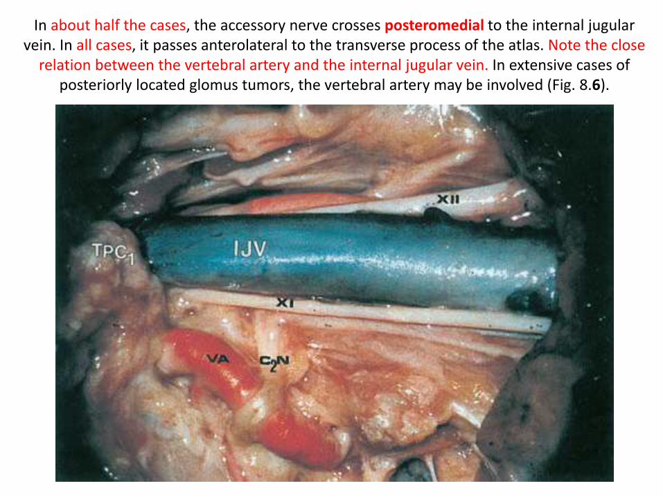
In about half the cases, the accessory nerve crosses posteromedial to the internal jugular vein. In all cases, it passes anterolateral to the transverse process of the atlas. Note the close
relation between the vertebral artery and the internal jugular vein. In extensive cases of posteriorly located glomus tumors, the vertebral artery may be involved (Fig. 8.6).
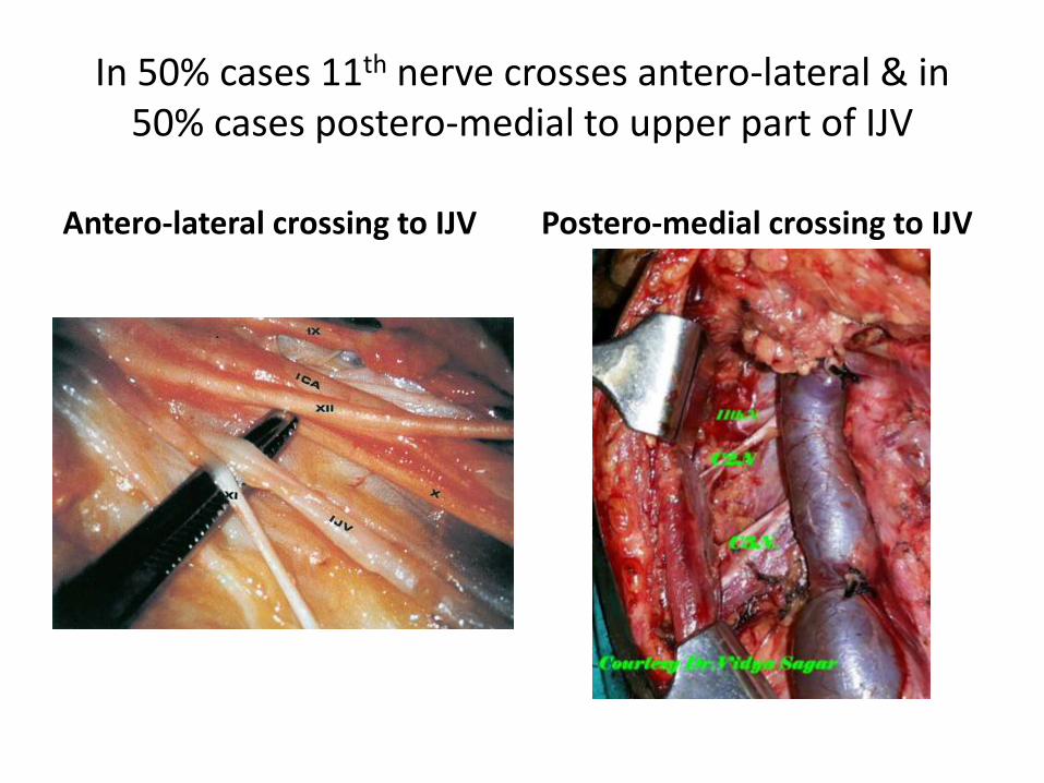
In 50% cases 11th nerve crosses antero-lateral & in 50% cases postero-medial to upper part of IJV
Antero-lateral crossing to IJV Postero-medial crossing to IJV
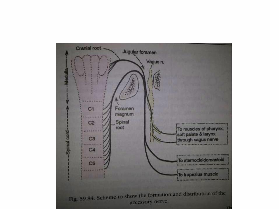

11th nerve
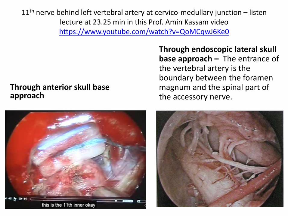
11th nerve behind left vertebral artery at cervico-medullary junction – listen lecture at 23.25 min in this Prof. Amin Kassam video https://www.youtube.com/watch?v=QoMCqwJ6Ke0
Through anterior skull base approach
Through endoscopic lateral skull base approach – The entrance of the vertebral artery is the boundary between the foramen magnum and the spinal part of the accessory nerve.
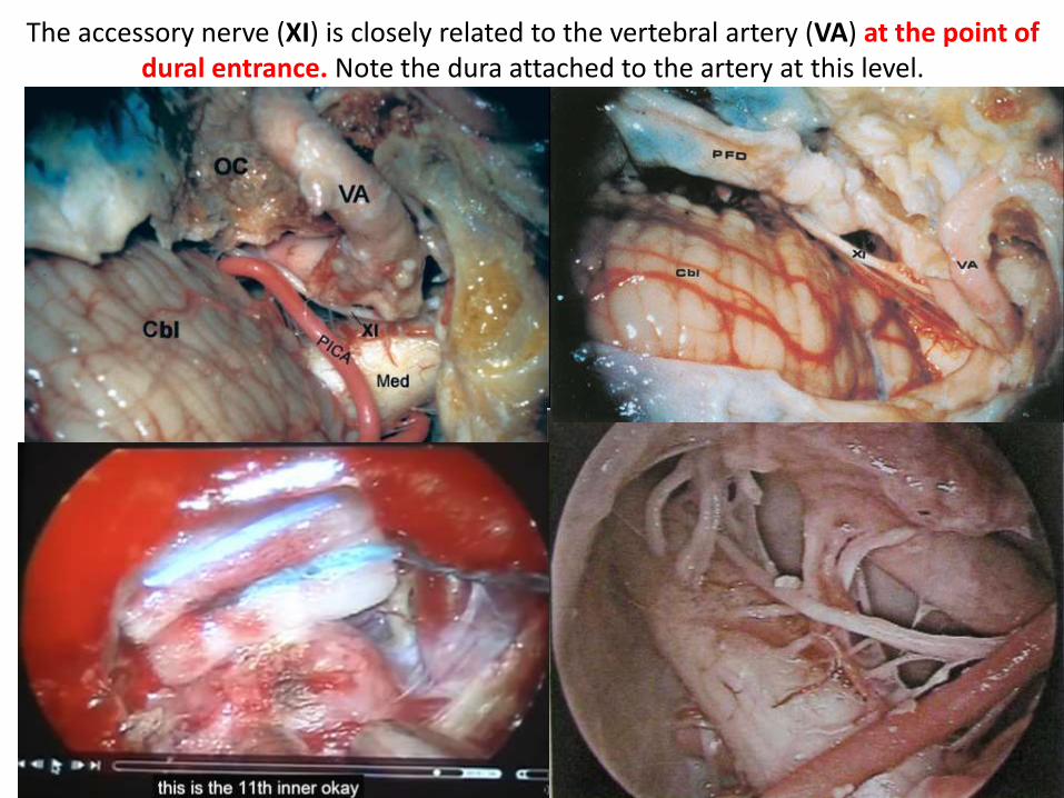
The accessory nerve (XI) is closely related to the vertebral artery (VA) at the point of dural entrance. Note the dura attached to the artery at this level.
Endoscopic lateral skull base approach
The accessory nerve (XI) is closely related to the vertebral artery (VA) at the point of dural entrance. Note the dura attached to the artery at this level.
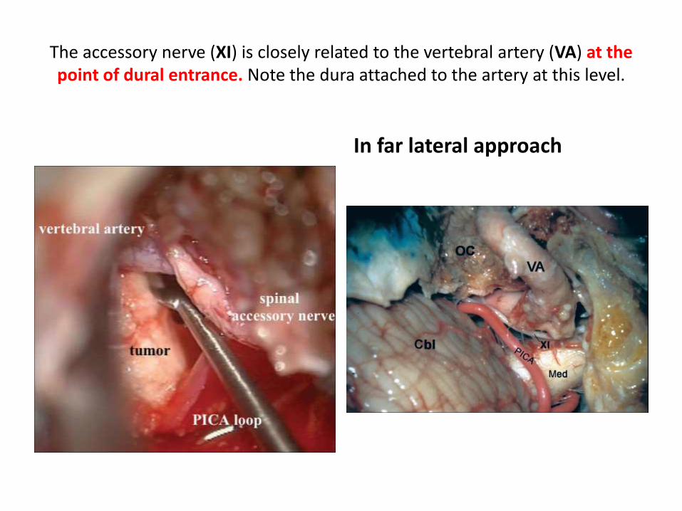
In far lateral approach
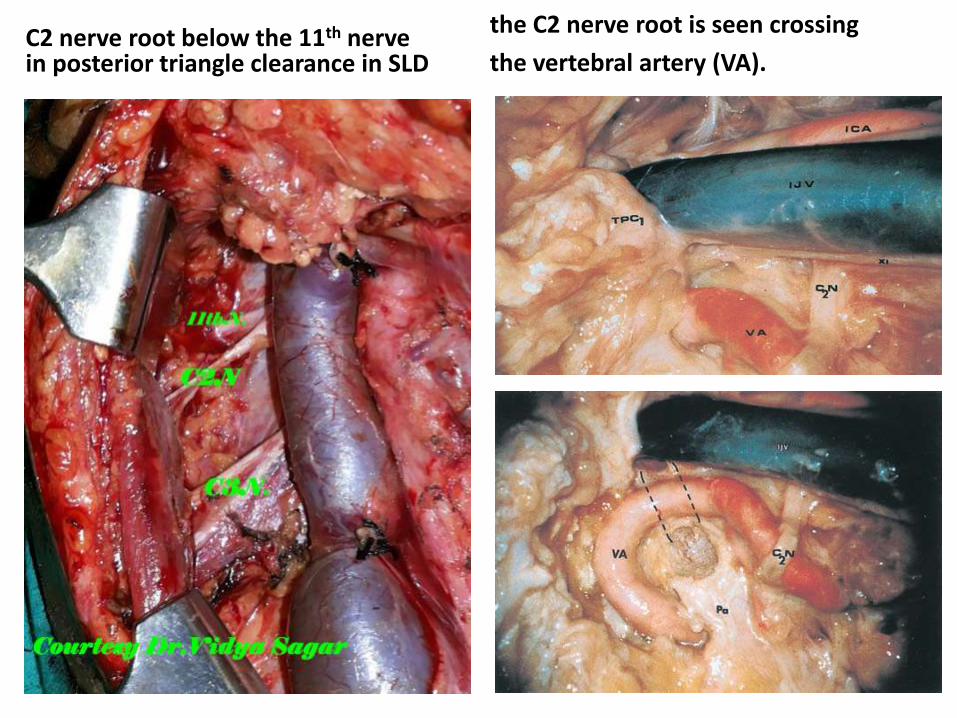
C2 nerve root below the 11th nerve in posterior triangle clearance in SLD
the C2 nerve root is seen crossing
the vertebral artery (VA).

In 50% cases 11th nerve crosses antero-lateral & in 50% cases postero-medial to upper part of IJV
Antero-lateral crossing to IJV Postero-medial crossing to IJV
12th nerve
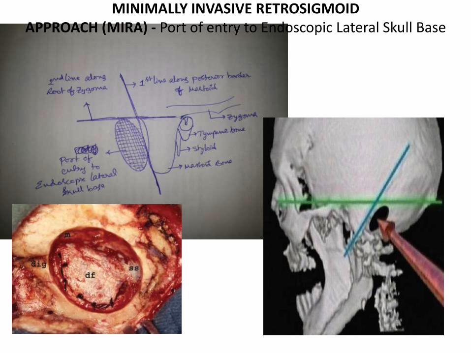
MINIMALLY INVASIVE RETROSIGMOIDAPPROACH (MIRA) - Port of entry to Endoscopic Lateral Skull Base

The pontomedullary junction.1. The exit zones of the hypoglossal and abducent nerves are at the same level [ same vertical line when view from Transclival
approah ( through lower clivus ) ] 2. The abducent nerve exits from the pontomedullary junction, and ascends
in a rostral and lateral direction toward the clivus.
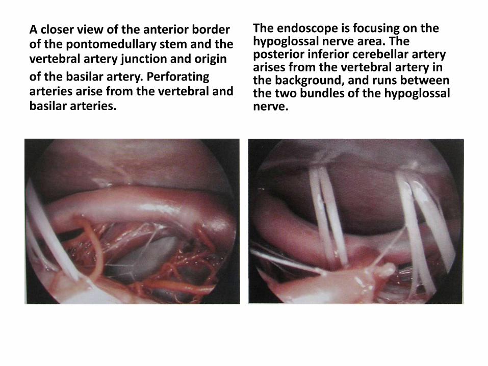
A closer view of the anterior border of the pontomedullary stem and the vertebral artery junction and origin
of the basilar artery. Perforating arteries arise from the vertebral and basilar arteries.
The endoscope is focusing on the hypoglossal nerve area. The posterior inferior cerebellar artery arises from the vertebral artery in the background, and runs between the two bundles of the hypoglossal nerve.
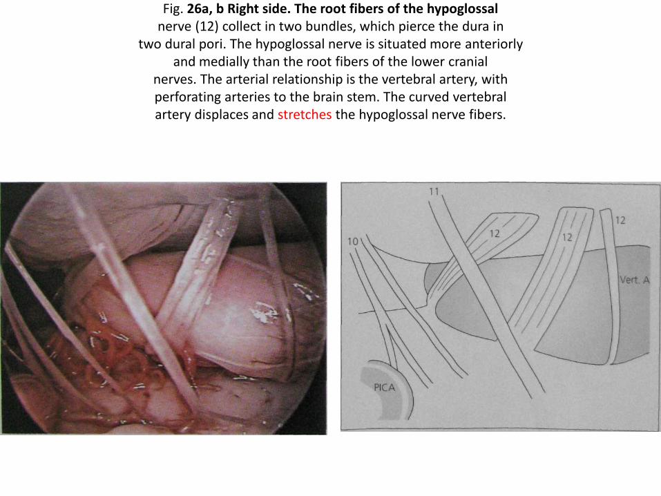
Fig. 26a, b Right side. The root fibers of the hypoglossalnerve (12) collect in two bundles, which pierce the dura in
two dural pori. The hypoglossal nerve is situated more anteriorlyand medially than the root fibers of the lower cranial
nerves. The arterial relationship is the vertebral artery, withperforating arteries to the brain stem. The curved vertebralartery displaces and stretches the hypoglossal nerve fibers.

90 degree turn of 12th nerve medial to medial wall of jugular bulb – Dr.Satish Jain

ITFA with Transcondylar [ = TC ] Transtubercular [ = TT ] approach
Here Transcondylar is through Occipital Condyle ;Transtubercular is through Jugular tubercle &
lateral pharyngeal tubercle
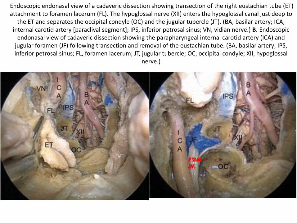
Endoscopic endonasal view of a cadaveric dissection showing transection of the right eustachian tube (ET) attachment to foramen lacerum (FL). The hypoglossal nerve (XII) enters the hypoglossal canal just deep to
the ET and separates the occipital condyle (OC) and the jugular tubercle (JT). (BA, basilar artery; ICA, internal carotid artery [paraclival segment]; IPS, inferior petrosal sinus; VN, vidian nerve.) B. Endoscopic
endonasal view of cadaveric dissection showing the parapharyngeal internal carotid artery (ICA) and jugular foramen (JF) following transection and removal of the eustachian tube. (BA, basilar artery; IPS, inferior petrosal sinus; FL, foramen lacerum; JT, jugular tubercle; OC, occipital condyle; XII, hypoglossal
nerve.)

Note 12th nerve in between JT ( Jugular tubercle ) & OC ( Occipital condyle ) in both lateral & anterior skull base
Lateral skull base Anterior skull base

The accessory nerve (XI) is closely related to the vertebral artery (VA) at the point of dural entrance. Note the dura attached to the artery at this level.
Endoscopic lateral skull base approach

Through endoscopic lateral skull base - The curved vertebral artery displaces and stretchesthe hypoglossal nerve fibers.
Through anterior skull base
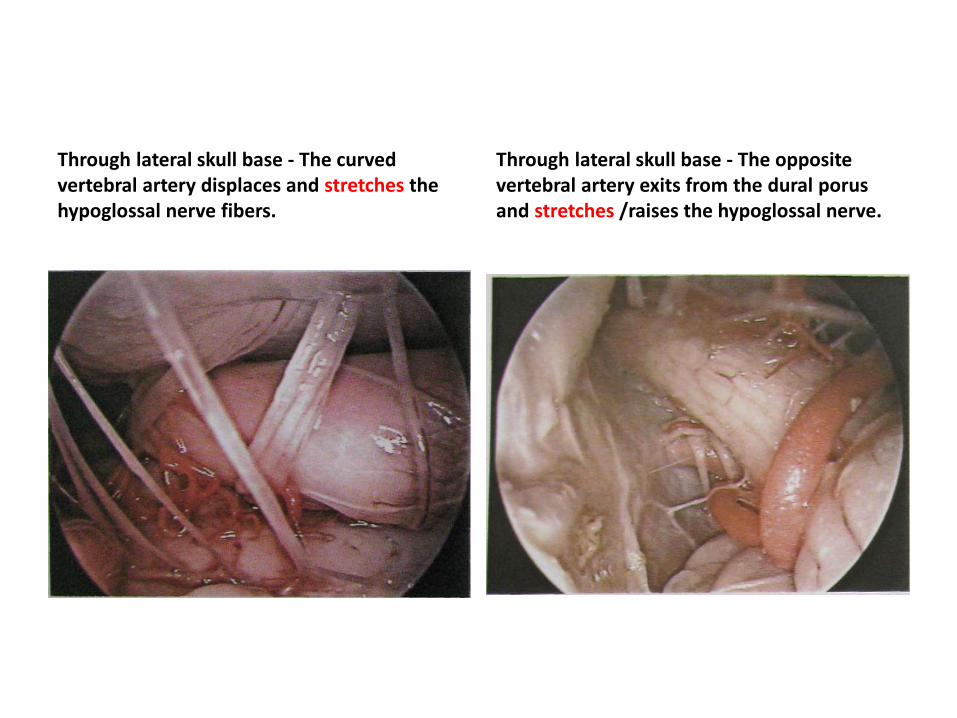
Through lateral skull base - The curved vertebral artery displaces and stretches the hypoglossal nerve fibers.
Through lateral skull base - The opposite vertebral artery exits from the dural porusand stretches /raises the hypoglossal nerve.
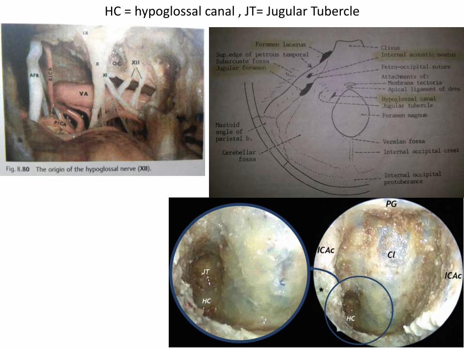
HC = hypoglossal canal , JT= Jugular Tubercle

SCG = Supracondylar groove – is an important landmark to hypoglossal
canal
Jugular fossa is just lateral to hypoglossal canal
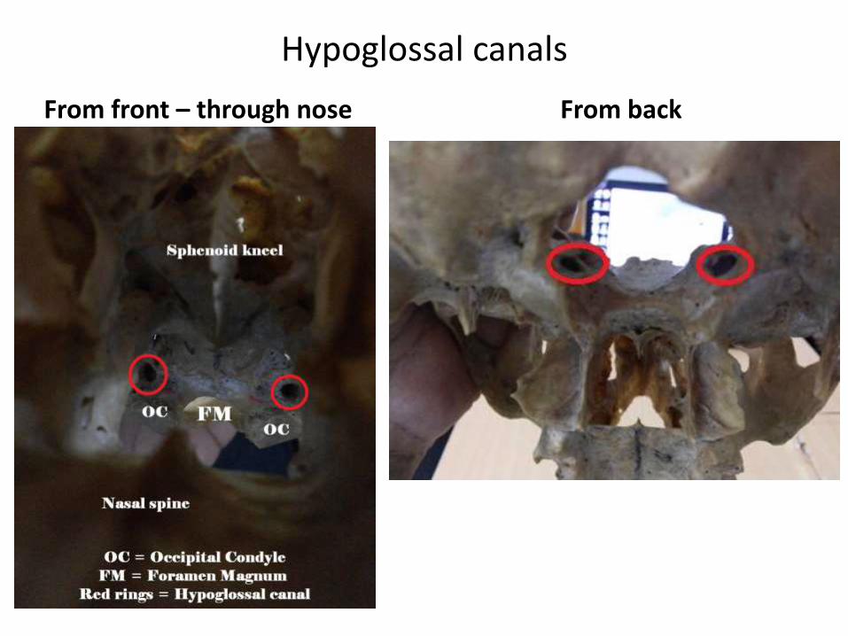
Hypoglossal canals
From front – through nose From back
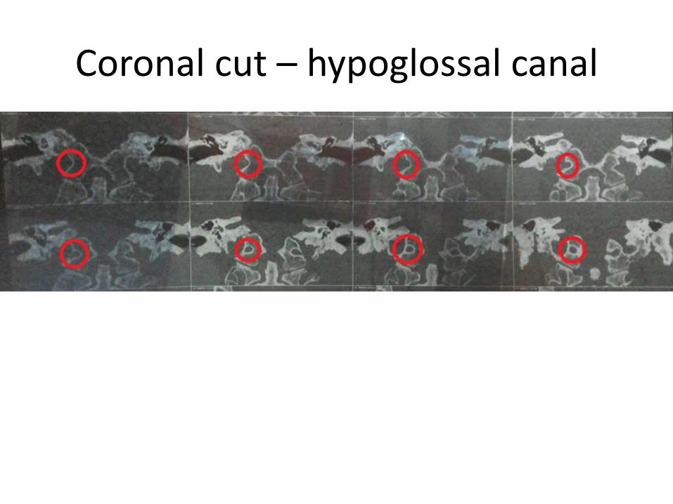
Coronal cut – hypoglossal canal
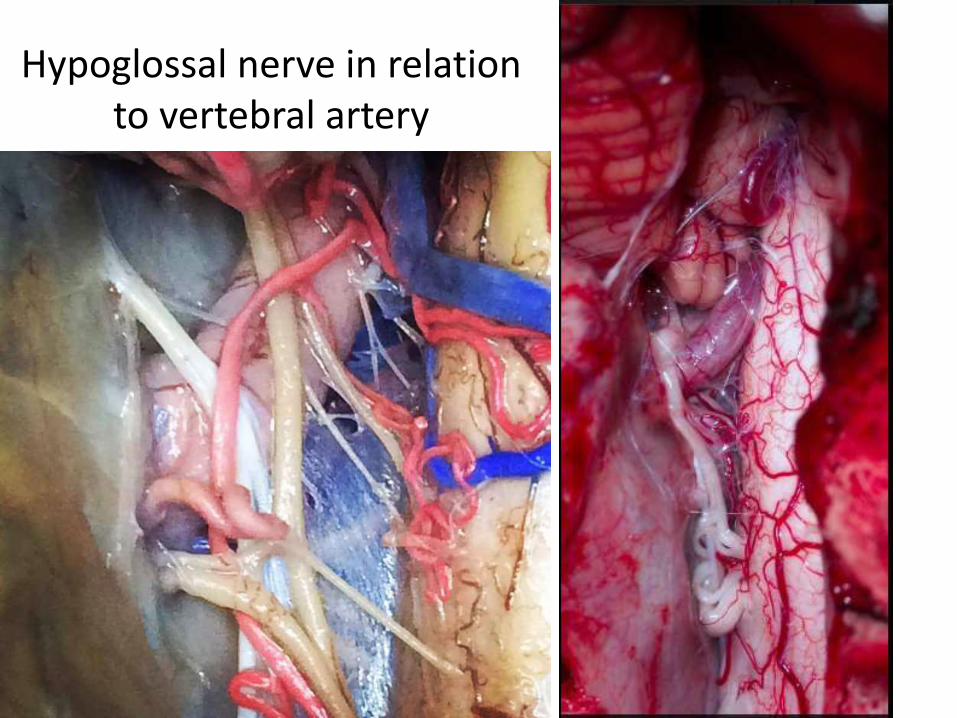
Hypoglossal nerve in relation to vertebral artery
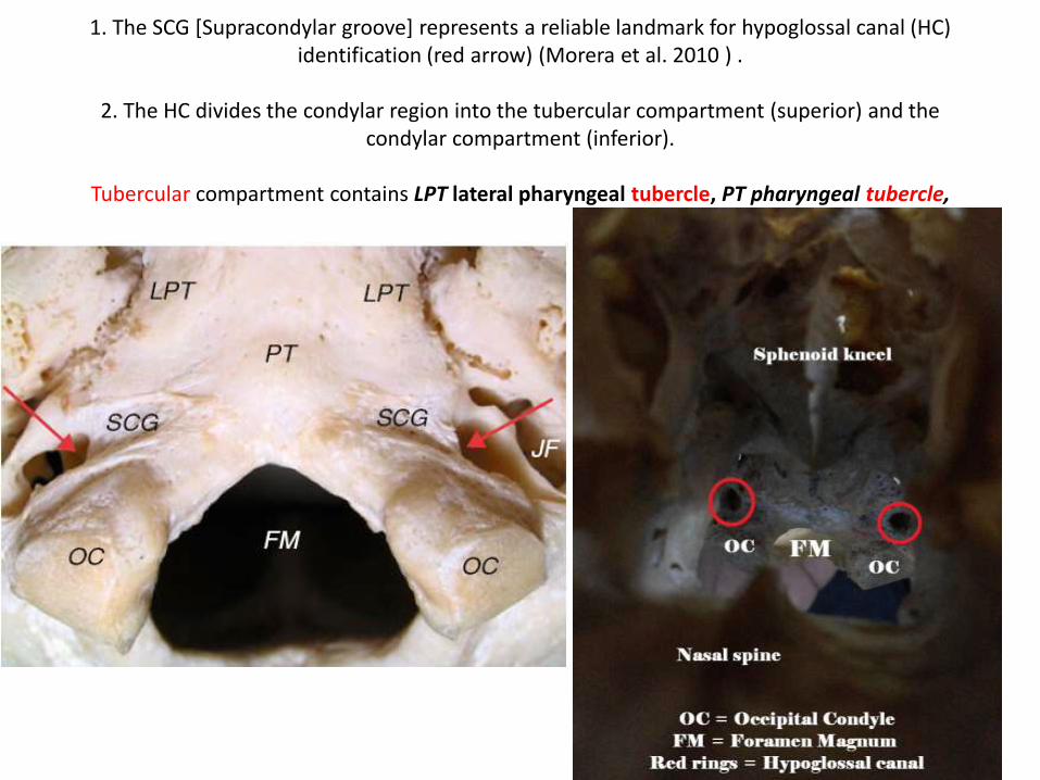
1. The SCG [Supracondylar groove] represents a reliable landmark for hypoglossal canal (HC) identification (red arrow) (Morera et al. 2010 ) .
2. The HC divides the condylar region into the tubercular compartment (superior) and the condylar compartment (inferior).
Tubercular compartment contains LPT lateral pharyngeal tubercle, PT pharyngeal tubercle,
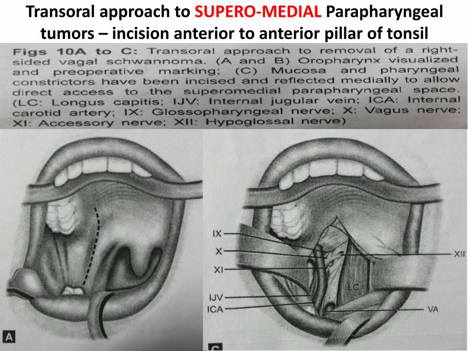
Transoral approach to SUPERO-MEDIAL Parapharyngealtumors – incision anterior to anterior pillar of tonsil
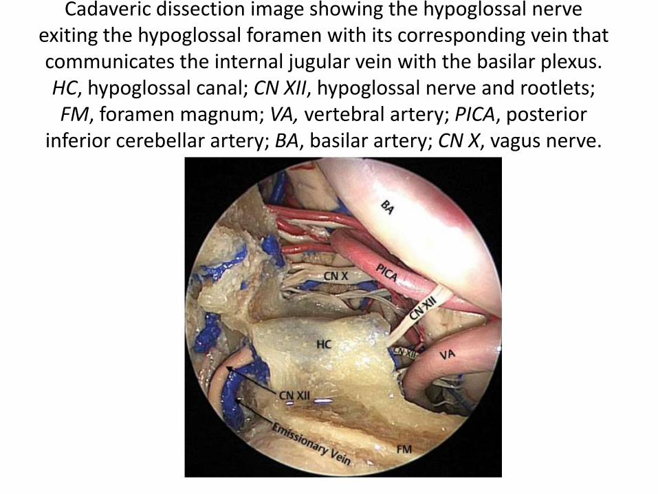
Cadaveric dissection image showing the hypoglossal nerve exiting the hypoglossal foramen with its corresponding vein that communicates the internal jugular vein with the basilar plexus. HC, hypoglossal canal; CN XII, hypoglossal nerve and rootlets; FM, foramen magnum; VA, vertebral artery; PICA, posterior
inferior cerebellar artery; BA, basilar artery; CN X, vagus nerve.
Note 1. Basillar artery is kinky , not always straight
2. observe bilateral hypoglossal canals
Cadaveric dissection following the removal of the apical and alar ligaments, and the odontoid process has been drilled away (OP). This re veals the strong and thick transverse portion of the
cruciform ligament (CL). Behind this is located the tectorial membrane (TM). ET, eustachiantube; SP, soft palate; HC, hypoglossal canal; VA, vertebral artery; BA, basilar artery.

IPS & HVP hypoglossalvenous plexus
Cadaveric dissection image showing the hypoglossal nerve exiting the hypoglossal foramen with its corresponding vein thatcommunicates the internal jugular vein with the basilar plexus
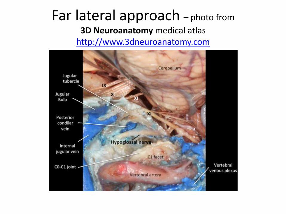
Far lateral approach – photo from
3D Neuroanatomy medical atlashttp://www.3dneuroanatomy.com

Hypoglossal is just behind the upper end of parapharyngel carotid – very easy way to
identify 12th nerve in paraphayrngeal space – Dr.Satish jain


In infrapetrous approach there are chances of injury to 6th nerve [ in dorello’scanal medial to paraclival carotid ] & 12th nerve
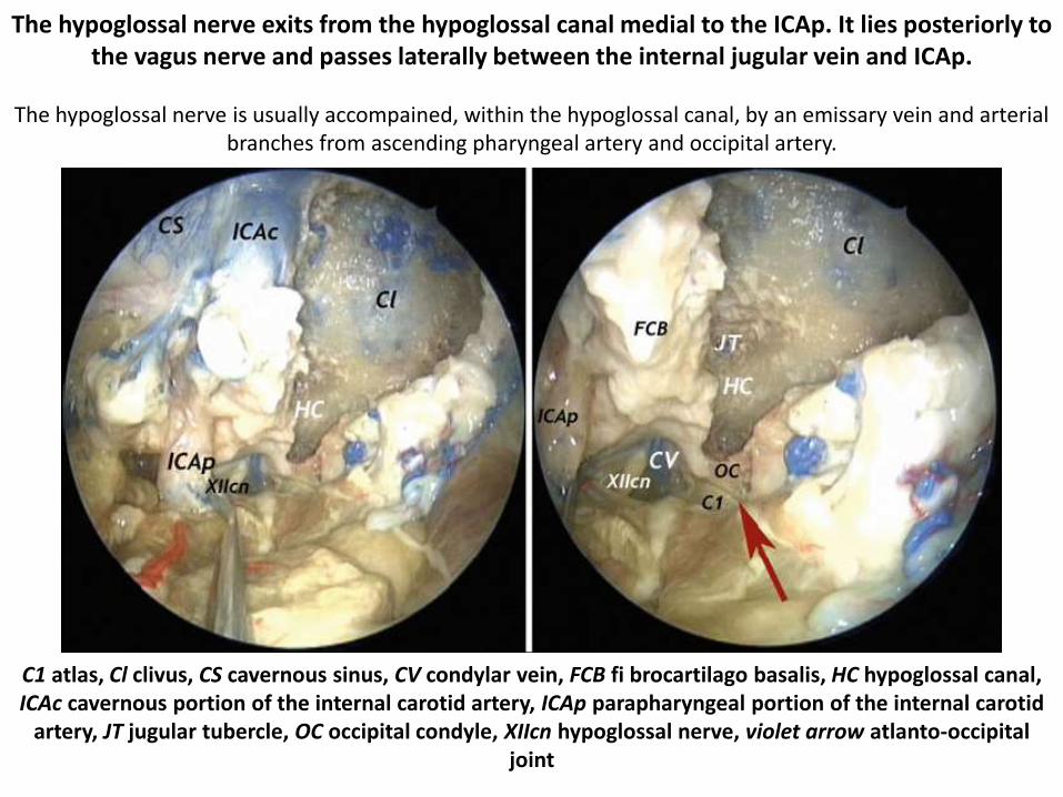
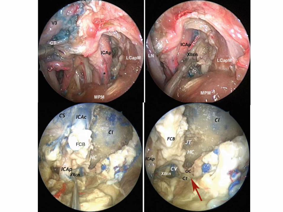
The hypoglossal nerve exits from the hypoglossal canal medial to the ICAp. It lies posteriorly to the vagus nerve and passes laterally between the internal jugular vein and ICAp.
The hypoglossal nerve is usually accompained, within the hypoglossal canal, by an emissary vein and arterial branches from ascending pharyngeal artery and occipital artery.
C1 atlas, Cl clivus, CS cavernous sinus, CV condylar vein, FCB fi brocartilago basalis, HC hypoglossal canal, ICAc cavernous portion of the internal carotid artery, ICAp parapharyngeal portion of the internal carotid
artery, JT jugular tubercle, OC occipital condyle, XIIcn hypoglossal nerve, violet arrow atlanto-occipital joint

Endoscopic endonasal view of a cadaveric dissection showing transection of the right eustachian tube (ET) attachment to foramen lacerum (FL). The hypoglossal nerve (XII) enters the hypoglossal canal just deep to
the ET and separates the occipital condyle (OC) and the jugular tubercle (JT). (BA, basilar artery; ICA, internal carotid artery [paraclival segment]; IPS, inferior petrosal sinus; VN, vidian nerve.) B. Endoscopic
endonasal view of cadaveric dissection showing the parapharyngeal internal carotid artery (ICA) and jugular foramen (JF) following transection and removal of the eustachian tube. (BA, basilar artery; IPS, inferior petrosal sinus; FL, foramen lacerum; JT, jugular tubercle; OC, occipital condyle; XII, hypoglossal
nerve.)
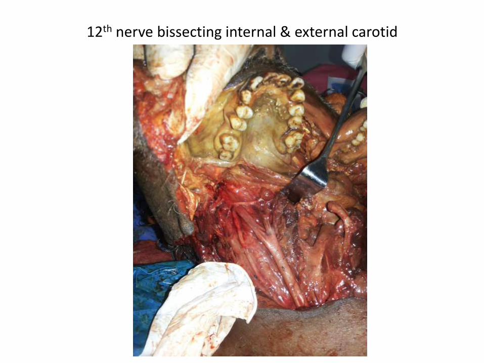
12th nerve bissecting internal & external carotid
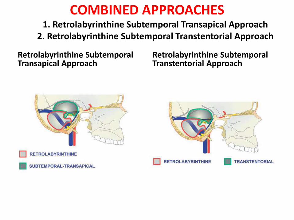
COMBINED APPROACHES 1. Retrolabyrinthine Subtemporal Transapical Approach
2. Retrolabyrinthine Subtemporal Transtentorial Approach
Retrolabyrinthine SubtemporalTransapical Approach
Retrolabyrinthine SubtemporalTranstentorial Approach
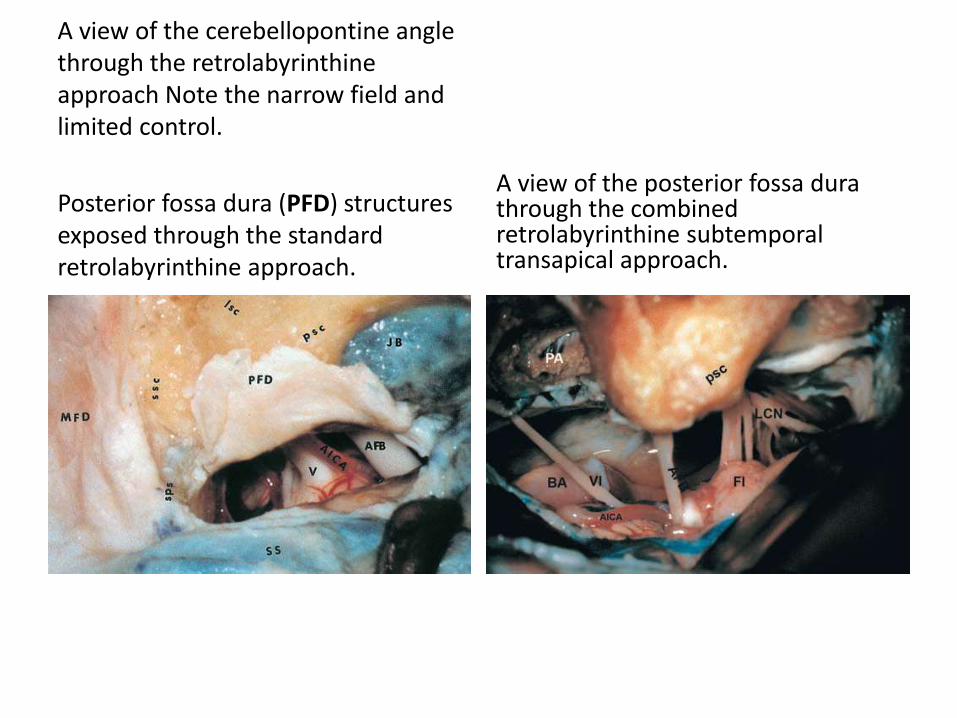
A view of the cerebellopontine angle through the retrolabyrinthineapproach Note the narrow field and limited control.
Posterior fossa dura (PFD) structures exposed through the standard retrolabyrinthine approach.
A view of the posterior fossa durathrough the combined retrolabyrinthine subtemporaltransapical approach.
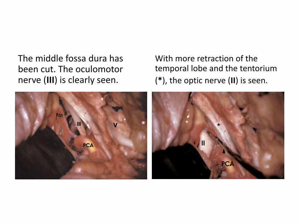
The middle fossa dura has been cut. The oculomotornerve (III) is clearly seen.
With more retraction of the temporal lobe and the tentorium
(*), the optic nerve (II) is seen.
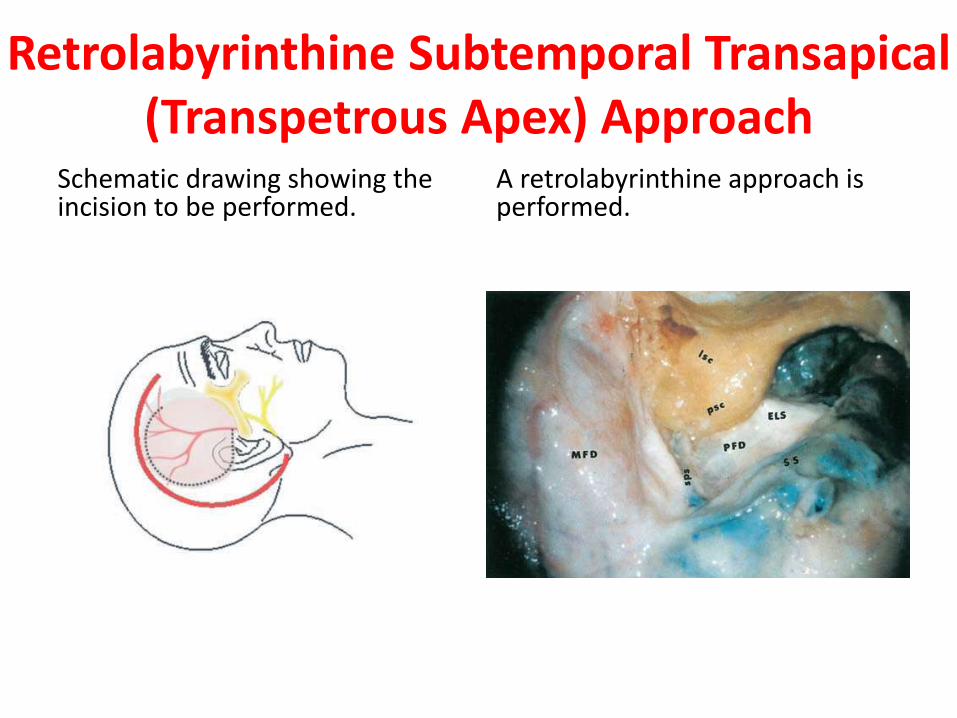
Retrolabyrinthine Subtemporal Transapical(Transpetrous Apex) Approach
Schematic drawing showing the incision to be performed.
A retrolabyrinthine approach is performed.

The dura of the middle fossa is detached from the superior surface of the temporal bone from posterior to anterior.
With further detachment of the dura, the middle meningeal (MMA) artery is clearly identified.
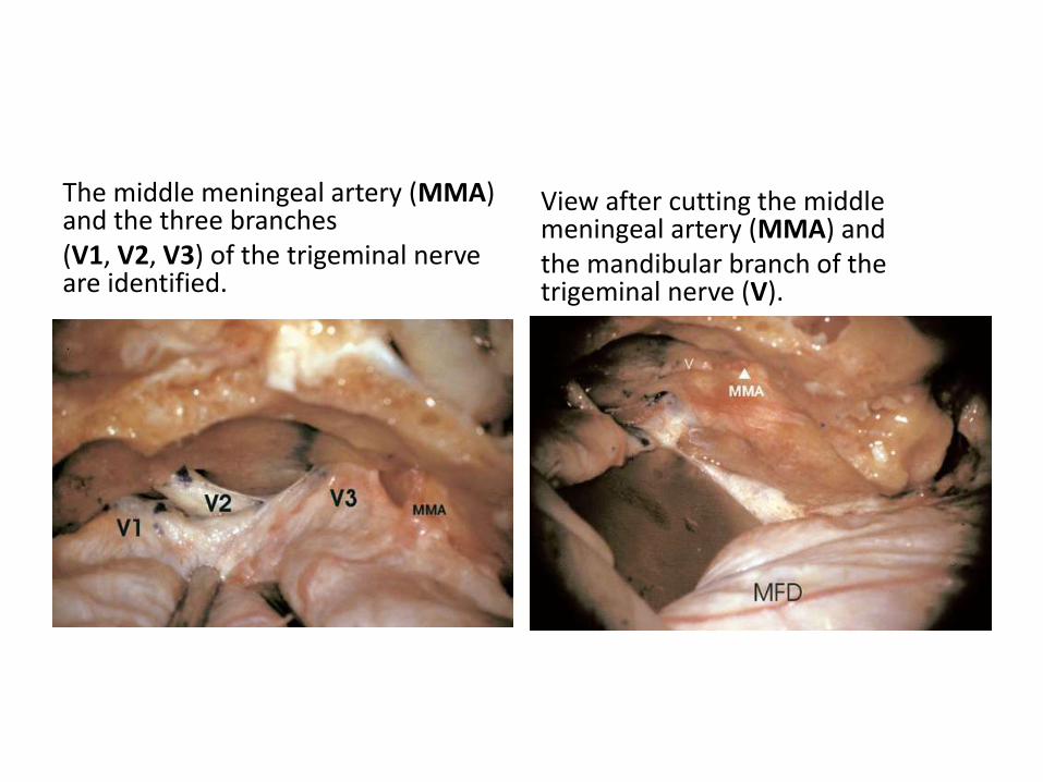
The middle meningeal artery (MMA) and the three branches(V1, V2, V3) of the trigeminal nerve are identified.
View after cutting the middle meningeal artery (MMA) andthe mandibular branch of the trigeminal nerve (V).
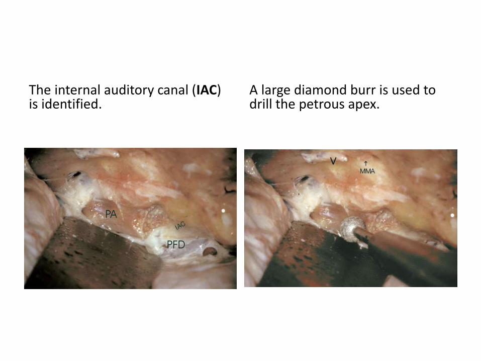
The internal auditory canal (IAC) is identified.
A large diamond burr is used to drill the petrous apex.
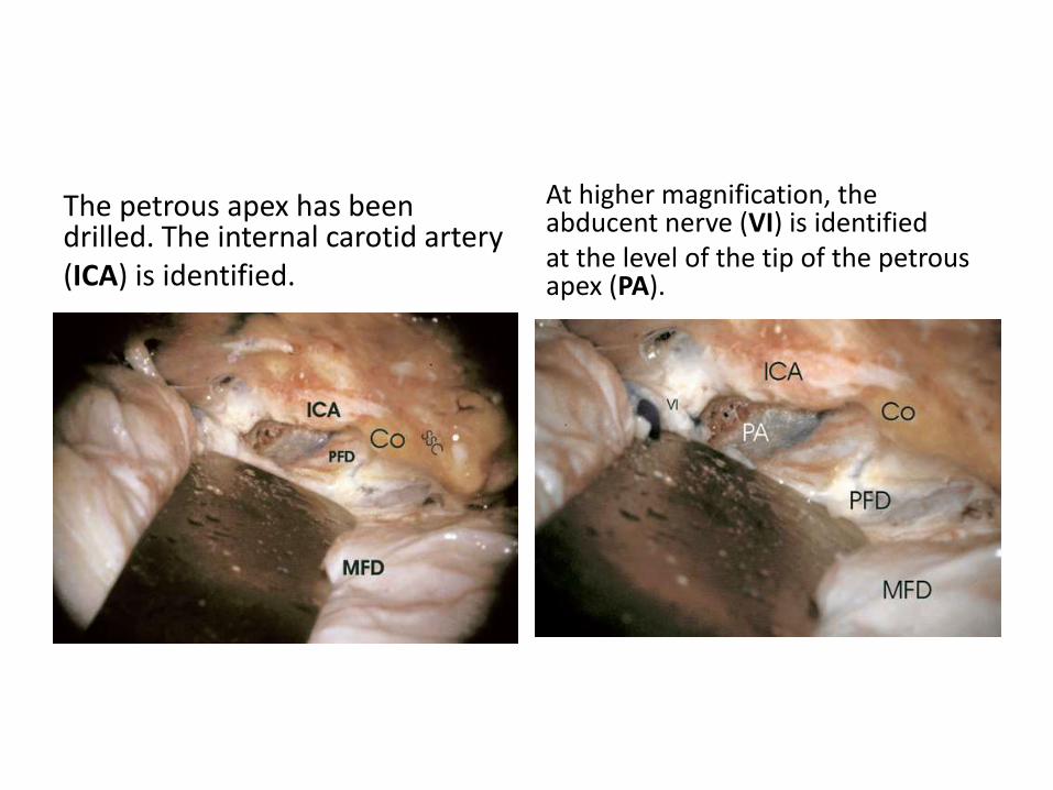
The petrous apex has been drilled. The internal carotid artery(ICA) is identified.
At higher magnification, the abducent nerve (VI) is identifiedat the level of the tip of the petrous apex (PA).

Panoramic view showing the structures after opening of the
posterior fossa dura.
At higher magnification, the anterior inferior cerebellar artery (AICA)is seen stemming from the basilar artery (BA) at the prepontine cistern. The artery is crossed by the abducent nerve (VI). Note the good control of the prepontine cistern through this approach.
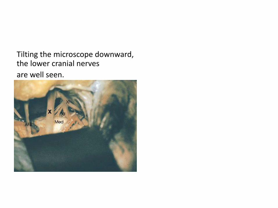
Tilting the microscope downward, the lower cranial nerves
are well seen.

Retrolabyrinthine SubtemporalTranstentorial Approach
The retrolabyrinthine craniotomy has been performed. The petrous apex has been partially drilled.
The middle fossa dura (*) is incised.

The tentorium (*) is cut, taking care not to injure thetrochlear nerve.
The tentorium is further cut until the tentorial notch isreached. With retraction of the temporal lobe the optic (II), oculomotor(III) and contralateral oculomotor(IIIc) nerves are seen.
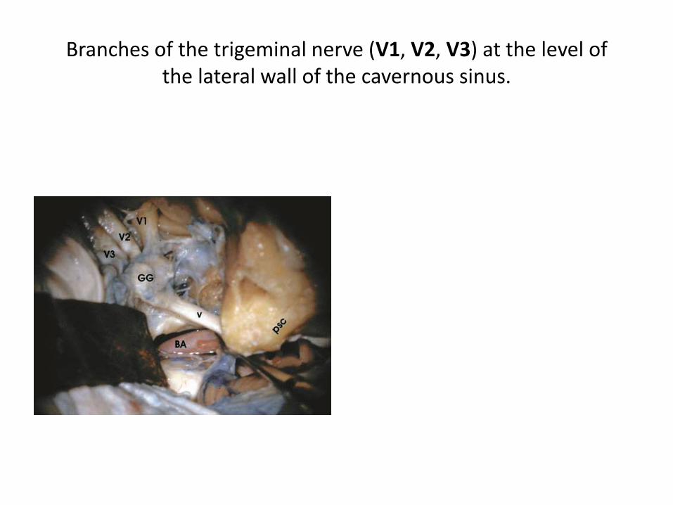
Branches of the trigeminal nerve (V1, V2, V3) at the level ofthe lateral wall of the cavernous sinus.

After this PPT must read “REZ 360” . Click http://www.slideshare.net/muralichandnal
lamothu/rez-360

For Other powerpoint presentatioinsof
“ Skull base 360° ”I will update continuosly with date tag at the end as I am
getting more & more information
click
www.skullbase360.in- you have to login to slideshare.net with Facebook
account for downloading.