covid directive for mgt. for co-infection with seasonal
TRANSCRIPT


if'k"V&1
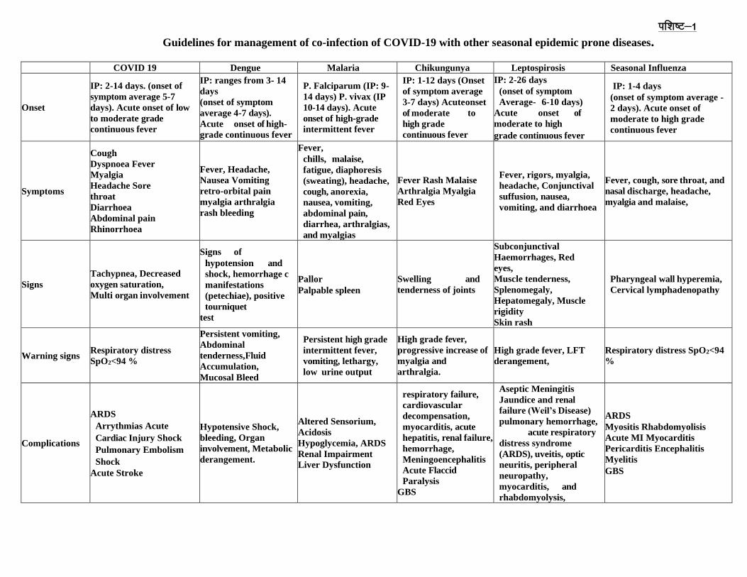
Guidelines for management of co-infection of COVID-19 with other seasonal epidemic prone diseases.
COVID 19 Dengue Malaria Chikungunya Leptospirosis Seasonal Influenza
Onset
IP: 2-14 days. (onset of
symptom average 5-7
days). Acute onset of low
to moderate grade
continuous fever
IP: ranges from 3- 14
days
(onset of symptom
average 4-7 days).
Acute onset of high-
grade continuous fever
P. Falciparum (IP: 9-
14 days) P. vivax (IP
10-14 days). Acute
onset of high-grade
intermittent fever
IP: 1-12 days (Onset
of symptom average
3-7 days) Acute onset
of moderate to
high grade
continuous fever
IP: 2-26 days
(onset of symptom
Average- 6-10 days)
Acute onset of
moderate to high
grade continuous fever
IP: 1-4 days
(onset of symptom average -
2 days). Acute onset of
moderate to high grade
continuous fever
Symptoms
Cough
Dyspnoea Fever
Myalgia
Headache Sore
throat
Diarrhoea
Abdominal pain
Rhinorrhoea
Fever, Headache,
Nausea Vomiting
retro-orbital pain
myalgia arthralgia
rash bleeding
Fever,
chills, malaise,
fatigue, diaphoresis
(sweating), headache,
cough, anorexia,
nausea, vomiting,
abdominal pain,
diarrhea, arthralgias,
and myalgias
Fever Rash Malaise
Arthralgia Myalgia
Red Eyes
Fever, rigors, myalgia,
headache, Conjunctival
suffusion, nausea,
vomiting, and diarrhoea
Fever, cough, sore throat, and
nasal discharge, headache,
myalgia and malaise,
Signs
Tachypnea, Decreased
oxygen saturation,
Multi organ involvement
Signs of
hypotension and
shock, hemorrhage c
manifestations
(petechiae), positive
tourniquet
test
Pallor
Palpable spleen
Swelling and
tenderness of joints
Subconjunctival
Haemorrhages, Red
eyes,
Muscle tenderness,
Splenomegaly,
Hepatomegaly, Muscle
rigidity
Skin rash
Pharyngeal wall hyperemia,
Cervical lymphadenopathy
Warning signs Respiratory distress
SpO2<94 %
Persistent vomiting,
Abdominal
tenderness,Fluid
Accumulation,
Mucosal Bleed
Persistent high grade
intermittent fever,
vomiting, lethargy,
low urine output
High grade fever,
progressive increase of
myalgia and
arthralgia.
High grade fever, LFT
derangement,
Respiratory distress SpO2<94
%
Complications
ARDS
Arrythmias Acute
Cardiac Injury Shock
Pulmonary Embolism
Shock
Acute Stroke
Hypotensive Shock,
bleeding, Organ
involvement, Metabolic
derangement.
Altered Sensorium,
Acidosis
Hypoglycemia, ARDS
Renal Impairment
Liver Dysfunction
respiratory failure,
cardiovascular
decompensation,
myocarditis, acute
hepatitis, renal failure,
hemorrhage,
Meningoencephalitis
Acute Flaccid
Paralysis
GBS
Aseptic Meningitis
Jaundice and renal
failure (Weil’s Disease)
pulmonary hemorrhage,
acute respiratory
distress syndrome
(ARDS), uveitis, optic
neuritis, peripheral
neuropathy,
myocarditis, and
rhabdomyolysis,
ARDS
Myositis Rhabdomyolisis
Acute MI Myocarditis
Pericarditis Encephalitis
Myelitis
GBS

Government of India Ministry of Health & Family Welfare Directorate
General of Health Services (EMR Division)
Guidelines for management of co-infection of COVID-19 with other seasonal epidemic prone
diseases
1. Background
Almost all States/UTs of the country are affected by COVID-19. Given the seasonal pattern of
epidemic prone diseases observed every year in our country, it diseases like Dengue, Malaria,
Seasonal Influenza, Leptospirosis, Chikungunya, Enteric fever, etc. can not only present as a
diagnostic dilemma but may co-exist in COVID cases. This poses challenges in clinical and
laboratory diagnosis of COVID , and have a bearing on clinical management and patient
outcomes.
2. Scope
The scope of this document is to provide clear guidelines on prevention and treatment of co-
infections of COVID with diseases like Dengue, Malaria, Seasonal Influenza (H1N1),
Leptospirosis, Chikungunya etc.
3. Clinical features As per the World Health Organization (WHO) case definition, a COVID case may present with:
• Acute onset of fever AND cough;
OR
• Acute onset of ANY THREE OR MORE of the following signs or symptoms: fever, cough,
general weakness/fatigue, headache, myalgia, sore throat, coryza, dyspnoea, anorexia/nausea/
vomiting, diarrhoea, altered mental status.
This case definition, although sensitive, is not very specific. Seasonal epidemic prone diseases,
as cited in the foregoing paragraphs may all present as febrile illness, with symptoms that
mimic COVID-19. If there is a co-infection, then apart from the febrile illness there may be
constellation of signs and symptoms that may lead to difficulty in diagnosis. A comparative
analysis of disease onset, symptoms, signs, warning signs, complications and diagnosis is given
at Annexure.
4. Approach to diagnosis of suspected co-infection
A high index of suspicion must be maintained for epidemic prone diseases (e.g. Dengue,
Malaria, Chikungunya, Seasonal influenza, Leptospirosis) prevalent in a particular geographic
region during monsoon and post-monsoon seasons. Bacterial co-infections must also be
suspected in moderate or severe cases of COVID-19 not responding to treatment.
• Malaria/Dengue: It must be borne in mind that malaria/dengue can coexist with other infections,
and thus confirmation of malaria/dengue infection does not rule out the possibility of the patient
not suffering from COVID-19. Similarly, a high index of suspicion of malaria/dengue must be there

when a fever case is diagnosed as COVID-19, particularly during the rainy and post rainy season
in areas endemic for these diseases.
• Seasonal Influenza: Both COVID-19 and Seasonal Influenza present as Influenza Like Illness
(ILI)/SARI, hence all ILI/SARI cases in areas reporting COVID-19 cases must be evaluated and
tested for both COVID-19 and Seasonal Influenza, if both viruses are circulating in population
under consideration.
• Chikungunya: Chikungunya presents with acute onset of moderate to high grade continuous fever
and malaise followed by rash, myalgia and arthralgia. Respiratory failure may ensue in late stages.
Co-infection with COVID-19 may be suspected in Chikungunya endemic areas, in the months of
monsoon.
• Leptospirosis: Leptospirosis apart from it presenting as febrile illness, has also the tendency to
manifest as acute respiratory illness, leading to respiratory distress and shock. In areas where
Leptospirosis is known to cause outbreaks during monsoon/ post monsoon, the possibility of co-
infection should be considered.
• Scrub Typhus: Scrub typhus is known to be prevalent in foothills of Himalayas viz Jammu &
Kashmir, Himachal Pradesh, Sikkim, Manipur, Nagaland, Meghalaya, etc. However, in recent
past, scrub typhus outbreaks have also been reported from Delhi, Haryana, Rajasthan, Maharashtra,
Uttarakhand, Chhattisgarh, Tamil Nadu and Kerala. The clinical picture consists of sudden high-
grade fever, severe headache, apathy, myalgia and generalized lymphadenopathy. A
maculopapular rash may appear first on the trunk and then on the extremities and blenches within
a few days. The patients may develop complications that include interstitial pneumonia (30 to 65%
of cases), meningoencephalitis and myocarditis. Scrub typhus infection may co-exist with COVID-
19.
• Bacterial infections: Few patients with COVID-19 experience a secondary bacterial infection. In
such cases, empiric antibiotic therapy as per local antibiogram needs to be considered.
Despite the possibility of above mentioned co-infections, in present times of the pandemic,
approach to diagnosis for COVID-19 essentially remains the same. Testing protocol as per
MoHFW/ICMR guidelines will be followed. However, in addition, further tests for a likely co-
infection will also be undertaken, whenever suspected.
5. Diagnostics
While each of these infections are antigenically distinct with specific serological responses, yet
in the eventuality of co-infections, cross-reactions (resulting in false-positive /false negative
results) cannot be totally ruled out, especially if the testing kits used are not having requisite
sensitivity and specificity. Hence the tests recommended by ICMR (for COVID-19) and that
recommended by the concerned programme divisions (NVBDCP for vector borne diseases
[Malaria, Dengue, Chikungunya]) and NCDC (Seasonal Influenza, Leptospirosis, Scrub
Typhus)] needs to be followed. Availability of rapid diagnostic kits for malaria, dengue, scrub
typhus should be ensured in such COVID treatment facilities.
The table below summarizes the various (confirmatory) test to be undertaken for possible co-
infections.

Laboratory Testing: Co-infection of COVID 19 with other seasonal epidemic prone diseases
Diseases Tests Sample
Dengue NS1 antigen ELISA or RT PCR: For < 5 days
of illness
IgM capture ELISA (MAC-ELISA): For >5
days of illness
Blood/Serum
Chikungunya Early disease: RT PCR
After first week of illness: IgM capture ELISA
Blood/Serum
H1N1 Acute phase: RT PCR Naso/Oropharyngeal swab
COVID 19 Acute phase: RT PCR Nasopharyngeal/
Oropharyngeal swab
Malaria RDT (bi-valent both Pf/Pv detection)
Quality microscopy for slide positivity
confirmation
Blood
Blood
Leptospirosis In endemic areas: IgM ELISA and MAT tests
Non-endemic areas: IgM ELISA followed by
MAT test for confirmation
Scrub Typhus Detection of IgM antibodies by Weil-Felix
Test (WFT)
Enzyme linked Immunosorbent assay
(ELISA)
Serum
Serum
Bacterial co-infections Gram stain and culture, Blood culture Sputum/Bronchial
aspirate/Blood
6. Case Management
Management of co-infection of COVID-19 with dengue, Influenza and bacterial co-infections
may however be challenging and are dealt with in greater detail here.
6.1 Management of COVID-19 and Dengue co-infection 6.1.1 Pathogenesis
Dengue Fever and COVID-19 share many pathogenic and clinical features which might make
it very difficult to differentiate the two infections (1). The phenomenon of ADE (Antibody
Dependent Enhancement) has been described for both dengue virus as well as for SARS-CoV-
2 virus resulting in escalation in degree of infection and number of complications. Both being
RNA viruses they share certain common features in pathogenesis, eventually leading to
subsequent cytokines and chemokine release and also affecting the integrity of the vascular
endothelium leading to vasculopathy, coagulopathy and capillary leak. Various mechanisms
can explain the signs and symptoms observed in co-infected patients but most will have the
following, (i) Antibody-dependent enhancement (ADE),
(ii) Cytokine Storm, (iii) Vasculopathy and (iv) Coagulopathy.
6.1.2 Clinical Features
The clinical features of both the infections are overlapping, both present as acute febrile illness
of short duration and may have thrombocytopenia and shortness of breath, although respiratory

symptoms are more common in COVID-19 and bleeding manifestations more common in
Dengue. Routine testing for both diseases shows leucopenia or normal leucocyte count.
Decrease in platelet count which is a defining feature of dengue infection but can also be seen
in significant number of covid cases. There are reports in literature, where dengue serology
was positive initially and later on, it was found that cases were positive by RT-PCR for
COVID-19 thereby suggesting that dengue serology can be falsely positive in COVID-19
patients. Therefore, there is a need to rely on more specific tests for each disease like throat
swab RT-PCR for COVID-19 and ELISA based Dengue NS1 Antigen or serology test for
dengue diagnosis. Serum sample for NS1 antigen within first 5 day of onset of fever were
negative in above study suggesting that positive dengue serology was more likely to be false
positive result and not co-infection. Hence, one needs to be careful while making diagnosis of
co-infection.
There are now enough evidences to support that severe dengue is associated with cytokine
storm and high levels of various circulating cytokine are associated with poor outcome in most
cases. COVID-
19 infects alveolar epithelial cells leading to pneumonia and ARDS, it also infects
monocytes/macrophages leading to cytokine storm associated with multi organ failure and
death. This cytokine storm seen in severe cases has led to increased use of steroids and other
immunosuppressive therapy in moderate to severe cases.
Both COVID-19 and Dengue infection are accompanied by coagulopathy and vasculopathy
with coagulopathy being predominant in formal leading to widespread use of Low Molecular
Weight Heparin (LMWH). There have been numerous evidences to suggest the increased
burden of thrombosis in COVID-19 based on which recommendations have been made for the
use of LMWH in moderate to severe cases. But in the presence of Dengue co-infection which
is usually accompanied by thrombocytopenia and increased risk of bleeding , the use of LMWH
becomes a challenging issue. Similarly, because of increased capillary leak and increased third
space fluid loss, fluid administration which forms the cornerstone in management of dengue
might not be recommended with clarity as conservative fluid administration has been
recommended for COVID-19 in absence of shock.
6.1.3 Clinical management consideration for Dengue and COVID-19 co-infection
Following are some general measures to followed in case of Dengue and COVID-19 co-
infection:
• Co-infection should be ruled out when suspected with proper diagnostic method at the early stage
to initiate proper specific management to reduce morbidity and mortality.
• Strengthening at the primary health care level is the key to manage dengue through early clinical
diagnosis and recognition of warning signs for severity of Dengue (such as abdominal pain or
tenderness, persistent vomiting, clinical fluid accumulation, mucosal bleed, lethargy or
restlessness, liver enlargement >2 cm, and increase in haematocrit).
• Mild to moderate Dengue and COVID co-infected patient should be monitored closely preferably
at hospital, as they may rapidly progress to severe stage therefore they should be referred to
higher centre at the early stage by recognizing warning signs.
• At the same time, all secondary and tertiary level hospitals should be prepared to manage severe
dengue and COVID cases.

These measures will help to prevent the progression of illness to severe dengue and deaths,
which in turn will also help to reduce the number of patients that need to be referred to
hospitals, thus avoiding saturation of these facilities as well as the intensive care units.
6.1.4 Specific therapeutic considerations
Points related with specific therapeutic options and their use in cases with co-infection:
• Fluid Therapy – Fluid therapy to be given in co-infection cases depends on hemodynamic status
of patient and degree of severity. One may follow national guidelines for clinical management of
dengue fever for most co-infection cases. It is only in the presence of SARI with COVID-19 that
we need to be careful with aggressive fluid administration as it leads to worsening of oxygenation.
Close clinical monitoring of fluid status is required in such cases. Aggressive fluid resuscitation is
recommended for COVID-19 patients in shock for initial resuscitation.
• LMWH – LMWH is being used and has been included in the National guidelines for the
management of moderate to severe covid-19 cases as it is associated with increased thrombosis.
Once the platelet count decreases to less than 1 lakh we need to be very careful with the use of
LMWH and it may be withheld based on clinical condition of the patient. Decision to administer
LMWH and the dosage for the same should be based on close monitoring with D-dimer
measurements. In any case of co-infection with active bleeding, LMWH needs to be stopped
immediately.
• Use of Corticosteroids – Steroids specially Dexamethasone have recently been shown to be
effective in severe covid-19 cases and have been recommended for the same. Dengue being a viral
illness, it’s course won’t be affected much. Hence, use of steroids can be continued as per COVID-
19 management guidelines.
• Tocilizumab –To be used as per national management guidelines for COVID-19 management.
• Antivirals – To be used as per COVID-19 management guidelines.
• Other supportive management to be continued as per the current guidelines.
6.2. Management of Seasonal influenza and COVID co-infection Co-infection with SARS-CoV-2 and other respiratory viruses has been described in a number
of studies. Most prominent among these are Respiratory Syncytial Virus, Enteroviruses and
Influenza A virus. With the approaching winter season, the seasonal Influenza cases may show
an upward trend, and there could be cases of coinfection with COVID-19.
6.2.1 Pathogenesis
COVID-19 and Influenza share many pathogenic feature. Both diseases involve the respiratory
system, manifesting widely from ILI to SARI. Both diseases cause pneumonitis. The
histopathological manifestation of Interstitial inflammation and diffuse alveolar damage and
intra- alveolar edema followed by fibrin deposition, hyaline membrane, and leukocyte
infiltration of the alveolar septa are seen in both COVID and Influenza. The radiological
appearance is not of much help either as both diseases may have presence of opacities or
consolidations.
Both being RNA viruses they share certain common features in pathogenesis, eventually
leading to subsequent cytokine release and acute respiratory distress syndrome.

6.2.2 Diagnosis
Whenever suspected, especially in areas reporting seasonal Influenza cases, samples should
also be sent and tested for SARS-CoV-2 and Influenza.
6.2.3 Clinical Features
The clinical features of both the infections are overlapping, both present as acute febrile illness
of short duration and may have fever, cough and shortness of breath. Similarly, laboratory
investigations are also not very helpful in differentiating between the two, both show
leucopenia or normal leucocyte count. Co-infection should be ruled out when suspected with
proper diagnostic method at the early stage to initiate proper specific management to reduce
morbidity and mortality.
6.2.4 Specific therapeutic considerations
Points related with specific therapeutic options and their use in cases with co-infection:
• The specific treatment as provided in the clinical management protocol of COVID needs to be
followed, as per severity of the disease.
• In addition to COVID management, for the treatment of influenza,
Oseltamivir needs to be administered in the prescribed dosages. • In case of an outbreak of Seasonal Influenza outbreak, Oseltamavir blanket therapy should be
considered in all patients of COVID-19.
• Other supportive management to be continued as per the current guidelines.
6.3 Management of Bacterial co-infections with COVID
Evidence shows that small proportion of COVID-19 patients may have coinfection with
bacteria. Patients with community-acquired co-infections and hospital-acquired
superinfections had worse outcomes. A recent systemic review on co-infections in people with
COVID-19 has found that the commonly associated pathogens in such cases are Mycoplasma
pnuemoniae, Psuedomonas aeroginosa, Hemophilus influenzae, Klebsiella pneumoniae etc.
The occurrence of healthcare associated infections like hospital acquired pneumonia
(particularly in ICU settings), urinary tract infection, skin/soft tissue infection, abdominal
infections, etc.) need to be considered.
Antibiotics should not be prescribed routinely unless there is clinical suspicion of a bacterial
infection. Consider empiric antibiotic therapy as per local antibiogram. For COVID-19 patients
with severe disease, also collect blood cultures, ideally prior to initiation of antimicrobial
therapy.
6.4 Management of Malaria and COVID-19 co-infection 6.4.1 Pathogenesis
Malaria is a potentially life-threatening parasitic disease caused by a protozoan having four
types: Plasmodium vivax (P. vivax), Plasmodium falciparum (P. falciparum), Plasmodium

malariae (P. malariae) and Plasmodium ovale (P. ovale). It is transmitted by the infective bite
of Anopheles female mosquito. Man develops disease after 10 to 14 days of being bitten by an
infective mosquito. Two types of parasites of human malaria, Plasmodium vivax (Pv), P.
falciparum (Pf), are commonly reported from India. Inside the human host, the parasite
undergoes a series of changes as part of its complex life cycle. The parasite completes life cycle
in liver cells (pre-erythrocytic schizogony) and red blood cells (erythrocytic schizogony).
Infection with P. falciparum is the deadliest form of malaria.
6.4.2 Diagnosis
Diagnosis of malaria may be made by the use of RDT (bivalent) or microscopic examination
of the blood smear. Early diagnosis and prompt initiation of treatment, as per national
guidelines, is the key in preventing the progression of uncomplicated malaria to severe forms
which can be fatal. In the current scenario, in endemic areas, all fever cases should be tested
for malaria using RDT kits.
6.4.3 Clinical Features
Typically, malaria produces fever, headache, vomiting and other flu-like symptoms. The
parasite infects and destroys red blood cells resulting in easy fatigue ability due to anemia,
fits/convulsions and loss of consciousness. Parasites are carried by blood to the brain (cerebral
malaria) and to other vital organs. Malaria in pregnancy poses a substantial risk to the mother,
the fetus and the newborn infant. Pregnant women are less capable of coping with and clearing
malaria infections, adversely affecting the unborn fetus.
6.4.4 Specific therapeutic considerations
Prompt malaria case management is very important for preventing serious cases and death due
to malaria.
Plasmodium vivax (Pv) cases should be treated with Chloroquine for three days (25 mg/kg
body weight divided over three days i.e. 10 mg/kg on day 1, 10 mg/kg on day 2 and 5 mg/kg
on day 3) and Primaquine (0.25 mg/kg body weight daily for 14 days). Primaquine is used to
prevent relapse but is contraindicated in pregnant women, infants and individuals with G6PD
deficiency.
Plasmodium falciparum (Pf) cases should be treated with ACT (Artesunate 3 days +
Sulphadoxine- Pyrimethamine 1 day) @ Artesunate 4 mg/kg body weight daily for 3 days plus
Sulfadoxine (25 mg/kg body weight) and Pyrimethamine (1.25 mg/kg body weight) on day 1.
This is to be accompanied by single dose of Primaquine (0.75 mg/kg body weight) preferably
on day 2. However, considering the reports of resistance to partner drug SP In North-Eastern
States, the Technical Advisory Committee has recommended to use the co-formulated tablet
of Artemether-Lumefantrine (ACT-AL) in North-Eastern States (Not recommended during the
first trimester of pregnancy and in children weighing <5 kg).
For details of treatment of uncomplicated and complicated malaria in certain endemic areas,
special population groups (pregnancy, children etc.) All healthcare providers should also
follow the NVBDCP National Guidelines for treatment of malaria (available at:
https://nvbdcp.gov.in/WriteReadData/l892s/20627628441542176662.pdf).

6.5 Management of Leptospirosis and COVID-19 co-infection 6.5.1 Pathogenesis
Leptospirosis is primarily a contagious disease of animals, occasionally infects humans caused
by pathogenic spirochete of the genus leptospira. Urinary shedding of organisms from infected
animals (reservoir and carrier hosts like rodents, cattle, etc.) is the most important source of
these bacterial pathogens. Contact with the organism via infected urine or urine-contaminated
media results in human infection.
The most consistent pathologic finding in leptospirosis is vasculitis of capillaries, manifested
by endothelial edema, necrosis, and lymphocytic infiltration. Capillary vasculitis is found in
every affected organ system.
6.4.2 Diagnosis
The definitive diagnosis of leptospirosis depends on sero-conversion or four fold rise in
antibody titer or isolation of leptospires from clinical specimen. Positive serology, preferably
Microscopic Agglutination Test (MAT), using a range of Leptospira strains for antigens that
should be representative of local strains.
a) Enzyme Linked Immuno Sorbent Assay (ELISA): ELISA is a sensitive and specific test for the
immunological diagnosis of Leptospirosis. It is of particular value as a serological screening test
because of its relative simplicity in comparison to the MAT (Microscopic Agglutination Test). ELISA
test can also be used in epidemiological studies to determine the sero-incidence/sero- prevalence of
Leptospirosis)
b) Rapid immunodiagnostics: Lepto dip-stick, Lepto lateral flow and Lepto Tek Dri Dot assays are
based on IgM detection. These are screening tests and the results require confirmation by MAT.
Isolation (and typing) from blood or other clinical materials through culture of pathogenic
leptospires. Culture and isolation can be very difficult requiring special media and longer
incubation.
6.4.3 Clinical Features
Leptospirosis has a varied clinical manifestation. Like COVID, the severity of illness ranges
from asymptomatic, mild self-limiting febrile illness to fulminant fatal disease. The disease
typically presents as one of the following clinical categories.
(i) Mild influenza like illness
(ii) Weil’s syndrome characterized by jaundice, renal failure, haemorrhagic manifestations and
myocarditis.
(iii) Meningitis/menigo-encephalitis
(iv) Pulmonary haemorrhage with respiratory failure
Clinical illness lasts for a few days to 3 weeks or longer. Generally, there are two phases in the
illness. The early phase illness is characterized by abrupt onset of high fever, myalgia (calves
and lumbar region) and headache (retro-orbital/frontal). Other early phase manifestations
include nausea, vomiting, abdominal pain, diarrhoea, cough and conjunctival suffusion (a

pathognomonic finding of leptospirosis). Late phase illness occurs, 4-9 days after onset of
symptoms, and is characterized by prolonged fever and systemic complications such as
jaundice, renal failure, bleeding, respiratory failure etc.

6.4.4 Specific therapeutic considerations
All clinically suspected leptospirosis patients in Leptospira endemic area during rainy season
should be given presumptive treatment of leptospirosis i.e. Tab. Doxycycline 100 mg twice
daily for 7 days.
Note: In children less than 6 years 30 to 50 mg/kg/day of Cap. Amoxycillin/Cap. Ampicillin
should be given in divided doses 6 hourly for 7 days.
Diagnosis and clinical management of leptospirosis in community setting should be in
accordance with national guidelines for prevention and control of leptospirosis (available at:
https://www.ncdc.gov.in/linkimages/Leptospirosis1232331086.pdf)
6.6 Management of Scrub Typhus and COVID-19 co-infection 6.6.1 Pathogenesis
Scrub typhus is transmitted by the mite Leptotrombidium deliense. The vector mites inhabit
sharply demarcated areas in the soil where the microecosystem is favourable (mite islands).
Human beings are infected when they trespass into these mite islands and are bitten by the mite
larvae (chiggers).
Scrub Typhus causes perivasculitis of the small blood vessels. O tsutsugamushi stimulates
phagocytosis by the immune cells, and then escapes the phagosome. Scrub typhus may
disseminate into multiple organs through endothelial cells and macrophages, resulting in the
development of fatal complications.
6.6.2 Diagnosis
Scrub typhus may be diagnosed in the laboratory by: (i) isolation of the organism (ii) serology
(iii) molecular diagnosis (PCR).
Several serological tests are currently available for the diagnosis of rickettsial diseases like
Weil-Felix Test (WFT), Indirect Immuno-flourescence (IIF), Enzyme linked Immunosorbent
assay (ELISA) etc. Although many techniques have been used successfully for rickettsial sero
diagnosis, rela-tively few are used regularly by most laboratories. BSL-3 Lab is not required
for performing serological tests.
Enzyme linked Immunosorbent Assay (ELISA): ELISA techniques, particularly
immunoglobulin M (IgM) capture assays, are probably the most sensitive tests available for
rickettsial diagnosis, and the presence of IgM antibodies, indicate recent infection with
rickettsial diseases. In cases of infecton with O. tsutsugamushi, a significant IgM antibody titer
is observed at the end of the first week, whereas IgG antibodies appear at the end of the second
week.
Molecular diagnosis (PCR) - For PCR, blood sample is collected in tubes containing EDTA or
sodium citrate. However, blood clot, whole blood or serum can also be used for the detection
of O. tsutsugamushi, R. rickettsii, R. typhi and R. prowazekii organisms by PCR test.

6.6.3 Clinical Features
Patients with scrub typhus may present early or later in the course of their disease. Inoculation
through the chigger bite is often painless and unnoticed. A small painless papule initially
appears at the site of infection and enlarges gradually. An area of central necrosis develops and
is followed by eschar formation. The eschar (if present) is well developed at the initiation of
the fevers, which may drive the patient to seek medical attention.
The incubation period lasts 6-20 days (average, 10 days). After incubation, persons may
experience headaches, shaking chills, lymphadenopathy, conjunctival infection, fever,
anorexia, and general apathy. The fever usually reaches 40-40.5°C (104-105°F).
6.6.4 Specific therapeutic considerations
If scrub typhus is suspected with COVID, treatment with Doxycycline (@ 200 mg/day in two
divided doses for duration of 7 days) or Azithromycin (@ 500 mg in a single oral dose for 5
days) should be administered.
Management of the individual complications should be done as per the existing practices.
7. Early warning signs
If the patient is in a primary care setup, the following criteria should be monitored to assess
patients clinical progress. Early warning signs for referral to higher centre are:
• Altered Mental Status (AVPU)
• Systolic blood pressure: <90mmHg or <20% of baseline in hypertensive patients
• Heart Rate/ Pulse Rate: <50 or >120 bpm
• SpO2: <94 % on room air
• Respiratory Rate: <10 or >30 bpm
• Temperature: persistently >38ºC
• Urine Output: <0.5 ml/Kg/Hr for consecutive 2 hrs
• Spontaneous bleeding/ haematuria
• Platelet count <50.000/cumm
8. Prevention
Even though the basic preventive strategies of COVID-19 and seasonal influenza are different
from diseases discussed in this document, it is desirable that there is synergy in the prevention
of these diseases. The States must make use of their resources effectively as staff is also
diverted to provide COVID-19 response. This can be achieved by combining prevention
activities.
7.1 Integrated surveillance: It must be ensured that IDSP networks are strengthened to include
surveillance of COVID-19 cases besides for dengue, malaria, chikungunya, leptospirosis, scrub typhus,
seasonal influenza to maximize the use of resources.
7.2 Basic preventive measures for COVID-19 and seasonal influenza, like avoiding large gatherings,
maintaining physical distance, hand hygiene and cough etiquette must be ensured at all times.
7.3 Vector control: Source reduction of mosquito breeding sites and adult control measures should be
implemented in areas affected by or at risk of these diseases, especially in and around treatment
facilities.

7.4 Use of approved insect repellents and ITN/LLINs is affective against vector borne diseases
including scrub typhus.
7.5 Preventive measures against leptospirosis include wearing protective clothing for people at
occupational risk (sanitation workers, rice-paddy and sugarcane workers etc.) and avoidance of
swimming in water that may be contaminated.
7.4 Vaccination for seasonal influenza for healthcare workers and other high-risk groups to be
implemented aggressively.
9. Community involvement Community support must be garnered and community awareness drives on COVID-19 and
other seasonal diseases must be ensured.
10. Conclusion
A concerted effort is required in prevention, surveillance, behaviour change communication
and management of such cases. Alert vigil, a high index of suspicion and constant awareness
of the possibility of co-infections can help physicians avert the adverse outcome of cases with
coinfection and improve clinical outcomes.

Annexure
Guidelines for management of co-infection of COVID-19 with other seasonal epidemic prone diseases.
COVID 19 Dengue Malaria Chikungunya Leptospirosis Seasonal Influenza
Onset
IP: 2-14 days. (onset of
symptom average 5-7
days). Acute onset of low
to moderate grade
continuous fever
IP: ranges from 3- 14
days
(onset of symptom
average 4-7 days).
Acute onset of high-
grade continuous fever
P. Falciparum (IP: 9-
14 days) P. vivax (IP
10-14 days). Acute
onset of high-grade
intermittent fever
IP: 1-12 days (Onset
of symptom average 3-
7 days) Acute onset
of moderate to
high grade continuous
fever
IP: 2-26 days
(onset of symptom
Average- 6-10 days)
Acute onset of
moderate to high
grade continuous fever
IP: 1-4 days
(onset of symptom average - 2
days). Acute onset of
moderate to high grade
continuous fever
Symptoms
Cough
Dyspnoea Fever
Myalgia
Headache Sore
throat
Diarrhoea
Abdominal pain
Rhinorrhoea
Fever, Headache,
Nausea Vomiting
retro-orbital pain
myalgia arthralgia
rash bleeding
Fever,
chills, malaise,
fatigue, diaphoresis
(sweating), headache,
cough, anorexia,
nausea, vomiting,
abdominal pain,
diarrhea, arthralgias,
and myalgias
Fever Rash Malaise
Arthralgia Myalgia
Red Eyes
Fever, rigors, myalgia,
headache, Conjunctival
suffusion, nausea,
vomiting, and diarrhoea
Fever, cough, sore throat, and
nasal discharge, headache,
myalgia and malaise,
Signs
Tachypnea, Decreased
oxygen saturation,
Multi organ involvement
Signs of
hypotension and
shock, hemorrhage c
manifestations
(petechiae), positive
tourniquet
test
Pallor
Palpable spleen
Swelling and
tenderness of joints
Subconjunctival
Haemorrhages, Red
eyes,
Muscle tenderness,
Splenomegaly,
Hepatomegaly, Muscle
rigidity
Skin rash
Pharyngeal wall hyperemia,
Cervical lymphadenopathy
Warning signs Respiratory distress
SpO2<94 %
Persistent vomiting,
Abdominal
tenderness,Fluid
Accumulation,
Mucosal Bleed
Persistent high grade
intermittent fever,
vomiting, lethargy, low
urine output
High grade fever,
progressive increase of
myalgia and
arthralgia.
High grade fever, LFT
derangement,
Respiratory distress SpO2<94
%
Complications
ARDS
Arrythmias Acute Cardiac
Injury Shock Pulmonary
Embolism Shock
Acute Stroke
Hypotensive Shock,
bleeding, Organ
involvement, Metabolic
derangement.
Altered Sensorium,
Acidosis
Hypoglycemia, ARDS
Renal Impairment
Liver Dysfunction
respiratory failure,
cardiovascular
decompensation,
myocarditis, acute
hepatitis, renal failure,
hemorrhage,
Meningoencephalitis
Acute Flaccid Paralysis
GBS
Aseptic Meningitis
Jaundice and renal failure
(Weil’s Disease)
pulmonary hemorrhage,
acute respiratory
distress syndrome
(ARDS), uveitis, optic
neuritis, peripheral
neuropathy, myocarditis,
and
rhabdomyolysis,
ARDS
Myositis Rhabdomyolisis
Acute MI Myocarditis
Pericarditis Encephalitis
Myelitis
GBS