covid-19: abnormal liver function tests
TRANSCRIPT

Research ArticleDILI, Autoimmune, Cholestatic and Genetic Diseases
COVID-19: Abnormal liver function tests
Graphical abstract
Abnormal liver function testsin patients with COVID-19
Liver injury associated with use of drugsin patients with COVID-19
91
326
417
85(93.4%)
233(71.5%)
318(76.3%)
4(46.2%)
47(14.4%)
90(21.5%)
0
50
100
150
200
250
300
350
400
450
Severe No-severe Total
Num
ber o
f pat
ient
s
Liver injuryAbnormal liver functionConfirmed case
2.15
1.69
1.341.48
2.21
0.99
4.44
1.7 1.691.51.6
1.23
0.73
5.03
0.3
1.0
3.0
10.0
Antibio
tics
Herbal
Interf
eron
Lopin
avir/
ritona
vir NSAIDOse
ltamivi
rRiba
virin
Drugs
OR
(95%
CI)
for l
iver
inju
ry
IPW modelMultivariable model
Highlights liver tests and liver injury were asso-
� Of 417 patients with COVID-19, 76.3% had abnormal liver tests and21.5% had liver injury during hospitalization.
� Patients with abnormal liver tests had significantly higher odds ofdeveloping severe pneumonia.
� The use of lopinavir/ritonavir increased the odds of liver injury by4-fold.
https://doi.org/10.1016/j.jhep.2020.04.006© 2020 European Association for the Study of the Liver. Published by Elsevier B.V. All rig
Authors
Qingxian Cai, Deliang Huang, Hong Yu,., Jun Chen, Lei Liu, Lin Xu
[email protected] (J. Chen), [email protected] (L. Liu), [email protected] (L. Xu).
Lay summaryData on liver tests in patients withCOVID-19 are scarce. We observed ahigh prevalence of liver test abnor-malities and liver injury in 417 pa-tients with COVID-19 admitted to ourreferral center, and the prevalenceincreased substantially during hospi-talization. The presence of abnormal
ciated with the progression to severepneumonia. The detrimental effectson liver injury were related to certainmedications used during hospitaliza-tion, which warrants frequent moni-toring and evaluation for thesepatients.
hts reserved. J. Hepatol. 2020, -, 1–9
Research ArticleDILI, Autoimmune, Cholestatic and Genetic Diseases
KeytestRecava
* CHosandFaxDiseSouChiHea873E-mxuli
†
#
http
COVID-19: Abnormal liver function tests
Qingxian Cai1,#, Deliang Huang1,#, Hong Yu1,#, Zhibin Zhu1, Zhang Xia1, Yinan Su1, Zhiwei Li1,Guangde Zhou1, Jizhou Gou1, Jiuxin Qu1, Yan Sun1, Yingxia Liu1, Qing He1, Jun Chen1,*,†,
Lei Liu1,*,†, Lin Xu2,*,†
1National Clinical Research Center for Infectious Diseases, The Third People’s Hospital of Shenzhen, The Second Affiliated Hospital of SouthernUniversity of Science and Technology, Shenzhen, Guangdong, 518100, China; 2School of Public Health, Sun Yat-sen University. Guangzhou,
Guangdong, China
Background & Aims: Recent data on the coronavirus disease liver injury mainly related to certain medications used during
2019 (COVID-19) outbreak caused by the severe acute respiratorysyndrome coronavirus 2 (SARS-CoV-2) has begun to shine lighton the impact of the disease on the liver. But no studies to datehave systematically described liver test abnormalities in patientswith COVID-19. We evaluated the clinical characteristics ofCOVID-19 in patients with abnormal liver test results.Methods: Clinical records and laboratory results were obtainedfrom 417 patients with laboratory-confirmed COVID-19 whowere admitted to the only referral hospital in Shenzhen, Chinafrom January 11 to February 21, 2020 and followed up to March 7,2020. Information on clinical features of patients with abnormalliver tests were collected for analysis.Results: Of 417 patients with COVID-19, 318 (76.3%) hadabnormal liver test results and 90 (21.5%) had liver injury duringhospitalization. The presence of abnormal liver tests becamemore pronounced during hospitalization within 2 weeks, with49 (23.4%), 31 (14.8%), 24 (11.5%) and 51 (24.4%) patients havingalanine aminotransferase, aspartate aminotransferase, total bili-rubin and gamma-glutamyl transferase levels elevated to morethan 3× the upper limit of normal, respectively. Patients withabnormal liver tests of hepatocellular type or mixed type atadmission had higher odds of progressing to severe disease(odds ratios [ORs] 2.73; 95% CI 1.19–6.3, and 4.44, 95% CI1.93–10.23, respectively). The use of lopinavir/ritonavir was alsofound to lead to increased odds of liver injury (OR from 4.44 to5.03, both p <0.01).Conclusion: Patients with abnormal liver tests were at higherrisk of progressing to severe disease. The detrimental effects onwords: 2019-nCoV; SARS-Cov-2; Pneumonia; Critical care; Liver injury; Livers; Bilirubin.eived 17 March 2020; received in revised form 7 April 2020; accepted 7 April 2020;ilable online xxxorresponding authors. Addresses: Department of Liver Diseases, The Third People’spital of Shenzhen, The Second Affiliated Hospital of Southern University of ScienceTechnology, Shenzhen, Guangdong, 518100, China. Tel: (86) 755-61222333;
: (86) 755-61238928 (J. Chen), or National Clinical Research Center for Infectiousases, The Third People’s Hospital of Shenzhen, The Second Affiliated Hospital ofthern University of Science and Technology, Shenzhen, Guangdong, 518100,na. Tel: (86) 755-61222333; Fax: (86) 755-61238928 (L. Liu), or School of Publiclth, Sun Yat-sen University, Guangzhou, Guangdong, China. Tel: (86) 20-35523; Fax: (86) 20-87330446 (L. Xu).ail addresses: [email protected] (J. Chen), [email protected] (L. Liu),[email protected] (L. Xu).These authors contributed equally to this manuscript.These authors contributed equally to this manuscript.s://doi.org/10.1016/j.jhep.2020.04.006
Journal of Hepatolog
hospitalization, which should be monitored and evaluatedfrequently.Lay summary: Data on liver tests in patients with COVID-19 arescarce. We observed a high prevalence of liver test abnormalitiesand liver injury in 417 patients with COVID-19 admitted to ourreferral center, and the prevalence increased substantially duringhospitalization. The presence of abnormal liver tests and liverinjury were associated with the progression to severe pneu-monia. The detrimental effects on liver injury were related tocertain medications used during hospitalization, which warrantsfrequent monitoring and evaluation for these patients.© 2020 European Association for the Study of the Liver. Published byElsevier B.V. All rights reserved.
IntroductionCoronaviruses are a family of viruses that are known to causeboth respiratory and intestinal diseases in various animal speciesand humans.1 These viruses tend to target the upper respiratorytract, causing anywhere from moderate to severe illnesses, suchas the cold or in more extreme cases, pneumonia. To date, 7human coronaviruses have been identified, including the 3epidemic viruses of severe acute respiratory syndrome (SARS)-CoV, middle east respiratory syndrome (MERS)-CoV and thenewest, severe acute respiratory syndrome coronavirus 2 (SARS-CoV-2).2 These 3 epidemic viruses have similar sequence iden-tity, sharing more than 50% genome sequences.3 In December of2019, a series of pneumonia cases of unknown origin began tospread in the central city of Wuhan, China. Now identified asSARS-CoV-2, the virus had gone on to infect more than 300,000people worldwide by March 2020.4 The coronavirus disease(COVID-19) has been labelled a pandemic by the World HealthOrganization (WHO) having led to thousands of deaths andhospitalizations worldwide. While most COVID-19 cases havebeen identified as mild, more extreme diagnoses have led torespiratory failure, septic shock, and/or multiple organdysfunction.5 As this infectious disease continues to spread,further clinical and epidemiological characteristics must beelucidated to improve our understanding of the true extent ofthe virus, in order to improve diagnostic and treatment capa-bilities and reduce its overall impact on morbidity and mortality.
Recently, there has been some insight into the impact ofCOVID-19 on other organs, as a number of reports have indicatedthat more than half of patients with COVID-19 showed varying
y 2020 vol. - j 1–9
Research Article DILI, Autoimmune, Cholestatic and Genetic Diseases
levels of liver disease.6 A new study found that the SARS-CoV-2virus may bind to angiotensin-converting enzyme 2 (ACE2) oncholangiocytes, leading to cholangiocyte dysfunction andinducing a systemic inflammatory response leading to liverinjury.7 As of March 10, 2020, 7 relatively large-scale hospital-based studies have reported the clinical characteristics of pa-tients with COVID-19, including some insights into other factors,which may lead to COVID-19 induced liver damage.8–14 In thesestudies, elevated levels of alanine aminotransferase (ALT) andaspartate aminotransferase (AST) were reported, ranging from14% to 53%.8,9,11,14 Additionally, a pathological study of liver bi-opsy specimens from a patient who died from COVID-19 showedmoderate microvesicular steatosis and mild lobular and portalactivity, indicating that SARS-CoV-2 may have led to this liverdamage.15 However, little data exists that has comprehensivelyanalyzed other liver enzymes and clinical characteristics of liverfailure among patients with COVID-19. Hence, the aim of thisstudy was to report the clinical course and liver test parametersin patients with COVID-19 admitted to the only referral hospitalin Shenzhen, China. With better knowledge of pathogenesis,more targeted therapies and holistic care models could bedeveloped, which may help to prevent severe liver injury orfailure in patients with COVID-19.
Patients and methodsStudy design and participant criteriaThis was a cross-sectional study among patients recruited fromthe Third People’s Hospital of Shenzhen, which is the onlyreferral hospital in Shenzhen, China. From January 11, 2020 toFebruary 21, 2020, 417 patients diagnosed with COVID-19 basedon the WHO interim guidance16 were identified. Those that had>−1 abnormal liver test result from admission to the end ofFebruary 2020 were enrolled in the study. This study wasapproved by the Ethics Committee of The Third People’s Hospitalof Shenzhen (2020 019). All patients provided signed informedconsent. It was not appropriate or possible to involve patients orthe public in the design, conduct, or reporting of our research.
Confirmation of COVID-19The presence of SARS-CoV-2 was detected by real-time reversetranscription PCR.17 Two pairs of primers targeting the openreading frame 1ab (ORF1ab) and the nucleocapsid protein (N)were amplified and examined. The corresponding sequences forORF1ab were 50-CCCTGTGGGTTTTACACTTAA-30 (F), 50-ACGATTGTGCATCAGCTGA-30 (R), and 50-CY3-CCGTCTGCGGTATGTGGAAAGGTTATGG-BHQ1-30 (probe), and those for N were 50-GGGGAACTTCTCCTGCTAGAAT-30 (F), 50-CAGACATTTTGCTCTCAAGCTG-30 (R), and 50-FAM-TTGCTGCTGCTTGACAGATT-TAMRA-30
(probe). Each sample was run in triplicate with positive andnegative control sets, as suggested. These diagnostic criteriawere based on the recommendations by the National Centers forDisease Control and Prevention of China (China CDC). TheShenzhen CDC reconfirmed samples that were identified aspositive for SARS-CoV-2 by the local laboratory.
Liver test parameters and abnormalitiesLiver test abnormalities were defined as the elevation of thefollowing liver enzymes in serum: ALT >40 U/L, AST >40 U/L,gamma-glutamyltransferase (GGT) >49 U/L, alkaline phosphatase(ALP) >135 U/L, and total bilirubin (TBIL) >17.1 lmol/L. As COVID-19 is a new, emerging infectious disease, guidance or consensus
2 Journal of Hepatolog
on liver injury classifications are lacking. Thus, we classified thepattern of these abnormalities as hepatocellular, cholestatic, ormixed. Patients who had raised ALT and/or AST more than 3× theupper limit unit of normal (ULN) were classified as hepatocytetype; patients who had raised ALP or GGT twice the ULN wereclassified as cholangiocyte type; and patients who had a combi-nation of both ALT/AST elevated more than 3× the ULN and ALP/GGT twice the ULN were classified as mixed type (Abnormalitytype (1)). To further describe liver test characteristics, we definedALT and/or AST over 3× ULN, ALP, GGT, and/or TBIL over 2× ULN asliver injury. Moreover, as the magnitude of the liver test eleva-tions in our patients ranged from mild to moderate, we alsodefined patterns of liver abnormality according to anothercriteria. Patients were classified as hepatocyte type when theAST/ALT activity was higher than the ALP/GGT activity, with theliver enzyme activities calculated by multiples of their ULN,respectively, and were classified as cholangiocyte type when thereverse occurred (Abnormality type (2)).
Severity of COVID-19As per the national guidelines for community-acquired pneu-monia and the diagnosis and treatment plan for the new coro-navirus in China,18,19 all patients were classified into severe ormild cases based on results from chest radiography, clinical ex-amination, and symptoms. Patients with mild symptoms (i.e.,fever, cough, expectoration, and other upper respiratory tractsymptoms), and without abnormalities, or with mild changes onchest radiography, were classified as non-severe types.8 A mildchange in chest radiography is defined by multiple small patchyshadows and interstitial changes, mainly in the outer zone of thelung and under the pleura. Severe pneumonia was defined by thepresence of any of the following conditions: i) significantlyincreased respiration rate (RR): RR >−30 times/minute; ii) hyp-oxia: oxygen saturation (resting state) <−93%; iii) blood gasanalysis: partial pressure of oxygen/fraction of inspired oxygen(PaO2) /FiO2) <−300 mmHg (millimeters of Mercury); or iv) theoccurrence of respiratory or other organ failure that requiresintensive care unit (ICU) monitoring and treatment, or shock.
We also investigated the pathological characteristics of a pa-tient who died from COVID-19 by obtaining biopsy samples atautopsy.
Statistical analysisCategorical variables were described as frequency and percent-ages, and continuous variables as mean and SD or median andIQR. Means for continuous variables were compared using in-dependent group t tests when the data were normally distrib-uted; otherwise, the Mann-Whitney U test was used.Comparison of categorical variables was done using the v2 test orthe Fisher exact test, if the cell counts were small. Multivariablelogistic regression was used to explore the association betweenliver test abnormalities and the severity of disease, and the as-sociation between drugs and liver injury, using odds ratios (ORs)and 95% CIs. As the presence of underlying liver disease may alsoplay a role in the association of liver tests with disease severity,we also conducted sensitivity analyses excluding patients withunderlying liver disease, including chronic hepatitis B andalcoholic/non-alcoholic fatty liver disease (NAFLD). NAFLD wasdefined by ultrasonographic detection or CT measurements ofsteatosis, with the exclusion of both secondary causes and of adaily alcohol consumption >−30 gram in men and >−20 gram in
y 2020 vol. - j 1–9

women.20 HBV infection was defined by the positive test forhepatitis B surface antigen.21 Furthermore, we used inverseprobability weighting (IPW) to adjust for potential confoundersand to account for possible selection bias induced by the severityof disease at admission when examining the effects of drugs onliver injury. All statistical analyses were performed using STATA/SE version 16.0 software (StataCorp. 2019. Stata Statistical Soft-ware: Release 16. College Station, TX: StataCorp LLC). A 2-sided aof less than 0.05 was considered statistically significant.
ResultsClinical features of patients with COVID-19Of 417 patients with COVID-19, 318 (76.3%) had abnormal livertest results and 90 (21.5%) had liver injury during hospitalization.Ninety-one (21.8%) developed severe disease and 326 (78.2%)had mild disease during hospitalization (Fig. 1).
Clinical features of patients with COVID-19 at admissionTable 1 shows that at admission, about half of the patients hadabnormal liver test results and 21 (5%) had liver injury. Patientswith abnormal liver test results were older, had a higher bodymass index (BMI), were male, and had no clear contact history (p<0.05). Patients also tended to have underlying liver diseases,including NAFLD, alcoholic liver disease, and chronic hepatitis B(p = 0.001), and had cough as an initial symptom (p = 0.04)(Table 1). Most of the patients had abnormal liver test resultswithin 1–2× ULN at admission and only a few (<4%) hadabnormal liver test results higher than 2× ULN. The increase inGGT at admission appeared to be more pronounced, with 53(12.71%) having 1–2× ULN, 5 (1.2%) having 2–3× ULN, and 10(2.4%) having more than 3× ULN (Table 1).
Clinical features of patients with COVID-19 and abnormalliver tests during hospitalizationTable 2 shows that, of the 318 patients with COVID-19 andabnormal liver test results during hospitalization, 26.7% pro-gressed to severe pneumonia. Regarding the patterns ofabnormal liver test results, 20.75% were hepatocyte type, 29.25%were cholestatic type, and 43.4% were mixed type (Table 2). Thepresence of liver test abnormalities became more pronouncedduring hospitalization for ALT, AST, TBIL and GGT, with 33(10.38%), 18 (5.66%), 9 (2.83%), and 37 (11.64%) patients exhibit-ing abnormal levels more than 3× ULN, respectively (Table 2).
91
326
417
85 (93.4%)
233 (71.5%)
318 (76.3%)
42 (46.2%)
47 (14.4%)
90 (21.5%)
0 50 100 150 200 250 300 350 400
Severe
Non-severe
Total
Number of patients
Liver injuryAbnormal liver functionConfirmed case
Fig. 1. Liver test abnormality during hospitalization in patients withCOVID-19 by severity of disease. (Bars represent number of patients).
Journal of Hepatolog
Table 2 also shows that, in the 318 patients with abnormalliver test results, those who progressed to severe cases tended tobe mixed type (p = 0.01), had higher ALT, AST, TBIL and GGT (p<0.005), but not ALP (Table 2). The use of drugs that may induceliver injury including antibiotics, non-steroidal anti-inflamma-tory drugs (NSAIDs), ribavirin, herbal medications, and inter-feron were associated with severity of disease (all p <−0.001) inpatients with abnormal liver test results, except for lopinavir/ritonavir (p = 0.66) and oseltamivir (p = 0.14).
Clinical features of patients with COVID-19 and liver injuryduring hospitalizationTable 3 shows that in 90 patients with COVID-19 and liver injuryduring hospitalization, about half were severe cases. Increases inALT and GGT were frequent (elevated to more than 3× ULN in 37%and 41% of patients, respectively), while increases in AST andTBIL were slightly less frequent (elevated to more than 3× ULN in20% and 10% of patients, respectively). The increase in ALP wasnot pronounced, with only 1 patient exhibiting an elevation tomore than 3× ULN. The use of antibiotics, NSAIDs, Chinese herbalmedications, and interferon were associated with progression tosevere disease (p from <0.001 to 0.04). Moreover, 10 (23.26%) ofthe severe cases presented with multi-organ failure. Of the 10patients who developed multiple organ failure, 3 patients died (1patient had liver failure). In addition to respiratory failure, these10 patients had other complications, including liver failure (2/10), septic shock (9/10), heart failure (4/10), renal failure (8/10),gastrointestinal hemorrhage (1/10), and disseminated intravas-cular coagulation (2/10). Except for respiratory failure, most ofthese complications, including liver failure, were related to se-vere secondary infections in the ICU.
Association of abnormal liver function tests and COVID-19disease severity by multivariate analysisTable 4 shows that, after adjustment for age, sex, epidemiologicalhistory, liver comorbidities, and initial symptoms, abnormality orinjury as indicated by liver tests at admission was not associatedwith disease severity. Using the definition of abnormality type(1), patients with hepatocellular type were at almost 3-foldgreater risk of severe COVID-19 compared to those withoutliver test abnormalities (OR 2.73; CI 1.19–6.30; p = 0.02) andthose with a mixed abnormality were at 4.44 higher odds ofsevere disease (OR 4.44; CI 1.93–10.23; p <0.001). Moreover,when using the definition of abnormality type (2), patients whowere classified as hepatocellular type and cholestatic type wereat �3-fold greater risk of developing severe COVID-19 (OR 3.83;95% CI 1.45–10.11; p = 0.007 and OR 3.45; 95% CI 1.25–9.5; p =0.02, respectively). Sensitivity analyses excluding 21 patientswith preexisting liver disease showed similar results, with theadjusted ORs (95% CIs) for severity being 1.73 (0.94–3.16) and1.86 (0.58–5.92) in patients with abnormal liver tests and liverinjury, respectively (Table not shown).
After similar adjustment, patients with liver injury were at a9-fold greater risk of severe COVID-19 (OR 9.04; 95% CI3.19–25.6; p <0.001). Having hepatocyte type or a mixed type(OR 3.19; 95% CI 1.15–8.84, and 11.22; 95% CI 4.42–28.45,respectively) also increased the odds of developing severe dis-ease. Sensitivity analyses excluding 21 patients with preexistingliver disease showed similar results, with the adjusted ORs (95%CIs) for severity being 2.41 (0.91–6.42) and 9.62 (3.34–27.7) in
y 2020 vol. - j 1–9 3

Table 1. Characteristics of 417 patients with COVID-19 at admission by liver tests.
Characteristics
Liver tests
Total p valueNormal Abnormal Injury
Number (%) 225 (54.0) 170 (41.0) 22 (5.0) 417Age, year, median (IQR) 47 (33–59) 47 (33–61) 53 (42–64) 47 (34–60) 0.04<10 5 (2.22) 15 (8.77) 0 (0) 20 (4.8) 0.0310–19 7 (3.11) 7 (4.09) 0 (0) 14 (3.36)20–39 75 (33.33) 50 (29.24) 3 (14.29) 128 (30.7)40–49 39 (17.33) 20 (11.7) 5 (23.81) 64 (15.35)>−50 99 (44) 79 (46.2) 13 (61.9) 191 (45.8)
Males, n (%) 81 (36) 103 (60.2) 14 (66.7) 198 (47.5) <0.001BMI, kg/m2, median (IQR) 22.6 (20.6–25) 23.7 (21.4–26.2) 25.8 (22.2–27) 23.1 (21.2–25.6) 0.004Epidemiology information, n (%)From Hubei 135 (60) 82 (47.95) 9 (42.86) 226 (54.2) 0.03Not been to Hubei, but infectedby individuals from Hubei
80 (35.56) 70 (40.94) 9 (42.86) 159 (38.13)
Without any clear contact history 10 (4.44) 19 (11.11) 3 (14.29) 32 (7.67)Comorbidities, n (%)Diabetes 12 (5.33) 10 (5.85) 1 (4.76) 23 (5.52) 0.96Hypertension 25 (11.11) 27 (15.79) 6 (28.57) 58 (13.91) 0.06Liver disease† 4 (1.78) 14 (8.19) 3 (14.29) 21 (5.04) 0.001
Initial symptoms, n (%)Fever 147 (65.3) 118 (69.0) 14 (66.7) 279 (66.9) 0.74Cough 73 (32.4) 73 (42.7) 11 (52.4) 157 (37.7) 0.04
Chest radiography, n (%)No Change 29 (12.89) 35 (20.47) 2 (9.52) 66 (15.83) 0.18Mild 36 (16) 20 (11.7) 1 (4.76) 57 (13.67)Advanced 140 (62.2) 96 (56.14) 15 (71.4) 251 (60.2)Severe 20 (8.89) 20 (11.7) 3 (14.29) 43 (10.31)
ALT, U/L, median (IQR) 17 (12–24) 27 (18–39.5) 47 (38–65.2) 21 (15–31) <0.001Normal 225 (100) 132 (77.19) 6 (28.57) 363 (87.1) <0.0011–2 ULN, n (%) 0 (0) 35 (20.47) 13 (61.9) 48 (11.51)2–3 ULN, n (%) 0 (0) 4 (2.34) 1 (4.76) 5 (1.2)>3 ULN, n (%) 0 (0) 0 (0) 1 (4.76) 1 (0.24)
AST, U/L, median (IQR) 23 (19–28) 34 (24.3–43) 47.2 (30.9–63.8) 26.5 (21–35) <0.001Normal 225 (100) 108 (63.16) 8 (38.1) 341 (81.77) <0.0011–2 ULN, n (%) 0 (0) 58 (33.92) 11 (52.38) 69 (16.55)2–3 ULN, n (%) 0 (0) 5 (2.92) 1 (4.76) 6 (1.44)>3 ULN, n (%) 0 (0) 0 (0) 1 (4.76) 1 (0.24)
TBIL, lmol/L, median (IQR) 9.55 (7.6–12.6) 16.8 (9.3–22) 17.2 (9.2–34.3) 10.9 (8.3–16.3) <0.001Normal 222 (100) 86 (50.29) 10 (47.62) 318 (76.81) <0.0011–2 ULN, n (%) 0 (0) 85 (49.71) 5 (23.81) 90 (21.74)2–3 ULN, n (%) 0 (0) 0 (0) 5 (23.81) 5 (1.21)>3 ULN, n (%) 0 (0) 0 (0) 1 (4.76) 1 (0.24)
ALP, U/L, median (IQR) 59 (48–70.5) 63 (52–79) 68 (56–81) 61 (50.5–74.5) <0.001Normal 180 (100) 120 (88.89) 16 (94.12) 316 (95.18) <0.0011–2 ULN, n (%) 0 (0) 14 (10.37) 1 (5.88) 15 (4.52)2–3 ULN, n (%) 0 (0) 1 (0.74) 0 (0) 1 (0.3)>3 ULN, n (%) 0 (0) 0 (0) 0 (0) 0 (0)
GGT, U/L, median (IQR) 21.71 (9.04) 36.45 (21.94) 134.91 (108.04) 33.45 (37.41) <0.001Normal 225 (100) 120 (70.18) 4 (19.05) 349 (83.69) <0.0011–2 ULN, n (%) 0 (0) 51 (29.82) 2 (9.52) 53 (12.71)2–3 ULN, n (%) 0 (0) 0 (0) 5 (23.81) 5 (1.2)>3 ULN, n (%) 0 (0) 0 (0) 10 (47.62) 10 (2.4)
ALT, alanine aminotransferase; AST, aspartate transaminase; ALP, alkaline phosphatase; GGT, gamma-glutamyltransferase; NSAIDs, non-steroidal anti-inflammatory drugs;TBIL, total bilirubin abnormal; ULN, upper limit of normal.†Liver comorbidities include non-alcoholic fatty liver disease, alcoholic liver disease and chronic hepatitis B.
Research Article DILI, Autoimmune, Cholestatic and Genetic Diseases
patients with abnormal liver tests and liver injury during hos-pitalization, respectively (Table not shown).
Moreover, after similar adjustment, patients treated withACE-inhibitors/angiotensin II receptor blockers (ARBs) did notshow differential odds for severe disease compared to patientstaking other antihypertensive drugs (i.e., nifedipine), with theadjusted OR being 0.70 (95% CI 0.20–2.36; p = 0.56) (Table notshown).
Compared to those without the use of the suspected drugsthat may lead to liver dysfunction (including antibiotics, NSAIDs,
4 Journal of Hepatolog
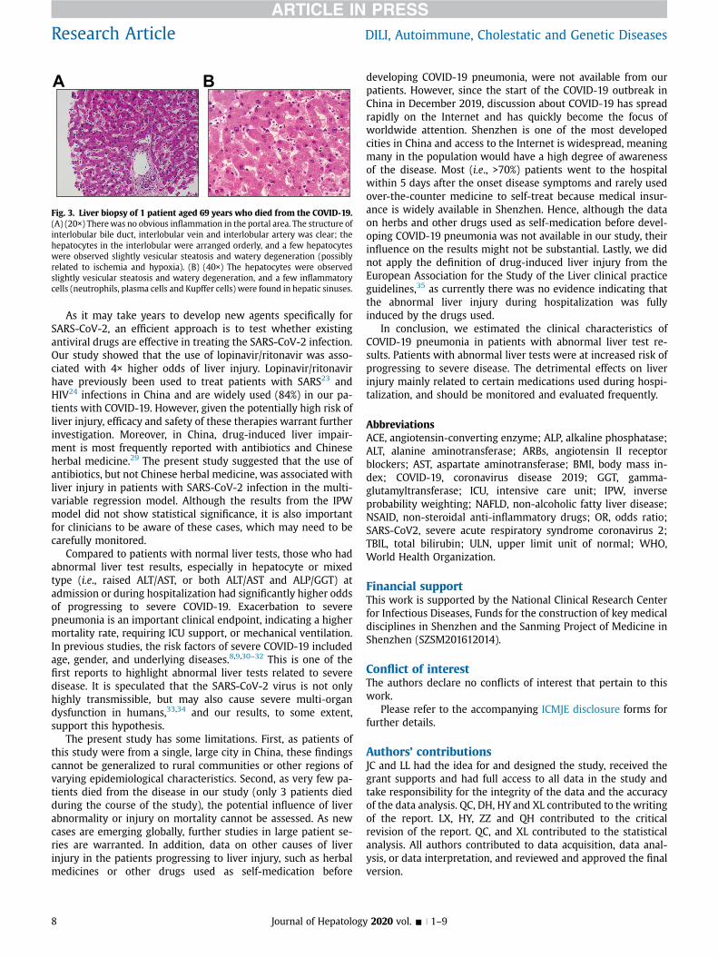
ribavirin, herbal medications, and interferon), no significant ev-idence showed the use of such drugs led to a higher risk of liverinjury (p >0.05 in IPW estimation), except for lopinavir/ritonavir(OR 4.44; 95% CI 1.50–13.17 in multivariable model and 5.03,1.78–14.23 in the IPW estimation; p <0.01) (Fig. 2). Patients whoused lopinavir/ritonavir had much higher levels of TBIL and GGTduring hospitalization (p from <0.004) (Table S1). One patientaged 69 years who died from COVID-19 had a liver biopsy. His-tological examination showed no obvious inflammation in theportal area (Fig. 3A). The structure of the interlobular bile duct,
y 2020 vol. - j 1–9
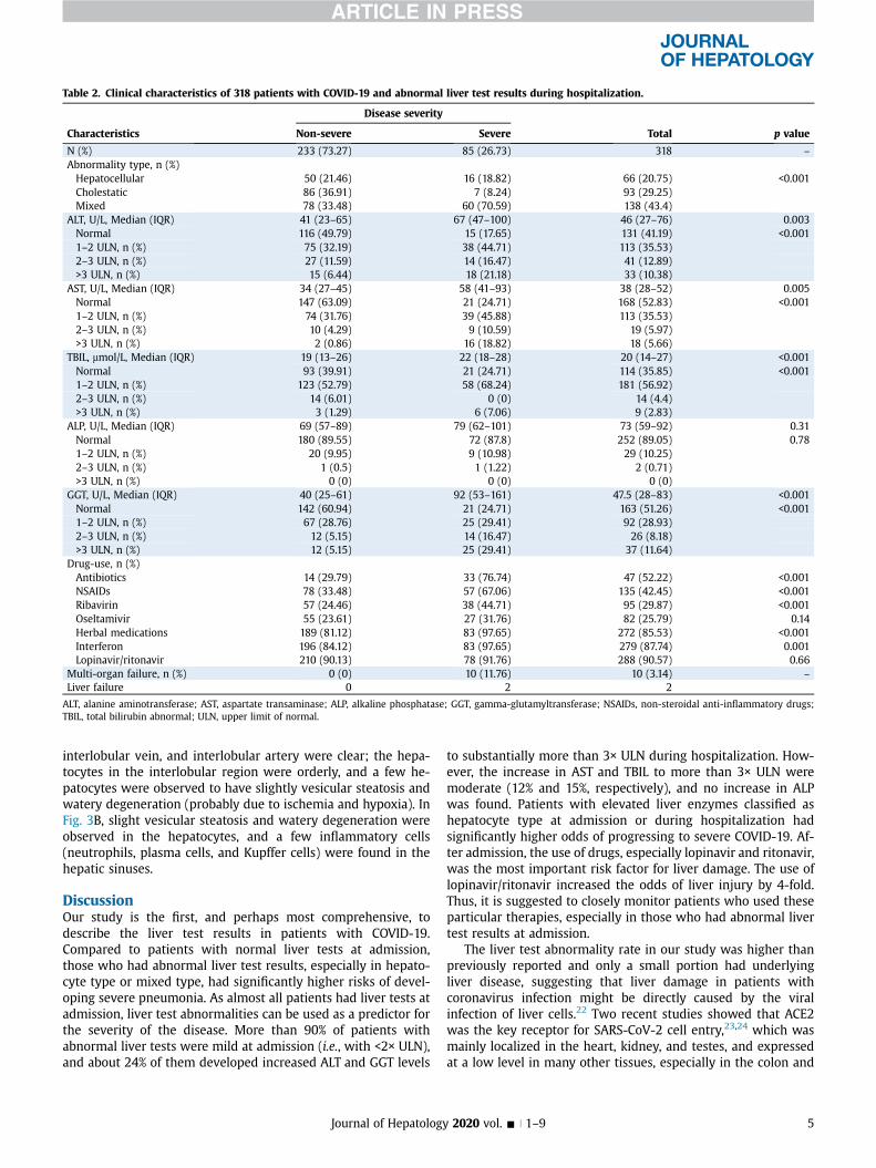
Table 2. Clinical characteristics of 318 patients with COVID-19 and abnormal liver test results during hospitalization.
Characteristics
Disease severity
Total p valueNon-severe Severe
N (%) 233 (73.27) 85 (26.73) 318 –
Abnormality type, n (%)Hepatocellular 50 (21.46) 16 (18.82) 66 (20.75) <0.001Cholestatic 86 (36.91) 7 (8.24) 93 (29.25)Mixed 78 (33.48) 60 (70.59) 138 (43.4)
ALT, U/L, Median (IQR) 41 (23–65) 67 (47–100) 46 (27–76) 0.003Normal 116 (49.79) 15 (17.65) 131 (41.19) <0.0011–2 ULN, n (%) 75 (32.19) 38 (44.71) 113 (35.53)2–3 ULN, n (%) 27 (11.59) 14 (16.47) 41 (12.89)>3 ULN, n (%) 15 (6.44) 18 (21.18) 33 (10.38)
AST, U/L, Median (IQR) 34 (27–45) 58 (41–93) 38 (28–52) 0.005Normal 147 (63.09) 21 (24.71) 168 (52.83) <0.0011–2 ULN, n (%) 74 (31.76) 39 (45.88) 113 (35.53)2–3 ULN, n (%) 10 (4.29) 9 (10.59) 19 (5.97)>3 ULN, n (%) 2 (0.86) 16 (18.82) 18 (5.66)
TBIL, lmol/L, Median (IQR) 19 (13–26) 22 (18–28) 20 (14–27) <0.001Normal 93 (39.91) 21 (24.71) 114 (35.85) <0.0011–2 ULN, n (%) 123 (52.79) 58 (68.24) 181 (56.92)2–3 ULN, n (%) 14 (6.01) 0 (0) 14 (4.4)>3 ULN, n (%) 3 (1.29) 6 (7.06) 9 (2.83)
ALP, U/L, Median (IQR) 69 (57–89) 79 (62–101) 73 (59–92) 0.31Normal 180 (89.55) 72 (87.8) 252 (89.05) 0.781–2 ULN, n (%) 20 (9.95) 9 (10.98) 29 (10.25)2–3 ULN, n (%) 1 (0.5) 1 (1.22) 2 (0.71)>3 ULN, n (%) 0 (0) 0 (0) 0 (0)
GGT, U/L, Median (IQR) 40 (25–61) 92 (53–161) 47.5 (28–83) <0.001Normal 142 (60.94) 21 (24.71) 163 (51.26) <0.0011–2 ULN, n (%) 67 (28.76) 25 (29.41) 92 (28.93)2–3 ULN, n (%) 12 (5.15) 14 (16.47) 26 (8.18)>3 ULN, n (%) 12 (5.15) 25 (29.41) 37 (11.64)
Drug-use, n (%)Antibiotics 14 (29.79) 33 (76.74) 47 (52.22) <0.001NSAIDs 78 (33.48) 57 (67.06) 135 (42.45) <0.001Ribavirin 57 (24.46) 38 (44.71) 95 (29.87) <0.001Oseltamivir 55 (23.61) 27 (31.76) 82 (25.79) 0.14Herbal medications 189 (81.12) 83 (97.65) 272 (85.53) <0.001Interferon 196 (84.12) 83 (97.65) 279 (87.74) 0.001Lopinavir/ritonavir 210 (90.13) 78 (91.76) 288 (90.57) 0.66
Multi-organ failure, n (%) 0 (0) 10 (11.76) 10 (3.14) –
Liver failure 0 2 2
ALT, alanine aminotransferase; AST, aspartate transaminase; ALP, alkaline phosphatase; GGT, gamma-glutamyltransferase; NSAIDs, non-steroidal anti-inflammatory drugs;TBIL, total bilirubin abnormal; ULN, upper limit of normal.
interlobular vein, and interlobular artery were clear; the hepa-tocytes in the interlobular region were orderly, and a few he-patocytes were observed to have slightly vesicular steatosis andwatery degeneration (probably due to ischemia and hypoxia). InFig. 3B, slight vesicular steatosis and watery degeneration wereobserved in the hepatocytes, and a few inflammatory cells(neutrophils, plasma cells, and Kupffer cells) were found in thehepatic sinuses.
DiscussionOur study is the first, and perhaps most comprehensive, todescribe the liver test results in patients with COVID-19.Compared to patients with normal liver tests at admission,those who had abnormal liver test results, especially in hepato-cyte type or mixed type, had significantly higher risks of devel-oping severe pneumonia. As almost all patients had liver tests atadmission, liver test abnormalities can be used as a predictor forthe severity of the disease. More than 90% of patients withabnormal liver tests were mild at admission (i.e., with <2× ULN),and about 24% of them developed increased ALT and GGT levels
Journal of Hepatolog
to substantially more than 3× ULN during hospitalization. How-ever, the increase in AST and TBIL to more than 3× ULN weremoderate (12% and 15%, respectively), and no increase in ALPwas found. Patients with elevated liver enzymes classified ashepatocyte type at admission or during hospitalization hadsignificantly higher odds of progressing to severe COVID-19. Af-ter admission, the use of drugs, especially lopinavir and ritonavir,was the most important risk factor for liver damage. The use oflopinavir/ritonavir increased the odds of liver injury by 4-fold.Thus, it is suggested to closely monitor patients who used theseparticular therapies, especially in those who had abnormal livertest results at admission.
The liver test abnormality rate in our study was higher thanpreviously reported and only a small portion had underlyingliver disease, suggesting that liver damage in patients withcoronavirus infection might be directly caused by the viralinfection of liver cells.22 Two recent studies showed that ACE2was the key receptor for SARS-CoV-2 cell entry,23,24 which wasmainly localized in the heart, kidney, and testes, and expressedat a low level in many other tissues, especially in the colon and
y 2020 vol. - j 1–9 5

Table 3. Clinical characteristics of patients with COVID-19 and liver injury.
Characteristics
Disease severity
Total p valueNon-severe Severe
N (%) 47 (52.22) 43 (47.78) 90 –
Abnormality type (1),† n (%)Hepatocellular 8 (17.02) 0 (0) 8 (8.89) 0.001Cholestatic 8 (17.02) 2 (4.65) 10 (11.11)Mixed 28 (59.57) 41 (95.35) 69 (76.67)
Abnormality type (2),† n (%)Hepatocellular 20 (42.55) 17 (39.53) 37 (41.11) 0.77Cholestatic 27 (57.45) 26 (60.47) 53 (58.89)
ALT, U/L, Median (IQR) 84 (42–136) 96 (63–159) 90.5 (53–145) 0.17Normal 10 (21.28) 2 (4.65) 12 (13.33) 0.141–2 ULN, n (%) 12 (25.53) 13 (30.23) 25 (27.78)2–3 ULN, n (%) 10 (21.28) 10 (23.26) 20 (22.22)>3 ULN, n (%) 15 (31.91) 18 (41.86) 33 (36.67)
AST, U/L, Median (IQR) 45 (29–81) 80 (56–145) 63 (36–101) 0.1Normal 20 (42.55) 4 (9.3) 24 (26.67) <0.0011–2 ULN, n (%) 15 (31.91) 16 (37.21) 31 (34.44)2–3 ULN, n (%) 10 (21.28) 7 (16.28) 17 (18.89)>3 ULN, n (%) 2 (4.26) 16 (37.21) 18 (20)
TBIL, lmol/L, Median (IQR) 22 (15–41) 25 (19–32) 23.5 (16–37) 0.13Normal 16 (34.04) 9 (20.93) 25 (27.78) <0.0011–2 ULN, n (%) 14 (29.79) 28 (65.12) 42 (46.67)2–3 ULN, n (%) 14 (29.79) 0 (0) 14 (15.56)>3 ULN, n (%) 3 (6.38) 6 (13.95) 9 (10)
ALP, U/L, Median (IQR) 77 (65–97) 92 (76–130) 83 (66–114.5) 0.01Normal 39 (95.12) 33 (76.74) 72 (85.71) 0.051–2 ULN, n (%) 2 (4.88) 9 (20.93) 11 (13.1)2–3 ULN, n (%) 0 (0) 1 (2.33) 1 (1.19)>3 ULN, n (%) 0 (0) 0 (0) 0 (0)
GGT, U/L, Median (IQR) 101 (39–148) 161 (122–211) 130.5 (77–187) <0.001Normal 16 (34.04) 0 (0) 16 (17.78) <0.0011–2 ULN, n (%) 7 (14.89) 4 (9.3) 11 (12.22)2–3 ULN, n (%) 12 (25.53) 14 (32.56) 26 (28.89)>3 ULN, n (%) 12 (25.53) 25 (58.14) 37 (41.11)
Drug-use, n (%)Antibiotics 14 (29.79) 33 (76.74) 47 (52.22) <0.001NSAIDs 18 (38.3) 29 (67.44) 47 (52.22) 0.006Ribavirin 16 (34.04) 17 (39.53) 33 (36.67) 0.59Oseltamivir 11 (23.4) 17 (39.53) 28 (31.11) 0.10Herbal 42 (89.36) 43 (100) 85 (94.44) 0.03Interferon 38 (80.85) 41 (95.35) 79 (87.78) 0.04Lopinavir/ritonavir 45 (95.74) 41 (95.35) 86 (95.56) 0.93
Multi-organ failure, n (%) 0 (0) 10 (23.26) 10 (11.11) –
ALT, alanine aminotransferase; AST, aspartate transaminase; ALP, alkaline phosphatase; GGT, gamma-glutamyltransferase; NSAIDs, non-steroidal anti-inflammatory drugs;TBIL, total bilirubin abnormal; ULN, upper limit of normal.†Abnormality type (1): Patients who had raised ALT and/or AST more than 3× the ULN were classified as hepatocyte type; patients who had raised ALP or GGT twice the ULNwere classified as cholangiocytes type; and patients who had a raised combination of both ALT/AST more 3 time the ULN and ALP/GGT twice the ULN were classified as mixedtype. Abnormality type (2): patients were classified as hepatocyte type when the AST/ALT activity was higher than the ALP/GGT activity and were classified as cholangiocytetype when the reverse occurred.
Research Article DILI, Autoimmune, Cholestatic and Genetic Diseases
lung.25 Another recent study showed that SARS-CoV-2 mightdirectly bind to ACE2 positive cholangiocytes and cause liverdamage,7 which may partially explain the contribution of SARS-CoV-2 infection to the liver test dysfunction in our patients.Moreover, the use of ACE-inhibitors and ARBs might also affectliver tests. All patients in this study with hypertension used ACE-inhibitors/ARBs at admission, subsequently, these medicationsmay have influenced their abnormal liver tests. However,although we found that those receiving ACE-inhibitors/ARBsappeared to have a higher percentage of abnormal liver tests atadmission (15.6–28.6% vs. 11.1%), the difference was not statisti-cally significant. Additionally, we excluded these 58 patientswith hypertension at admission and found that the prevalence ofabnormal liver function tests remained similar (from 46% to
6 Journal of Hepatolog
44%), suggesting that the influence of ACE-Is/ARBs drugs on livertests at admission, if any, should be minor. Moreover, our studyshowed that patients treated with ACE-inhibitors/ARBs were notat increased odds of progressing to severe disease compared topatients taking other antihypertensive drugs. The prevalence ofabnormal liver tests increased to 76% during hospitalization,which could be due to a more frequent examination (i.e., every 3to 5 days) and drugs used during hospitalization. Note that 84%of patients used lopinavir/ritonavir during hospitalization, drugswhich have been reported to cause liver damage and affect livertests.26
In our study, GGT was elevated substantially at admission andincreased to a much higher level during hospitalization, whereasthe increase in ALP was not pronounced. Both GGT and ALP were
y 2020 vol. - j 1–9
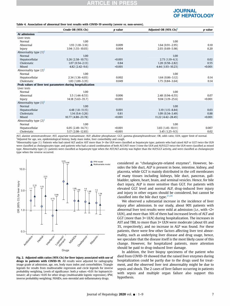
Table 4. Association of abnormal liver test results with COVID-19 severity (severe vs. non-severe).
Crude OR (95% CIs) p value Adjusted OR (95% CIs)† p value
At admissionLiver testsNormal 1.00 1.00Abnormal 1.93 (1.18–3.16) 0.009 1.64 (0.91–2.95) 0.10Injury 3.94 (1.55–10.03) 0.004 2.03 (0.69–5.98) 0.20
Abnormality type (1)‡
Normal 1.00 1.00Hepatocellular 5.26 (2.58–10.75) <0.001 2.73 (1.19–6.3) 0.02Cholestatic 1.07 (0.54–2.13) 0.84 1.28 (0.58–2.82) 0.55Mixed 4.82 (2.42–9.6) <0.001 4.44 (1.93–10.23) <0.001
Abnormality type (2)‡
Normal 1.00 1.00Hepatocellular 2.34 (1.36–4.03) 0.002 1.64 (0.86–3.12) 0.14Cholestatic 1.83 (1.00–3.35) 0.049 1.75 (0.84–3.64) 0.14
Peak values of liver test parameters during hospitalizationLiver testsNormal 1.00 1.00Abnormal 3.5 (1.44–8.53) 0.006 2.48 (0.94–6.55) 0.07Injury 14.18 (5.63–35.7) <0.001 9.04 (3.19–25.6) <0.001
Abnormality type (1)‡
Normal 1.00 1.00Hepatocellular 4.48 (1.8–11.15) 0.001 3.19 (1.15–8.84) 0.03Cholestatic 1.14 (0.4–3.26) 0.81 1.09 (0.34–3.49) 0.88Mixed 10.77 (4.88–23.78) <0.001 11.22 (4.42–28.45) <0.001
Abnormality type (2)‡
Normal 1.00 1.00Hepatocellular 6.05 (2.49–14.71) <0.001 3.83 (1.45–10.11) 0.007Cholestatic 5.17 (2.08–12.83) <0.001 3.45 (1.25–9.5) 0.02
ALT, alanine aminotransferase; AST, aspartate transaminase; ALP, alkaline phosphatase; GGT, gamma-glutamyltransferase; OR, odds ratio; ULN, upper limit of normal.†Adjusted for age, sex, epidemiological history, body mass index, liver comorbidity and cough.‡Abnormality type (1): Patients who had raised ALT and/or AST more than 3× the ULN were classified as hepatocyte type; patients who had raised ALP or GGT twice the ULNwere classified as cholangiocytes type; and patients who had a raised combination of both ALT/AST more 3 time the ULN and ALP/GGT twice the ULN were classified as mixedtype. Abnormality type (2): patients were classified as hepatocyte type when the AST/ALT activity was higher than the ALP/GGT activity, and were classified as cholangiocytetype when the reverse occurred.
2.15
1.69
1.341.48
2.21
0.99
4.44
1.7 1.691.51.6
1.23
0.73
5.03
0.3
1.0
3.0
10.0
Antibio
tics
Herbal
Interf
eron
Lopin
avir/
ritona
vir
NSAIDs
Oselta
mivir
Ribavir
in
Drugs
OR
(95%
CI)
for l
iver
inju
ry
IPW modelMultivariable model
Fig. 2. Adjusted odds ratios (95% CIs) for liver injury associated with use ofdrugs in patients with COVID-19. All results were adjusted for radiographyimage grade at admission, age, sex, body mass index and comorbidities. Trianglelegends for results from multivariable regression and circle legends for inverseprobability weighting. Levels of significance: both p values <0.01 for lopinavir/ri-tonavir; all p values >0.05 for other drugs (multivariable logistic regression). IPW,inverse probability weighting; NSAIDs, non-steroidal anti-inflammatory drugs.
Journal of Hepatolog
considered as “cholangiocyte-related enzymes”. However, be-sides the bile duct, ALP is present in bone, intestine, kidney, andplacenta, while GGT is mainly distributed in the cell membranesof many tissues including kidneys, bile duct, pancreas, gall-bladder, spleen, heart, brain, and seminal vesicles. Hence, for bileduct injury, ALP is more sensitive than GGT. For patients withelevated GGT level and normal ALP, drug-induced liver injuryand injury in other organs should be considered, but cannot beclassified into the bile duct type.27,28
We observed a substantial increase in the incidence of liverinjury after admission. In our study, about 90% patients withabnormal liver test results were mild at admission (i.e., with <2×ULN), and more than 10% of them had increased levels of ALT andGGT (more than 3× ULN) during hospitalization. The increases inAST and TBIL to more than 3× ULN were moderate (about 6% and3%, respectively), and no increase in ALP was found. For thesepatients, there were few other factors affecting liver test abnor-mality, such as underlying liver disease and drug usage, hence,we speculate that the disease itself is the most likely cause of thischange. However, for hospitalized patients, more attentionshould be paid to drug-induced liver damage.
In addition, the liver biopsy specimens of the patient whodied from COVID-19 showed that the raised liver enzymes duringhospitalization could be partly due to the drugs used for treat-ment, and the observed liver test abnormality might be due tosepsis and shock. The 2 cases of liver failure occurring in patientswith sepsis and multiple organ failure also support thishypothesis.
y 2020 vol. - j 1–9 7
A B
Fig. 3. Liver biopsy of 1 patient aged 69 years who died from the COVID-19.(A) (20×) Therewas no obvious inflammation in the portal area. The structure ofinterlobular bile duct, interlobular vein and interlobular artery was clear; thehepatocytes in the interlobular were arranged orderly, and a few hepatocyteswere observed slightly vesicular steatosis and watery degeneration (possiblyrelated to ischemia and hypoxia). (B) (40×) The hepatocytes were observedslightly vesicular steatosis and watery degeneration, and a few inflammatorycells (neutrophils, plasma cells and Kupffer cells) were found in hepatic sinuses.
Research Article DILI, Autoimmune, Cholestatic and Genetic Diseases
As it may take years to develop new agents specifically forSARS-CoV-2, an efficient approach is to test whether existingantiviral drugs are effective in treating the SARS-CoV-2 infection.Our study showed that the use of lopinavir/ritonavir was asso-ciated with 4× higher odds of liver injury. Lopinavir/ritonavirhave previously been used to treat patients with SARS23 andHIV24 infections in China and are widely used (84%) in our pa-tients with COVID-19. However, given the potentially high risk ofliver injury, efficacy and safety of these therapies warrant furtherinvestigation. Moreover, in China, drug-induced liver impair-ment is most frequently reported with antibiotics and Chineseherbal medicine.29 The present study suggested that the use ofantibiotics, but not Chinese herbal medicine, was associated withliver injury in patients with SARS-CoV-2 infection in the multi-variable regression model. Although the results from the IPWmodel did not show statistical significance, it is also importantfor clinicians to be aware of these cases, which may need to becarefully monitored.
Compared to patients with normal liver tests, those who hadabnormal liver test results, especially in hepatocyte or mixedtype (i.e., raised ALT/AST, or both ALT/AST and ALP/GGT) atadmission or during hospitalization had significantly higher oddsof progressing to severe COVID-19. Exacerbation to severepneumonia is an important clinical endpoint, indicating a highermortality rate, requiring ICU support, or mechanical ventilation.In previous studies, the risk factors of severe COVID-19 includedage, gender, and underlying diseases.8,9,30–32 This is one of thefirst reports to highlight abnormal liver tests related to severedisease. It is speculated that the SARS-CoV-2 virus is not onlyhighly transmissible, but may also cause severe multi-organdysfunction in humans,33,34 and our results, to some extent,support this hypothesis.
The present study has some limitations. First, as patients ofthis study were from a single, large city in China, these findingscannot be generalized to rural communities or other regions ofvarying epidemiological characteristics. Second, as very few pa-tients died from the disease in our study (only 3 patients diedduring the course of the study), the potential influence of liverabnormality or injury on mortality cannot be assessed. As newcases are emerging globally, further studies in large patient se-ries are warranted. In addition, data on other causes of liverinjury in the patients progressing to liver injury, such as herbalmedicines or other drugs used as self-medication before
8 Journal of Hepatolog
developing COVID-19 pneumonia, were not available from ourpatients. However, since the start of the COVID-19 outbreak inChina in December 2019, discussion about COVID-19 has spreadrapidly on the Internet and has quickly become the focus ofworldwide attention. Shenzhen is one of the most developedcities in China and access to the Internet is widespread, meaningmany in the population would have a high degree of awarenessof the disease. Most (i.e., >70%) patients went to the hospitalwithin 5 days after the onset disease symptoms and rarely usedover-the-counter medicine to self-treat because medical insur-ance is widely available in Shenzhen. Hence, although the dataon herbs and other drugs used as self-medication before devel-oping COVID-19 pneumonia was not available in our study, theirinfluence on the results might not be substantial. Lastly, we didnot apply the definition of drug-induced liver injury from theEuropean Association for the Study of the Liver clinical practiceguidelines,35 as currently there was no evidence indicating thatthe abnormal liver injury during hospitalization was fullyinduced by the drugs used.
In conclusion, we estimated the clinical characteristics ofCOVID-19 pneumonia in patients with abnormal liver test re-sults. Patients with abnormal liver tests were at increased risk ofprogressing to severe disease. The detrimental effects on liverinjury mainly related to certain medications used during hospi-talization, and should be monitored and evaluated frequently.
AbbreviationsACE, angiotensin-converting enzyme; ALP, alkaline phosphatase;ALT, alanine aminotransferase; ARBs, angiotensin II receptorblockers; AST, aspartate aminotransferase; BMI, body mass in-dex; COVID-19, coronavirus disease 2019; GGT, gamma-glutamyltransferase; ICU, intensive care unit; IPW, inverseprobability weighting; NAFLD, non-alcoholic fatty liver disease;NSAID, non-steroidal anti-inflammatory drugs; OR, odds ratio;SARS-CoV2, severe acute respiratory syndrome coronavirus 2;TBIL, total bilirubin; ULN, upper limit unit of normal; WHO,World Health Organization.
Financial supportThis work is supported by the National Clinical Research Centerfor Infectious Diseases, Funds for the construction of key medicaldisciplines in Shenzhen and the Sanming Project of Medicine inShenzhen (SZSM201612014).
Conflict of interestThe authors declare no conflicts of interest that pertain to thiswork.
Please refer to the accompanying ICMJE disclosure forms forfurther details.
Authors’ contributionsJC and LL had the idea for and designed the study, received thegrant supports and had full access to all data in the study andtake responsibility for the integrity of the data and the accuracyof the data analysis. QC, DH, HY and XL contributed to the writingof the report. LX, HY, ZZ and QH contributed to the criticalrevision of the report. QC, and XL contributed to the statisticalanalysis. All authors contributed to data acquisition, data anal-ysis, or data interpretation, and reviewed and approved the finalversion.
y 2020 vol. - j 1–9

AcknowledgementsWe thank Sarah Robbins Scott, an independent consultant for herhelp in English writing.
Supplementary dataSupplementary data to this article can be found online at https://doi.org/10.1016/j.jhep.2020.04.006.
ReferencesAuthor names in bold designate shared co-first authorship
[1] Dong Y, Liang X, Yu X. Prognostic value of the dynamic changes in extravascular lung water index and angiopoietin-2 in severe multiple traumapatients with acute respiratory distress syndrome. Zhonghua Wei ZhongBing Ji Jiu Yi Xue 2019;31:571–576.
[2] Niu P, Shen J, Zhu N, Lu R, Tan W. Two-tube multiplex real-time reversetranscription PCR to detect six human coronaviruses. Virologica Sinica2016;31:85–88.
[3] Lu R, Zhao X, Li J, Niu P, Yang B, Wu H, et al. Genomic characterisation andepidemiology of 2019 novel coronavirus: implications for virus originsand receptor binding. Lancet 2020;395:565–574.
[4] Jia Z, Yan L, Ren Z, Wu L, Wang J, Guo J, et al. Delicate structural coordi-nation of the Severe Acute Respiratory Syndrome coronavirus Nsp13 uponATP hydrolysis. Nucleic Acids Res 2019;47:6538–6550.
[5] Wu Z, McGoogan JM. Characteristics of and important lessons from thecoronavirus disease 2019 (COVID-19) outbreak in China: summary of areport of 72 314 cases from the Chinese Center for Disease Control andPrevention. JAMA 2020. https://doi.org/10.1001/jama.2020.2648. Pub-lished online February 24, 2020.
[6] Chau TN, Lee KC, Yao H, Tsang TY, Chow TC, Yeung YC, et al. SARS-asso-ciated viral hepatitis caused by a novel coronavirus: report of three cases.Hepatology 2004;39:302–310.
[7] Chai X, Hu L, Zhang Y, Han W, Lu Z, Ke A, et al. Specific ACE2 expression incholangiocytes may cause liver damage after 2019-nCoV infection. bio-Rxiv 2020. https://doi.org/10.1101/2020.02.03.931766.
[8] Chen N, Zhou M, Dong X, Qu J, Gong F, Han Y, et al. Epidemiological andclinical characteristics of 99 cases of 2019 novel coronavirus pneumoniain Wuhan, China: a descriptive study. Lancet 2020;395:507–513.
[9] Huang C, Wang Y, Li X, Ren L, Zhao J, Hu Y, et al. Clinical features ofpatients infected with 2019 novel coronavirus in Wuhan, China. Lancet2020;395:497–506.
[10] Tian S, Hu N, Lou J, Chen K, Kang X, Xiang Z, et al. Characteristics ofCOVID-19 infection in Beijing. J Infect 2020;80:401–406.
[11] Guan WJ, Ni ZY, Hu Y, Liang WH, Ou CQ, He JX, et al. Clinical charac-teristics of coronavirus disease 2019 in China. N Engl J Med 2020. https://doi.org/10.1056/NEJMoa2002032. Published online February 28, 2020.
[12] Yang W, Cao Q, Qin L, Wang X, Cheng Z, Pan A, et al. Clinical character-istics and imaging manifestations of the 2019 novel coronavirus disease(COVID-19): a multi-center study in Wenzhou city, Zhejiang, China.J Infect 2020;80:388–393.
[13] Zhang JJ, Dong X, Cao YY, Yuan YD, Yang YB, Yan YQ, et al. Clinical char-acteristics of 140 patients infected with SARS-CoV-2 in Wuhan, China.Allergy 2020. https://doi.org/10.1111/all.14238. Published online 19February 2020.
[14] Wang D, Hu B, Hu C, Zhu F, Liu X, Zhang J, et al. Clinical characteristics of138 hospitalized patients with 2019 novel coronavirus–infected pneu-monia in Wuhan, China. JAMA 2020;323:1061–1069.
[15] Xu Z, Shi L,Wang Y, Zhang J, Huang L, Zhang C, et al. Pathological findingsof COVID-19 associated with acute respiratory distress syndrome. LancetRespir Med 2020. https://doi.org/10.1016/S2213-2600(20)30076-X. Pub-lished online February 18, 2020.
[16] World Health Organization. Clinical management of severe acute respi-ratory infection when novel coronavirus (nCoV) infection is suspected:interim guidance. Available at, 2020. https://www.who.int/publications-
Journal of Hepatolog
detail/clinical-management-of-severe-acute-respiratory-infection-when-novel-coronavirus-(ncov)-infection-is-suspected. [Accessed 31 January2020].
[17] Bai Y, Yao L, Wei T, Tian F, Jin DY, Chen L, et al. Presumed AsymptomaticCarrier transmission of COVID-19. JAMA 2020. https://doi.org/10.1001/jama.2020.2565. Published online February 21, 2020.
[18] Metlay JP, Waterer GW, Long AC, Anzueto A, Brozek J, Crothers K, et al.Diagnosis and treatment of Adults with community-acquired pneumonia.An Official Clinical Practice Guideline of the American Thoracic Societyand Infectious Diseases Society of America. Am J Respir Crit Care Med2019;200:e45–e67.
[19] National Health Commission of the People’s Republic of China. Handbookof Prevention and Treatment of the Pneumonia Caused by the NovelCoronavirus (2019-nCoV) (in Chinese). Updated: February 6, 2020.Available at: 2020. http://en.nhc.gov.cn/2020-02/06/c_76295.htm.[Accessed 23 February 2020].
[20] European Association for the Study of the Liver, European Association forthe Study of Diabetes. EASL-EASD-EASO Clinical Practice Guidelines forthe management of non-alcoholic fatty liver disease. Obes Facts2016;9:65–90.
[21] European Association for the Study of the Liver. EASL 2017 Clinical Prac-tice Guidelines on the management of hepatitis B virus infection.J Hepatol 2017;67:370–398.
[22] Zhang C, Shi L, Wang FS. Liver injury in COVID-19: management andchallenges. Lancet Gastroenterol Hepatol 2020. https://doi.org/10.1016/S2468-1253(20)30057-1. Published online March 04, 2020.
[23] Hoffmann M, Kleine-Weber H, Schroeder S, Krüger N, Herrler T,Erichsen S, et al. SARS-CoV-2 cell entry depends on ACE2 and TMPRSS2and is blocked by a clinically proven protease inhibitor. Cell 2020.https://doi.org/10.1016/j.cell.2020.02.052. Published online March 05,2020.
[24] Yan R, Zhang Y, Li Y, Xia L, Guo Y, Zhou Q. Structural basis for therecognition of SARS-CoV-2 by full-length human ACE2. Science2020;367:1444–1448.
[25] Clarke NE, Turner AJ. Angiotensin-converting enzyme 2: the first decade.Int J Hypertens 2012;2012:307315.
[26] Meraviglia P, Schiavini M, Castagna A, Viganò P, Bini T, Landonio S, et al.Lopinavir/ritonavir treatment in HIV antiretroviral-experienced patients:evaluation of risk factors for liver enzyme elevation. HIV Med2004;5:334–343.
[27] Dillon JF, Miller MH. Gamma glutamyl transferase ‘To be or not to be’aliver function test? London, England: SAGE Publications Sage UK; 2016.
[28] Fernandez NJ, Kidney BA. Alkaline phosphatase: beyond the liver. Vet ClinPathol 2007;36:223–233.
[29] Zhu Y, Niu M, Chen J, Zou Z-s, Ma Z-j, Liu S-h, et al. Hepatobiliary andpancreatic: comparison between Chinese herbal medicine and Westernmedicine-induced liver injury of 1985 patients. J Gastroenterol Hepatol2016;31:1476–1482.
[30] Wang D, Hu B, Hu C, Zhu F, Liu X, Zhang J, et al. Clinical characteristics of138 hospitalized patients with 2019 novel coronavirus-infected pneu-monia in Wuhan, China. JAMA 2020.
[31] Xu XW, Wu XX, Jiang XG, Xu KJ, Ying LJ, Ma CL, et al. Clinical findings in agroup of patients infected with the 2019 novel coronavirus (SARS-Cov-2)outside of Wuhan, China: retrospective case series. BMJ 2020;368:m606.
[32] Zhu N, Zhang D, Wang W, Li X, Yang B, Song J, et al. A novel coronavirusfrom patients with pneumonia in China, 2019. N Engl J Med2020;382:727–733.
[33] MacLaren G, Fisher D, Brodie D. Preparing for the most critically ill pa-tients with COVID-19: the potential role of extracorporeal membraneoxygenation. JAMA 2020. https://doi.org/10.1001/jama.2020.2342. Pub-lished online February 19, 2020.
[34] Zhang W. Imaging changes of severe COVID-19 pneumonia in advancedstage. Intensive Care Med 2020:1–3.
[35] Andrade RJ, Aithal GP, Bjornsson ES, Kaplowitz N, Kullak-Ublick GA,Karlsen TH, et al. EASL clinical practice guidelines: drug-induced liverinjury. J Hepatol 2019;70:1222–1261.
y 2020 vol. - j 1–9 9