cov-2 infection month outcomes after hospitalization for
TRANSCRIPT

Page 1/26
“Long COVID” follow-up pathway: results oflongitudinal cohort study to investigate 6- and 12-month outcomes after hospitalization for SARS-CoV-2 infectionMarta Rigoni ( [email protected] )
University of TrentoEmanuele Torri
Provincia Autonoma di TrentoGiandomenico Nollo
University of TrentoLivia Delle Donne
U.O. di Medicina InternaSebastiano Rizzardo
U.O. di Medicina InternaLorenza Lenzi
U.O. di Medicina InternaAndrea Falzone
Unità Operativa multizonale di RadiologiaSusanna Cozzio
U.O. di Medicina Interna
Research Article
Keywords: SARS-CoV-2, long COVID, post-COVID, 12 months follow-up, pathway, chest images
Posted Date: September 21st, 2021
DOI: https://doi.org/10.21203/rs.3.rs-908078/v1
License: This work is licensed under a Creative Commons Attribution 4.0 International License. Read Full License

Page 2/26
AbstractLong-term sequelae of symptomatic infection caused by SARS-CoV-2 are largely undiscovered. Weperformed a prospective cohort study on consecutively hospitalized Sars-CoV-2 patients (March-May2020) for evaluating COVID-19 outcomes at 6 and 12 months. After hospital discharge, patients wereaddressed to two follow-up pathways based on respiratory support needed during hospitalization.Outcomes were assessed by telephone consultation or ambulatory visit. Among 471 patients, 81.6%received no respiratory support during hospitalization; 8.2% received non-invasive ventilation; 0.2%required invasive mechanical ventilation (IMV). 64 patients died during hospitalization, therefore 407were enrolled for follow-up. At 6 months, among 355patients, the 27.0% had any symptoms, 19.4%dyspnea, 5.4% neurological symptoms. Fifty-two out of 104 had major damages in interstitial ComputedTomography images. IMV patients had higher probability to suffer of neurological symptoms (OR=4.12,p=0.01). At 12 months, among 344, the 24.4% suffered on any symptoms, 14.0% dyspnea, 10.0%neurological symptoms. Severe interstitial lesions were present in 37 out of 47 investigated patients. IMVpatients in respect to no respiratory support, had higher probability of experiencing symptoms (OR=3.51,p<0.01), dyspnea (OR=3.08, p<0.01 ), neurological symptoms (OR= 11.50, p<0.01). COVID-19 patientsshowed prolonged sequelae up to 12 months, highlighting the need of follow-up pathways for post-COVID-19 syndrome.
IntroductionCoronavirus disease 2019 (COVID-19) is a severe acute respiratory infection caused by the emergentcoronavirus, SARS-CoV-2. Though most infected individuals are asymptomatic, SARS-CoV-2 infectionmay cause symptoms ranging from mild to severe acute respiratory distress, with a substantial fractionof patients requiring hospitalization (estimated to be around 10%) and many patients experiencingprolonged symptoms and complications for weeks or months after the initial period of acute illness 1–6.
The long COVID-19 phase or post-acute sequelae (signs and symptoms that continue or develop afteracute COVID-19) includes both ongoing symptomatic COVID-19 (from 4 to 12 weeks) and post-COVID-19syndrome (12 weeks or more) 6,7 and are not explained by an alternative diagnosis. Therefore, patientsshould be followed up to detect and manage sequelae and functional impairment 8,9.
Since COVID-19 has spread globally, and long COVID became a burgeoning health concern 10, a growingnumber of studies has been focused on “long-term effects of COVID-19” 11–18. However, to date only afew studies 19–24 really addressed long term (up to 12 months) sequelae for hospitalized patientpopulations, as well as temporal trends and longstanding health consequences for “long-haulers” with awhole patient pathway perspective in different populations, timepoints, settings and countries. The fullrange of long-term health consequences of COVID-19 in patients who were discharged from hospital islargely unclear 25.
Page 3/26
We implemented a strati�ed follow-up pathway and performed a prospective observational cohort studyin patients who survived hospitalization for acute COVID-19 with the aim to:
Investigate the prevalence of COVID-related symptoms and additional clinical �ndings at 6 and 12months after hospital discharge;
Identify the association of hospital respiratory support needed to help deal with the long-term effectsof COVID-19.
Methods
Study design and participantsWe conducted a prospective observational cohort study in adult patients with COVID-19 who had beenadmitted to the Internal Medicine ward of the Santa Maria del Carmine Hospital of Rovereto (Italy),between March 1, 2020, and May 31, 2020. The setting was a public general hospital with 300 beds thatwas identi�ed in the �rst phase of the Italian COVID-19 outbreak as a regional hub hospital for COVIDpatients in the Autonomous Province of Trento (northeastern Italy, around 543,000 inhabitants).
All COVID diagnoses were con�rmed using molecular diagnostic tests for SARS-CoV-2 (ReverseTranscription Polymerase Chain Reaction, RT-PCR) performed with a nasopharyngeal swab,oropharyngeal swab, or bronchoalveolar lavage using the standard protocols determined by the ItalianHealth Ministry, consistent with the World Health Organization’s interim guidance diagnostic criteria foradults with severe COVID-19 pneumonia 26,27.
We set up a long-term strati�ed follow-up pathway supported by a telehealth solution (a structured andrecorded call) that enabled patient monitoring and evaluation and the recording of long-lastingsymptoms and clinical �ndings in patients hospitalized with COVID-19.
We implemented two different follow-up routes targeting two different patient groups on the basis of theseverity of COVID disease assessed during hospitalization through the Brescia-COVID RespiratorySeverity Scale (BCRSS), a scoring system developed in Italy during the early phase of the COVID-19pandemic to aid the assessment and management of patients with COVID-19 pneumonia 28. The scorewas calculated for each patient at the time of admission and in the case of clinical worsening duringhospital stay; for patient strati�cation, we used the worst score recorded.
We further categorized and routed patients as follows:
cohort 1: patients with mild COVID-19, treated without oxygen or with nasal cannula or mask toadminister supplemental low �ow oxygen-therapy < 6 l/minute (BCRSS 0–1); independently of theseverity of COVID-19 during hospitalization, patients with severe cognitive or motor disabilities,residents of long-term care facilities and those refusing (i.e. because living far away from thehospital) the ambulatory follow up were included in this group;

Page 4/26
cohort 2: patients with moderate to severe disease requiring oxygen-therapy > 6 l/min, High-FlowNasal Cannula (HFNC), Non-Invasive Ventilation (NIV) including Continuous positive airway pressure(CPAP), or invasive mechanical ventilation (IMV) (BCRSS ≥ 2). All patients in IMV previously failed torecover on HFNC or NIV.
The time frames for the follow-up visits were as follows: 6 months (25–27 weeks post symptom onset),and 12 months (50–54 weeks post symptom onset).
ProceduresAll patients pertaining to cohort 1 and 2 were assessed at 6 months and 12 months after discharge andwere investigated for serum levels of SARS-CoV-2 IgG antibodies (6 and 12 months) and status ofvaccination anti-SARS-CoV-2 (fully vaccinated individuals) at 12-months, when vaccination was availableand indicated for infected individuals according to Italian guidance on vaccination of COVID survivors 29.
Patients enrolled in route one (cohort 1) have been phone interviewed by physicians of the generalmedicine ward following a structured questionnaire (Table 1S Supplementary material) conceivedsimilarly to other published tools 30 investigating clinical recovery and presence of COVID relatedsymptoms. Additionally, all patients were assessed with the modi�ed British Medical Research Council(mMRC) dyspnea scale, a �ve-category scale to characterize the level of dyspnea with physical activity inwhich higher scores correspond with increased dyspnea (from 0 to 4) 31. In case of mMRC scale ≥ 1 thepatient was shifted to ambulatory follow up at the same timepoint. For people with disabilities or long-term care facilities residents the interview was supported by caregiver or treating physician.
Patients enrolled in route two (cohort 2) have been evaluated with follow-up in person. We performed acomplete ambulatory visit where patients were interviewed with the same questionnaire used at distanceand undertaken the mMRC score. In addition, the same day of the visit, patients performed a functionalassessment carried out with Six-Minute-Walking test (6MWT) 32 which was performed on all patients at 6months and at 12 months only on patients showing altered 6MWT test at 6 months; as part of thepathway, patients with altered 6MWT were referred to pneumological specialistic consultation to performadditional lung tests. In addition, we performed a chest non-contrast high resolution ComputedTomography (CT) scan on all patients at 6 months and only on survivors with interstitial abnormalities ofdegree 3 and 4 (see below) at 6 months for the 12-month follow-up.
The focus was whether lesions identi�ed on the CT, involved one or both lungs and several lung lobes.The evaluation of the imaging appearance included: lesion density (ground-glass opacity, consolidation),distribution (unilateral, bilateral), interlobular septa thickening, crazy-paving patterns, bronchiolectasisand bronchiectasis. The degree of chest imaging changes of the lung parenchyma and lung interstitiumwere graded on a 5-point scale based on the severity of the abnormal �nding and the extent of lunginvolvement.
For parenchymal CT the score was categorized as follows:
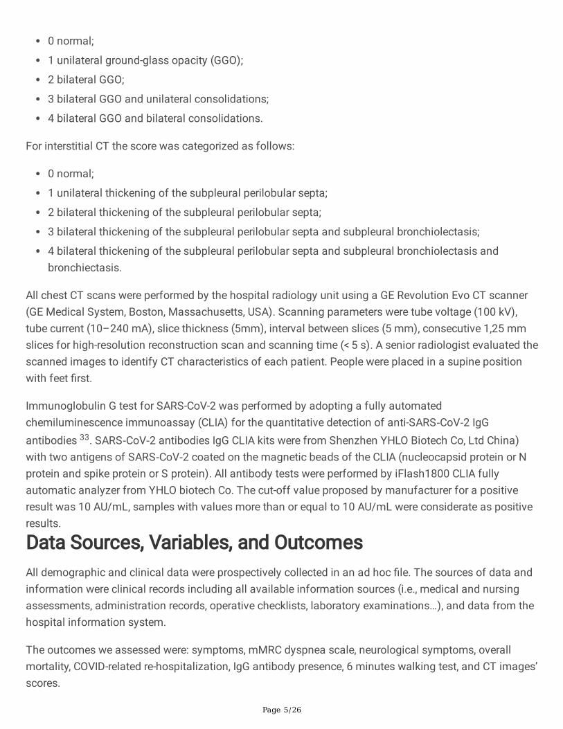
Page 5/26
0 normal;
1 unilateral ground-glass opacity (GGO);
2 bilateral GGO;
3 bilateral GGO and unilateral consolidations;
4 bilateral GGO and bilateral consolidations.
For interstitial CT the score was categorized as follows:
0 normal;
1 unilateral thickening of the subpleural perilobular septa;
2 bilateral thickening of the subpleural perilobular septa;
3 bilateral thickening of the subpleural perilobular septa and subpleural bronchiolectasis;
4 bilateral thickening of the subpleural perilobular septa and subpleural bronchiolectasis andbronchiectasis.
All chest CT scans were performed by the hospital radiology unit using a GE Revolution Evo CT scanner(GE Medical System, Boston, Massachusetts, USA). Scanning parameters were tube voltage (100 kV),tube current (10–240 mA), slice thickness (5mm), interval between slices (5 mm), consecutive 1,25 mmslices for high-resolution reconstruction scan and scanning time (< 5 s). A senior radiologist evaluated thescanned images to identify CT characteristics of each patient. People were placed in a supine positionwith feet �rst.
Immunoglobulin G test for SARS-CoV-2 was performed by adopting a fully automatedchemiluminescence immunoassay (CLIA) for the quantitative detection of anti-SARS‐CoV‐2 IgGantibodies 33. SARS‐CoV‐2 antibodies IgG CLIA kits were from Shenzhen YHLO Biotech Co, Ltd China)with two antigens of SARS‐CoV‐2 coated on the magnetic beads of the CLIA (nucleocapsid protein or Nprotein and spike protein or S protein). All antibody tests were performed by iFlash1800 CLIA fullyautomatic analyzer from YHLO biotech Co. The cut-off value proposed by manufacturer for a positiveresult was 10 AU/mL, samples with values more than or equal to 10 AU/mL were considerate as positiveresults.
Data Sources, Variables, and OutcomesAll demographic and clinical data were prospectively collected in an ad hoc �le. The sources of data andinformation were clinical records including all available information sources (i.e., medical and nursingassessments, administration records, operative checklists, laboratory examinations…), and data from thehospital information system.
The outcomes we assessed were: symptoms, mMRC dyspnea scale, neurological symptoms, overallmortality, COVID-related re-hospitalization, IgG antibody presence, 6 minutes walking test, and CT images’scores.
Page 6/26
The study was approved by the Ethics Committee of the Autonomous Province of Trento (referencenumber 4659) and followed the Declaration of Helsinki Ethical Principles for Medical Research InvolvingHuman Subjects. Informed consent was obtained from all individual participants.
Statistical analysesDichotomous variables or scores were expressed as frequencies and percentages of occurrence.Continuous variables were checked for normality using Shapiro–Wilk test and, since their distributionwas non-normal, they were expressed by median and �rst and third quartiles (q1-q3). Differences amonggroups were tested using the Fisher’s exact test. We performed univariate logistic regression models toassess whether patients with greater respiratory needs during hospitalization had higher risk of havingsymptoms at the follow-up. Therefore, for each outcome we calculated the Odds Ratio (OR) for IMVpatients in respect to no therapy of low �ow oxygen, and the OR for HFNC/NIV patients in respect to notherapy of low �ow oxygen. A P-value < 0.05 was considered to be statistically signi�cant. Statisticalanalyses were performed using the Stata software, (StataCorp, College Station, Texas USA).
ResultsThe demographic and clinical characteristics of the 471 eligible hospitalized patients who were admittedto hospital with COVID-19 are shown in Table 1.

Page 7/26
Table 1Characteristics of hospitalized patients.
Characteristics 471 patients
Male, n (%) 300/471 (63.7)
Female, n (%) 171/471 (36.3)
Age, median (Q1-Q3) years 71 (58–81)
Age male, median (Q1-Q3) years 68 (56–78)
Age female, median (Q1-Q3) years 76 (61–85)
BCRSS at admission 0, n (%) 227/471 (48.2)
BCRSS at admission 1, n (%) 112/471 (23.8)
BCRSS at admission 2, n (%) 47/471 (10.0)
BCRSS at admission 3, n (%) 45/471 (9.5)
BCRSS at admission 4, n (%) 38/471 (8.1)
BCRSS at admission 5, n (%) 2/471 (0.4)
P/F ratio at admission, median (q1-q3) 310 (191–368)
P/F ratio < 300, n (%) 218/469 (46.5)
NEWS2 at admission, median (q1-q3) 4 (2–7)
NEWS2 0–4, n (%) 258/471 (54.9)
NEWS2 5–6, n (%) 83/471 (17.7)
NEWS2 ≥ 7, n (%) 129/471 (27.4)
Comorbidities:
Cardiovascular (including hypertension), n (%) 289/471 (61.4)
Diabetes, n (%) 82/471 (17.4)
Gastrointestinal, n (%) 73/471 (15.5)
Autoimmune, n (%) 63/471 (13.4)
Obesity, n (%) 55/471 (11.7)
Pulmonary, n (%) 58/471 (12.3)
Renal, n (%) 51/471 (10.8)
Legend: BCRSS = Brescia COVID Respiratory Severity Scale; HFNC = High-Flow Nasal Cannula; IMV = Invasive Mechanical Ventilation; NEWS2 = National Early Warning Score 2; NIV = Non-invasiveVentilation; P/F ratio = PiO2/FiO2 ratio; Q1 = �rst quartile; Q3 = third quartile; TI = tracheal intubation.
Page 8/26
Characteristics 471 patients
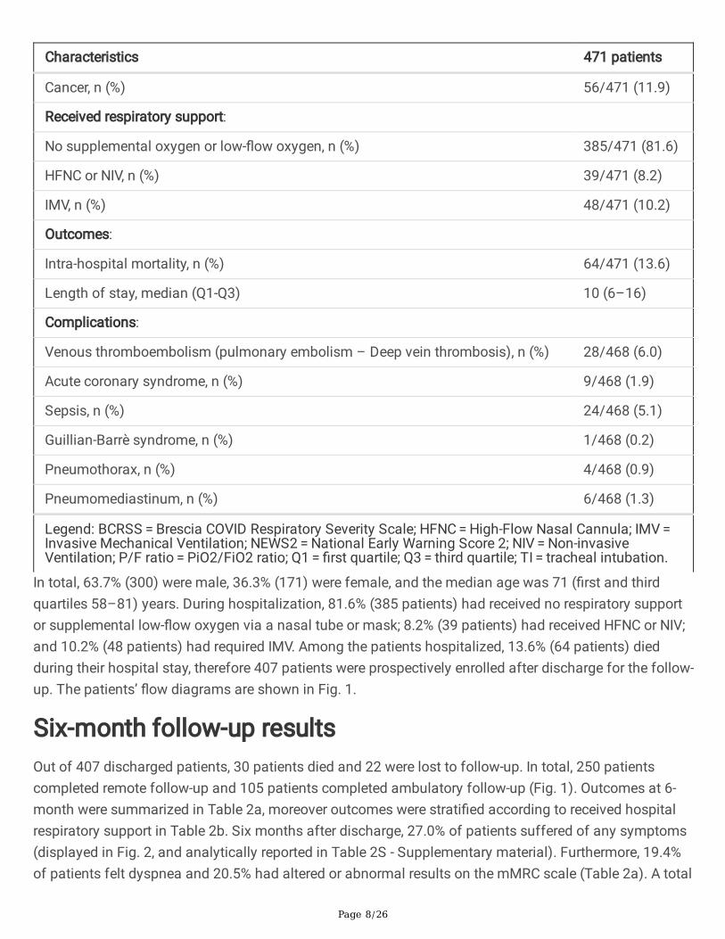
Cancer, n (%) 56/471 (11.9)
Received respiratory support:
No supplemental oxygen or low-�ow oxygen, n (%) 385/471 (81.6)
HFNC or NIV, n (%) 39/471 (8.2)
IMV, n (%) 48/471 (10.2)
Outcomes:
Intra-hospital mortality, n (%) 64/471 (13.6)
Length of stay, median (Q1-Q3) 10 (6–16)
Complications:
Venous thromboembolism (pulmonary embolism – Deep vein thrombosis), n (%) 28/468 (6.0)
Acute coronary syndrome, n (%) 9/468 (1.9)
Sepsis, n (%) 24/468 (5.1)
Guillian-Barrè syndrome, n (%) 1/468 (0.2)
Pneumothorax, n (%) 4/468 (0.9)
Pneumomediastinum, n (%) 6/468 (1.3)
Legend: BCRSS = Brescia COVID Respiratory Severity Scale; HFNC = High-Flow Nasal Cannula; IMV = Invasive Mechanical Ventilation; NEWS2 = National Early Warning Score 2; NIV = Non-invasiveVentilation; P/F ratio = PiO2/FiO2 ratio; Q1 = �rst quartile; Q3 = third quartile; TI = tracheal intubation.
In total, 63.7% (300) were male, 36.3% (171) were female, and the median age was 71 (�rst and thirdquartiles 58–81) years. During hospitalization, 81.6% (385 patients) had received no respiratory supportor supplemental low-�ow oxygen via a nasal tube or mask; 8.2% (39 patients) had received HFNC or NIV;and 10.2% (48 patients) had required IMV. Among the patients hospitalized, 13.6% (64 patients) diedduring their hospital stay, therefore 407 patients were prospectively enrolled after discharge for the follow-up. The patients’ �ow diagrams are shown in Fig. 1.
Six-month follow-up resultsOut of 407 discharged patients, 30 patients died and 22 were lost to follow-up. In total, 250 patientscompleted remote follow-up and 105 patients completed ambulatory follow-up (Fig. 1). Outcomes at 6-month were summarized in Table 2a, moreover outcomes were strati�ed according to received hospitalrespiratory support in Table 2b. Six months after discharge, 27.0% of patients suffered of any symptoms(displayed in Fig. 2, and analytically reported in Table 2S - Supplementary material). Furthermore, 19.4%of patients felt dyspnea and 20.5% had altered or abnormal results on the mMRC scale (Table 2a). A total
Page 9/26
of 5.4% of patients experienced neurological symptoms (see Fig. 2 and Table 2S for details). Globally, 20different general symptoms/neurological symptoms were reported by patients. The COVID-related 6-month re-hospitalization was 2.8%, and overall mortality was 20.9%. The IgG were found in the 95.3% ofthe tested patients, while 13.4% of patients showed an altered or scarce walking test. At 6 months 86.5%of 104 patients investigated showed different degrees of chest imaging abnormalities of the lung:including parenchymal GGO in 61.5% of patients (with bilateral damage in 50.0% of cases and 2.9% withconsolidation). The interstitium abnormalities recorded included thickening of the subpleural perilobularsepta in 86.5% of patients, bilateral thickening was present in 78.8% of cases, 33.6% also with bilateralsubpleural bronchiolectasis and 16.3% with bronchiectasis in addition to brochiolectasis.

Page 10/26
Table 2a. Outcomes at 6 months.
Outcomes n (%)
Symptoms 96/355 (27.0)
Dyspnea 69/355 (19.4)
mMRC scale 0 282/355 (79.5)
mMRC scale 1 59/355 (16.6)
mMRC scale 2 9/355 (2.5)
mMRC scale 3 4/355 (1.1)
mMRC scale 4 1/355 (0.3)
Neurological symptoms 19/355 (5.4)
COVID-related re-hospitalization 10/355 (2.8)
Overall 6-month mortality 94/449 (20.9)
IgG 163/171 (95.3)
Walking test 0 90/104 (86.6)
Walking test 1 12/104 (11.5)
Walking test 2 2/104 (1.9)
Parenchymal CT 0 40/104 (38.5)
Parenchymal CT 1 9/104 (8.6)
Parenchymal CT 2 52/104 (50.0)
Parenchymal CT 3 1/104 (1.0)
Parenchymal CT 4 2/104 (1.9)
Interstitial CT 0 14/104 (13.5)
Interstitial CT 1 8/104 (7.7)
Interstitial CT 2 30/104 (28.9)
Interstitial CT 3 35/104 (33.6)
Interstitial CT 4 17/104 (16.3)
Legend: CT = computed tomography; IgG = Immunoglobulin G; mMRC = Modi�ed Medical ResearchCouncil.
As far as outcomes strati�ed by received respiratory support during hospitalization are concerned (Table2b), patients treated with IMV had higher percentage of neurological symptoms (15.1%) in comparison to

Page 11/26
HFNC/NIV (6.2%) and to no therapy or low-�ow oxygen (4.1%), p = 0.03 (Odds Ratio IMV versus notherapy = 4.12 (1.35–12.55), p = 0.01, Odds Ratio HFNC/NIV versus no therapy = 1.54 (0.33–7.20), p = 0.58). No statistical differences in the CT scores based on patients’ respiratory support were noted.

Page 12/26
Table 2b. Outcomes at 6 months strati�ed by received respiratory support.
No therapy or low-�ow oxygen, n(%)
HFNC/NIV, n(%)
IMV, n (%) p-value
Outcomes
Symptoms 74/290 (25.5) 10/32 (31.2) 12/33(36.4)
0.34
Dyspnea 53/290 (18.3) 8/32 (25.0) 8/33 (24.2) 0.44
mMRC scale 0 236/290 (81.4) 22/32 (68.8) 24/33(72.7)
0.24
mMRC scale 1 42/290 (14.5) 10/32 (31.2) 7/33 (21.2)
mMRC scale 2 7/290 (2.4) 0/32 (0.0) 2/33 (6.1)
mMRC scale 3 4/290 (1.4) 0/32 (0.0) 0/33 (0.0)
mMRC scale 4 1/290 (0.3) 0/32 (0.0) 0/33 (0.0)
Neurologicalsymptoms
12/289 (4.1) 2/32 (6.2) 5/33 (15.1) 0.03
Walking test 0 70/78 (89.7) 9/11 (81.8) 11/15(73.3)
0.05
Walking test 1 8/78 (10.3) 2/11 (18.2) 2/15 (13.3)
Walking test 2 0/78 (0.0) 0/11 (0.0) 2/15 (13.3)
Parenchymal CT 0 32/78 (41.0) 5/12 (41.7) 4/15 (26.7) 0.37
Parenchymal CT 1 4/78 (5.1) 3/12 (25.0) 2/15 (13.3)
Parenchymal CT 2 39/78 (50.0) 4/12 (33.3) 9/15 (60.0)
Parenchymal CT 3 1/78 (1.3) 0/12 (0.0) 0/15 (0.0)
Parenchymal CT 4 2/78 (2.6) 0/12 (0.0) 0/15 (0.0)
Interstitial CT 0 12/78 (15.6) 1/12 (8.3) 1/15 (6.7) 0.42
Interstitial CT 1 7/78 (9.1) 0/12 (0.0) 1/15 (6.7)
Interstitial CT 2 20/78 (26.0) 4/12 (33.3) 6/15 (40.0)
Interstitial CT 3 28/78 (36.4) 5/12 (41.7) 2/15 (13.3)
Interstitial CT 4 10/78 (13.0) 2/12 (16.7) 5/15 (33.3)
Legend: HFNC = High-Flow Nasal Cannula; IgG = Immunoglobulin G; IMV = Invasive MechanicalVentilation; mMRC = Modi�ed Medical Research Council; NIV = Non-invasive Mechanical Ventilation;TC = Computed Tomography.

Page 13/26
Twelve-month follow-up resultsBy the 12-month follow-up, 5 more patients died, 237 patients performed a phone follow-up, 107 patientsperformed an ambulatory follow-up, and 6 more patients were lost to follow-up (Fig. 1). Outcomes at 12months were summarized in Table 3a, moreover outcomes were strati�ed according to received hospitalrespiratory support in Table 3b. Twelve months after discharge, 24.4% of patients suffered of anysymptoms (see Fig. 2, and Table 2S for details); 14.0% of patients felt dyspnea, and 14.5% of patientshad altered or abnormal results on the mMRC scale (Table 3a). In total, the 10.0% of patients experiencedneurological symptoms (see Fig. 2, and Table 2S for details). Globally, 23 differentsymptoms/neurological symptoms were reported by patients. The COVID-related 12-month re-hospitalization was 1.4%, and overall mortality was 22.3%. IgG antibodies were found in the 85.7% of thepatients tested and 41.0% of the patients followed up had been vaccinated. Among the patientsinvestigated with CT images at 12 months (47), GGO was still present in 51.0% of patients (with bilateraldamage in 46.8 % of cases), while 2.1% continued to show consolidation (score > 2). Interstitiumabnormalities in this group of patients included thickening of the subpleural perilobular septa in 95.8% ofpatients, bilateral thickening was found in 12.8 % of cases, 51.1% also with bilateral subpleuralbronchiolectasis and 27.2% with bronchiectasis in addition to brochiolectasis. Among patients followedup with CT at 6 months, it could be estimated that any form of GGO was found in 23.1% of cases, whileinterstitial severe damages (score > 2) were still present in 35.6% of patients followed up with CT at 6months.

Page 14/26
Table 3a. Outcomes at 12 months.
Outcomes n (%)
Symptoms 84/344 (24.4)
Dyspnea 48/344 (14.0)
mMRC scale 0 294/344 (85.5)
mMRC scale 1 38/344 (11.0)
mMRC scale 2 7/344 (2.0)
mMRC scale 3 4/344 (1.2)
mMRC scale 4 1/344 (0.3)
Neurological symptoms 34/341 (10.0)
COVID-related re-hospitalization 5/344 (1.4)
Overall 12-month mortality 99/443 (22.3)
IgG 192/224 (85.7)
Vaccinated 132/322 (41.0)
Walking test 0 7/11 (63.6)
Walking test 1 4/11 (36.4)
Walking test 2 0/11 (0.0)
Parenchymal CT 0 23/47 (49.0)
Parenchymal CT 1 1/47 (2.1)
Parenchymal CT 2 22/47 (46.8)
Parenchymal CT 3 1/47 (2.1)
Parenchymal CT 4 0/47 (0.0)
Interstitial CT 0 2/47 (4.2)
Interstitial CT 1 2/47 (4.2)
Interstitial CT 2 6/47 (12.8)
Interstitial CT 3 24/47 (51.1)
Interstitial CT 4 13/47 (27.7)
Legend: CT = computed tomography; IgG = Immunoglobulin G; mMRC = Modi�ed Medical ResearchCouncil.

Page 15/26
As far as outcomes strati�ed by received respiratory support during hospitalization are concerned (Table3b), patients treated with IMV and HFNC/NIV had higher probability of experiencing symptoms in respectto no therapy (Odds Ratio IMV versus no therapy = 3.51 (1.68–7.31), p < 0.01, Odds Ratio HFNC/NIVversus no therapy = 2.25 (1.05–4.86), p = 0.04). Similarly, patients treated with IMV and HFNC/NIV hadhigher probability of experiencing dyspnea in respect to no therapy (Odds Ratio IMV versus no therapy = 3.08 (1.35–7.01), p < 0.01; Odds Ratio HFNC/NIV versus no therapy = 1.32 (0.48–3.66), p = 0.59). Indeed,mMRC scale showed worst score for IMV in respect to HFNC/NIV and no therapy, p = 0.02 (Table 3b).Moreover, patients treated with IMV and HFNC/NIV had higher probability of experiencing neurologicalsymptoms in comparison to no therapy or low-�ow oxygen (Odds Ratio IMV versus no therapy = 11.50(4.79–27.61), p < 0.01; Odds Ratio HFNC/NIV versus no therapy = 5.00 (1.85–13.49), p < 0.01). Nostatistical differences were noted in CT scores based on patients’ respiratory support.

Page 16/26
Table 3b. Outcomes at 12 months strati�ed by received respiratory support.
No therapy or low-�ow oxygen, n(%)
HFNC/NIV, n(%)
IMV, n (%) p-value
Outcomes
Symptoms 56/277 (20.2) 12/33 (36.4) 16/34(47.1)
< 0.01
Dyspnea 33/277 (11.9) 5/33 (15.2) 10/34(29.4)
0.03
mMRC scale 0 243/277 (87.7) 28/33 (84.9) 23/34(67.7)
0.02
mMRC scale 1 26/277 (9.4) 4/33 (12.1) 8/34 (23.5)
mMRC scale 2 5/277 (1.8) 0/33 (0.0) 2/34 (5.9)
mMRC scale 3 3/277 (1.1) 1/33 (3.0) 0/34 (0.0)
mMRC scale 4 0/277 (0.0) 0/33 (0.0) 1/34 (2.9)
Neurologicalsymptoms
14/274 (5.1) 7/33 (21.2) 13/34(38.2)
< 0.01
Walking test 0 6/8 (75.0) 0/0 (0.0) 1/3 (33.3) 0.49
Walking test 1 2/8 (25.0) 0/0 (0.0) 2/3 (66.7)
Walking test 2 0/8 (0.0) 0/0 (0.0) 0/3 (0.0)
Parenchymal CT 0 6/13 (46.1) 9/14 (64.3) 8/20 (40.0) 0.38
Parenchymal CT 1 0/13 (0.0) 1/14 (7.1) 0/20 (0.0)
Parenchymal CT 2 7/13 (53.9) 4/14 (28.6) 11/20(55.0)
Parenchymal CT 3 0/13 (0.0) 0/14 (0.0) 1/20 (5.0)
Parenchymal CT 4 0/13 (0.0) 0/14 (0.0) 0/20 (0.0)
Interstitial CT 0 0/13 (0.0) 2/14 (14.3) 0/20 (0.0) 0.15
Interstitial CT 1 2/13 (15.4) 0/14 (0.0) 0/20 (0.0)
Interstitial CT 2 2/13 (15.4) 3/14 (21.4) 1/20 (5.0)
Interstitial CT 3 5/13 (38.4) 6/14 (42.9) 13/20(65.0)
Legend: CT = computed tomography; HFNC = High-Flow Nasal Cannula; IgG = Immunoglobulin G; IMV = Invasive Mechanical Ventilation; mMRC = Modi�ed Medical Research Council; NIV = Non-invasiveventilation; TI = tracheal intubation.
Page 17/26
No therapy or low-�ow oxygen, n(%)
HFNC/NIV, n(%)
IMV, n (%) p-value
Interstitial CT 4 4/13 (30.8) 3/14 (21.4) 6/20 (30.0)
Legend: CT = computed tomography; HFNC = High-Flow Nasal Cannula; IgG = Immunoglobulin G; IMV = Invasive Mechanical Ventilation; mMRC = Modi�ed Medical Research Council; NIV = Non-invasiveventilation; TI = tracheal intubation.
DiscussionWe implemented a long-term strati�ed follow-up path that adhered to the international proposed clinicalguidance for the assessment and management of COVID-19 patients 7,8,34,35, in the context of the limitedavailability of resources and the operational pressure faced by hospitals during the pandemic. Thisproject was part of a broad set of strategies and paths implemented within the Healthcare Trust of theAutonomous Province of Trento for reorganizing care and managing patients at a distance, includingroutine services and the continuation of care after hospital discharge 36, as well assessing the impact ofpandemic burden and related change in practice 37. In our study, we targeted the same cohort of patients(excluding six patients with multiple admissions) to investigate in-hospital outcomes during the COVID-19 pandemic 38.
Overall, in our study any long COVID symptoms were reported at 6 months in 27.0% of patients and at 12months in 24.4%, but we noted an increased number of different symptoms at 12 months. Alterations inthe mMRC scale were reported in almost 20.5% of patients at 6 months and in 14.5% of patients at 12months. The frequency of neurocognitive symptoms increased from 5.4–10.0% from the 6-month to the12-month timepoints. At both 6 and 12 months, neurocognitive symptoms were markedly more commonin patients who had been treated with mechanical ventilation during hospital admission. Furthermore,patients who had been treated with tracheal intubation in comparison to HFNC or NIV and who hadreceived no therapy or low-�ow oxygen reported a signi�cantly higher prevalence of experiencing anysymptom and having dyspnea at 12 months.
Published studies investigating the highly heterogeneous and poorly understood post-COVID-19syndrome show the relevance of medical and psychological sequelae for several months after activeinfection, with more than 50 long-term effects of COVID-19 having been reported 39. Pooled prevalencedata show that the 10 most prevalent symptoms are fatigue, shortness of breath, muscle pain, joint pain,headache, cough, chest pain, altered smell, altered taste, and diarrhea 40. The increasingly evident long-term neurological effects include the impact of the virus on cognition, autonomic function, and mentalwellbeing 41. Patients with long COVID present with prolonged multisystem involvement and signi�cantdisability 13. The development of long COVID symptoms may be linked to symptomatic COVID-19infection, hospitalization (with mechanical ventilation being required), severity of illness, and sex (womenmay have a higher incidence) 42–44. However, any patient with COVID-19 may develop long COVID,regardless of the severity of their infection and the intensity of the treatment they received 43.

Page 18/26
The few published prospective studies using a 12-month timepoint providing an overview of the clinicalsymptoms and quality of life of adult patients show that, although decreasing over time, a meaningfulportion of patients still report persistent symptoms one year after infection 20,22−25. Overall, our �ndingsare in line with such results, although with a lower proportion of patients experiencing persistentsymptoms, with a meaningful proportion of patients of experiencing dyspnea and patterns of neurologicsymptoms. Clinical impairment can persist at least until one year after COVID-19 symptom onset andreduce patients’ quality of life signi�cantly 22. The persistence of neuropsychiatric long COVID symptoms(which can reduce quality of life signi�cantly) one year after COVID-19 symptom onset may be partiallyexplained by the in�uence of the extended pandemic situation and consequent psychological impact 22.
Our �ndings seem consistent with the literature showing the persistence of chest imaging manifestationsmonths after hospitalization for COVID-19 pneumonia 12,45−47 and the persistence (although decreasingover time) of pulmonary alterations up to 12 months later 21,25. However, we did not �nd any associationbetween lung structural abnormalities and the severity of the disease during hospital stay. Lung imagingpatterns at 12 months may be associated with lung diffusion impairment, although further studies areneeded to explore the effect of these persistent abnormalities on physical function and quality of life 25.Inour study, we found a decreased detection of IgG antibodies in SARS-CoV-2-infected patients, droppingfrom 95.3% at six months to 85.7% at 12 months. The detectability of antibodies 1 year after infection,although with different temporal trends and magnitudes, seems to con�rm the �ndings of other studiesthat have carried out comparable long-term follow-ups 48–50. The relationship between the antibody leveland protection against COVID-19 is still unclear; however, the one-year follow-up data show that patientswho have recovered from COVID-19 have a very low risk of reinfection. Natural immunity to SARS-CoV-2appears to confer a protective effect for at least a year 51. The rate of full vaccine coverage at 12 monthsseem to mirror the progress of the Italian vaccination campaign, which is still underway at the timeframeof the writing, re�ecting the need of speci�c logistic organization 52–54.
All in all, our study shows the high clinical burden of long COVID-19 12 months after acute infection anda�rms the importance of understanding the natural course of long COVID as a long-term chroniccondition with symptoms persisting beyond 12 months after the onset of illness. Identifying patients atmajor risk of sequelae from the early post-acute phase; setting up appropriate and patient-centeredpathways supported by online support tools; and the implementation of surveillance systems andspecialized multidisciplinary care, including rehabilitation, are critical in order to understand and treatpatients suffering from long COVID 40,55−58.
The development and implementation of clinical guidelines 59 as well as use of tools and methods forHealth Technology Assessment 60,61 in order to evaluate the effects of decisions and actions related toresource allocation, models of care, professional practice, drugs, and medical devices in response to thecomplex and evolving challenges of long COVID may enable value-based decisions to be made 62.
Strenghts And Limitations

Page 19/26
To the best of our knowledge, our research represents one of the few 12-month follow-up studies onCOVID-19 reported to date, addressing a unique mix of symptoms and clinical �ndings and including theinvestigation of a broad range of clinical symptoms, serum antibodies titers, pulmonary functions, andCT imaging and vaccination statuses.
The strength of our study is its long-term follow-up of a well-characterized patient population from the�rst peak of the pandemic in Italy (with almost no missing data and no risk of any selection bias) withina real world standardized pathway in which we prioritized instrumental assessments and investigationsdirected at survivors by exploiting telehealth solution to reduce ambulatory burden and patient risk.Following appropriateness principle, in deep diagnosis as CT at 12 months was performed only onsurvivors who had had interstitial abnormalities of degree 3 and 4 at 6 months, and the Six-MinuteWalking test at 12 months was performed only on patients who had had altered test results at 6 months.Considering the diversity of the initial target population and the duration of the pathway, we believe thatour drop-out rate was very low, which may be attributable to the bene�t of engaging patients within astructured and patient-oriented follow-up path.
We acknowledge the potential bias caused by not performing the same investigations on all patientsprospectively followed by either comparing cohort one and two as well as within cohort two (see above).In addition, our �ndings apply to a population selected in the early phase of the pandemic, in which wedid not have a clinical assessment for use before acute infection. Both presenting patient characteristicsand clinical management have evolved since. The generalizability of our results may be limited by thepotential patient population selection bias, lack of standardized validated questionnaires for reporting theprevalence of COVID-19 symptoms at the international level and the self-reporting of symptoms based ona remote interview (19) as well as the study’s single-center, not-blinded, and not-randomized design.
ConclusionsOur study may provide valuable information enabling relevant professionals to understand the clinicalneeds of long COVID patients and identify practical and feasible approaches for the routine follow-up andmanagement of patients with long-term COVID-19 complications. Large and long-ranging observationalstudies and clinical trials for investigating long-term sequelae, as well as follow-up pathways for lookingafter people with long-term complications after acute COVID-19, should be considered.
DeclarationsFunding
The authors received no funding for this work.
ETHICS STATEMENT
Page 20/26
The study protocol was approved by the ethic committee of the Autonomous Province of Trento(reference number 4659) and followed the Declaration of Helsinki Ethical Principles for Medical ResearchInvolving Human Subjects. Informed consent was obtained from all individual participants for whomidentifying information is included in this article.
COMPETING INTERESTS STATEMENT
The authors declare no competing interests.
ACKNOWLEDGEMENTS
We acknowledge all patients who participated in this study and their families. We would also like to thankthe staff of this follow-up study team conducted at Santa Maria del Carmine Hospital.
AUTHOR CONTRIBUTIONS
All authors have made substantial contributions to all of the following: (1) the conception and design ofthe study (SC, LDD, GN, MR and ET), or acquisition of data (LL, SR, LDD, and AF), or analysis andinterpretation of data (MR, GN, and ET), (2) drafting the article or revising it critically for importantintellectual content (ET, MR, GN, LDD, and SC), (3) �nal approval of the version to be submitted (allauthors).
DATA AVAILABILITY STATEMENT
Data are available on reasonable requests.
References1. World Health Organization. Coronavirus disease (COVID-19) – World Health Organization.
https://www.who.int/emergencies/diseases/novel-coronavirus-2019.
2. World Health Organization. Critical preparedness, readiness and response actions for COVID-19:interim guidance, 22 March 2020. https://apps.who.int/iris/handle/10665/331511 (2020).
3. Wu, Z. & McGoogan, J. M. Characteristics of and Important Lessons From the Coronavirus Disease2019 (COVID-19) Outbreak in China: Summary of a Report of 72 314 Cases From the Chinese Centerfor Disease Control and Prevention. JAMA, 323, 1239–1242 (2020).
4. Scherlinger, M. et al. Re�ning “Long-COVID” by a Prospective Multimodal Evaluation of Patients withLong-Term Symptoms Attributed to SARS-CoV-2 Infection. Infect. Dis. Ther, 10, 1747–1763 (2021).
5. Nalbandian, A. et al. Post-acute COVID-19 syndrome. Nat. Med, 27, 601–615 (2021).
�. Datta, S. D., Talwar, A. & Lee, J. T. A Proposed Framework and Timeline of the Spectrum of DiseaseDue to SARS-CoV-2 Infection: Illness Beyond Acute Infection and Public Health Implications. JAMA,324, 2251–2252 (2020).

Page 21/26
7. COVID-19 rapid guideline: managing the long-term effects of COVID-19. (National Institute for Healthand Care Excellence (UK), 2020).
�. CDC. Healthcare Workers. Centers for Disease Control and Preventionhttps://www.cdc.gov/coronavirus/2019-ncov/hcp/clinical-care/post-covid-conditions.html (2020).
9. Venkatesan, P. NICE guideline on long COVID. Lancet Respir. Med, 9, 129 (2021).
10. The Lancet. null. Facing up to long COVID. Lancet Lond. Engl, 396, 1861 (2020).
11. Logue, J. K. et al. Sequelae in Adults at 6 Months After COVID-19 Infection. JAMA Netw. Open, 4,e210830 (2021).
12. Huang, C. et al. 6-month consequences of COVID-19 in patients discharged from hospital: a cohortstudy. Lancet Lond. Engl, 397, 220–232 (2021).
13. Davis, H. E. et al. Characterizing long COVID in an international cohort: 7 months of symptoms andtheir impact. EClinicalMedicine, 38, 101019 (2021).
14. Havervall, S. et al. Symptoms and Functional Impairment Assessed 8 Months After Mild COVID-19Among Health Care Workers. JAMA, 325, 2015–2016 (2021).
15. Carfì, A., Bernabei, R. & Landi, F. & Gemelli Against COVID-19 Post-Acute Care Study Group. PersistentSymptoms in Patients After Acute COVID-19. JAMA, 324, 603–605 (2020).
1�. Munblit, D. et al. Incidence and risk factors for persistent symptoms in adults previously hospitalizedfor COVID-19. Clin. Exp. Allergy J. Br. Soc. Allergy Clin. Immunol, https://doi.org/10.1111/cea.13997(2021).
17. Akbarialiabad, H. et al. Long COVID, a comprehensive systematic scoping review. Infection 1–24(2021) doi:10.1007/s15010-021-01666-x.
1�. Günster, C. et al. 6-month mortality and readmissions of hospitalized COVID-19 patients: Anationwide cohort study of 8,679 patients in Germany. PLoS ONE, 16, e0255427 (2021).
19. Boscolo-Rizzo, P. et al. Sequelae in adults at 12 months after mild-to-moderate coronavirus disease2019 (COVID-19). Int. Forum Allergy Rhinol, https://doi.org/10.1002/alr.22832 (2021).
20. Wu, X. et al. 6-month, 9-month, and 12-month respiratory outcomes in patients following COVID-19-related hospitalisation: a prospective study. Lancet Respir. Med, 9, 3–3 (2021).
21. Yan, X. et al. Follow-up study of pulmonary function among COVID-19 survivors 1 year after recovery.J. Infect, 83, 381–412 (2021).
22. Seeßle, J. et al. Persistent symptoms in adult patients one year after COVID-19: a prospective cohortstudy. Clin. Infect. Dis. Off. Publ. Infect. Dis. Soc. Am, ciab611, https://doi.org/10.1093/cid/ciab611(2021).
23. Budhiraja, S. et al. Long Term Health Consequences of COVID-19 in Hospitalized Patients from NorthIndia: A follow up study of upto 12 months 2021.06.21.21258543https://www.medrxiv.org/content/10.1101/2021.06.21.21258543v1 (2021)doi:10.1101/2021.06.21.21258543.
Page 22/26
24. Sigfrid, L. et al. Long Covid in adults discharged from UK hospitals after Covid-19: A prospective,multicentre cohort study using the ISARIC WHO Clinical Characterisation Protocol. Lancet Reg.Health Eur, 100186, https://doi.org/10.1016/j.lanepe.2021.100186 (2021).
25. Huang, L. et al. 1-year outcomes in hospital survivors with COVID-19: a longitudinal cohort study.Lancet Lond. Engl, 398, 747–758 (2021).
2�. World Health Organization. Clinical management of severe acute respiratory infection (SARI) whenCOVID-19 disease is suspected. Interim guidance. Pediatr. Med. Rodz, 16, 9–26 (2020).
27. Italian Health Ministry. General Directorate of Health Prevention. Circular letter no. 11715 of April 1st2021. Pandemia di COVID-19 – Aggiornamento delle indicazioni sui test diagnostici e sui criteri daadottare nella determinazione delle priorità.Aggiornamento delle indicazioni relative alla diagnosi dilaboratorio.
2�. Duca, A., Piva, S., Focà, E., Latronico, N. & Rizzi, M. Calculated Decisions: Brescia-COVID RespiratorySeverity Scale (BCRSS)/Algorithm. Emerg. Med. Pract, 22, CD1–CD2 (2020).
29. Italian Health Ministry. General Directorate of Health Prevention. Circular letter no. 8248 of March 3rd2021. Vaccinazione dei soggetti che hanno avuto un’infezione da SARS-CoV-2.
30. Genecand, L. et al. [Diagnostic and therapeutic management of medium and long-term sequelae ofSARS-CoV-2 infection]. Rev. Med. Suisse, 17, 842–849 (2021).
31. Bestall, J. C. et al. Usefulness of the Medical Research Council (MRC) dyspnoea scale as a measureof disability in patients with chronic obstructive pulmonary disease., 54, 581–586 (1999).
32. Enright, P. L. & Sherrill, D. L. Reference equations for the six-minute walk in healthy adults. Am. J.Respir. Crit. Care Med, 158, 1384–1387 (1998).
33. Infantino, M. et al. Diagnostic accuracy of an automated chemiluminescent immunoassay for anti-SARS-CoV-2 IgM and IgG antibodies: an Italian experience. J. Med. Virol, 92, 1671–1675 (2020).
34. Raghu, G. & Wilson, K. C. COVID-19 interstitial pneumonia: monitoring the clinical course in survivors.Lancet Respir. Med, 8, 839–842 (2020).
35. Greenhalgh, T., Knight, M., A’Court, C., Buxton, M. & Husain, L. Management of post-acute covid-19 inprimary care. BMJ, 370, m3026 (2020).
3�. Testa, S. et al. Implementation of tele visit healthcare services triggered by the COVID-19 emergency:the Trentino Province experience. Z. Gesundheitswissenschaften J. Public Health, 1–16,https://doi.org/10.1007/s10389-021-01609-8 (2021).
37. Ciarleglio, F. A. et al. The negative effects of COVID-19 and national lockdown on emergency surgerymorbidity due to delayed access. World J. Emerg. Surg. WJES, 16, 37 (2021).
3�. Rigoni, M., Torri, E., Nollo, G., Delle Donne, L. & Cozzio, S. NEWS2 is a valuable tool for appropriateclinical management of COVID-19 patients. Eur. J. Intern. Med, 85, 118–120 (2021).
39. Lopez-Leon, S. et al. More than 50 long-term effects of COVID-19: a systematic review and meta-analysis. Sci. Rep, 11, 16144 (2021).

Page 23/26
40. Aiyegbusi, O. L. et al. Symptoms, complications and management of long COVID: a review. J. R. Soc.Med, 1410768211032850, https://doi.org/10.1177/01410768211032850 (2021).
41. Jesuthasan, A., Massey, F., Manji, H., Zandi, M. S. & Wiethoff, S. Emerging potential mechanisms andpredispositions to the neurological manifestations of COVID-19. J. Neurol. Sci, 428, 117608 (2021).
42. Baratta, J. M., Tompary, A., Siano, S., Floris-Moore, M. & Weber, D. J. Postacute Sequelae of COVID-19Infection and Development of a Physiatry-Led Recovery Clinic. Am. J. Phys. Med. Rehabil, 100, 633–634 (2021).
43. Crook, H., Raza, S., Nowell, J., Young, M. & Edison, P. Long covid-mechanisms, risk factors, andmanagement. BMJ, 374, n1648 (2021).
44. Fernández-de-Las-Peñas, C. et al. Prevalence of post-COVID-19 symptoms in hospitalized and non-hospitalized COVID-19 survivors: A systematic review and meta-analysis. Eur. J. Intern. Med, S0953-6205 (21), 00208–00209 https://doi.org/10.1016/j.ejim.2021.06.009 (2021).
45. Blanco, J. R. et al. Pulmonary long-term consequences of COVID-19 infections after hospitaldischarge. Clin. Microbiol. Infect. Off. Publ. Eur. Soc. Clin. Microbiol. Infect. Dis, 27, 892–896 (2021).
4�. Sideris, G. A. et al. Imaging in the COVID-19 era: Lessons learned during a pandemic. World J. Radiol,13, 192–222 (2021).
47. Zhang, S. et al. Eight months follow-up study on pulmonary function, lung radiographic, and relatedphysiological characteristics in COVID-19 survivors. Sci. Rep, 11, 13854 (2021).
4�. Laing, E. D. et al. SARS-CoV-2 antibodies remain detectable 12 months after infection and antibodymagnitude is associated with age and COVID-19 severity 2021.04.27.21256207https://www.medrxiv.org/content/10.1101/2021.04.27.21256207v1 (2021)doi:10.1101/2021.04.27.21256207.
49. Xiao, K. et al. Antibodies Can Last for More Than 1 Year After SARS-CoV-2 Infection: A Follow-UpStudy From Survivors of COVID-19. Front. Med, 8, 684864 (2021).
50. Haveri, A. et al. Persistence of neutralizing antibodies a year after SARS-CoV-2 infection 2021.07.13.21260426https://www.medrxiv.org/content/10.1101/2021.07.13.21260426v1 (2021)doi:10.1101/2021.07.13.21260426.
51. Vitale, J. et al. Assessment of SARS-CoV-2 Reinfection 1 Year After Primary Infection in a Populationin Lombardy, Italy. JAMA Intern. Med, https://doi.org/10.1001/jamainternmed.2021.2959 (2021).
52. COVID-19 Vaccine Tracker | European Centre for Disease Prevention and Control.https://vaccinetracker.ecdc.europa.eu/public/extensions/COVID-19/vaccine-tracker.html#uptake-tab.
53. Pilati, F., Tronconi, R., Nollo, G., Heragu, S. S. & Zerzer, F. Digital Twin of COVID-19 Mass VaccinationCenters. Sustainability, 13, 7396 (2021).
54. Nollo, G., Pilati, F., Tronconi, F. & Rigoni, M. R. & Industry 4.0 at the service of public health against theCOVID-19 pandemic. Disaster Med. Public Health Prep. In press.

Page 24/26
55. Garg, M. et al. The Conundrum of ‘Long-COVID-19’: A Narrative Review. Int. J. Gen. Med, 14, 2491–2506 (2021).
5�. Menges, D. et al. Burden of post-COVID-19 syndrome and implications for healthcare serviceplanning: A population-based cohort study. PloS One, 16, e0254523 (2021).
57. Rajan, S. et al. In the wake of the pandemic: Preparing for Long COVID (European Observatory onHealth Systems and Policies, 2021).
5�. Herrera, J. E. et al. Multidisciplinary collaborative consensus guidance statement on the assessmentand treatment of fatigue in postacute sequelae of SARS-CoV-2 infection (PASC) patients. PM R,https://doi.org/10.1002/pmrj.12684 (2021).
59. Schünemann, H. J. et al. Using GRADE in situations of emergencies and urgencies: certainty inevidence and recommendations matters during the COVID-19 pandemic, now more than ever and nomatter what. J. Clin. Epidemiol, 127, 202–207 (2020).
�0. Miglietta, A. et al. Health technology assessment applied to emergency preparedness: a newperspective. Int. J. Technol. Assess. Health Care, 37, e77 (2021).
�1. Grossi, A. et al. Hospital contextual factors affecting the implementation of health technologies: asystematic review. BMC Health Serv. Res, 21, 407 (2021).
�2. Torri, E. & Nollo, G. Public Health Decision-Making in the Real World: Four Points to Reshape It AfterCOVID-19.Disaster Med. Public Health Prep.1–2doi:10.1017/dmp.2020.108.
Figures

Page 25/26
Figure 1
Patients’ �ow diagram.

Page 26/26
Figure 2
General (blue color) and neurological symptoms (green color) at 6 (left) and 12 months (right) afterdischarge for hospitalized SARS-CoV-2 patients.
Supplementary Files
This is a list of supplementary �les associated with this preprint. Click to download.
Supplementarytables.docx