course- diseases of field crop and their management (dfcm)
TRANSCRIPT

Laboratory Manual
Course- Diseases of Field Crop and their Management
(DFCM)
B.Sc.(Hons.) Agriculture Vth
Semester
Department of Agriculture,
Faculty of Science & Engineering
Jharkhand Rai University, Ranchi.
Contents Sl No. Experiments Page No.
1. Rice Diseases 1
2. Sorghum smuts & Bajra Diseases 5
3. Diseases of wheat 12
4. Maize & Sugarcane Diseases 25 5. Turmeric & Tobacco Diseases 39 6. Groundnut & Sunflower Diseases 44
7. Sesamum & Cotton Diseases 54
8. Red gram, Green gram & Black gram diseases 65
9 Bengal gram & Bean Diseases 71
10. Field visit for Disease Survey 86
11. Preparation of Herbarium 87

Experiment-1: Rice Diseases (Blast, Brown spot & Sheath blight)
1. Rice blast
Symptoms
On the leaves the lesion/ spots first appear as minute brown specks, and then grow to become spindle-
shaped, pointed at both ends. The center of the spots is usually gray or whitish with brown or reddish-
brown margin. Fully developed lesions reach 1-1.5 cm long, 0.3-0.5 broad. Under favorable conditions,
lesions enlarge and coalesce; eventually kill the leaves.
Infection at the junction of the leaf blade and sheath in the typical brown ―collar rot‖ symptom. A severe
collar rot can cause the leaf to die completely. When collar rot kill the flag or penultimate leaf it may have
a significant impact on yield.
The pathogen also causes brown lesions on the branches on the panicles and on the spikelets pedicles,
resulting in “panicle blast”.Infection of the neck, panicle branches, and spikelets pedicles may occur
together or may occur separately.
Causal organism
Magnaporthegrisea is a filamentous, heterothallic Ascomycotina that collectively causes disease on
many species of the grass (Poaceae) family. M. grisea is the teleomorph corresponding to the previously
distinct anamorphs Pyriculariaoryzae, infecting rice (Oryza sativa), and P. grisea, infecting other
grasses. However, P. oryzae and P. grisea have now been synonymized, with the earlier name, P. grisea,
having priority.
1

Management Cultural
Remove collateral weed hosts from bunds and channels
Use only disease free seedlings
Avoid excess nitrogen
Apply N in three split doses (50% basal, 25% in tillering phase and 25% N in panicle initiation
stage)
Chemical
In endemic areas treat seeds with Tricyclazole (Beam or Sevic) @ 1 g/ kg seed or Carbendazim
50 WP (Bavistin @ 2 g /Kg seed.
When the Leaf blast symptoms appear in the field Spray the crop with Tricyclazole 75 WP @ 0.6
g/Litre or Ediphenphos 50 EC (Hinosan) @ 1 ml per Litre or Kasugamycin 3 SL (Kasu B) @ 2.5
ml per Litre
2

2. Brown Spot
Symptoms
Small brown lesions are ellipsoidal or oval, most conspicuous (around 2.8 mm x 0.5 mm) on the
aerial parts namely, coleoptiles, leaf blades, leaf sheaths and glumes. Lesions with a light-brown
or greyish centre and a dark-reddish brown margin were observed on foliage at maturity. In cases
of severe infection, spotting of grains and discolouration of seeds are observed and the affected
grains some times get shrivelled.
CAUSAL ORGANISM
The disease is caused by Drechsleraoryzae (previously Helminthosporiumoryzae).The perfect
stage of the fungus is Cochliobolusrniyabeanus.
Management
1. Use disease free seed for sowing.
2. Avoid high doses of nitrogenous fertilizers.
3. If the disease appears in the field spray Mancozeb (2 g per litre of water) or Zineb
@ 2.25 g per Litre of water.
3

3. Sheath blight
Symptoms
Initial symptoms are noticed on leaf sheaths near water level. On the leaf sheath oval or elliptical
or irregular greenish grey spots are formed. As the spots enlarge, the centre becomes greyish
white with an irregular blackish brown or purple brown border. Lesions on the upper parts of
plants extend rapidly coalescing with each other to cover entire tillers from the water line to the
flag leaf. The presence of several large lesions on a leaf sheath usually causes death of the whole
leaf, and in severe cases all the leaves of a plant may be blighted in this way. The infection
extends to the inner sheaths resulting in death of the entire plant. Older plants are highly
susceptible. Five to six week old leaf sheaths are highly susceptible. Plants heavily infected in
the early heading and grain filling growth stages produce poorly filled grain, especially in the
lower part of the panicle.
CAUSAL ORGANISM
Rhizoctoniasolani (Sexual stage: Thanetophoruscucumeris)
Management
Avoid excess doses of fertilizers.
Eliminate weed hosts.
Avoid flow of irrigation water from infected fields to healthy fields.
Deep ploughing in summer and burning of stubbles.
Spray [email protected]% or [email protected]% or [email protected]%
Seed treatment with Pseudomonas fluorescens @ of 10g/kg of seed followed by seedling
dip @ of 2.5 kg of product/ha dissolved in 100 litres and dipping for 30 minutes.
Soil application of P.fluorescens @ of 2.5 kg/ha after 30 days of transplanting
(This product should be mixed with 50 kg of FYM/Sand and then applied.
4

Experiment-2: Sorghum Smuts and Bajra diseases
Sorghum smuts
Smuts are one of the most important diseases of sorghum. Three sorghum smuts are common
covered l smut, smut, and head smut. Each one is caused by a different species of the fungus Sporisorium.
Symptoms
Covered smut
All of the kernels in a smutted head are destroyed and replaced by dark brown, powdery masses of smut spores (teliospores or chlamydospores) covered with a tough, grayish white or brown membrane (persistent peridium). The membrane usually ruptures at harvest time. The smut sori break, and the microscopic spores adhere to the surface of healthy seeds where they overwinter. Only seeedborne spores cause infection. Smut sori are slightly longer than grains.
Loose Smut Symptoms
The affected plants can be detected before the ears come out. They are shorter than the healthy plants with thinner
stalks and marked tillering. The ears come out much earlier than the healthy. The glumes
are hypertrophied and the earhead gives a loose appearance than healthy. The sorus is covered by a thin membrane
which ruptures very early, exposing the spores even as the head emerges from the sheath.
5

Normally, all kernels in an infected panicle are smutted. Partial destruction is rare. Some kernels may
be transformed into leafy structures or escape infection completely. Individual kernels are replaced by small smut galls (or sori) that are 2.5 cm or longer, pointed and surrounded by a thin gray membrane. This membrane usually ruptures when or soon after the panicle emerges from the boot.
The powdery, dark brown to black spores (teliospores) are soon blown away, leaving a long, black, pointed, conical, often curved structure (columella) in the center of what was the gall. Some smut spores (6 to 10 microns in diameter) adhere to the surface of healthy kernels on neighboring plants in the same field or ones nearby before and during harvest.
Unlike covered kernel smut, plants affected with loose kernel smut are stunted, have thin stalks, and heads emerge earlier than healthy plants. Abundant side branches (tillers) also may develop. Occasionally, the tillers are smutted, while the primary head is not.
Secondary infection may occur in loose kernel smut when spores from a smutted head infect late-developing heads of healthy sorghum plants, causing them to become smutted. Localized infection of floral parts from airborne spores may occur.
Head Smut
Head smut has increased in severity proportionately to intensive cultivation of susceptible hybrids.
Infection first appears when the young head, enclosed in the boot, is usually completely replaced by a large smut gall covered by a thick whitish membrane. The membrane soon ruptures, often before the head emerges, exposing a mass of dark brown to black, powdery teliospores intermingled with a network of long, thin, dark, broom-like filaments of vascular tissue.The head may be totally smutted
with characteristic "witches' brooms," i.e., many small, rolled leaves protruding from the heads of suckers at the nodes or joints.
6

Causal organism
Covered smut of sorghum is caused by Sporisoriumsorghi .
Loosel smut, caused by the fungus Sporisoriumcruentum(synonymSphacelothecacruenta) isless widespread than covered kernel smut.
Head smut caused by Sporisoriumholci-sorghi (synonyms S. reilianum and Sphacelothecareiliana) is
not so widespread and damaging.
Management
1. Covered and loose kernel smuts are easily and effectively controlled by treating the seed with a protectant fungicide (Carboxin @ 2 gm per kg see/ Thiram @ 3 g per kg seed). Seed treatment prevents introducing the head smut fungus into un-nfested fields.
2. Where feasible, promptly remove and burn head smut galls before the spores are scattered.
3. Since the head smut fungus may live in the soil for several years grow sorghum in the same field only once in 4 years. Such a rotation also helps to control other diseases that attack the leaves, heads, stalks, and roots.
7

Bajra and Sorghum diseases
1. Downy mildew and Green ear
Symptoms
Two stages of the symptoms of the disease have been recorded: the downy mildew stage
prominent on the leaves and the green ear stage affecting the inflorescence (ears).Leaf symptoms
begin as chlorosis (yellowing) at the base of the lamina and successively top leaf show greater
leaf coverage by symptoms.When ear head is infected, the floral parts are transformed into leaf
like structures, which can be total or partial, hence the name, green ear.
Causal Organism
Downy mildew & Green ear disease of Bajra is caused by Sclerosporagraminicola
Management
1. Use certified disease-free seed
2. Remove and destroy infected plants
3. Follow crop rotation of 4-5 years
4. Treat seeds with Thiram @ 3 g per kg of seed
5. Spray Mancozeb @ 2.5 Kg per h (First spray- 2-3 wk after sowing and second spray at
the time of ear formation.
8

Ergot of Bajra
Economic importance of Ergot Disease
Ergot sometimes destructive disease on pearl millet grain production in India. Although the disease has been known for a long time, possibly over
100 years, the first ergot epidemic was not reported until 1957. The
importance of ergot as a major threat to pearl millet production was recognized in late 1960s with the cultivation of commercial hybrids. Losses
in grain yield due to ergot have been estimated to be as high as 58 to 70% in hybrids. This disease assumes special importance because grain is easily
contaminated by fungal bodies (sclerotia) which affect the health of human beings and animals. Normal grain contaminated with ergot infested grain
when consumed by human beings induces nausea, vomiting, giddiness and in extreme cases may be fatal also.
Contamination of pearl millet seed with ergot infested ones
Symptoms of Ergot Diseases
The ergot causing fungus infects the florets and develops in the ovaries, producing initially copious creamy, pink, or red colored sweet sticky liquid
called honey dew.
9

The honeydew can drip down onto the upper leaves making them sticky. Often pollen and anther sacs adhere to the honeydew. Subsequently long dark colored
hard structures, sclerotia, develop from infected florets, first dark at the tip and then completely black.
Management Of Ergot Diseases
The major source of primary inoculum is sclerotia already in soil from the previous crop or added at sowing with the use of contaminated seed. Disease development
and spread depends on prevailing weather conditions during flowering and the timely availability of pollen.
Cultural control
Deep ploughing soon after harvest helps bury sclerotia in a soil at a depth which prevents their germination, thus reducing primary inoculum. Separate infested seed
from normal seed by soaking in 10% salt solution. Floating light weight infested seeds are separated from normal grain which sinks to the bottom. In India, two
perennial grass weeds Cenchrus ciliaris and Panicum antidotale were found to harbor the pearl millet ergot fungus
10

Eradicating these two weeds from around pearl millet fields during early May / June might help reduce the inoculum.
Chemical control
A practical and economical fungicide spray schedule for farmers is yet to be demonstrated.
Host –plant Resistance
Use of resistant cultivars is the most cost-effective method for the control of
Ergot Disease. Four open pollinated varieties, WC-C75, ICMS 7703, ICTP 8203, and ICMV 155 released in India are resistant to Ergot Disease.
11

Experiment-3: Diseases of wheat
1.Rusts of Wheat
In India wheat is one of the most important staple food crops and it is cultivated over 13 million
hectares. Three types of wheat rusts are known and all of them frequently occur in India
These are: 1. Black or stem rust caused by Puccinia graminis tritici.
2. Orange – Brown rust caused by P. recondita (= syn. P. triticina)
3. Yellow stripe rust caused by P. striiformis (= syn. P. glumarum)
All the three rusts have an alternate host to complete their life cycles.
These are
Rust Alternate host
P. graminis tritici Berberis vulgaris, B. lyceum, Mahonia sp.
P. recondita Thalictrum flavum
P. striiformis Muehelengerbia hugeli
Symptoms
Black or Stem rust
Symptoms
Symptoms are produced on almost all aerial parts of the wheat plant but are most common on
stem, leaf sheaths and upper and lower leaf surfaces.
Pustules (containing masses of urediospores) are dark reddish brown - occur on both sides of the
leaves, on the stems, and on the spikes.
Pustules are usually separate and scattered, heavy infections -coalesce.
Prior to pustule formation, "flecks" may appear. Before the spore masses break through the
epidermis, the infection sites feel rough to the touch.
As the spore masses break through, the surface tissues take on a ragged and torn appearance.
12

Survival and spread: Both survive on stubbles and volunteer crops, alternate host: Berberis spp. and primary spread
occur through uredospores from southern hills
Favourable conditions Moisture and temperature above 20° C favours the development of disease.
13

Life Cycle of Puccinia graminis tritici
P. graminis is an obligate biotroph (it colonizes living plant cells) and has a complex life cycle
featuring alternation of generations. The fungus is heteroecious, requiring two hosts to complete
its life cycle - the cereal host and the alternate host. There are many species
in Berberis and Mahonia that are susceptible to stem rust, but the common barberry is considered
to be the most important alternate host. P. graminis is macrocyclic (exhibits all five of the spore
types that are known for rust fungi).
P. graminis can complete its life cycle either with or without barberry (the alternate host).
14

Life cycle on barberry
Due to its cyclical nature, there is no true 'start point' for this process. Here, the production
of urediniospores is arbitrarily chosen as a start point.
Urediniospores are formed in structures called uredinia, which are produced by
fungal mycelia on the cereal host 1–2 weeks after infection. The urediniospores
are dikaryotic (contain two un-fused, haploid nuclei in one cell) and are formed on individual
stalks within the uredinium. They are spiny and brick-red. Urediniospores are the only type of
spores in the rust fungus life cycle which are capable of infecting the host on which they are
produced, and this is therefore referred to as the 'repeating stage' of the life cycle. It is the spread
of urediniospores which allows infection to spread from one cereal plant to another. This phase
can rapidly spread the infection over a wide area.
Towards the end of the cereal host's growing season, the mycelia produce structures called
telia. Telia produce a type of spore called teliospores. These black, thick-walled spores
are dikaryotic. They are the only form in which Puccinia graminis is able to overwinter
independently of a host.
Each teliospore undergoes karyogamy (fusion of nuclei) and meiosis to form four haploid spores
called basidiospores. This is an important source of genetic recombination in the life
cycle. Basidiospores are thin-walled and colourless. They cannot infect the cereal host, but can
infect the alternate host (usually barberry). They are usually carried to the alternate host by wind.
Once basidiospores arrive on a leaf of the alternate host, they germinate to produce
a haploid mycelium which directly penetrates the epidermis and colonises the leaf. Once inside
the leaf the mycelium produces specialized infection structures called pycnia. The pycnia
produce two types of haploid gametes, the pycniospores and the receptive hyphae. The
pycniospores are produced in a sticky honeydew which attracts insects. The insects carry
pycniospores from one leaf to another. Splashing raindrops can also spread pycniospores. A
pycniospore can fertilize a receptive hypha of the opposite mating type, leading to the production
of a dikaryotic mycelium. This is the sexual stage of the life cycle and cross-fertilization
provides an important source of genetic recombination.
This dikaryotic mycelium then forms structures called aecia, which produce a type of dikaryotic
spores called aeciospores. These have a warty appearance and are formed in chains - unlike the
urediniospores which are spiny and are produced on individual stalks. The aeciospores are able
to germinate on the cereal host but not on the alternate host (they are produced on the alternate
host, which is usually barberry). They are carried by wind to the cereal host where they
germinate and the germ tubes penetrate into the plant. The fungus grows inside the plant as a
dikaryotic mycelium. Within 1–2 weeks the mycelium produces uredinia and the cycle is
complete.
15

Life cycle without barberry
Since the urediniospores are produced on the cereal host and can infect the cereal host, it is
possible for the infection to pass from one year's crop to the next without infecting the alternate
host (barberry). For example, infected volunteer wheat plants can serve as a bridge from one
growing season to another. In other cases the fungus passes between winter wheat and spring
wheat, meaning that it has a cereal host all year round. Since the urediniospores are wind
dispersed, this can occur over large distances. It may be mentioned that this cycle consists simply
of vegetative propagation - urediniospores infect one wheat plant, leading to the production of
more urediniospores which then infect other wheat plants.
Brown rust
Symptoms
The most common site for symptoms is on upper leaf blades, however, sheaths, glumes and awns
may occasionally become infected and exhibit symptoms.
The pustules are circular or slightly elliptical, smaller than those of stem rust, usually do not
coalesce, and contain masses of orange to orange-brown Urediospores.
Survival and spread Pathogen over-summers in low and mid altitudes of Himalayas and Nilgiris. Primary infections
develop from wind deposited urediospores in eastern Indo-gangetic plains in middle of January
where it multiplies and moves westwards by March
Alternate host is Thalictrum sp.
Favourable conditions Temperatures of 20-25° C with free moisture (rain or dew) cause epidemics. Severe infection
causes upto 30 percent yield losses.
16

Yellow or Stripe rust
Symptoms Mainly occur on leaves than the leaf sheaths and stem. Bright yellow pustules (Uredia) appear on
leaves at early stage of crop and pustules are arranged in linear rows as stripes.
The stripes are yellow to orange yellow. The teliospores are also arranged in long stripes and are
dull black in colour.
The pustules of stripe rust, which, contain yellow to orange-yellow urediospores, usually form
narrow stripes on the leaves.
Pustules also can be found on leaf sheaths, necks, and glumes.
Control Measures of Rust Disease:
The most effective method of control of rust is to grow rust resistant varieties. Biodiversity
among wheat cultivars can also effectively check the rust problem. Use of 3-4 varities at a time
on each farm is recommended. Late sowing and late maturing varieties should be avoided.
(1) Resistant Varieties:
It is one of the most effective and practical methods to control the rusts of wheat. Some varieties
of wheat like N. P. 120, N. P. 52, N. P. 4, N. P. 165 Pb., C. 591 showed good tolerance to rusts
Sonara 64 and Lerma Rojo are highly resistant to black rust.
(2) Mixed Cropping:
In this method the mixed crop of barley and wheat is grown in the field. This method gives a
good crop insurance even if the main crop fails.
(3) Eradication of Barberry Bushes:
The eradication of barberry plants may control the disease by cutting down the life cycle of
fungus. It is the most effective method in those countries (e.g., U. S. A.) where the pathogen
completes its life cycle on alternate host (heteroecious).
17
However, in India the source of primary infection (uredospore’s) lives in the hills and in plains it
is brought through winds. Here this method is of no except one way that the absence of barberry
bushes will exclude the chances of dikaryotization and new genetic varieties of the fungus.
(4) Effect of Fertilizers:
Higher dose of the nitrogenous fertilizers makes the crop more susceptible to rusts. The
potassium has the opposite effect. Reduction of N in the proportion of NPK ratio helps in
reducing the incidence of rust in a susceptible variety.
(5) Rotation of Crops:
This method is used on hills. In this method the cultivation of barley and wheat plant is replaced
by oat.
(6) Chemical Control:
Spraying of sulphur (13-6 kg. per acre) over young healthy wheat plants, checks the rust
infection to a great extent. Four to five applications of Nabam and Zinc sulphate gave efficient
control of wheat rusts. Chemicals such as Propiconazole (0.1%), Plantavax and RH-124 have
given quite encouraging results to control wheat rusts.
Annual Recurrence of Wheat Rusts in India
In Indo-gangetic plains of India, wheat is a winter crop. It is sown in October, November and
December and is harvested in March and April. In Hilly regions of Western Himalyas, the wheat
crop is sown in the month of September and October and harvested in April and May.
In plains, the wheat crop shows the infection of rust in the month of January to March. How this
disease appears regularly every year on wheat crop?
This annual recurrence of rust in plains may be due to the following:
18

1.Role of Urediniospores from Plains Urediniospores are considered as the possible cause of infection. The infection of wheat rust
(uredinial stage) appears in the month of January and February when the plants are one to two
feet high. In plains wheat-crop is harvested in the month of March and April.
After the harvesting there is the possibility that uredospore’s may survive in the soil to infect the
wheat plants next year. But it has been proved that uredospores cannot remain viable at 35°C
temperature. The temperature in the Indo-gangetic plains even in shady places is 46°C.
Therefore, there arises no question of survival of uredospore’s in the summer season in plains
and they have nothing to do with the recurrence of rust.
2. Role of Barberry Plant (Alternate Host):
It was also considered that the aecidiospores produced on Barbery plant may cause the annual
recurrence of the rust disease, but it was observed that eradication of Barberry bushes does not
help in controlling the annual recurrence of rust disease.
Moreover, the barbery plants are usually found growing on high altitude of 3000-8000 feet,
several thousand kilometers away from the crops grown in the plains. So, there are much less
chances that alternate host play any role in the recurrence of rust disease. (In the hills the
alternate host plays an important role in the continuation of the life cycle as both the hosts are
found near each other).
3. Role of Basidiospores:
Basidiospores can infect wheat plants but the experiments show that the spores are totally
incapable of infecting wheat and they grow only on Barberry leaves and flowers.
In 1933, late professor K. C. Mehta worked on the problem of annual recurrence of rust in India
and solved the mystery. He proved that the uredospore’s produced on the hills are responsible for
the annual recurrence of rust disease in the plains of India. According to him:
(a) Uredospore’s can survive in the summer in hills (at higher altitude of 1300-2500 metres).
(b) They survive on self sown wheat plants and tillers. The atmospheric conditions on high
altitude and the low temperature help for the survival of uredospores.
19

(c) In the hills the wheat crop is sown in September and October, its gets infected very soon
whereas in the plains of India the wheat crop is sown in the months of October and November.
At that time the hilly crop is already heavily infected by rust disease. The wheat plants are
infected by the uredospores surviving during the summer season in hills.
Epidemiological studies of rusts of wheat were first taken up in India by Mehta who showed
that due to intense summer heat the inoculum of rusts in any form is completely destroyed in the
plains during the summer months. But the rust survives in the hills of North and South India.
Recent work has identified different foci of infection and has shown that the primary source of
stem rust lies mainly in the South Indian hills and that the North Indian hills contribute very
little, if at all. Stripe rust, on the other hand, comes mainly from the northern hills while leaf
rust is contributed both by southern and northern hills. This view is supported by detailed
studies of temperature profile, incubation period, disease gradient etc. Moreover, it has been
shown that the cyclones in the Bay of Bengal play a very vital role in dissemination of stem and
leaf rusts from Nilgiri and Pulney hills. The ground survey data and information collected
through rain sampler, satellite television cloud photography etc. are being utilized for
developing bioclimatic models and linear prediction equations.
2. Loose Smut
Symptoms
It is a seed borne disease; infection occurs during flowering through wind-borne spores.
The infection remains dormant inside the otherwise healthy looking seed but the plants grown
from such seeds bear infected inflorescence.
At this time, infected heads emerge earlier than normal heads. The entire inflorescence is
commonly affected and appears as a mass of olive-black spores, initially covered by a thin gray
membrane.
Once the membrane ruptures, the black powdery spores are blown away leaving the bare rachis
behind.
20

The disease is caused by Ustilago segetum tritici
Spores are carried by the wind.Moist conditions during flowering with temperatures between 16
and 220C favour infection.Seed from infested crops produces heavy infestations in subsequent
crops.
Life Cycle:
Spores released as infected heads emerge are released and carried by the wind to infect cereal
florets. The fungus grows into the developing cereal seed and the seed looks normal at harvest.
After the planting the fungus grows through the plant to form spore masses in the cereal heads.
The disease is prevalent in areas with an average rainfall of more than 450 mm. Yield loss is proportional to the number of ear heads infected.
Management
Seeds should be obtained from uninfected crops/areas. Seeds from the crop where more than 5%
ear heads are infected should be avoided.
Seed treatment with systemic chemicals (Vitavax (0.2%; Tebuconazole (Raxil (0.15%)
Carbendazim (0.25%) is recommended.
Solar Energy Treatment of seeds is also recommended (Soaking seeds from 10 am to 1 .00 pm
noon in cold water followed by spreading the seeds in a thin layer from 2.00 pm to 4 .00 pm).
Varieties differ in their tolerance to Loose Smut. If persistent problems are occurring then
consider changing varieties.
21

3. Karnal Bunt
Symptoms
Karnal bunt, or partial bunt, is a fungal disease of wheat, durum wheat, and triticale (a hybrid of
wheat and rye). Typically, only a portion of the kernel is affected; this is why the disease is
sometimes called partial bunt. Climatic conditions determine the extent of the disease. The
damage may be twofold: infected plants may produce less grain, and the quality of the grain
itself may be lessened.
The disease was first reported in 1931 in wheat growing areas near the city of Karnal in the
Indian State of Haryana. Since then, it has been found in all major wheat-growing States of India Karnal bunt is spread mainly by the planting of infected seeds. Infection occurs during the
flowering stage of the host plant, when its developing ovary comes into contact with infectious
sporidia, a stage in the lifecycle of the pathogen Tilletia indica. The ideal conditions for infection
are cool weather, rainfall, and high humidity at the time of heading of wheat. In soil, the spores
may be able to survive as long as 5 years. The spores can be carried on a variety of surfaces—
plants and plant parts, seeds, soil, elevators, buildings, farm equipment, tools, and even vehicles.
T. indica survives in the soil. In certain areas a 2-year period free from wheat reduces, but does
not eliminate, the disease. However, survival and spread of the fungus can occur by transport of
infested and infected seed. Teliospores germinate at or near the soil surface in response to
temperature and moisture, normally at temperatures between 20 and 25°C. Teliospores produce a
promycelium bearing many filiform primary sporidia. Primary sporidia give rise to allantoid or
filiform secondary sporidia, or hyphae that can also produce secondary sporidia. Two types of
secondary sporidia are produced: allantoid sporidia and filiform sporidia, of which only the
allantoid type is thought to be able to infect and cause the disease. Allantoid secondary sporidia
Are ballistospores (i.e. forcibly discharged).
22

Primary and secondary sporidia are dispersed by wind or rainsplash to the wheat ears and act as
the primary source of infection. Germ tubes arise from secondary sporidia and grow towards
stomatal openings of the glume, lemma or palea, where they enter. The hyphae grow
intercellularly within the glume, lemma, palea and possibly rachis, entering the base of the ovary
from these tissues and leading to infection of the seed, which is normally limited to the pericarp
.Spread of the pathogen then appears to take place systemically from primary infection sites to
adjacent spikelet and florets Another factor regarding floral infection is the finding that sporidia
develop on the outer glumes of florets, indicating that repeated cycles of sporidial production in
spikes provide secondary inoculums. Sporidia also develop on leaves and other plant parts. the
fungus colonizes the surfaces of lower plant leaves to produce more secondary sporidia. These
are splashed or blown to higher leaves. In this way, the pathogen moves in steps up the plant to
infect the spike.
23

The disease seems to be favoured by cool temperatures and high relative humidity at heading .
Temperature, rainfall and humidity factors have been utilized in models proposed to predict the
development of Karnal bunt. Temperatures of 8-20°C and high humidity associated with light
rain showers and cloudy weather are most favourable for infection of the ears. Environmental
conditions are considered to play a decisive role in infection, with dry weather, high
temperatures (20-25°C) and bright sunlight being unfavourable.
Seed- or soil-borne teliospores and their subsequent germination are believed to play only a
starting role in Karnal bunt epidemics. Repeated cycles of sporidial production in the ears
provide more inoculum than soil-borne teliospores of T. indica. Secondary sporidia were also
able to germinate and multiply on surface-sterilized leaves and in sterile soil as well as on
glumes and leaves of resistant wheats, thus providing a large inoculum for airborne infection.
These secondary sporidia have been shown to be very durable and can remain dormant and then
regenerate very rapidly under conditions conducive for the disease.
Management
1. In field trials in India, a single protective application of propiconazole controlled the incidence
of T. indica on wheat by 71.4%; three sprays reduced disease by 100%. Triadimefon and
carbendazim controlled the disease by 87 and 84%, respectively. Carboxin + thiram, and
Chlorothalonil have been used as seed treatments.
2. Exposing infected seeds to sonication has also been tested as a potential seed treatment. This
killed the teliospores without affecting germination of the wheat seed. Feeding diseased seeds to
animals showed that ingestion by chickens or passage through the intestinal tract of a cow
reduced teliospore germination of these fungi, but did not prevent it. Soaking seeds in water
could help eradicate teliospores of T. indica without an adverse effect on wheat seed
germination. Soaking at 35°C for 12 h was the most effective treatment.
3. Washing contaminated wheat seeds in 0.005% SDS, 0.001% Tween-20 or 0.05% Triton-X-
100 removed 90-95% of teliospores on the seeds. The detergents Triton-X-100 and SDS also
increased germination rate.
4. Treatment of wheat seeds with Tilt (propiconazole, 25% EC) and Hexaconazole, 5% EC) gave
94.7 and 88.0% control against karnal bunt; however, as a seed treatment Tilt is phytotoxic,
resulting in stunting, curling and yellowing of emerging seedlings.
5. For organic farming, seed treatment with extracts of Canabis sativa, Eucalyptus
globulus, Thuja sinensis and Datura stramonium are fully effective against T. indica in the field .
6. Solar energy treatment can also significantly reduce karnal bunt incidence. Soaking wheat
seeds in water (1:1 w/v) in a galvanised tub that was tightly covered with a transparent polythene
sheet. The tub was left out in the sun for six hours (08.00-16.00) during September in north-
western India. The treatment gave 73.64% control of T. indica and is recommended for farmers
in the north-western plains of India.
24

Experiment-4: Maize and Sugarcane diseases
Leaf Blights of Maize
Northern corn leaf blight (NCLB) is a foliar disease of maize caused by Exserohilumturcicum, the
anamorph of the ascomycete,Setosphaeriaturcica. With its characteristic cigar-shaped lesions, this
disease can cause significant yield loss in susceptible corn hybrids.
Causal Organism
There are several host-specific forms of E. turcicum. The most economically important host is corn, but
other forms may infect sorghum, Johnson grass, or sudangrass. The most common diagnostic symptom of
the disease on corn is cigar-shaped or elliptical necrotic gray-green lesions on the leaves that range from
one to seven inches long. These lesions may first appear as narrow, tan streaks that run parallel to the leaf
veins. Fully developed lesions typically have a sooty appearance during humid weather, as a result of
spore (conidia) formation. As the disease progresses, the lesions grow together and create large areas of
dead leaf tissue. The lesions found in Northern corn leaf blight are more acute if the leaves above the ear
are infected during or soon after flowering of the plant .In susceptible corn hybrids, lesions are also found
on the husk of ears or leaf sheaths. In partially resistant hybrids, these lesions tend to be smaller due to
reduced spore formation. In highly resistant hybrids, the only visible disease symptoms may be minute
yellow spots.
25

Disease Management
1. Growing of Resistant Varieties.
2. Management of overwintering infected crop residues will reduce the amount of available
inoculums at the onset of the subsequent growing season.
3. Fungicidal sprays (Propiconazole (Tilt-0.1%; Mancozeb-0.25%; Carbendazim + Mancozeb(
SAAF-0.25%) can effectively control Turcicum leaf blight when applied at the right time i.e.
when the lesion first become visible on the lower leaves.
Southern Corn Leaf Blight or Maydis leaf blight
Southern Corn Leaf Blight orMaydis leaf blight (MLB), a fungal disease caused by
Helminthosporiummaydis (Syn. Bipolarismaydis syn. Drechsleramaydis (teleomorph :
Cochliobolusheterostrophus )is an important and serious foliar disease in almost all the maize
growing regions of India. The maize growing regions in Karnataka, Andhra Pradesh, Bihar,
Maharashtra, Uttaranchal and Tamil Nadu have been identified as endemic areas for the
disease. Losses up to 40 per cent or more have been demonstrated in inoculated yield trials. In
India, it is commonly known as Maydis leaf blight. And crops affected by this disease are Corn
(Zea mays), Sorghum and Teosinte. The disease is prevalent in warm humid temperate to
tropical regions, where the temperature ranges from 20-300C during cropping period.
Symptoms
Once infected, host plants will develop small, diamond-shaped lesions on their surface. Lesions
are tan in color with brownish borders. With age, these lesions will stretch out and appear longer.
Lesions may combine together causing large portions of leaf tissue to be burned. In the T strain
of the disease, lesions are more ovular and larger in size compared to those of the O strain.
Typically lesions will appear on leaves near the base of the plant before other parts of the
canopy.
26

The primary host for Southern corn leaf blight is Zea mays, or maize, known as corn in the
United States.
Various types of corn with normal cytoplasm (N), while vulnerable to Race O, have cytoplasmic
resistance to the T-toxin of Bipolarismaydis (produced by Race T). This resistance owes itself to
the absence of a gene found only in plants with Texas male sterile cytoplasm.[1] Corn plants with
T-cms cytoplasm have maternally inherited the gene T-urf 13, which encodes for a protein
component of the inner mitochondrial membrane. This portion of the mitochondria functions as
T-toxin's site of action.[4] In a similar manner, Race C is only pathogenic to hosts with cytoplasm
male-sterile C.
The disease cycle of Cocholiobolusheterostrophus is cyclical and releases either asexual conidia or
sexual ascospores to infect corn plants. The asexual cycle is known to occur in nature and is of primary
concern. Upon favorable moist and warm conditions, conidia (the primary inoculum) are released from
lesions of an infected corn plant and carried to nearby plants via wind or splashing rain. Once conidia
have landed on the leaf or sheath of a healthy plant, Bipolarismaydis will germinate on the tissue by way
of polar germ tubes. The germ tubes either penetrate through the leaf or enter through a natural opening
such as the stomata. The parenchymatous leaf tissue is invaded by the mycelium of the fungus; cells of
the leaf tissue subsequently begin to turn brown and collapse. These lesions give rise to conidiophores
which, upon favorable conditions, can either further infect the original host plant (kernels, husks, stalks,
leaves) or release conidia to infect other nearby plants. The term 'favorable conditions' implies that water
is present on the leaf surface and temperature of the environment is between 60 and 80 degrees
Fahrenheit. Under these conditions, spores germinate and penetrate the plant in 6 hours. The fungus
overwinters in the corn debris as mycelium and spores, waiting once again for these favorable spring
conditions. The generation time for new inoculum is only 51 hours.
As previously mentioned, Bipolarismaydis also has a sexual stage with ascospores, but this has only been
observed in laboratory culture. Its ascospores (within asci) are found in the ascocarp Cochiobolus, a type
of perithecium rare in nature. Thus, the main route of SCLB infection is asexual via conidial infection.
27

Race Overview
Race / Toxin
produced Susceptible Host
Race O / O-toxin Maize with normal cytoplasm (N)- most maize plants
Race T / T-toxin Maize with Texas male sterile cytoplasm (T-cms)- these plants have gene T-urf 13, which
encodes for T-toxin's site of action
Race C / C-toxin Maize with cytoplasm male sterile C (C-cms)- currently found only in China
28

Management
The best practice for management of Southern Corn Leaf Blight is breeding for host resistance.
Hybrids and inbreds are available with both monogenic and polygenic resistance and should be
used whenever possible. Normal cytoplasm maize hybrid can resist both Race T and Race C,
hence the more widespread presence of Race O. In some resistant hybrids flecking may be
found, but is only a reaction to resistance and will not cause loss of economic significance.
Other methods of control can prevent the spread of all races. For example, it is important to
manage crop debris between growing seasons, as B. maydis overwinters in the leaf and sheath
debris. Tillage can be used to help encourage breakdown of any remaining debris. It has been
observed that burying residues by ploughing has reduced the occurrence of SCLB as opposed to
minimal tillage, which can leave residue on soil surface. Another form of cultural control used to
limit southern corn leaf blight is crop rotation with non host crops.
Additionally, foliar fungicides may be used. Foliar disease control is critical from 14 days before
to 21 days after tasseling, this is the most susceptible time for damages from leaf blight to
Occur. The fungicides (Propiconazole (Tilt-0.1% ); Mancozen-0.25%; Carbendazim + Mancozeb
(SAAF 0.25%) should be applied to plants infected by SCLB immediately once lesions become
apparent. Depending on the environmental conditions, re-application may be necessary during
the growing season.
29

Diseases of Sugarcane
1.Red rot of Sugarcane
Symptoms
Red rot is one of the oldest known diseases of sugarcane. It occurs in most sugarcane-growing
countries and, in India, where it causes important yield losses, it is considered a disease of major
importance Red rot occurs in various parts of the sugarcane plant but it is usually considered a
stalk and a seed piece disease. Symptoms of red rot are highly variable depending upon the
susceptibility of the sugarcane variety and the environment. Symptoms may not be readily
apparent in the field, especially in the early stages of the disease. In the later stages of the
disease, red rot may cause standing cane to ―break down‖. Diagnostic symptoms can best be
observed by splitting the stalk lengthwise. The infected tissues have a dull red color interrupted
by occasional whitish patches across the stalk. These white patches are specific to the disease
and are of significance in distinguishing red rot from other stalk rots. Reddened vascular bundles
may also pass through to healthy tissues. In susceptible varieties, the red color may be seen
throughout the length of the stalk, and sometimes also with some gray color. The infection is
largely confined to the internodes in resistant varieties. Red rot infected sugarcane may be
distinguished from pineapple disease, another sugarcane disease causing internal stalk reddening
by its rather sour odor. By contrast, sugarcane infected with pineapple disease emits a sweet
scent, smelling like ripened pineapples. On the leaves, the pathogen may produce elongated red
lesions on the midribs reddish patches on the leaf sheaths, and, infrequently, small dark spots on
the leaf.
30

Causal Organism
Causal Agent Red rot disease is caused by the fungus, Glomerellatucumanensis. An older name,
Colletotrichumfalcatum, is still preferred by some pathologists. The red rot fungus can be readily
isolated from infected tissues. The pathogen produces specialized structures known as acervuli, which
support profuse sporulation. Spores (conidia) are hyaline (clear), oblong, single-celled, and produced in a
slimy matrix. They rely heavily on water, particularly rainfall, for dissemination. At least six races of the
red rot pathogen have been identified. Fungal growth is affected by temperature, pH, nutrition and
environmental conditions.
31

Spread of the disease
Midrib lesions are probably the major source of inoculum during the growing season. Diseased
stalks generate a great deal of inoculum. Dissemination of the inoculum takes place by wind,
rain, heavy dews, and irrigation water. Infected plant material can readily spread or cause
secondary infections. Crop debris or stubble may also provide inoculum to infect a new crop.
Although the fungus is not a true soil-borne organism, spores washed into the soil may produce
infection in planted seed pieces. Hosts other than sugarcane are not considered important
32

The sources of primary inoculum are the old fragmented stalks and leaves and other rubbish on
which the fungus grows saprophytically; and unknowingly planted diseased stock during
cultivation. Ratoon crops also serve as a source of primary inoculum. Opinions differ whether
the fungus is strictly saprophytic or parasitic.
The conidia that are produced in the acervuli developed along the midribs of the diseased leaves
during primary infection, form the secondary inoculum. They are disseminated by wind, rain
splashes, irrigation water and also by insects. The conidia germinate readily by germ tube which
on coming in contact with any hard surface, e.g., soil particles or plant parts, forms appressorium
from-which infection hypha is produced.
The pathogen may gain entrance through the nodes at the leaf scars, through any kind of wound,
through root primordia and seed-cuttings. The diseased canes are frequently found to be injured
by insects, especially borers, and no doubt these wounds facilitate the entrance of the fungus,
which in turn does much more damage than the insects.
33

Red rot is not a root disease, though roots are often infected by the fungus. High humidity due to
water-logging, weak growth of host plant for want of proper cultural operations, continuous
cultivation of the same variety of sugarcane in a particular locality, and cultivation of susceptible
cane variety in the neighbouring areas are some of the aspects that help disease incidence and
often to epiphytotic.
Management
1. The use of resistant sugarcane varieties is the most effective method of prevention and
control.Some of the resistant varieties are: Co. 975, 1148, 1158, 1336 and 6611; Co. S 561, 574;
B.O. 3, 10, 47.The factors determining resistance to red rot are not fully understood. There are
two kinds of resistance: (a) morphological, which may prevent or retard the infection process,
and (b) physiological, in which the living cells of the plant suppress or prevent pathogen growth.
Physiological resistance is considered to be of greater importance.
2.The incidence of red rot can be reduced through good cultural practices, such as clearing fields
of excessive trash and efficient drainage.
3. Agronomic practices that hasten germination are important in reducing seed rotting and
obtaining good stands. The avoidance of planting susceptible cultivars during excessively cool
and wet weather has been effective.
4. Regular roguing of diseased plants, burning of trash, plowing out badly affected fields,
maintenance of proper soil moisture, and prompt harvesting of infected or susceptible crops are
other management practices recommended for red rot control
5. Foliar fungicides have not been effective in the control of red rot. However, better crop stands
have been achieved from enhanced germination obtained by treating seed pieces with a fungicide
before planting. This treatment reduces the incidence of red rot infection in the treated seed
pieces.
6.Heat treating of seed cane has also been effective in controlling seed piece infection of red rot,
but is impractical in most situations.
7. In recent years, antagonistic biological control agents, such as Trichoderma and Pseudomonas
species, have been used to successfully reduce losses due to red rot. For this reason, their use is
expanding in regions where cultivar resistance has fallen short with respect to management.
34

2. Whip Smut of Sugarcane
Causal Organism: Ustilagoscitaminea Primary spread of the disease is through infected setts and the secondary spread is through wind
borne teliospore.
Symptoms:
The production of long whip like structure from the terminal bud of the stalk, which is black in
colour covered by thin silvery membrane. This silvery membrane ruptures releasing millions of
reproductive spores of smut fungus, present in the form of a powdery mass.
Losses due to smut range from 30 - 40% in plant crops and even up to 70% in ratoons. Sucrose
content of infected cane is reduced to 3 - 7%.
35

Whip smut, one of the easily recognized disease of sugarcane is caused by the Basidiocetous
fungus, Ustilagoscitaminea. Recently its name has been changed to Sporisoriumscitamineum.
It is present throughout India but its damage is severe in the tropical region, especially in the four
southern States of Andhra Pradesh, Maharashtra, Tamil Nadu, Karnataka and Gujarat.
This seed- piece transmissible disease has bimodal flush of producing whips. The first flush arise
due to primary infection, usually appears in May-June after the harvest of the February-March
planted/ harvested crop. The second flush appears during October-November, mostly due to
secondary infection which takes place during the summer months. In this situation whip emerges
from the side buds.
Prior to the formation of whip, the infected plants start elongating (outgrowing others); leaves
become narrow with pointed tips and come out at an acute angle at the long spaced nodes. From
cane apex a long whip-like black structure comes out. The whip is initially covered by a thin
silvery membrane of host origin, which eventually get ruptured exposing the black spore mass in
the air. The exposed spores get disseminated by air current. Although the pathogen produces
billions of teliospores per whip, the success rate of of infection is very low and it seldom exceeds
more than 3 per cent under normal cultivation practices. However, in a highly susceptible
variety, if the planting material is fully infected, it may result a total failure of the crop. Incidence
of smut invariably increases in the ratoon crop due to the infection of subterranean buds which
germinates to from the ratoon tillers.
36

Management
Planting disease-free sets.
Inspection and rouguing of smutted clumps / stools in the field to be done regularly.
Before rouguing of smutted clumps / stools, whip of smuts should be collected in plastic
bags.
Discouraging rationing of diseased crop if the incidence is more than 20 per cent.
Use of resistant variety.
Treatment of sets with fungitoxicants ().1% Carbendazim or 0.1% Bayleton- 100 g of
fungicide in 100 Litres of water, set dipping for 10-15 minutes.
37

Use of heat therapy for breeder seed production (Moist Heat- 540C for 150 minutes or
Hot water treatment at 500C for 2 hrs.
Crop rotation.
38

Experiment-5: Turmeric and Tobacco diseases
1. Leaf Spot and 2. Leaf Blotch of Turmeric
Symptoms
Leaf Spot
Symptom appears as brown spots of various sizes on the upper surface of the young leaves.
The spots are irregular in shape and white or grey in the centre. Later, spots may coalesce and
form an irregular patch covering almost the whole leaf.The centre of spots contains fruit head
shaped fruiting structures.
Disease is soil borne and survives in plant debris.
The disease spreads through rain splashes during intermittent showers. The incidence of the
disease is severe in turmeric grown under exposed conditions
Favourable conditions High soil moisture, temperature 25° C and leaf wetness.
The fungus is carried on the scales of rhizomes which are the source of primary infection during
sowing. The secondary spread is by wind, water and other physical and biological agents. The same fungus is also reported to cause leaf-spot and fruit rot of chilli where it is transmitted
through seed borne infections. If chilli is grown in nearby fields or used in crop rotation with turmeric, fungus perpetuates
easily, building up inoculum potential for epiphytotic outbreaks.
Control measures
Seed material should be selected from disease free areas. Seed material should be treated with Dithane M -45 @ 3g/litre of water or Bavistin @ 1 g/litre of
water. Seed material should be dipped for 30 minutes in the fungicidal solution and should be shade-
dried before sowing. The disease is effectively controlled by spraying Dithane M-45 @ 2.5 g/litre of water or Bavistin
1g/litre , 2-3 sprayings should be given at fortnightly intervals The infected and dried leaves should be collected and burnt in order to reduce the inoculum
source in the field.
39
Spraying Blitox or Blue copper at 3 g/l of water was found effective against leaf spot. Crop rotations should be followed whenever possible. Cultivate tolerant varieties like suguna and sudarshan.
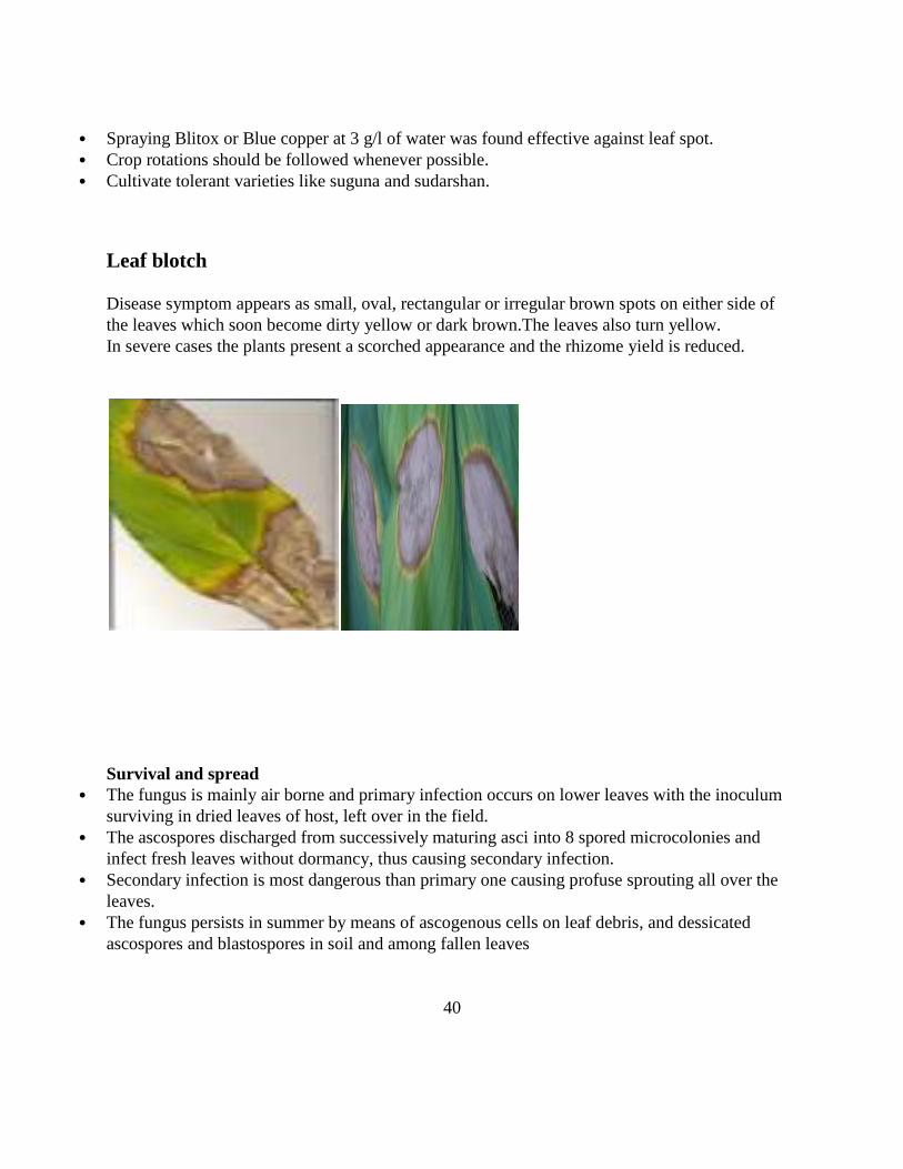
Leaf blotch
Disease symptom appears as small, oval, rectangular or irregular brown spots on either side of
the leaves which soon become dirty yellow or dark brown.The leaves also turn yellow.
In severe cases the plants present a scorched appearance and the rhizome yield is reduced.
Survival and spread The fungus is mainly air borne and primary infection occurs on lower leaves with the inoculum
surviving in dried leaves of host, left over in the field. The ascospores discharged from successively maturing asci into 8 spored microcolonies and
infect fresh leaves without dormancy, thus causing secondary infection. Secondary infection is most dangerous than primary one causing profuse sprouting all over the
leaves. The fungus persists in summer by means of ascogenous cells on leaf debris, and dessicated
ascospores and blastospores in soil and among fallen leaves
40

Control measures
Seed material should be selected from disease free areas. Seed material should be treated with Dithane M - 45 @ 3g/litre of water or Bavistin @ 1 g/litre
of water. Seed material should be dipped for 30 minutes in the fungicidal solution and should be shade-
dried before sowing. The disease is effectively controlled by spraying Dithane M - 45 @ 2.5 g/litre of water or
Bavistin 1g/litre , 2-3 sprayings should be given at fortnightly intervals or Carbendazim (1g/lit)
or Mancozeb (2.5g/lit) mixed wits 1ml Sandovit can be sprayed 2-3 times. The infected and dried leaves should be collected and burnt in order to reduce the inoculum
source in the field. Spraying Blitox or blue copper at 3 g/l of water was found effective against leaf blotch. Crop rotations should be followed whenever possible.
Tobacco mosaic virus Diseases Caused by Tobamoviruses:
Tobacco Mosaic
Named after tobacco mosaic virus, the genus Tobamoviruscontains more than a dozen rod-
shaped viruses measuring 18 by 300 nanometers. Their genome consists of one positive single-
stranded RNA [(+) ssRNA] of approximately 6,400 nucleotides (6.4 kb).Their protein coat
consists of a single species of protein subunit arranged in a helix. There are two closely related
viruses of economic importance in the genus: tobacco mosaic virus, which
infects tobacco and many other, mostly solanaceous hosts, and tomato mosaic virus, which
infects tomato.
Tobamoviruses cause serious losses in their hosts by damaging the leaves, flowers, and fruits and
by causingstunting of the plant. The losses are greatest when the plants are infected young.
Infections at later stages ofgrowth cause smaller losses. Tobamoviruses are easily transmitted
mechanically, and in nature they are spreadby incidental contact and wounding. They do not
seem to be transmitted by any vectors.
41

Symptoms consist of various degrees of mottling, chlorosis, curling, distortion, and dwarfing of
leaves, flowers, and entire plants. In some plants, necrotic areas develop on the leaves. On
tomato, leaflets may become long and pointed and, sometimes, shoe-string-like. Infections of
young plants reduce fruit set and may occasionally cause blemishes and internal browning on the
fruit that does form. Infected cells contain virus particles seen easily with an electron microscope
and sometimes visible as crystalline aggregates or amorphous bodies with a
compoundmicroscope.
The pathogen is tobacco mosaic virus. The virus particle measures 18 by 300 nanometers
and weighs 39 million daltons. Its protein coat consists of approximately 2,130 protein subunits,
and each subunit consists of 158 amino acids. Its ssRNA consists of 6,400 nucleotides. TMV
exists in numerous strains, which differ from one another in one or more characteristics.
Tobacco mosaic virus is exceptionally stable. It overseasons in infected tobacco stalks and leaves
in the soil,on the surface of contaminated seeds, and for many years in cigarettes, cigars, and so
on made with infectedtobacco. TMV is very prevalent in many ornamentals in greenhouses and
botanical gardens as a result of transmission from tobacco products.
The virus is transmitted easily by handling contaminated tobacco products or implements, or
infected tobacco plants, and then healthy susceptible plants. From the point of entrance (wound)
the virus moves from cell to cell through plasmodesmata, multiplies in and infects each cell,and
when it reaches the phloem, travels systemicallythrough it and infects the entire plant.
The control of tobacco mosaic virus depends on sanitation and the use of resistant varieties.
Sanitation includes removing infected plants and then washing hands with soap and avoiding
planting susceptible hostsfor two years in fields or seedbeds where a diseased crop was grown. In
some countries, tomatoes in greenhouseswere protected from severe strains of TMV by
inoculating them while young with a mild strain of the virus.. Infections of young plants reduce
fruit set and may occasionally cause blemishes and internal browning
on the fruit that does form. Infected cells containvirus particles (Figs. 14-4A and 14-30D) seen
easily withan electron microscope and sometimes visible as crystalline aggregates or amorphous
bodies with a compoundmicroscope.
42

The pathogen is tobacco mosaic virus(TMV).The virus particle measures 18 by 300
nanometersand weighs 39 million daltons. Its protein coat consistsof approximately 2,130
protein subunits, and each subunit consists of 158 amino acids. Its ssRNAconsists of 6,400
nucleotides.
In the past 10 years promising experimental control has been obtained by genetically engineering
tobacco andtomato plants with the gene coding for the TMV coat protein. Some control of TMV
is also obtained byspraying the plants with or dipping them in milk, which inhibits infection by
TMV.
43

Experiment-6: Groundnut and Sunflower diseases
1. Groundnut Leaf Spot (Tikka disease)
(Early and Late Leaf spots)
1.Tikka Disease The groundnut leaf spots (early leaf spot and late leaf spot) commonly called as ―Tikka Disease‖
cause nearly complete defoliation and yield loss up to 50 per cent or more depending upon
disease severity.
Early leaf is caused by Cercosporaarachidicola (Anamorph)( Teleomorph-
Mycospherellaarachidis)while Late leaf spot is caused by
Phaeoisariopsispersonatum(Anamorph) (Teleomorph-Mycospherellaberkeleyii).
Symptoms
In case of Early leaf spot, the infection starts about 1 month after sowing.Small chlorotic spots
appear on leaflets, with time they enlarge and turn brown to black and assume sub circular shape
on upper leaf surface.On lower surface of leaves light brown colouration is seen.Lesions also
appear on petioles, stems, stipules.
44

In case of early leaf spot, the spots are present on the upper surface of the leaflet and are
circular to irregular in shape and the colour is reddish brown while in case of late leaf spot, the
spots are present on the lower leaf surface in concentric rings and the spota are dark brown to
black in colour. Yellow halo is conspicuous in early leaf spot but dull and limited to margins of
spot in late leaf spot.
The disease perpetuates through conidia lying in the soil on diseased plant debris and through
conidia being carried in the shells of ground nut.
When the new crop of ground nut starts growing, the viable conidia are brought to the host
surface by various agencies, germinate under favourable conditions and cause primary infection.
The mycelium of C. arachidicola is inter and intracellular, brown, septate and without haustoria;
conidiophores are geniculate, 22-44 micron llong and 3-5 mm broad, continuous or 1-2 septate
Phaeoisariopsispersonatumproduces an intercellular branched myselium. Conidiophore are 25-
54 micron long, 5-8 micron broad, continuous or 1-2 septate. Conidia are terminal and each
conidiophores bear single conidium at the apex.
45

Management
Leaf spot infected diseased plant materials should be properly destroyed.
Resistant genotypes should be sown.
Seed treatment with talc-based powder formulation of Pseudomonas fluorescens should be done.
Spraying of Trichodermaviride (5 per cent) and Verticilliumlecani (5 per cent) reduces disease
severity,
Neem leaf extract (5 %), Mehandi (2%), Neem oil (1%), Neem kernel extract (3%) effectively
contain the disease.
Two sprays of Hexaconazole (0.2%), Carbendazim (0.1%) + Mancozeb (0.2%), Tebuconazole
(0.15%) reduces disease severity.
First spray of Carbendazim (0.1%) followed by 2 % Neem Leaf extract + 1% K2O has been
found to be efficacious in controlling the disease.
46

2. Rust (Puccinia arachidis)
Symptom
Rust can be readily recognized as orange colouredpustules (uredinia) that appear on the lower
leaflet surface and rupture to expose masses of reddish brown urediniospores. Rust pustules
appear first on the lower surface and in highly susceptible cultivars the original pustules may be
surrounded by colonies of secondary pustules.Pustules may also appear on the upper surface of
the leaflet.The pustules are usually circular and range from 0.5 to 1.4 mm in diameter.They may
be formed on all aerial plant parts apart from flower and pegs.
The groundnut rust disease is an economically important biotic stress that significantly reduces
the pod and fodder yield and oil quality. It is caused by the basidiomycete
fungus,Pucciniaarachidis, which belongs to class Pucciniomycetes like other rust fungus but has
fewer occurrences in teliospore form. The P. arachidis predominantly spreads by the repeated
cycle of uredospores in the field. The disease is prevalent in most of the countries where
groundnut is cultivated and favored by warm and humid climatic conditions. Despite its
economic importance, very limited work has been carried out on host-fungus interaction, fungal
genetic diversity, and physiological specialization.
Rust becomes devastating under conditions of high rainfall and humidity. In India, a continuous
dry period characterized by high temperature (>26°C) and low relative humidity (<70%) is
reported to delay rust occurrence and severity, whereas intermittent rain, high relative humidity
and 20 to 26°C temperature favor disease development.
47

Management
1. A cereal-cereal-groundnut crop rotation and removal of volunteer groundnut plants from the
field will help to check rust inoculum build-up.
2.Adjustment of the sowing time is recommended to avoid the most conducive environmental
conditions for rust development (i.e., high humidity, cloudy weather).Intercropping pearl millet
or sorghum with groundnut (1 :3) is useful in reducing the intensity of rust.
3. Sprays of Dithiocarbamate have been found effective to control rust. Chlorothalonil 0.2%,
Hexaconazole @ 0.1%, Propiconazole @ 0.1% spray have been found effective against rust.
when sprayed 30 days after germination at 10-15 day intervals.
4. The spray combination of carbendazim @ 0.1% + Mancozeb @ 0.1% effective in reducing
leaf rust disease.
5. Foliar application of aqueous neem leaf extract @ 2-5% is useful and economical for the
control of rust.
6. Growing of resistant cultivars, viz., ICGV 87160 or ICGV 86590.
3. Bud necrosis
Symptoms
Bud necrosis disease is an important disease of Groundnut. It is caused by Tomato Spotted
Wiltvirus and vectored by Thripspalmi in a propagative manner.
The primary symptom of bud necrosis is a mild chlorotic mottle or specks on young,
quadrifoliate leaves, which develop into chlorotic and necrotic rings and streaks. Necrosis from
the leaf extends to the petiole and terminal bud and results in necrosis of the terminal bud.
Secondary symptoms include stunting, auxiliary shoot proliferation, and malformation of
leaflets. The virus can cause severe crop losses, especially when the plants are infected before
they are a month old. Seeds from such plants are small, shriveled, mottled, and discolored.
48

It is caused by Tomato Spotted Wiltvirus and vectored by Thripspalmi in a propagative manner.
TSWV causes serious disease in many economically important plants representing many families
including dicots and monocots.Weed hosts are important reservoirs of the virus.
TSWV virus particles are membrane bound, spherical in shape measuring 70-90 nm in diameter.
The tospoviruses are a genus (Tospovirus) of negative RNA virus found within
the family Bunyaviridae.
This virus has a single stranded RNA genomewith negative polarity ((-) ssRNA )
Management
1.Intercropping of groundnut with bajra, sorghum, pigeonpea and maize in the ratio of 3:1
significantly reduces the disease incidence.
2. The elimination of weeds that are the primary source of tomato spotted wilt virus inoculum
from the vicinity of groundnut fields is recommended.
3. Clean cultivation, Seed treatment with imidacloprid 70 WS @ 10-15 g ai per kg seed followed
by spraying of a systemic insecticides(Fipronil @ 0.15 ml per litre of water) for the control of
thrips vector is the most effective method.
49
Sunflower diseases
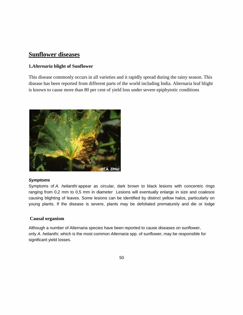
1.Alternaria blight of Sunflower
This disease commonly occurs in all varieties and it rapidly spread during the rainy season. This
disease has been reported from different parts of the world including India. Alternaria leaf blight
is known to cause more than 80 per cent of yield loss under severe epiphytotic conditions
Symptoms
Symptoms of A. helianthi appear as circular, dark brown to black lesions with concentric rings
ranging from 0,2 mm to 0,5 mm in diameter Lesions will eventually enlarge in size and coalesce
causing blighting of leaves. Some lesions can be identified by distinct yellow halos, particularly on
young plants. If the disease is severe, plants may be defoliated prematurely and die or lodge
Causal organism
Although a number of Alternaria species have been reported to cause diseases on sunflower,
only A. helianthi, which is the most common Alternaria spp. of sunflower, may be responsible for
significant yield losses.
50

Alternaria occurs in all sunflower producing areas and is currently a potential disease threat to
sunflower production in South Africa. Alternaria survives between sunflower crops in and on infested
crop debris, on weed hosts and on seed. The disease starts when spores land on leaves or stems,
germinate in the presence of moisture, and directly penetrate and infect the leaves.
The conidiophores are cylindrical, scattered or gregarious, pale grey yellow, straight or curved,
geniculate, simple or branched, up to 5 septate, 25-80 x 8-11µ. The conidia are cylindrical to
long ellipsoid, straight or slightly curved pale grey yellow to pale brown.
Disease cycle
Disease occurs when spores land on leaves or stems, germinate in the presence of free moisture,
and directly penetrate and infect the plant. Plants are most susceptible to infection beginning at
flowering and continuing through maturity. Plant stress also predisposes plants to the disease.
Spores are readily disseminated in and among fields by splashing irrigation water, wind, and
perhaps insects. The pathogen survives between sunflower crops in and on infested crop debris,
as a pathogen of safflower and cocklebur, and on seed.
Wet, warm weather promotes Alternaria disease growth. Regions prone to high humidity and
warmer temperatures are susceptible to this disease. Fields that are planted early are susceptible
to more severe losses from the affects of the disease than those planted later. Disease
development is favoured by 25-27o C temperatures with at least 12 hours of wet foliage.
Management
Cultural control
Deep summer ploughing
Clean cultivation and field sanitation
Use of resistant variety
Planting on mid-September
Chemical control
Seed treatment
Mancozeb 75 WP – 2g/kg of seed
51

Spray
Zineb 80 WP – 2g/lit
Mancozeb 75 WP – 2g/lit
Hexaconazole 5 EC – 1 ml/lit
Spray at 40, 55 and 65 days of crop.
2. Head Rot of Sunflower
Symptoms
Symptoms first become noticeable as dark spots on the back of ripening heads, followed by a
watery soft rot that later turns brown. As disease progresses, heads dry prematurely, shrivel, and
tissues appear to shred. Inside shredded tissues, coarse, thread-like mycelial strands are observed,
followed by the appearance of small black dots (sporangia). Sporangia are filled with spores that
are easily released and wind-blown to other plants. Symptoms on the flower side of heads
include the appearance of mycelium, a grayish, fuzzy substance that is covered with sporangia.
Causal Pathogen
Fungal structures: mycelium, sporangia, and sporangiospores. Several species of the
genus Rhizopus have been implicated in causing head rot, including R. arrhizus A.
R. stoloniferand R. microsporus It has historically been considered to be of minor importance,
however, it was documented as causing severe losses in Israel, and a recent survey of sunflower
diseases in California found that Rhizopus head rot was the most common disease of sunflower.
Under favorable conditions, it caused 100% losses in certain fields in the High Plains. Infection
is initiated in heads through wounds created by hail, birds, or insects.
52

Epidemiology
Some type of mechanical injury on the head in combination with high temperatures and high
relative humidity are required for infection and disease progress. Damage and economic losses
are dependent upon time of the season that wounding and infection occurs. Infection rarely
occurs before flowering, and greatest yield reductions result when infection occurs before seeds
are properly filled. Infection is primarily attributed to wounds from bird feeding. head moth
infestations and severe storms with hail.
Management
No resistant cultivars are available, but cultivars with more upright heads are more susceptible to
infection.
53

Experiment-7 :Sesamum and Cotton diseases
1. SesamumPhyllody
Symptoms
All floral parts are transformed into green leafy structures followed by abundant vein clearing in
different flower parts.In severe infection, the entire inflorescences is replaced by short twisted
leaves closely arranged on a stem with short internodes, abundant abnormal branches bend
down.Finally, plants look like witches broom.If capsules are formed on lower portion of plant
they do not yield quality seeds
Causal organism
Phytoplasmas (Mycoplasma - like organisms) are specialized bacteria which do not have cell
wall are obligate parasites found in sieve elements of plants and some insect vector.
They are transmitted from one plant to another by phloem-feeding insects.Sesamephyllody is
transmitted by the leafhopper vector,Orosiusalbicinctus.
The leaf hopper , O.albicinctussuccessfully transmittedsesamumphytoplasma when allowed
minimum acquisition access period, inoculation feeding period and incubation period in the
vector and plants for 4 hours, 30 minutes, 15 to 23 days and 13 to 25 days,respectively.
54

Management
1. Sesamum phyllody phytoplasma has a very large host range. The highly infected phyllody
plants should be uprooted and burnt.
2. Seed treatment with Imidacloprid (7.5/kgai per kg of seed)
3.Folior Sprays of the aboveinsecticides(Imidachlorprid 17.8SL) (0.02%) was most effective for
management of phyllody disease.
Diseases of Crucifers
1.White rust of Crucifers
White rust is a disease in plants caused by the oomycete, Albugo candida. Plants susceptible to
this disease generally include members of the Brassica family.White rust has been known to
cause agricultural losses in fields cultivating members of this family
including broccoli, cauliflower, and Indian mustard. Despite the name, it is not considered
true rusts.
Symptoms
Both local and systemic infections are observed. In case of local infection, white chalky, creamy
yellow raised pustules appear on the leaves which later coalesce to form patches.In systemic
infection and during humid weather, mixed infection of white rust and downy mildew cause
swelling and distortion of the stem and floral parts due to hypertrophy and hyperplasia and
develop ―stag head‖ structure.
55

Life cycle
The vegetative pant body is composed of non-septate coenocytic hyphae that grow in the inter-
cellular spaces of the host. the hyphae bear haustoria. it. The hyphae accumulate just beneath the
epidermis of the infected leaf. From these hyphae, certain thick-walled, clavate aerial
sporangiophores come out. The sporangium is formed in chains.. When the sporangia are formed
in abundance on innumerable sporangiophores, the pressure is caused; the host epidermis
ruptures and hundreds of sporangia are seen on the surface of the host in the form of white
creamy powder forming pustules. The sporangia are transferred from one place to another by
various agencies such as wind, insects, water, etc. The sporangium bursts anteriorly and the
zoospores liberate in the film of water .Zoospores are naked, biflagellate, uninucleate, reniform
and vacuolate zoospore. The sporangium bursts anteriorly and the zoospores liberate in the film
of water. The zoospores after moving about in a film of water encysts. The zoospores germinate
through germ tube which infect the host plant.
Sexual reproduction takes place when the growing season comes to an end. The mycelium
penetrates into the deeper tissues of the host. The sexual reproduction is highly oogamous type.
56

The antheridium and oogonium develops deeper in the host tissue in close association within the
intercellular spaces. Its formation is externally indicated by hypertrophy. Oogonium is spherical
and multinucleate The oogonium develops a papilla like out growth at the point of contact with
the antheridium. This is called as receptive papilla. Soon it disappears, and the antheridium
develops a fertilization tube. On fusion oospores are formed. Zoospores are formed in side
vesicle formed on germination of oospore.
57

58

Survival and spread
The pathogen survives through oospores in affected host tissues and soil.
Secondary infection is carried out by sporangia and zoospores which produce new infection.
Favourable conditions
Moist (more than 70% relative humidity) coupled with warm weather (12-25 °C) and
intermittent rains favours disease development.
Management
(i) Growing resistant varieties.
(ii) Eradication of infected plant and their complete destruction.
(iii) Rotation of crucifers plants with non cruciferous plants.
(iv) Three sprays of metalaxyl + mancozeb (Ridomil MZ) 0.2% at 50, 65 and 80 days after
sowing or seed treatment with metalaxyl (2 g./kg seed plus two sprays of metalaxyl + mancozeb
at 0.144% a.i. at 50 and 65 days after sowing.
59

2. Alternaria leaf spot
Symptoms
. The most common symptom of Alternaria diseases is yellow, dark brown to black circular leaf
spots with target like, concentric rings. Lesion centers may fall out, giving the leaf spots a shot-
hole appearance. Individual spots coalesce into large necrotic areas and leaf drop can occur.
Lesions can occur on petioles, stems, flowers, flower pedicels, and seed pods.
Causal organism
Three species of Alternaria cause serious damage to brassicas: Alternaria brassicicola, A.
brassicae, and A. raphani. Alternaria brassicicola and A. brassicae infect broccoli, Brussels
sprouts, cabbage, cauliflower, turnip etc. A. raphani is most often found on radish, but can infect
other brassica crops.
1..Symptoms2.Conidia of A. brassicae and 3. A.
brassicicola
60

Life Cycle
Alternaria species are simple parasites that survive saprophytically outside the host. Diseased
crop debris is the primary site of survival from year to year. Resting spores (chlamydospores,
microsclerotia) have been reported.
Conidiophores of A. brassicae produce asexual spores (conidia) .Continuous moisture of 24
hours or longer practically guarantees infection. Relative humidity of 91.5% result in the
production of large numbers of mature spores in 24 hours.
A. brassicae conidia germinates within 3 hours, when the temperature is between 21 and 280
C. A. raphani and A. brassicicola germinate at higher temperatures. A. brassicae and A.
brassicicola remain viable for a long period of time as spores on seed coat or as mycelium in
seed as well as in infected plant debris. A.brassicae and A. brassicicola also survive in the form
of microsclerotia and chlamydospores which appear after infected leaves have partially decayed.
Microsclerotia and chlamydspores of both pathogens can be formed within conidial cells.
Survival and spread
The disease is externally and internally seed borne. The pathogen survives through spores or
mycelium in diseased plant debris or weed.
Favourable conditions
Moist (more than 70% relative humidity) coupled with warm weather (12-25 °C ) and
intermittent rains favours disease development
Management
Use seed certified as disease-free or treat seed with hot water.
Practice long rotations with non-cruciferous crops.
Incorporate diseased plant debris into the soil.
Control cruciferous weeds.
Minimize the length of leaf wetness periods by reducing plant density, orienting rows
with prevailing winds, and irrigating in the morning when leaves can dry quickly.
Avoid overhead irrigation during head development.
Keep seedbeds disease-free to prevent the spread of disease and locate seedbeds so as to
avoid wind-borne inoculums.
Spraying of Dithane M-45 0.2% on appearance of the disease to be repeated at 15 days
interval.
61

Diseases of Cotton
Angular leaf spot and black arm disease of cotton
The most common and conspicuous symptom is angular leaf spot which begins with dark-green,
water-soaked spots, initially more clearly visible on the underside of the leaf lamina; the spots
are angular in shape, being delimited by the smaller veins. Older spots become dark-brown or
black and are visible on the upper surface of the leaves. The angular spots may be few in number
in more resistant host material but, on susceptible cultivars, they can cover much of the leaf,
causing chlorosis, followed by necrosis and distortion of the lamina. Similar spots may be found
on the cotyledons of young seedlings where infection occurs from the soil or seed during
germination and emergence. Under favourable conditions, infection may spread from the
seedling cotyledon or the leaf onto the petiole and then to the main stem, leading to seedling
mortality insusceptible cultivars.
In older plants, the lesions can girdle the main branches causing them to break, with the loss of
leaves and fruiting branches. This phase of the bacterial blight syndrome is known as blackarm
because of the blackened appearance of the affected petioles and branches.
Sometimes infection on the leaf occurs as water-soaked tissue, which later turns black, on either
side of the main veins. This is referred to as vein blight and can occur together with, or
Occasionally in the absence of angular leaf spot.
In older plants, water-soaked lesions can occur on the bracts of the epicalyx and more commonly
on the developing boll. Bacterial boll rot begins as roughly spherical water-soaked spots on the
boll surface which can expand to >1 cm in diameter on susceptible cultivars, becoming black as
they age and penetrating the boll cortex to cause internal boll rot. The blight bacterium can also
be introduced into the young boll during feeding on the seed by the cotton stainer (Dysdercus
spp.). This causes the lint to become stained yellow or brown and sometimes leads to internal
boll rot.
62

Phases in the disease- Angular leaf spot, Black arm phase, Boll rot phase and seedling blight
phage.
Hosts other than cotton- Thurbaria, Eriodendrone, Lochnera pusilla Jatropha curcas etc.
The bacterium, X. axonopodis pv. Malvacearum is gram (-), possesses single polar flagellum
and is non-spore forming.
Hosts other than cotton- Thurbaria, Eriodendrone, Lochnera pusilla Jatropha curcas etc.
Management
Removal of weed and other hosts.
Seed delinting- To remove seed-borne inoculums to eradicate externally seed-borne
inoculums. Dip in conc. H2 SO4
Seed treatment -with Vitavax/ plantvax @ 2 g perkg of seed/
Spraying - Copper fungicides (Copper oxy chloride ( seed treatment & foliar sprayers.
63
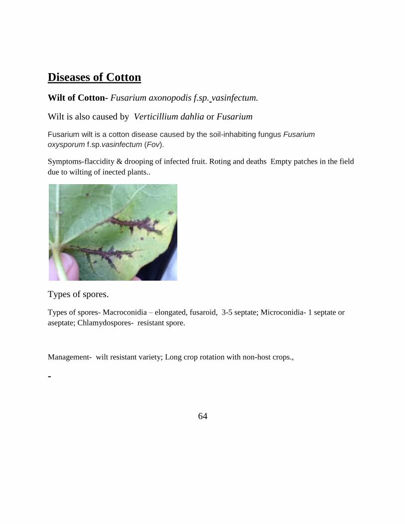
Diseases of Cotton
Wilt of Cotton- Fusarium axonopodis f.sp. vasinfectum.
Wilt is also caused by Verticillium dahlia or Fusarium
Fusarium wilt is a cotton disease caused by the soil-inhabiting fungus Fusarium
oxysporum f.sp.vasinfectum (Fov).
Symptoms-flaccidity & drooping of infected fruit. Roting and deaths Empty patches in the field
due to wilting of inected plants..
Types of spores.
Types of spores- Macroconidia – elongated, fusaroid, 3-5 septate; Microconidia- 1 septate or
aseptate; Chlamydospores- resistant spore.
Management- wilt resistant variety; Long crop rotation with non-host crops.,
-
64

Experiment-8 :Red gram Green gram and Black gram diseases
1. Wilt of Red gram (Pigeon pea)
Symptoms
Symptoms can appear 4 to 6 weeks after sowing. The initial visible symptoms are loss of
turgidity in leaves, and slight inter-veinal clearing.The foliage shows slight chlorosis and
sometimes becomes bright yellow before wilting.Leaves are retained on wilted plants. The initial
characteristic internal symptom of wilt is the browning of the xylem vessels from the root system
to the stems.The xylem gradually develops black streaks, and brown or dark purple bands appear
on the stem surface of partially wilted plants extending upwards from the base.
When the bark of such bands is peeled off , browning or blackening of the wood beneath can be
seen. in wilt-tolerant genotypes these bands are confined to the basal part of the plant.
Sometimes, especially in the later stages of crop growth, the branches dry from the top
downwards, but symptoms are not seen on the lower portions of the main stem or branches.
Small branches on the lower part of the plant also dry.
When the main stem of such plants is split open, intensive blackening of the xylem can be seen.
In humid weather, a pinkish mycelial growth is commonly observed the basal portions of the
wilted plants.Partial wilting is usually associated with lateral root infection. Tap root infection
results in complete wilting.
65

The disease is soil-borne. The infected plant may be either infected partly or wholly.
Three types of spores are formed- Macroconidia (elongated, fusaroid, having 3-5 septum;
Microconidia- 1 or no septa. Chlamydospores (resistant spores)
Management- Crop rotation, Mixed cropping, , intercropping. Soil amendment.with saw dust,
66

Diseases of Black gram
1.Powdery mildew
Symptoms
White powdery patches appear on leaves and other green parts which later become dull coloured.
These patches gradually increase in size and become circular covering the lower surface also.
When the infection is severe, both the surfaces of the leaves are completely covered by whitish
powdery growth. Severely affected parts get shrivelled and distorted.
In severe infections, foliage becomes yellow causing premature defoliation. The disease also
creates forced maturity of the infected plants which results in heavy yield losses.The pathogen
has a wide host range and survives in oidial form on various hosts in off-season.
Secondary spread is through air-borne oidia produced in the season.
67

Causal organism
The disease is caused by Erysiphe polygoni
Life cycle of E. polygoni
The disease powdery mildew, caused by the pathogen Erysiphepisi, oversummers on infected
black gramtrash and produces spores which are blown by wind into new crops. The disease may
also be seed-borne, but this source of infection is least important.
Under favourable conditions, the disease may completely colonize a plant in five to six days.
Once a few plants become infected, the disease rapidly spreads to adjacent areas. Warm (15-
25°C), humid (over 70 per cent RH) conditions for four to five days late in the growing season,
during flowering and pod filling, favour disease development. However, heavy rainfall is not
favourable for the disease as it will actually wash spores off plants. Night time dews are
sufficient for the disease to develop.
Management
The seeds must be sown early in the month of June to avoid early incidence of the disease on
the crop
Powdery mildew could be controlled by spraying Carbendazim 1g/lit or Tridemorph 1 ml /lit.
The maximum reduction of disease incidence was recorded in wettablesulphur 0.25% followed
by carbendazim 0.1% and castor oil 1% with Ampelomyces quisqualis.
68
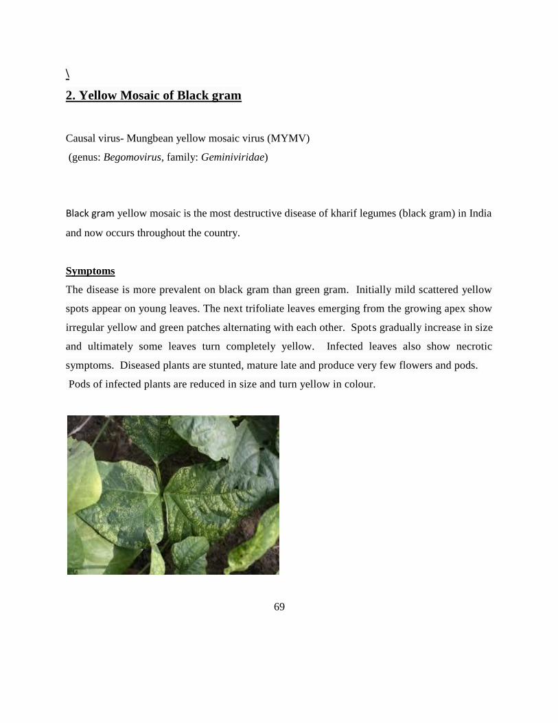
\
2. Yellow Mosaic of Black gram
Causal virus- Mungbean yellow mosaic virus (MYMV)
(genus: Begomovirus, family: Geminiviridae)
Black gram yellow mosaic is the most destructive disease of kharif legumes (black gram) in India
and now occurs throughout the country.
Symptoms
The disease is more prevalent on black gram than green gram. Initially mild scattered yellow
spots appear on young leaves. The next trifoliate leaves emerging from the growing apex show
irregular yellow and green patches alternating with each other. Spots gradually increase in size
and ultimately some leaves turn completely yellow. Infected leaves also show necrotic
symptoms. Diseased plants are stunted, mature late and produce very few flowers and pods.
Pods of infected plants are reduced in size and turn yellow in colour.
69

Black gram yellow mosaic is the most destructive disease of kharif legumes (black gram) in
India and now occurs throughout the country. The disease is transmitted by the whitefly
vector Bemisiatabaci.
Management
Infected plants should be removed. A barrier crop, such as sorghum, maize can be grown.
Insecticides can be sprayed to control the whitefly vector.
Grow seven rows of sorghum as border crop.\Treat seeds with Imidacloprid 70 WS @ 5ml/kg
to control vector. Rogue out infected plants early in the season to eliminate the source of
Inoculum. Give one foliar spray of systemic insecticide (Dimethoate @ 750 ml/ha) on 30 days
after sowing.
70
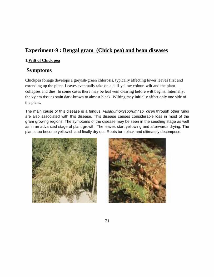
Experiment-9 : Bengal gram (Chick pea) and bean diseases
1.Wilt of Chick pea
Symptoms
Chickpea foliage develops a greyish-green chlorosis, typically affecting lower leaves first and
extending up the plant. Leaves eventually take on a dull-yellow colour, wilt and the plant
collapses and dies. In some cases there may be leaf vein clearing before wilt begins. Internally,
the xylem tissues stain dark-brown to almost black. Wilting may initially affect only one side of
the plant. The main cause of this disease is a fungus, Fusariumoxysporumf.sp. ciceri through other fungi
are also associated with this disease. This disease causes considerable loss in most of the
gram growing regions. The symptoms of the disease may be seen in the seedling stage as well
as in an advanced stage of plant growth. The leaves start yellowing and afterwards drying. The
plants too become yellowish and finally dry out. Roots turn black and ultimately decompose.
71

Fusariumoxysporum is a common soil inhabitant and produces three types of asexual spores;
Macroconidia, microconidia and chlamydospores. The macroconidia are straight to slightly curved,
slender, thin walled usually with three or four septa, a foot-shaped basal cell and a tapered and
curved apical cell. They are generally produced from phialides on conidiophores by basipetal
division. They are important in secondary infection. The microconidia are ellipsoidal and either have
no septum or a single one. They are formed from phialides in false heads by basipetal division. They
are important in secondary infection.The chlamydospores are globose and have thick walls. They
are formed from hyphae or alternatively by the modification of hyphal cells. They are important as
endurance organs in soils where they act as inocula in primary infection.
The teleomorph or sexual reproductive stage, of F. oxysporum is unknown.
Management
Control measures of Wilt of Chickpea
Seed Treatment with carbendazim@ 2.5 gram per Kg. of seed OR
Carboxin + Thiram 1:2 at the rate of 3 gram per Kg. Seed.
Grow the resistant varieties like C-214, Avrodhi, Uday, BG-244; Pusa-362, JG-315,
Phule G-5 etc.
In fields having heavy incidence of gram wilt, the cultivation of chick pea should be
avoided for three to four years.
72

As far as possible sowing of chick pea should not be done before third week of October.
Deep planting of chick pea about 8-10 centimeters deep in the light soils reduces the
gram wilt incidence.
2. Collar rot: Sclerotium rolfsii
Symptom
It comes in the early stages i.e up to six weeks from sowing.
Drying plants whose foliage turns slightly yellow before death, scattered in the field is an
indication of the disease.
Seedling become chlorotic.
The joint of stem & root turns soft slightly contracts and begins to decay.
Infected parts turn brown white.
Black dots, like mustard in shape known as sclerotia are seen appearing on the white
infected plant parts.
73

The fungus is microscopic when inside the plant tissue, but eventually the organism
forms sclerotia, which are an asexual dormant body which are visible to the naked eye,
and look like white, yellow, or brown mustard seeds.
The fungus overwinters as a sclerotium, which is a dense mass of hyphae with a hard outer shell. When
Warm, humid conditions are present, the sclerotia "germinate", and infect plant parts. The pathogen is
not systemic on the plant, so removing infected parts can help control the disease
Affected plants show signs including wilted leaves and soft, rotten crowns, often followed
by the death of the infected plant.
The fungus is microscopic when inside the plant tissue, but eventually the organism
forms sclerotia, which are an asexual dormant body which are visible to the naked eye,
and look like white, yellow, or brown mustard seeds.
The fungus overwinters as a sclerotium which a dense mass of hyphae with a hard outer
shell. When warm, humid conditions are present, the sclerotia germinate and infect plant
parts. The pathogen is not systemic on the plant, so removing infected parts can help
control the disease.
Management
Exclusion:
Once established, this pathogen is very difficult to get rid of, so care should be taken when introducing
susceptible plants to the garden, and infected plants should be removed as soon as the infection is
discovered.
Bulbs and corms can be treated with hot water for 30 minutes
When working around infected plants, tools and shoes should be cleaned.
74
Cultivation: The sclerotia only become active when on the surface of the soil, so a deep top-dressing
with compost may help control the disease. Removal of the mulch layer in winter is also
recommended, as this both physically removed the sclerotia and exposes the soil to winter
dessication.
Adding composts raises the level of antagonistic organisms.
Deep plowing can also provide some control by burying the sclerotia.
Plastic mulches can serve as a barrier between the innoculant and plant tissues.
Cultural Controls
Crop rotation is sometimes tried as a control, but this can be difficult to to the wide host
range, however rotation with resistant Allium species
Avoid dense leaf canopies, in order to let the soil surface remain dry.
Keep litter and weeds out of the garden.
Solarization can kill the fungus in the top layer of soil, in regions where solarization can
bring about high enough temperatures.
Physical Removal: Removal of infected plant and soil is strongly recommended. Mulch around infected
plants should be removed at the end of the season.
Chemical Controls (synthetic)
Methyl Bromide and Methyl Bromide/Chloropicrin have been applied as soil fumigants,
however both are extremely dangerous chemicals and are banned in many regions.
PCNB (Terraclor) at 0.5-1 lba.i./1000 sqft can be incorporated into the soil surface.
Management
Deep ploughing in summer.
Avoid high moisture at the sowing time.
75

Seedlings should be protected from excessive moisture.
Destroy the residues of last crop and weed before sowing and after harvest.
All undecomposed matter should be removed from the field before land
preparation.
Treat the seeds with a mixture of Carbendazim 2g per kgseed.
Dry root rot: Rhizoctoniabataticola/Macrophominaphaseolina
Symptom
The disease appears from flowering to podding stage as scattered dried plants.
The leaves and stem are become straw colored.
Affected plants wither and spread across the entire field.
The roots of infected plants become brittle and dry.
76

Causal organism
M. phaseolina has a monocyclic disease cycle. Survival. The
M. phaseolina fungus has aggregates of hyphal cells, which form microsclerotia
within the taproots and stems of the host plants. The microsclerotia overwinter in
the soil and crop residue and are the primary source of inoculum .
Management
Deep ploughing in summer
Grow cultivars resistant to dry root rot.
Drought should be avoided.
Sowing should always be done on the recommended time.
Germinating and young seedlings should be saved from high temperatures.
Seed treatment with T. viride @4g/kg or P. fluorescens @ 10g/ kg of seed or
Carbendazim or Thiram 2g/kg of seed.
Spot drenching with Carbendazim 1g/lit or P. fluorescens / T. viride 2.5 kg/ha with
50 kg FYM.
77

Pea diseases
1.Rust of pea Causal Organism: Uromycespisi and U. fabae
The disease is serious in northern India. It is serious on the late sown crop.
All the green parts of the plants are affected.Under cool and wet climate in northern India entire
plant may be killed.
Symptoms:
The earliest symptoms are the yellow spots having aecia in round or elongated clusters.Pustules
develop which are powdery and orange brown in appearance.
Uromycesviciae-fabae is an autoecious fungus, completing its lifecycle on a single host.
The resting stage (telia) survives in a semi-dormant stage over summer in crop
residues.contaminations. It may also perpetuate on weed hosts from where it may infect lentil or
field pea crops by windborne spores. Teliospores produced on residues are blown by wind and
infect volunteer plants and seedlings. Infection leads to the production of aecia. Aecia produce
aeciospores, which infect leaves and will spread the rust within the crop and to other crops.
78

Aeciospores germinate at 17-22oC and infect other plants forming either secondary aecia or
uredia at 25oC. In turn, this leads to the production of urediniospores, which can be carried long
distances to produce new infections. Severe epidemics are caused by the production of several
generations of urediniospores. Uridinia develop late in the season and are rapidly followed by
telia .Teliospores develop in late summer (Hall 2003) on stems and leaves. After harvest, aecia
and uredia present on the plant die out, but teliospores are more persistent.
At lower temperatures, uredospores are probably an important means of survival in the absence
of the host. Uredomycelium is, on the other hand, highly resistant to heat and sunlight and is
probably important for continued development and survival of rust in hot, dry conditions. The
fungus survives on debris and dispersal of inoculum can occur in several forms, namely infested
debris, dust and sand.
Control:
Follow 1-2 years of crop rotation avoiding beans
After harvest, the affected plant trash should be burnt.
Spray the crop with Dithane M-45 at the rate of 2 kg per hectare in 1000 liters of water. Two to
three sprays at 10 days interval are sufficient.
79

2. Powdery mildew of pea
Powdery mildew, caused by the pathogen Erysiphepisi, can be a serious disease of peas in South Australia and
Victoria. Severe infections can significantly reduce yield in susceptible varieties. Powdery mildew is most
prevalent late in the season.
Symptoms
Infected plants are covered with a white powdery film. Severely infected foliage turns blue-white
in colour; tissue below these infected areas may turn purple, (Figures 7.12 and 7.13). Symptoms
first appear on the upper surfaces of the oldest leaves. Leaves, stems and pods may all become
infected resulting in withering of the whole plant. Severe pod infection can cause a grey-brown
discolouration of the seeds.
80

Life cycle
Management
Varietal Selection
Growing a resistant variety is the most effective means of controlling powdery mildew.
Crop Rotation
Leave a four year break between growing field pea crops in the same plot.. Control volunteer field peas which
can harbour disease. Avoid sowing field pea crops adjacent to last season’s stubble. Incorporate or burn
infected pea stubble soon after harvest where practicable.
Seed Treatment
Seed treatments can be beneficial and are recommended where powdery mildew frequently occurs.
81

Foliar Fungicides
Monitor crops from flowering onwards for signs of powdery mildew. If the disease is present the application of
a foliar spray may be warranted. Fungicides need to be applied prior to disease development to be most
effective. Fungicides for powdery mildew have limited systemic activity and will not protect the new growth
following spraying. Good plant coverage with the fungicides is essential.
Soybean diseases
1. Bacterial pustule
Bacterial pustule, which is caused by Xanthomonasaxonopodis(syn. campestris)
pvglycines, can cause premature defoliation and reduced seed size and quantity. This disease
has been reported in most parts of the world where soybeans are grown and the climate is
warm with frequent rain. It is most prevalent later in the growing season.
Symptoms
Initial symptoms are the appearance of tiny pale green spots on leaves. These spots have
raised centers that may develop on either surface of the leaf but are more common on the
lower surface. Lesions are often associated with main leaf veins.As the disease progresses,
small, light-colored pustules will form in the center of the spots. Spots may merge together
to form irregular lesions. This disease can easily be confused with soybean rust. Mature
soybean rust pustules have a small, round opening at the top for spore release. Bacterial
pustule lesions normally lack an opening and do not produce spores. If an opening is present
with bacterial pustule, it is typically a linear crack across the surface of the pustule. These
features can only be seen under magnification. Symptoms of bacterial pustule are very
similar to those of bacterial blight during the early stages of development, but water soaking
will not be present with bacterial pustule, and bacterial pustule lesions will have raised
centers as they develop.
82

Epidemiology
Like bacterial blight, bacterial pustule overwinters in crop residue and is carried by wind-driven
rain or water droplets splashing from the ground to the plant. In addition, the disease can be
spread during cultivation while the foliage is wet. The bacterium will enter the plant through
natural openings and wounds. Warm weather with frequent showers promotes the development
of this disease. Unlike bacterial blight, warm temperatures do not limit development of bacterial
pustule. Its optimal temperature range for development is 86-92oF.
Bacterial Disease Management
While these diseases are not typically yield limiting in Nebraska, producers should consider the
following options in problem fields:
• Resistance. Cultivars that are not highly susceptible to the disease should be considered
in fields with a history of bacterial disease problems.
• Crop rotation. Crop rotation can be an effective method to avoid inoculum from a
previously infected crop.
• Tillage. Incorporating crop residue by tillage will reduce the amount of inoculum
available in the spring to infect plants.
• Cultivation. To prevent the spread of disease, limit cultivation to times when the foliage
is dry.
• Fungicides. Copper fungicides are labeled for control of bacterial blight on soybeans but
need to be applied early in the disease cycle to be effective.
2.Yellow mosaic of Soybean
Soybean plants were found to be infested with whiteflies (Bemisiatabaci) suggesting
begomovirus etiology. The disease agent was transmitted experimentally by whiteflies, and
symptoms developed after 23 days.
Yellow mosaic disease caused by whitefly-transmitted bipartite Geminiviruses is one of the
major constraints on productivity of a number of pulse crops. We have cloned the bipartite
genome of Mungbean Yellow Mosaic India Virus.
Soyabean is susceptible to yellow mosaic virus. The virus affected plant turns yellowish and
stunted in growth. The affected plant should be immediately removed and destroyed.
83

Control
To stop spread of yellow mosaic disease in the field the crop may be sprayed with Rogor at the
rate of 1-1.5 litres in 800-1000 litres water per hectare.
2.Bud necrosis of soybean
Bud blight disease (BBD) of soybean ( Glycine max L. Merr.) showed little or no transmission
via seeds from naturally infected plants. This is consistent with the presence of Peanut Bud
Necrosis Tospovirus (PBNV) which has been investigated recently in the Chhattisgarh region of
India and is not seed-borne. Various host species were tested in host range studies. Of these, the
bud blight pathogen infected only four species, producing chlorotic/necrotic rings/spots on
Chenopodiumamaranticolor,Nicotianaglutinosa L., Vignaunguiculata (Land systemic infection
in Arachis hypogea . The number of plants with bud blight infections was significantly high
when the crop was at the flowering and pod initiation stages. However, the numberof infected
plants was significantly lower when the crop was at the pre-bloom and podding stages. Similarly,
infected plants were significantly more common in JS 75-46 than in JS 335.
Symptoms
Symptoms first appear on young leaflets as faint chloroticspots or mottling that may develop into
chlorotic andnecrotic rings and streaks. Occasionally,the leaflets may show a general chlorosis
with greenislands. Petioles bearing fully expanded leaflets with initialsymptoms usually become
flaccid and droop. Necrosisof the terminal bud soon follows. The bud necrosis symptom is
common on crops
grown in the dry (summer) and rainy seasons in India,indicating that this symptom is probably
associated withhigh temperatures. If bud necrosis occurs on plants lessthan 1 month old. total
necrosis of the plant may follow.
The stunting and proliferation of axillary shoots are common secondary symptoms.
The structure of TSWV is unique among plant viruses. The particles are 70-90 nm in diameter
and are surroundedby a double membrane of protein and lipid.
84
Both TSWV and the vector thrips have wide host ranges that include crop plants, ornamentals,
and weeds.
Studies with Thrips palmi, Frankliniella schultzei and Scirtothrips dorsalis showed that only T.
palmi transmitted PBNV. T. palmi acquired the virus as larvae, requiring a 5-min acquisition
period, but only transmitted it as adults. Adults needed an inoculation feeding period of > 1 h
to transmit, and the majority transmitted the virus throughout their life. In field situations, a
virus-susceptible peanut genotype had higher densities of T. palmi than resistant genotypes. In
peanuts, S. dorsalis was the dominant foliar species, T. palmi was on foliage and flowers,
and F. schultzei was mostly on flowers. Several common weed species in peanut were infected
with PBNV and were colonized by T. palmi.
85

Experiment No. 10 Field visit(s)
The purpose of field visit is to collect information on total area under the crop, the varieties
grown and to record the occurrence and severity of the disease using a Z-shaped area for
recording observations. Sufficient number of observations as per variety of standing crop.
1. Incidence and severity of the disease is to be recorded.
2. A Disease Rating scale is to be adopted.
3. Diseased Plant specimen should be properly labeled including the following:
• Name of the Collector
• Collection date
• Crops
• Variety
• Locality.
The specimen should be brought to the laboratories. Examined under Compound Microscope.
Objectives
Students should be able to diagnose important plant diseases and identify the pathogen
associated with a specific disease.
Students should have an understanding of the principle techniques used in plant disease
diagnostics and have the ability to select and use the most appropriate technique for the
diagnosis.
Students should be able to write disease diagnosis reports.
Students will be able to describe in oral form their experiences in diagnosing an unknown
plant disease.
86

Experiment No. 11: Preparation of Herbarium of Crop diseases
Herbarium, collection of dried diseased plant specimens mounted on sheets of paper. The plants
are usually collected in situ (e.g., where they were growing in nature), identified by experts,
pressed, and then carefully mounted to archival paper in such a way that all major morphological
characteristics are visible (i.e. both sides of the leaves and the floral structure). The mounted
plants are labeled with their proper scientific names, the name of the collector, and usually,
information about from where they were collected and how they grew and general observations.
The specimen are commonly filed in cases according to families and genera and are available for
ready reference.
Plant Collection Procedures and specimen preservation
The following is a guide to the various items of equipment and techniques required to make and
preserve plant collections.
.Equipments
Field press
A press typically consists of 2 hard wood frames with each frame made from-
4 wood strips c. 20 x 12 x 450 mm and
6 wood strips c. 20 x 12 x 300 mm
2 pieces of 12 mm plywood cut to 300 x 450 mm may be used. Some holes drilled in each piece
will assist circulation of air. If using the hard wood frame type of press, it is useful to have 2
inner pieces of corrugated card board, 300 x 450 mm. Corrugated card board may also be used
elsewhere in the press to separate specimens and allow greater circulation of air.
87

Newspaper, card board and foam
A newspaper folded in half, is excellent. Corrugated card board can be used to separate
woody or bulky specimens from delicate ones. Using foam (c. 10 mm thick) in the press
results in evenly pressed specimens, especially bulky specimens.
Press straps
A pair of strong webbing straps with claw buckles is excellent. Sash cord may also be
used. In either case, the minimum length is 1.5 m.
Field note book
A pocket sized note book which will stand up to the wet. Use a pencil which is water-
proof- both at the time and later.
Tie on tags
Large enough to take your name (or initials) and fiels number. They may also be used to
label collecting bags.
Clippers
A pair of secateurs.
Diggers
A trowel, preferably with a steel shank.
Scrappers
A large spatula is excellent for scrapping up mosses and lichens.
Collecting bags
Plastic bags, in a couple of sizes, and rubber bands to close them. Small brown bags for
collecting fruits, seeds etc.
88

Hand lens
At least 10X.
Rucksack
Big enough to carry all of the above specimen.
.
89