courrent pinion right heart failure in the … right heart failure in the intensive care unit...
TRANSCRIPT

Copyright © Lippincott Williams & Wilkins. Unauthorized reproduction of this article is prohibited.
CURRENTOPINION Right heart failure in the intensive care unit
Clifford R. Greyson
Purpose of reviewThis review summarizes the approach to and recent developments in the evaluation and treatment of acuteright heart failure in the ICU. Right heart failure, defined as failure of the right ventricle to provide sufficientblood flow through the pulmonary circulation at normal central venous pressure, is a common problemcaused by a combination of increased right-ventricular afterload and right-ventricular contractiledysfunction.
Recent findingsManagement of acute right heart failure continues to be challenging because of insufficient understandingof its pathophysiology, a lack of guidelines, and few available tools. Recent research has contributed to animproved understanding of its mechanisms, helping to guide therapy and suggest future options. Right-ventricular assist devices are emerging as a promising approach to treatment when optimization ofhemodynamics and conventional medical therapy fail.
SummaryRight heart failure causes venous congestion and systemic hypoperfusion. Once right heart failure isidentified, the primary goal is to alleviate any reversible cause of excessive load or right-ventricularcontractile failure. When the underlying abnormalities cannot be alleviated, trials of diuretic, vasodilator,or inotropic therapy may be required. Invasive monitoring helps guide therapy. Medically refractory rightheart failure may potentially be treated with right-ventricular assist devices.
Keywordscor pulmonale, pulmonary hypertension, right ventricle, ventricular assist device
INTRODUCTIONRight heart failure (RHF) is defined as the clinicalsyndrome in which the right ventricle (RV) of theheart fails to deliver adequate blood flow throughthe pulmonary circulation at a normal centralvenous pressure (CVP). Clinical RHF is identifiedby signs and symptoms of venous congestion dueto elevated CVP along with evidence of right-ven-tricular contractile dysfunction and right-ventricu-lar pressure overload; progressive RHF can causesystemic hypoperfusion. However, RHF is not syn-onymous with right-ventricular contractile dysfunc-tion, and is not necessarily associated with severepulmonary hypertension, although pulmonary arte-rial pressure is usually at least mildly elevated.
The development, normal physiology, and path-ophysiology of the RV and the RHF were previouslyreviewed elsewhere [1,2]. Treatment of chronic pul-monary hypertension, venous thromboembolic dis-ease, interventions for right-ventricular myocardialinfarction, and general issues of resuscitation will notbe discussed in this review. This review will focus onthe cause, evaluation, and management of RHF in theICU. Experimental studies that provide insight into
the pathophysiology of RHF and suggest potentialfuture therapies will also be discussed.
From a practical point of view, management ofRHF in the ICU consists of identifying abnormalitiesof right-ventricular function or pulmonary circula-tion, addressing any underlying reversible pro-blems, optimizing preload, and utilizing inotropicagents. If these measures fail, the most promisingemerging therapy is the use of right-ventricularassist devices (RVADs).
PATHOPHYSIOLOGY AND CAUSE OFRIGHT HEART FAILUREThe RV of the heart differs structurally and func-tionally from the left ventricle, necessitating a
Department of Veterans Affairs Medical Center and the University ofColorado School of Medicine, Denver, Colorado, USA
Correspondence to Clifford R. Greyson, MD, Cardiology 111B, DenverVAMC, 1055 Clermont Street, Denver, CO 80220, USA. Tel: +1 303393 2826; fax: +1 303 393 5054; e-mail: [email protected]
Curr Opin Crit Care 2012, 18:424–431
DOI:10.1097/MCC.0b013e3283577070
www.co-criticalcare.com Volume 18 ! Number 5 ! October 2012
REVIEW
Copyright © Lippincott Williams & Wilkins. Unauthorized reproduction of this article is prohibited.
fundamentally different approach to RHF from theusual approach to left heart failure. RHF is almostinvariably a consequence of a combination of elev-ated right-ventricular afterload and depressed right-ventricular contractile function, with the relativecontribution of these two factors determining theappropriate therapy. Table 1 lists the major con-ditions that cause elevated RV afterload and RVcontractile dysfunction in the ICU.
In contrast to the left ventricle, the RV isoptimized for low-pressure flow and to accommo-date large dynamic changes in venous return with
minimal change in generated pressure or output [3].Because the pulmonary circulation is normally avery low resistance circuit, moderate or even severeright-ventricular contractile dysfunction does notnecessarily cause RHF in the absence of elevatedpulmonary arterial pressure. Conversely, in thesetting of right-ventricular contractile dysfunction,moderate increases in right-ventricular afterloadcause RHF [4]. Once the normal compensatorymechanisms of right-ventricular function reachtheir limits, CVP rises, leading to venous congestionas the primary manifestation of RHF.
Sudden increases in right-ventricular pressureare poorly tolerated [5]. Even when right-ventricularcontractile function is initially normal, severeright-ventricular pressure overload can cause a pro-gressive and persistent decline in right-ventricularfunction after as little as 90 min, likely due toactivation of endogenous proteases [6] or inductionof apoptosis [7]. Whereas the RV can in some casesadapt to slowly rising pulmonary pressure, chroni-cally elevated right-ventricular pressure more typi-cally causes progressive adverse remodeling andright-ventricular contractile dysfunction [8]. Pro-longed high pulmonary arterial flow can also causeprogressive right-ventricular contractile dysfunc-tion and RHF independent of changes in the pul-monary vasculature, possibly due to activation ofinflammatory and apoptotic factors in the RV [9&].
Recently, an increased appreciation of theimportance of ventricular–vascular coupling hasdeveloped. Abnormal right-ventricular afterloadfrom changes in pulmonary artery stiffness (or com-pliance) may be as important as abnormalities ofpulmonary vascular resistance [10&,11&], with a lesscompliant pulmonary circulation leading to anincrease in pulsatile load on the RV, and excessivelycompliant pulmonary circulation causing dissipa-tion of right-ventricular contractile energy: Vanagtet al. [12&] describe a case of RHF occurring despiteapparently normal right-ventricular function andnormal pulmonary vascular resistance when amassively dilated pulmonary homograft absorbedrather than transmitted right-ventricular outputfollowing a truncus repair in an infant. The problemwas corrected by replacement of the pulmonaryhomograft with a less compliant aortic homograft.
Once RHF develops, elevated intrathoracic pres-sure from ventilator therapy may exacerbate it;use of high-frequency ventilation with high meanairway pressure is particularly detrimental to right-ventricular function [13].
ASSESSMENT OF RIGHT HEART FAILUREJust as the single most important finding in leftheart failure is elevated left-ventricular diastolic
KEY POINTS
! Clinical right heart failure is defined as elevated centralvenous pressure due to a combination of right-ventricular contractile dysfunction and increased right-ventricular afterload.
! The primary approach to management of right heartfailure is alleviation of underlying causes of right-ventricular contractile dysfunction and abnormalright-ventricular afterload, followed by optimization ofpreload and afterload, then judicious use of inotropicagents.
! Invasive hemodynamic monitoring may be necessary tocorrectly identify reversible causes of right heart failureand to optimize management.
! Right-ventricular assist devices are the most promisingemerging therapeutic modality for treatment ofmedically refractory right heart failure.
Table 1. Causes of right heart failure
Major causes of right-ventricular contractile dysfunction
Coronary ischemia (usually from right coronary artery disease)
Chronic pulmonary hypertension
Acute pulmonary hypertension
Systemic inflammatory states and sepsis
Drug toxicity
Right-ventricular cardiomyopathy
Major causes of elevated right-ventricular afterload(e.g. ‘pulmonary hypertension’)
Abnormalities of the pulmonary arterial circulation (WHO type 1pulmonary hypertension)
Left-ventricular systolic or diastolic dysfunction
Mitral valve disease (mitral stenosis or mitral regurgitation)
Ventricular septal defect
Hypoxic pulmonary vasoconstriction and lung injury
Venous thromboembolic disease
Right heart failure in the intensive care unit Greyson
1070-5295 ! 2012 Wolters Kluwer Health | Lippincott Williams & Wilkins www.co-criticalcare.com 425

Copyright © Lippincott Williams & Wilkins. Unauthorized reproduction of this article is prohibited.
pressure, the single most important finding in RHFis elevated right-ventricular diastolic pressure,which, in the absence of tricuspid valve stenosis,is effectively equal to CVP. When CVP is normal,RHF should rarely, if ever, be diagnosed, even ifright-ventricular contractile function appearsabnormal on imaging studies. Thus, the first stepin the assessment of suspected RHF is determinationof CVP.
Estimation of CVP is usually easily accom-plished through inspection of the jugular veins,but correct estimation of CVP requires practiceand can be challenging [14]. When the jugular veinscannot be clearly identified (such as in very obeseor instrumented patients), adjunctive tools such asvascular ultrasound devices can be helpful [15,16&].In the absence of elevated CVP, peripheral andvisceral edema should not generally be attributedto RHF, and alternative potential causes should beinvestigated, such as impairment of venous or lym-phatic return, hypoalbuminemia, renal failure,capillary leak syndromes, and side effects of drugtherapy such as calcium channel blockers. Causes ofelevated CVP other than RHF, such as simplevolume overload, pericardial tamponade or con-striction, left heart failure (either systolic ordiastolic), or factitious causes such as superior venacava thrombosis, should also be excluded.
Absence of pulmonary congestion in the settingof elevated CVP is often considered to be the mostspecific finding of isolated right-ventricular failure;however, severe pulmonary hypertension may causeelevated left-ventricular end-diastolic pressure andpulmonary edema through interventricular septalshift [17]. Moreover, conditions that are common inthe ICU commonly cause physical findings thatmimic pulmonary congestion, potentially leadingto missed diagnoses. Thus, other more reliablemeans of assessing left-sided filling pressures areoften required.
ImagingImaging studies are essential for excluding alterna-tive causes of elevated CVP and assessing right-ventricular size and function. The most readilyavailable imaging technique in the ICU is echo-cardiography, but in critically ill patients obtainingsuitable imaging windows is challenging, and inter-observer agreement on qualitative measures ofright-ventricular function is poor [18]. Other modal-ities, such as magnetic resonance (MR) and cardiac-gated computed tomography (CT), although moreaccurate, are not always feasible for use in criticallyill patients. Several quantitative echocardiographicmeasurements of right-ventricular contractile
function have been shown to correlate more closelywith MRI-derived estimates of right-ventricular ejec-tion fraction than visual estimates using 2D imag-ing, but may be difficult to obtain reliably incritically ill patients [19&]. At the very least, it shouldgenerally be possible to identify significant right-ventricular dilation or contractile dysfunction froma limited number of views and to exclude the majoralternative causes of elevated CVP.
Unfortunately, even when good imaging isobtained, interpretation can be difficult: becauseof the RV’s extreme sensitivity to loading con-ditions, the correlation between right-ventricularejection fraction and intrinsic right-ventricularfunction is poor, often leaving in doubt whetherthe primary cause of RHF is intrinsic right-ventric-ular contractile dysfunction or abnormal right-ventricular afterload.
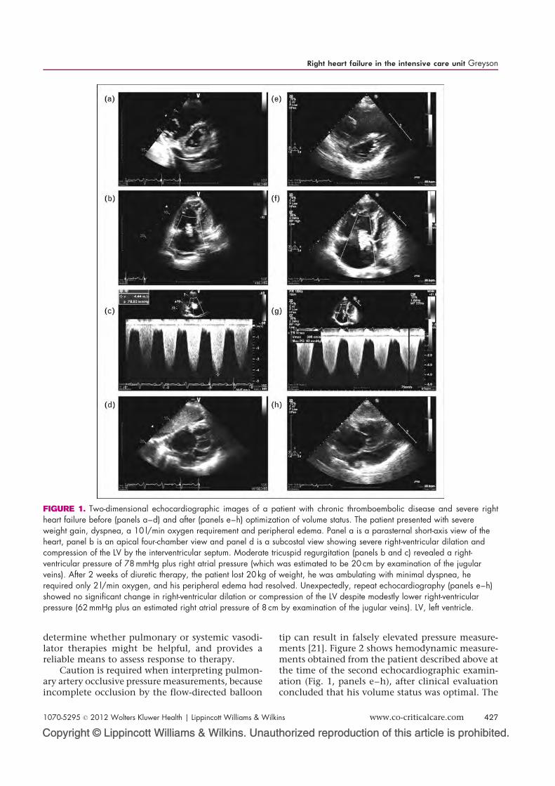
Moreover, echocardiography may not bereliable for establishing the relationship betweenright-ventricular function and systemic hemody-namics. Figure 1 (panels a–d) shows echocardio-graphic images from a patient who presented withRHF due to chronic thromboembolic disease. Onadmission, he had severe dyspnea and orthopnea,bilateral lower extremity edema, and a requirementfor 10 l O2 by mask; his SBP was 120 mmHg, and hisestimated CVP was 20 mmHg. He was treated aggres-sively with diuretic therapy, and over the next 2weeks his weight declined 20 kg with no change inrenal function, his exercise tolerance improved, hisSBP stabilized at 100 mmHg, and his oxygen require-ment declined to 2 l by nasal cannula. Surprisingly,follow-up echocardiography (Fig. 1, panels e–h)showed no apparent improvement in right-ventric-ular size or function and no change in the para-doxical interventricular septal motion. In this case,it was simple clinical evaluation, rather thanadvanced imaging, that provided the most reliableguide to ongoing management.
Invasive hemodynamic assessmentBecause of challenges in interpreting imaging stud-ies, invasive hemodynamic assessment is frequentlynecessary in management of RHF. This is particu-larly true because measurement variability limitsthe reliability of echocardiographic evaluation ofright heart hemodynamics over time [20]. Rightheart catheterization provides a direct assessmentof CVP and right-ventricular filling pressure andan indirect measurement of left atrial pressure(pulmonary artery occlusive or wedge pressure),helps determine the contribution of left-sidedcardiovascular disease to pulmonary hypertension,is used to measure pulmonary vascular resistance to
Cardiovascular system
426 www.co-criticalcare.com Volume 18 ! Number 5 ! October 2012
Copyright © Lippincott Williams & Wilkins. Unauthorized reproduction of this article is prohibited.
determine whether pulmonary or systemic vasodi-lator therapies might be helpful, and provides areliable means to assess response to therapy.
Caution is required when interpreting pulmon-ary artery occlusive pressure measurements, becauseincomplete occlusion by the flow-directed balloon
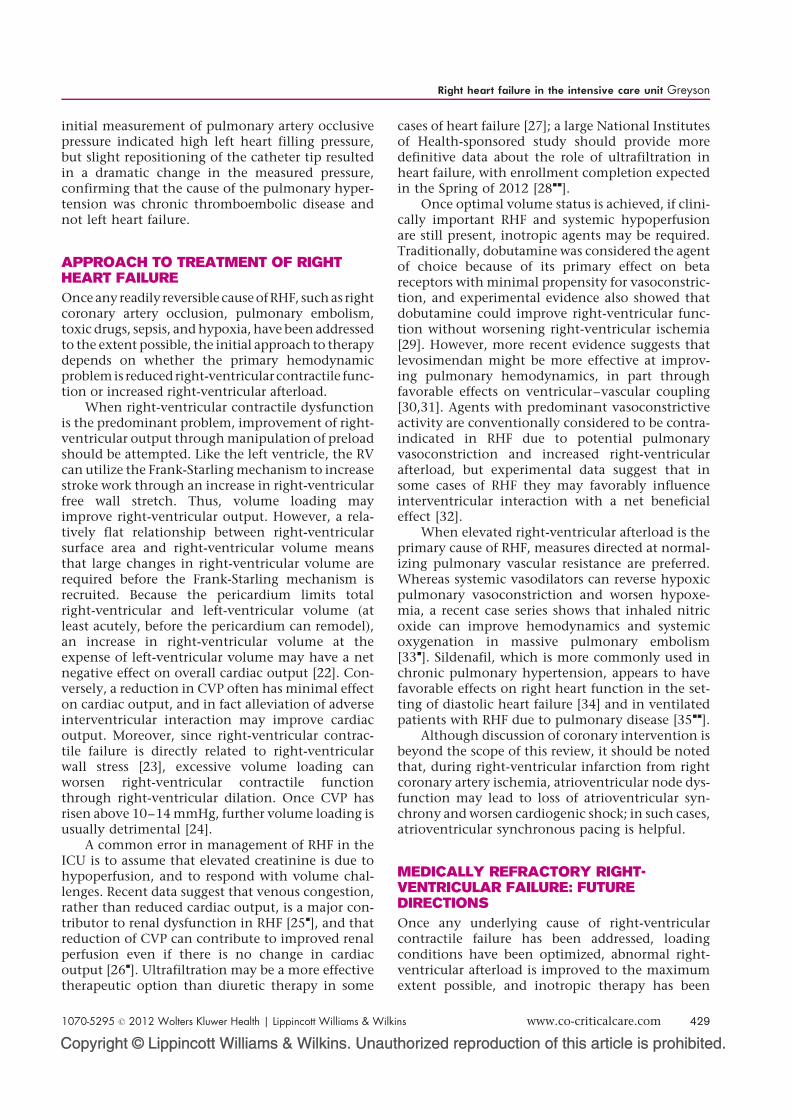
tip can result in falsely elevated pressure measure-ments [21]. Figure 2 shows hemodynamic measure-ments obtained from the patient described above atthe time of the second echocardiographic examin-ation (Fig. 1, panels e–h), after clinical evaluationconcluded that his volume status was optimal. The
FIGURE 1. Two-dimensional echocardiographic images of a patient with chronic thromboembolic disease and severe rightheart failure before (panels a–d) and after (panels e–h) optimization of volume status. The patient presented with severeweight gain, dyspnea, a 10 l/min oxygen requirement and peripheral edema. Panel a is a parasternal short-axis view of theheart, panel b is an apical four-chamber view and panel d is a subcostal view showing severe right-ventricular dilation andcompression of the LV by the interventricular septum. Moderate tricuspid regurgitation (panels b and c) revealed a right-ventricular pressure of 78 mmHg plus right atrial pressure (which was estimated to be 20 cm by examination of the jugularveins). After 2 weeks of diuretic therapy, the patient lost 20 kg of weight, he was ambulating with minimal dyspnea, herequired only 2 l/min oxygen, and his peripheral edema had resolved. Unexpectedly, repeat echocardiography (panels e–h)showed no significant change in right-ventricular dilation or compression of the LV despite modestly lower right-ventricularpressure (62 mmHg plus an estimated right atrial pressure of 8 cm by examination of the jugular veins). LV, left ventricle.
Right heart failure in the intensive care unit Greyson
1070-5295 ! 2012 Wolters Kluwer Health | Lippincott Williams & Wilkins www.co-criticalcare.com 427

Copyright © Lippincott Williams & Wilkins. Unauthorized reproduction of this article is prohibited.
86MonitorBPM
87MonitorBPM
60/1, 10RV 87MonitorBPM62/27 (39)PA(a)
11/11 (10)RA(c)
(b)
100
50
0
Ÿ
⁄
100
50
0
Ÿ
⁄
50
25
0
Ÿ
⁄
1 1
1
vvvvv v v v v v vaaa
a
r r r rr
r r r r r r r
d d d d d d d d d d d
e e e e e e e e e e
ss
s
ss
s s ss s s
s
rrrrrrrr r rr r r r r r r r r r r
r r
sss
ssss
sss
d dd
d dd d d
d d
85Monitor
BPM6/5 (1)PW
(e)
50
25
0
Ÿ
⁄
1
r r r r r r r r r r r r
aa a
vvav
a vaa vvv
86MonitorBPM
r r r rrrrrrr rr
23/28 (24)PW(d)
100
50
0
Ÿ
⁄
1
vvvvv
vv
v v v vaa
aaaaa
a aa
FIGURE 2. Hemodynamic tracings obtained during right heart catheterization from the patient described in Fig. 1 after2 weeks of diuretic therapy, at a time when clinical evaluation concluded optimization of volume status had been achieved.Panels a–c show right-ventricular pulmonary artery and right atrial pressure tracings. Right atrial mean pressure was 8 mmHgand peak right-ventricular pressure was 62 mmHg, confirming findings by physical examination and echocardiography(Fig. 1, panel g). Panel d shows a tracing obtained after inflation of the pulmonary artery catheter balloon. Initially, itappeared that left atrial pressure was very high (end-expiratory pulmonary artery occlusion pressure was 20 mmHg). Slightrepositioning of the catheter resulted in a dramatically different finding shown in panel e (end-expiratory pulmonary arteryocclusion pressure of only 8 mmHg), highlighting the difficulty of obtaining reliable estimates of left atrial pressure in patientswith severe pulmonary hypertension. Balloon inflation for measurement of pulmonary artery occlusion pressure should be donewith caution in patients with severe pulmonary hypertension.
Cardiovascular system
428 www.co-criticalcare.com Volume 18 ! Number 5 ! October 2012
Copyright © Lippincott Williams & Wilkins. Unauthorized reproduction of this article is prohibited.
initial measurement of pulmonary artery occlusivepressure indicated high left heart filling pressure,but slight repositioning of the catheter tip resultedin a dramatic change in the measured pressure,confirming that the cause of the pulmonary hyper-tension was chronic thromboembolic disease andnot left heart failure.
APPROACH TO TREATMENT OF RIGHTHEART FAILUREOnce any readily reversible cause ofRHF, such as rightcoronary artery occlusion, pulmonary embolism,toxic drugs, sepsis, and hypoxia, have been addressedto the extent possible, the initial approach to therapydepends on whether the primary hemodynamicproblem is reduced right-ventricularcontractile func-tion or increased right-ventricular afterload.
When right-ventricular contractile dysfunctionis the predominant problem, improvement of right-ventricular output through manipulation of preloadshould be attempted. Like the left ventricle, the RVcan utilize the Frank-Starling mechanism to increasestroke work through an increase in right-ventricularfree wall stretch. Thus, volume loading mayimprove right-ventricular output. However, a rela-tively flat relationship between right-ventricularsurface area and right-ventricular volume meansthat large changes in right-ventricular volume arerequired before the Frank-Starling mechanism isrecruited. Because the pericardium limits totalright-ventricular and left-ventricular volume (atleast acutely, before the pericardium can remodel),an increase in right-ventricular volume at theexpense of left-ventricular volume may have a netnegative effect on overall cardiac output [22]. Con-versely, a reduction in CVP often has minimal effecton cardiac output, and in fact alleviation of adverseinterventricular interaction may improve cardiacoutput. Moreover, since right-ventricular contrac-tile failure is directly related to right-ventricularwall stress [23], excessive volume loading canworsen right-ventricular contractile functionthrough right-ventricular dilation. Once CVP hasrisen above 10–14 mmHg, further volume loading isusually detrimental [24].
A common error in management of RHF in theICU is to assume that elevated creatinine is due tohypoperfusion, and to respond with volume chal-lenges. Recent data suggest that venous congestion,rather than reduced cardiac output, is a major con-tributor to renal dysfunction in RHF [25&], and thatreduction of CVP can contribute to improved renalperfusion even if there is no change in cardiacoutput [26&]. Ultrafiltration may be a more effectivetherapeutic option than diuretic therapy in some
cases of heart failure [27]; a large National Institutesof Health-sponsored study should provide moredefinitive data about the role of ultrafiltration inheart failure, with enrollment completion expectedin the Spring of 2012 [28&&].
Once optimal volume status is achieved, if clini-cally important RHF and systemic hypoperfusionare still present, inotropic agents may be required.Traditionally, dobutamine was considered the agentof choice because of its primary effect on betareceptors with minimal propensity for vasoconstric-tion, and experimental evidence also showed thatdobutamine could improve right-ventricular func-tion without worsening right-ventricular ischemia[29]. However, more recent evidence suggests thatlevosimendan might be more effective at improv-ing pulmonary hemodynamics, in part throughfavorable effects on ventricular–vascular coupling[30,31]. Agents with predominant vasoconstrictiveactivity are conventionally considered to be contra-indicated in RHF due to potential pulmonaryvasoconstriction and increased right-ventricularafterload, but experimental data suggest that insome cases of RHF they may favorably influenceinterventricular interaction with a net beneficialeffect [32].
When elevated right-ventricular afterload is theprimary cause of RHF, measures directed at normal-izing pulmonary vascular resistance are preferred.Whereas systemic vasodilators can reverse hypoxicpulmonary vasoconstriction and worsen hypoxe-mia, a recent case series shows that inhaled nitricoxide can improve hemodynamics and systemicoxygenation in massive pulmonary embolism[33&]. Sildenafil, which is more commonly used inchronic pulmonary hypertension, appears to havefavorable effects on right heart function in the set-ting of diastolic heart failure [34] and in ventilatedpatients with RHF due to pulmonary disease [35&&].
Although discussion of coronary intervention isbeyond the scope of this review, it should be notedthat, during right-ventricular infarction from rightcoronary artery ischemia, atrioventricular node dys-function may lead to loss of atrioventricular syn-chrony and worsen cardiogenic shock; in such cases,atrioventricular synchronous pacing is helpful.
MEDICALLY REFRACTORY RIGHT-VENTRICULAR FAILURE: FUTUREDIRECTIONSOnce any underlying cause of right-ventricularcontractile failure has been addressed, loadingconditions have been optimized, abnormal right-ventricular afterload is improved to the maximumextent possible, and inotropic therapy has been
Right heart failure in the intensive care unit Greyson
1070-5295 ! 2012 Wolters Kluwer Health | Lippincott Williams & Wilkins www.co-criticalcare.com 429

Copyright © Lippincott Williams & Wilkins. Unauthorized reproduction of this article is prohibited.
employed, few additional interventions have estab-lished efficacy.
In theory, bypassing the pulmonary circulationmight improve cardiac output in cases of elevatedright-ventricular afterload. Atrial septostomy hasbeen promoted as a potential therapy for RHF insevere pulmonary hypertension, but, whereascardiac output may improve, systemic oxygenationprobably does not [36,37]. Selectively redirectingdeoxygenated venous blood from the pulmonaryartery to the lower half of the body via a left pul-monary artery to descending thoracic aorta right toleft shunt (Potts anastomosis) has been used success-fully in pediatric patients with chronic suprasyste-mic pulmonary pressure, but, if pulmonary pressurefalls, left to right shunt develops, causing an increasein pulmonary flow and worsened pulmonary hyper-tension. To avoid this problem, Bui et al. [38] createda Potts anastomosis with a one-way valve in a por-cine experimental model of pulmonary hyperten-sion to allow right to left flow only during episodicincreases in pulmonary artery pressure (such asduring exercise).
Because right-ventricular pressure overloadcauses dyssynchronous contraction of the RV andleft ventricle with paradoxical septal motion contri-buting to diminished cardiac output, it would seemthat some form of resynchronization therapy mightbe beneficial, just as it is in left heart failure with awide QRS complex from left bundle branch block.Preliminary data suggest just such a possibility [39&&].
The most promising new approach to RHF is theuse of mechanical assist devices. When RHF devel-ops after left-ventricular assist device (LVAD)implantation, several different devices have beenemployed successfully using either surgical [40&]or percutaneous [41] implantation. Percutaneouslyimplanted RVADs have also been used as single-chamber device therapy in the setting of primaryright-ventricular contractile failure [42&]; whetherthese devices are effective in right-ventricular pres-sure overload failure is not yet established. RV tosystemic bypass might be more effective if thebypassed blood were oxygenated. Spillner et al.[43&] tested extracorporeal oxygenation using anRVAD in an ovine model; the clinical feasibility ofa similar technique was demonstrated when used incombination with a LVAD [44&].
Although only animal experimental data areavailable so far, there are indications that activationof proteases or apoptosis contributes to right-ventricular contractile dysfunction in acute right-ventricular pressure overload, and that certainprotease inhibitors attenuate this process [45,46&].Whether or not these agents have clinical efficacy isyet to be determined.
CONCLUSIONRight heart failure occurs when the RV of the heartis unable to provide adequate blood flow throughthe pulmonary circulation at a normal CVP. Carefulphysical examination, imaging studies, and invasivehemodynamic monitoring are key to identificationof reversible underlying causes of right-ventricularcontractile dysfunction or elevated right-ventricularafterload. When optimization of preload fails,judicious use of inotropic agents is effective. RVADsare currently employed in some centers and showpromise for patients with medically refractory RHF.Further data on the biochemical mechanisms of RHFmay lead to new medical therapies in the future.
Acknowledgements
Supported by the Department of Veterans AffairsMedical Research Service.
Conflicts of interest
There are no conflicts of interest.
REFERENCES AND RECOMMENDEDREADINGPapers of particular interest, published within the annual period of review, havebeen highlighted as:& of special interest&& of outstanding interestAdditional references related to this topic can also be found in the CurrentWorld Literature section in this issue (pp. 562–563).
1. Greyson CR. The right ventricle and pulmonary circulation: basic concepts.Rev Esp Cardiol 2010; 63:81–95.
2. Greyson CR. Pathophysiology of right ventricular failure. Crit Care Med 2008;36:S57–65.
3. Santamore WP, Amoore JN. Buffering of respiratory variations in venousreturn by right ventricle: a theoretical analysis. Am J Physiol 1994; 267:H2163–H2170.
4. Brooks H, Kirk ES, Vokonas PS, et al. Performance of the right ventricle understress: relation to right coronary flow. J Clin Invest 1971; 50:2176–2183.
5. Guyton AC, Lindsey AW, Gilluly JL. The limits of right ventricular compensa-tion following acute increase in pulmonary circulatory resistance. Circ Res1954; 2:326–332.
6. Greyson CR, Schwartz GG, Lu L, et al. Calpain inhibition attenuates rightventricular contractile dysfunction after acute pressure overload. J Mol CellCardiol 2008; 44:59–68.
7. Dewachter C, Dewachter L, Rondelet B, et al. Activation of apoptotic path-ways in experimental acute afterload-induced right ventricular failure. CritCare Med 2010; 38:1405–1413.
8. Bogaard HJ, Abe K, Vonk Noordegraaf A, Voelkel NF. The right ventricle underpressure: cellular and molecular mechanisms of right-heart failure in pulmon-ary hypertension. Chest 2009; 135:794–804.
9.&
Rondelet B, Dewachter C, Kerbaul F, et al. Prolonged overcirculation-inducedpulmonary arterial hypertension as a cause of right ventricular failure. EurHeart J 2012; 33:1017–1026.
This experimental study provides new insight into the mechanisms that contributeto RHF in the setting of excessive pulmonary arterial flow.10.&
Stevens GR, Garcia-Alvarez A, Sahni S, et al. RV dysfunction in pulmonaryhypertension is independently related to pulmonary artery stiffness. JACCCardiovasc Imaging 2012; 5:378–387.
This clinical study shows that right-ventricular function is as dependent onpulmonary arterial stiffness as it is on pulmonary vascular resistance; this mayhave implications for clinical evaluation of patients with right heart failure notentirely explained by elevated PVR.11.&
Wang Z, Chesler NC. Pulmonary vascular wall stiffness: an important con-tributor to the increased right ventricular afterload with pulmonary hyperten-sion. Pulm Circ 2011; 1:212–223.
Nice review of methods for investigating mechanical characteristics of the pul-monary circulation that go beyond simple assessment of pulmonary vascularresistance, written by and for biomedical engineers.
Cardiovascular system
430 www.co-criticalcare.com Volume 18 ! Number 5 ! October 2012
Copyright © Lippincott Williams & Wilkins. Unauthorized reproduction of this article is prohibited.
12.&
Vanagt WY, Famaey N, Rega F, Gewillig M. Extreme windkessel effect cancause right heart failure early after truncus repair. Interact Cardiovasc ThoracSurg 2012; 15:181–182.
The physiology of RHF is incompletely understood; this interesting case reportunderlines the fact that RHF can occur for reasons not commonly appreciated.13. Guervilly C, Forel JM, Hraiech S, et al. Right ventricular function during high-
frequency oscillatory ventilation in adults with acute respiratory distresssyndrome. Crit Care Med 2012; 40:1539–1545.
14. Eisenberg PR, Jaffe AS, Schuster DP. Clinical evaluation compared topulmonary artery catheterization in the hemodynamic assessment of criticallyill patients. Crit Care Med 1984; 12:549–553.
15. Jang T, Aubin C, Naunheim R, Char D. Ultrasonography of the internal jugularvein in patients with dyspnea without jugular venous distention on physicalexamination. Ann Emerg Med 2004; 44:160–168.
16.&
Jang T, Aubin C, Naunheim R, et al. Jugular venous distension on ultrasound:sensitivity and specificity for heart failure in patients with dyspnea. Am J EmergMed 2011; 29:1198–1202.
A widely available portable ultrasound unit can provide useful measurements ofCVP noninvasively when physical examination is inconclusive; this is simpler andprobably more accurate than estimation of CVP using inferior vena cava dynamicsas commonly performed during a full echocardiographic examination.17. Allemann Y, Rotter M, Hutter D, et al. Impact of acute hypoxic pulmonary
hypertension on LV diastolic function in healthy mountaineers at high altitude.Am J Physiol Heart Circ Physiol 2004; 286:H856–H862.
18. Ling LF, Obuchowski NA, Rodriguez L, et al. Accuracy and interobserverconcordance of echocardiographic assessment of right ventricular size andsystolic function: a quality control exercise. J Am Soc Echocardiogr 2012;25:709–713.
19.&
Mitoff PR, Beauchesne L, Dick AJ, et al. Imaging the failing right ventricle. CurrOpin Cardiol 2012; 27:148–153.
This is a very readable and concise review of imaging techniques available forevaluation of the RV.20. Farber HW, Foreman AJ, Miller DP, McGoon MD. REVEAL Registry: correla-
tion of right heart catheterization and echocardiography in patients withpulmonary arterial hypertension. Congest Heart Fail 2011; 17:56–64.
21. Leatherman JW, Shapiro RS. Overestimation of pulmonary artery occlusionpressure in pulmonary hypertension due to partial occlusion. Crit Care Med2003; 31:93–97.
22. Belenkie I, Dani R, Smith ER, Tyberg JV. Effects of volume loading duringexperimental acute pulmonary embolism. Circulation 1989; 80:178–188.
23. Greyson C, Xu Y, Lu L, Schwartz GG. Right ventricular pressure and dilationduring pressure overload determine dysfunction after pressure overload. Am JPhysiol Heart Circ Physiol 2000; 278:H1414–H1420.
24. Berisha S, Kastrati A, Goda A, Popa Y. Optimal value of filling pressure in theright side of the heart in acute right ventricular infarction. Br Heart J 1990;63:98–102.
25.&
Costanzo MR, Jessup M. Treatment of congestion in heart failure withdiuretics and extracorporeal therapies: effects on symptoms, renal function,and prognosis. Heart Fail Rev 2012; 17:313–324.
This comprehensive review summarizes what is known about the effects of venouscongestion and renal failure, and outlines the therapeutic options.26.&
Testani JM, Khera AV, St John Sutton MG, et al. Effect of right ventricularfunction and venous congestion on cardiorenal interactions during the treat-ment of decompensated heart failure. Am J Cardiol 2010; 105:511–516.
This very provocative study concludes that venous congestion is a more importantcause of renal dysfunction than low cardiac output in RHF.27. Giglioli C, Landi D, Cecchi E, et al. Effects of ULTRAfiltration vs. DIureticS on
clinical, biohumoral and haemodynamic variables in patients with deCOm-pensated heart failure: the ULTRADISCO study. Eur J Heart Fail 2011;13:337–346.
28.&&
Bart BA, Goldsmith SR, Lee KL, et al. Cardiorenal rescue study in acutedecompensated heart failure: rationale and design of CARRESS-HF, for theHeart Failure Clinical Research Network. J Card Fail 2012; 18:176–182.
Venous congestion is a major cause of cardiorenal syndrome, but patients withheart failure are often diuretic resistant. RHF is a major cause of renal dysfunctionfrom venous congestion. Smaller studies have suggested that ultrafiltration is aneffective alternative to loop diuretics in left heart failure. This study, when pub-lished, should provide conclusive data about whether ultrafiltration should be first-line therapy.29. Greyson C, Garcia J, Mayr M, Schwartz GG. Effects of inotropic stimulation
on energy metabolism and systolic function of ischemic right ventricle. Am JPhysiol 1995; 268:H1821–H1828.
30. Kerbaul F, Rondelet B, Demester JP, et al. Effects of levosimendan versusdobutamine on pressure load-induced right ventricular failure. Crit Care Med2006; 34:2814–2819.
31. Morelli A, Teboul JL, Maggiore SM, et al. Effects of levosimendan on rightventricular afterload in patients with acute respiratory distress syndrome: apilot study. Crit Care Med 2006; 34:2287–2293.
32. Apitz C, Honjo O, Friedberg MK, et al. Beneficial effects of vasopressors onright ventricular function in experimental acute right ventricular failure in arabbit model. Thorac Cardiovasc Surg 2012; 60:17–23.
33.&
Summerfield DT, Desai H, Levitov A, et al. Inhaled nitric oxide as salvagetherapy in massive pulmonary embolism: a case series. Respir Care 2012;57:444–448.
This short case series suggests that inhaled nitric oxide may be effective forstabilizing patients with massive pulmonary embolism until more definitive therapyis available.34. Guazzi M, Vicenzi M, Arena R, Guazzi MD. Pulmonary hypertension in heart
failure with preserved ejection fraction: a target of phosphodiesterase-5inhibition in a 1-year study. Circulation 2011; 124:164–174.
35.&&
Karakitsos D, Papanikolaou J, Karabinis A, et al. Acute effect of sildenafil oncentral hemodynamics in mechanically ventilated patients with WHO group IIIpulmonary hypertension and right ventricular failure necessitating adminis-tration of dobutamine. Int J Cardiol 2012. [Epub ahead of print]
Sildenafil is inexpensive and well tolerated. This small study suggests it may haveefficacy in RHF from pulmonary hypertension, and is worth trying in RHF that fails toimprove on conventional medical therapy. Ventilated patients dependent ondobutamine were treated with oral sildenafil, and in many cases could besuccessfully weaned from dobutamine and ventilatory support.36. Diller GP, Lammers AE, Haworth SG, et al. A modelling study of atrial
septostomy for pulmonary arterial hypertension, and its effect on the state oftissue oxygenation and systemic blood flow. Cardiol Young 2010; 20:25–32.
37. Koeken Y, Kuijpers NH, Lumens J, et al. Atrial septostomy benefits severepulmonary hypertension patients by increase of left ventricular preloadreserve. Am J Physiol Heart Circ Physiol 2012; 302:H2654–H2662.
38. Bui MT, Grollmus O, Ly M, et al. Surgical palliation of primary pulmonaryarterial hypertension by a unidirectional valved Potts anastomosis in an animalmodel. J Thorac Cardiovasc Surg 2011; 142:1223–1228.
39.&&
Hardziyenka M, Surie S, de Groot JR, et al. Right ventricular pacing improveshaemodynamics in right ventricular failure from pressure overload: an openobservational proof-of-principle study in patients with chronic thromboem-bolic pulmonary hypertension. Europace 2011; 13:1753–1759.
Resynchronization therapy has been proven to improve left heart failure in severallarge clinical studies. This study sets the stage for similar trials to be conducted inpatients with RHF.40.&
Fukamachi K, Shiose A, Massiello AL, et al. Implantable continuous-flow rightventricular assist device: lessons learned in the development of a Clevelandclinic device. Ann Thorac Surg 2012; 93:1746–1752.
This is a comprehensive review of one large center’s experience with the technicaldevelopment of RVAD therapy, and surveys the current state of the art.41. Haneya A, Philipp A, Puehler T, et al. Temporary percutaneous right ventricular
support using a centrifugal pump in patients with postoperative acuterefractory right ventricular failure after left ventricular assist device implanta-tion. Eur J Cardiothorac Surg 2012; 41:219–223.
42.&
Kapur NK, Paruchuri V, Korabathina R, et al. Effects of a percutaneousmechanical circulatory support device for medically refractory right ventricularfailure. J Heart Lung Transplant 2011; 30:1360–1367.
This is a case series describing the use of percutaneously implanted RVADs. Itprovides useful guidance for clinicians interested in using this technique.43.&
Spillner J, Stoppe C, Hatam N, et al. Feasibility and efficacy of bypassing theright ventricle and pulmonary circulation to treat right ventricular failure: anexperimental study. J Cardiothorac Surg 2012; 7:15.
This experimental animal study shows the potential for RVADs in conjunction withextracorporeal oxygenation as single-chamber therapy for treatment of RHF.44.&
De Silva RJ, Soto C, Spratt P. Extra corporeal membrane oxygenation as rightheart support following left ventricular assist device placement: a newcannulation technique. Heart Lung Circ 2012; 21:218–220.
This clinical report describes a surgical technique that allows RVADs to be used inconjunction with extracorporeal oxygenation in closed chest patients.45. Mani SK, Shiraishi H, Balasubramanian S, et al. In vivo administration of
calpeptin attenuates calpain activation and cardiomyocyte loss in pressure-overloaded feline myocardium. Am J Physiol Heart Circ Physiol 2008;295:H314–H326.
46.&
Ahmad HA, Lu L, Ye S, et al. Calpain inhibition preserves talin and attenuatesright heart failure in acute pulmonary hypertension. Am J Respir Cell Mol Biol2012. [Epub ahead of print]
This is the first study of any agent that attenuates the hemodynamic severity ofacute right-ventricular pressure overload-induced RHF by modulating biochemicalchanges within the right-ventricular myocardium in a large animal model of RHF.Whether or not such agents are useful in clinical RHF remains to be determined.
Right heart failure in the intensive care unit Greyson
1070-5295 ! 2012 Wolters Kluwer Health | Lippincott Williams & Wilkins www.co-criticalcare.com 431