coupling of hedgehog and hippo pathways promotes stem cell
TRANSCRIPT

JCB: Article
The Rockefeller University Press $30.00J. Cell Biol. Vol. 205 No. 3 325–338www.jcb.org/cgi/doi/10.1083/jcb.201309141 JCB 325
Correspondence to Daniel Kalderon: [email protected] used in this paper: act, actin; AP, anterior–posterior; BMP, bone morphogenetic protein; Ci, Cubitus interruptus; CycE, Cyclin E; Dpp, Decapen-taplegic; EC, escort cell; EdU, 5-ethynyl-2-deoxyuridine; Ex, Expanded; Fas3, Fasciclin3; FC, follicle cell; FSC, follicle stem cell; GSC, germline stem cell; Hh, Hedgehog; Hpo, Hippo; HSC, hematopoietic stem cell; JAK-STAT, Janus kinase–signal transducer and activator of transcription; Mam, Mastermind; MARCM, mosaic analysis with a repressible cell marker; Mer, Merlin; ptc, patched; Sav, Salvador; smo, smoothened; tub, tubulin; ubi, ubiquitin; Wts, Warts; YAP, Yes-associated protein; Yki, Yorkie.
IntroductionSelf-renewal of adult stem cells is generally supported by a spe-cialized niche microenvironment, which can serve to maintain stem cells in an appropriate location, in an undifferentiated state or in an appropriate proliferative state. For example, a short-range bone morphogenetic protein (BMP) signal produced in directly adjacent cap cells (Fig. 1 A) promotes germline stem cell (GSC) maintenance in the Drosophila melanogaster ovary by repressing a key differentiation factor (Chen et al., 2011b). BMP similarly represses differentiation in the male GSC lin-eage, whereas a Janus kinase–signal transducer and activator of transcription (JAK-STAT) pathway ligand produced in directly adjacent hub cells represses differentiation of somatic stem cells in the Drosophila testis (Losick et al., 2011; Matunis et al., 2012). These and other findings firmly established repression of differentiation as a, likely widespread, mechanism for niche sig-nals to maintain stem cells (Losick et al., 2011). Other potential mechanisms for niche signals to support stem cells have not been fully explored and validated.
Although stem cells must proliferate to serve their regen-erative function, relative quiescence was once thought to be universally important for long-term maintenance of stem cells. Several types of stem cells, including mammalian hematopoietic stem cells (HSCs) and muscle stem cells, indeed divide infre-quently, whereas normal cycles of hair growth depend on main-taining quiescence of hair follicle stem cells (FSCs) in between brief periods of activation (Pietras et al., 2011; Chakkalakal et al., 2012; Hsu and Fuchs, 2012). However, it is now clear that some adult stem cells, including mammalian epidermal and gut stem cells proliferate constitutively and extensively (Simons and Clevers, 2011; Hsu and Fuchs, 2012). In all of these cases, there are likely specific niche signals, some of which have been identified, that stimulate or inhibit stem cell proliferation to produce an appropriate supply of tissue-replenishing daughters. However, an important question that has been hard to resolve is whether regulation of the rate of stem cell proliferation by niche factors is also a major mechanism for regulating the mainte-nance of the stem cells themselves.
Drosophila FSCs provide a particularly informative stem cell paradigm for understanding niche function because FSC maintenance depends on multiple known extracellular signals
I t is essential to define the mechanisms by which exter-nal signals regulate adult stem cell numbers, stem cell maintenance, and stem cell proliferation to guide re-
generative stem cell therapies and to understand better how cancers originate in stem cells. In this paper, we show that Hedgehog (Hh) signaling in Drosophila melanogaster ovarian follicle stem cells (FSCs) induces the activity of Yorkie (Yki), the transcriptional coactivator of the Hippo pathway, by inducing yki transcription. Moreover, both Hh signaling and Yki positively regulate the rate of FSC
proliferation, both are essential for FSC maintenance, and both promote increased FSC longevity and FSC duplica-tion when in excess. We also found that responses to acti-vated Yki depend on Cyclin E induction while responses to excess Hh signaling depend on Yki induction, and excess Yki can compensate for defective Hh signaling. These causal connections provide the most rigorous evidence to date that a niche signal can promote stem cell mainte-nance principally by stimulating stem cell proliferation.
Coupling of Hedgehog and Hippo pathways promotes stem cell maintenance by stimulating proliferation
Jianhua Huang and Daniel Kalderon
Department of Biological Sciences, Columbia University, New York, NY 10027
© 2014 Huang and Kalderon This article is distributed under the terms of an Attribution–Noncommercial–Share Alike–No Mirror Sites license for the first six months after the publi-cation date (see http://www.rupress.org/terms). After six months it is available under a Creative Commons License (Attribution–Noncommercial–Share Alike 3.0 Unported license, as described at http://creativecommons.org/licenses/by-nc-sa/3.0/).
TH
EJ
OU
RN
AL
OF
CE
LL
BIO
LO
GY
Dow
nloaded from http://rupress.org/jcb/article-pdf/205/3/325/1364738/jcb_201309141.pdf by guest on 15 February 2022

JCB • VOLUME 205 • NUMBER 3 • 2014 326
FSCs are constitutively active in well-fed flies, supporting the production of up to two egg chambers per day from each germarium (Margolis and Spradling, 1995), and are therefore a good model for highly proliferative mammalian stem cells. We had previously suggested that FSC proliferation promotes FSC maintenance based on artificial genetic conditions: several homozygous mutations expected to decrease cell proliferation, including partial loss of function cyclin E (cycE) mutations, caused cell-autonomous loss of FSCs, whereas excess CycE compensated for the loss of several otherwise essential FSC fac-tors (Wang and Kalderon, 2009; Wang et al., 2012). We there-fore sought to understand how proliferation is regulated in FSCs and whether regulation of the rate of FSC proliferation by niche signals affects stem cell maintenance or stem cell numbers. Here, we show that Hh signaling induces the Hippo (Hpo) path-way coactivator Yorkie (Yki) in FSCs and that Yki, in turn, reg-ulates FSC maintenance and expansion by stimulating FSC proliferation via induction of CycE and by preventing apoptosis. These causal connections provide clear evidence that a niche signal can regulate stem cell maintenance by controlling the rate of stem cell proliferation.
and FSCs compete for niche occupation (Nystul and Spradling, 2007, 2010; Vied et al., 2012). FSCs are maintained midway along the anterior–posterior (AP) axis of the germarium, at the region 2a/2b border; here, rounded stage 2a germline cysts shorten along the AP axis to form lens-shaped stage 2b cysts that span the width of the germarium for the first time (Fig. 1 A; Margolis and Spradling, 1995). FSCs self-renew and produce follicle cell (FC) daughters, which proliferate as they form a monolayer epi-thelium around passing germline cysts and also become special-ized stalk cells that separate egg chambers as they bud from the posterior of the germarium (Fig. 1 A; Nystul and Spradling, 2007, 2010; Wu et al., 2008b). FSC maintenance depends on Wnt and BMP niche signals, but the Hedgehog (Hh) and JAK-STAT path-ways are the most potent regulators of FSC maintenance and expansion (Song and Xie, 2003; Kirilly et al., 2005; Vied et al., 2012). Hh and the JAK-STAT pathway ligand, Unpaired, derive from opposite ends of the germarium, producing reciprocal AP gradients that overlap in region 2a/b to induce sufficiently high activity in each pathway to support FSCs (Vied et al., 2012). How those signals act within FSCs to promote their maintenance or duplication has not yet been determined.
Figure 1. Yki is required for FSC maintenance. (A) Drosophila germarium: germline stem cells (GSC) produce cystoblast (CB) daughters, which proliferate to form 16-cell cysts (black) while surrounded by somatic escort cells (ECs). Follicle stem cells (FSC) just anterior (left) to region 2b cysts, which span the germarium, produce follicle cells (FC), which express Fas3 (red) at their surface and envelop germline cysts. A typical single FSC clone lineage is indicated by green nuclei. Hh is expressed strongly in anterior terminal filament (TF) and cap cells (CC), whereas the JAK-STAT pathway ligand is expressed strongly in polar FCs at the posterior of the germarium. (B) Percentage of ovarioles that contain wild-type (WT) or ykiB5 FSC or GSC clones 9–18 d after clone induc-tion in 2-d-old adults. (C) The percentage of ovarioles that maintain ykiB5 FSC clones 12 d after induction in larvae was greatly increased by coexpression of both UAS-diap1 and UAS-cycE transgenes. Error bars in B and C show SDs; n = 3, with ≥100 ovarioles scored in each measurement. (D–G) D and E show clones marked by the loss of GFP, whereas F and G show MARCM clones marked by expression of GFP. In all cases, clones were induced 12 d earlier, ovarioles were stained with Fas3, and close-ups of the FSC region are shown (D, E, F, and G). Red bars, 10 µm. (D) The wild-type FSC clone includes an FSC (arrowhead) just anterior (left) to Fas3 staining and many FC derivatives (white lines). (E) In most ykiB5 ovarioles, all FSCs (arrowhead) and FCs express GFP, indicating the absence of yki mutant FSCs, but GFP-negative germline cysts (asterisks) and GSCs were frequently present, indicating continued activity of yki mutant GSCs. (F) yki mutant FSCs expressing transgenic DIAP1 were generally not maintained for 12 d, but some ovarioles include a patch of GFP-marked FCs (white line), indicating that the marked FSC (arrowhead) was lost within the last 5 d. (G) Excess DIAP1 and CycE together rescued the maintenance of yki mutant FSCs in many ovarioles to produce clones with a marked FSC (arrowhead) and FC derivatives (white lines).
Dow
nloaded from http://rupress.org/jcb/article-pdf/205/3/325/1364738/jcb_201309141.pdf by guest on 15 February 2022

327Yorkie relays Hedgehog stem cell niche signal • Huang and Kalderon
excess CycE did not alter yki mutant FC clone size (Fig. 2, D–H). Thus, net expansion of yki mutant FC clones is limited principally by cell death.
Coexpression of DIAP1 in MARCM clones did not, how-ever, prevent the rapid loss of yki mutant FSCs (Fig. 1, C and F). Excess CycE was similarly ineffective. However, excess CycE and DIAP1 together substantially rescued yki FSC maintenance (Fig. 1, C and G). Hence, yki has an important antiapoptotic function throughout the FSC lineage, but it also has a selective function in FSCs connected with stimulating cell cycling that is not critical for FCs or indeed for GSCs.
FSCs with increased Yki activity compete betterIn Drosophila and mammals, upstream components of the Hpo pathway regulate the specific activity of Yki proteins. A major mechanism involves regulation of the nuclear entry and activity of Yki orthologues by phosphorylation at conserved sites, in-cluding S168 in Drosophila Yki (Pan, 2010; Staley and Irvine, 2012). The protein kinase directly responsible, Warts (Wts), is itself phosphorylated and activated by the Hpo protein kinase in a complex with the scaffolding protein Salvador (Sav). Hence, loss of hpo, sav, or wts generally activates Yki protein. We found that hpo, wts, and sav mutant FSC clones produced striking overrepresentation phenotypes, indicating enhanced FSC ac-tivity (Fig. 3, A and C; and Fig. S2 B). Each germarium con-tains more than one FSC. Consequently, marked wild-type FSC clones are almost always accompanied by unmarked FSC de-rivatives in mosaic ovarioles (Fig. 1, A and D). However, a significant proportion of hpo, wts, and sav mutant FSC clones occupied the entire ovariole (all-marked clones; Fig. 3, A and C; and Fig. S2 B), as seen previously for genetic changes (patched [ptc] mutation) that increase Hh (Fig. 3 D), JAK-STAT, or phos-phatidyl inositol 3-kinase pathway activities (Vied et al., 2012; Wang et al., 2012). Production of an all-marked clone from a single FSC implies that the marked FSC duplicated and out-competed unmarked FSCs in the same germarium (Nystul and Spradling, 2007; Vied et al., 2012). The frequency of all-marked clones continued to increase over time (36% for hpo with 28% mosaic and 39% for ptc with 34% mosaic at 26 d after clone in-duction in adults), as might be expected for FSCs with a stable competitive advantage. hpo, wts, and sav mutant FSC clones were also maintained better than wild-type FSC clones (Fig. 3 A). The frequency of all-marked ovarioles for hpo, wts, and sav was significantly higher for FSC clones induced in larvae rather than in adults (Fig. 3 A), as observed previously for excess Hh, phos-phatidyl inositol 3-kinase, or JAK-STAT pathway activity, con-sistent with the idea that competition among potential stem cells is particularly intense during FSC establishment (Vied et al., 2012; Wang et al., 2012).
The all-marked hpo, wts, and sav FSC clones also pro-duced an excessive number of FCs, which accumulated between egg chambers or formed multilayered epithelia (Fig. 3 C and Fig. S2 B). Those phenotypes are reminiscent of ptc mutant FSC clones (Fig. 3 D), which have elevated Hh pathway activity, and differ from the phenotypes produced by FSCs with increased
ResultsYki is required for FSC maintenanceThe Hpo pathway has recently emerged as a major regulator of organ size and of the growth, cycling, and survival of individual cells in Drosophila and mammals, motivating exploration of its potential roles governing stem cell proliferation (Pan, 2010; Zhao et al., 2011; Ramos and Camargo, 2012). To investigate Hpo pathway function in FSCs, we induced marked homozy-gous mutant FSCs at a fixed time by using a heat shock–inducible flp recombinase and FRT recombination targets located at the base of the appropriate chromosome arm. We then scored the percentage of ovarioles that retained marked (GFP negative) FSC and FC descendants (an FSC clone; Fig. 1, A and D) over time. The earliest time interval used in this well-established assay is generally 7 d (Vied and Kalderon, 2009; Wang and Kalderon, 2009; Vied et al., 2012; Wang et al., 2012) to ensure that “all-marked” cells originated from an FSC. Derivatives of FRT-mediated recombination in FCs (transient FC clones) normally pass through the ovariole in 5 d or less (Margolis and Spradling, 1995). FSC clones lacking activity of Yki, the key regulated transcriptional coactivator of the Hpo pathway, were lost very rapidly relative to controls when clones were induced in adults or in larvae (shortly before FSC establishment; Fig. 1, B–E; Vied et al., 2012).
We examined earlier time points by focusing only on the germarium, which should not retain transient FC clones beyond 3 d. At 4 d after clone induction, yki mutant FSCs were present in 13% of germaria compared with 71% for controls. Moreover, only 5% of germaria contained marked wild-type FCs but no FSC, whereas 30% of germaria contained yki mutant FCs but no FSC, directly revealing accelerated yki mutant FSC loss (Fig. S1 A).
In contrast, yki mutant GSCs, which also reside in the ger-marium and can be assayed in parallel by exactly analogous methodology, were maintained normally over 18 d (Fig. 1, B and E). A selective requirement in FSCs but not GSCs has been observed previously for several other regulators of FSC behavior (Wang et al., 2012).
Most FSC daughters divide about eight times over a 3–4-d period before differentiating as mature FCs and dying 2–3 d later (Margolis and Spradling, 1995). The proliferation of these FCs is not impaired by partial loss-of-function cycE mutations or by several other mutations in FSC-selective factors that dras-tically reduce FSC persistence (Wang and Kalderon, 2009; Wang et al., 2012). However, yki mutant clones (marked by loss of GFP) induced in FCs and captured five or fewer days after induction were much smaller than the “twin spot” wild-type clones (marked by two copies of the ubiquitin [ubi]-GFP gene), which are generated simultaneously (Fig. 2, A–C). Yki com-monly regulates both cell cycling and apoptosis. Apoptosis was detected in yki mutant FCs by activated Capase-3 staining in >15% of yki mutant FSC clones 4 d after induction compared with <2% of control FSC clones (Fig. S1, B–D). We also found that expression of a DIAP1 transgene, which inhibits apoptosis, in (GFP positive) yki mutant FC clones made by the mosaic analy-sis with a repressible cell marker (MARCM) method (Lee and Luo, 2001), restored their size to normal; in contrast, expressing
Dow
nloaded from http://rupress.org/jcb/article-pdf/205/3/325/1364738/jcb_201309141.pdf by guest on 15 February 2022

JCB • VOLUME 205 • NUMBER 3 • 2014 328
hpo, sav, or wts FSC clones, whereas kibra mutant phenotypes were the strongest of all (Fig. 3, A and B; and Fig. S2, C–E). Thus, Kibra, Ex, and the NF2 tumor suppressor orthologue Mer are key transducers of Hpo signaling in FSCs. The transmem-brane proteins, Crumbs, Echinoid, and Fat are potential up-stream regulators of the Hpo pathway that limit Yki activity, but their involvement in different Drosophila epithelial tissues is varied (Boggiano and Fehon, 2012; Staley and Irvine, 2012). Mutations affecting these three proteins each failed to pro-duce elevated frequencies of all-marked ovarioles (Fig. 3 B and Fig. S2 A), suggesting that they might act redundantly or that different, currently unknown, transmembrane sensors regulate the Hpo pathway in FSCs. Scalloped likely collaborates with Yki in the nucleus of FSCs as in several other cell types because scalloped mutant FSCs were lost significantly faster than con-trol FSCs (Fig. 3 B).
Hpo–Yki pathway regulates FSCs by altering the rate of FSC proliferationWe then wished to test whether the Hpo pathway regulates FSC maintenance and FSC expansion by influencing cell prolif-eration, as suggested by our earlier finding that excess CycE
phosphatidyl inositol 3-kinase or JAK-STAT pathway activity (Vied et al., 2012; Wang et al., 2012).
FSC clones lacking both Hpo and Yki activity were lost just as rapidly as yki mutant FSCs (Fig. 3 A), consistent with the expectation that hpo mutant phenotypes are induced entirely by excessive activation of Yki. Indeed, phenotypes analogous to those of hpo, wts, and sav were produced by expression of Yki with an activating S168A alteration in GFP-positive MARCM FSC clones (Fig. 4 D and Fig. S2, F and G). Thus, FSCs with elevated Yki activity outcompete wild-type FSCs for retention in the germarium and become overrepresented.
Merlin (Mer), Expanded (Ex), and Kibra are key upstream regulators of the Hpo pathway in FSCsKibra can associate with the cortical FERM (4.1 protein, Ezrin, Radixin, and Moesin) domain proteins Ex and Mer to connect external stimuli to activation of core Hpo pathway components (Boggiano and Fehon, 2012; Staley and Irvine, 2012). Both ex and mer mutant FSC clones survived better than wild-type clones and produced a significant frequency of all-marked ovar-ioles (Fig. 3, A and B). Those phenotypes were weaker than for
Figure 2. Yki is important for net FC amplification principally by preventing apoptosis. (A and B) Stage 10 egg chambers with control (wild type [WT]) or yki mutant clones lacking GFP (green, outlined in yellow) and adjacent wild-type twin spot clones with two copies of the GFP transgene (brighter green, outlined in white). Control clones were of similar size to simultaneously generated twin spots (A), but yki mutant clones were much smaller (B). (C) Mean number of cells in a GFP-negative clone divided by the number of cells in a twin spot, calculated for 20 (wild type) or 25 (yki) clones. Error bars show SDs. (D–G) Clones derived from single FCs formed patches (green nuclei, outlined in yellow) in stage 10 egg chambers that were much smaller than wild type (D) for yki mutant FCs (E); normal clone size was restored by expressing excess DIAP1 (G), but not CycE (F), in the clone. (H) Mean number of cells in a clone for wild-type and yki FCs expressing the indicated transgenes in the clone. Error bars show SDs, and number of clones measured is indicated in parentheses. Red bars, 15 µm.
Dow
nloaded from http://rupress.org/jcb/article-pdf/205/3/325/1364738/jcb_201309141.pdf by guest on 15 February 2022

329Yorkie relays Hedgehog stem cell niche signal • Huang and Kalderon
We then tested whether the regulation of FSC mainte-nance and competition by the Hpo pathway depends on CycE, which is commonly induced by Yki to stimulate cell cycling (Pan, 2010). Here, we used ex mutations to alter Hpo pathway activity because ex and cycE are on the same chromosome arm, facilitating testing of double mutants. These tests used both a null allele of cycE (cycEAR95) and an allele (cycEWX) that retains enough activity to support FC proliferation (Wang et al., 2012). Although ex mutant FSCs induced in larvae normally survive better than control FSC clones and produce an elevated fre-quency of all-marked clones (Fig. 4, D and E), cycE ex double mutant FSCs were instead lost prematurely (Fig. 4 D). Simi-larly, expression of an activated form of Yki (UAS-YkiS168A) alone produced a strong all-marked FSC clone phenotype but failed even to rescue FSC maintenance in cycE mutant FSCs (Fig. 4 D). FSC maintenance can be rescued for both cycE alleles by constitutive expression of UASGAL4-cycE (driven by constitutive GAL4 transgenes tubulin [tub]-GAL4 and actin [act]-GAL4; Wang and Kalderon, 2009), whereas expression of UAS-cycE in otherwise wild-type FSC clones has no effect
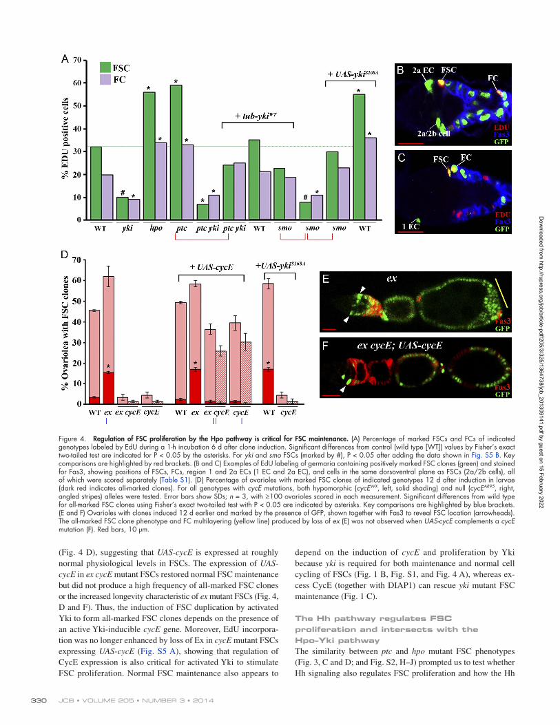
together with DIAP1 substantially restored yki FSC mainte-nance (Fig. 1, C and G). To examine whether the Hpo pathway reg-ulates proliferation in the FSC lineage, we labeled GFP-positive MARCM FSC clones of various genotypes with 5-ethynyl-2- deoxyuridine (EdU) for 1 h in vitro and measured the EdU- labeling DNA replication index. A typical wild-type FSC clone necessarily contains a marked FSC (scored as situated adjacent to the germarial wall immediately anterior to Fasciclin3 [Fas3] staining), together with marked Fas3-positive FCs, which we scored in regions 2b and 3 of the germarium (Fig. 1 A and Fig. 4, B and C). We also scored marked Fas3-negative cells close to the FSC (region 2a/b cells) and marked escort cells (ECs), which occupy regions 1 and 2a of the germarium and are normally quiescent (Fig. 4, B and C; and Table S1; Kirilly et al., 2011). The EdU labeling index, measured 6 d after clone induction, was greatly reduced for yki mutant FSCs (to 30% of wild type) and increased almost twofold for hpo mutant FSCs and FCs (Fig. 4 A and Table S1). Region 1 ECs were quiescent in all cases (Table S1). Thus, Yki activity is a major regulator of the rate of FSC proliferation.
Figure 3. FSCs with increased Yki activity displace wild-type FSCs and produce FC hypertrophy. (A and B) Percentage of ovarioles con-taining marked FSC clones 12 d after induction in larvae or adults. Dark red and blue columns show the percentage of all-marked (A.M.) ovarioles, containing only marked FSCs and FCs. To the left of each group of mutant genotypes is the control (wild type [WT]) for the appropriate chromosome arm (2R, 3R, X, or 2L) that becomes homo-zygous in clones. Error bars show SDs; n = 3, with ≥100 ovarioles scored in each measurement. Values for adult FSC clones are from one experiment. Significant differences from WT all-marked values for all-marked FSC clones using Fisher’s exact two-tailed test with P < 0.05 are indicated by asterisks. (C–D) All-marked ovarioles (C and D) and enlarged germarial regions (C and D), stained for Fas3, con-tained only GFP-negative FSCs (arrowheads) and FCs, which are homozygous for the indicated mutations, and included multilayering of FCs (yellow lines) and accumulation of FCs between egg chambers (white lines). Red bars, 10 µm.
Dow
nloaded from http://rupress.org/jcb/article-pdf/205/3/325/1364738/jcb_201309141.pdf by guest on 15 February 2022
JCB • VOLUME 205 • NUMBER 3 • 2014 330
depend on the induction of cycE and proliferation by Yki because yki is required for both maintenance and normal cell cycling of FSCs (Fig. 1 B, Fig. S1, and Fig. 4 A), whereas ex-cess CycE (together with DIAP1) can rescue yki mutant FSC maintenance (Fig. 1 C).
The Hh pathway regulates FSC proliferation and intersects with the Hpo–Yki pathwayThe similarity between ptc and hpo mutant FSC phenotypes (Fig. 3, C and D; and Fig. S2, H–J) prompted us to test whether Hh signaling also regulates FSC proliferation and how the Hh
(Fig. 4 D), suggesting that UAS-cycE is expressed at roughly normal physiological levels in FSCs. The expression of UAS-cycE in ex cycE mutant FSCs restored normal FSC maintenance but did not produce a high frequency of all-marked FSC clones or the increased longevity characteristic of ex mutant FSCs (Fig. 4, D and F). Thus, the induction of FSC duplication by activated Yki to form all-marked FSC clones depends on the presence of an active Yki-inducible cycE gene. Moreover, EdU incorpora-tion was no longer enhanced by loss of Ex in cycE mutant FSCs expressing UAS-cycE (Fig. S5 A), showing that regulation of CycE expression is also critical for activated Yki to stimulate FSC proliferation. Normal FSC maintenance also appears to
Figure 4. Regulation of FSC proliferation by the Hpo pathway is critical for FSC maintenance. (A) Percentage of marked FSCs and FCs of indicated genotypes labeled by EdU during a 1-h incubation 6 d after clone induction. Significant differences from control (wild type [WT]) values by Fisher’s exact two-tailed test are indicated for P < 0.05 by the asterisks. For yki and smo FSCs (marked by #), P < 0.05 after adding the data shown in Fig. S5 B. Key comparisons are highlighted by red brackets. (B and C) Examples of EdU labeling of germaria containing positively marked FSC clones (green) and stained for Fas3, showing positions of FSCs, FCs, region 1 and 2a ECs (1 EC and 2a EC), and cells in the same dorsoventral plane as FSCs (2a/2b cells), all of which were scored separately (Table S1). (D) Percentage of ovarioles with marked FSC clones of indicated genotypes 12 d after induction in larvae (dark red indicates all-marked clones). For all genotypes with cycE mutations, both hypomorphic (cycEWX, left, solid shading) and null (cycEAR95, right, angled stripes) alleles were tested. Error bars show SDs; n = 3, with ≥100 ovarioles scored in each measurement. Significant differences from wild type for all-marked FSC clones using Fisher’s exact two-tailed test with P < 0.05 are indicated by asterisks. Key comparisons are highlighted by blue brackets. (E and F) Ovarioles with clones induced 12 d earlier and marked by the presence of GFP, shown together with Fas3 to reveal FSC location (arrowheads). The all-marked FSC clone phenotype and FC multilayering (yellow line) produced by loss of ex (E) was not observed when UAS-cycE complements a cycE mutation (F). Red bars, 10 µm.
Dow
nloaded from http://rupress.org/jcb/article-pdf/205/3/325/1364738/jcb_201309141.pdf by guest on 15 February 2022

331Yorkie relays Hedgehog stem cell niche signal • Huang and Kalderon
expression of activated Yki (Fig. 6, A and F). Thus, Mam ap-pears to be critical for excess Hh signaling to activate Yki in the FSC lineage, and mam mutant FSC loss may be caused by in-sufficient Yki activity.
Hh induces yki transcription in the FSC lineageHh signaling normally influences cells by transcriptionally in-ducing cell type–specific target genes. We therefore tested whether Yki activation by Hh signaling in FSCs might be medi-ated by transcriptional induction of yki itself. Because reporters of Yki activity are activated by excess Hh pathway activity in both FSCs and FCs, we reasoned that the underlying mecha-nism was probably common to all cells of the FSC lineage and could be studied in samples containing both FSCs and FCs. We therefore isolated RNA from samples that included the germar-ium and all egg chambers of an ovariole up to stage 9. Analo-gous samples were collected from wild-type ovarioles and from ovarioles that were populated mostly by derivatives of either ptc or hpo mutant FSCs. We then measured the relative levels of key RNAs by quantitative RT-PCR, using rp49 mRNA as an in-ternal control. All three common Yki target genes tested (DIAP1, cycE, and ex) were induced in extracts from hpo mutant ovari-oles and, to almost the same extent, in extracts from ptc mutant ovarioles (Fig. 5 L). Those observations confirmed our previous findings with ex-lacZ and DIAP1-lacZ reporter genes and also showed directly that both Hh and Hpo–Yki pathways induce cycE in the FSC lineage. Crucially, yki RNA was also induced in samples from ptc mutant FSC lineages (Fig. 5 L), showing that Hh signaling does indeed induce yki expression in the FSC lineage. In contrast, yki RNA was not induced in hpo mutant FSC lineages (Fig. 5 L), as expected because Hpo regulates Yki activity posttranscriptionally.
Hh regulates FSCs by inducing Yki transcriptionally and thereby stimulating FSC proliferationIf Hh regulates FSCs by inducing yki transcription, excess Yki should mimic the effects of excess Hh signaling. Conversely, in the absence of Hh signaling, Yki protein levels may be too low to respond to inactivation of upstream Hpo pathway regulators. We found that excess wild-type Yki was indeed sufficient to produce increased FSC longevity and a significant frequency of all-marked clones (Fig. 6, A and D) as well as increased EdU incorporation (Fig. S5 B). The percentage of all-marked FSC clones was similar for FSCs with increased Hh pathway activity (ptc FSCs), increased Yki expression (from UAS-yki), or both alterations together (ptc + UAS-yki), consistent with the idea that Hh acts by increasing yki expression (Fig. 6 A). Likewise, the phenotype of ptc hpo double mutant FSCs was similar to the phenotype of ptc or hpo single mutant FSCs (Fig. S5 C). These results are consistent with the idea that maximal Hh signaling, inactivation of upstream Hpo pathway regulators of Yki, and excess Yki expression provide three routes to activate Yki, each of which produces a saturatingly high input from Yki into the proliferation rate and competitive behavior of FSCs.
and Hpo signaling pathways intersect in FSCs. The EdU label-ing index was increased almost twofold for both ptc mutant FSCs and germarial FCs, whereas region 1 ECs remained qui-escent (Fig. 4 A and Table S1). FSC proliferation has previously been shown to be increased by excess Hh signaling (Hartman et al., 2010). Conversely, the EdU labeling index was very low in smoothened (smo) mutant FSCs and germarial FCs, which cannot respond to Hh (Fig. 4 A). Thus, FSC proliferation is highly dependent on the level of Hh pathway activity. Further-more, double mutant ptc yki FSC clones showed strongly re-duced EdU labeling in FSCs (Fig. 4 A). Double mutant ptc yki FSC clones were also lost just as rapidly as yki FSC clones (see Fig. 6 A), showing that the effects of excess Hh on FSC duplica-tion, maintenance, and proliferation all depend absolutely on Yki activity.
Hh signaling induces Yki activity specifically in the FSC lineageTo test whether Hh signaling might directly stimulate Yki activity, we examined two Yki target gene reporters, DIAP1-lacZ and ex-lacZ (Karpowicz et al., 2010). Both reporters were readily detected in somatic cells in the ovary but not in the germline and showed increased expression cell autonomously in hpo mutant clones, confirming that they do report Yki activity (Fig. 5, A, B, F, G, and K; and Fig. S3, A, B, D, E, and G). Interestingly, FSCs expressed significantly lower levels of both DIAP1-lacZ and ex-lacZ than more posterior FC daughters or more anterior ECs (Fig. 5, A, F, and K; and Fig. S3, A, D, and G), implying that up-stream Hpo pathway components (Kibra, Ex, Mer, Sav, Wts, and Hpo) are particularly active (limiting Yki protein activity) in FSCs. Loss of yki activity reduced DIAP1-lacZ expression in FCs to a level similar to that of FSCs (Fig. S4, A, C, and I), supporting the inference that Yki activity is normally higher in FCs than FSCs.
Strikingly, loss of ptc activity markedly increased DIAP1-lacZ and ex-lacZ expression in both FSCs and FCs (Fig. 5, A, C, F, H, and K; and Fig. S3, A, C, D, F, and G), indicating that excess Hh pathway activity significantly stimulates Yki activity. The cell-autonomous activation of Yki by Hh signaling seen in the FSC lineage was not observed in wing discs, where ex-lacZ was instead induced in a ring around each anterior ptc mutant clone (Fig. S3, H–M). The nonautonomous induction of ex-lacZ is consistent with the recent observation that Decapentaplegic (Dpp) signaling activates Yki in wing discs (Kagey et al., 2012). Dpp is induced cell autonomously by Hh pathway activity in wing discs, but the ectopic Dpp acts principally on surrounding cells because Hh pathway activity also cell-autonomously inhibits expression of the Dpp receptor Thickveins (Funakoshi et al., 2001).
A previous study suggested that the transcriptional coacti-vator Mastermind (Mam), which normally contributes to Notch signaling, has an atypical role in FSCs, in which it collaborates closely with the Hh pathway (Vied and Kalderon, 2009). We therefore tested whether DIAP1-lacZ expression was induced by excess Hh pathway activity in the absence of Mam function. We found that DIAP1-lacZ was not increased in ptc mam double mutant FSCs or FCs (Fig. 5, E, J, and K). We also found that mam mutant FSCs have reduced EdU incorporation (Fig. S5 B) and that the maintenance of mam mutant FSCs was rescued by
Dow
nloaded from http://rupress.org/jcb/article-pdf/205/3/325/1364738/jcb_201309141.pdf by guest on 15 February 2022

JCB • VOLUME 205 • NUMBER 3 • 2014 332
to smo single mutant FSCs (Fig. S5 D). Thus, the requirement for Hh pathway input is downstream of, or parallel to, the input of conventional Hpo pathway regulators, consistent with our demonstration that Hh acts by inducing yki RNA, whereas up-stream Hpo pathway components regulate the activity of Yki protein. It also appears that regulation of Yki activity by the Hpo pathway in FSCs is only effective when normal Hh signaling provides sufficient, but not excessive, yki transcription.
Conversely, inactivation of the upstream Hpo pathway regulator Ex, which normally leads to posttranscriptional acti-vation of Yki, enhanced FSC maintenance, and frequent all-marked FSC clones did not induce all-marked FSC clones in the absence of smo activity and did not even counter the rapid loss of smo7.6.6 (Fig. 6 B) or smo2 mutant FSCs (Fig. S5 F). Similarly, smo hpo and smo wts double mutant FSCs did not produce all-marked clones but instead exhibited accelerated FSC loss, similar
Figure 5. Hh signaling increases Yki activity at the transcriptional level in the FSC lineage. (A–J) FSC clones of the indicated genotypes, marked by loss of GFP in germaria stained for Fas3 and -galactosidase (red) encoded by a diap1-lacZ reporter. diap1-lacZ expression was lower in wild-type (WT) FSCs (arrowhead; A) than FCs (white lines) and was increased in both FSCs (arrowheads) and FCs (arrows in F–J) for hpo and ptc genotypes (B, C, G, and H) but not in ptc yki; tub-yki (D and I) or ptc mam (E and J) genotypes. Red bars, 10 µm. (K) Quantification of diap1-lacZ expression levels in FSCs and FCs of the indicated genotypes. Error bars show SDs from three measurements of the same samples, as described in the Materials and methods (n > 18 for FSCs and n > 300 for FCs). Dotted lines indicate values for wild-type FSCs and FCs. (L) Relative mRNA levels of diap1, cycE, ex, yki, and ptc (indicated on top) in ovarioles containing control (wild type, blue bars), hpo (red bars), or ptc (green bars) FSC clones induced 12 d earlier in larvae. rp49 mRNA was used to normalize total mRNA concentration for each genotype. Error bars show SDs; n = 3. Values significantly different from wild type at P < 0.05 by Student’s t test in K and L are indicated by asterisks.
Dow
nloaded from http://rupress.org/jcb/article-pdf/205/3/325/1364738/jcb_201309141.pdf by guest on 15 February 2022

333Yorkie relays Hedgehog stem cell niche signal • Huang and Kalderon
by excess Hh pathway activity in otherwise normal FSCs is achieved by inducing yki expression.
Crucially, ptc yki; tub-yki FSCs, which have elevated Hh pathway activity but do not have elevated Yki activity, did not have an increased EdU incorporation frequency (Fig. 4 A) and did not produce all-marked clones (Fig. 6, A and E). Thus, all responses to excess Hh pathway activity seen in ptc mutant FSC clones (Yki activation, increased proliferation, and increased FSC competitiveness) were lost when normal regulation of Yki was substituted by constitutive Yki expression at a level appro-priate for normal FSC function.
We also tested whether upstream Hpo pathway compo-nents can regulate Yki activity when normal transcriptional reg-ulation of yki is prevented. We found that the phenotype of hpo yki; tub-yki FSCs was very similar to that of hpo mutant FSCs, producing many all-marked FSC clones (Fig. S5 C). Thus, upstream Hpo pathway components do not regulate Yki activity by modulating yki transcription, in accordance with precedent in other tissues and with our observation that yki RNA levels are unaltered in hpo mutant FSC clones (Fig. 5 L). Thus, substitu-tion of yki with tub-yki clearly outlines the contrast between
To test definitively whether yki induction by Hh is criti-cal for FSC responses to Hh, we eliminated the transcriptional induction of yki by Hh signaling. To achieve this, we used a tub-yki transgene, in which the yki coding sequence is expressed from a heterologous, constitutive low-level promoter, to replace normal yki gene function. This transgene has previously been shown to rescue viability of yki mutant animals and must there-fore be expressed at roughly physiological levels (Huang et al., 2005). Indeed, tub-yki almost fully rescued yki mutant FSC clone maintenance (Fig. 6 A) and EdU incorporation (Fig. S5 B) and restored normal DIAP1-lacZ expression to yki mutant FCs (Fig. S4, C, D, and I). Also, tub-yki did not detectably increase DIAP1-lacZ expression in wild-type FCs (Fig. S4, A, B, and I), and unlike UAS-yki or UAS-ykiS168 expression, tub-yki did not cause any increase in proliferation of wild-type FSCs (Fig. S5 B). Hence, tub-yki must produce almost the same levels of Yki as in normal FSCs. When the constitutive tub-yki transgene was used to complement yki in FSCs, excess Hh signaling did not elevate DIAP1-lacZ reporter activity (in ptc yki FSC clones in animals with one copy of tub-yki in all cells, ptc yki; tub-yki; Fig. 5, D, I, and K). We therefore conclude that elevation of Yki activity
Figure 6. Regulation of FSCs by Hh is mediated principally by induction of Yki. (A–C) Percentage of ovarioles containing positively marked FSC clones 12 d after larval heat shock induction; dark red indicates all-marked clones. Each set of genotypes has the appropriate control (wild type [WT]) to the left. Horizontal black lines clarify correspondence between bars and genotypes. Blue brackets indicate critical comparisons. (B and C) All tests used a hypo-morphic smo allele (smo7.6.6, right, angled stripes) and some (C) also used a strong, effectively null allele (smo2, left, solid shading). Error bars show SDs; n = 3, with ≥100 ovarioles scored in each measurement. Significant differences from wild type for all-marked FSC clones using Fisher’s exact two-tailed test with P < 0.05 are indicated by asterisks. (D–G) Ovarioles with clones induced 12 d earlier and marked by the presence of GFP, shown together with Fas3 and with FSCs indicated (arrowheads). All-marked FSC clones were induced by excess Yki (D) but not by ptc when yki is replaced by a tub-yki transgene (E). mam FSC clones were rescued by activated Yki (F), and smo FSC clones were rescued by excess CycE together with DIAP1 (G). Red bars, 10 µm.
Dow
nloaded from http://rupress.org/jcb/article-pdf/205/3/325/1364738/jcb_201309141.pdf by guest on 15 February 2022

JCB • VOLUME 205 • NUMBER 3 • 2014 334
germarium. It is also clear that excess Hh pathway activity in-creases Yki activity in FSCs and that the increased proliferation, longevity, and competitive strength of such FSCs is completely suppressed when the endogenous yki gene is inactivated in ani-mals that have a tub-yki transgene. We argue that the latter ob-servation shows that all responses to excess Hh pathway activity depend on inducing Yki activity.
We measured a three- to fourfold increase in FSC Yki activity using a DIAP1-lacZ reporter and an induction of less than twofold for both yki and diap1 RNA in quantitative RT-PCR measurements using ovarioles with ptc mutant FSC clones. The latter experiment almost certainly underestimates induction in FSCs because the samples included some wild-type FSC lineages and many ptc mutant FCs, which show less induction of Yki activity than FSCs (Fig. 5 K). We therefore surmise that a three- to fourfold increase in Yki activity gives an FSC a large competitive advantage.
FSCs expressing UAS-Yki and UAS-YkiS168A presumably also reached this threshold of excessive Yki activity because they induced many all-marked FSC clones and increased EdU incorporation in FSCs. In contrast, the tub-yki transgene likely produces Yki levels similar to those in wild-type FSCs because DIAP1-lacZ activity and EdU incorporation in yki mutant FSCs expressing tub-yki were not significantly higher or lower than in wild-type FSCs (Fig. S4 and Fig. S5). Hence, complementation of yki loss of function with tub-yki provided a suitable, normal level of yki expression to test the effects of elevating Hh path-way activity. In those tests, there was no measurable response at all to loss of ptc, even though ptc mutations in a wild-type back-ground strongly increase the expression of reporters of Yki activity (DIAP1-lacZ and ex-lacZ) and produce very strong phe-notypes of increased FSC proliferation, FSC longevity, and FSC duplication. Hence, we have no reservations in concluding that excess Hh pathway activity makes an FSC more competitive by increasing yki transcription and hence Yki activity.
The next critical question is whether increased Yki activ-ity makes an FSC more competitive by increasing its prolifera-tion rate. We showed that the competitive advantage of FSCs with increased Yki activity (in ex mutant FSC clones) was lost when induction of CycE activity was lost (cycEAR95) or greatly reduced (cycEWX) by complementing cycE mutations with a UAS-cycE transgene. The same UAS-cycE transgene almost fully complemented the FSC maintenance defects of both cycE alleles without inducing all-marked FSC clones in these or other-wise normal FSCs, as shown previously (Wang and Kalderon, 2009) and repeated here. We therefore conclude that CycE ac-tivity provided by the UAS-cycE transgene is likely slightly lower than in wild-type FSCs. Hence, CycE activity induced by excess Yki activity acting on a normal cycE gene must be much higher than the activity provided by UAS-cycE. The failure of Yki activa-tion to induce all-marked FSCs, or indeed to alter the longevity of cycE; UAS-cycE FSCs at all, can therefore be attributed, logi-cally and reasonably, to a failure to induce cycE transcriptionally. We conclude that increased CycE activity is critical for increased Yki activity, and therefore, by inference, for excess Hh pathway activity, to make an FSC more competitive. Whether increased DIAP1 activity is also critical was not tested.
the effects of the Hh pathway on yki RNA levels and Hpo path-way components on Yki protein activity and defines Yki as the point of convergence of these two pathways in FSCs.
To investigate the importance of yki induction for FSC re-sponses to normal levels of Hh signaling, we first tested whether excess Yki activity could restore deficits of smo mutant FSCs. We found that expression of activated Yki restored EdU incor-poration in smo mutant FSCs and FCs to almost normal levels (Fig. 4 A) and substantially rescued smo7.6.6 (Fig. 6 B) and smo2 mutant FSC maintenance (Fig. S5 F). Thus, both the deficits in FSC proliferation and FSC maintenance as a result of loss of Hh pathway activity are substantially complemented by provision of excess Yki activity. We also found that smo mutant FSC maintenance was substantially rescued by the combined expres-sion of excess CycE and DIAP1, two key targets of Yki in FSCs, but not by either alone (Fig. 6, C and G), just as observed for rescue of yki mutant FSCs (Fig. 1 C). Finally, we saw that DIAP1-lacZ activity in FCs was reduced in smo mutant FCs and restored by tub-yki (Fig. S4, G–I). DIAP1-lacZ expression in FSCs is too low to determine whether it can be reduced further by yki or smo mutations. These results support the idea that induction of Yki is an essential response to Hh signaling in normal FSCs.
However, some observations indicate that Yki induction is not the only way that Hh normally influences FSCs. Most convincing, excess wild-type or activated Yki induced many all-marked FSC clones, even in yki mutant FSCs but not in smo mutant FSCs (Fig. S5 E). Second, low levels of Yki, supplied by the tub-yki transgene, did not rescue smo FSC maintenance completely. In fact, rescue was slightly less effective than for yki mutant FSC maintenance (Fig. 6, A and B), even though Yki activity is compromised more by yki than by smo mutations (Fig. S4 I). Thus, activation of Yki and stimulation of cell cy-cling appear to be critical components of the response to normal Hh pathway activity in FSCs, whereas excessive induction of Yki and hyperproliferation are essential for the duplication and increased competitiveness of FSCs responding to excess Hh pathway activity.
DiscussionRegulation of proliferation is clearly critical for all types of adult stem cells to ensure adequate, but not excessive, production of daughter cells for tissue maintenance. Our studies with FSCs have revealed a specific mechanism for regulating stem cell proliferation that centers on the Hpo–Yki pathway, a major conserved pathway that was initially recognized for its roles in regulating tissue size. Most importantly, the inductive connections among Hh, Yki, and CycE, coupled to their tightly correlated effects on FSC prolif-eration and maintenance, provide compelling evidence that the longevity and competitive strength of an FSC are regulated by controlling the rate of FSC proliferation.
Our results clearly show that an FSC lacking Yki or Hh pathway activity has reduced proliferation and is rapidly lost, whereas an FSC with excessive Yki or Hh pathway activity has an increased proliferation rate, extended longevity, and fre-quently duplicates and outcompetes wild-type FSCs in the same
Dow
nloaded from http://rupress.org/jcb/article-pdf/205/3/325/1364738/jcb_201309141.pdf by guest on 15 February 2022

335Yorkie relays Hedgehog stem cell niche signal • Huang and Kalderon
Importantly, FSCs have provided an opportunity to under-stand how an essential proliferative action of the Hpo path-way is integrated into the normal physiological regulation of a stem cell population, revealing two key relationships. First, for FSCs, we find that regulated stem cell proliferation and stem cell maintenance are causally and positively linked. That relation-ship was originally considered unlikely for any stem cell based on considerations of the potential for replicative DNA damage and evidence of HSC loss in response to enforced proliferation. However, even for HSCs, the direct impact of regulated prolif-eration has been difficult to assess definitively because a few genetic manipulations that increase HSC division rates (such as loss of Cdkn2c/p18 or c-Myb) do not impair HSC maintenance, whereas the few direct manipulations of niche factor signaling responses that affect both stem cell proliferation and mainte-nance do not prove a causal connection between these two responses (Orford and Scadden, 2008; Pietras et al., 2011; Yamazaki et al., 2011; Rossi et al., 2012). In contrast, it is likely that maintenance of highly proliferative stem cells is, at worst, indifferent to high rates of stem cell division. Indeed, Wnt sig-naling can promote proliferation of mammalian intestinal stem cells, and loss of Wnt signaling pathway components or a key transcriptional target, c-Myc, implicated in growth control was shown to result in stem cell loss, suggesting that proliferation likely stimulates intestinal stem cell maintenance (Clevers, 2013). However, even in this well-studied paradigm, it remains possible that responses to Wnt or c-Myc other than changes in stem cell proliferation rate play an important role in regulating stem cell maintenance (Hsu and Fuchs, 2012; Clevers, 2013). A key strength of our experiments was the demonstration that it is specifically the proliferative response to Hh signaling, which acts directly in a cell-autonomous manner to regulate the main-tenance and competitive behavior of a stem cell. It will be very important to establish rigorously whether regulated stem cell proliferation and maintenance are indeed causally linked in some mammalian stem cells, as in FSCs, because this has im-portant consequences for cancer biology. As illustrated by ptc or hpo mutations in FSCs, activation of either regulatory pathway in a single stem cell would, in one step, induce expansion and increased longevity of the activated stem cell and confer inher-ited hyperproliferation on daughter cells.
Second, we discovered how stem cells can be regulated by tissue-specific coupling of two widely used and conserved sig-naling pathways, Hh and Hpo. Normal FSC behavior was previ-ously shown to require a precise level of Hh pathway activity (Vied et al., 2012), which we now find is translated into Yki and CycE activities sufficient to stimulate appropriate, but not ex-cessive, FSC proliferation. This is achieved by linking Hh and Hpo pathways through transcriptional induction of yki, involv-ing a noncanonical role of the Notch coactivator Mam. This mode of coupling is inherently versatile because the transcrip-tional targets of Hh and other signaling pathways vary exten-sively according to cell type; indeed, the robust coupling we observed in the FSC lineage was not evident in wing disc cells. An analogous tissue-specific linkage in mammals would pro-vide a mechanism for selectively regulating a specific stem cell or progenitor cell type, with the accompanying susceptibility to
The substantial rescue of smo FSC maintenance and smo FSC EdU incorporation by a tub-yki transgene that approxi-mates normal levels of yki expression in FSCs (Fig. 4 A and Fig. 6 B) suggests that yki induction by Hh signaling is also im-portant for the proliferation and maintenance of normal FSCs. The logical extrapolation that cycE induction (via Yki) is also a critical consequence of normal Hh signaling is further sup-ported by the finding that smo mutant FSC clones are substan-tially rescued by coexpression of DIAP1 and a CycE transgene at a level that produces approximately normal levels of CycE activity (Fig. 6 C). As discussed earlier, Yki does not appear to be the only significant target for normal Hh signaling in FSCs because excessive Yki activity supplied by a transgene (UAS-yki or UAS-ykiS168A) provides a greater competitive advantage for yki mutant FSCs than for smo mutant FSCs (Fig. S5 E).
We have not determined whether the Hh pathway tran-scriptional effector Cubitus interruptus (Ci) induces Yki directly or indirectly in FSCs. However, yki is induced transcriptionally in response to Hh signaling on the basis of direct RNA measure-ments and suppression of Yki activity induction by excess Hh pathway activity in yki; tub-yki FSCs. Similarly, we do not know whether the transcriptional coactivator Mam acts directly at the yki locus or whether it interacts physically with Ci. How-ever, the observation that Mam is required for excess Hh signal-ing to induce Yki activity in FSCs (Fig. 5, E, J, and K) suggests that Mam is required for transcriptional induction of yki. Mam is not required for induction of known Hh target genes in wing discs (Vied and Kalderon, 2009), and Hh does not induce Yki activity in wing discs (Fig. S3, H–M), suggesting that the role of Mam in FSCs is tissue specific. The role of Mam in FSCs has previously been shown to be independent of Notch signaling (Vied and Kalderon, 2009), and hence, the simplest hypothesis is that Mam collaborates directly with Ci specifically in the FSC lineage to induce yki.
The Hpo pathway has attracted great interest as a con-served regulator of cell proliferation that can influence tissue size, prompting investigation of whether it sometimes acts at the level of stem cells (Ramos and Camargo, 2012). In both fly and mammalian gut stem cells Yki, or its orthologue Yes-associated protein (YAP), is required in the stem cell for damage-induced pro-liferation but not for normal stem cell maintenance (Cai et al., 2010; Karpowicz et al., 2010; Ren et al., 2010; Shaw et al., 2010). Under stress conditions, the Hpo pathway also regulates gut stem cell proliferation indirectly by altering the differentiation or behavior of critical niche cells and hence the production of ligands for multiple other influential signaling pathways, in-cluding Wnt, JAK-STAT, and Notch (Irvine, 2012; Barry et al., 2013; Li and Clevers, 2013). Yki/YAP proteins are also appar-ently dispensable for mammalian HSC (Jansson and Larsson, 2012) and Drosophila GSC maintenance (this study) under nor-mal physiological conditions. The niche-regulated, direct, and constitutive proliferative role we uncovered for Yki in FSCs may, however, be mirrored in mammalian epidermal stem cells, in which YAP directly supports normal stem cell proliferation and induces dramatic hyperplasia when released from restraint by upstream components that include -catenin (Schlegelmilch et al., 2011).
Dow
nloaded from http://rupress.org/jcb/article-pdf/205/3/325/1364738/jcb_201309141.pdf by guest on 15 February 2022
JCB • VOLUME 205 • NUMBER 3 • 2014 336
washed three times in PBST for 20 min each and incubated with Alexa Fluor 488, Alexa Fluor 595, or Alexa Fluor 647 secondary antibodies (1:1,000; Molecular Probes) for 2 h at room temperature. Ovaries were washed twice in PBST for 20 min each and once in PBS for 10 min and mounted in Aqua-Poly/Mount (Polysciences, Inc.). Fluorescence images were captured at room temperature (22°C) using 40×, 1.3 NA or 63×, 1.4 NA oil immersion lenses on a confocal microscope (LSM 700; Carl Zeiss). Images were processed using ImageJ (National Institutes of Health) and Photoshop (Adobe). For measurement of DIAP1-lacZ and ex-lacZ staining, ImageJ software was used to find a mean intensity from three areas within each cell, sampling ≥18 FSCs and ≥300 FCs in each case, to produce a single mean intensity value. These measurements were repeated three times to avoid sampling area bias, and SDs were calculated from the three sets of mean intensity values obtained.
EdU labelingEdU labeling was performed using the EdU imaging kit (Click-iT; Invitrogen). Ovaries dissected in PBS were rinsed twice in 3% BSA in PBS, incubated in 15 µM EdU solution in PBS for 1 h, rinsed twice with 3% BSA, and fixed in 4% paraformaldehyde for 10 min. Ovaries were washed 10 min with 3% BSA and 20 min with PBST and then stained with the primary antibody for 1 h at room temperature. After washing once in PBST for 20 min and twice in 3% BSA for 10 min each, ovaries were incubated in Click-iT reaction cocktail for 30 min at room temperature. Ovaries were rinsed once in 3% BSA and washed twice in PBST for 10 min each and then incubated with secondary antibodies for 2 h at room temperature. Ovaries were washed twice in PBST for 20 min each followed by PBS for 10 min and mounted.
Quantitative RT-PCRFRT42D sha (control), FRT42D ptcS2, and FRT42D hpo42-47 flies were crossed with hs-Flp; FRT42D ubi-GFP.nls flies to induce negatively marked clones in ovaries. The larvae of the appropriate genotype were heat shocked for 1 h at 37°C and then dissected 12 d after heat shock. The ger-marium and early egg chambers (up to stage 9) were collected using the hyperplastic ptc and hpo phenotypes to identify ovarioles harboring those mutant FSC clones. RNA was isolated using the RNeasy Mini kit (QIAGEN) according to the manufacturer’s protocol. Quantitative RT-PCR was per-formed in the StepOnePlus Real-Time PCR System with Power SYBR Green RNA-to-CT 1-Step Kit (Applied Biosystems). Primers for amplifying 100–200 bp of each PCR product are listed in Table S2. RT-PCR reactions were per-formed for 30 min at 48°C followed by 10 min at 95°C and then followed by 40 cycles of two-step PCR for 15 s at 95°C and 1 min at 60°C. Each sam-ple was performed in triplicate. The mRNA levels of target genes (DIAP1, cycE, ex, yki, and ptc) were normalized to rp49 mRNAs, and the relative quantification in gene expression between mutants and control was deter-mined using the 2Ct method (Livak and Schmittgen, 2001).
Online supplemental materialFig. S1 shows that yki mutant FSCs are lost very rapidly and that yki mutant FCs have increased apoptosis. Fig. S2 shows that core components of the Hpo pathway regulate FSCs, whereas several known upstream regulators do not. Fig. S3 shows that excess Hh activity induces Yki activity, measured with an ex-lacZ reporter, cell autonomously in FSCs but not in wing disc cells. Fig. S4 shows that a tub-yki transgene rescues Yki activity in yki, ptc yki, and smo mutant FCs but does not confer excessive Yki activity on wild-type cells. Fig. S5 shows that EdU incorporation is no longer enhanced by excess Yki activity in FSCs when cycE is not inducible, that Yki trans-genes complement proliferation defects and FSC maintenance defects in yki, smo, and mam mutant FSCs to different degrees, that the hpo mutant phenotype is not altered by additional loss of ptc or substitution of yki with a constitutive yki transgene, and that smo mutant FSC maintenance is restored by a hyperactive Yki transgene but not by activation of Yki in response to loss of the upstream regulators Hpo, Wts, or Ex. Table S1 shows fractions of GFP-labeled cells of the indicated genotypes and cell types with EdU labeling. Table S2 shows primers used for quantitative real-time PCR. Online supplemental material is available at http://www.jcb .org/cgi/content/full/jcb.201309141/DC1.
We thank Ken Irvine, Laura Johnston, Nicolas Tapon, Duojia Pan, Norbert Perrimon, Nicholas Baker, Jui-Chou Hsu, Laura Nilson, and the Bloomington Stock Center for providing critical Drosophila stocks. We also thank Amy Reilein, Elisa Garcia, and Jamie Little for advice and discussion.
This work was supported by National Institutes of Health grant GM079351 to D. Kalderon and benefited from core imaging facilities pro-vided by the Department of Biological Sciences.
The authors declare no competing financial interests.
cancer-initiating mutations. In fact, it has already been found that Sonic Hh stimulates both transcription of the Yki ortho-logue YAP1 and proliferation in human cerebellar granule neu-ral precursors and that elevated YAP1 activity contributes to the cancerous phenotype of some murine medulloblastomas in-duced by excess Hh pathway activity (Fernandez-L et al., 2009). It remains to be established whether YAP1 is involved in the regulation of other Hh-responsive stem cells or Hh-initiated tu-mors in mammals.
Materials and methodsDrosophila stocks and clonal analysisFor clonal analyses, we used FRT19A (control), scalloped[47M] FRT19A, mer3 FRT19A, mer4 FRT19A, NM FRT40A (control), exNY1 FRT40A, exe1 FRT40A, edIF20 FRT40A, edF72 FRT40A, fat8 FRT40A, dG13 FRT40A, FRT42D sha (control), FRT42D ykiB5, FRT42D hpo42-47, FRT42D hpoKC202, FRT42D hpo42-47ykiB5, FRT42D ptcS2, FRT82B NM (control), FRT82B dRassfX36, FRT82B crb11A22, FRT82B crb82-04, FRT82B sav3, FRT82B wts×1, FRT82B kibra32, and FRT82Bkibradel. These flies were crossed to hs-Flp; ubi-mRFP.nls FRT19A, hs-Flp; ubi-GFP.nls FRT40A, hs-Flp; FRT42D ubi-GFP.nls, or hs-Flp; FRT82B ubi-GFP.nls flies for negatively marked clones. For positively marked clones, we either used alleles on an FRT40A chromosome (NM, smo2, smo7.6.6, exNY1, cycEAR95, cycEWX, exNY1cycEAR95, exNY1cycEWX, exNY1smo2, or exNY1smo7.6.6) crossed to hs-Flp, UAS-GFP, tub-GAL4; tub-GAL80 FRT40A/Cyo; act>CD2>Gal4 or alleles on an FRT42D chromo-some (sha, ykiB5, hpo42-47, ptcS2, ptcS2ykiB5, mam8, or ptcS2mam8) crossed to hs-Flp, UAS-GFP, tub-GAL4; FRT42D tub-GAL80/Cyo; act>CD2>Gal4. UAS-cycE, UAS-DIAP1, UAS-DIAP1 + UAS-cycE, UAS-ykiS168A, UAS-ykiWT, tub-ykiWT, UAS-yki RNAi, UAS-fj RNAi, and UAS-zyx RNAi were added to the third chromosomes of these crosses when needed. Third chromosome Hpo pathway reporters DIAP1-lacZ and ex-lacZ were added to stocks for negative clone marking. To induce smo hpo double mutant clones, we used flies of the genotype yw hs-Flp; smo3 FRT42D hpo42-47/smo3 FRT42D P[smo+] ubi-GFP.nls and yw hs-Flp; FRT42D hpo42-47 (or hpo+)/smo3 FRT42D P[smo+] ubi-GFP.nls to generate hpo mutant and control clones in a smo/+ background. To induce smo wts double mutant clones, we used flies of the genotype yw hs-Flp; smo2 FRT40A/ubi-GFP FRT40A; FRT82B wts×1/FRT82B H2Av-RFP with controls lacking the smo mutation. Larvae or adult flies of the appropriate genotype were heat shocked for 1 h at 37°C to induce negatively or positively marked clones. For positive marking, flies were in-cubated at 29°C for ≥2 d before dissection to increase the expression of GFP. Drosophila stocks were obtained from the Bloomington Stock Center and from D. Pan (Johns Hopkins Medical School, Baltimore, MD; kibradel [Yu et al., 2010], scalloped[47M] [Wu et al., 2008a], crb82-04 [Ling et al., 2010], ykiB5, and hpo42-47 [Huang et al., 2005]), N. Tapon (London Research Institute, London, England, UK; kibra32 [Genevet et al., 2010], dRassfX36 [Polesello et al., 2006], and sav3 [Tapon et al., 2002]), N. Perrimon (Harvard Medical School, Boston, MA; UAS-yki, DIAP1-lacZ, and ex-lacZ; Karpowicz et al., 2010), L. Johnston (Columbia University Medical School, New York, NY; tub-yki [ykiTub84B.PD in FlyBase]; Neto-Silva et al., 2010), K. Irvine (Columbia University Medical School, New York, NY; UAS-zyx RNAi [zyxdsRNA.Scer\UAS in FlyBase]; Rauskolb et al., 2011), N. Baker (Albert Einstein College of Medicine, New York, NY; exNY12; Tyler et al., 2007), K. Basler (University of Zurich, Zurich, Switzerland; P[smo+] [smo+tMa in FlyBase]; Méthot and Basler, 1999), J.-C. Hsu (National Tsing Hua Univer-sity, Hsinchu, Taiwan; edIF20; Escudero et al., 2003), and L. Nilson (McGill University, Montreal, Quebec, Canada; edF72; Laplante and Nilson, 2006). All alleles and transgenes are described in FlyBase except for smo7.6.6, which carries an unknown ethyl methanesulfonate–induced mutation and is described in Wang et al. (2012). Specific Bloomington Stock Center lines used for dsRNA transgenes were UAS-yki RNAi (BL#31965) and UAS-fj RNAi (BL#28009).
Immunohistochemistry and microscopyOvaries were dissected in PBS and fixed in 4% paraformaldehyde in PBS for 20 min, rinsed twice with PBST (PBS containing 0.1% Triton X-100 and 0.05% Tween 20), blocked with 5% normal goat serum (Jackson Immuno-Research Laboratories, Inc.) in PBST for 1 h, and stained with the following primary antibodies: mouse anti-Fas3 (1:250; Developmental Studies Hy-bridoma Bank), rabbit anti–-galactosidase (1:500; Promega), and rabbit anti-GFP (1:1,000; Molecular Probes) overnight at 4°C. Ovaries were then
Dow
nloaded from http://rupress.org/jcb/article-pdf/205/3/325/1364738/jcb_201309141.pdf by guest on 15 February 2022

337Yorkie relays Hedgehog stem cell niche signal • Huang and Kalderon
Li, V.S., and H. Clevers. 2013. Intestinal regeneration: YAP-tumor suppressor and oncoprotein? Curr. Biol. 23:R110–R112. http://dx.doi.org/10.1016/ j.cub.2012.12.021
Ling, C., Y. Zheng, F. Yin, J. Yu, J. Huang, Y. Hong, S. Wu, and D. Pan. 2010. The apical transmembrane protein Crumbs functions as a tumor suppressor that regulates Hippo signaling by binding to Expanded. Proc. Natl. Acad. Sci. USA. 107:10532–10537. http://dx.doi.org/10.1073/pnas.1004279107
Livak, K.J., and T.D. Schmittgen. 2001. Analysis of relative gene expression data using real-time quantitative PCR and the 2(-Delta Delta C(T)) Method. Methods. 25:402–408. http://dx.doi.org/10.1006/meth.2001.1262
Losick, V.P., L.X. Morris, D.T. Fox, and A. Spradling. 2011. Drosophila stem cell niches: a decade of discovery suggests a unified view of stem cell regulation. Dev. Cell. 21:159–171. http://dx.doi.org/10.1016/j.devcel.2011.06.018
Margolis, J., and A. Spradling. 1995. Identification and behavior of epithelial stem cells in the Drosophila ovary. Development. 121:3797–3807.
Matunis, E.L., R.R. Stine, and M. de Cuevas. 2012. Recent advances in Drosophila male germline stem cell biology. Spermatogenesis. 2:137–144. http://dx.doi .org/10.4161/spmg.21763
Méthot, N., and K. Basler. 1999. Hedgehog controls limb development by regulating the activities of distinct transcriptional activator and repres-sor forms of Cubitus interruptus. Cell. 96:819–831. http://dx.doi.org/ 10.1016/S0092-8674(00)80592-9
Neto-Silva, R.M., S. de Beco, and L.A. Johnston. 2010. Evidence for a growth-stabilizing regulatory feedback mechanism between Myc and Yorkie, the Drosophila homolog of Yap. Dev. Cell. 19:507–520. http://dx.doi .org/10.1016/j.devcel.2010.09.009
Nystul, T., and A. Spradling. 2007. An epithelial niche in the Drosophila ovary undergoes long-range stem cell replacement. Cell Stem Cell. 1:277–285. http://dx.doi.org/10.1016/j.stem.2007.07.009
Nystul, T., and A. Spradling. 2010. Regulation of epithelial stem cell replace-ment and follicle formation in the Drosophila ovary. Genetics. 184:503–515. http://dx.doi.org/10.1534/genetics.109.109538
Orford, K.W., and D.T. Scadden. 2008. Deconstructing stem cell self-renewal: genetic insights into cell-cycle regulation. Nat. Rev. Genet. 9:115–128. http://dx.doi.org/10.1038/nrg2269
Pan, D. 2010. The hippo signaling pathway in development and cancer. Dev. Cell. 19:491–505. http://dx.doi.org/10.1016/j.devcel.2010.09.011
Pietras, E.M., M.R. Warr, and E. Passegué. 2011. Cell cycle regulation in hema-topoietic stem cells. J. Cell Biol. 195:709–720. http://dx.doi.org/10.1083/ jcb.201102131
Polesello, C., S. Huelsmann, N.H. Brown, and N. Tapon. 2006. The Drosophila RASSF homolog antagonizes the hippo pathway. Curr. Biol. 16:2459–2465. http://dx.doi.org/10.1016/j.cub.2006.10.060
Ramos, A., and F.D. Camargo. 2012. The Hippo signaling pathway and stem cell biology. Trends Cell Biol. 22:339–346. http://dx.doi.org/10.1016/ j.tcb.2012.04.006
Rauskolb, C., G. Pan, B.V. Reddy, H. Oh, and K.D. Irvine. 2011. Zyxin links fat signaling to the hippo pathway. PLoS Biol. 9:e1000624. http://dx.doi .org/10.1371/journal.pbio.1000624
Ren, F., B. Wang, T. Yue, E.Y. Yun, Y.T. Ip, and J. Jiang. 2010. Hippo signaling regulates Drosophila intestine stem cell proliferation through multiple pathways. Proc. Natl. Acad. Sci. USA. 107:21064–21069. http://dx.doi .org/10.1073/pnas.1012759107
Rossi, L., K.K. Lin, N.C. Boles, L. Yang, K.Y. King, M. Jeong, A. Mayle, and M.A. Goodell. 2012. Less is more: unveiling the functional core of hema-topoietic stem cells through knockout mice. Cell Stem Cell. 11:302–317. http://dx.doi.org/10.1016/j.stem.2012.08.006
Schlegelmilch, K., M. Mohseni, O. Kirak, J. Pruszak, J.R. Rodriguez, D. Zhou, B.T. Kreger, V. Vasioukhin, J. Avruch, T.R. Brummelkamp, and F.D. Camargo. 2011. Yap1 acts downstream of -catenin to control epider-mal proliferation. Cell. 144:782–795. http://dx.doi.org/10.1016/j.cell.2011 .02.031
Shaw, R.L., A. Kohlmaier, C. Polesello, C. Veelken, B.A. Edgar, and N. Tapon. 2010. The Hippo pathway regulates intestinal stem cell proliferation dur-ing Drosophila adult midgut regeneration. Development. 137:4147–4158. http://dx.doi.org/10.1242/dev.052506
Simons, B.D., and H. Clevers. 2011. Strategies for homeostatic stem cell self-renewal in adult tissues. Cell. 145:851–862. http://dx.doi.org/10.1016/ j.cell.2011.05.033
Song, X., and T. Xie. 2003. Wingless signaling regulates the maintenance of ovarian somatic stem cells in Drosophila. Development. 130:3259–3268. http://dx.doi.org/10.1242/dev.00524
Staley, B.K., and K.D. Irvine. 2012. Hippo signaling in Drosophila: recent advances and insights. Dev. Dyn. 241:3–15. http://dx.doi.org/10.1002/ dvdy.22723
Tapon, N., K.F. Harvey, D.W. Bell, D.C. Wahrer, T.A. Schiripo, D. Haber, and I.K. Hariharan. 2002. salvador promotes both cell cycle exit and
Submitted: 27 September 2013Accepted: 28 March 2014
ReferencesBarry, E.R., T. Morikawa, B.L. Butler, K. Shrestha, R. de la Rosa, K.S. Yan, C.S.
Fuchs, S.T. Magness, R. Smits, S. Ogino, et al. 2013. Restriction of intes-tinal stem cell expansion and the regenerative response by YAP. Nature. 493:106–110. http://dx.doi.org/10.1038/nature11693
Boggiano, J.C., and R.G. Fehon. 2012. Growth control by committee: intercel-lular junctions, cell polarity, and the cytoskeleton regulate Hippo signaling. Dev. Cell. 22:695–702. http://dx.doi.org/10.1016/j.devcel.2012.03.013
Cai, J., N. Zhang, Y. Zheng, R.F. de Wilde, A. Maitra, and D. Pan. 2010. The Hippo signaling pathway restricts the oncogenic potential of an intesti-nal regeneration program. Genes Dev. 24:2383–2388. http://dx.doi.org/ 10.1101/gad.1978810
Chakkalakal, J.V., K.M. Jones, M.A. Basson, and A.S. Brack. 2012. The aged niche disrupts muscle stem cell quiescence. Nature. 490:355–360. http://dx.doi.org/10.1038/nature11438
Chen, S., S. Wang, and T. Xie. 2011b. Restricting self-renewal signals within the stem cell niche: multiple levels of control. Curr. Opin. Genet. Dev. 21:684–689. http://dx.doi.org/10.1016/j.gde.2011.07.008
Clevers, H. 2013. The intestinal crypt, a prototype stem cell compartment. Cell. 154:274–284. http://dx.doi.org/10.1016/j.cell.2013.07.004
Escudero, L.M., S.Y. Wei, W.H. Chiu, J. Modolell, and J.C. Hsu. 2003. Echinoid synergizes with the Notch signaling pathway in Drosophila mesothorax bristle patterning. Development. 130:6305–6316. http://dx.doi.org/10.1242/ dev.00869
Fernandez-L, A., P.A. Northcott, J. Dalton, C. Fraga, D. Ellison, S. Angers, M.D. Taylor, and A.M. Kenney. 2009. YAP1 is amplified and up-regulated in hedgehog-associated medulloblastomas and mediates Sonic hedgehog-driven neural precursor proliferation. Genes Dev. 23:2729–2741. http://dx.doi.org/10.1101/gad.1824509
Funakoshi, Y., M. Minami, and T. Tabata. 2001. mtv shapes the activity gradient of the Dpp morphogen through regulation of thickveins. Development. 128:67–74.
Genevet, A., M.C. Wehr, R. Brain, B.J. Thompson, and N. Tapon. 2010. Kibra is a regulator of the Salvador/Warts/Hippo signaling network. Dev. Cell. 18:300–308. http://dx.doi.org/10.1016/j.devcel.2009.12.011
Hartman, T.R., D. Zinshteyn, H.K. Schofield, E. Nicolas, A. Okada, and A.M. O’Reilly. 2010. Drosophila Boi limits Hedgehog levels to suppress fol-licle stem cell proliferation. J. Cell Biol. 191:943–952. http://dx.doi.org/ 10.1083/jcb.201007142
Hsu, Y.C., and E. Fuchs. 2012. A family business: stem cell progeny join the niche to regulate homeostasis. Nat. Rev. Mol. Cell Biol. 13:103–114. http://dx.doi.org/10.1038/nrm3272
Huang, J., S. Wu, J. Barrera, K. Matthews, and D. Pan. 2005. The Hippo signal-ing pathway coordinately regulates cell proliferation and apoptosis by in-activating Yorkie, the Drosophila Homolog of YAP. Cell. 122:421–434. http://dx.doi.org/10.1016/j.cell.2005.06.007
Irvine, K.D. 2012. Integration of intercellular signaling through the Hippo pathway. Semin. Cell Dev. Biol. 23:812–817. http://dx.doi.org/10.1016/ j.semcdb.2012.04.006
Jansson, L., and J. Larsson. 2012. Normal hematopoietic stem cell function in mice with enforced expression of the Hippo signaling effector YAP1. PLoS ONE. 7:e32013. http://dx.doi.org/10.1371/journal.pone.0032013
Kagey, J.D., J.A. Brown, and K.H. Moberg. 2012. Regulation of Yorkie activity in Drosophila imaginal discs by the Hedgehog receptor gene patched. Mech. Dev. 129:339–349. http://dx.doi.org/10.1016/j.mod.2012.05.007
Karpowicz, P., J. Perez, and N. Perrimon. 2010. The Hippo tumor suppressor pathway regulates intestinal stem cell regeneration. Development. 137: 4135–4145. http://dx.doi.org/10.1242/dev.060483
Kirilly, D., E.P. Spana, N. Perrimon, R.W. Padgett, and T. Xie. 2005. BMP sig-naling is required for controlling somatic stem cell self-renewal in the Drosophila ovary. Dev. Cell. 9:651–662. http://dx.doi.org/10.1016/j.devcel .2005.09.013
Kirilly, D., S. Wang, and T. Xie. 2011. Self-maintained escort cells form a germ-line stem cell differentiation niche. Development. 138:5087–5097. http://dx.doi.org/10.1242/dev.067850
Laplante, C., and L.A. Nilson. 2006. Differential expression of the adhesion molecule Echinoid drives epithelial morphogenesis in Drosophila. Development. 133:3255–3264. http://dx.doi.org/10.1242/dev.02492
Lee, T., and L. Luo. 2001. Mosaic analysis with a repressible cell marker (MARCM) for Drosophila neural development. Trends Neurosci. 24:251–254. http://dx.doi.org/10.1016/S0166-2236(00)01791-4
Dow
nloaded from http://rupress.org/jcb/article-pdf/205/3/325/1364738/jcb_201309141.pdf by guest on 15 February 2022

JCB • VOLUME 205 • NUMBER 3 • 2014 338
apoptosis in Drosophila and is mutated in human cancer cell lines. Cell. 110:467–478. http://dx.doi.org/10.1016/S0092-8674(02)00824-3
Tyler, D.M., W. Li, N. Zhuo, B. Pellock, and N.E. Baker. 2007. Genes affect-ing cell competition in Drosophila. Genetics. 175:643–657. http://dx.doi .org/10.1534/genetics.106.061929
Vied, C., and D. Kalderon. 2009. Hedgehog-stimulated stem cells depend on non-canonical activity of the Notch co-activator Mastermind. Development. 136:2177–2186. http://dx.doi.org/10.1242/dev.035329
Vied, C., A. Reilein, N.S. Field, and D. Kalderon. 2012. Regulation of stem cells by intersecting gradients of long-range niche signals. Dev. Cell. 23:836–848. http://dx.doi.org/10.1016/j.devcel.2012.09.010
Wang, Z.A., and D. Kalderon. 2009. Cyclin E-dependent protein kinase activity regulates niche retention of Drosophila ovarian follicle stem cells. Proc. Natl. Acad. Sci. USA. 106:21701–21706. http://dx.doi.org/ 10.1073/pnas.0909272106
Wang, Z.A., J. Huang, and D. Kalderon. 2012. Drosophila follicle stem cells are regulated by proliferation and niche adhesion as well as mitochondria and ROS. Nat Commun. 3:769. http://dx.doi.org/10.1038/ncomms1765
Wu, S., Y. Liu, Y. Zheng, J. Dong, and D. Pan. 2008a. The TEAD/TEF family protein Scalloped mediates transcriptional output of the Hippo growth-regulatory pathway. Dev. Cell. 14:388–398. http://dx.doi.org/10.1016/ j.devcel.2008.01.007
Wu, X., P.S. Tanwar, and L.A. Raftery. 2008b. Drosophila follicle cells: mor-phogenesis in an eggshell. Semin. Cell Dev. Biol. 19:271–282. http://dx.doi.org/10.1016/j.semcdb.2008.01.004
Yamazaki, S., H. Ema, G. Karlsson, T. Yamaguchi, H. Miyoshi, S. Shioda, M.M. Taketo, S. Karlsson, A. Iwama, and H. Nakauchi. 2011. Nonmyelinating Schwann cells maintain hematopoietic stem cell hibernation in the bone marrow niche. Cell. 147:1146–1158. http://dx.doi.org/10.1016/j.cell.2011 .09.053
Yu, J., Y. Zheng, J. Dong, S. Klusza, W.M. Deng, and D. Pan. 2010. Kibra func-tions as a tumor suppressor protein that regulates Hippo signaling in con-junction with Merlin and Expanded. Dev. Cell. 18:288–299. http://dx.doi .org/10.1016/j.devcel.2009.12.012
Zhao, B., K. Tumaneng, and K.L. Guan. 2011. The Hippo pathway in organ size control, tissue regeneration and stem cell self-renewal. Nat. Cell Biol. 13:877–883. http://dx.doi.org/10.1038/ncb2303
Dow
nloaded from http://rupress.org/jcb/article-pdf/205/3/325/1364738/jcb_201309141.pdf by guest on 15 February 2022