could mdma promote stemness characteristics in mouse
TRANSCRIPT

CELL JOURNAL(Yakhteh), Vol 14, No 3, Autumn 2012 185
Original Article
Could MDMA Promote Stemness Characteristics in Mouse Embryonic Stem Cells via mGlu5 Metabotropic Glutamate
Receptors?
Rokhsareh Meamar, MD, Ph.D1,2,3, Fereshte Karamali, M.Sc1, Seyed Ali Mousavi, M.D2, Hossein Baharvand, Ph.D4,5, Mohammad Hossein Nasr-Esfahani, Ph.D.1, 4*
1. Department of Cell and Molecular Biology, Cell Science Research Center, Royan Institute for Animal Biotechnology, ACECR, Isfahan, Iran
2. Isfahan Neurosciences research center, Isfahan University of Medical Sciences, Isfahan, Iran3. Islamic Azad University, Najafabad Branch, Isfahan, Iran
4. Department of Stem Cells and Developmental Biology, Cell Science Research Center, Royan Institute for Stem Cell Biology and Technology, ACECR, Tehran, Iran
5. Department of Developmental Biology, University of Science and Culture, ACECR, Tehran, Iran
* Corresponding Address: P.O.Box: 8158968433, Department of Cell and Molecular Biology, Cell Science Research Center, Royan Institute for Animal Biotechnology, ACECR, Isfahan, Iran
Email: [email protected]
Received: 20/Dec/2011, Accepted: 7/May/2012 AbstractObjective: Ecstasy, or 3, 4 (±) methylenedioxymethamphetamine (MDMA), is a potent neurotoxic drug. One of the mechanisms for its toxicity is the secondary release of glu-tamate. Mouse embryonic stem cells (mESCs) express only one glutamate receptor, the metabotropic glutamate receptor 5 (mGlu5), which is involved in the maintenance and self-renewal of mESCs. This study aims to investigate whether MDMA could influence self-renewal via the mGlu5 receptor in mESCs.
Materials and Methods: In this expremental study, we used immunocytochemistry and reverse transcription-polymerase chain reaction (RT-PCR) to determine the pres-ence of the mGlu5 receptor in mESCs. The expression of mGlu5 was evaluated after MDMA was added to mESCs throughout neural precursor cell formation as group 1and during neural precursor cell differentiation as group 2. The stemness characteris-tic in treated mESCs by immunofluorescence and flow cytometry was studied. Finally, caspase activity was evaluated by fluorescence staining in the treated group. One-way ANOVA or repeated measure of ANOVA according to the experimental design was used for statistical analyses.
Results: In this study mGlu5 expression was shown in mESCs. In terms of neuronal differentiation, MDMA affected mGlu5 expression during neural precursor cell forma-tion (group 1) and not during neural precursor differentiation (group 2). MDMA (450 µM) induced a significant increment in self-renewal properties in mESCs but did not reverse 2-methyl-6(phenylethynyl) pyridine (MPEP, 1 µM), a non-competitive selec-tive mGlu5 antagonist. Fluorescence staining with anti-caspase 3 showed a signifi-cant increase in the number of apoptotic cells in the MDMA group.
Conclusion: We observed a dual role for MDMA on mESCs: reduced proliferation and maintenance of self-renewal. The lack of decreasing stemness characteristic in pres-ence of MPEP suggests that MDMA mediates its role through a different mechanism that requires further investigation. In conclusion, despite being toxic, MDMA maintains stemness characteristics.
Keywords: Embryonic Stem Cells, Ecstasy, MDMA, Self Renewal, mGlu Receptor Cell Journal(Yakhteh), Vol 14, No 3, Autumn 2012, Pages: 185-192
Citation: Meamar R, Karamali F, Mousavi SA, Baharvand H, Nasr-Esfahani MH. Could MDMA promote stemness characteristics in mouse embryonic stem cells via mGlu5 metabotropic glutamate receptors?. Cell Journal(Yakhteh). 2012; 14(3): 185-192.

CELL JOURNAL(Yakhteh), Vol 14, No 3, Autumn 2012 186
Could MDMA Promote Stemness Characteristic?
IntroductionEcstasy or 3, 4 (±) methylenedioxymetham-
phetamine (MDMA) is a designer drug that has caused a worldwide dilemma due to its psychotic effects of euphoria, empathy, and increased self-esteem (1, 3). These drugs induce a rapid release of 5-hydroxytryptamine (5-HT) and dopamine (DA) from presynaptic vesicles and inhibit the reuptake of these neurotransmitters, which sub-sequently results in neuronal toxicity that may last for months in rats and years in primates and humans (4-7). Consequently DA motivates the release of glutamate, an excitatory amino acid that binds to the metabotropic glutamate recep-tor (mGlu5), which is coupled with the NMDA receptor to induce neurotoxicity by the release of calcium (8-11).
Ecstasy or MDMA is used primarily as a rec-reational drug during the child-bearing years. By using embryonic stem cells (ESCs) in vitro to ex-amine pharmacologic and toxicological effects of drugs (12-15) we have shown that MDMA is a moderate or weak teratogen (16).
The only glutamate receptor expressed by ESCs is mGlu5, which is involved in the maintenance and self-renewal of mouse ESCs (mESCs). The differentiation of ESCs via formation of aggre-gates or embryoid bodies (EB) progressively decreases the expression of mGlu5 (17-19). Ac-cording to previous studies MDMA, despite being a toxic drug, enhances the differentiation and sur-vival of DA neurons both in vivo and in vitro (20, 21); however no report regarding the effects of this compound in the early embryonic stage has been published. Considering the role of MDMA in releasing glutamate and mGlu5 in the mainte-nance and self-renewal of mESCs, this study aims to investigate whether MDMA could influence self-renewal via the mGlu5 receptor.
Materials and MethodsCulture of ESCs
Royan B1(RB1) mESCs were cultured on inac-tivated embryonic fibroblasts in an ESC medium that contained Knockout™ Dulbecco’s Modi-fied Eagle’s Medium (KDMEM, Gibco, 10829-018, Germany, supplemented with 15% ES-fetal calf serum (Gibco, 16141-079, Germany), 0.1
mM β-mercaptoethanol (Sigma-Aldrich, M7522, USA), 2 mM glutamine (Gibco, 15039-027, Ger-many), 0.1 mM non-essential amino acids (Sigma-Aldrich, M7145, USA), and 1000 IU/ml leuke-mia inhibitory factor (LIF, Chemicon, ESGRO, ESG1107, Germany) according to Baharvand et al. (22). In order to obtain neural differentiation, after EB formation with 1000 cells per 20 µl hang-ing drops for two days, we treated EBs with 1 µM retinoic acid (RA, Sigma-Aldrich, R2625, USA) for an additional four days in suspension. The EBs were individually plated on day six in 1% gelatin-coated wells of a 24-well tissue culture plate and allowed to grow for four days after plating, ac-cording to Meamar et al. (16).
Experimental designmESCs at a density of 2500 cells/cm2 were cul-
tured on gelatin (0.1%, Sigma-Aldrich, G2500, USA) for four days in the presence of 450 µM MDMA (Sigma-Aldrich, M6403, USA), 1 µM 2-methyl-6 (phenylethynyl) pyridine (MPEP) a non-competitive selective mGlu5 antagonist (Sig-ma-Aldrich M5435, USA), MDMA/MPEP, and 30 µM glutamate (Sigma-Aldrich,G3291, USA). In another design, ESCs were treated with MDMA either throughout the process of neural differentia-tion (group 1), or only during post-plating (group 2). We assessed the ID50 for neuronal differentia-tion in groups 1 (60 µM) and 2 (130 µM) (16), and used the concentrations before and after ID50 for both groups.
Immunocytochemistry and flow cytometry analysis
ESCs were fixed with 4% paraformaldehyde (Sigma-Aldrich, P6148, USA) and permeabi-lized with 0.2% Triton X100. Cells were then treated with 10 mg/ml bovine serum albumin (BSA; Sigma-Aldrich, A3311, USA) and incu-bated overnight at 4˚C with primary antibodies and anti-mouse IgG against mGlu5 (1:100, Ab-cam, ab53090) or anti-mouse IgM against SSEA1 (1:100, Chemicon, MAB4301, Germany). Cells were subsequently labeled with FITC-conjugated secondary antibody, goat anti-rabbit (1:80, Sig-ma-Aldrich, F1262, USA), and goat anti-mouse (1:125, Sigma-Aldrich, F9259, USA) for 60 min-utes at room temperature, respectively. Nuclei were counterstained with DAPI (3 ng/ml, Sigma-Aldrich, D9564, USA) and cells were analyzed

CELL JOURNAL(Yakhteh), Vol 14, No 3, Autumn 2012 187
Meamar et al.
for mGlu5 expression with a fluorescent micro-scope (Olympus, BX51, Japan). Quantitative ex-pression of SSEA1 was determined with a FACS Calibur flow cytometer (Becton Dickinson, San Jose, CA, USA) equipped with a 488 nm laser. For each sample, 10000 cells were calculated and single cells were selected by forward and side scatter gate from debris and aggregates. An isotype antibody was used as a negative control. Green fluorescence from the FITC-conjugated secondary antibody was collected in fluorescence detector 1 (FL-1) with a 530/30 nm band-pass filter. Each experiment was repeated in triplicate and the acquired data analyzed with CellQuest Pro software (23).
Reverse transcription-polymerase chain reaction (RT-PCR) analysis
Total RNA was extracted using the RNeasy Mini Kit (Qiagen, Spain). RNA samples were then digested with DNase I (Fermentas; EN0521, USA) to remove any genomic DNA contamina-tion. Reverse transcription (RT) was carried out using 2 μg total RNA, oligo (dT), and the Re-vertAidTM H Minus First Strand cDNA Synthesis Kit (Fermentas, K1622, USA) according to good manufacturing practice (GMP). Complementary DNA was exposed to PCR amplification using mouse-specific primers according to the follow-ing conditions: initial denaturation at 94˚C for 5 minutes, denaturation at 94˚C for 30 seconds, an-nealing at 55-70˚C for 45 seconds, extension for 45 seconds at 72˚C (35 cycles), and a final polym-erization at 72˚C for 10 minutes. The PCRs were performed in triplicate. PCR products were elec-trophoresed in 1.7% agarose gels that contained ethidium bromide (10 μg/ml) and bands were visualized using a UV transilluminator (SynGene, Korea). Finally, the amount of cDNA normalized was based on β-tubulin mRNA expression using Gene Tools software to estimate the semi-quan-titative expression of different mRNAs. Gels of three independent replicates were analyzed (24). The sequences of primers were: mGlu5 (forward: 5'-ATTGCAGCTGTGTTTGCC -3' and reverse: 5'-AGTGACAACCCCTAGGTTGG -3'); Oct3/4 (forward: 5'-CACGAGTGGAAAGCAACTCAG -3' and reverse: 5'-CTGGGAAAGGTGTCCCT-GTAG -3'); and β-tubulin (forward: 5'-GTTC-CCACGTCTCCACTTCTTC-3' and reverse: 5'-CCAGGTCATTCATGTTGCTCTC-3').
Analysis of apoptosis induced by MDMAMDMA-treated (450 µM) and untreated stem
cells were assessed for caspase activity accord-ing to GMP (Chemicon, APT403, Germany) and visualized with a fluorescence microscope. The labeled fluoromethyl ketone peptide inhibitor of caspase-3 (FAM-DEVD-FMK) produces a green fluorescence when it covalently binds to active caspase (25).
Statistical analysesStudies utilized either a one-way ANOVA or re-
peated measure of ANOVA according to the ex-perimental design. p<0.05 was considered statisti-cally significant.
ResultsFigure 1A confirmed the presence of mGlu5
in the mESCs. As seen by RT-PCR analysis, the expression of mGlu5 increased when cells were treated with RA for neural differentiation and af-ter plating when neurons were formed (Fig 1B). Treatment with 100 µM MDMA throughout the culture compared to the control and 10 µM MDMA significantly decreased the expression of mGlu5 (p<0.002). Treatment with 100 µM or 200 µM MDMA applied post-plating did not affect ex-pression of mGlu5 (Fig 2).
Fig 1: Expression of metabotropic glutamate receptor 5 (mGlu5) on embryonic stem cells (ESCs) using immunocy-tochemistry (A) and RT-PCR analysis (B). The nuclei were counterstained with DAPI (This figure has also been printed in full-color at the end of this issue).
A
B
CELL JOURNAL(Yakhteh), Vol 14, No 3, Autumn 2012 188
Could MDMA Promote Stemness Characteristic?
A
B
C
*
1.2
1
0.8
0.6
0.4
0.2
0
Rel
ativ
e ge
ne e
xpre
ssio
n
Control 10 100
a
Drug concentration (µM)
a
Control 10 µM 100 µM mGlu5
β-tublin
1.2
1
0.8
0.6
0.4
0.2
0
Rel
ativ
e ge
ne e
xpre
ssio
n
Control 100 200
Drug concentration (µM)
Control 100 µM 200 µM mGlu5
β-tublin
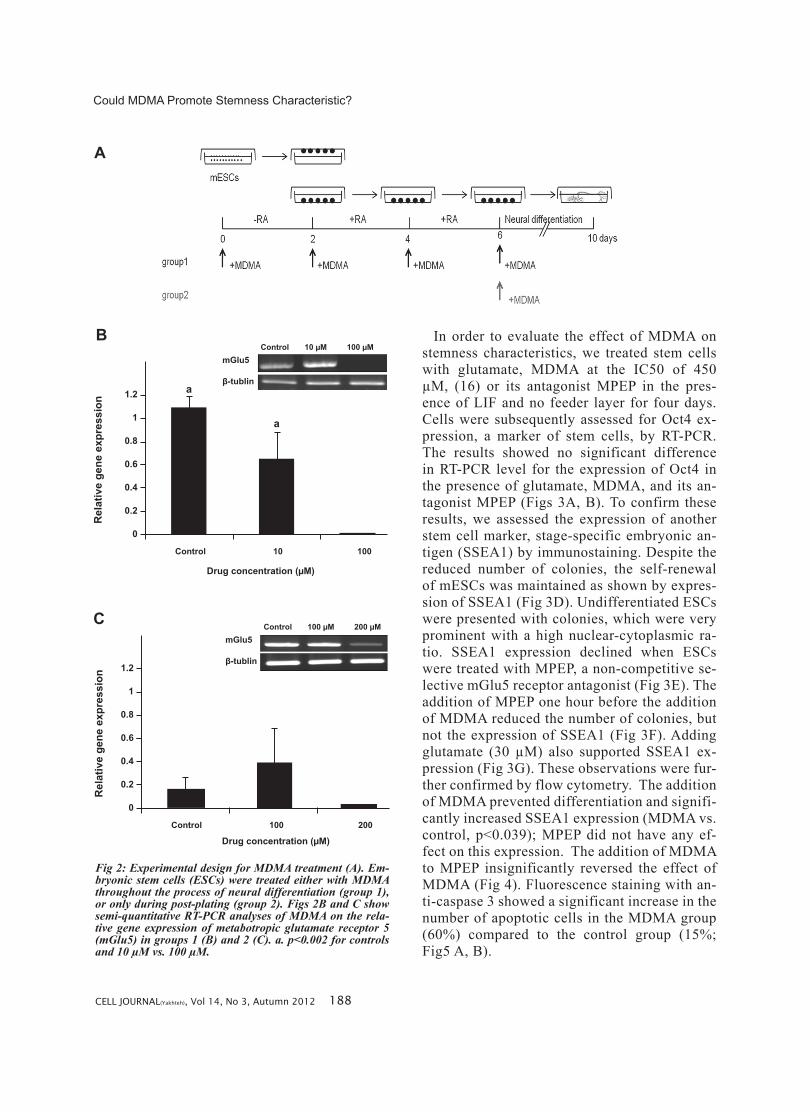
Fig 2: Experimental design for MDMA treatment (A). Em-bryonic stem cells (ESCs) were treated either with MDMA throughout the process of neural differentiation (group 1), or only during post-plating (group 2). Figs 2B and C show semi-quantitative RT-PCR analyses of MDMA on the rela-tive gene expression of metabotropic glutamate receptor 5 (mGlu5) in groups 1 (B) and 2 (C). a. p<0.002 for controls and 10 µM vs. 100 µM.
In order to evaluate the effect of MDMA on stemness characteristics, we treated stem cells with glutamate, MDMA at the IC50 of 450 µM, (16) or its antagonist MPEP in the pres-ence of LIF and no feeder layer for four days. Cells were subsequently assessed for Oct4 ex-pression, a marker of stem cells, by RT-PCR. The results showed no significant difference in RT-PCR level for the expression of Oct4 in the presence of glutamate, MDMA, and its an-tagonist MPEP (Figs 3A, B). To confirm these results, we assessed the expression of another stem cell marker, stage-specific embryonic an-tigen (SSEA1) by immunostaining. Despite the reduced number of colonies, the self-renewal of mESCs was maintained as shown by expres-sion of SSEA1 (Fig 3D). Undifferentiated ESCs were presented with colonies, which were very prominent with a high nuclear-cytoplasmic ra-tio. SSEA1 expression declined when ESCs were treated with MPEP, a non-competitive se-lective mGlu5 receptor antagonist (Fig 3E). The addition of MPEP one hour before the addition of MDMA reduced the number of colonies, but not the expression of SSEA1 (Fig 3F). Adding glutamate (30 µM) also supported SSEA1 ex-pression (Fig 3G). These observations were fur-ther confirmed by flow cytometry. The addition of MDMA prevented differentiation and signifi-cantly increased SSEA1 expression (MDMA vs. control, p<0.039); MPEP did not have any ef-fect on this expression. The addition of MDMA to MPEP insignificantly reversed the effect of MDMA (Fig 4). Fluorescence staining with an-ti-caspase 3 showed a significant increase in the number of apoptotic cells in the MDMA group (60%) compared to the control group (15%; Fig5 A, B).

CELL JOURNAL(Yakhteh), Vol 14, No 3, Autumn 2012 189
Meamar et al.
A
B
Oct4
β-tublin
Control MDMA MPEP MDMA/MPEP Glutamate
*
1.8
1.6
1.4
1.2
1
0.8
0.6
0.4
0.2
0
Rel
ativ
e ge
ne e
xpre
ssio
n
Control MDMA MPEP MDMA/MPEP Glutamate
Drug concentration (µM)
C
D
Fig 3: RT-PCR and semi-quantitative analyses of MDMA on the relative gene expression of Oct4 (A, B). Immunofluo-rescence staining of SSEA1 expression in control (C), and treated culture with MDMA (D), MPEP (E), MDMA/MPEP (F) and Glutamate (G) (This figure has also been printed in full-color at the end of this issue).
E
F
G

CELL JOURNAL(Yakhteh), Vol 14, No 3, Autumn 2012 190
Could MDMA Promote Stemness Characteristic?
A
B
80
70
60
50
40
30
20
10
0
SSEA
1 ex
pres
sion
Control MDMA MPEP MDMA/MPEP
Drug concentration (µM)
a
Fig 4: Percentage of cells expressing SSEA1 in control and after treatment with MDMA, MPEP, and MDMA/MPEP as assessed by flow cytometry. a. p<0.039 for MDMA vs. control.
Fig 5: Fluorescence staining with anti-caspase 3 (FLICA) in control (A) and MDMA (B) groups for apoptosis. The nu-clei were counterstained with Hoechst (This figure has also been printed in full-color at the end of this issue).
DiscussionmGlu receptors are neurotransmitter receptors in
the nervous system because they respond to synap-tic glutamate and are involved in the regulation of synaptic plasticity (19, 26). However, the presence of this receptor in mESCs and its down-regulation during differentiation and up-regulation during neurogenesis suggests an additional role for mGlu receptors (27). For example, the endogenous acti-vation of this receptor is necessary for maintenance of self-renewal properties in mESCs (17, 18). Ini-tially the presence of mGlu5 has been investigated
by immunostaining and RT-PCR analysis to prove its presence in mESCs. These observations are in agreement with previous reports that have con-firmed the expression of mGlu5 on mESCs.
Previous studies have suggested that expression of mGlu5 decreases during EB formation, fol-lowed by an increase during neurolation (17, 18). The results of mGlu5 in the current study are con-sistent with these reports. In this study we have also shown that treatment with the IC50 of MDMA (16) during neural precursor formation and differ-entiation significantly decreased expression of the mGlu5 receptor. However treatment with MDMA during neural differentiation (post-plating) did not affect the expression of mGlu5. These results have suggested that MDMA may affect mGlu5 expres-sion during neural precursor cell formation but not during differentiation. It is important to note that in our previous study we have also shown that the IC50 of MDMA significantly decreased neural markers, MAP2 and nestin expression only during neural precursor cell formation (group 1) and not during differentiation (group 2) (16). Therefore, this suggests that MDMA may induce inhibition of neural precursor formation via down-regulation of the mGlu5 receptor, but this requires additional confirmation.
To further investigate the influence of this recep-tor and MDMA on stemness characteristics we ap-plied glutamate, it’s natural agonist, during stem cell culture in the presence of LIF and absence of a feeder layer. The results revealed a higher expres-sion of SSEA1. In a preliminary study we showed that glutamate depletion for 24 hours by glutamate transaminase reduced Oct4-GFP expression in mESCs. We showed that the IC50 dose of MDMA decreased the numbers of colonies despite the in-creased expression of SSEA1. These observations were further confirmed by flow cytometry. No sub-stantial changes in Oct4 expression were noted, possibly because reduction of Oct4 requires more than four days (28). Therefore, these results may imply a binary role for MDMA: reducing ESCs proliferation while maintaining or supporting self-renewal characteristics. In concordance with our results, such a role has been described for DA neu-rons. Previous studies have shown that MDMA, despite being a toxic drug, enhances the differen-tiation and survival of DA neurons both in vivo and

CELL JOURNAL(Yakhteh), Vol 14, No 3, Autumn 2012 191
Meamar et al.
in vitro (20, 21). In other studies, such results have been obtained for glutamate in MSCs and neural progenitor cells in the brain (29, 30).
In order to confirm whether MDMA’s effects on stemness characteristic were mediated by the mGlu5 receptor, MPEP was applied prior to MDMA. MPEP partially reduced SSEA1 expres-sion, which thereby suggested that this effect of MDMA was possibly mediated by the mGlu5 re-ceptor in addition to other possible pathways. It has been reported that methamphetamines and ecstasy could motivate Janus kinas/signal transducers and activators of transcription (JAK/STAT) pathways; this effect is independent of the mGlu5 receptor and could not be reversed by MPEP (31-33).
Previous research has reported that MPEP, it-self, inhibited stemness characteristics (17). In our experiment the expression of stemness markers in the MPEP group was similar to the culture of mESCs in the absence of a feeder layer and pres-ence of LIF. The difference observed between the two studies could be attributed to different types of stem cells as the current study’s cells were feeder-dependent.
The reduced cell proliferation, despite the main-tenance of stemness characteristics may be related to the toxic effect of MDMA. This effect might be induced by the activation of the caspase pathway, as observed by increased caspase activity in the MDMA group. It has been reported that MDMA can modulate an apoptotic pathway and induce ox-idative stress both in vivo and in vitro (34, 35). A study of the literature suggests that the activation of the mGlu5 receptor via protein kinase c (PKC) inhibits glycogen synthase kinase-3β (GSK3-β) (36), thus mimicking the actions of the Wnt sign-aling pathway (37) which regulates SSEA1 ex-pression (38, 39) by inhibiting β-catenin phos-phorylation. It may be expected that the addition of glutamate and MDMA increases the level of β-catenin and decreases its phosphorylated form, thereby maintaining self-renewal. However, this proposed mechanism needs further investigation. This is the first report which shows that MDMA can increase stemness characteristic in mESCs, elucidating the pathway that begins with MDMA-induced self-renewal and may allow us to better understand how MDMA reduces normal neuron loss that occurs during development.
ConclusionIn this study we reported the binary role for
MDMA on mESCs; reduced proliferation and maintenance of self-renewal. Pharmacological blockade of the mGlu5 receptor with MPEP before addition of MDMA only partially reduced this ef-fect. This suggests that MDMA mediated its role through a different mechanism, which requires fur-ther investigation. In conclusion, despite its toxic-ity, MDMA maintains stemness characteristics.
AcknowledgmentsThis study was funded by a grant provided by
Royan Institute. There is no potential conflict of interest.
References1. Mohamed WM, Ben Hamida S, Cassel JC, De Vascon-
celos AP, Jones BC. MDMA: interactions with other psy-choactive drugs. Pharmacol Biochem Behav. 2011; 99(4): 759-774.
2. Mohamed WM, Hamida SB, De Vasconcelos AP, Cassel JC, Jones BC. Interactions between 3, 4-methylenediox-ymethamphetamine and ethanol in humans and rodents. Neuropsychobiology. 2009; 60 (3-4): 188-194.
3. Seger D. Cocaine, metamfetamine, and MDMA abuse: the role and clinical importance of neuroadaptation. Clin Toxi-col (Phila). 2010; 48(7): 695-708.
4. Bowyer JF, Young JF, Slikker W, Itzak Y, Mayorga AJ, Newport GD, et al. Plasma levels of parent compound and metabolites after doses of either d-fenfluramine or d-3, 4-methylenedioxymethamphetamine (MDMA) that pro-duce long-term serotonergic alterations. Neurotoxicology. 2003; 24(3): 379-390.
5. Green AR, Mechan AO, Elliott JM, O’shea E, Colado MI. The pharmacology and clinical pharmacology of 3, 4- methylenedioxymethamphetamine (MDMA, ‘‘ecstasy”). Pharmacol Rev. 2003; 55(3): 463-508.
6. Hatzidimitriou G, McCann UD, Ricaurte GA. Altered sero-tonin innervations patterns in the forebrain of monkeys treated with (+/_) 3, 4- methylenedioxymethamphetamine seven years previously: factors influencing abnormal re-covery. J Neurosci.1999; 19(12): 5096-5107.
7. McCann UD, Szabo Z, Scheffel U, Dannals RF, Ricaurte GA. Positron emission tomographic evidence of toxic ef-fect of MDMA ("Ecstasy") on brain serotonin neurons in human beings. Lancet, 1998; 352(9138): 1433-1437.
8. Awad H, Hubert GW, Smith Y, Levey AI, Conn PJ Activa-tion of metabotropic glutamate receptor 5 has direct ex-citatory effects and potentiates NMDA receptor currents in neurons of the subthalamic nucleus. J Neurosci. 2000; 20(21): 7871-7879.
9. Battaglia G, Fornai F, Busceti CL, Aloisi G, Cerrito F, De Blasi A, et al. Selective blockade of mGlu5 metabotropic glutamate receptors is protective against methampheta-mine neurotoxicity. J Neurosci. 2002; 22 (6): 2135-2141.
10. Kita T, Wagner GC, Nakashima T. Current research on meth-amphetamine-induced neurotoxicity: animal models of mon-oamine disruption. J Pharmacol Sci. 2003; 92(3): 178-195.

CELL JOURNAL(Yakhteh), Vol 14, No 3, Autumn 2012 192
Could MDMA Promote Stemness Characteristic?
11. Sarihi A, Komaki A, Tsumoto T. Calcium signaling path-ways involved in long-term potentiation at excitatory syn-apses on parvalbumin positive fast-spiking GABAergic neurons in the mouse visual cortex. Cell Journal (Yakhteh). 2009; 11(3): 285-292.
12. Farokhpour M, Karbalaie K, Tanhaei S, Nematollahi M, Etebari M, Sadeghi HM, et al. Embryonic stem cell-derived cardiomyocytes as a model system to study cardioprotec-tive effects of dexamethasone in doxorubicin cardiotoxic-ity. Toxicol In Vitro. 2009; 23(7): 1422-1428.
13. Paquette JA, Kumpf SW, Streck RD, Thomson JJ, Chapin RE, Stedman DB. Assessment of the embryonic stem cell test and application and use in the pharmaceutical indus-try. Birth Defects Res B Dev Reprod Toxicol. 2008; 83(2): 104-111.
14. Rohwedel J, Guan K, Hegert C, Wobus AM. Embryonic stem cells as an in vitro model for mutagenicity, cytotox-icity and embryotoxicity studies: present state and future prospects. Toxicol In Vitro. 2001; 15(6): 741-753.
15. Whitlow S, Bürgin H, Clemann N. The embryonic stem cell test for the early selection of pharmaceutical compounds. ALTEX. 2007; 24(1): 3-7.
16. Meamar R, Karamali F, Sadeghi HM, Etebari M, Nasr-Esfahani MH, Baharvand H. Toxicity of ecstasy (MDMA) towards embryonic stem cell-derived cardiac and neural cells. Toxicol In Vitro. 2010; 24(4): 1133-1138.
17. Cappuccio I, Spinsanti P, Porcellini A, Desiderati F, De Vita T, Storto M, et al. Endogenous activation of mGlu5 metabotropic glutamate receptors supports self-renewal of cultured mouse embryonic stem cells. Neuropharma-cology. 2005; 49 Suppl 1: 196-205.
18. Cappuccio I, Verani R, Spinsanti P, Niccolini C, Gradini R, Costantino S, et al. Context-dependent regulation of embryonic stem cell differentiation by mGlu4 metabotropic glutamate receptors. Neuropharmacology. 2006; 51(3): 606-611.
19. Melchiorri D, Cappuccio I, Ciceroni C, Spinsanti P, Mosillo P, Sarichelou I, et al. Metabotropic glutamate receptors in stem/progenitor cells. Neuropharmacology. 2007; 53(4): 473-480.
20. Koprich JB, Chen EY, Kanaan NM, Campbell NG, Kor-dower JH, Lipton JW. Prenatal 3,4-methylenedioxymeth-amphetamine (ecstasy) alters exploratory behavior, re-duces monoamine metabolism, and increases forebrain tyrosine hydroxylase fiber density of juvenile rats.. Neuro-toxicol Teratol. 2003; 25(5): 509-517.
21. Lipton JW, Tolod EG, Thompson VB, Pei L, Paumier KL, Terpstra BT, et al. 3,4-Methylenedioxy-N-methampheta-mine (ecstasy) promotes the survival of fetal dopamine neurons in culture. Neuropharmacology. 2008; 55(5): 851-859.
22. Baharvand H, Matthaei KI. Culture condition difference for establishment of new embryonic stem cell lines from the C57BL/6 and BALB/c mouse strains. In Vitro Cell Dev Biol Anim. 2004; 40(3-4): 76-81.
23. Niapour A, Karamali F, Karbalaie K, Kiani A, Mardani M, Nasr-Esfahani MH, et al. Novel method to obtain highly enriched cultures of adult rat Schwann cells. Biotechnol Lett. 2010; 32(6): 781-786.
24. Sagha M, Karbalaie K, Tanhaee S, Esfandiari E, Salehi H, Sadeghi-Aliabadi H, et al. Neural induction in mouse em-
bryonic stem cells by co-culturing with chicken somites. Stem Cells Dev. 2009; 18(9): 1351-1360.
25. Ekert PG, Silke J, Vaux DL. Caspase inhibitors. Cell Death Differ. 1999; 6(11): 1081-1086.
26. De Blasi A, Conn PJ, Pin J, Nicoletti F. Molecular deter-minants of metabotropic glutamate receptor signaling. Trends Pharmacol Sci. 2001; 22(3): 114-120.
27. Nicoletti F, Arcella A, Iacovelli L, Battaglia G, Giangaspero F, Melchiorri D. Metabotropic glutamate receptors: new targets for the control of tumor growth? Trends Pharmacol Sci. 2007; 28(5): 206-213.
28. He H, McHaney M, Hong J, Weiss ML. Cloning and Charac-terization of 3.1kb Promoter Region of the Oct4 Gene from the Fischer 344 Rat. Open Stem Cell J. 2009; 1(1): 30-39.
29. Iemata M, Takarada T, Hinoi E, Taniura H, Yoneda Y. Suppression by glutamate of proliferative activity through glutathione depletion mediated by the cystine/glutamate antiporter in mesenchymal C3H10T1/2 stem cells. J Cell Physiol. 2007; 213(3): 721-729.
30. Luk KC, Kennedy TE, Sadikot AF. Glutamate promotes proliferation of striatal neuronal progenitors by an NMDA receptor-mediated mechanism. J Neurosci. 2003; 23(6): 2239 -2250.
31. Hebert M, O'Callaghan JP. Protein phosphorylation cas-cades associated with methamphetamine-induced glial activation. Ann N Y Acad Sci. 2000; 914: 238-262.
32. Miguel-Hidalgo JJ. The role of Glial cells in Drug Abuse. Curr Drug Abuse Rev. 2009; 2(1): 76-82.
33. Narita M, Miyatake M, Narita M, Shibasaki M, Shindo K, Nakamura A, et al. Direct evidence of astrocytic modula-tion in the development of rewarding effects induced by drugs of abuse. Neuropsychopharmacology. 2006; 31 (11): 2476-2488.
34. Jiménez A, Jordà EG, Verdaguer E, Pubill D, Sureda FX, Canudas AM, et al. Neurotoxicity of amphetamine deriva-tives is mediated by caspase pathway activation in rat cer-ebellar granule cells. Toxicol Appl Pharmacol. 2004; 196 (2): 223- 234.
35. Stumm G, Schlegel J, Schäfer T, Würz C, Mennel HD, Krieg JC, et al. Amphetamines induce apoptosis and regulation of bcl-x splice variants in neocortical neurons. FASEB J. 1999; 13(9): 1065-1072.
36. Spinsanti P, De Vita T, Di Castro S, Storto M, Formisano P, Nicoletti F, et al. Endogenously activated mGlu5 metabo-tropic glutamate receptors sustain the increase in c-Myc expression induced by leukaemia inhibitory factor in cul-tured mouse embryonic stem cells. J Neurochem., 2006; 99(1): 299-307.
37. Stewart R, Stojkovic M, Lako M. Mechanisms of self-renewal in human embryonic stem cells. Eur J Cancer. 2006; 42(9): 1257-1272.
38. Capela A, Temple S. LeX is expressed by principle pro-genitor cells in the embryonic nervous system, is secreted into their environment and binds Wnt-1.. Dev Biol. 2006; 291(2): 300-313.
39. Koso H, Ouchi Y, Tabata Y, Aoki Y, Satoh S, Arai K, et al. SSEA-1 marks regionally restricted immature subpopula-tions of embryonic retinal progenitor cells that are regu-lated by the Wnt signaling pathway. Dev Bio. 2006; 292 (1): 265-276.