correspondence open access a tool to ... - bmc medicine
TRANSCRIPT

CORRESPONDENCE Open Access
A tool to facilitate clinical biomarker studies - atissue dictionary based on the Human ProteinAtlasCaroline Kampf1, Julia Bergman1, Per Oksvold2, Anna Asplund1, Sanjay Navani3, Mikaela Wiking2, Emma Lundberg2,Mathias Uhlén2 and Fredrik Ponten1*
Abstract
The complexity of tissue and the alterations thatdistinguish normal from cancer remain a challenge fortranslating results from tumor biological studies intoclinical medicine. This has generated an unmet needto exploit the findings from studies based on celllines and model organisms to develop, validate andclinically apply novel diagnostic, prognostic andtreatment predictive markers. As one step to meetthis challenge, the Human Protein Atlas project hasbeen set up to produce antibodies towards humanprotein targets corresponding to all human proteincoding genes and to map protein expression innormal human tissues, cancer and cells. Here, wepresent a dictionary based on microscopy imagescreated as an amendment to the Human ProteinAtlas. The aim of the dictionary is to facilitate theinterpretation and use of the image-based dataavailable in the Human Protein Atlas, but also to serveas a tool for training and understanding tissuehistology, pathology and cell biology. The dictionarycontains three main parts, normal tissues, cancertissues and cells, and is based on high-resolutionimages at different magnifications of full tissuesections stained with H & E. The cell atlas is centeredon immunofluorescence and confocal microscopyimages, using different color channels to highlight theorganelle structure of a cell. Here, we explain howthis dictionary can be used as a tool to aid cliniciansand scientists in understanding the use of tissuehistology and cancer pathology in diagnostics andbiomarker studies.
Keywords: Antibody-based proteomics, cancer bio-markers, tissue and cell dictionary, immunohistochem-istry, protein expression, histology, pathology
BackgroundThe Human Protein Atlas project, launched in 2003, wasinitiated as a natural extension of the Human GenomeProject, with the objective to explore the proteins encodedby the human genome. The primary focus was to analyzethe distribution and relative abundance of all proteins inhuman normal cells and tissues, and to determine the sub-cellular localization of each protein. One main goal in thiseffort was to contribute to biomedical and clinicalresearch, and because cancer is a major disease wherediagnostics, classification and prognostic stratification isbased on tissue morphology, a multitude of clinical cancertissue samples were included in the comprehensive proteinprofiling. This has allowed researchers to utilize the pro-tein profiling data for both biomarker discovery effortsand for validation of altered gene expression patterns atthe protein level in both normal and cancer tissue.The Human Protein Atlas project pursues a systematic
high-throughput generation of affinity-purified polyclonalantibodies with the aim of generating a map of proteinexpression patterns on a proteome-wide scale in bothhuman normal cells, tissues and organs, and in cancer tis-sues [1]. Immunohistochemistry (IHC) is performed ontissue micro arrays (TMA), containing a multitude of dif-ferent normal tissues and tumors, to enable a comprehen-sive mapping of protein expression patterns at cellularresolution in a tissue context. Altogether 144 different nor-mal tissues are analyzed together with 216 different tumorsrepresenting the 20 most common forms of human cancer[2]. Immunofluorescence (IF)-based profiling of proteinexpression in cell lines is performed to generate a map ofsubcellular localization patterns [3]. All protein expression
* Correspondence: [email protected] of Immunology, Genetics, Pathology, Science for LifeLaboratory, Uppsala University, Dag Hammarskjölds väg 20, Uppsala, 751 85SwedenFull list of author information is available at the end of the article
Kampf et al. BMC Medicine 2012, 10:103http://www.biomedcentral.com/1741-7015/10/103
Clinical Biomarkers
© 2012 Kampf et al; licensee BioMed Central Ltd. This is an Open Access article distributed under the terms of the Creative CommonsAttribution License (http://creativecommons.org/licenses/by/2.0), which permits unrestricted use, distribution, and reproduction inany medium, provided the original work is properly cited.

data, including the underlying images, are made publiclyavailable at the Human Protein Atlas web portal (http://www.proteinatlas.org) [4]. The current version of theHuman Protein Atlas contains data for more than 14,000unique proteins. This corresponds to more than 70% of allhuman protein encoding genes [5].As the cell constitutes the smallest living entity, it is
required to harbor specialized and distinct subcellularstructures. Cells vary considerably in function and mor-phology and these differences form the basis for the con-cept of different cellular phenotypes. On a higher level,cell types with their distinct phenotypes are organized intotissues, commonly categorized as epithelial, muscle, vascu-lar, nervous and connective tissue, and hematopoieticcells. Genetic changes leading to dysregulated signalingpathways with altered protein expression patterns cause atransformation from normal to the phenotypes and mor-phology that signifies cancer. Cancer is a heterogeneousdisease associated with alterations in protein expressionpatterns leading to cell growth and ‘anti-social behavior’ oftumor cells. The deregulated expression patterns in tumorcells are caused by genetic and epigenetic alterations lead-ing to distortion of multiple proteins and signaling path-ways. Despite the complexity of cancer, microscopicevaluation of tissue morphology remains the gold standardfor determining a cancer diagnosis in a clinical setting.Although morphology is crucial, adding a layer of informa-tion regarding protein expression on top of morphologyappears to be beneficial for the stratification of differenttumor types. Immunohistochemistry prevails as an invalu-able method to provide such a tool for visualization ofprotein expression patterns in cells from a section oftumor tissue.
The Dictionary - a tool for biomarker studiesThe dictionary contains three main parts: normal tissues,cancer tissues, and cells (http://www.proteinatlas.org/dic-tionary) (Figure 1). All images and examples includedescriptive text boxes and supporting text with back-ground information, to facilitate interpretation of the com-plex patterns underlying normal tissue histology, tumorpathology and cell biology. H & E-stained tissue sectionshave been scanned at 40 × magnification and both normaland cancer tissues are shown at three different levels ofmagnification.Altogether 45 normal tissue types (represented by 173
images), 20 different cancer types (represented by 193images) and 18 subcellular structures (represented by103 images) are included in the dictionary. Examples ofnormal tissue show colon (Figure 2A) and breast (Figure2B) at the three levels of magnification. For cancer, onecase of low-grade (Figure 2C) and one case of high-grade (Figure 2D) ductal breast cancer is shown. IF andIHC images representing antibodies targeting proteins
in the nucleoli and the mitochondria demonstrate thecell structure part (Figure 3). In addition to high resolu-tion images, there are summarizing descriptive textparagraphs to supplement the images.As one of the main goals of this project is to identify
novel biomarkers that can be developed for clinical use,the 20 types of human cancers illustrated in the dictionaryhave also been used for protein profiling in the HumanProtein Atlas. Using the search function at the HumanProtein Atlas portal [6], search strings can be created toidentify candidates for cell or tumor type specific markersand also proteins differentially expressed within a giventumor type, thus representing potential prognosticindicators.
Clinical impactSuccessful identification and translation of informativebiomarkers to aid clinical decision-making is a prerequisitefor the implementation of personalized cancer therapeuticregimens. The antibody-based proteomics strategyemployed in the Human Protein Atlas plays a key role inthe cancer biomarker discovery and validation pipeline,facilitating evaluation of candidate markers [7]. The newlylaunched dictionary provides a useful tool to interpret andevaluate biomarker candidates identified through varioussearch strategies in the Human Protein Atlas. The apprai-sal of protein expression patterns in tumor tissue is a cru-cial step to select the most promising candidates forextended experiments, including clinical studies in largercohorts, functional studies and in-depth validation ofexpression patterns.The Human Protein Atlas has already been used in
several clinical biomarker studies as a starting point forexploring both diagnostic and prognostic factors. Celland tumor type specific protein expression, critical fordeveloping diagnostic markers, is exceedingly rare [8],and only a few such markers exist for clinical use. As anexample, the DNA-binding protein SATB2 was identi-fied in the Human Protein Atlas as a potential noveldiagnostic marker for colorectal cancer and in anextended study including more than 2,400 tumors,SATB2 was found to be both a sensitive and highly spe-cific marker for colorectal cancer [9]. The basic proteinprofiling data available in the Human Protein Atlas hasalso allowed for several potential prognostic cancer bio-markers to be identified for different types of cancer.This is exemplified by the RNA-binding protein RBM3,discovered to be a prognostic marker for several differ-ent forms of cancer [10-12], and also a potential treat-ment predictive marker for platinum-based therapies[13]. Understanding of tumor tissue composition is alsofundamental for studies regarding tumor stroma com-partments. In a recent tumor biology study [14] using amouse model, large numbers of granulin-expressing
Kampf et al. BMC Medicine 2012, 10:103http://www.biomedcentral.com/1741-7015/10/103
Page 2 of 6

bone marrow-derived hematopoietic cells were found inthe tumor stroma of breast cancers responding to insti-gating signals. This study also showed that the expres-sion of granulin in human breast cancer was stronglycorrelated with the triple negative/basal-like breasttumor subtypes, and that breast cancer patients with
tumors positive for granulin staining had a significantlyworse outcome in terms of overall survival. The pre-sented dictionary and Human Protein Atlas can also beutilized in other clinical research fields, exemplified bythe identification of targets for in vivo imaging of pan-creatic beta-cells cells in diabetes research [15,16].
Figure 1 Schematic showing the starting page for the dictionary. The three main parts, normal tissues, cancer tissues and cell structures, aredisplayed side by side with alphabetical lists below showing the contents of each part to facilitate navigation. All figures are original andavailable at the Human Protein Atlas web portal (www.proteinatlas.org/dictionary). Published with permission from the Human Protein Atlas.
Kampf et al. BMC Medicine 2012, 10:103http://www.biomedcentral.com/1741-7015/10/103
Page 3 of 6
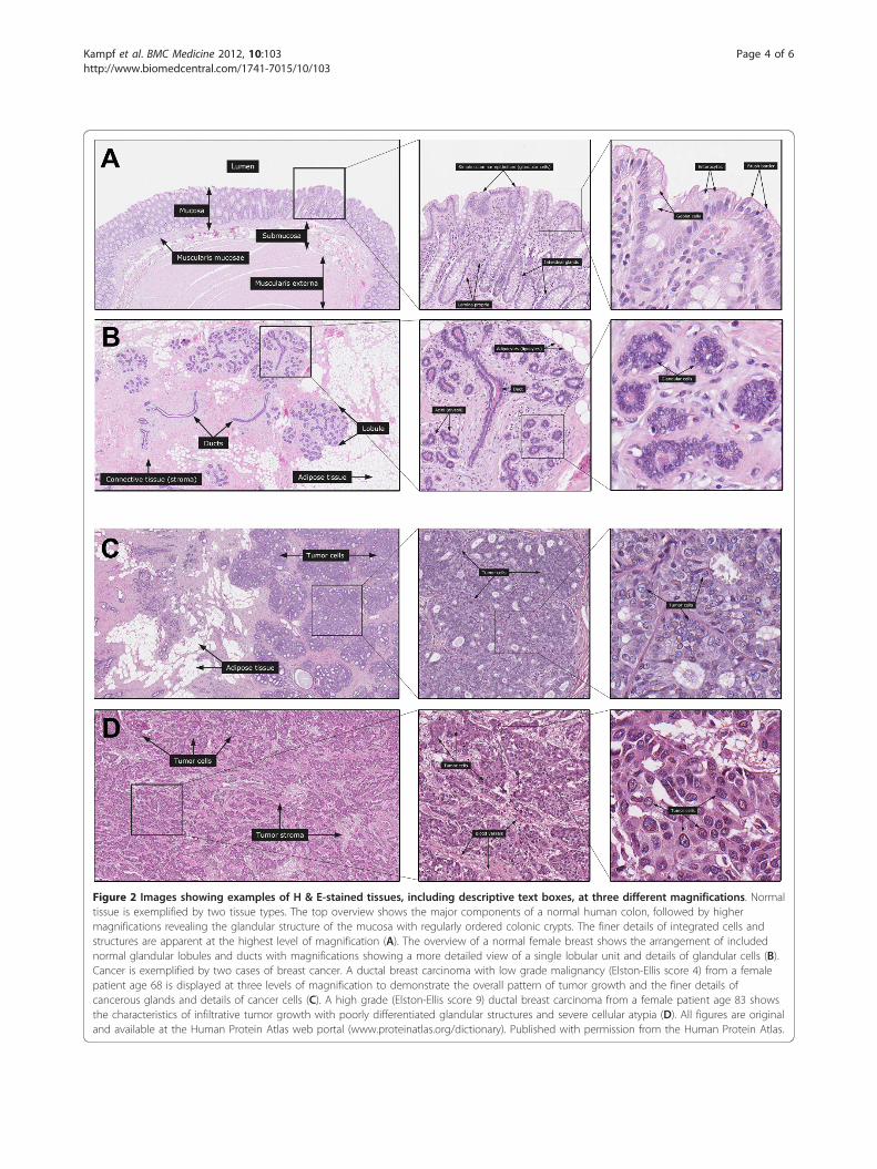
Figure 2 Images showing examples of H & E-stained tissues, including descriptive text boxes, at three different magnifications. Normaltissue is exemplified by two tissue types. The top overview shows the major components of a normal human colon, followed by highermagnifications revealing the glandular structure of the mucosa with regularly ordered colonic crypts. The finer details of integrated cells andstructures are apparent at the highest level of magnification (A). The overview of a normal female breast shows the arrangement of includednormal glandular lobules and ducts with magnifications showing a more detailed view of a single lobular unit and details of glandular cells (B).Cancer is exemplified by two cases of breast cancer. A ductal breast carcinoma with low grade malignancy (Elston-Ellis score 4) from a femalepatient age 68 is displayed at three levels of magnification to demonstrate the overall pattern of tumor growth and the finer details ofcancerous glands and details of cancer cells (C). A high grade (Elston-Ellis score 9) ductal breast carcinoma from a female patient age 83 showsthe characteristics of infiltrative tumor growth with poorly differentiated glandular structures and severe cellular atypia (D). All figures are originaland available at the Human Protein Atlas web portal (www.proteinatlas.org/dictionary). Published with permission from the Human Protein Atlas.
Kampf et al. BMC Medicine 2012, 10:103http://www.biomedcentral.com/1741-7015/10/103
Page 4 of 6

Concluding remarksHere we present a freely available cell and tissue dictionaryas an amendment to the Human Protein Atlas (reviewed in[17]) that can be used to facilitate the interpretation of clin-ical tissue biomarkers. A multitude of high quality imageswith supporting short text paragraphs are displayed on theHuman Protein Atlas web portal (http://www.proteinatlas.org/dictionary) to provide a useful guide for researcherswho are not familiar with the microscopical landscape thatforms the home ground for histologists and pathologists.In this first version of the dictionary, H & E stained tissuesections are presented for visualization of normal tissueand cancer morphology. The essential background neededto interpret and understand expression data retrieved fromtissues and cells is presented. The aim is to expand thecontents of the dictionary to also include additional levelsof information regarding protein expression so that variouscell populations not distinguishable from morphologyalone can be visualized. Established antibodies can be used
for IHC on consecutive sections of selected tissues todemonstrate different cell types, for example, B-lympho-cytes, T-lymphocytes and endothelial cells, and differentcell states, for example, proliferation and differentiation.Moreover, updates with additional links and text para-graphs can be added as well as including more examples ofboth normal and diseased tissues. For educational pur-poses, show/hide functionality for text boxes could befurther developed together with sets of relevant ‘Questions& Answers’. We anticipate that a content rich and knowl-edge-based cell and tissue dictionary, combined with thecomprehensive map of protein expression patterns in nor-mal and cancer tissues available through the Human Pro-tein Atlas, will provide a significant foundation for bothbasic and clinical research projects.
AbbreviationsH & E: haematoxylin and eosin; IF: immunofluorescence; IHC:immunohistochemistry; TMA: tissue micro arrays.
Figure 3 Examples of images demonstrating different organelles in cells. The upper panel shows IF (left and middle) and IHC (right)images representing the nucleoli, visualized by antibodies targeting proteins expressed in the nucleoli. The nucleoli are shown as a green colorin the IF example and brown color in the IHC example. The lower panel shows images representing mitochondria, visualized by antibodiesexpressed in mitochondrion. IF: green - antibody (HPA026512, HPA027999); blue - nucleus (DAPI), red - microtubule. IHC: brown - antibody(HPA005768, HPA004016). Scalebar 10 μm. IF, immunofluorescence; IHC, immunohistochemistry. All figures are original and available at theHuman Protein Atlas web portal (www.proteinatlas.org/dictionary). Published with permission from the Human Protein Atlas.
Kampf et al. BMC Medicine 2012, 10:103http://www.biomedcentral.com/1741-7015/10/103
Page 5 of 6

AcknowledgementsThis work was supported by grants from the Knut and Alice WallenbergFoundation. The pathologists and staff at the Pathology Clinic, UppsalaUniversity Hospital, Uppsala, Sweden, are gratefully acknowledged for allefforts regarding the handling and diagnostics of the tissues used in theHuman Protein Atlas. The authors wish to especially thank DijanaDjureinovic, Sofie Gustafsson, Elene Karlberg, Dijana Cerjan and John Juterfor assistance with the dictionary work.
Author details1Department of Immunology, Genetics, Pathology, Science for LifeLaboratory, Uppsala University, Dag Hammarskjölds väg 20, Uppsala, 751 85Sweden. 2Science for Life Laboratory, Royal Institute of Technology,Tomtebodavägen 23A, Solna, 171 65, Sweden. 3Lab Surgpath, 204 BombayMarket, Tardeo Main Road, Mumbai, 400 034, India.
Authors’ contributionsCK, MU and FP contributed to the conception and the supervision of theproject. CK, JB, AA and FP wrote the manuscript. JB, MW, EL, SN and FPwrote the text corresponding to all normal, cancer and cell images. POdeveloped the database and website. All authors were involved in editingand the final approval of the paper.
Authors’ informationCK: associate professor and site Director for the tissue protein profilingfacility, JB: PhD student (tissue biomarkers), PO: IT developer, AA: post-doctoral (immunohistochemistry-based cell profiling), SN: senior pathologist(immunohistochemistry-based tissue profiling), MW: PhD student(immunofluorescence-based cell profiling), EL: associate professor andresponsible for the subcellular profiling unit, MU: professor and programDirector for the Human Protein Atlas and FP: professor, senior pathologistand clinical Director for the Human Protein Atlas.
Competing interestsThe authors declare that they have no competing interests.
Received: 7 August 2012 Accepted: 12 September 2012Published: 12 September 2012
References1. Uhlen M, Ponten F: Antibody-based proteomics for human tissue
profiling. Mol Cell Proteomics 2005, 4:384-393.2. Kampf C, Olsson I, Ryberg U, Sjostedt E, Ponten F: Production of tissue
microarrays, immunohistochemistry staining and digitalization withinthe human protein atlas. J Vis Exp 2012, , 63: e3620, DOI: 10.3791/3620.
3. Barbe L, Lundberg E, Oksvold P, Stenius A, Lewin E, Bjorling E, Asplund A,Ponten F, Brismar H, Uhlen M, Andersson-Svahn H: Toward a confocalsubcellular atlas of the human proteome. Mol Cell Proteomics 2008,7:499-508.
4. Uhlen M, Bjorling E, Agaton C, Szigyarto CA, Amini B, Andersen E,Andersson AC, Angelidou P, Asplund A, Asplund C, Berglund L,Bergström K, Brumer H, Cerjan D, Ekström M, Elobeid A, Eriksson C,Fagerberg L, Falk R, Fall J, Forsberg M, Björklund MG, Gumbel K, Halimi A,Hallin I, Hamsten C, Hansson M, Hedhammar M, Hercules G, et al: A humanprotein atlas for normal and cancer tissues based on antibodyproteomics. Mol Cell Proteomics 2005, 4:1920-1932.
5. Uhlen M, Oksvold P, Fagerberg L, Lundberg E, Jonasson K, Forsberg M,Zwahlen M, Kampf C, Wester K, Hober S, Wernerus H, Björling L, Ponten F:Towards a knowledge-based Human Protein Atlas. Nat Biotechnol 2010,28:1248-1250.
6. Bjorling E, Lindskog C, Oksvold P, Linne J, Kampf C, Hober S, Uhlen M,Ponten F: A web-based tool for in silico biomarker discovery based ontissue-specific protein profiles in normal and cancer tissues. Mol CellProteomics 2007, 7:825-44.
7. Brennan DJ, O’Connor DP, Rexhepaj E, Ponten F, Gallagher WM: Antibody-based proteomics: fast-tracking molecular diagnostics in oncology. NatRev Cancer 2010, 10:605-617.
8. Ponten F, Gry M, Fagerberg L, Lundberg E, Asplund A, Berglund L,Oksvold P, Bjorling E, Hober S, Kampf C, Navani S, Nilsson P, Ottosson J,Persson A, Wernérus H, Wester K, Uhlén M: A global view of proteinexpression in human cells, tissues, and organs. Mol Syst Biol 2009, 5:337.
9. Magnusson K, De Wit M, Brennan DJ, Johnson LB, McGee SF, Lundberg E,Naicker K, Klinger R, Kampf C, Asplund A, Wester K, Gry M, Bjartell A,Gallagher WM, Rexhepaj E, Kilpinen S, Kallioniemi OP, Belt E, Goos J,Meijer G, Birgisson H, Glimelius B, Borrebaeck CA, Navani S, Uhlén M,O’Connor DP, Jirström K, Pontén F: SATB2 in combination withcytokeratin 20 identifies over 95% of all colorectal carcinomas. Am J SurgPathol 2011, 35:937-948.
10. Jogi A, Brennan DJ, Ryden L, Magnusson K, Ferno M, Stal O, Borgquist S,Uhlen M, Landberg G, Pahlman S, Pontén F, Jirström K: Nuclear expressionof the RNA-binding protein RBM3 is associated with an improvedclinical outcome in breast cancer. Mod Pathol 2009, 22:1564-1574.
11. Hjelm B, Brennan DJ, Zendehrokh N, Eberhard J, Nodin B, Gaber A,Ponten F, Johannesson H, Smaragdi K, Frantz C, Hober S, Johnson LB,Påhlman S, Jirström K, Uhlen M: High nuclear RBM3 expression isassociated with an improved prognosis in colorectal cancer. ProteomicsClin Appl 2011, 5:624-635.
12. Jonsson L, Bergman J, Nodin B, Manjer J, Ponten F, Uhlen M, Jirstrom K:Low RBM3 protein expression correlates with tumour progression andpoor prognosis in malignant melanoma: an analysis of 215 cases fromthe Malmo Diet and Cancer Study. J Transl Med 2011, 9:114.
13. Ehlen A, Brennan DJ, Nodin B, O’Connor DP, Eberhard J, Alvarado-Kristensson M, Jeffrey IB, Manjer J, Brandstedt J, Uhlen M, Pontén F,Jirström K: Expression of the RNA-binding protein RBM3 is associatedwith a favourable prognosis and cisplatin sensitivity in epithelial ovariancancer. J Transl Med 2010, 8:78.
14. Elkabets M, Gifford AM, Scheel C, Nilsson B, Reinhardt F, Bray MA,Carpenter AE, Jirstrom K, Magnusson K, Ebert BL, Pontén F, Weinberg RA,McAllister SS: Human tumors instigate granulin-expressing hematopoieticcells that promote malignancy by activating stromal fibroblasts in mice.J Clin Invest 2011, 121:784-799.
15. Lindskog C, Asplund A, Engkvist M, Uhlen M, Korsgren O, Ponten F:Antibody-based proteomics for discovery and exploration of proteinsexpressed in pancreatic islets. Discov Med 2010, 9:565-578.
16. Lindskog C, Korsgren O, Ponten F, Eriksson JW, Johansson L, Danielsson A:Novel pancreatic beta cell-specific proteins: antibody-based proteomicsfor identification of new biomarker candidates. J Proteomics 2012,75:2611-2620.
17. Ponten F, Schwenk JM, Asplund A, Edqvist PH: The Human Protein Atlasas a proteomic resource for biomarker discovery. J Intern Med 2011,270:428-446.
Pre-publication historyThe pre-publication history for this paper can be accessed here:http://www.biomedcentral.com/1741-7015/10/103/prepub
doi:10.1186/1741-7015-10-103Cite this article as: Kampf et al.: A tool to facilitate clinical biomarkerstudies - a tissue dictionary based on the Human Protein Atlas. BMCMedicine 2012 10:103.
Submit your next manuscript to BioMed Centraland take full advantage of:
• Convenient online submission
• Thorough peer review
• No space constraints or color figure charges
• Immediate publication on acceptance
• Inclusion in PubMed, CAS, Scopus and Google Scholar
• Research which is freely available for redistribution
Submit your manuscript at www.biomedcentral.com/submit
Kampf et al. BMC Medicine 2012, 10:103http://www.biomedcentral.com/1741-7015/10/103
Page 6 of 6