correlations between terasaki’s hla class ii epitopes … · correlations between terasaki’s...
TRANSCRIPT

Tissue Antigens ISSN 0001-2815
Correlations between Terasaki’s HLA class II epitopes andHLAMatchmaker-defined eplets on HLA-DR and -DQ antigensM. Marrari & R. J. Duquesnoy
Division of Transplantation Pathology, Thomas E. Starzl Transplantation Institute, University of Pittsburgh Medical Center, Pittsburgh, PA, USA
Key words
epitope; eplet; HLAMatchmaker; human
leukocyte antigen-DR, -DQ; human leukocyte
antigen antibody
Correspondence
Rene J. Duquesnoy, PhD
University of Pittsburgh Medical Center
Thomas E. Starzl Biomedical Science Tower
Room W1552
Pittsburgh
PA 15261
USA
Tel: 412 647 6148
Mobile: 412 860 8083
Fax: 412 647 1755
e-mail: [email protected]
Received 31 January 2009; accepted 22
March 2009
doi: 10.1111/j.1399-0039.2009.01272.x
Abstract
Human leukocyte antigen (HLA) class II-specific antibodies increase the risk of
transplant failure, and their characterization must consider epitopes rather than
antigens. There are two strategies to determine HLA epitope structure. Terasaki’s
group has analyzed antibody reactivity patterns with single antigen panels with
a computer program based on shared amino acid residues of reactive alleles.
HLAMatchmaker is a theoretical algorithm that predicts HLA epitopes on the
HLA molecular surface from stereochemical modeling of epitope–paratope
interfaces of antigen–antibody complexes. Our epitope repertoire is based on so-
called ‘eplets’ representing 3-A patches of at least one polymorphic residue on the
molecular surface. This report describes how 49 of 53 Terasaki’s HLA-DR epitopes
correspond to HLAMatchmaker-defined eplets. Most of them are equivalent to
single eplets (n ¼ 33) or two or more possible eplets (n ¼ 10), but six had
corresponding eplet pairs. There were 10 cases whereby eplets have permissible
residue combinations, and in 5 cases, we found that eplet specificity might be
influenced by nearby hidden residues.We could assign corresponding eplets to 17 of
18 Terasaki’s HLA-DQ epitopes. This study demonstrates how the HLAMatch-
maker interpretation of amino acid residues shared between antibody-reactive
antigens can increase our understanding of the structural basis of HLA epitopes.
Introduction
Human leukocyte antigen (HLA) class II-specific anti-
bodies increase the risk of transplant failure because they
lead toa higher incidence of acute and chronic rejection (1–6).
Antibodies react with epitopes on antigenic molecules, and
a characterization of the antibody response to class II
epitopes rather than antigens seems important for the
management of sensitized patients considered for trans-
plantation. Sensitive antibody detection methods such as
the Luminex assays with single antigen panels are com-
monly used to assess humoral sensitization against HLA-
DR and -DQ mismatches. The interpretation of antibody
reactivity patterns requires an understanding of the
repertoire of class II epitopes that can now be structurally
defined with amino acid polymorphisms.
There are two strategies to determine the structural basis
of HLA epitopes. One is based on the analysis of antibody
reactivity patterns with HLA panels and a determination of
amino acid residues exclusively shared by antibody-reactive
alleles. Terasaki’s group has recently published a series of
papers that describe the amino acid configurations of HLA
class II epitopes (7–12). Another strategy is a theoretical
algorithm called HLAMatchmaker that predicts epitope
structure onHLAmolecules from stereochemical models of
protein antigen–antibody complexes (13–15). The annota-
tion ‘eplet’ refers to a patch of amino acids within a 3 A
radius of polymorphic residues on the molecular surface.
The Web site http://HLAMatchmaker.net has more details
about this algorithm.
An accompanying paper in this issue describes our
strategy inmore detail and addresses a comparative analysis
of Terasaki’s epitopes (TerEps) and HLAMatchmaker-
defined eplets on HLA-A, -B and -C antigens (16). This
report describes a similar analysis of 60 HLA-DR and 18
HLA-DQ epitopes reported by Terasaki’s group (7–11).
Methods
TerEps on class II antigens are described by amino acid
residues shared between alleles that react with mouse
134 ª 2009 John Wiley & Sons A/S � Tissue Antigens 74, 134–146

monoclonal antibodies (mAbs) or human allosera from
renal transplant recipients. Antibody testing was performed
with Luminex-based binding assays, and Table 1 shows the
composition of a typical panel of 28 single DRB alleles and
18 DQ heterodimers. This report addresses 60 TerEps on
DRB (#1001–1043, #1401–1411 and #1601–1606), 15 on
DQB (#2001–2015) and 3 on DQA (#2017–2019). Their
residue compositions have the standard notation system of
sequence positions and single letter amino acid codes.
Combinations of two or more residues are separated by a1
sign, i.e. 98E1104A; hidden residues under the molecular
surface are shown between parentheses, i.e. (9W) and (10Q),
and two or more possible amino acid combinations are
separated by a slash, i.e. 70G171A/116I/125S.
For each TerEp, we searched HLAMatchmaker for one
ormore eplets present on the same group of antigens and/or
alleles of the Luminex panel and with similar amino acid
residue compositions. We conducted this analysis in a step-
wise manner as previously described (16). We searched for
TerEps that are equivalent to single eplets with comparable
amino acid compositions or correspond to pairs of eplets on
the molecular surface locations separated far enough for
contact by two different complementarity-determining
regions (CDRs) of antibody. The locations of these eplet
pairs were determined on crystallographic structures of
DRB and DQ molecules downloaded from the http://
www.ncbi.nlm.nih.gov/Structure Web site and viewed with
the CN3D structure and sequence alignment software
program (17). This program has a ‘select by distance’ (in
Angstroms) command that permits an assessment of the
distances between eplets. From the 700–900 A2 surface area
of a structural epitope, one can estimate that two eplets
contacted by two different CDRs cannot be further apart
than about 15 A.
We have also considered polymorphic residues in
unexposed locations below themolecular surface. Although
such hidden residues cannot make direct contact with
antibody, they may alter the conformation of a nearby
epitope (18–22). Current definitions of eplets include hidden
residues within a 3 A radius, but it is also possible that
hidden residues somewhat further away have an effect.
Therefore, we have also considered hidden residues up to
6 A away from an eplet.
Conversely, two or more eplets in the same molecular
location may define the same epitope because their amino
acid differences do not significantly influence the binding
with specific antibody. This permissible residue variability
reflects the structural type of cross-reactivity, and such eplet
configuration has surface residues that dominate epitope
specificity. Our eplet notations use an asterisk to indicate
permissible residue combinations. For instance, residues in
positions 73, 77 and 78 define the 77ATY and 77GTV eplets
recognized by the same antibody. An epitope shared
between these eplets has permissible differences between
73A and 73G and between 78V and 78Y, whereas its
specificity is dominated by 77T.We annotate this epitope as
77T*, whereby asterisk represents the permissible 73A/G
and 78V/Y combinations.
Results
DRB TerEpswith surface residue descriptions and their
equivalent eplets
There are 60 DRB TerEps described by residues on and/or
below themolecular surface. Our analysis separated TerEps
described exclusively by hidden residues (see below).
Table 2 summarizes how 30 DRB TerEps with surface
residue descriptions correspond to eplets, and Figure 1A–F
depicts their locations on informative DRB molecules.
Residues in sequence position 96 in the b2 domain
describe four TerEps. Each one corresponds to an eplet that
has a polymorphic residue at that position and another
polymorphic residue in position 180 about 3 A away: #1031
is 96EV (Figure 1F), #1032 is 96HV (Figure 1C,E) and
#1033 is 96QK (Figure 1D). The Luminex panel in Table 1
has three DR4 alleles, DRB1*0401, *0404 and *0405.
Although their residue descriptions are different, the DR4-
specific #1034 seems to be the same as the DR4-specific
#1042 and #1605. This TerEp corresponds to 96YL and two
adjoining eplets, 98EN and 180LT, as well as 32FYHon the
a-helix (Figure 1A,B).
The DR52-specific #1036 is equivalent to 96QV in the b2domain (Figure 1E). DR17, DR18 and DR52 share #1027
with the 77N description and is equivalent to 77GNY
on the a-helix (Figure 1C,E). The DR17- and DR18 (or
Table 1 High-resolution types of DR and DQ antigens in the Luminex
panela
DRB alleles DQ heterodimers
DR1 DRB1*0101 DRB1*0102 DRB1*0103 DQB1*0201 DQA1*0201
DR4 DRB1*0401 DRB1*0404 DRB1*0405 DQB1*0201 DQA1*0301
DR7 DRB1*0701 DQB1*0201 DQA1*0501
DR8 DRB1*0801 DQB1*0202 DQA1*0201
DR9 DRB1*0901 DQB1*0401 DQA1*0201
DR10 DRB1*1001 DQB1*0402 DQA1*0201
DR11 DRB1*1101 DQB1*0402 DQA1*0401
DR12 DRB1*1201 DRB1*1202 DQB1*0501 DQA1*0101
DR13 DRB1*1301 DRB1*1303 DQB1*0502 DQA1*0102
DR14 DRB1*1401 DQB1*0601 DQA1*0103
DR15 DRB1*1501 DRB1*1502 DQB1*0602 DQA1*0102
DR16 DRB1*1601 DQB1*0301 DQA1*0301
DR17 DRB1*0301 DQB1*0301 DQA1*0505
DR18 DRB1*0302 DQB1*0301 DQA1*0601
DR51 DRB5*0101 DRB5*0202 DQB1*0302 DQA1*0101
DR52 DRB3*0101 DRB3*0202 DRB3*0301 DQB1*0302 DQA1*0301
DR53 DRB4*0101 DRB4*0103 DQB1*0303 DQA1*0301
DQB1*0303 DQA1*0201
a Composition of the single class II allele kit, lot 6 by One Lambda Inc.,
Canoga Park, CA, USA.
M. Marrari & R. J. Duquesnoy Eplets and Terasaki’s HLA class II epitopes
ª 2009 John Wiley & Sons A/S � Tissue Antigens 74, 134–146 135

DR3)-specific #1026 is described by 74R, but this residue is
also onDRB3*0101. The best descriptionof aDR3 epitope is
a pair of eplets: 77GNY and 60Y, which are about 14 A
apart, a distance sufficient for contact by two CDRs of
antibody (Figure 1C). TerEps #1040 and #1041 are on the
same groupofDRBantigens except forDR4 andDR10; they
correspond to 140TV (Figure 1B,C) and 149H (Figure 1C),
respectively. These eplets are also on DRB3*0301.
The DR15- and DR16 (or DR2)-specific #1603 corre-
sponds to 133L and140AM in the b2 domain; residues (11P)
Table 2 Thirty DRB TerEps with surface residue descriptions and their corresponding eplets
TerEp Defined by
Antibody-reactive
antigens
Residue
descriptiona Comments Epletsb Model
#1031 Allo DR1; DR51 96E 96EV Figure 1F
#1032 Allo DR7, 8, 9, 10, 11, 12,
13, 14, 17, 18; DR52
96H 96H not on
DR10
96HV Figure 1C,E
#1033 Allo DR10, 15, 16; DR53 96Q 96QK Figure 1D
#1034 Allo DR4 96Y 32FYH/96YL/98EN/180LT Figure 1A,B
#1042 Allo DR4 180L 32FYH/96YL/98EN/180LT Same as #1034
#1605 mAb, Allo DR4 (13H), 33H,
96Y, 180L
32FYH/96YL/98EN 180LT Same as #1034
#1036 mAb, Allo DR52 98Q 98QS Figure 1E
#1027 mAb, Allo DR17, 18; DR52 77N 77GNY Figure 1C,E
#1026 mAb, Allo DR17, 18 74R 74R also on
DRB3*0101
71QKGR 1 60Y Figure 1C (eplets
are 14 A apart)
#1040 Allo DR4, 8, 10, 11, 12,
13, 14, 17, 18
140T 140T also on
DRB3*0301
140TV Figure 1B,C
#1041 Allo DR8, 11, 12, 13, 14,
17, 18
149H 149H also on
DRB3*0301
149H Figure 1C
#1603 Allo DR15, 16 (11P), (13R),
133L, 142M
133L/140AM Figure 1D
#1020 mAb, Allo DR15 71A 71A also on
DRB5*0202
71QAA Figure 1D
#1402 mAb DR51 (9Q), 108T 108T Figure 1F
#1017 mAb DR11 58E 57DE
#1008 Allo DR7 25Q 26QKF/71DRG
#1602 mAb DR7 (11G), 14K,
25Q, (30L)
26QKF/71DRG Same as #1008
#1001 mAb, Allo DR7, 9; DR53 4Q 4Q
#1019 mAb, Allo DR9, 10, 14; DR53 70R 71RRA
#1029 Allo DR7, 9 78V 77TV/98ES
#1043 Allo DR7, 9, 10 181M 181M also on
DR53
180VM
#1410 mAb, Allo DR7, 9, 12 (57V), 60S both also on
DRB3*0101
and *0301
60S171R 11 A apart
#1606 mAb DR14 (57A), 60H, 112Y 57AA/60H/112Y
#1409 Allo DR13 (57S)171K 71K only on
DRB1*1303
71DKA
#1030 Allo DR1, 12 85A 85A only on
DRB1*0102 also
on DRB5*0202
85AV
#1407 mAb, Allo DR8, 12 (13G), 16Y 14GEY/71DRA-(13G) 13G is 4.5 A away
from 71DRA
#1024 mAb, Allo DR8 74L 25YRF/73ALDT
#1023 mAb, Allo DR9, 14; DR53 74E 71RAE Also 77T
#1014 mAb DR10; DR53 40Y 40Y171RRA Eplets are 4 A apart
#1408 Allo DR10; DR53 (38A), 40Y 38A also on DRB3*0202 40Y171RRA Same as #1014
Allo, alloserum or eluted alloantibody; mAb, monoclonal antibody; TerEp, Terasaki’s epitope.a Amino acids in human leukocyte antigen protein sequence positions are listed with the standard single letter code. Hidden residues not exposed on
surface of molecule are in parenthesis. Combinations of residues are shown with 1 sign.b Two and three unique eplets are separated with slash.
136 ª 2009 John Wiley & Sons A/S � Tissue Antigens 74, 134–146
Eplets and Terasaki’s HLA class II epitopes M. Marrari & R. J. Duquesnoy
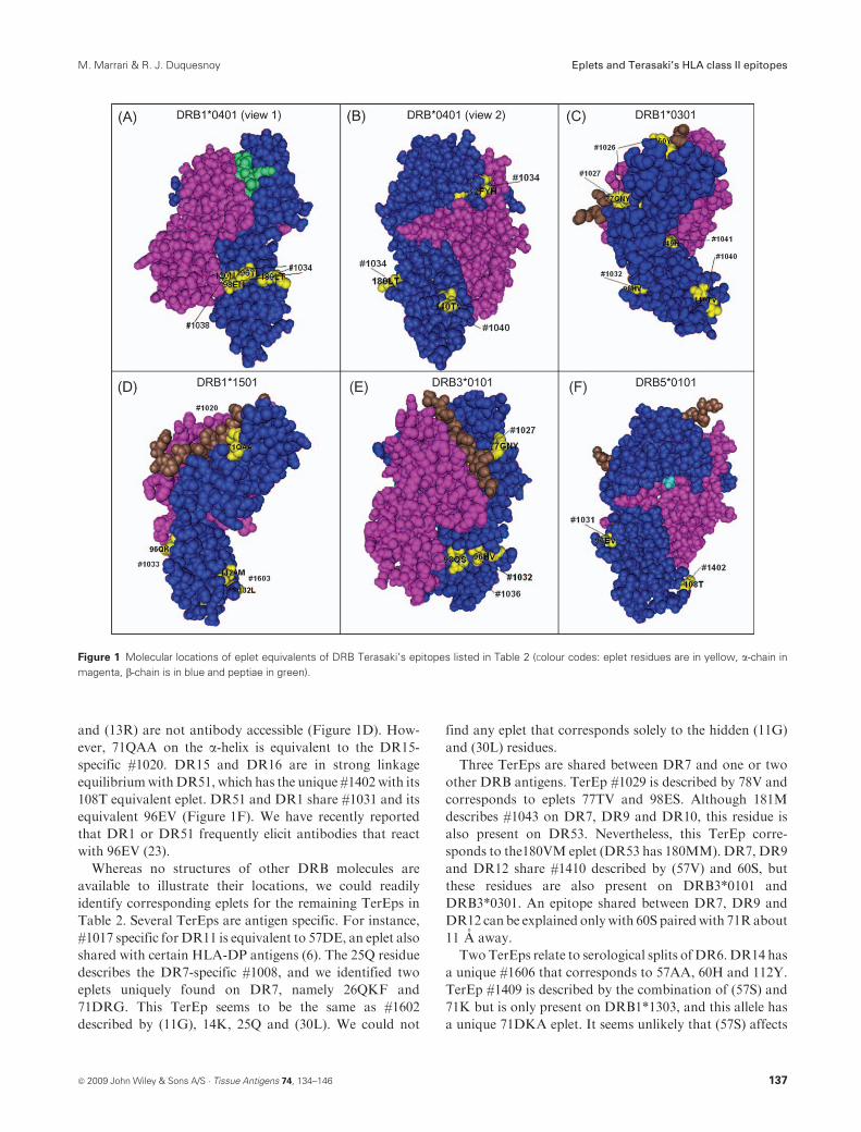
and (13R) are not antibody accessible (Figure 1D). How-
ever, 71QAA on the a-helix is equivalent to the DR15-
specific #1020. DR15 and DR16 are in strong linkage
equilibriumwithDR51, which has the unique #1402 with its
108T equivalent eplet. DR51 and DR1 share #1031 and its
equivalent 96EV (Figure 1F). We have recently reported
that DR1 or DR51 frequently elicit antibodies that react
with 96EV (23).
Whereas no structures of other DRB molecules are
available to illustrate their locations, we could readily
identify corresponding eplets for the remaining TerEps in
Table 2. Several TerEps are antigen specific. For instance,
#1017 specific for DR11 is equivalent to 57DE, an eplet also
shared with certain HLA-DP antigens (6). The 25Q residue
describes the DR7-specific #1008, and we identified two
eplets uniquely found on DR7, namely 26QKF and
71DRG. This TerEp seems to be the same as #1602
described by (11G), 14K, 25Q and (30L). We could not
find any eplet that corresponds solely to the hidden (11G)
and (30L) residues.
Three TerEps are shared between DR7 and one or two
other DRB antigens. TerEp #1029 is described by 78V and
corresponds to eplets 77TV and 98ES. Although 181M
describes #1043 on DR7, DR9 and DR10, this residue is
also present on DR53. Nevertheless, this TerEp corre-
sponds to the180VM eplet (DR53 has 180MM). DR7, DR9
and DR12 share #1410 described by (57V) and 60S, but
these residues are also present on DRB3*0101 and
DRB3*0301. An epitope shared between DR7, DR9 and
DR12 can be explained onlywith 60S pairedwith 71Rabout
11 A away.
TwoTerEps relate to serological splits ofDR6.DR14 has
a unique #1606 that corresponds to 57AA, 60H and 112Y.
TerEp #1409 is described by the combination of (57S) and
71K but is only present on DRB1*1303, and this allele has
a unique 71DKA eplet. It seems unlikely that (57S) affects
Figure 1 Molecular locations of eplet equivalents of DRB Terasaki’s epitopes listed in Table 2 (Colour codes: eplet residues are in yellow, a-chain in
magenta, b-chain is in blue and peptiae in green).
ª 2009 John Wiley & Sons A/S � Tissue Antigens 74, 134–146 137
M. Marrari & R. J. Duquesnoy Eplets and Terasaki’s HLA class II epitopes

this epitope because this hidden residue is more than 10 A
away from 71K.
The DR8-specific #1024 is defined by 74L and has two
equivalent eplets: 73ALDT and 25YRF. DR8 shares #1407
with DR12; this TerEp is described by (13G) and 16Y and
corresponds to 14GEY. It is also possible that #1407 is
equivalent to 71DRA whose conformation is influenced by
13G below the molecular surface about 4.5 A away. DR1
and DR12 share #1030 described as 85A, but this residue is
absent on DRB1*0101 or DRB1*0103 but present on
DRB5*0202. The 85AV eplet corresponds to an epitope
shared between DRB1*0102, DR12 and DRB5*0202.
A pair of eplets 71RRA and 40Y separated by about 4 A
can also explain #1014 on DR10 and DR53. This TerEp
appears to be same as #1408.
TerEps equivalent to eplets with permissible amino
acid combinations
Table 3 lists 10 TerEps with surface residue descriptions
that correspond to eplets with permissible residue combi-
nations; 5 of them (#1018, #1021, #1022, #1025 and #1028)
are located in the highly variable 67–78 sequence of the
a-helix of DRB. Each one has a unique amino acid
description, and Figure 2A–F shows the amino acids within
a 3 A radius on informative DR molecules that carry these
TerEps. The upper part (yellow) shows the residues of a 3-A
patch. The lower part depicts the patch in detail: poly-
morphic residues are colored yellow, permissible residue
combinations are recorded next to residues colored in
magenta and monomorphic residues in yellow are marked
only with their sequence position number.
The 77T residue describes #1028, and a visualization of
a corresponding 3-A patch on DRB*0101 (Figure 2A) and
DRB5*0101 (Figure 2B) shows that 77T is surrounded by
a monomorphic 76D and two residues with permissible
combinations. They are 73A (on all #1028-carrying antigens
exceptDR7,which has 73G) and 78Y (on all #1028-carrying
antigens except DR7 and DR9, which have 78V). These
patch configurations illustrate that #1028 may have
a dominant 77T contiguous with the monomorphic 76D
and that the A/G and V/Y interchanges in flanking
positions 73 and 78 do not significantly affect the specificity
of this epitope. We have assigned a corresponding eplet as
77T*, whereby asterisk represents the permissible 73A/G
and 78V/Y combinations.
As shown in Figure 2C, onDRB1*0301, the 3-A patch of
the #1022-describing 73G has the monomorphic 69E, 72R
and 76D and the permissible 74Q/R and 77N/T combina-
tions. This modeling suggests a dominant role of 73G in this
epitope, and the alignment of 69E, 72R and 76D might
generate a contiguous configuration as a potential contact
site for the specificity-determining CDR loop of antibody.
We assigned the corresponding eplet as 73G*, whereby
asterisk represents 74Q/R and 77N/T.
TerEp #1025 on DR7 and DR52 (except DRB3*0301) is
described by 74Q that forms a 3-A patch with the mono-
morphic 75V, 76D and 79C and with the polymorphic 73G
and 78V/Y (Figure 2D). The corresponding eplet is 74GQ*,
whereby asterisk represents the permissible 78V/Y combi-
nation. The 3-A patch of the #1018 describing 70D has the
monomorphic 69E and 67H/I/L, 71E/K/R and 73A/G
(Figure 2E). This TerEp corresponds to 70D*, whereby
Table 3 Ten DRB TerEps that correspond to eplets with permissible residue combinations
TerEp Defined by
Antibody-reactive
antigens
Residue
description Comments Eplets Comments Model
#1028 Allo DR1, 4, 7, 9, 10, 11,
12, 13, 14, 15, 16;
DR51; DR53
77T 77T also on
DR8
77T* * ¼ 73A/G and 78V/Y Figure 2A,B
#1025 mAb, Allo DR7; DR52 74Q 74Q not on
DRB3*0101
74GQ* * ¼ 78V/Y Figure 2C
#1018 Allo DR7, 8, 11, 12, 13,
16, 103; DR51
70D 70D not on
DRB5*0202
70D* * ¼ 67H/I/L, 71E/K/R
and 73A/G
Figure 2D
#1022 Allo DR7, 17, 18; DR52 73G 73G* * ¼ 74Q/R and 77N/T Figure 2E
#1021 Allo DR4, 13, 17, 18;
DR52
71K 71K not on
DRB1*0404/5
and *1301
71K* (?) * ¼ 70D/Q Figure 2F
#1035 Allo DR4, 7, 9 98E 98E* * ¼ 120N/S Figure 2G
#1411 mAb, Allo DR4, 7, 9 98E, 104A 104A also on DR51
and DR52
98E* * ¼ 120N/S same
as #1035
Figure 2H
#1038 Allo DR4, 10; DR51; DR53 120N 120N* * ¼ 98E/K Figure 2I
#1037 mAb, Allo DR4, 7, 9; DR51; DR52 104A 104A* * ¼ 105K/R Figure 2J
#1039 Allo DR1, 7, 9, 15, 16; DR51;
DR52; DR53
140A 140A not on DRB3*0301 140A* * ¼ 105K/R Figure 2K,L
Allo, alloserum or eluted alloantibody; mAb, monoclonal antibody; TerEp, Terasaki’s epitope.
138 ª 2009 John Wiley & Sons A/S � Tissue Antigens 74, 134–146
Eplets and Terasaki’s HLA class II epitopes M. Marrari & R. J. Duquesnoy
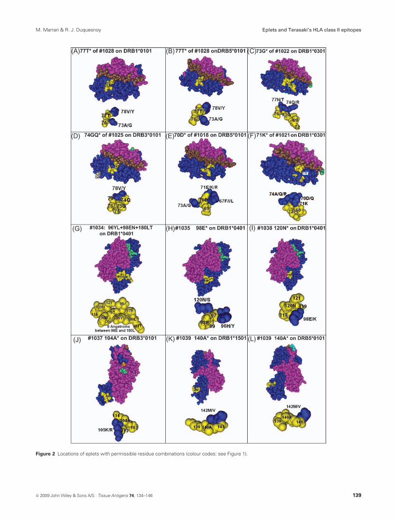
Figure 2 Locations of eplets with permissible residue combinations (colour codes: see Figure 1).
ª 2009 John Wiley & Sons A/S � Tissue Antigens 74, 134–146 139
M. Marrari & R. J. Duquesnoy Eplets and Terasaki’s HLA class II epitopes
asterisk represents these three permissible polymorphic
residue combinations.
The 71K residue that describes #1021 forms a 3-A patch
with the monomorphic 68L and 72R and the polymorphic
70D/Q and 74A/Q/R combinations (Figure 2F). Because
71K is not well expressed on the molecular surface, it is
uncertain how this residue can play a dominant role in the
specificity of #1021. Moreover, #1021 is on DR4, DR13,
DR17, DR18 andDR52, butDRB1*0403, *0405 and *1301
do not have 71K. At this time, the assignment of 71K*must
be considered tentative.
As noted above, three DR4-specific eplets 96YL, 98EN
and 180LT are in adjoining positions in the b2 domain
(Figure 1A). The polymorphic residues of these eplets are
96Y, 98E, 120N, 180L and 181T. Their 3-A patches put
together form a large complex of adjoining residues that
may comprise additionalDR4-specific eplets, such as 96YE,
i.e. 96Y plus 98E (Figure 2G). Moreover, the contiguous
alignments of residues within these eplets such as 99V-98E-
120N and 97D-98Y-180L-181T seem to meet criteria for
contact with the loops of specificity-determining CDRs of
antibody. Residues 98E and 180L are about 9 A apart, and
it is also possible that the DR4-specific epitope requires an
eplet pair such as 98EN1180LT or 96YL198EN. Alto-
gether, these findings suggest that DR4-specific antibodies
may recognize multiple configurations in that part of the b2domain.
In this large structural complex, we found two eplets with
permissible residue combinations. DR4, DR7 and DR9
share #1035 described by 98E; this residue is in a 3-A patch
that has themonomorphic 97D and 99V and the permissible
96H/Yand120N/S combinations (Figure 2H). It seems that
98E dominates the specificity of #1035, but the mono-
morphic 97D and 99V might also be part of this epitope.
Although the residue description of #1411 includes 104A
also on DR51 and DR52, it appears that #1411 is identical
to #1035 because both TerEps are on the same group of
DRB antigens. The second example is #1038 on DR4,
DR10, DR51 and DR53 and corresponds to 120N*,
whereby asterisk is 98E/K (Figure 2I).
TerEp #1037 onDR4,DR7,DR9,DR51 andDR52 has a
corresponding 104A*, whereby asterisk represents 105K/R
(Figure 2J). All antigens in this group have 105K except
DR51, which has 105R. A large group of DRB antigens
have #1039 described by 140A (not on DRB3*0301) and
that corresponds to 140A*, whereby asterisk is 142M/V
(Figure 2K,L).
Eplet assignments to DRB TerEps described
by hidden residues
Table 4 lists 20 TerEps described solely by hidden poly-
morphic residues below the molecular surface and that
cannot make direct contact with antibody. These residues
are in 14 different sequence locations highlighted in yellow
in the DR4 structural model (Figure 3). All these hidden
positions are in the peptide-binding groove, and a top view
shows their approximate primary locations: below the
a-helix of the monomorphic DRA chain (positions 9, 10
and 11), the bound peptide (positions 12, 13, 30, 31, 37, 38
and 57) and the a-helix of the polymorphic DRB chain
(positions 14, 28 and 47). A side view suggests a relative
prominence of positions 13, 14, 28 and 47 immediately
below the molecular surface where epitopes are present.
This analysis addresses the possibility that hidden
residues might significantly influence the conformation of
an epitope, thereby altering its specificity. Their distance
must be close enough for any significant interaction,
probably no further away than 6 A. Several eplets such as
14FEH incorporate hidden residues because they are within
3 A of the polymorphic surface residues, but we also have
searched for eplets that might be influenced by hidden
residues up to 6 A away. We annotated corresponding
eplets with hidden residues between parentheses, for
example 48YR-(37F) or 32IYN-(9K and 30G). We also
searched for hidden-residue-independent eplets (single ones
or pairs) expressed by the same DR antigens as those
reported for TerEps.
Table 3 lists the DR7-specific #1405 and the DR4-
specific #1406 with hidden residue descriptions, but these
TerEps appear to be the same as #1008 and #1602 (onDR7)
and #1034, #1042 and #1605 (on DR4) shown in Table 2.
Five TerEps are equivalent to eplets that have hidden
residues. Residues in hidden position 13 describe three
TerEps. For two of them, we readily found corresponding
eplets, namely 14FEH for #1005 with (13F) and 14SEH for
#1006 with (13S). However, #1007 corresponds to an eplet
48YR,whose conformation seems to be dependent on (13Y)
about 5.5 A away. TerEp #1015 with (47F) corresponds to
48FR. Although #1016 is on DR4, DR8 and DR13, we
noted that the descriptive (57S) is only on DRB1*0405,
DRB1*0801 and DRB1*1303; this TerEp seems equivalent
to 57SA. TerEp #1010 onDR1, DR9, DR51 andDR53 and
described by (31I) is equivalent to 32IYN.
The DR12-specific #1012 is described by (37L) and may
correspond to 48YR-(37L), but there are other DR12-
specific eplets such as 25YRL and 71DRA185AV, which
do not require (37L). DR9 and DR51 share #1404 and they
uniquely have (11D) and (28H). Both antigens share 32IYN,
and (11D) seems close enough (5.5 A) to alter the
conformation of this eplet; (28H) is more than 9 A away,
too far for any effect. Therefore, #1404 may correspond to
32IYN-(11D).
DR9 has a distinct mAb-defined epitope #1401 and two
unique residues (9K) and (30G) hidden in the peptide-
binding groove. These residues are about 4 A from 32IYN
sufficiently close for a conformational effect that would
render this epitope as DR9 specific. Although #1401 may
140 ª 2009 John Wiley & Sons A/S � Tissue Antigens 74, 134–146
Eplets and Terasaki’s HLA class II epitopes M. Marrari & R. J. Duquesnoy
correspond to 32IYN-(9K and 30G), there are other DR9-
specific eplet configurations including 26KYH and
74RAE177TV that do not require these hidden residues.
TerEps #1002 and #1403 (both onDR1, DR7, DR15 and
DR16) appear to be the same, and the hidden residues 9W
and 10Q are closest (3.8 and 4.1 A, respectively) to 32YN*,
whereby asterisk represents the permissible F/I combina-
tion in position 31.Accordingly, theseTerEps are annotated
as 32YN*-(9W and 10Q).
Two TerEps are described by combinations of hidden
residues: #1604 has the (11S)1(12T)1(13S) description
and corresponds to 14SEH, whose conformation is
dependent on (11S) and (12T). The combination of
(9E)1(10Y)1(11S)1(12T)1(13S) described #1601. DR8
and DR12 are listed as #1601-carrying antigens, but they
do not have (13S), and this residue should probably
be excluded from the residue description of #1602. We
have not found a corresponding eplet affected by
(9E)1(10Y)1(11S)1(12T) with or without (13S). However,
the 96HK eplet is equivalent to #1602.
DR7 and DR14 carry #1011 described by (37F); this
residue is also on DRB3*0101 and DRB3*0301, and the
closest eplet is 48YR about 4.5 A away. Another residue is
necessary to explain the presence of #1011 on only DR7 and
DR14: 77T is 12 A away from 48YR far enough for contact
with a second CDR. DRB3*0101 and DRB3*0301 have
77N. Therefore, #1011 may correspond to 48YR-
(37F)177T.
Four TerEps with hidden residue descriptions do not
have informative eplet configurations shared between
antibody-reactive antigens. A large group of DR antigens
have #1003 described by (10Q), and the most nearby eplets
Table 4 Eplet equivalents of 20 DRB TerEps described solely by hidden residues
TerEp
Defined
by
Antibody-reactive
antigens
Residue
description Comments Eplets Comments
#1405 Allo DR7 (11G), (14K) 26QKF/71DRG Same as #1008
and #1602
#1406 Allo DR4 (11V), (13H) 32FYH/96YL/98EN/180LT Same as #1034,
#1042 and #1605
#1005 Allo DR1, 9, 10 (13F) 14FEH
#1006 Allo DR11, 13, 14, 17,
18; DR52
(13S) 14SEH
#1007 Allo DR7; DR51 (13Y) 48YR-(13Y)
#1010 Allo DR1, 9; DR51;
DR53
(31I) 32IYN
#1015 Allo DR11, 12, 13,
15, 17
(47F) Not on DRB1*1303 48FR
#1016 Allo DR4, 8, 13 (57S) Only on DRB1*0405,
*0801 and *1303
57SA
#1012 mAb DR12 (37L) 48FR-(37L)/25YRL/
71DRA185AV
#1401 mAb DR9 (9K), (30G) 32IYN-(9K, 30G)/
26KYH/74RAE177TV
#1404 Allo DR9; DR51 (11D), (28H) 32IYN-(11D) (11D) is 5.5 A away
#1002 Allo DR1, 7, 15, 16 (9W) 32YN*-(9W10Q) * ¼ 31I/F, 9W is 3.8
A away, 10Q is 44
A away
#1403 Allo DR1, 7, 15, 16 (9W), (10Q) 32YN*-(9W10Q) Same as #1002
#1604 Allo DR11, 13, 14,
17, 18
(11S)1(12T)1(13S) 14SEH-(11S, 12T)
#1601 mAb,
Allo
DR8, 11, 12,
13, 14, 17, 18
(9E)1(10Y)1(11S)1
(12T)1(13S)
DR8 and DR12 have
13G, not 13S
96HK
#1011 Allo DR7, 14 (37F) 37F also on DRB3*0101
and *0301
48YR-(37F)177T 48YR and 77T are
12 A apart
#1004 Allo DR4, 10 (11V) 140TV1149Q? Eplets are 19 A apart
#1003 Allo DR1, 4, 7, 9, 15,
16; DR51; DR53
(10Q) 32Y*-(10Q)?
#1009 Allo DR4, 8, 11, 13, 14,
15, 16, 17
(28D) 28D also on DRB3*0101 ?
#1013 Allo DR1, 103, 14, 15 (37S) 37S not on DR14 but
on DR16
?
Allo, alloserum or eluted alloantibody; mAb, monoclonal antibody; TerEp, Terasaki’s epitope.
ª 2009 John Wiley & Sons A/S � Tissue Antigens 74, 134–146 141
M. Marrari & R. J. Duquesnoy Eplets and Terasaki’s HLA class II epitopes

are in position 32 about 5 A away. Three different eplets are
on DR antigens with (10Q), namely 32IYN (on DR1, DR9,
DR51 and DR53), 32FYH (on DR4) and 32FYN (on
DR7), and each one has 32Y. It is possible that #1003
reflects an epitope dominated by 32Y and significantly
influenced by 10Q, which is located under the DRA a-helix.However, we question why so many closely located
combinations like 31F/I, 33H/N as well as the nearby 9E/
K/Q/W combination would not affect the specificity of
#1003. We must conclude that the eplet equivalent of this
TerEp remains elusive.
There are no eplets even within a 10 A radius of the
hidden (11V), which describes #1004 on DR4 and DR10.
This TerEp might correspond to the 140TV1149Q pair,
although these eplets are 19 A apart, probably too far to
serve as contact sites for two CDRs of antibody. TerEp
#1009 on a large group of antigens is described by (28D), but
this residue is also on DRB3*0101. TerEp #1013 on DR1,
DR103, DR14 and DR15 and has (37S), but this residue is
on DR16 rather than on DR14.
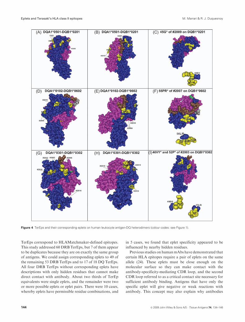
DQ TerEps with corresponding eplets
Table 5 lists 15 DQB TerEps and their residue descriptions,
and Figure 4 shows the locations of corresponding eplets on
the crystalline structures of DQ2, DQ6 and DQ8 hetero-
dimers. Six TerEps define serological DQ specificities. The
DQ1 (i.e. DQ51DQ6)-specific #2004 corresponds to three
eplets: 52PQ and the proximally located 84EV and 89GI
(Figure 4D,E). TerEp #2015 is specific for DQ5 and
corresponds to 70GA and 116I. A mAb defines #2011 on
DQ5 and DQB1*0601, which have (38V), but not on
DQB1*0602, which has (38A). This hidden residue is>10 A
away from 46V listed in the description of #2011, and it
seems unlikely that it affects the conformation of a 46V-
defined epitope.However, the distance between the 38V and
the DQ1-defining 52PQ eplet is 5.8 A, and it is possible that
#2011 corresponds to 52PQ-(38V).
Three hidden residues (28S), (30S) and (37I) and two
surface residues 52L and 55L describe the DQ2-specific
#2001. DQ2 has four more unique residues 46E, 47F, 71K
and 74A, and consequently, there are several eplets that
correspond to #2001 including 45GE, 52LL and 70RKA
(Figure 4A,B).
The DQ3 (i.e. DQ71DQ81DQ9)-specific #2006
described by 55P is equivalent to 55PPP (Figure 4G).
DQ7 has a unique 45E residue that describes #2005, and
45EV defines this specificity. DQ4 is defined by #2002
described by 56L and has also the distinct 70E and 71D
residues; this TerEp corresponds to 56LD and 70ED. DQ3
and DQ4 share #2014 that has five possible residue
Figure 3 Views of hidden polymorphic residues on the DR4 molecular structure (colour codes: see Figure 1).
142 ª 2009 John Wiley & Sons A/S � Tissue Antigens 74, 134–146
Eplets and Terasaki’s HLA class II epitopes M. Marrari & R. J. Duquesnoy

descriptions; there are three corresponding eplets: 52PL and
in the b2 domain, 140T and 182N (Figure 4G).
All DQ antigens except DQ1 have #2013 described by
multiple residues; six of them 84Q, 85L, 86E, 87L, 89T and
90T are in a patch of about 5 A. A single eplet 84QL
describes this patch (Figure 4H). The 220H and 221H
descriptions of #2013 are not on the external DQ domains
and are therefore not considered as epitopes.
The descriptions of three TerEps include 45G. TerEp
#2010 on DQ4, DQ5, DQ6, DQ8 and DQ9 is equivalent to
45GV (Figure 4D,E,G). TerEp #2012 with 45G155P is on
DQ8 and DQ9 and corresponds to 45GV155PPP separated
by about 12 A (Figure 4G). AmAb defines #2009 on all DQ
molecules except DQ7; 45G and the monomorphic 34R
describe this TerEp. The 3-A patch of 45Ghas 46E/V and the
monomorphic 41D, 44V and 71R (Figure 4C) and 45G*,
whereby the asterisk represents 45E/V corresponds to #2009.
There are two additional DQB TerEps corresponding to
eplets with permissible residue combinations. TerEp #2007
on DQ4, DQ5 and DQ6 has a unique 55R, whereas 52P is
also onDQ3. The 3-A patch of 55R has 52P, 56L/P and 58E
(monomorphic), and#2007 corresponds to 55PR*,whereby
asterisk represents the permissible 56L/P combination. This
eplet has a contiguous sequence structure flanked by the
permissible residue combination (Figure 4F). All DQ
antigens except DQ2 have #2003 described by 46V and 52P
that are 15 A apart; the hidden (28T) is more than 7 A away,
which seems too far for a conformational effect. The 3-A
patch of 46V has 45E/G and 47Y, whereas the 3-A patch of
52P has the monomorphic 51T and 53L/Q. Figure 4I shows
the structural configurations of 46VY* (whereby asterisk is
45E/G) and 52P* (whereby asterisk is 53L/Q).
Two hidden residues (9Y) and the monomorphic (11F)
describe the mAb-defined #2008; we could not identify an
eplet shared byDQ2,DQ3 andDQ5, and it seems likely that
#2008 is a xenoepitope rather than an alloepitope.
Table 4 lists also three DQA TerEps, each of them are
defined by alloantibodies eluted from informative DQ
heterodimers used to separate DQA from DQB antibodies
(8). TheDQA1*02-specific #2017 is equivalent to 47KL and
53HRL, which are about 5 A apart. The DQA1*03-specific
#2019 corresponds to 47QL, 52FRRand 187T (Figure 4H).
The rather common #2018 on DQA1*04, DQA1*05 and
DQA1*06 is analogous to 41GR and 51VLQ (Figure 4B).
DR and DQ eplets without corresponding TerEps
Table 6 represents a list of DRB, DQB and DQA eplets for
which no corresponding TerEps have been reported. Our
serum screening experience has indicated antibodies with
DRB specificities associated with these eplets (data not
shown). A recent analysis of sensitized kidney transplant
patients has shown rather common antibodies against
several DQB eplets such as 57PA and 70GT and DQA
eplets such as 169AE and 75SL (6).
Discussion
Similar to our experience with the class I TerEps (16), this
analysis has shown that the majority of HLA-DR and -DQ
Table 5 Eighteen DQ TerEps and their corresponding eplets
TerEp Defined by Antigens Residue description Eplet Comments
#2004 mAb, allo DQ5, 6 (DQ1) 84E/85V/86A/89G/90I/221Q 52PQ/84EV/89GI Figure 4D,E
#2015 allo DQ5 70G171A/116I/125S 70GA/116I
#2011 mAb DQ5, DQB1*0601 (38V)146V 52PQ-(38V)
#2001 mAb, allo DQ2 (28S)/(30S)/(37I)/52L/55L 45GE/52LL/71RKA
#2006 mAb, allo DQ7, 8, 9 (DQ3) 55P 55PPP Figure 4A,B
#2005 mAb, allo DQ7 45E 45EV
#2002 mAb, allo DQ4 56L 57LD/70ED
#2014 allo DQ4, 7, 8, 9 77T184Q/77T185L/77T186E/77T187T/182N 52PL/140T/182N Figure 4G,H
#2013 allo DQ2, 4, 7, 8, 9 84Q/85L/86E/(87L)/89T/220H/221H 84QL Figure 4H
#2010 mAb DQ4, 5, 6, 8, 9 45G146V 45GV Figure 4D,E,G
#2012 mAb DQ8, 9 45G155P 45GV155PPP Figure 4G
#2009 mAb DQ2, 4, 5, 6, 8, 9 34R (is monomorphic)145G 45G* * ¼ 46E/V, Figure 4C
#2007 mAb, allo DQ4, 5, 6 52P155R 55PR* * ¼ 56L/P, Figure 4F
#2003 mAb, allo DQ4, 5, 6, 7, 8, 9 (28T)/46V/52P 46VY*/52P* * ¼ 45E/G/* ¼ 53L/Q,
Figure 4I
#2008 mAb DQ2, 5, 7, 8, 9 (9Y)1(11F) (is monomorphic) Undefined Xenoepitope?
#2017 allo DQA1*02 47K/52H/54L 47KL/53HRL
#2019 allo DQA1*03 (26S)/47Q/56R/187T 47QL/52FRR/187T Figure 4H
#2018 allo DQA1*04, 05, 06 40G/47C 41GR/51VLQ Figure 4B
Allo, alloserum or eluted alloantibody; mAb, monoclonal antibody; TerEp, Terasaki’s epitope.
ª 2009 John Wiley & Sons A/S � Tissue Antigens 74, 134–146 143
M. Marrari & R. J. Duquesnoy Eplets and Terasaki’s HLA class II epitopes
TerEps correspond to HLAMatchmaker-defined epitopes.
This study addressed 60 DRB TerEps, but 7 of them appear
to be duplicates because they are on exactly the same group
of antigens. We could assign corresponding eplets to 49 of
the remaining 53 DRB TerEps and to 17 of 18 DQ TerEps.
All four DRB TerEps without corresponding eplets have
descriptions with only hidden residues that cannot make
direct contact with antibody. About two thirds of TerEp
equivalents were single eplets, and the remainder were two
or more possible eplets or eplet pairs. There were 10 cases,
whereby eplets have permissible residue combinations, and
in 5 cases, we found that eplet specificity appeared to be
influenced by nearby hidden residues.
Previous studies on humanmAbs have demonstrated that
certain HLA epitopes require a pair of eplets on the same
allele (24). These eplets must be close enough on the
molecular surface so they can make contact with the
antibody-specificity-mediating CDR loop, and the second
CDR loop referred to as a critical contact site necessary for
sufficient antibody binding. Antigens that have only the
specific eplet will give negative or weak reactions with
antibody. This concept may also explain why antibodies
Figure 4 TerEps and their corresponding eplets on human leukocyte antigen-DQ heterodimers (colour codes: see Figure 1).
144 ª 2009 John Wiley & Sons A/S � Tissue Antigens 74, 134–146
Eplets and Terasaki’s HLA class II epitopes M. Marrari & R. J. Duquesnoy

react with certain antigens in binding assays but not in
complement-dependent lymphocytotoxicity (25).
This analysis indicates that only about 10% of DRB
TerEps are equivalent to eplet pairs, which is much less than
the 35% incidence of paired eplet equivalents of TerEps on
HLA-A, -B and -C antigens (16). The number of poly-
morphic or locus-specific residue positions may explain this
difference. Virtually, all class I eplet pairs involve the a1 anda2 domains, which have more than 40 such positions. In
contrast, there are less than 20 polymorphic positions in b1domain of DRB1 and none onDRA chains. Although both
DQA and DQB chains are polymorphic, we identified only
1/18 DQ TerEps equivalent to an eplet pair. It is possible
that the size of the Luminex panel is too small for
informative HLA-DQ heterodimers to identify eplet pairs
on or shared by DQA and DQB antigens.
DRB antigens appear to have two large regions with
overlapping eplets, namely the highly variable 67–78a-helicalsequence of the b1 domain and a discontinuous sequence
configuration in the b2 domain dominated by residues in
polymorphic positions 96, 98, 120, 180 and 182. Together
they comprise almost one half of the eplets that correspond to
TerEps. As illustrated in Figure 2, the eplets with permissible
residue combinations appear to have contiguous alignments
of dominant residues with monomorphic residues, and such
structures seem suitable as potential contact sites for CDR
loops of antibody. We have found similar structural align-
ments for HLA class I eplets (16). This concept expands the
eplet definition, especially in relation to the antibody analysis
version of HLAMatchmaker.
This analysis is not without limitations. The eplet
assignments were based on amino acid descriptions of
TerEps and/or antigens that reacted with a given mAb or
alloserum.Our comparisons ofDRB sequences showed that
several TerEps had somewhat different residue descriptions
as reported by Terasaki’s group. This created aminor pitfall
because we could choose either the reactive antigens or the
reported residue(s) as criteria in the determination of eplets
that correspond to a given TerEp. The residue descriptions
of TerEps do not consider HLA information about the
immunizer and antibody producer, which would have
permitted a distinction between nonself and self amino acid
residues shared between the immunizing antigen and the
antigens in the Luminex panel.
Nevertheless, this study demonstrates that how the
HLAMatchmaker interpretation of amino acid residues
shared between antibody-reactive antigens can increase our
understanding of the structural basis of HLA epitopes.
More studies are needed to fully evaluate the HLA epitope
repertoire.
Acknowledgments
This study was possible because of the extensive laboratory
testing and detailed information about HLA epitopes
generated by Paul Terasaki and his group. We gratefully
acknowledge their contributions. This study was supported
by RO1 grant AI-55933 from the National Institutes of
Health.
References
1. Takemoto S, Zeevi A, Feng S et al. National conference to
assess antibody-mediated rejection in solid organ
transplantation. Am J Transplant 2004: 4: 1033–41.
2. Gebel H, Bray R, Nickerson P. Pre-transplant assessment of
donor-reactive, HLA-specific antibodies in renal
transplantation: contraindication vs. risk. Am J Transplant
2003: 3: 1488–500.
Table 6 HLA-DR and HLA-DQ eplets without corresponding TerEps
DRB eplet Antigensa
DQB
eplet Antigens
12LKF DR1 14GM DQ2, 4, 6, 8
25HRL DR1, 10 52PL DQ3, 4
26RL DR1, 10, 12 57PA DQ2, 8
120S non-DR4, 51,
53
70GT DQB1*0602/3
98KS DR1, 2, 3, 5,
6, 8
74EL DQ6, 7, 8, 9
104SK DR1, 2, 3, 5,
6, 8, 53
74SV DQ4, 5
149Q non-DR3, 5,
6, 8, 52c
77DR DQ2, 5
71Qk/rA DR1, 4 140A DQ2, 6
98KN DR10, 51, 53
81HA DR12, 51b
26KFD DR2, 4 DQA
eplet
25HRF DR2, 4, 6, 11,
18, 51, 52
48LF DQA1*02, 03
26RF DR2, 4, 6, 8,
11, 51, 52bc
50EF DQA1*01
32FHN DR3, 6, 12, 52 56RB DQA1*02, 04,
05, 06
6C DR51b 60QF non-DQA1*01
51R DR52b 69L DQA1*02, 03, 05
48YQ DR53 75ILR DQA1*02, 04, 06
67FR DR8, 9, 11,
16, 51a
75SL DQA1*05
189S DR8, DR52b, 53 80IRS DQA1*02, 03,
04, 06
14ER non-DR7 160AE DQA1*0501/5
4R non-DR7, 9, 53
189R non-DR8, 52b, 53
a DR51a ¼ DRB5*0101; DR51b ¼ DRB5*0202; DR52a ¼ DRB3*0101;
DR52b ¼ DRB3*0202; DR52c ¼ DRB3*0301; non, all DRB antigens in
the Luminex panel except those listed.
ª 2009 John Wiley & Sons A/S � Tissue Antigens 74, 134–146 145
M. Marrari & R. J. Duquesnoy Eplets and Terasaki’s HLA class II epitopes

3. Susal C, Opelz G. Kidney graft failure and presensitization
against HLA class I and class II antigens. Transplantation
2002: 73: 1269–73.
4. Pollinger HS, Stegall MD, Gloor JM et al. Kidney
transplantation in patients with antibodies against donorHLA
class II. Am J Transplant 2007: 7: 857–63.
5. Terasaki PI. Humoral theory of transplantation. Am J
Transplant 2003: 3: 665–73.
6. Duquesnoy R, Awadalla Y, Lomago J et al. Retransplant
candidates have donor-specific antibodies that react with
structurally defined HLA-DR,DQ,DP epitopes. Transpl
Immunol 2008: 18: 352–60.
7. DengCT, Cai J, Tarsitani C, El-AwarN, LachmannN,Ozawa
M. HLA class II DQ epitopes. Clin Transpl 2006: 115–22.
8. DengCT,El-AwarN,OzawaMet al.Human leukocyte antigen
class II DQ alpha and beta epitopes identified from sera of
kidney allograft recipients. Transplantation 2008: 86: 452–9.
9. El-AwarN, Terasaki PI, Cai J et al. Epitopes of theHLA-A, B,
C, DR, DQ and MICA antigens. Clin Transpl 2007: 175–94.
10. Cai J, Terasaki PI, Mao Q et al. Development of
nondonor-specific HLA-DR antibodies in allograft recipients
is associated with shared epitopes with mismatched donor DR
antigens. Am J Transplant 2006: 6: 2947–54.
11. Cai J, Kohanof S, Terasaki P. HLA-DR Antibody Epitopes.
Clin Transpl 2006: 103–14.
12. Cai J, Terasaki P. Post-transplantation antibody monitoring
and HLA antibody epitope identification. Curr Opin Immunol
2008: 20: 602–6.
13. Duquesnoy R. Clinical usefulness of HLAMatchmaker in
HLA epitope matching for organ transplantation. Curr Opin
Immunol 2008: 20: 594–601.
14. Duquesnoy RJ, Askar M. HLAMatchmaker: a molecularly
based algorithm for histocompatibility determination V. Eplet
matching for HLA-DR, HLA-DQ and HLA-DP. Hum
Immunol 2007: 68: 12–25.
15. Duquesnoy RJ. A structurally based approach to determine
HLA compatibility at the humoral immune level. Hum
Immunol 2006: 67: 847–62.
16. Duquesnoy R, Marrari M. Correlations between Terasaki’s
HLA class I epitopes and HLAMatchmaker-defined eplets
on HLA-A, -B and -C antigens. Tissue Antigens 2009:
Submitted.
17. Hogue C. Cn3D: a new generation of three-dimensional
molecular structure viewer. Trends Biochem Sci 1997: 22:
314–6.
18. Marsh SG, Bodmer JG. HLA-DR and -DQ epitopes and
monoclonal antibody specificity. Immunol Today 1989: 10:
305–12.
19. Klohe E, Fu XT, Ballas M, Karr RW. HLA-DR beta chain
residues that are predicted to be located in the floor of the
peptide-binding groove contribute to antibody-binding
epitopes. Hum Immunol 1993: 37: 51–8.
20. Drover S, Marshall WH, Kwok WW, Nepom GT, Karr RW.
Amino acids in the peptide-binding groove influence an
antibody-defined, disease-associated HLA-DR epitope. Scand
J Immunol 1994: 39: 539–50.
21. Fu X-T, Drover S, Marshall WH, Karr RW. HLA-DR
residues accessible under the peptide-binding groove
contribute to polymorphic antibody epitopes. Hum Immunol
1995: 43: 243–50.
22. Smith KD, Mace BE, Valenzuela A et al. Probing HLA-B7
conformational shifts induced by peptide-binding groove
mutations and bound peptide with anti-HLA monoclonal
antibodies. J Immunol 1996: 157: 2470–8.
23. Marrari M, Duquesnoy R. Why can sensitization by
a HLA-DR2 mismatch lead to antibodies that react also with
HLA-DR1? Hum Immunol 2009 (in press).
24. Duquesnoy RJ, Mulder A, Askar M, Fernandez-Vina M,
Claas FHJ. HLAMatchmaker-based analysis of human
monoclonal antibody reactivity demonstrates the
importance of an additional contact site for specific
recognition of triplet-defined epitopes. Hum Immunol 2005:
66: 749–61.
25. Duquesnoy RJ. HLAMatchmaker: a molecularly based
algorithm for histocompatibility determination. ASHI
Quarterly 2002: 60–2.
146 ª 2009 John Wiley & Sons A/S � Tissue Antigens 74, 134–146
Eplets and Terasaki’s HLA class II epitopes M. Marrari & R. J. Duquesnoy