correlation of radiographic measurements and pulmonary function tests in chronic obstructive...
TRANSCRIPT
695
Correlation of RadiographicMeasurements andPulmonary Function Tests inChronic ObstructivePulmonary Disease
Stanley B. Reich1Albert Weinshelbaum1
John Yee2
Received March 1 4, 1984; accepted after revi-sin November 13, 1984.
Presented at the annual meeting of the Societyof Thoracic Radiology, Orlando, FL, March 1984.
‘Department of Radiology, University of Califor-nia, Davis, CA 95616, and Veterans AdministrationMedical Center, Martinez, CA 94553. Address re-print requests to S. B. Reich, Diagnostic Radiology(1 14A), VA Medical Center, 150 Muir Rd., Martinez,CA 94553.
2Department of Medicine, Pulmonary Section,Lkiiversity of California, Davis, CA 95616, and Vet-erans Administration Medical Center, Martinez, CA94553.
AJR 144:695-699, April 19850361-803X/85/1444-0695© American Roentgen Ray Society
Measurements on standard frontal and lateral radiographs that reliably predict thepresence or absence of chronic obstructive lung disease would be useful to the clinicalradiologist when no other clinical data are available. Therefore, statistical correlationsof pulmonary function tests and measurements of chest films were made in 104 menchosen from 1000 cases referred for pulmonary function tests in whom no obviousabnormalfty was present on the chest film. Two measurements were significantlycorrelated (p < 0.001) without requiring correction for body surface area: (1) The heightof the arc of the right diaphragm in the lateral projection. When 2.6 cm or less it identifies67.7% of all patients with abnormal pulmonary function tests and 78.3% of patients with
moderately or severely abnormal pulmonary function tests. (2) The height of the rightlung in the posteroanterior projection. When this is 29.9 cm or more it will identify 69.8%of all patients with abnormal pulmonary function tests and 79.7% of patients withmoderately to severely abnormal pulmonary function tests. These simple measurementswill assist the radiologist to judge from standard chest radiographs whether a patientmay or may not have chronic obstructive lung disease.
Clinical evaluation of lung function primarily concerns functional rather thananatomic features. Pulmonary function tests measure air trapping, lung volume,obstruction to air flow, and capillary volume [1 -4], but do not measure emphysemadirectly. Pulmonary emphysema can be diagnosed and graded only postmortem[5-i 0].
Previous studies differ as to which radiographic criteria of emphysema are useful,and one study indicated that no criteria could be evolved that exceeded thesubjective diagnosis [8]. All showed that advanced disease would usually berecognized, while mild to moderate disease could escape radiographic detection.To attempt further refinement of the relation of radiographic features to lungfunction in chronic obstructive airways disease, we made multiple chest radi-ographic measurements and pulmonary function tests. Associations that might beused to evaluate borderline radiographs-particularly where clinical information isunavailable or equivocal-were sought. Such associations could aid the radiologistin estimating the chances for the presence or absence of obstructive lung disease.
Materials and Methods
We reviewed 1000 patients undergoing pulmonary function testing over a 5 year period.After excluding women (as too small a group to analyze), patients without appropriate films,and patients with pleural changes, bullous disease, prior surgery, neoplasms, and granuloma-tous disease, 104 cases remained to be analyzed. The patients had normal films except forchanges relating to chronic obstructive lung disease.
The group comprised men of 22-96 years of age (mean, 59.1 years). Their weights variedfrom 49 to 150 kg (mean, 82.1 kg). The standard diagnoses were chronic obstructivepulmonary disease, 57%; chronic bronchitis, 8%; asthma, 3%; sleep apnea, 3%; and nopulmonary disease, 29%.
Dow
nloa
ded
from
ww
w.a
jron
line.
org
by 1
58.1
21.2
47.6
0 on
11/
09/1
4 fr
om I
P ad
dres
s 15
8.12
1.24
7.60
. Cop
yrig
ht A
RR
S. F
or p
erso
nal u
se o
nly;
all
righ
ts r
eser
ved

696 REICH ET AL. AJR:144, April 1985
I
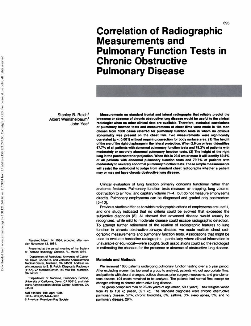
Ag. 1 -Measurements made onchest radiographs. Descriptions of meas-urements are in text.
The radiographs were frontal and left lateral films taken at the timeof pulmonary function tests at 3 m with no air gaps. Thirty caseswere repeated at 1 .8 m to determine the factor to correct measure-ments from the longer to the shorter target-film distance. This variedfrom +iO.2% to +10.8% (mean, +10.5%).
The measurements were made with an ordinary transparent rulerhaving 1 mm increments. In 20 cases this was checked by comparingfilms taken at different times-and in 30 cases the same film wasmeasured at different times by two different observers (S.B.A. andA.W.) with good agreement. Eleven different radiographic measure-
ments (fig. 1) were obtained: (A) lung length-distance from thetubercle of the first rib to the top of the dome of the right diaphragm;
(B) lung width-lateral diameter of the thorax between the inner ribsurfaces at the level of the top of the right diaphragm; (C) anteropos-tenor chest diameter-horizontal distance at the level of the top ofthe right diaphragm from the posterior sternum to the midpointbetween the anterior borders of the posterior ribs; (0) cardiac diam-eter-transverse diameter of the heart obtained by adding the far-thest projections of the heart to the right and left of midline; (E)retrosternal space width-horizontal distance from the posterior as-
pect of the sternum 3 cm below the stemoclavicular junction to theanterior margin of the aorta measured on the lateral film; (F) dia-phragm level-level of the apex of the right diaphragm in relation tothe anterior and posterior ribs; (G) frontal and lateral tracheal diame-ter-measurement of the airway 2 cm below the thoracic inlet,yielding a frontal/lateral ratio; (H)right diaphragmatic arc on the frontalfilm-measurement of a line normal to the curve at its apex andnormal to a line drawn between the lateral costophrenic angle andthe cardiophrenic angle; (I) right diaphragmatic arc on the lateralfilm-measurement of a line normal to the curve at its apex andnormal to a line drawn between the posterior and anterior costo-phrenic angles (which might be obtained by continuing the arc of thediaphragm anteriorly); (J) right pulmonary artery diameter-trans-verse diameter of the right descending pulmonary artery just beforebranching; and (K) left pulmonary artery diameter-largest diameterof the pulmonary artery as it arcs over the left main-stem bronchus.
The pulmonary function tests were performed in our pulmonaryfunction laboratory [3] using standard procedures [1 , 2] with the
patient sitting in an upright position. During the forced vital capacity(FVC) maneuver, the technician inspected the flow volume curve onthe X-Y plotter and coached the patients until an “accurate” curvewas obtained. Several measurements were obtained: (1 ) Forcedexpiratory flow rate in 1 sec (FEV1) and FVC. These two tests wereobtained with a wedge spirometer and assessed the severity ofairway obstruction. (2) Diffusing capacity. This was performed withthe single-breath carbon monoxide method and estimated functionalalveolar capillary volume, which is usually decreased in patients withemphysema. (3) Alveolar volume was obtained by helium dilutionduring the diffusing capacity maneuver. (4) Residual volume/alveolarvolume ratio. This assessed the severity of air-trapping.
The patients were split into three groups based on the FEV1 andFEV1/FVC ratio according to accepted clinical conventions. The nor-mal patients (group 1) had an FEV1 greater than 80% and FEV1/FVCratio greater than 70% of predicted. Patients with mild obstructivelung disease (group 2) had an FEV1 greater than 65%-80% and anFEV1/FVC ratio greater than 60% of predicted. Patients with moder-
ate to severe obstructive lung disease (group 3) had an FEV1 lessthan 65% and an FEV1/FVC ratio less than 60% of predicted. Thesemeasurements were evaluated for mean and standard deviations anda correlation matrix was made. These were analyzed with and without
correlation for body surface area, height, weight, and age.
Results
Table 1 shows the means and standard deviations of thevariables measured in all subjects. Table 2 shows the corre-lation coefficients for all groups. The dimensions correlatingbest with normal function were (1) the length of the lung withand without correction for body surface area, (2) the arc ofthe right diaphragm in the lateral projection with and withoutcorrection for body surface area, and (3) the level of thediaphragm relative to the posterior ribs when corrected forbody surface area. The last measurement is not always usefulto the clinical radiologist since the patient’s height and weight,
Dow
nloa
ded
from
ww
w.a
jron
line.
org
by 1
58.1
21.2
47.6
0 on
11/
09/1
4 fr
om I
P ad
dres
s 15
8.12
1.24
7.60
. Cop
yrig
ht A
RR
S. F
or p
erso
nal u
se o
nly;
all
righ
ts r
eser
ved

TABLE 1: Findings in 104 Individuals Undergoing PulmonaryFunction Testing
Variable Mean Value
1 , Age (years) 59.0 (11.8)2, Height (cm) 175.8 (7.9)3,Weight(kg) 82.1 (19.3)4, Body surface area (ma) 2.0 (0.2)5, Frontal lung length (cm) 24.9 (3.1)6, Frontal lung width (cm) 30.2 (2.3)7, Lateral chest diameter (cm) 21 .8 (2.5)8, Frontal cardiac diameter (cm) 13.8 (1.9)9, Retrostemal air (cm) 3.0 (0.8)
10, Anterior rib level 6.1 (1.0)1 1 , Posterior rib level 1 0.6 (0.8)12, Frontal trachea diameter (cm) 1 .8 (0.2)13, Lateral trachea diameter (cm) 2.1 (0.3)14, Frontal diaphragm arc (cm) 2.3 (0.8)15, Lateral diaphragm arc (cm) 2.4 (1.4)16, Right pulmonary artery diameter (cm) 1 .6 (0.3)17, Left pulmonary artery diameter (cm) 2.3 (0.4)18, FEy1 (% predicted) 61 .0 (28.4)19, FVC (% predicted) 79.3 (21.4)20, FEV1/FVC (% predicted) 73.3 (18.9)21 , Diffusing volume (% predicted) 88.3 (43.5)22, Alveolar volume (% predicted) 100.4 (18.1)23, Residual volume/alveolar volume ratio (%
predicted) 43.4 (16.0)
IC�)
tO i- �- 0 �LO � CO0000Cl)U)0#{216}#{216}00#{216}#{216}
0000ZZOZZooZZ
,�, CsJ ,- CO I-”.‘�‘ C”) 10 0 0) C”) 0) CO
0000000000000
I I I I I ILOL()
O 0 CO Cl) COCOCOCOCl) COCi) CO CO0OZZZZZZZZZZZ
‘-‘-�N-C)COCOC’)C”)O)Cc�J C�J 0 0 ‘- 0 0 0 ‘- 0 0 0�
0000000cr0000?
0Z0ZZZ0Zoo�ooLt) LC) ‘� ‘- ‘- C”) 0 0 C”) CO ‘- CO LI)�qqc�c�C”)0000000000000
I I I I I
0 �LI) �L() (0 0 ‘- �Lt) 0 LI) 0< 0U)OCOQQQOCOOOQO
�. 2.�.2,�,2,2.2,2,�.2.2.2.2.�
0 0 0 cr o o 0000000
-�---.-.-.. LI)
ZZZZZZZZZoZZZ<> �
. � 0 0 � C�J ‘-000000000000000
II I III I
0LO0��0�OCOCOOCOU)OCOCOCOOCOU)S
�,�. ‘- ‘- C’%J� . CV) 0 ‘- �- � �
0000000000000
I I I I I I I I I
0 00000Co00CO0000
�> � ‘- CO N. � C”) C�J � CO 0) CD r- c’, ��
0 c� � o 000000000I I I I I I I I I
0 LI) LI) �0 �-�0 LI) � � LI)�Q0QCOOC000C00000
0
�A. � C) LI) C”) 0 C�J 0 It) N. ,- � �
�
0 0 ‘D 0 0 0 0 0 0 0 � 00
I I I I I I I I I
� � � � � �
>� 000ZoZooZooooUiU. LI) CO CO F- CO CO N. � � CD C�4 t�-CO C’J C�J ‘- C”) ,- If) C�J ,- C”) CO C�J C”)
0000000000000� I I I I I I I I I
Note.-FEV1 = forced expiratory flow rate in 1 sac: FVC = forced vital capacity.
necessary for this determination, are not always available at
the time of interpretation.Table 3 shows the t test and false-positive and false-
negative errors for lung length and lateral diaphragm arctesting group 1 (normal pulmonary function tests) againstgroups 2 and 3 (mild and moderate or severely abnormalpulmonary function tests). This shows that an arc of the rightdiaphragm in the lateral projection of 2.6 cm or less onstandard 1 .8 m radiographs will include 5% of normals and67% of abnormals.
Table 3 also shows the results if the groups are split intogroups 1 and 2 (normal and mildly abnormal pulmonary func-tion tests) against group 3 (moderately to severely abnormalpulmonary function tests).
The results of plotting the length of the lung (in centimeters)and the arc of the diaphragm against FEV1/FVC are shownin figures 2 and 3, respectively. Also included are the regres-sion lines from this data.
Discussion
The clinical diagnosis of emphysema implies a high proba-bility that the signs and symptoms may ascribe to morphologicemphysema. These signs and symptoms include both tho-racic hyperexpansion and airflow obstruction clinically, radi-ographically, and on pulmonary function tests. As a conse-quence, we usually refer to chronic airflow obstruction ratherthan emphysema.
Emphysema can be defined in morphologic terms only.Radiologic-pathologic correlation has been performed, and,in British literature, arterial deficiency has been considered
2
U)
I>C
AJR:144, April 1985 RADIOGRAPHIC PULMONARY FUNCTION CORRELATION 697
0)
C
U)
I!C0
.�
UC
IL
a)C0E
a-a)C
0a)a)�0C
U)
a)
‘D
‘OC
0
C
U)C0
UC
IL
a)
0z�0Ca)
U)C0U)Ca)
E0Ca)a)
�2.� �a) � � . . . : .EECUt
.� .� 0)�cCV . .
� a ��5’50�� .
0 - -.� �
(_)
C�J 22o�#{248}, .�V�3E
CCC)C�C)�#{231}C)CC).C
22�2C)coE�2�.2�wI- U-LLJLU-�<OLLJLL.J�J
Dow
nloa
ded
from
ww
w.a
jron
line.
org
by 1
58.1
21.2
47.6
0 on
11/
09/1
4 fr
om I
P ad
dres
s 15
8.12
1.24
7.60
. Cop
yrig
ht A
RR
S. F
or p
erso
nal u
se o
nly;
all
righ
ts r
eser
ved

120
100
80C
C
;, : �C #{149}� ++t C
76 51
25.8 22.9
2.8 2.6
1.9 3.11.2 1.2
60
C C53
26.8
2.3
1.61.2
C CC C
C C
40<0.001
530.2
<0.0015
32.3
<0.0015
20.3
<0.0015
22.7
C
C
20
0
LATERAl. ARC OF THE DIAPHRAGM
120
100
80
Fig. 3.-Correlation between arc of diaphragm on lateral view and FEy1!ivc. Crosses represent measurements in 104 patients. Solid line is linear
regression (V = 7.84X + 54.65; r = 0.57; p < 0.001).
60
CC
C C
CC
16 18 20 22 24 26 28 30 32
698 REICH ET AL. AJR:144, April 1985
TABLE 3: False-Positive and False-Negative Findings inGroups of Patients Undergoing Pulmonary Function Tests
Total no. of subjects 28Frontal lung length at 27.2
cm:Mean 22.5SD 2.6ttest%False-positive
%False-negativeLateral diaphragm arc at 2.4
cm:
Mean 3.7SD 0.8ttest%False-positive
%False-negative
Groupi vs.2+3 Groupsi +2vs.3
1 2-1-3 1+2 3
Note-Group 1 subjects had normal pulmonary function tests; group 2 had mildlyabnormal tests; and group 3 had moderately to severely abnormal tests.
40
20
LENGTH OF THE LUNG IN AP (cM)
Fig. 2.-correlation between length of lung on frontal view and FEV1/FVc.Crosses represent measurements in 104 patients. Solid line is linear regression(V = -4.29X + 180.12; r = -0.71 ; p < 0.001). AP = anteroposterior.
the best criterion for emphysema [2]. This criterion waseffective in moderately severe and severe grades of emphy-sema, while mild and moderate cases were usually unde-tected. Recently, the importance of midzonal pulmonary ar-terial narrowing in suggesting carbon monoxide diffusing ca-pacity abnormality has been emphasized [1 1].
Other studies have emphasized the value of radiographicsigns of overinfiation while indicating that the arterial defi-ciency pattern was not reliable [5, 7]. One study found thatthe diaphragm was flat [5] in only 4% of patients with little orno emphysema while present in 94% of patients with severe
emphysema. Another study [8] found that the objective radio-logic measurements could not predict the amount of emphy-
sema in the lungs better than the subjective impression.A variety of pulmonary function tests are available to mea-
sure airflow, airflow obstruction, functional capillary volume,alveolar volume, and air-trapping. The most reliable of thesetests in mild to moderate obstructive lung disease is FEV1
and the FEV1/FVC ratio [1 -4]. Good correlation of these testswith radiologic findings has been demonstrated in advancedobstructive lung disease but not in mild or moderate disease[5-10, 12].
With advanced disease, which is already apparent clinically,the usual function of radiologists is to indicate unsuspectedunilateral predominance or the possible presence of pulmo-nary hypertension. When symptoms exacerbate, the radi-
ograph is useful in evaluating new infection and congestiveheart failure. However, to search intelligently for these fea-tures, we would like to know the probability of chronic oh-structive lung disease when we are not informed of clinicalfindings. Certain signs are reliable in advanced disease, but itwould be advantageous in less obvious disease to know ifobstructive lung disease were likely or not. Our study mdi-cates some statistically significant signs.
The right lung length and height of the arc of the rightdiaphragm in the lateral projection correlated at high orderwith FEV1 and FEV1/FVC. This was also true of the level ofthe right diaphragm relative to the posterior ribs, but only ifcorrected for body surface area. Right lung length of 29.9 cmor more will identify 69.8% of patients with abnormal pulmo-nary function tests. The height of the arc of the right dia-phragm in the lateral projection of 2.6 cm or less will identify67.7% of patients with abnormal pulmonary function tests.Both these measurements apply to a 1 .8 m target-film dis-tance without air gap.
There have been many other studies evaluating overinfia-tion of lungs. Thuribeck et al. [1 0] used a 1 -4 scale for
overinflation. They found that diaphragmatic flattening (severeif the diaphragm was flat or concave superiorly) was moreuseful than retrosternal space widening or alteration of pe-ripheral vasculature when correlated with postmortemchanges of emphysema. These criteria were evaluated sub-jectively rather than morphometrically. They considered agroup of patients with postmortem emphysema who had an“increased vascular pattern” as opposed to the “critical defi-ciency pattern.” Most other investigators have not found theconcept of “increased markings pattern” useful.
Dow
nloa
ded
from
ww
w.a
jron
line.
org
by 1
58.1
21.2
47.6
0 on
11/
09/1
4 fr
om I
P ad
dres
s 15
8.12
1.24
7.60
. Cop
yrig
ht A
RR
S. F
or p
erso
nal u
se o
nly;
all
righ
ts r
eser
ved

AJR:144, April 1985 RADIOGRAPHIC PULMONARY FUNCTION CORRELATION 699
In his classic monograph Thurlbeck [9] reviewed the criteriasuggested in eight different reports. These included flatteneddepressed diaphragms with flattened costophrenic angles in
the posteroanterior projection, the level of the diaphragm ator below the seventh anterior rib, an elongated slender heart,retrostemal space more than 2.5 cm, and flattening andconcavity of the diaphragm in the lateral projection. Theevaluations of hyperinflation were based on subjective criteriain general, and there was no agreement as to which criteria
were most useful.Musk [1 2] compared the plain chest radiograph with pul-
monary function tests using the criteria of the right diaphragmbelow the right anterior seventh rib and midzonal vascularattenuation. Using these criteria, two observers were able toidentify eight of the nine people from a group of 1 25 surveyedwho had moderately severe pulmonary function tests.
Our investigation is an attempt to evaluate morphometriccriteria of hyperinflation of the lung compared with pulmonary
function tests of airflow obstruction. These measurementsmay prove useful in cases where the clinical assessment ofchronic obstructive lung disease is not available to the radiol-ogist.
ACKNOWLEDGMENT
We thank Valerie Williams for help in manuscript preparation.
REFERENCES
1 . American Thoracic Society statement. Snowbird workshop onstandardization of spirometry. Am Rev Respir Dis 1978;119:831-838
2. Gaensler EA. Epidemiology standardization project Ill. Recom-mended standardized procedures for pulmonary function testing.Am Rev Respir Dis 1978;1 18:55-787
3. Krumpe P, Weigt G, Martinez N, et al. Computerized rapidanalysis of pulmonary function test: use of least mean squarescorrelation for interpretation of data. Comp.it Biol Med
1982;12:295-3074. Ogilvie C, Forster RE, Blackmore WS, et al. A standardized
breathholding technique for the clinical measurement of thediffusing capacity of the lung for carbon monoxide. J Gun Invest1957;36: 1-7
5. Nicklaus TM, Stowell DW, Christianson WR, Renzetti AD Jr. Theaccuracy of the roentgenologic diagnosis of chronic pulmonaryemphysema. Am Rev Respir Dis 1966;93:889-899
6. Reid L, Millaid FJC. Correlations between radiological changesand structural lung changes in emphysema. C/in Radiol1964;15:307-31 1
7. Sutinens 5, Christoforidis AJ, Klugh GA, Pratt DC. Roentgeno-logic criteria for the recognition of non-symptomatic pulmonaryemphysema: a correlation between roentgenologic findings andpulmonary pathology. Am Rev Respir Dis 1965;91 :69-76
8. Thurlbeck WM, Simon G. Radiographic appearance of the chestin emphysema. AJR 1978;130:429-440
9. Thurlbeck WM. Chronic airflow obstruction in lung disease. Phil-adelphia: Saunders, 1976
10. Thurlbeck WM, Henderson JA, Fraser RG, Bates JH. Chronicobstructive lung disease: a comparison between dinical, roent-genologic, functional and morphologic criteria in chronic bron-chitis, emphysema, asthma and bronchiectasis. Medicine (Balti-
more) 1970;49:81 -14511 . Musk AW. Relation of pulmonary vessel size to transfer factor
in subjects with airflow obstruction. MR 1983;41 :915-91812. Musk AW. Validation of the plain chest radiograph for epide-
miological studies of airflow obstruction. Am J Epidemiol1982;1 16:801 -807
Dow
nloa
ded
from
ww
w.a
jron
line.
org
by 1
58.1
21.2
47.6
0 on
11/
09/1
4 fr
om I
P ad
dres
s 15
8.12
1.24
7.60
. Cop
yrig
ht A
RR
S. F
or p
erso
nal u
se o
nly;
all
righ
ts r
eser
ved