correlation of pap smear and colposcopy in relation … · correlation of pap smear and colposcopy...
TRANSCRIPT
55 International Journal of Scientific Study | November 2015 | Vol 3 | Issue 8
Correlation of Pap Smear and Colposcopy in Relation to Histopathological Findings in Detection of Premalignant Lesions of Cervix in A Tertiary Care CentreChandrakala Joshi1, Pratima Kujur2, Nitya Thakur3
1Associate Professor, Department of Pathology, Pt. Jawaharlal Nehru Memorial Medical College, Raipur, Chhattisgarh, India, 2Associate Professor, Department of Pathology, Pt. Jawaharlal Nehru Memorial Medical College, Raipur, Chhattisgarh, India, 3Post Graduate Candidate, Department of Pathology, Pt. Jawaharlal Nehru Memorial Medical College, Raipur, Chhattisgarh, India
invasive lesion is effective.1 It has been well-established that well-organized screening by conventional cytology has substantially reduced the incidence of morbidity and mortality from cervical cancer in developed countries.1
In developed countries such as the USA, 85% of women had at least one papanicolaou (PAP) test through their lifetime, but this rate is only 5% in the developing countries.4 The goal of screening of carcinoma of cervix is to diagnose and treat carcinoma cervix in early pre-invasive states make the disease ideal for screening procedures.1 The PAP smear is a simple, safe, non-invasive and effective method for detection of precancerous and noncancerous changes in the cervix and vagina.5 In 1925 Hinsellman 1st hypothesized visualization of cervical epithelium under the magnification. Colposcopy provides a unique method to study the benign and premalignant lesions.5 It is a simple noninvasive procedure which helps in determining the
INTRODUCTION
According to the World Health Organization (WHO), cervical cancer is the second most common type of cancer among women’s.1 The main cause of cervical cancer is a sexually transmitted infection by human papillomaviruses.2 The worldwide human papiloma virus prevalence in cervical cancer is 99.7%.3 Cancer cervix has been considered preventable because it has a long pre-invasive state and availability of screening programs and treatment of pre-
Original Article
AbstractObjective: Correlation of papanicolaou (PAP) smear and colposcopy in the detection of premalignant lesions of cervix.
Materials and Methods: A prospective observational study was conducted in a tertiary care referral institute in 100 symptomatic, sexually active women of 20-65 years. PAP smears were performed by the conventional method and colposcopy was done for all 100 women who came with complaints of white discharge per vagina, intermenstrual, or postcoital bleeding, etc. Final correlation of the PAP smear and colposcopy were based on histopathology reports.
Results: In cytology and colposcopy-directed biopsy sensitivity is 65.38%, specificity is 95.83%. Positive predictive value 94.4%, negative predictive value 71.8% and accuracy are 80%.
Conclusion: In the present study, incidence of cervical intraepithelial neoplasia I (CIN I) was 28%, CIN II 11%, CIN III 4%, carcinoma in situ 2%, squamous cell carcinoma 5%, and adenocarcinoma 2%. This emphasizes the use of all 3 methods PAP cytology (conventional method), colposcopy, and histology is complementary to each other and helps to reduce false negative cases.
Key words: Adenocarcinoma, Colposcopy, Histopathology, Papanicolaou cytology, Squamous cell carcinoma
Access this article online
www.ijss-sn.com
Month of Submission : 09-2015 Month of Peer Review : 10-2015 Month of Acceptance : 11-2015 Month of Publishing : 11-2015
Corresponding Author: Dr. Chandrakala Joshi, Department of Pathology, A/210 Ekta Parisar Malviya Nagar, Durg - 491 001, Chhattisgarh, India. E-mail: [email protected]
DOI: 10.17354/ijss/2015/508
Joshi, et al.: Correlation of Pap smear and Colposcopy in Relation to histopathological findings in detection of pre - malignant and malignant lesions of cervix
56International Journal of Scientific Study | November 2015 | Vol 3 | Issue 8
location, size and extent of abnormal cervical lesions and serves for detecting the site for biopsies. Colposcopy is complementary to cytology.6 Cytology (PAP smear) is the lab method while the colposcopy is the clinical method of detection.6 The final diagnosis must be made on histopathological examination.6 PAP smear were interpreted according to The New Bethesda System 2014.7 Histopathological slides were interpreted according to the WHO classification 2003.8
The aim of this study was to find a correlation of PAP smear and colposcopy in detecting the premalignant lesions of the cervix.
MATERIALS AND METHODS
This prospective study was conducted in the Department of Pathology Pt. J. N. M. Medical College, Raipur, Chhattisgarh, India, and Dr. Bhim Rao Ambedkar Memorial Hospital, Raipur, Chhattisgarh, India, from 15th July 2014 to 15th June 2015 after taking approval from Institutional Ethical Committee.
The material of present study was collected from women who met the inclusion criteria and gave the consent for colposcopy and directed biopsy from the Department of Obstetrics and Gynecology, Dr. Bhim Rao Ambedkar Memorial Hospital, Raipur, Chhattisgarh, India.
Inclusion Criteria• Sexually active women of age group of 20-65 years• Abnormal vaginal discharge, abdominal pain, irregular
menstrual bleeding, post-menopausal bleeding, postcoital bleeding, prolapse, and burning micturition.
Exclusion Criteria• Women >65 years and <20 years, women with frank
cancer, pregnant women, and post total hysterectomy patients
• Unsatisfactory smears for evaluation.
Written and informed consent was taken from all the patients after a brief explanation of the procedure. A careful history including demographic data like age, socioeconomic status, education, parity, age at marriage of the patient, was taken. General examination and systemic examination was done. Information is noted on pretested proforma.
Prepared PAP smear slides were received fixed in 95% ethyl alcohol and ether. All the women were subjected to colposcopy and cervical biopsy. Biopsy specimens were received in 10% formalin fixative. The prepared PAP smears slides were then stained according to the
conventional PAP technique and examined under a light microscope. The cytological interpretation of the smears was made according to the Bethesda system 2014.
Colposcopy-directed biopsies were processed, histopathological slides prepared and stained with hematoxylin and eosin and examined under a light microscope. Biopsy results were categorized as chronic cervicitis, cervical intraepithelial neoplasia I (CIN I), CIN II, CIN III, carcinoma in situ, squamous cell carcinoma (SCC) and adenocarcinoma according to WHO.
Statistical analysis was carried out by for calculating sensitivity, specificity and positive and negative predictive value (NPV) of PAP smear, colposcopy, and histopathological examination.
RESULTS
In the present study, women attending gynecology outpatient department had PAP smears were subjected to colposcopy and directed biopsy. The results of histopathology were compared and analyzed.
Total of 874 PAP smears were taken from 15th July 2014 to 15th June 2015, which is the time period of our study. Out of these 100 (11.4%) patients had abnormal PAP smear were interpreted. Colposcopy findings and colposcopic-directed biopsy were received from the Department of Obstetrics and Gynecology, and histopathological examination was done. The peak age group was between 41 and 50 years, 57% were menopausal cases, 93% women were from rural areas, and 20% were literate (Figure 1-6).
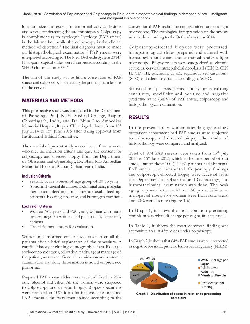
In Graph 1, it shows the most common presenting complaint was white discharge per vagina in 40% cases.
In Table 1, it shows the most common finding was acetowhite area in 43% cases under colposcopy.
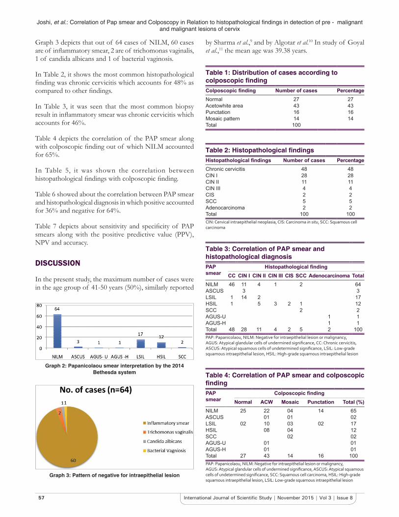
In Graph 2, it shows that 64% PAP smears were interpreted as negative for intraepithelial lesion or malignancy (NILM).
Graph 1: Distribution of cases in relation to presenting complaint
Joshi, et al.: Correlation of Pap smear and Colposcopy in Relation to histopathological findings in detection of pre - malignant and malignant lesions of cervix
57 International Journal of Scientific Study | November 2015 | Vol 3 | Issue 8
Graph 3 depicts that out of 64 cases of NILM, 60 cases are of inflammatory smear, 2 are of trichomonas vaginalis, 1 of candida albicans and 1 of bacterial vaginosis.
In Table 2, it shows the most common histopathological finding was chronic cervicitis which accounts for 48% as compared to other findings.
In Table 3, it was seen that the most common biopsy result in inflammatory smear was chronic cervicitis which accounts for 46%.
Table 4 depicts the correlation of the PAP smear along with colposcopic finding out of which NILM accounted for 65%.
In Table 5, it was shown the correlation between histopathological findings with colposcopic finding.
Table 6 showed about the correlation between PAP smear and histopathological diagnosis in which positive accounted for 36% and negative for 64%.
Table 7 depicts about sensitivity and specificity of PAP smears along with the positive predictive value (PPV), NPV and accuracy.
DISCUSSION
In the present study, the maximum number of cases were in the age group of 41-50 years (50%), similarly reported
by Sharma et al.,9 and by Algotar et al.10 In study of Goyal et al.,11 the mean age was 39.38 years.
Graph 2: Papanicolaou smear interpretation by the 2014 Bethesda system
Graph 3: Pattern of negative for intraepithelial lesion
Table 1: Distribution of cases according to colposcopic findingColposcopic finding Number of cases PercentageNormal 27 27Acetowhite area 43 43Punctation 16 16Mosaic pattern 14 14Total 100
Table 2: Histopathological findingsHistopathological findings Number of cases PercentageChronic cervicitis 48 48CIN I 28 28CIN II 11 11CIN III 4 4CIS 2 2SCC 5 5Adenocarcinoma 2 2Total 100 100CIN: Cervical intraepithelial neoplasia, CIS: Carcinoma in situ, SCC: Squamous cell carcinoma
Table 3: Correlation of PAP smear and histopathological diagnosisPAP smear
Histopathological findingCC CIN I CIN II CIN III CIS SCC Adenocarcinoma Total
NILM 46 11 4 1 2 64ASCUS 3 3LSIL 1 14 2 17HSIL 1 5 3 2 1 12SCC 2 2AGUS-U 1 1AGUS-H 1 1Total 48 28 11 4 2 5 2 100PAP: Papanicolaou, NILM: Negative for intraepithelial lesion or malignancy, AGUS: Atypical glandular cells of undermined significance, CC: Chronic cervicitis, ASCUS: Atypical squamous cells of undetermined significance, LSIL: Low‑grade squamous intraepithelial lesion, HSIL: High‑grade squamous intraepithelial lesion
Table 4: Correlation of PAP smear and colposcopic findingPAP smear
Colposcopic findingNormal ACW Mosaic Punctation Total (%)
NILM 25 22 04 14 65ASCUS 01 01 02LSIL 02 10 03 02 17HSIL 08 04 12SCC 02 02AGUS-U 01 01AGUS-H 01 01Total 27 43 14 16 100PAP: Papanicolaou, NILM: Negative for intraepithelial lesion or malignancy, AGUS: Atypical glandular cells of undermined significance, ASCUS: Atypical squamous cells of undetermined significance, SCC: Squamous cell carcinoma, HSIL: High‑grade squamous intraepithelial lesion, LSIL: Low‑grade squamous intraepithelial lesion
Joshi, et al.: Correlation of Pap smear and Colposcopy in Relation to histopathological findings in detection of pre - malignant and malignant lesions of cervix
58International Journal of Scientific Study | November 2015 | Vol 3 | Issue 8
In present study white discharge per vaginum (40%) was most common complaint similarly reported by Chaudhary et al.,6 39%.
In present study, the most common colposcopy finding was acetowhite area (43%), similar study reported by Krishnegowda and Veena12 22%.
On PAP smear 64% were reported NILM, and frank malignancy was reported as 2% cases, low-grade squamous intraepithelial lesion and high-grade squamous intraepithelial lesion was reported 17% and 12%, respectively (Graph 2 and Table 8).
A maximum number of cases on histopathological examination were those of infection among them majority had chronic cerivicitis (48%). Cervical Intraepithelial lesions were seen in 43 cases. CIN I were seen in 28 cases and CIN II and CIN III were reported 15%, and SCC and adenocarcinoma were reported 2% cases, respectively. Similar study reported by Bodal and Brar18 reported adenocarcinoma in 2% cases only (Tables 9 and 10).
14% cases were malignant in PAP smear turned out to malignant in histopathology showing strong correlation between PAP smear and histopathology (P < 0.0001) by Pearson correlation coefficient factor.
Some of the cases were obscured by blood and inflammation which were missed on PAP smear but proved to be malignant on histopathology.
Table 8 shows sensitivity, specificity, PPV and NPV compared with other studies.
Table 9 shows accuracy of PAP smear compared with other studies.
Table 10 shows correlation between PAP smear and colposcopy on comparison with other studies.
CONCLUSION
The result on current study support, PAP smear demonstrates of premalignant and malignant lesions,
Table 5: Correlation between histopathological finding with colposcopic findingHistopathological findings
Colposcopic findingNormal ACW Mosaic
patternPunctuation Total
Chronic cervicitis 25 17 02 09 51CIN I 04 12 04 06 26CIN II 07 03 01 11CIN III 03 03CIS 02 02SCC 03 05Adenocarcinoma 02 02Total 29 43 14 16 100CIN: Cervical intraepithelial neoplasia, CIS: Carcinoma in situ, SCC: Squamous cell carcinoma
Table 6: Correlation between PAP smear and histopathological diagnosisHistopathology Positive Negative TotalPAP smear
Positive 34 02 36Negative 18 46 64Total 52 48 100
PAP: Papanicolaou
Table 7: Sensitivity and specificity of PAP smearSensitivity TP/TP+FN 65.38%Specificity TN/TN+FP 95.83%PPV TP/TP+FP 94.44%NPV TN/TN+FN 71.86%Accuracy TP+TN/TP+TN+FP+FN 80.00%PAP: Papanicolaou, PPV: Positive predictive value, NPV: Negative predictive value
Table 8: On comparison with other studies the following results were obtainedStudy Sensitivity
(%)Specificity
(%)PPV (%)
NPV (%)
Present study (2015) 65.38 95.83 94.44 80Chaudhary et al.6 79.37 81.02 65.79 89.52Ashmita et al.13 90.24 72.73 66.6 86.54Mallur et al.14 80 81.54 66.67 89.83Pimple et al.15 74.5 92.9Goyal et al.10 86 40.5 66.18 66.18Kushtagi et al.16 78PPV: Positive predictive value, NPV: Negative predictive value
Table 9: Accuracy of PAP smearStudy Accuracy (%)Present study (2015) 80Chaudhary et al.6 80.5Ashmita et al.13 86.54Mallur et al.14 80Boicea et al.17 98.3PAP: Papanicolaou
Table 10: Correlation between PAP smear and colposcopy on comparison with other studiesStudy ASCUS
(%)AGUS
(%)LSIL (%)
HSIL (%)
SCC (%)
Present (2015) 3 (3.0) 2 (2.0) 17 (17) 12 (12) 2 (2.0)Goyal et al.10 9 (3.0) 1 (0.33) 17 (5.67) 1 (0.33)Chaudhary et al.6 17 (8.5) 10 (5.0) 5 (2.5) 2 (1.0)Sharma et al.9 1 (0.04) 214 (9.28) 5 (0.21)PAP: Papanicolaou, ASCUS: Atypical squamous cells of undetermined significance, SCC: Squamous cell carcinoma, AGUS: Atypical glandular cells of undermined significance, LSIL: Low‑grade squamous intraepithelial lesion, HSIL: High‑grade squamous intraepithelial lesion
Joshi, et al.: Correlation of Pap smear and Colposcopy in Relation to histopathological findings in detection of pre - malignant and malignant lesions of cervix
59 International Journal of Scientific Study | November 2015 | Vol 3 | Issue 8
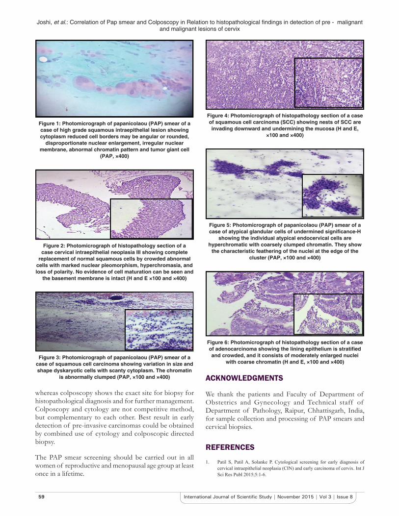
Figure 1: Photomicrograph of papanicolaou (PAP) smear of a case of high grade squamous intraepithelial lesion showing cytoplasm reduced cell borders may be angular or rounded,
disproportionate nuclear enlargement, irregular nuclear membrane, abnormal chromatin pattern and tumor giant cell
(PAP, ×400)
Figure 2: Photomicrograph of histopathology section of a case cervical intraepithelial neoplasia III showing complete
replacement of normal squamous cells by crowded abnormal cells with marked nuclear pleomorphism, hyperchromasia, and loss of polarity. No evidence of cell maturation can be seen and
the basement membrane is intact (H and E ×100 and ×400)
Figure 3: Photomicrograph of papanicolaou (PAP) smear of a case of squamous cell carcinoma showing variation in size and shape dyskaryotic cells with scanty cytoplasm. The chromatin
is abnormally clumped (PAP, ×100 and ×400)
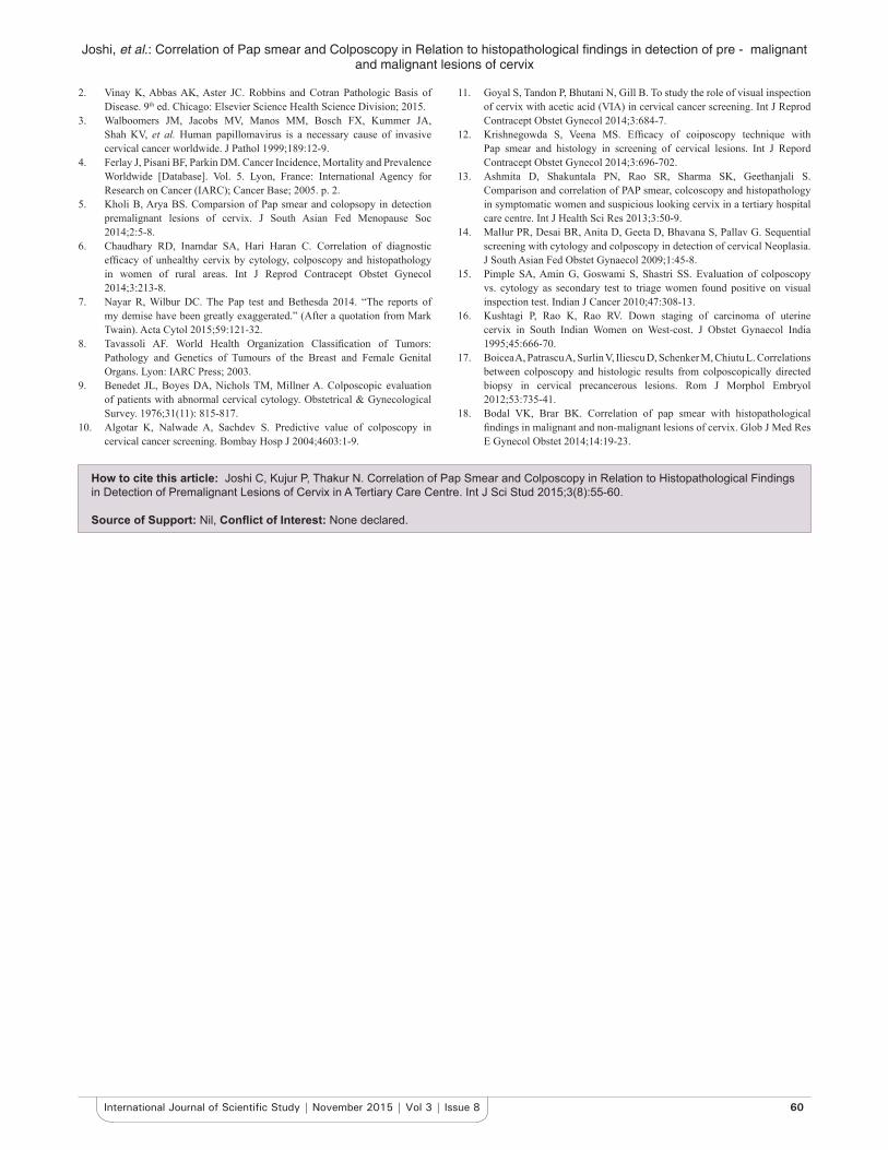
Figure 4: Photomicrograph of histopathology section of a case of squamous cell carcinoma (SCC) showing nests of SCC are invading downward and undermining the mucosa (H and E,
×100 and ×400)
Figure 5: Photomicrograph of papanicolaou (PAP) smear of a case of atypical glandular cells of undermined significance-H
showing the individual atypical endocervical cells are hyperchromatic with coarsely clumped chromatin. They show
the characteristic feathering of the nuclei at the edge of the cluster (PAP, ×100 and ×400)
Figure 6: Photomicrograph of histopathology section of a case of adenocarcinoma showing the lining epithelium is stratified and crowded, and it consists of moderately enlarged nuclei
with coarse chromatin (H and E, ×100 and ×400)
whereas colposcopy shows the exact site for biopsy for histopathological diagnosis and for further management. Colposcopy and cytology are not competitive method, but complementary to each other. Best result in early detection of pre-invasive carcinomas could be obtained by combined use of cytology and colposcopic directed biopsy.
The PAP smear screening should be carried out in all women of reproductive and menopausal age group at least once in a lifetime.
ACKNOWLEDGMENTS
We thank the patients and Faculty of Department of Obstetrics and Gynecology and Technical staff of Department of Pathology, Raipur, Chhattisgarh, India, for sample collection and processing of PAP smears and cervical biopsies.
REFERENCES
1. Patil S, Patil A, Solanke P. Cytological screening for early diagnosis of cervical intraepithelial neoplasia (CIN) and early carcinoma of cervix. Int J Sci Res Publ 2015;5:1-6.
Joshi, et al.: Correlation of Pap smear and Colposcopy in Relation to histopathological findings in detection of pre - malignant and malignant lesions of cervix
60International Journal of Scientific Study | November 2015 | Vol 3 | Issue 8
2. Vinay K, Abbas AK, Aster JC. Robbins and Cotran Pathologic Basis of Disease. 9th ed. Chicago: Elsevier Science Health Science Division; 2015.
3. Walboomers JM, Jacobs MV, Manos MM, Bosch FX, Kummer JA, Shah KV, et al. Human papillomavirus is a necessary cause of invasive cervical cancer worldwide. J Pathol 1999;189:12-9.
4. Ferlay J, Pisani BF, Parkin DM. Cancer Incidence, Mortality and Prevalence Worldwide [Database]. Vol. 5. Lyon, France: International Agency for Research on Cancer (IARC); Cancer Base; 2005. p. 2.
5. Kholi B, Arya BS. Comparsion of Pap smear and colopsopy in detection premalignant lesions of cervix. J South Asian Fed Menopause Soc 2014;2:5-8.
6. Chaudhary RD, Inamdar SA, Hari Haran C. Correlation of diagnostic efficacy of unhealthy cervix by cytology, colposcopy and histopathology in women of rural areas. Int J Reprod Contracept Obstet Gynecol 2014;3:213-8.
7. Nayar R, Wilbur DC. The Pap test and Bethesda 2014. “The reports of my demise have been greatly exaggerated.” (After a quotation from Mark Twain). Acta Cytol 2015;59:121-32.
8. Tavassoli AF. World Health Organization Classification of Tumors: Pathology and Genetics of Tumours of the Breast and Female Genital Organs. Lyon: IARC Press; 2003.
9. Benedet JL, Boyes DA, Nichols TM, Millner A. Colposcopic evaluation of patients with abnormal cervical cytology. Obstetrical & Gynecological Survey. 1976;31(11): 815-817.
10. Algotar K, Nalwade A, Sachdev S. Predictive value of colposcopy in cervical cancer screening. Bombay Hosp J 2004;4603:1-9.
11. Goyal S, Tandon P, Bhutani N, Gill B. To study the role of visual inspection of cervix with acetic acid (VIA) in cervical cancer screening. Int J Reprod Contracept Obstet Gynecol 2014;3:684-7.
12. Krishnegowda S, Veena MS. Efficacy of coiposcopy technique with Pap smear and histology in screening of cervical lesions. Int J Repord Contracept Obstet Gynecol 2014;3:696-702.
13. Ashmita D, Shakuntala PN, Rao SR, Sharma SK, Geethanjali S. Comparison and correlation of PAP smear, colcoscopy and histopathology in symptomatic women and suspicious looking cervix in a tertiary hospital care centre. Int J Health Sci Res 2013;3:50-9.
14. Mallur PR, Desai BR, Anita D, Geeta D, Bhavana S, Pallav G. Sequential screening with cytology and colposcopy in detection of cervical Neoplasia. J South Asian Fed Obstet Gynaecol 2009;1:45-8.
15. Pimple SA, Amin G, Goswami S, Shastri SS. Evaluation of colposcopy vs. cytology as secondary test to triage women found positive on visual inspection test. Indian J Cancer 2010;47:308-13.
16. Kushtagi P, Rao K, Rao RV. Down staging of carcinoma of uterine cervix in South Indian Women on West-cost. J Obstet Gynaecol India 1995;45:666-70.
17. Boicea A, Patrascu A, Surlin V, Iliescu D, Schenker M, Chiutu L. Correlations between colposcopy and histologic results from colposcopically directed biopsy in cervical precancerous lesions. Rom J Morphol Embryol 2012;53:735-41.
18. Bodal VK, Brar BK. Correlation of pap smear with histopathological findings in malignant and non-malignant lesions of cervix. Glob J Med Res E Gynecol Obstet 2014;14:19-23.
How to cite this article: Joshi C, Kujur P, Thakur N. Correlation of Pap Smear and Colposcopy in Relation to Histopathological Findings in Detection of Premalignant Lesions of Cervix in A Tertiary Care Centre. Int J Sci Stud 2015;3(8):55-60.
Source of Support: Nil, Conflict of Interest: None declared.