coronary artery spasm as a frequent cause of acute coronary syndrome: the caspar (coronary artery...
TRANSCRIPT

Cpceaebcef
Pa
FS
2
Journal of the American College of Cardiology Vol. 52, No. 7, 2008© 2008 by the American College of Cardiology Foundation ISSN 0735-1097/08/$34.00P
Coronary Artery Spasm as aFrequent Cause of Acute Coronary SyndromeThe CASPAR (Coronary Artery Spasm inPatients With Acute Coronary Syndrome) Study
Peter Ong, MD, Anastasios Athanasiadis, MD, Stephan Hill, MD, Holger Vogelsberg, MD,Matthias Voehringer, MD, Udo Sechtem, MD
Stuttgart, Germany
Objectives This study was conducted to clarify the incidence of coronary spasm in emergency patients with suspected acutecoronary syndrome (ACS) and acute chest pain at rest.
Background Chest pain at rest is a frequent symptom in the emergency room. Acute coronary syndrome is suspected in pa-tients with elevation of cardiac markers, ischemic electrocardiographic changes, or simply typical clinical symp-toms of unstable (usually resting) angina. However, of all patients with suspected ACS who undergo coronaryangiography, up to 30% have nonobstructed coronary arteries. We sought to clarify how many of these patientssuffer from coronary spasm as a possible cause of their chest pain.
Methods In a prospective study from June to December 2006, all patients with suspected ACS who underwent coronaryangiography and had no culprit lesion underwent intracoronary provocation with acetylcholine. The ACH testingwas considered positive at a vasoconstriction of �75% relative to the diameter after intracoronary nitroglycerinewhen the initially reported symptoms could be reproduced.
Results Of 488 consecutive patients, 138 had no culprit lesion (28%). Twenty-two were found to have another diagnosis. TheACH testing was performed in 86 of the remaining 116 patients. In 42 patients, coronary spasm was verified (49%).
Conclusions Every fourth patient with ACS had no culprit lesion. Coronary spasm could be documented in nearly 50% of thepatients tested by ACH. Coronary spasm is a frequent cause of ACS and should regularly be considered as a dif-ferential diagnosis. (J Am Coll Cardiol 2008;52:523–7) © 2008 by the American College of CardiologyFoundation
ublished by Elsevier Inc. doi:10.1016/j.jacc.2008.04.050
ndd
AeApwC(fatfsnc
hest pain at rest is one of the most frequent symptoms ofatients in the emergency room (1). The diagnosis of acuteoronary syndrome (ACS) is suspected in these patients,specially when typical electrocardiographic (ECG) changesnd/or elevation of cardiac markers can be detected. How-ver, of all patients who undergo coronary angiographyecause of suspected ACS, up to 30% have unobstructedoronary arteries or at least no culprit lesion that couldxplain the patient’s discomfort (2,3). Potential other causesor the clinical symptoms encompass a variety of diagnoses.
See page 528
ossible extracardial causes can be pulmonary, gastroesoph-geal, musculoskeletal, or psychiatric (4). Apart from coro-
rom the Department of Cardiology and Pulmology, Robert-Bosch-Krankenhaus,tuttgart, Germany.
(Manuscript received November 20, 2007; revised manuscript received April 24,
008, accepted April 29, 2008.
ary embolism or thrombosis (5), diseases such as myocar-itis (6) or tako-tsubo-cardiomyopathy (7) have also beenescribed in these patients.Coronary spasm is another well-defined mechanism for
CS causing ischemia at rest (8). However, only 2 studiesxamined the frequency of coronary spasm in patients withCS and unobstructed coronary arteries. Whereas a highrevalence (74%) of ergonovine-provoked coronary spasmas found in a Taiwanese population (8), a similar study inaucasian patients documented coronary spasm only in 16%
9). Therefore, we sought to prospectively investigate therequency of coronary spasm in patients with ACS includingll presentations (ST-segment elevation myocardial infarc-ion [STEMI], non–ST-segment elevation myocardial in-arction [NSTEMI], unstable angina pectoris [UAP]) whohowed no culprit lesion or hemodynamically relevant ste-osis (�50%) at coronary angiography. Intracoronary provo-ation for coronary spasm was done using acetylcholine
ACH) (10).
m(cbmw1ttistj
tcprsi
QatvawgtSaFquoe(
R
BidcohhTgAcm0Ptc(t
524 Ong et al. JACC Vol. 52, No. 7, 2008Coronary Spasm as Frequent Cause of ACS August 12, 2008:523–7
Methods
Patient population and ACHtest. In a prospective study fromJune to December 2006, 488consecutive patients who under-went coronary angiography be-cause of ACS were registered andwritten informed consent for anadditional intracoronary ACHprovocation was obtained. Thestudy complied with the Decla-ration of Helsinki. We definedACS as acute chest pain (i.e.,chest pain at rest �20 minwithin the last 48 h) togetherwith ECG changes suggesting
yocardial ischemia and/or elevation of cardiac markers11). In case of detection of a culprit lesion, percutaneousoronary intervention was performed or coronary arteryypass grafting or conservative treatment was recom-ended. If coronary angiography revealed no culprit lesion,e conducted an ACH test. Incremental doses of 2, 20, and00 �g ACH were injected into the left coronary artery viahe diagnostic catheter for 3 min each (10,12). If the test inhe left coronary artery was negative, 80 �g ACH wasnjected into the right coronary artery. When coronarypasm was demonstrated, 0.2 mg of Perlinganit (glycerol-rinitrate, Schwarz Pharma, Monheim, Germany) was in-ected into the responsible vessel.
The ACH test was performed either at primary cathe-erization or in a second session depending on the patient’sondition and the administered medication. It was noterformed in one of the following conditions: patientefusal, suspected myocarditis, tako-tsubo-cardiomyopathy,evere chronic obstructive pulmonary disease, severe renalnsufficiency, allergy to iodinated contrast media.
Abbreviationsand Acronyms
ACH � acetylcholine
ACS � acute coronarysyndrome
ECG � electrocardiogram
LCA � left coronary artery
NSTEMI � non–ST-segmentelevation myocardialinfarction
STEMI � ST-segmentelevation myocardialinfarction
UAP � unstable anginapectoris
Patient Main Clinical Characteristics
Table 1 Patient Main Clinical Characteristic
All Patients
n 488
Gender, male 315 (64.5%)
Age, yrs (mean � SD) 66 (�12)
Risk factors
Hypertension 310 (70.6%)
Diabetes mellitus 126 (28.8%)
Hypercholesterolemia 262 (59.8%)
Smokers 118 (26.7%)
Obesity 69 (15.8%)
Positive family history of CVD 141 (32.3%)
Cardiac markers and LVEF
LVEF, % (IQR) 61 (47–71)
TnI, �g/l, n � 0.16 (IQR) 1.70 (0.04–22.01)
CK, U/l, n � 180 (IQR) 143 (69–599)
BNP, pg/ml, n � 80 (IQR) 193 (67–600)
BNP � B-type natriuretic peptide; CK � creatine kinase; CVD � cardiovasculafraction; NS � not significant; TnI � troponin I.
uantitative analysis. All ACH tests were quantitativelynalyzed with QCA-CMS 7.0 (Medis-Software, Leiden,he Netherlands). The ACH test was positive when theisual findings at coronary angiography and the quantitativenalysis confirmed a vasoconstriction of �75% comparedith the relaxed state after intracoronary administration oflyceroltrinitrate together with a reproduction of the pa-ient’s initial symptoms (10,13,14).tatistics. Results are expressed as mean � standard devi-tion. The t test was used to compare continuous variables.or values without normal distribution, median and inter-uartile ranges are stated and Mann-Whitney U test wassed. The chi-square test was used for categorical andrdinal variables. A two-tailed p value �0.05 was consid-red significant. Data analysis was done with SPSS 14.0SPSS Inc., Chicago, Illinois).
esults
aseline characteristics. A total of 488 patients werencluded (Table 1). They were divided into 2 groupsepending on the presence or absence of a culprit lesion atoronary angiography. Patients with culprit lesion werelder, were more often male, and showed significantlyigher levels of cardiac markers. In addition, these patientsad a significantly lower left ventricular ejection fraction.here was a higher prevalence of diabetes mellitus in theroup with culprit lesion (p � 0.002). The distribution ofCS in the 2 groups is shown in Figure 1. In patients with
ulprit lesion, STEMI and NSTEMI were significantlyore prevalent than in patients without culprit lesion (p �
.001).atients without culprit lesion. We identified 138 pa-
ients without culprit lesion. In 22 patients, the diagnosisould be determined before the ACH test. Nine patients6.5% of all patients without culprit lesion) had myocarditishat was diagnosed by cardiac magnetic resonance imaging
t Lesion Present Culprit Lesion Absent p Value
350 138
(71.1%) 66 (47.8%) �0.0001
(�12) 62 (�13) �0.0001
(71.9%) 87 (67.4%) NS
(33%) 24 (18.6%) 0.002
(61.2%) 73 (56.6%) NS
(28.7%) 28 (21.9%) NS
(15.5%) 21 (16.4%) NS
(28.1%) 54 (42.2%) 0.001
(44–69) 59 (39–69) �0.001
(0.49–51.76) 0.02 (0.02–0.28) �0.001
(115–714) 83 (56–128) �0.001
(80–1018) 59 (25–178) �0.001
s
Culpri
249
67
223
102
189
90
48
87
56
6.41
332
247
r diseases; IQR � interquartile range; LVEF � left ventricular ejection
ahapwt
Apsulp2A
ewp(tArwtdp3oiEbEtb
D
AhprostpA7iocc
CC
A
525JACC Vol. 52, No. 7, 2008 Ong et al.August 12, 2008:523–7 Coronary Spasm as Frequent Cause of ACS
nd later confirmed by endomyocardial biopsy, 7 patientsad tako-tsubo-cardiomyopathy (5%), 1 patient had ancute bleeding, and 1 had pulmonary embolism. Four otheratients already had a positive previous ACH test result andere hence assumed to have coronary spasm as the cause of
heir recurrent chest pain.Eighty-six of the remaining 116 patients underwent the
CH test (74%). In 30 patients the ACH test could not beerformed because of the abovementioned reasons. Coronarypasm was verified in 42 patients (49% of the patients whonderwent the test and 30% of all patients without culpritesion) (Table 2). The remaining 44 patients showed differentatterns of reproduction of symptoms and vasoreaction (Fig.). Of all 86 patients without culprit lesion who underwentCH testing, 7 patients showed significant troponin I
Figure 1 Presentation of ACS (n � 488)
Distribution of different presentations of ACS according to presence orabsence of culprit lesion. ACS � acute coronary syndrome; NSTEMI � non–ST-segment elevation myocardial infarction; STEMI � ST-segment elevation myo-cardial infarction; UAP � unstable angina pectoris.
linical Characteristics of Patients Withoutulprit Lesion Who Had an Acetylcholine Test
Table 2 Clinical Characteristics of Patients WithoutCulprit Lesion Who Had an Acetylcholine Test
SpasmPresent
SpasmAbsent
pValue
n 42 44
Gender, male 21 (50%) 25 (56.8%) NS
Age, yrs (median) 60 (�12) 61 (�11) NS
Risk factors
Hypertension 27 (64.2%) 29 (65.9%) NS
Diabetes mellitus 8 (19%) 7 (15.9%) NS
Hypercholesterolemia 26 (61.9%) 22 (50%) NS
Smokers 10 (23.8%) 8 (18.1%) NS
Obesity 6 (14.2%) 6 (13.6%) NS
Positive family history of CVD 22 (52.3%) 21 (47.7%) NS
Cardiac markers and LVEF
LVEF, % (IQR) 70 (62–79) 71% (62–77) NS
TnI, �g/l, n � 0.16 (IQR) 0.02 (0.02–0.03) 0.02 (0.02–0.02) NS
CK, U/l, n � 180 (IQR) 73.5 (55.5–134.5) 79 (49–120) NS
BNP, pg/ml, n � 80 (IQR) 48 (23–68) 60 (22.5–176) NS
ebbreviations as in Table 1.
levation. Coronary spasm was found in 4 (57%) of them,hich was not statistically different from the 43% (3 of 7)atients who had coronary spasm without marker elevationp � 0.696). Thus, there was no association betweenroponin levels and spasm.
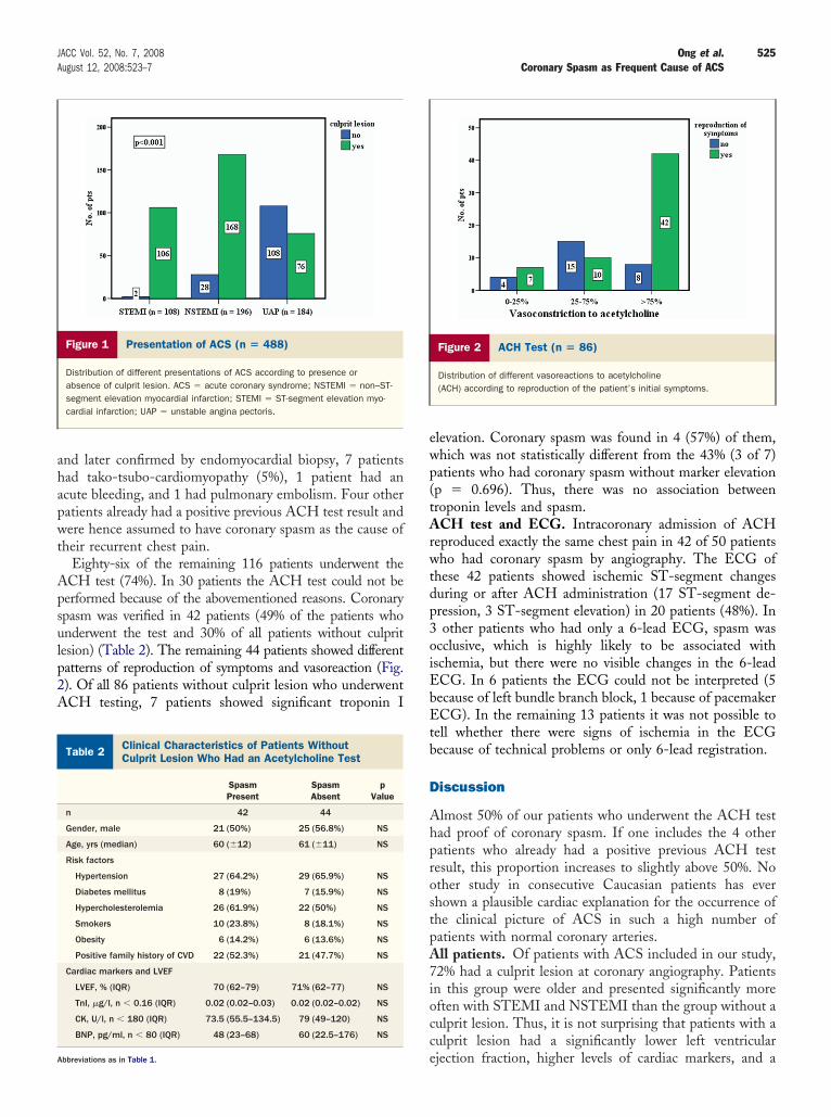
CH test and ECG. Intracoronary admission of ACHeproduced exactly the same chest pain in 42 of 50 patientsho had coronary spasm by angiography. The ECG of
hese 42 patients showed ischemic ST-segment changesuring or after ACH administration (17 ST-segment de-ression, 3 ST-segment elevation) in 20 patients (48%). Inother patients who had only a 6-lead ECG, spasm was
cclusive, which is highly likely to be associated withschemia, but there were no visible changes in the 6-leadCG. In 6 patients the ECG could not be interpreted (5ecause of left bundle branch block, 1 because of pacemakerCG). In the remaining 13 patients it was not possible to
ell whether there were signs of ischemia in the ECGecause of technical problems or only 6-lead registration.
iscussion
lmost 50% of our patients who underwent the ACH testad proof of coronary spasm. If one includes the 4 otheratients who already had a positive previous ACH testesult, this proportion increases to slightly above 50%. Nother study in consecutive Caucasian patients has everhown a plausible cardiac explanation for the occurrence ofhe clinical picture of ACS in such a high number ofatients with normal coronary arteries.ll patients. Of patients with ACS included in our study,2% had a culprit lesion at coronary angiography. Patientsn this group were older and presented significantly moreften with STEMI and NSTEMI than the group without aulprit lesion. Thus, it is not surprising that patients with aulprit lesion had a significantly lower left ventricular
Figure 2 ACH Test (n � 86)
Distribution of different vasoreactions to acetylcholine(ACH) according to reproduction of the patient’s initial symptoms.
jection fraction, higher levels of cardiac markers, and a

hlvpdPDwetTitiHcpf
vvAvAtCcww
ciatavas0fi(cp1i
iCppcpthCm
bbcl
ptnoc4Mwmlaiip
letAtdEassFamvoaapsSmcaprfwp
C
Ecsf
526 Ong et al. JACC Vol. 52, No. 7, 2008Coronary Spasm as Frequent Cause of ACS August 12, 2008:523–7
igher prevalence of diabetes mellitus. Patients with culpritesions were more often male, confirming a previous obser-ation of a higher prevalence of women in the group ofatients with ACS but without significant coronary arteryisease (15).atients without culprit lesion. In contrast to our results,a Costa et al. (9) found that only 16% of French patientsith myocardial infarction and normal coronary arteries had
rgonovine-induced coronary spasm. A potential explana-ion for this discrepancy is the different inclusion criteria.heir patients had to have chest pain, ECG changes, and
ncreased plasma enzyme activity, whereas our patients hado have chest pain at rest �20 min and only one of eitherschemic ECG changes or elevation of cardiac markers.
owever, after matching our patients with the inclusionriteria of Da Costa et al. (9), we still found 50% of ouratients with coronary spasm, which is 3 times morerequent than in their study.
Could the different method of application used to pro-oke coronary spasm explain the different proportions ofasospasm in the study by Da Costa et al. (9) and our study?pplication of ergonovine in their patients was done intra-
enously, whereas we used intracoronary application ofCH. This methodical difference could have contributed to
he lower percentage of positive tests in the study by Daosta et al. (9). Goto et al. (16) showed that in patients with
hest pain who underwent intravenous ergonovine testingith a negative result, additional intracoronary provocationith ACH could reveal coronary spasm in 79%.In contrast to the Da Costa et al. (9) findings and more
ongruent with our observations, coronary spasm is anmportant differential diagnosis in Asian patients with ACSnd unobstructed coronary arteries. Miwa et al. (17) studiedhe role of coronary spasm in Japanese patients with stablend unstable angina. Intracoronary infusion of ACH pro-oked coronary spasm in 93% of patients with unstablengina, which was significantly higher than in patients withtable angina (20%), who had organic stenoses �50% (p �.01). However, 30% of their unstable patients also hadxed coronary stenoses (�50%) (17). Finally, Wang et al.8) found in a Taiwanese population a high frequency oforonary spasm, especially in patients with troponin I–ositive ACS and insignificant coronary artery disease. In7 of 23 patients (74%), coronary spasm was provoked byntracoronary administration of ergonovine (8).
Although we found a higher incidence of coronary spasmn our population compared with a previous study inaucasian patients, we could not reproduce the high pro-ortion of patients with coronary spasm in the Asianopulations with ACS. Because the higher prevalence oforonary vasospasm has also been documented in Asianopulations with stable effort angina (18), this finding leadso the assumption that racial differences could explain theigher prevalence of spasm in the Asian populations.linically it has been observed that Japanese patients have a
ore diffuse coronary hyperreactivity with an increased aasal tonus. This might be a reason why coronary spasm cane observed in Japanese patients more often than in Cau-asians, in whom coronary spasm is reported to be of a moreocalized nature (18).
One could therefore speculate that coronary spasm in ouratients might be a response to some local arterial injuryhat could vanish after healing. Indeed, patients with coro-ary spasm had a significantly shorter time interval betweennset of symptoms and ACH test than those withoutoronary spasm (mean: 47 h, interquartile range: 24 h to8 h vs. mean: 65 h, interquartile range: 24 h to 96 h,ann-Whitney U test). However, in the 16 patients in
hom we performed ACH testing in all 3 coronaries,ultivessel spasm was found in 69%. This indicates that a
ocal mechanism may not be the only cause for the overre-ction of the coronary vessels. One possible transient injurys impairment of endothelial function by virus-inducednflammation, which was recently shown to occur witharvovirus B19 infection (19).Because the clinical situation in which the chest pain
eading to emergent presentation of the patient cannot bexactly reproduced, some uncertainty about the causal rela-ionship between the observation of coronary spasm duringCH administration and the clinical event remains. We
hink, however, that the combination of angiographicallyocumented spasm combined with signs of ischemia in theCG plus exact reproduction of the symptoms leading to
dmission (i.e., chest pain, 42 of 50 patients with coronarypasm in the ACH test) strongly suggests a causal relation-hip between spasm and the clinical occurrence of ACS.luctuation of symptoms is well known in patients withngina caused by epicardial coronary stenosis. Hence, oneight expect that the same might be true in coronary
asospasm. Therefore, asymptomatic coronary spasm in 8 ofur patients may still be related to the acute chest pain, but
causal relationship is less convincing. Nevertheless,symptomatic coronary vasospasm may be a clinically im-ortant finding because serious complications may occur inuch a setting (20).tudy limitations. We did not evaluate coagulation abnor-alities that were previously reported in some cases as a
ause of ACS (21). However, our aim was not to providell-encompassing information about possible causes of chestain in patients without culprit lesion but to focus on theole of coronary spasm reproduced by ACH testing. There-ore, other possible causes of chest pain such as myocarditisere not systematically ruled out, although in a subgroup ofatients this may have been the underlying cause.
onclusions
very fourth patient with ACS had no culprit lesion atoronary angiography. In these patients epicardial coronarypasm could be documented in 50%. Coronary spasm is arequent cause of ACS and should regularly be considered as
differential diagnosis. Intracoronary provocation with

Art
ATtm
RRmU
R
1
1
1
1
1
1
1
1
1
1
2
2
K
527JACC Vol. 52, No. 7, 2008 Ong et al.August 12, 2008:523–7 Coronary Spasm as Frequent Cause of ACS
CH is the gold standard for establishing the diagnosis. Iteliably and safely detects coronary spasm to guide institu-ion of appropriate therapy.
cknowledgmentshe authors greatly appreciate the support of the nurses and
echnicians in the catheterization laboratories and all staffembers.
eprint requests and correspondence: Dr. Udo Sechtem,obert-Bosch-Krankenhaus, Department of Cardiology and Pul-ology, Auerbachstrasse 110, 70376 Stuttgart, Germany. E-mail:[email protected].
EFERENCES
1. Lee TH, Goldman L. Evaluation of the patient with acute chest pain.N Engl J Med 2000;342:1187–95.
2. Hochman JS, Tamis JE, Thompson TD, et al. Sex, clinical presentation,and outcome in patients with acute coronary syndromes. Global Use ofStrategies to Open Occluded Coronary Arteries in Acute CoronarySyndromes IIb Investigators. N Engl J Med 1999;341:226–32.
3. Yang EH, Lerman A. Angina pectoris with a normal coronaryangiogram. Herz 2005;30:17–25.
4. Cayley WE. Diagnosing the cause of chest pain. Am Fam Physician2005,15;72:2012–21.
5. Tun A, Khan IA. Myocardial infarction with normal coronary arteries:the pathologic and clinical perspectives. Angiology 2001;52:299–304.
6. Kuhl U, Pauschinger M, Bock T, et al. Parvovirus B19 infectionmimicking acute myocardial infarction. Circulation 2003;108:945–50.
7. Kurisu S, Sato H, Kawagoe T, et al. Tako-tsubo-like left ventriculardysfunction with ST-segment elevation: a novel cardiac syndromemimicking acute myocardial infarction. Am Heart J 2002;143:448–55.
8. Wang CH, Kuo LT, Hung MJ, et al. Coronary vasospasm as apossible cause of elevated cardiac troponin I in patients with acutecoronary syndrome and insignificant coronary artery disease. AmHeart J 2002;144:275–81.
9. Da Costa A, Isaaz K, Faure E, et al. Clinical characteristics, aetio-logical factors and long-term prognosis of myocardial infarction withan absolutely normal coronary angiogram; a 3-year follow-up study of
91 patients. Eur Heart J 2001;22:1459–65. a0. Sueda S, Ochi N, Kawada H, et al. Frequency of provoked coronaryvasospasm in patients undergoing coronary arteriography with spasmprovocation test of acetylcholine. Am J Cardiol 1999;83:1186–90.
1. Bertrand ME, Simoons ML, Fox KA, et al. Task Force on theManagement of Acute Coronary Syndromes of the European Societyof Cardiology. Management of acute coronary syndromes in patientspresenting without persistent ST-segment elevation. Eur Heart J2002;23:1809–40.
2. ENCORE Investigators. Effect of nifedipine and cerivastatin oncoronary endothelial function in patients with coronary artery disease:the ENCORE I Study (Evaluation of Nifedipine and Cerivastatin OnRecovery of coronary Endothelial function). Circulation 2003;107:422–8.
3. Mohri M, Koyanagi M, Egashira K, et al. Angina pectoris caused bycoronary microvascular spasm. Lancet 1998;351:1165–9.
4. Bertrand ME, LaBlanche JM, Tilmant PY, et al. Frequency ofprovoked coronary arterial spasm in 1089 consecutive patients under-going coronary arteriography. Circulation 1982;65:1299–306.
5. Patel MR, Chen AY, Peterson ED, et al. Prevalence, predictors, andoutcomes of patients with non-ST-segment elevation myocardialinfarction and insignificant coronary artery disease: results from theCan Rapid risk stratification of Unstable angina patients SuppressADverse outcomes with Early implementation of the ACC/AHAGuidelines (CRUSADE) initiative. Am Heart J 2006;152:641–7.
6. Goto A, Ito S, Kondo H, et al. Evaluation of adjunctive intracoronaryadministration of acetylcholine following intravenous infusion of ergono-vine to provoke coronary artery spasm. J Cardiol 1999;34:309–16.
7. Miwa K, Fujita M, Ejiri M, et al. The sensitivity of intracoronaryinjection of acetylcholine in inducing coronary spasm differs in patientswith stable and unstable angina. Int J Cardiol 1992;36:329–39.
8. Beltrame JF, Sasayama S, Maseri A. Racial heterogeneity in coronaryartery vasomotor reactivity: differences between Japanese and Cauca-sian patients. J Am Coll Cardiol 1999;33:1442–52.
9. Yilmaz A, Mahrholdt H, Athanasiadis A, et al. Coronary vasospasm asthe underlying cause for chest pain in patients with PVB19-myocarditis. Heart 2008; Jan 29 [Epub ahead of print].
0. Nishizaki M, Arita M, Sakurada H, et al. Polymorphic ventriculartachycardia in patients with vasospastic angina—clinical and electro-cardiographic characteristics and long-term outcome. Jpn Circ J2001;65:519–25.
1. Da Costa A, Tardy-Poncet B, Isaaz K, et al. Prevalence of factor VLeiden (APCR) and other inherited thrombophilias in young patientswith myocardial infarction and normal coronary arteries. Heart 1998;80:338–40.
ey Words: acute coronary syndrome y coronary artery spasm y
cetylcholine y culprit lesion y chest pain.