core protein-dependence of epimerization of … · dcn, a small leucine-rich proteoglycan carrying...
TRANSCRIPT
Core protein-dependence of epimerization of glucuronosyl residues in
galactosaminoglycans∗
Daniela G. Seidler1,2, Egon Breuer1,2, K. Jane Grande-Allen3, Vincent C. Hascall3,
and Hans Kresse1,3
From the Institute of Physiological Chemistry and Pathobiochemistry1, University of Münster,
48149 Münster, Germany and the 3Department of Biomedical Engineering, Cleveland Clinic
Foundation, Cleveland, OH 44106, USA
Running title: Epimerization of Glucuronosyl Residues
2These authors contributed equally to this work
Correspondence: Dr. Hans Kresse, Institute of Physiological Chemistry and Pathobiochemistry, Waldeyerstr. 15, D-48149 Münster, Germany Phone: +49-251-8355581 Fax: +49-251-8355596 E-mail: [email protected]
∗ This work was supported in part by the Deutsche Forschungsgemeinschaft (SFB 496, Project A6)
Copyright 2002 by The American Society for Biochemistry and Molecular Biology, Inc.
JBC Papers in Press. Published on August 30, 2002 as Manuscript M208442200 by guest on O
ctober 5, 2018http://w
ww
.jbc.org/D
ownloaded from

2
Synopsis
Chondroitin sulfate and dermatan sulfate proteoglycans are distinguished by
differences in their proportion of D-glucuronosyl and L-iduronosyl residues, the latter being
formed by chondroitin-glucuronate 5-epimerase during or after glycosaminoglycan chain
polymerization. To investigate the influence of the core protein on the extent of epimerization,
we expressed chimeric proteins in 293 HEK cells constructed from intact or modified Met-1 -
Gln-153 of decorin (DCN), which normally has a single dermatan sulfate chain at Ser 34, in
combination with intact or modified Leu-241 - Ser-353 of CSF-1, which has a chondroitin
sulfate attachment site at Ser-309. Transfected DCNM1-Q153, like full length DCN, contained
~20 % L-iduronate. Conversely, transfected CSF-1L242-S353, attached C-terminally on the DCN
prepropeptide, contained almost exclusively D-glucuronate. Transfected intact chimeric
DCNM1-Q153 – CSF-1L241-S353, with 2 glycosaminoglycan chains also contained almost
exclusively D-glucuronate in chains at both sites, as did chimeras in which alanine was
substituted for serine at either of the glycosaminoglycan attachment sites. Nevertheless,
undersulfated intact chimeric proteoglycan was an effective substrate for epimerization of
glucuronate to iduronate residues when incubated with microsomal proteins and 3'-
phosphoadenylylphosphosulfate. C-Terminal truncation constructs were prepared from the
full length chimera with an alanine substitution at the CSF-1 glycosaminoglycan attachment
site. Transfected truncations retaining the alanine blocked site contained chains with
essentially only glucuronate, while those further truncated by 49 or more amino acids and
missing the modified attachment site contained chains with ~15% iduronate. This 49 amino
acid region contains a 7 amino acid motif that appears to be conserved in several chondroitin
sulfate proteoglycans. The results are consistent with a model in which the core protein,
possibly via this motif, is responsible for routing to subcellular compartments with or without
sufficient access to chondroitin-glucuronate 5-epimerase for addition of chains with or
without iduronate residues respectively.
by guest on October 5, 2018
http://ww
w.jbc.org/
Dow
nloaded from
3
Introduction
The assembly of glycosaminoglycan chains on core proteins is a multistep process that
begins most often with the formation of a specific carbohydrate-protein linkage region and
continues with the alternating addition of D-GlcUA1 and either D-GlcNAc or D-GalNAc
followed by sulfation and additional polymer modifications (see 1,2 for reviews). The two
types of galactosaminoglycans, chondroitin sulfate and dermatan sulfate, are primarily
distinguished by the absence or presence of L-IdoUA residues, respectively. The C-5
inversion of D-GlcUA to L-IdoUA occurs on the polymer level (3) and involves an abstraction
of the C-5 hydrogen of the uronosyl residues (4). Sulfation at carbon atom 4 of adjacent
galactosamine residues prevents back-epimerization to D-GlcUA (3,5). The responsible
enzyme, chondroitin-glucuronate 5-epimerase (EC 5.3.1.19), has not yet been characterized at
the molecular level. However, a two-base reaction mechanism was postulated, involving a
monoprotic L-ido-specific base and a polyprotic D-gluco-specific base, during the action of
the enzyme on an E. coli-derived capsular polysaccharide (6). The C-5 epimerization of the
�1-4 linked GlcUA residues in heparan sulfate is accomplished by the action of a different
enzyme (7), named heparosan N-sulfate-glucuronate 5-epimerase (EC 5.3.1.17), which has
been characterized by cDNA cloning (8) and kinetic studies (9).
Iduronic acid-containing glycosaminoglycans have a greater conformational flexibility
than glycosaminoglycans composed only of GlcUA as the hexuronic acid moiety (10-14).
Additionally, L-IdoUA residues are more frequently 2-O-sulfated than D-GlcUA residues,
allowing the appearance of clustered, highly charged domains along the glycosaminoglycan
chain. Hence, the degree of epimerization is of functional relevance. For example, only
dermatan sulfate and not chondroitin sulfate can stimulate thrombin inhibition by heparin
1 Non-standard abbreviations are: DCN, decorin; GlcUA, glucuronic acid; IdoUA, iduronic acid; PAGE, polyacrylamide gel electrophoresis.
by guest on October 5, 2018
http://ww
w.jbc.org/
Dow
nloaded from

4
cofactor II (15-17), and an altered dermatan sulfate structure may be of pathophysiological
importance during the development of arteriosclerosis (18).
Various cell types synthesize simultaneously proteoglycans of the lectican family
(reviewed in 19), which carry multiple chondroitin sulfate chains, and proteoglycans of the
small leucine-rich protein family (reviewed in 20). DCN and biglycan are the most intensely
studied members of the latter family. They carry either only one or two galactosaminoglycan
chains, which, with some exceptions, are rich in L-IdoUA residues (21,22). Whereas the
variability of epimerization within the family of small leucine-rich proteoglycans may be
explained by differences in the activity of chondroitin-glucuronate 5-epimerase, the reasons
for the different extent of epimerization of proteoglycans being synthesized by the very same
cell are not yet understood. Several not mutually exclusive hypotheses may explain the
experimental findings. The respective core proteins may influence epimerase activity, or may
alter the degree of modification of the linkage region components GlcUA-Gal-Gal-Xyl by
phosphorylation and/or sulfation, which may be important for subsequent biosynthetic events
(23). Alternatively, different proteoglycans may encounter different multi-component
complexes of synthesizing enzymes with different epimerase activities along their transport
from the endoplasmic reticulum to the trans-Golgi network. To address these possibilities, we
generated chimeric core proteins composed of truncated forms of two proteoglycans, each
characterized by the presence of a single glycosaminoglycan chain. The N-terminal part of
DCN, a small leucine-rich proteoglycan carrying a dermatan sulfate chain, was linked with
the central portion of the proteoglycan form of CSF-1 (M-CSF), which is characterized by the
presence of a single chondroitin sulfate chain (24-27). Additional mutants, truncated in the
CSF-1 moiety were also generated. It was shown previously that individual MG-63
osteosarcoma cells can express simultaneously DCN and the proteoglycan form of CSF-1,
which differ greatly in their D-GlcUA/ L-IdoUA ratios (21).
by guest on October 5, 2018
http://ww
w.jbc.org/
Dow
nloaded from
5
EXPERIMENTAL PROCEDURES
Construction of chimeric cDNA - First, the cDNA for DCNM1-Q153 including 120 bp of
the 5'-untranslated sequence and a stop codon was amplified by PCR from full-length human
cDNA (28) in pGem3Z, by using the primer pair 5’-TAATACGACTCACTATAG-3’ (T7-
forward primer) and 5'-CTGAAGAGTTTTGGGCATTTTTTC-3' (reverse and complement
primer). Similarly, the cDNA for L241-V554 of CSF-1 in pGem-T (27) including a
downstream located part of the vector sequence was amplified by using the T7-forward
primer mentioned above and the reverse and complement primer 5'-
CTGCACACAGTGGATCCAGG-3'. The purified amplicons were ligated after treatment
with T4-kinase and used as template for the amplification of the chimeric cDNA with the
primer pair 5'-ATGAAGGCCACTATCATCCTC-3' and 5'-
CGTCTATGATGCAGGGAGTGGAGA-3', thereby introducing a stop codon after the codon
for S353 of CSF-1. The PCR product was directly ligated via its A overhangs into the pCR3-
Uni vector (Invitrogen) and used to transform competent E. coli DH5.� 7KH F'1$V IRU
truncated DCNM1-Q153, truncated and mutant chimeras were generated by using the
QuikChange Site Directed Mutagenesis Kit (Stratagene), with the cDNA of the chimera
serving as template and HPSF quality oligonucleotides (MWG Biotech) as primers. The
method uses forward and reverse primers that carry the base exchange in the center of their
sequence and anneal to identical positions of the plasmid. High fidelity Pfu Turbo DNA
polymerase was used for amplification. The PCR conditions were 30 sec at 94 °C, 1 min at 55
°C and 14 min at 69 °C. After 30 PCR cycles, the methylated template was degraded by DpnI,
and XL1-Blue supercompetent cells were transformed without prior ligation of the PCR
product. The forward primers used to introduce stop codons or amino acid exchanges in the
original chimera (full-length DCN cDNA in case of DCNM1-Q153); respectively, were as
follows. Generation of DCNM1-Q153: 5´GCCCAAAACTCTTCAGTAGCTGCGTGCCCATG;
mutation S34A of the DCN moiety to eliminate the first glycosaminoglycan attachment site:
by guest on October 5, 2018
http://ww
w.jbc.org/
Dow
nloaded from

6
5’CTAGAAGATGAGGCTGCTGGGATAGGCCCAG-3; mutation S309A of the CSF-1
sequence (designated S222A in the chimera) to remove the second glycosaminoglycan
attachment site: 5’-CCCAGAAGAAGCCGCTGGAGAGGCCAG -3'. Truncated chimeras
were generated by using the cDNA of the last mentioned chimera as template. The respective
forward primers were as follows. Truncation of the CSF-1 moiety at K250/K163 (the first
number of the amino acid indicates the number in the full-length sequence of CSF-1 and the
second one the number appearing in the chimeric protein): 5’-
GCAGTGCCAAGTAGCGGCCACCCAG-3'; truncation at V269/V182: 5’-
AGACCCCAGTTGTCTAGGACAGCACCATC-3'; at G290/G203: 5’-
GGGGCCTCCAACCCCTAGATGGAGGATATTCTT-3'; at P318/P231 5’-
GATTCCCGTACCCTAGGGGACAGAGCTTTC-3'; at G329/G242 5’-
CTCCAGGCCAGGATAGGGCAGCATGCAGAC-3', and at S341/S254 5’-
CCCGCCAGACCCAGCTAATTCCTCTCAGCATC-3'. For investigating the
glycosaminoglycan chain of an isolated CSF-1L241-S353 fragment, the corresponding cDNA
sequence was fused to the prepro cDNA sequence of DCN to attach a proper secretory signal.
First, the prepro sequence of DCN was amplified with the primer pair 5’-
ATGAAGGCCACTATCATCCTCC-3’ and 5’-TTCTAGCATAAAGTCAAATAAGCCTCT-
3’. For amplifying the CSF-1 moiety the forward primer comprised at its 5’end 28 nt of the
3’sequence of the DCN prepro sequence: 5’-
GAGAGGCTTATTTGACTTTATGCTAGAACTGCACACAGTGGATCCAG-3’. The
second primer was 5’-TGATGCAGGGAGTGGAGAA-3’. Both amplicons served as
template in a subsequent PCR reaction in which the primers were the forward primer for the
amplification of prepro DCN and the reverse and complement primer the one used for CSF-1
amplification. Upon purification by agarose gel electrophoresis, the PCR product was ligated
into pGemT and subcloned into the expression vector pcDNA3.1 (Invitrogen). Sequencing of
all cDNAs employed in the present study verified their predicted structure and the absence of
by guest on October 5, 2018
http://ww
w.jbc.org/
Dow
nloaded from

7
unwanted mutations. Table I shows the protein sequence derived from the individual cDNA
constructs.
Expression of Recombinant Proteins – Cultured human 293 embryonic kidney cells,
maintained in Eagle’s Minimum Essential Medium supplemented with non-essential amino
acids, 10 % fetal bovine serum and antibiotics, were transfected by using Lipofectin or
Lipofectamine 2000 (GibcoBRL) according to the instructions of the manufacturer. The cells
were selected for neomycin resistance by adding 750 µg/ml G418 (Life Technologies) and
maintained in culture without further subcloning. The expression of human DCN cDNA in
293 cells has been described (29).
Metabolic Labeling and Proteoglycan Isolation - Subconfluent 293 cells were
metabolically labelled with [35S]sulfate by incubation for 48 h as described previously (29).
Secreted macromolecules were concentrated from the medium by ammonium sulfate
precipitation (70 % saturation). The precipitate obtained after ultracentrifugation was
dissolved in 20 mM Tris/HCl, pH 7.4, containing 0.15 M NaCl and protease inhibitors and re-
centrifuged. Proteoglycans were isolated from the supernatant first by chromatography on
DEAE-Trisacryl and then by high-performance ion exchange chromatography on a BioGel
SEC DEAE-5-PV column (BioRad) with a discontinuous NaCl gradient exactly as described
(30). For controls, aliquots of the preparations were subjected to immunoprecipitation with
polyclonal rabbit antisera against human DCN (30) or CSF-1 (24), as appropriate. In all cases
complete immunoprecipitation of 35S-labelled proteoglycans could be achieved, thereby
verifying the absence of other proteoglycan species. However, some constructs were
substituted with heparan sulfate instead of chondroitin/dermatan sulfate chains (up to 15 % of
35S-radioactivity). This is analogous to the observation that biglycan can also carry a heparan
sulfate chain when expressed in 293 cells (31). Additionally, not all potential
glycosaminoglycan chain attachment sites were completely used. Therefore, chimeric proteins
with two such sites were subjected to SDS-PAGE (7.5 % total acrylamide in the separation
by guest on October 5, 2018
http://ww
w.jbc.org/
Dow
nloaded from

8
gel), and a separate lane was used to identify by fluorography proteoglycan isoforms with
either two or only one glycosaminoglycan chain. Areas containing the two chain forms were
cut out, mixed with three volumes of water, and thoroughly homogenized by pressing the gel
several times through two syringes interconnected with a small Teflon tubing. The gel
suspension was stirred for 12 h at 4 °C before the extract was rechromatographed on DEAE-
Trisacryl M (Serva) to recover the proteoglycans as described (30).
Isolation of DCN- and CSF-1-Attached Glycosaminoglycan Chains - The principle for
the separation of DCN- and CSF-1-attached glycosaminoglycan chains was to block all free
amino groups, then to digest the proteoglycan proteolytically and to biotinylate the newly
formed amino groups. A protease was chosen that could not act N-terminally to the DCN-
specific attachment site. After protease cleavage, therefore, only chains on the CSF-1 site will
have newly exposed N-termini available for biotinylation. After biotinylation the two
glycosaminoglycan chains were separated by affinity chromatography on avidin-Sepaphose.
In detail, the extracted proteoglycan was made 0.1 % with SDS, dialyzed against water and
concentrated 10 fold under reduced pressure. A 100 µl sample of each was mixed with 100 µl
N-ethylmorpholine and 150 µl freshly prepared dansyl chloride (25 mg/ml N,N-
dimethylformamide). The sample was mixed end over end for 3 h at room temperature before
the proteoglycan was precipitated with 1 ml of acetone. Upon centrifugation, the pellet was
washed with 80 % acetone and dried in air. Subsequently, the sample was redissolved in SDS
sample buffer and subjected to gel electrophoresis, gel extraction and DEAE-chromatography
as described above. Appropriate aliquots were exhaustively digested with 1000 units of
trypsin, and the radioactive degradation products were rechromatographed on DEAE-
Trisacryl M. Glycosaminoglycan-containing peptides (50 µl) were mixed with 420 µl H2O, 60
µl 10 fold concentrated PBS and 70 µl N-hydroxysuccinimidyl-6-(biotinamido)-6-hexane
amidohexanoate (8 mg/ml N,N-dimethylformamide) and allowed to react for 16 h at 0°C. The
excess of reagents was removed by anion exchange chromatography. Appropriate fractions
by guest on October 5, 2018
http://ww
w.jbc.org/
Dow
nloaded from
9
were then treated with 50 µl streptavidin agarose (Sigma) and allowed to react for 3 h at 4°C.
The gel was then transferred into a Pasteur pipet and extensively washed with 20 mM
Tris/HCl, pH 7.4, containing 1 M NaCl, 0.5 % Triton-X-100 and 0.5 % sodium deoxycholate
to yield the unbound fraction. Bound (from the CSF-1 site) and unbound (from the DCN site)
gl\FRVDPLQRJO\FDQV ZHUH VXEVHTXHQWO\ UHOHDVHG E\ D �-elimination reaction using 0.1 M
NaOH/1 M NaBH4 for 48 h at ambient temperature.
Glycosaminoglycan Analysis – [35S]Sulfate-labelled proteoglycans were treated in
parallel for 2 h at 37°C with chondroitin ABC lyase (EC 4.2.2.4) from Proteus vulgaris (50
mU/assay), chondroitin ACI lyase (from Flavobacterium heparinum) or chondroitin ACII
lyase (from Arthrobacter ausrescens; for both enzymes the EC number is 4.2.2.5), (50
mU/assay), chondroitin B lyase (unclassified), also from Flavobacterium heparinum (20
mU/assay), and with buffer alone, respectively, under the conditions described by the
manufacturer (Seikagaku Kogyo, Tokyo, Japan). The final volume was 50 µl. Chondroitin
ABC lyase represents a mixture of an endo- and an exolyase, effectively cleaving glycosidic
linkages of N-acetylgalactosamine to either D-GlcUA or L-IdoUA (32). Chondroitin ACI
lyase is better suited than chondroitin ACII lyase to cleave glycosidic bonds of N-
acetylgalactosamine to D-GlcUA due to its endolytic action (33). In a complementary manner
to the chondroitin AC lyases chondroitin B lyase eliminates selectively linkages between
sulfated N-acetylated galactosamine and L-IdoUA (34). Completeness of enzyme action was
ascertained by analyzing parallel incubations being performed where either standard
conditions were employed or where the degrading enzyme was added a second time after the
first incubation step and incubation continued for an additional 2 h period. At the end of the
incubation, heparin was added as carrier (50 µg per assay), and the samples were subjected
either to size fractionation or to ethanol precipitation as described below.
Size fractionation of oligosaccharides was done on a Superdex Peptide HR 10/30
column (Pharmacia Biotech) equilibrated and eluted with 0.5 M NH4HCO3 at a flow rate of
by guest on October 5, 2018
http://ww
w.jbc.org/
Dow
nloaded from
10
0.5 ml/min. Fractions of 150 µl were collected, and small aliquots were taken for liquid-
scintillation counting or hexuronic acid determination. The column was calibrated with a
chondroitin sulfate disaccharide, a trisulfated heparan sulfate trisaccharide and a heparan
sulfate dodecasaccharide fraction as described (35). The total amount of disaccharides
released by chondroitin ABC lyase was taken as 100 % of chondroitin sulfate and dermatan
sulfate disaccharide units. In chondroitin AC lyase digests, the percentage of chondroitin
sulfate disaccharide units was obtained by adding all radioactivity in disaccharides, 50 % of
the radioactivity in tetra-, and 33 % of that in hexasaccharides, and comparing this quantity
with the amount of chondroitin ABC lyase-generated disaccharides. Analogous calculations
were made for the dermatan sulfate disaccharide units in the products of chondroitin B lyase
digestions.
During the course of the study it became apparent that a much simpler method yielded
the same results with a variation of less than ±5 %. The lyase-digested proteoglycan solution
was mixed with 4 volumes of ethanol and stored over night at -20°C. After thawing and
centrifugation for 10 min at 10,000 x g, an aliquot of the supernatant was carefully removed
and subjected to liquid scintillation counting. After 2 washing steps with ethanol, the pellet
was solubilized in 1 M NaOH, and its radioactivity was also determined. When the two
fractions were separately subjected to high performance gel filtration as described above, the
ethanol-soluble fraction contained to at least 95 % di- and tetrasaccharides, while the ethanol-
insoluble material was composed of hexa- and higher saccharides. The radioactivities of the
ethanol supernate and of the pellet were related to each other without using corrective factors.
Fluorophore-assisted carbohydrate electrophoresis (36) after chondroitin ABC, ACII,
and B lyase digestion, respectively, was used to determine the ratio of 4- and 6-sulfated N-
acetylgalactosamine-containing disaccharides in the chondroitin sulfate- and dermatan
sulfate-containing domains of the glycosaminoglycan chains of the various chimeric proteins.
by guest on October 5, 2018
http://ww
w.jbc.org/
Dow
nloaded from

11
In-vitro-Assay of 5-Glucuronate Epimerase Activity – Undersulfated DCN was
prepared from the conditioned medium of 293 cells expressing the chimeric DCN/CSF-1
proteoglycan as described (37), and used as a substrate for 5-glucuronate epimerase.
Confluent cells in a 75 cm2 flask were incubated for 48 h with 8 ml of Waymouth MAB 87/3
medium, as formulated in the catalogue of GIBCO, which was modified as follows. FeSO4,
CuSO4, MnSO4 and ZnSO4 were omitted; MgSO4 was replaced by MgCl2; the glucose
concentration was reduced to 1 mM, and the concentrations of cysteine and methionine to 1 %
of the original concentration. The medium was supplemented with 10 mM NaClO3, [6-
3H]glucosamine (15 µCi/ml), 4 % dialyzed fetal bovine serum, transferrin (2 µg/ml), sodium
selenite (30 nM), and penicillin. The medium was then digested with 300 U hyaluronate lyase
from Streptomyces hyalurolyticus (Sigma) for 2 h at 37°C and subjected to ammonium sulfate
precipitation and chromatography on 0.5 ml of DEAE-Trisacryl M (SERVA) column as
described except that the starting buffer contained only 50 mM NaCl. The column was
washed with buffer containing 100 mM NaCl, and proteoglycans were desorbed from the
resin with 1.0 ml of 300 mM NaCl in 20 mM Tris/HCl, pH 7.2.
The source of 5-glucuronate epimerase was a freshly prepared suspension in 50 mM
HEPES, pH 6.5, of the 10,000 g pellet obtained from the post-nuclear supernatant of 293 cells
as described (7). The crude enzyme preparation (3.5 mg) was incubated for 3 h at 37 °C in a
total volume of 200 µl containing 50 mM HEPES, pH 6.5, 10 mM MnCl2, 0.5 % Nonidet
P40, 0.5 mM 3'-phosphoadenylylphosphosulfate (PAPS, Sigma) and 100,000 c.p.m. of the
[3H]glucosamine-labelled substrate (6). The same amount of PAPS was added twice during
the course of the incubation.
Other Methods - Polyacrylamide gel electrophoresis in the presence of SDS followed
by fluorography was done as previously described (30).
by guest on October 5, 2018
http://ww
w.jbc.org/
Dow
nloaded from
12
RESULTS
The DCN/CSF-1 Chimera Contains Either One or Two Glycosaminoglycan Chains –
During the course of the present study several cDNAs were prepared that encoded for
chimeric proteins composed of DCN and CSF-1 moieties. For better orientation, the general
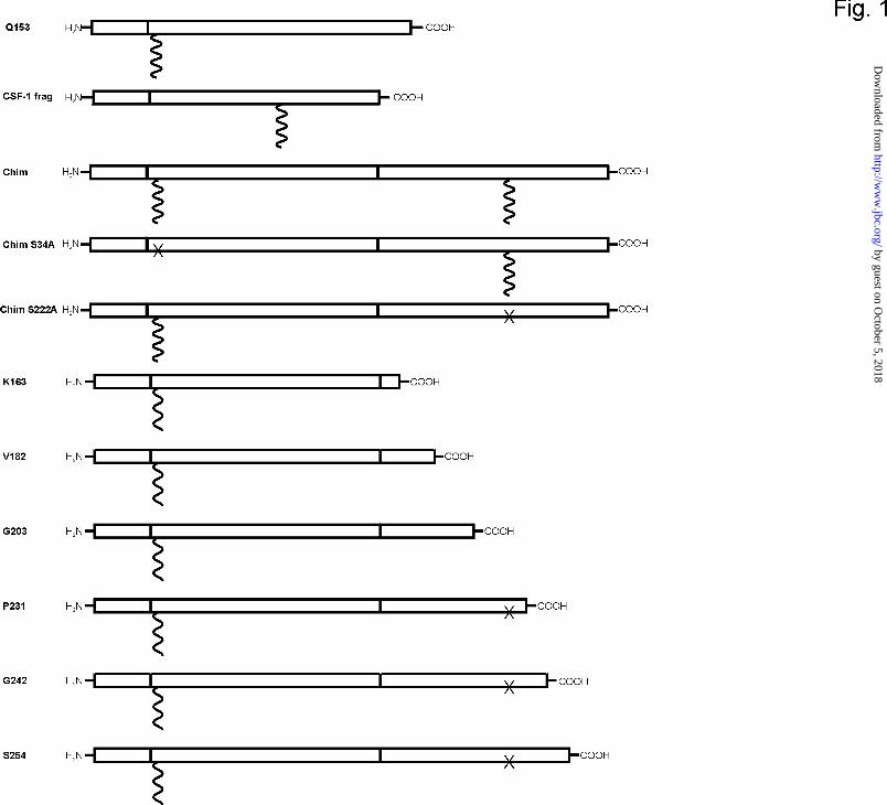
structures of the chimeras are schematically depicted in Fig. 1. The precise alignment of the
respective moieties is given in Tab. I.
In a first set of experiments stably transfected 293 HEK cells expressing the full-
length DCN/CSF-1 chimera were incubated in the presence of [35S]sulfate for 21 h or with
[35S]methionine for 8 h. Proteins from conditioned media were then precipitated with
ammonium sulfate and redissolved as described in the Methods section. Subsequently, the
dissolved proteins were subjected to immunoprecipitation with polyclonal antibodies against
DCN or the proteoglycan form of CSF-1. The immune complexes were subjected to SDS-
PAGE followed by fluorography. Fig. 2 shows that the fluorogram of the chimeric
proteoglycan yielded two broad bands migrating somewhat faster than globular proteins of
200 kDa and 97 kDa, respectively. Upon elimination of either one of the two potential
glycosaminoglycan attachment sites only the faster moving proteoglycan species was
obtained. Thus, it appears that the chimeric core protein contains either one or two
glycosaminoglycan chains. No attempt was made to investigate a potential preference for
using one of the two glycosaminoglycan attachment sites. It is known, however, that DCN is
always linked with a glycosaminoglycan chain while CSF-1 has to be considered as a part-
time proteoglycan, i.e. with a proportion lacking a glycosaminoglycan chain (25,26).
Digestion of [35S]methionine-labelled proteoglycan preparations resulted in the appearance of
core proteins of 29 kDa in accordance with the prediction derived from the cDNA of the
constructs used for transfection.
by guest on October 5, 2018
http://ww
w.jbc.org/
Dow
nloaded from

13
The two core protein fragments used to obtain the chimera were also expressed
individually whereby the CSF-moiety was combined with the DCN prepro sequence to allow
secretion into the culture medium. For better yields, DCNQ153 and the chimeras shown in Fig.
3C were purified by anion exchange chromatography on DEAE Trisacryl. This resin exhibits
some molecular sieve properties; e.g. it does not retain the large proteoglycan aggrecan
(unpublished observation). Therefore, the chimera could be separated from other high
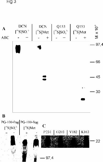
molecular weight proteoglycans. DCNQ153 could be obtained in large quantities. It migrated
during SDS-PAGE as a broad smear suggesting the presence of glycosaminoglycan chains of
varying length. The core protein, obtained after chondroitin ABC lyase digestion, migrated
like a monomer (~16 kDa) and a dimer (~34 kDa), respectively (Fig. 3A). DCN migrates also
as a doublet but this is due to the presence of either 2 or 3 N-glycosidically linked
oligosaccharides (30). Parallel incubations of cells expressing either DCNQ153 or CSF-1L241-
S353 with [35S]sulfate yielded about ten times higher radioactivies in the medium of the former
than of the latter cell strain. During SDS-PAGE, CSF-1L241-S353 appeared as a high molecular
weight aggregate as was true for the chondroitin ABC lyase-treated [35S]methionine-labelled
core protein (Fig. 3B). This observation is a strong argument for inappropriate folding of the
CSF-1 moiety when expressed without the DCN fragment M1-Q153. However, as indicated
above, the full-length chimera showed neither inappropriate secretion nor abnormal
electrophoretic mobility. Furthermore, pulse-chase experiments of 293 cells transfected either
with the chimera or with full-length DCN using [35S]methionine as a radioactive precursor did
not reveal significant differences in the secretion kinetics (data not shown). Other chimeric
proteins behaved as expected during SDS-PAGE (Fig. 3C).
The Glycosaminoglycan Chains of the Chimera Contain Minute Amounts of L-IdoUA
Residues – The [35S]sulfate-labelled biglycanated form of the chimera was isolated from
conditioned medium by ion exchange chromatography and SDS-PAGE. The proteoglycan
by guest on October 5, 2018
http://ww
w.jbc.org/
Dow
nloaded from

14
extracted from the gel was subsequently subjected to parallel treatments with chondroitin
lyases ABC, ACI, and B, respectively. Each digest was then subjected to chromatography on
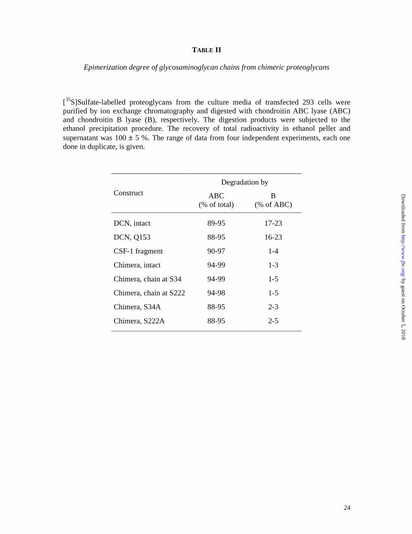
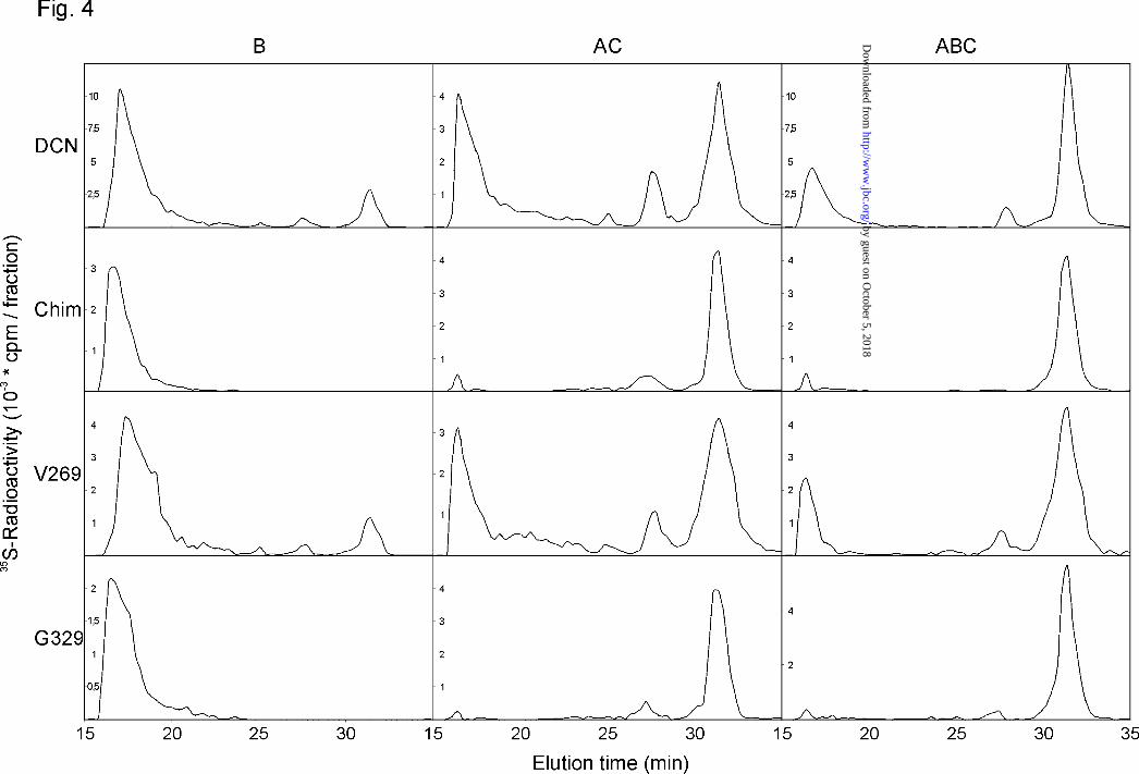
a Superdex Peptide HR 10/30 column. It is evident from Fig. 4 and Table II that the
glycosaminoglycan chains of the chimera were almost completely resistant towards
chondroitin B lyase, while similar chromatographic profiles were obtained after treatments
with chondrotin ACI and ABC lyase, respectively. Thus, the glycosaminoglycan chains
attached to the chimeric protein are apparently devoid of significant quantities of L-IdoUA
residues. The ratio of 4- and 6-sulfated galactosamine residues was apparently not
significantly altered when one takes into account the technical problem of quantifying minute
amounts of L-IdoUA-containing disaccharides released upon chondroitin B lyase digestion
(Table III). It should be noted that some labelled material was resistant to exhaustive digestion
with chondroitin ABC lyase and appeared in the Vo of the gel filtration column (right graphs
in Fig. 4). In case of glycosamonoglycans with minute amounts of L-IdoUA-containing
disaccharide units, similar quantities of label in the Vo fraction were also found after
chondroitin ACI lyase treatment. Digestion of the chondroitin ABC lyase-resistant material
with heparinases I and III indicated that it represents core proteins with heparan sulfate
substitution, analogous to the occasional linkage of biglycan with heparan sulfate chains that
occurs in 293 cells (31).
The almost complete absence of conversion of D-GlcUA into L-IdoUA residues in the
glycosaminoglycan chains of the chimeric proteoglycan was ascertained by analyzing the
individual glycosaminoglycan chains, which were either linked at S34 (DCN moiety) or at
S222 (CSF-1 moiety). The methodology to obtain the individual glycosaminoglycan chains is
described in the Methods section. The strategy relies on the proposal that upon blocking all
free amino groups followed subsequently by proteolytic digestion, only the
glycosaminoglycan chain linked to the CSF-1 moiety (at S222), but not the one attached to
the DCN core protein part (at S34), of the chimera can be linked with biotin and can
by guest on October 5, 2018
http://ww
w.jbc.org/
Dow
nloaded from
15
subsequently interact with avidin or streptavidin. However, about 15 % of non-biotinylated
chains bound non-specifically to streptavidin, thereby contaminating the CSF-1-linked
glycosaminoglycan fraction. Nevertheless, both fractions were almost completely insensitive
towards chondroitin B lyase and sensitive towards chondroitin ACI lyase (Table II). This
result corroborates the finding of the extremely low epimerization degree in both
glycosaminoglycan chains of the chimeric proteoglycan. Mutating either one of the serine
residues, which serve as attachment sites for glycosaminoglycan chains, into alanine residues
also does not change the epimerization pattern of the monoglycanated proteoglycan (Table II).
The Core Protein of the Chimera is Not Responsible for the Low Degree of Epimerization -
To investigate the possibility that the chondroitin-glucuronate 5-epimerase may require
distinct structural features on the core protein that are absent in the chimera, undersulfated
[3H]glucosamine-labelled proteoglycans (DCN, chimera, mutant full-length chimeras, see
Tab. IV) were prepared and incubated in vitro with microsomal proteins and
3´phosphoadenylyl 5´phosphosulfate. Under the assumption that the core protein exhibits a
major influence on epimerase activity, one would expect that maximal epimerization would
be observed only with DCN and with the chimera S222A as substrates. Half-maximal
epimerization may be found in the intact chimera, whereas the mutant chimera S34A should
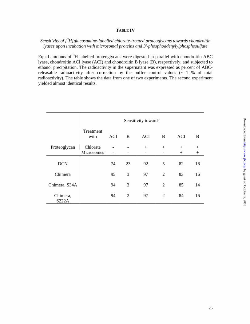
be an unsuited substrate. The data shown in Table IV clearly indicate that extensive
epimerization of all non-epimerized and undersulfated proteoglycans can be achieved in the
presence of microsomes. In case of DCN the proportion of chondroitin B lyase-sensitive
material rose from 5 % to 16 % upon incubation with microsomal proteins, whereas the
proportion of chondroitin ACI lyase-sensitive radioactivity declined from 92 % to 82 %. Very
similar data were obtained for all chimeric proteins, where the sensitivity towards chondroitin
B lyase rose from 2 % to 14-16 % and the sensitivity towards chondroitin ACI lyase declined
by guest on October 5, 2018
http://ww
w.jbc.org/
Dow
nloaded from
16
from 97 % to 83-85 %. These findings do not support the assumption that the core protein
regulates the extent of epimerization directly.
A Short Peptide Sequence at the N-Terminal Part of the CSF-1-Derived Fragment is Required
for Epimerization - In a further attempt to define the prerequisites for epimerization, we used
the S222A chimera, which allows only the attachment of a glycosaminoglycan chain in the
DCN-derived moiety, and introduced stop codons at various places along the CSF-1-derived
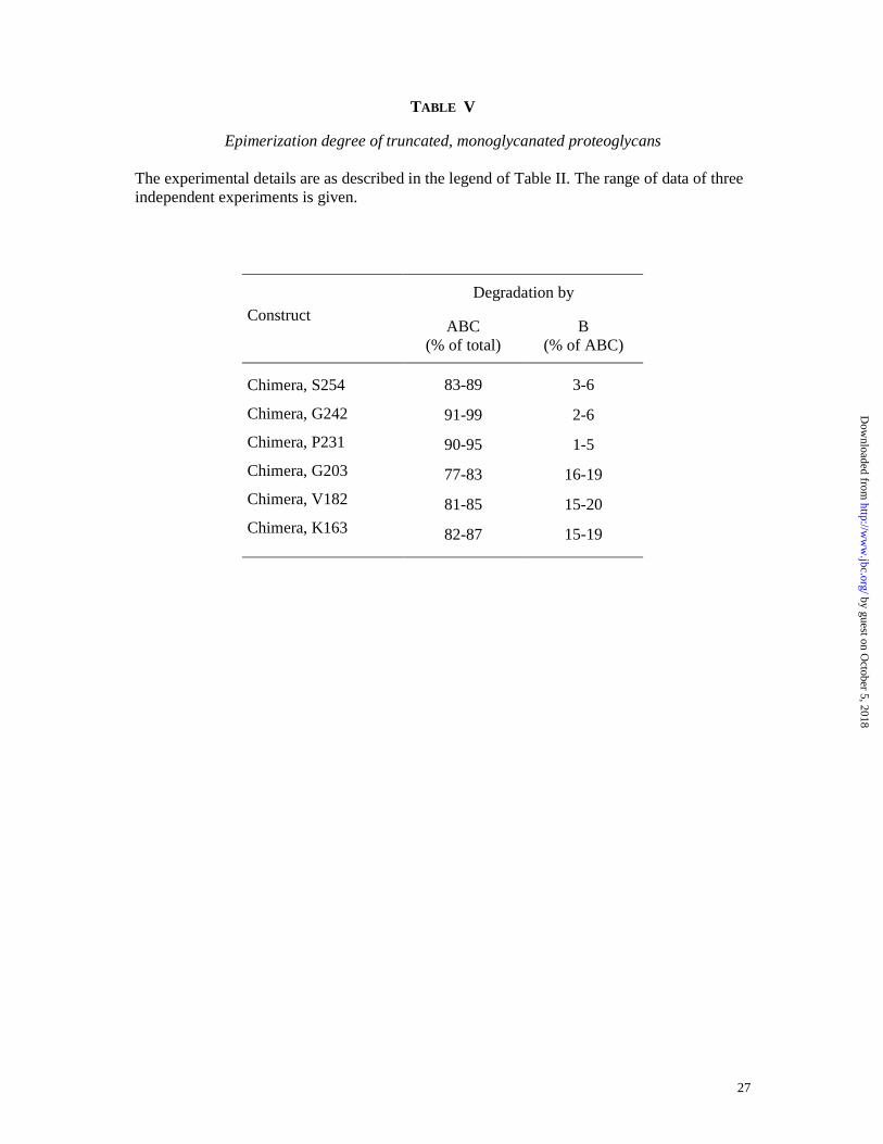
moiety of the core protein to obtain various truncated proteins. The data in Table V clearly
indicate that constructs with 50 or less N-terminal amino acids of the CSF-1 sequence allow
epimerization to occur at almost physiological levels (see chimeras G203, V182, K163), while
those with 78 or more amino acids do not, i.e. their glycosaminoglycan chains contain only a
very small percentage of L-IdoUA residues (see chimera P231 and longer constructs).
by guest on October 5, 2018
http://ww
w.jbc.org/
Dow
nloaded from
17
DISCUSSION
The data presented in this paper demonstrate that in 293 human kidney cells the
occurrence or absence of polymer-bound L-IdoUA residues (i.e. dermatan sulfate) depends on
the structure of the core protein. This finding is not specific for 293 cells since preliminary
data indicate that transiently transfected CHO cells modify the glycosaminoglycan chains of
the chimeric proteins similarly as do 293 cells (data not shown). COS cells, on the other hand,
do not epimerize chondroitin sulfate chains at all, even with full-length DCN; this is most
likely due to a missing chondroitin-glucuronate 5-epimerase (D.G. Seidler, unpublished
result) as holds probably true, for example, for DCN and biglycan in bone, which also does
not contain L-IdoUA residues (38). Our conclusion about the importance of the structure of
the core protein on the extent of epimerization is in some contrast to the observation by
Fransson et al. (39) who showed that protein-free glycosaminoglycan chains initiated on p-
nitrophenyl-�-D-xyloside may have a copolymeric structure of chondroitin sulfate and
dermatan sulfate building blocks when the xyloside concentration does not exceed 50
µmoles/litre. However, the intracellular distribution of p-nitrophenyl-�-D-xyloside is not
known, and it may well be that the relative concentration of the compound in different cellular
compartments depends on its extracellular concentration (see below).
An unexpected finding was our observation that the removal of only 28 amino acids of
a truncated DCN/CSF-1 chimera (converting chimera P231 into chimera G203, see Fig. 1)
enabled the cells to epimerize the chimera´s glycosaminoglycan to an extent resembling that
of wild-type DCN. It appears, therefore, that there is a signal within the sequence of these 28
amino acids which is responsible for the avoidance of epimerization. This signal is needed for
maintaining the initially synthesized chondroitin sulfate structure, while the formation of
dermatan sulfate follows a default pathway. An alternative explanation could be the formation
within the transfected cells of non-physiological complexes between the C-terminal part of
the CSF-1 moiety and the glycosaminoglycan chains with a subsequent inability of the
by guest on October 5, 2018
http://ww
w.jbc.org/
Dow
nloaded from
18
complex to become modified by the epimerase. We consider this as an unlikely event since it
is hard to imagine how a peptide of 28 amino acids masks simultaneously the two high
molecular weight chondroitin sulfate chains of the intact chimera.
Considering the core protein as a major determinant for glucuronide C5-epimerization,
the question arises about its mode of action. The possibility of a direct influence of the core
protein on chondroitin-glucuronate 5-epimerase activity does not fit with the observation that
microsomal proteins from 293 cells are able to convert undersulfated chondroitin sulfate
chains of chimeric proteoglycans into dermatan sulfate when incubated in the presence of 3´-
phosphoadenylylphosphosulfate. The second possibility, that the core protein sequence
determines modification reactions of the polysaccharide protein linkage region, which in turn
influence at a later stage the polymer modification reactions, could not be tested reliably since
the incorporation of [35S]sulfate or [32P]phosphate into the linkage region was at or below the
limit of detection. However, this hypothesis seems unlikely because the DCN-derived part of
the chimera contained a chondroitin and not a dermatan sulfate sulfate chain in contrast to
DCN alone or the truncated chimeras. In contrast, in case of syndecan-1, the linkage regions
for chondroitin and heparan sulfate chains were differently modified within the same
proteoglycan molecule (23). These findings should be seen in the context that also the
truncated DCNQ153 molecule carries a dermatan sulfate chain, thus providing evidence, that
the truncated part of the molecule itself has no negative influence on epimerization.
An alternate explanation is based on the assumption that the core protein may be
responsible for the transport into different subcellular compartments that can be distinguished
by their content of chondroitin-glucuronate 5-epimerase. Glycosaminoglycan chain
polymerization is generally considered to take place in trans-Golgi cisternae and/or in the
trans-Golgi network (see 1, 40 for reviews). Xylose addition may begin in the endoplasmic
reticulum and continue in the Golgi apparatus (41,42). So far, each individual Golgi cisterna
has been considered as a distinct entity. However, considering the observations that some
by guest on October 5, 2018
http://ww
w.jbc.org/
Dow
nloaded from
19
secretory proteins are transported by progressive maturation of Golgi cisternae while other
molecules require intra-Golgi transport vesicles (43,44), one might speculate that there could
be an unequal distribution of enzyme complexes even within a given Golgi cisterna. If this
were the case, our data could be explained by the existence of complexes of
glycosaminoglycan-synthesizing enzymes that differ in their content of chondroitin-
glucuronate 5-epimerase. The core protein would then fulfill a sorting function, targeting
chondroitin sulfate proteoglycans to complexes poorly equipped with the epimerase.
Under the assumption that chondroitin sulfate synthesis without concomitant
epimerization follows a regulated pathway, other chondroitin sulfate proteoglycans should
carry a similar sorting signal as the one that appears to be within the 28 amino acids that are
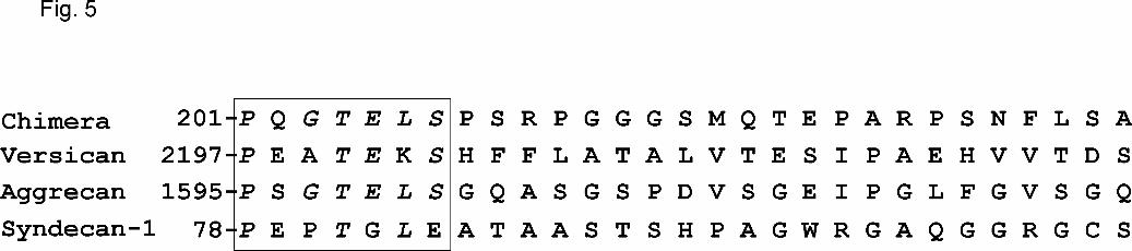
present in chimera P231 but not in chimera G203. A database search revealed indeed a
sequence of 7 amino acids (P201-P207 of the chimera, corresponding to P288-P294 of the
CSF-1 moiety of the construct) that are highly homologous to sequences found in the
chondroitin sulfate-containing proteoglycans aggrecan (located N-terminally of the first
chondroitin sulfate attachment block; ref. 45) , versican (located in between the two possible
glycosaminoglycan attachment sites; ref. 46), and syndecan-1 (located N-terminally of the
heparan sulfate attachment site, but further away from the chondroitin sulfate attachment
sites; ref. 47; see Fig. 5). No such homology was detected in the primary structure of neurocan
and brevican. However, the glycosaminoglycan structures of these two proteoglycans have
not yet been well established, and the presence of L-IdoUA residues within the respective
glycosaminoglycan chains cannot be excluded.
In summary, our data provide evidence that dermatan sulfate is synthesized by a
default pathway. A putative signal sequence preventing epimerization by the chondroitin-
glucuronate 5-epimerase has been identified. Further studies, however, are required, e.g. by
deleting the putative signal sequence in chondroitin sulfate proteoglycans, before the function
of this sequence for glycosaminoglycan modification can be unambiguously defined.
by guest on October 5, 2018
http://ww
w.jbc.org/
Dow
nloaded from
20
References
1. Silbert, J.E. 1996. Glycoconjugate J. 13:907-912.
2. Prydz, K., and Dalen, K.T. 2000. J. Cell Sci. 113:193-205.
3. Malmström, A., and Fransson, L.-Å. 1975. J. Biol. Chem. 250:3419-3425.
4. Malmström, A. 1981. Biochem. J. 198:669-675.
5. Malmström, A. 1984. J. Biol. Chem. 259:161-165.
6. Hannesson, H.H., Hagner-McWhirter, A., Tiedemann, K., Lindahl, U., and Malmström, A.
1996. Biochem. J. 313:589-596.
7. Malmström, A., and Åberg, L. 1982. Biochem. J. 201:489-493.
8. Li, J., Hagner-McWhirter, A., Kjéllen, L., Palgi, J., Jalkanen, M., and Lindahl, U. 1997. J.
Biol. Chem. 272:28158-28163.
9. Hagner-McWhirter, A., Hannesson, H.H., Campbell, P., Westley, J., Rodén, L., Lindahl,
U., and Li, J.P. 2000. Glycobiology 10:159-171.
10. Ferro, D.R., Provasoli, A., Ragazzi, M., Casu, B., Torri, G., Bossennec, V., Perly, B.,
Sinay, P., Petitou, M., and Choay, J. 1990. Carbohydr. Res. 195:157-167.
11. Inoue, Y., Inouye, Y., and Nagasawa, K. 1990. Biochem. J. 265:533-538.
12. Rao, V.S., Balaji, P.V., and Qasba, P.K. 1995. Glycobiology 5:273-279.
13. Mikhailov, D., Linhardt, R.J., and Mayo, K.H. 1997. Biochem. J. 328:51-61.
14. Mulloy, B., and Forster, M.J. 2000. Glycobiology 10:1147-1156.
15. Linhardt, R.J., al Hakim, A., Liu, J.A., Hoppensteadt, D., Mascellani, G., Bianchini, P.,
and Fareed, J. 1991. Biochem. Pharmacol. 42:1609-1619.
16. Mascellani, G., Liverani, L., Bianchini, P., Parma, B., Torri, G., Bisio, A., Guerrini, M.,
and Casu, B. 1993. Biochem. J. 296:639-648.
by guest on October 5, 2018
http://ww
w.jbc.org/
Dow
nloaded from
21
17. Mascellani, G., Liverani, L., Prete, A., Bergonzini, G., Bianchini,P., Torri, G., Bisio, A.,
Guerrini, M., and Casu, B. 1994. Anal. Biochem. 223:135-141.
18. Shirk, R.A., Parthasarathy, N., San Antonio, J.D., Church, F.C., and Wagner, W.D. 2000.
J. Biol. Chem. 275:18085-18092.
19. Aspberg, A., Miura, R., Bourdoulous, S., Shimonaka, M., Heinegård, D., Schachner, M.,
Ruoslahti, E., and Yamaguchi., Y. 1997. Proc. Natl. Acad. Sci. U. S. A. 94:10116-10121.
20. Iozzo, R.V. 1999. J. Biol. Chem. 274:18843-18846.
21. Hausser, H., Schönherr, E., and Kresse, H. 1993. Glycobiology 3:557-562.
22. Cheng, F., Heinegård, D., Malmström, A., Schmidtchen, A., Yoshida, K., and Fransson,
L.-Å. 1994. Glycobiology 4:685-696.
23. Ueno, M., Yamada, S., Zako, M., Bernfield, M., and Sugahara, K. 2001. J. Biol. Chem.
276:29134-29140.
24. Schwarz, K., Breuer, B., and Kresse, H. 1990. J. Biol. Chem. 265:22023-22028.
25. Price, L.K., Choi, H.U., Rosenberg, L., and Stanley, E.R. 1992. J. Biol. Chem. 267:2190-
2199.
26. Suzu, S., Ohtsuki, T., Yanai, N., Takatsu, Z., Kawashima, T., Takaku, F., Nagata, N., and
Motoyoshi, K. 1992. J. Biol. Chem. 267:4345-4348.
27. Partenheimer, A., Schwarz, K., Wrocklage, C., Kölsch, E., and Kresse, H. 1995. J.
Immunol. 155:5557-5565.
28. Beavan, L.A., Quentin-Hoffmann, E., Schönherr, E., Snigula, F., Leroy, J.G., and Kresse,
H. 1993. J. Biol. Chem. 268: 9856-9862.
29. Kresse, H., Liszio, C., Schönherr, E., and Fisher, L.W. 1997. J. Biol. Chem. 272:18404-
18410.
30. Glössl, J., Beck, M., and Kresse, H. 1984. J. Biol. Chem. 259:14144-14150.
31. Kresse, H., Seidler, D.G., Müller, M., Breuer, E., Hausser, H., Roughley, P.J., and
Schönherr, E. 2001. J. Biol. Chem. 276: 13411-13416.
by guest on October 5, 2018
http://ww
w.jbc.org/
Dow
nloaded from
22
32. Hamai, A., Hashimoto, N., Mochizuki, H., Kato, F., Makiguchi, Y., Horie, K., and
Suzuki, S. 1997. J. Biol. Chem. 272:9123-9130.
33. Hiyama, K., and Okada, S. 1976. J. Biochem. (Tokyo) 80:1201-1207.
34. Ototani, N., and Yosizawa, Z. 1979. Carbohydrate Res. 70:295-306.
35. Hausser, H., and Kresse, H. 1999. Biochem. J. 344:827-835.
36. Calabro, A., Benavides, M., Tammi, M., Hascall, V.C., and Midura, R.J. 2000.
Glycobiology 10:273-281.
37. Greve, H., Cully, Z., Blumberg, P., and Kresse, H. 1988. J. Biol. Chem. 263:12886-12892.
38. Boskey, A.L., Spevak, L., Doty, S.B., and Rosenberg, L. 1997. Calci. Tissue Int. 61:298-
305.
39. Fransson, L.-Å., Schmidtchen, A., Cöster, L., and Malmström, A. 1991. Glycoconj. J.
8:108-115.
40. Calabro, A., Midura, R.J., Hascall, V.C., Plaas, A., Goodstone, N.J., and Rodén, L.
(2000), in Proteoglycans (Iozzo, R.V., ed), pp. 5-26, Marcel Dekker, New York.
41. Kearns, A.E., Vertel, B.M., and Schwartz, N.B. 1993. J. Biol. Chem. 268:11097-11104.
42. Bai, X., Zhou, D., Brown, J.R., Crawford, B.E., Hennet, T., and Esko, J.D. 2001. J. Biol.
Chem. 276:48189-48195.
43. Bonfanti, L., Mironov, Jr., A.A., Martínez-Menárguez, J.A., Martella, O., Fusella, A.,
Baldassarre, M., Buccione, R., Geuze, H.J., Mironov, A.A., and Luini, A. 1998. Cell
95:993-1003.
44. Pelham, H.R.B., and Rothman, J.E. 2000. Cell 102:713-719.
45. Doege, K.J., Sasaki, M., Kimura, T., and Yamada, Y. 1991. J. Biol. Chem. 266:894-902.
46. Zimmermann, D.R., and Ruoslahti, E. 1989. EMBO J. 8:2975-2981.
47. Kokenyesi, R., and Bernfield, M. 1994. J. Biol. Chem. 269:12304-12309.
by guest on October 5, 2018
http://ww
w.jbc.org/
Dow
nloaded from

23
TABLE I
Overview of recombinant proteins In all chimeric proteins the numbering of amino acids is based on the sequence of the construct and not on the sequence of the natural precursor proteins.
Construct
DCN moiety
CSF-1 moiety
DCN, intact DCN, Q153 CSF-1, GAG site Chimera, intact Chimera, S34A Chimera, S222A Chimera, S254 Chimera, G242 Chimera, P231 Chimera, G203 Chimera, V182 Chimera, K163
M1 - K359
M1 - Q153
M1 - E30
M1 - Q153
M1 - Q153
S34A
M1 - Q153
M1 - Q153
M1 - Q153
M1 - Q153
M1 - Q153
M1 - Q153
M1 - Q153
none
none
L241 - S353
L241 - S353
L241 - S353
L241 - S353 S309A
L241 - S341
L241 - G329
L241 - P318
L241 - G290
L241 - V269
L241 - K250
by guest on October 5, 2018
http://ww
w.jbc.org/
Dow
nloaded from
24
TABLE II
Epimerization degree of glycosaminoglycan chains from chimeric proteoglycans
[35S]Sulfate-labelled proteoglycans from the culture media of transfected 293 cells were purified by ion exchange chromatography and digested with chondroitin ABC lyase (ABC) and chondroitin B lyase (B), respectively. The digestion products were subjected to the ethanol precipitation procedure. The recovery of total radioactivity in ethanol pellet and supernatant was 100 ± 5 %. The range of data from four independent experiments, each one done in duplicate, is given.
Degradation by
Construct ABC (% of total)
B (% of ABC)
DCN, intact DCN, Q153 CSF-1 fragment Chimera, intact Chimera, chain at S34 Chimera, chain at S222 Chimera, S34A Chimera, S222A
89-95
88-95
90-97
94-99
94-99
94-98
88-95
88-95
17-23
16-23
1-4
1-3
1-5
1-5
2-3
2-5
by guest on October 5, 2018
http://ww
w.jbc.org/
Dow
nloaded from

25
TABLE III
Disaccharide composition of GAG chains from selected chimeric constructs Proteoglycan samples from the culture media of transfected 293 cells were purified by ion exchange chromatography and subjected to fluorophore-assisted carbohydrate electrophoresis after exhaustive treatment with either chondroitin ABC lyase (ABC), chondroitin ACII lyase (ACII) or chondroitin B lyase (B) for quantification of unsaturated disaccharides. In brackets the percentage of released disaccharides on the total radioactivity is given. Note that most of the values in the B column are based on the quantification of very weak bands obtained after electrophoretic separation of the released disaccharides.
Ratio of 4-sulfated and 6-sulfated disaccharides after
digestion with chondroitinase
Construct
ABC
ACII
B
DCN, intact DCN, Q153 Chimera, intact Chimera, S222A Chimera, P231 Chimera, V182
5.6
4.7
4.1
3.8
5.1
3.9
4.9
4.3
3.7
3.5
4.8
3.7
13.4
16.3
10.2
7.3
6.1
6.2
by guest on October 5, 2018
http://ww
w.jbc.org/
Dow
nloaded from
26
TABLE IV
Sensitivity of [3H]glucosamine-labelled chlorate-treated proteoglycans towards chondroitin lyases upon incubation with microsomal proteins and 3'-phosphoadenylylphosphosulfate
Equal amounts of 3H-labelled proteoglycans were digested in parallel with chondroitin ABC lyase, chondroitin ACI lyase (ACI) and chondroitin B lyase (B), respectively, and subjected to ethanol precipitation. The radioactivity in the supernatant was expressed as percent of ABC-releasable radioactivity after correction by the buffer control values (~ 1 % of total radioactivity). The table shows the data from one of two experiments. The second experiment yielded almost identical results.
Sensitivity towards
Treatment
with
ACI
B
ACI
B
ACI
B
Proteoglycan Chlorate - - + + + + Microsomes - - - - + +
DCN
Chimera
Chimera, S34A
Chimera, S222A
74
95
94
94
23
3 3 2
92
97
97
97
5 2 2 2
82
83
85
84
16
16
14
16
by guest on October 5, 2018
http://ww
w.jbc.org/
Dow
nloaded from
27
TABLE V
Epimerization degree of truncated, monoglycanated proteoglycans The experimental details are as described in the legend of Table II. The range of data of three independent experiments is given.
Degradation by
Construct ABC
(% of total) B
(% of ABC)
Chimera, S254
Chimera, G242
Chimera, P231
Chimera, G203
Chimera, V182
Chimera, K163
83-89
91-99
90-95
77-83
81-85
82-87
3-6
2-6
1-5
16-19
15-20
15-19
by guest on October 5, 2018
http://ww
w.jbc.org/
Dow
nloaded from

28
FIG. 1. Schematic representation of the proteoglycans encoded by cDNA constructs used in this study. Glycosaminoglycans are indicated by the wavy line; X indicates a mutant glycosaminoglycan attachment site; , represents the prepro sequence of DCN, , the N-terminal fragment of DCN, and , CSF-1 fragments.
by guest on October 5, 2018
http://ww
w.jbc.org/
Dow
nloaded from
29
FIG. 2 Immune precipitation of chimeric proteins with antibodies against DCN or CSF-1 prior to SDS-gel electrophoresis and fluorography. 293 HEK cells, either transfected with a control vector (control) or with a cDNA encoding the intact DCN/CSF-1 chimera (Chim), and chimeras containing a deleted glycosaminoglycan attachment site (Chim S34A and Chim S222A, respectively) were incubated with radioactive precursors prior to immune precipitation (PASA) with antisera raised against DCN and CSF-1, respectively. The immune complexes were treated with chondroitin ABC lyase (ABC) or buffer alone and then subjected to SDS-polyacrylamide gel electrophoresis in a 12.5 % separation gel. Migration of molecular weight markers is indicated on the right margin.
by guest on October 5, 2018
http://ww
w.jbc.org/
Dow
nloaded from
30
FIG. 3. Fluorogram of recombinant proteoglycans after separation in a 12.5 % polyacrylamide gel. The truncated form, DCNM1-Q153, is abbreviated by Q153. ABC + denotes digestion with chondroitin ABC lyase prior to SDS-PAGE. In B the recombinant fragment (frag) of CSF-1 was isolated from the culture medium by immunoprecipitation with a polyclonal antibody against the growth factor. Other proteoglycans were obtained after chromatography on DEAE-Trisacryl. In C all samples had been treated with chondroitin ABC lyase after immune precipitation.
by guest on October 5, 2018
http://ww
w.jbc.org/
Dow
nloaded from
31
FIG. 4. Size fractionation on a Superdex Peptide HR 10/30 column of recombinant [35S]sulfate-labelled proteoglycans. Proteoglycans were isolated by anion exchange FKURPDWRJUDSK\ DQG VXEMHFWHG WR D �-elimination reaction. The isolated glycosaminoglycans were then digested with either chondroitin B lyase (B), chondroitin ACI lyase (AC) or chondroitin ABC lyase (ABC) prior to gel filtration. Vo corresponds to an elution time of 16 min, and Vt to 35.5 min.
by guest on October 5, 2018
http://ww
w.jbc.org/
Dow
nloaded from

32
FIG. 5. Alignment of amino acids P201-A229 of the chimera with core protein sequences of chondroitin sulfate-containing proteoglycans. The peptide sequences were from the GeneBank accession numbers NP_000748 (CSF-1 part of the chimera), NP_004376 (versican), NP_001126 (aggrecan), NP_002988 (syndecan-1). No homologous region was detected in neurocan and brevican, respectively.
by guest on October 5, 2018
http://ww
w.jbc.org/
Dow
nloaded from


Daniela G. Seidler, Egon Breuer, Jane Grande-Allen, Vincent C. Hascall and Hans Kressegalactosaminoglycans
Core protein-dependence of epimerization of glucuronosyl residues in
published online August 30, 2002J. Biol. Chem.
10.1074/jbc.M208442200Access the most updated version of this article at doi:
Alerts:
When a correction for this article is posted•
When this article is cited•
to choose from all of JBC's e-mail alertsClick here
by guest on October 5, 2018
http://ww
w.jbc.org/
Dow
nloaded from