copyright © 2012 pearson education, inc. chapter 14 the respiratory system betty mcguire cornell...
TRANSCRIPT

Copyright © 2012 Pearson Education, Inc.
Chapter 14
The Respiratory
System
Betty McGuireCornell University
Lecture Presentation

Copyright © 2012 Pearson Education, Inc.
The Respiratory System
Structures of the respiratory system Mechanism of breathing Transport of gases between the lungs and
the cells Respiratory centers in the brain Respiratory disorders
Copyright © 2012 Pearson Education, Inc.
Structures of the Respiratory System
Overview of the respiratory system Function
Provides the body with essential oxygen and disposes of carbon dioxide This exchange regulates the acidity
of body fluids

Copyright © 2012 Pearson Education, Inc.
Structures of the Respiratory System
Overview of the respiratory system (cont.) Four processes play a part in respiration:
1. Breathing (ventilating)
2. External respiration
3. Gas transport
4. Internal respiration

Copyright © 2012 Pearson Education, Inc.

Copyright © 2012 Pearson Education, Inc.
Structures of the Respiratory System
Regions of the respiratory system Upper
Nose and pharynx Lower
Larynx, epiglottis, trachea, bronchi, bronchioles, and lungs

Copyright © 2012 Pearson Education, Inc.

Copyright © 2012 Pearson Education, Inc.

Copyright © 2012 Pearson Education, Inc.
Structures of the Respiratory System
Nose Structure
Nasal septum divides the inside into two nasal cavities
Mucous membrane covers inner surfaces Functions
Cleans incoming air Warms and moistens air Provides for the sense of smell

Copyright © 2012 Pearson Education, Inc.
Copyright © 2012 Pearson Education, Inc.
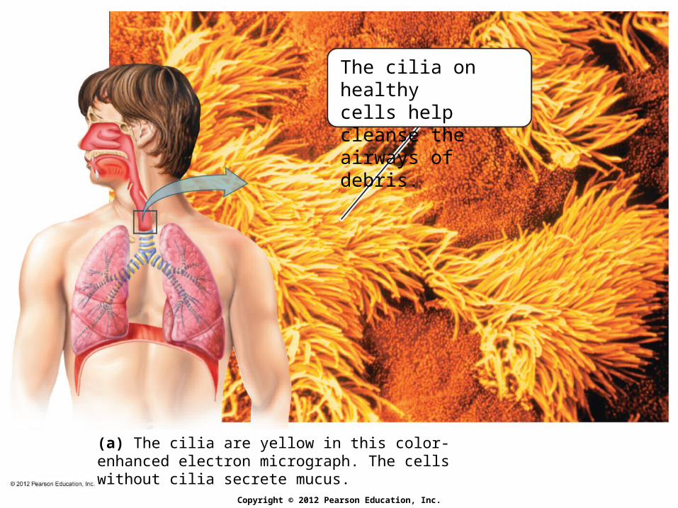
The cilia on healthycells help cleanse the airways of debris.
(a) The cilia are yellow in this color-enhanced electron micrograph. The cells without cilia secrete mucus.

Copyright © 2012 Pearson Education, Inc.
Cigarette smokedestroys the ciliain airways.
(b) Cigarette smoke first paralyzes and then destroys the cilia. As a result, hazardous materials can accumulate on the surfaces of the air passageways.

Copyright © 2012 Pearson Education, Inc.
Structures of the Respiratory System
Sinuses
Structure Large air-filled spaces in the bones
of the face Connect to nasal cavities
Functions Lighten head Warm and moisten air Part of the resonating chamber that
affects voice Sinusitis = inflammation of the mucous
membranes of the sinuses

Copyright © 2012 Pearson Education, Inc.
Structures of the Respiratory System
Pharynx (throat) Space behind the nose and mouth Passageway for food, drink, and air Connected to the middle ear via auditory
(Eustachian) tubes Help equalize pressure

Copyright © 2012 Pearson Education, Inc.
Structures of the Respiratory System
Larynx (voice box or Adam’s apple) Structure
Boxlike Made primarily of cartilage
Functions Serves as a selective entrance to the
lower respiratory system Source of the voice

Copyright © 2012 Pearson Education, Inc.

Copyright © 2012 Pearson Education, Inc.
(a) The epiglottis is open during breathing but covers the opening to the larynx during swallowing to prevent food or drink from entering the trachea.
Uppertrachea
Larynx
Front view
Epiglottis

Copyright © 2012 Pearson Education, Inc.
During quiet breathing, the vocalcords are near the sides of thelarynx, and the glottis is open.
During speech, the vocal cordsare stretched over the glottis andvibrate as air passes throughthem, producing the voice.
Top view of larynxTop view of larynx
Vocal cords
Glottis
(b) The vocal cords are the folds of connective tissue above the opening of the larynx (the glottis) that produce the voice.

Copyright © 2012 Pearson Education, Inc.
Structures of the Respiratory System
The larynx as a selective entrance to the lower respiratory system During swallowing, the larynx rises up and
causes the epiglottis (a flap of cartilage) to cover the glottis (opening in the larynx through which air passes)
If this mechanism fails and food or drink accidentally enter the trachea, then Coughing may expel material Heimlich maneuver may dislodge material

Copyright © 2012 Pearson Education, Inc.

Copyright © 2012 Pearson Education, Inc.
Structures of the Respiratory System
The larynx as the source of the voice Vocal cords (two thick stands of tissue
stretched over the glottis) vibrate and produce the voice Tension of vocal cords determines pitch
Stretched and thin cords = higher pitch Laryngitis
Inflammation of the larynx Vocal cords become swollen and thick,
causing voice to deepen

Copyright © 2012 Pearson Education, Inc.
Structures of the Respiratory System
Trachea (windpipe) Structure
Tube held open by C-shaped rings of cartilage
Function Conducts air between environment
and lungs

Copyright © 2012 Pearson Education, Inc.
Structures of the Respiratory System
Bronchial tree Network of progressively smaller air tubes
Trachea divides into two air tubes called primary bronchi, each of which leads to a lung
Bronchi branch repeatedly within each lung, eventually forming bronchioles
Bronchioles terminate in alveoli (air sacs) Bronchi are held open by cartilage; the amount
of cartilage decreases as tubes get smaller Bronchioles lack cartilage and have smooth
muscle

Copyright © 2012 Pearson Education, Inc.

Copyright © 2012 Pearson Education, Inc.
Structures of the Respiratory System
Asthma Spasms of the bronchial muscles that
severely restrict air flow Characterized by recurring attacks of
wheezing and difficult breathing and persistent inflammation of the airways
Inhalants Relax bronchial muscles Reduce inflammation of air tubules

Copyright © 2012 Pearson Education, Inc.
Structures of the Respiratory System
PLAY | Asthma

Copyright © 2012 Pearson Education, Inc.
Structures of the Respiratory System
Alveoli Minute sacs where
Oxygen diffuses from the inhaled air into the blood
Carbon dioxide diffuses from the blood into the alveolar air to be exhaled

Copyright © 2012 Pearson Education, Inc.
Structures of the Respiratory System
Surfactant Phospholipid molecules that coat alveoli and
keep them open Respiratory distress syndrome (RDS) occurs
in some premature babies due to insufficient production of surfactant

Copyright © 2012 Pearson Education, Inc.
Copyright © 2012 Pearson Education, Inc.
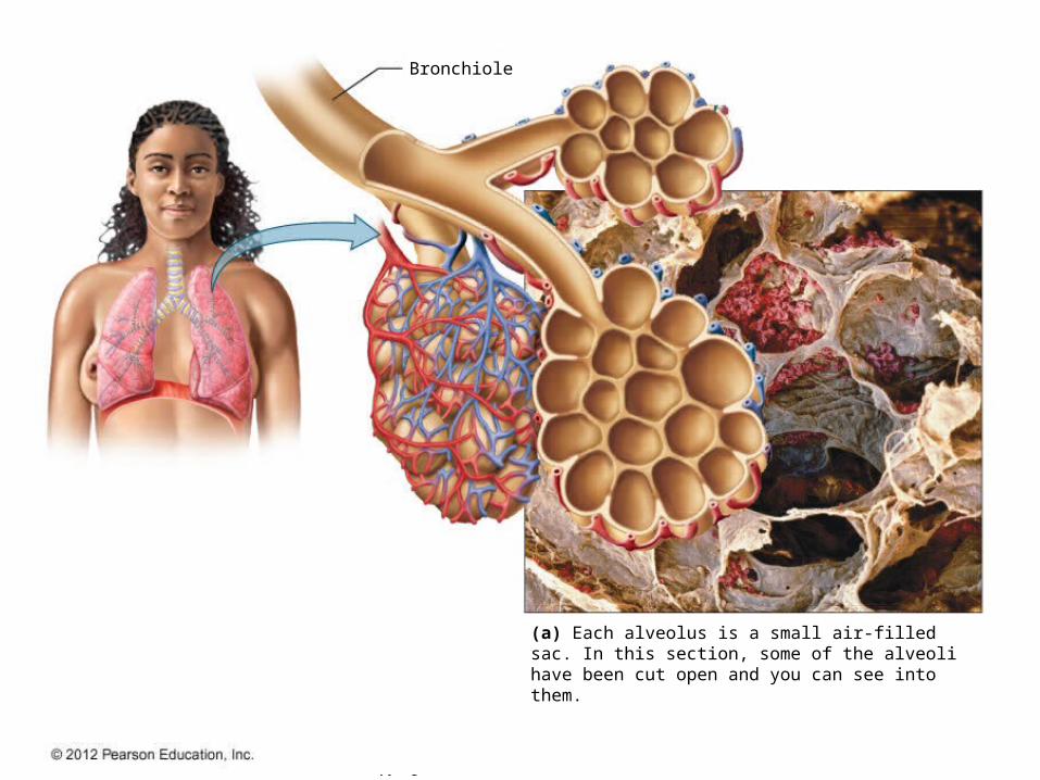
Bronchiole
(a) Each alveolus is a small air-filled sac. In this section, some of the alveoli have been cut open and you can see into them.

Copyright © 2012 Pearson Education, Inc.
(b) Much of the surface of each alveolus is covered with capillaries. The interface provides a vast surface area for the exchange of gases between the alveoli and the blood.

Copyright © 2012 Pearson Education, Inc.
Mechanism of Breathing
Air moves between the atmosphere and the lungs in response to pressure gradients Air moves into lungs when pressure in
atmosphere > pressure in lungs Air moves out of lungs when pressure in
lungs > pressure in atmosphere
Pressure changes in lungs are created by changes in volume of the thoracic cavity

Copyright © 2012 Pearson Education, Inc.
Mechanism of Breathing
Inhalation Air moves into the lungs when the thoracic
cavity increases in volume due to contraction of the diaphragm and intercostal muscles
Air rushes in because pressure in lungs < pressure in atmosphere
Also called inspiration Active process involving muscle contraction
Copyright © 2012 Pearson Education, Inc.
Mechanism of Breathing
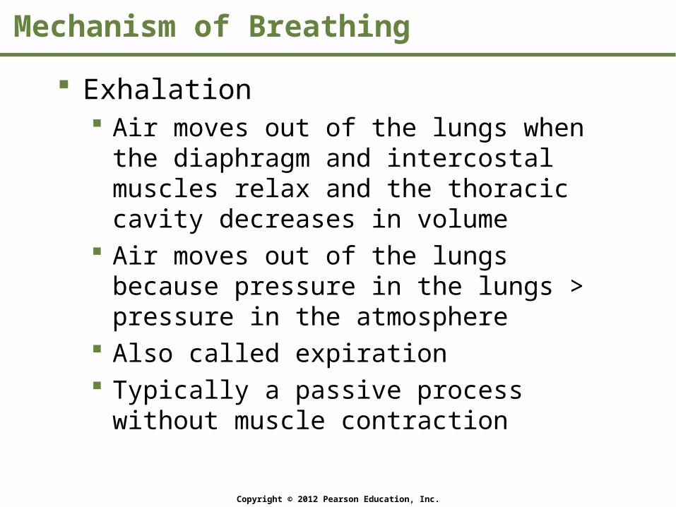
Exhalation Air moves out of the lungs when the
diaphragm and intercostal muscles relax and the thoracic cavity decreases in volume
Air moves out of the lungs because pressure in the lungs > pressure in the atmosphere
Also called expiration Typically a passive process without muscle
contraction

Copyright © 2012 Pearson Education, Inc.

Copyright © 2012 Pearson Education, Inc.
The chest cavity increases In size, and pressure withinthe lungs decreases.
Diaphragmcontractsand flattens
Intercostalmusclescontract
Diaphragmcontracts
Rib cagemoves upand out
Inhalation
Air flow
(a)
The lungs expand, and air moves in.

Copyright © 2012 Pearson Education, Inc.
The chest cavity decreasesin size, and pressure withinthe lungs increases.
The lungs recoil,and air moves out.
Diaphragmrelaxes andmoves upward
Intercostalmuscles relax
Rib cagemoves downand inward
Diaphragmrelaxes
Air flow
Exhalation
(b)

Copyright © 2012 Pearson Education, Inc.
Mechanism of Breathing
Tidal volume Volume of air inhaled or exhaled during a
normal breath
Inspiratory reserve volume Volume of air that can be inhaled in addition
to a normal breath
Expiratory reserve volume Volume of air that can be exhaled in addition
to a normal breath

Copyright © 2012 Pearson Education, Inc.
Mechanism of Breathing
Vital capacity Maximum volume of air that can be inhaled or
exhaled in a single forced breath tidal volume + inspiratory reserve volume +
expiratory reserve volume Residual volume
Volume of air remaining in lungs after maximum exhalation
Total lung capacity Total volume of air in lungs after maximal
inhalation vital capacity + residual volume

Copyright © 2012 Pearson Education, Inc.

Copyright © 2012 Pearson Education, Inc.
Transport of Gases between the Lungs and the Cells
Three processes (review) External respiration
Occurs in alveoli Oxygen diffuses into blood and carbon
dioxide diffuses from blood Gas transport by the blood Internal respiration
Occurs in tissues Oxygen diffuses out of blood and into
cells, and carbon dioxide diffuses out of cells and into blood

Copyright © 2012 Pearson Education, Inc.

Copyright © 2012 Pearson Education, Inc.
Transport of Gases between the Lungs and the Cells
Most oxygen carried in the blood is bound to hemoglobin, a protein in RBCs Hemoglobin bound to oxygen is called
oxyhemoglobin

Copyright © 2012 Pearson Education, Inc.
Transport of Gases between the Lungs and the Cells
Carbon dioxide is removed by the blood in one of three ways1. Dissolved in blood plasma
2. Carried by hemoglobin (carbaminohemoglobin)
3. As a bicarbonate ion

Copyright © 2012 Pearson Education, Inc.
Transport of Gases between the Lungs and the Cells
Most carbon dioxide is transported as bicarbonate ion
Bicarbonate ions are an important part of the body’s acid-base buffering system

Copyright © 2012 Pearson Education, Inc.
Respiratory Centers in the Brain
Basic breathing rhythm Controlled by a breathing center located in
the medulla Within the breathing center is an
inspiratory area and an expiratory area
Pattern of breathing can be voluntarily altered through impulses originating in the cerebral cortex

Copyright © 2012 Pearson Education, Inc.

Copyright © 2012 Pearson Education, Inc.
Respiratory Centers in the Brain
Carbon dioxide Most important chemical influencing
breathing rate Chemoreceptors located in the medulla,
aortic bodies, and carotid bodies Increased carbon dioxide prompts increased
breathing rate

Copyright © 2012 Pearson Education, Inc.

Copyright © 2012 Pearson Education, Inc.
Respiratory Centers in the Brain
Oxygen Does not influence breathing rate unless its
blood levels fall dangerously low Chemoreceptors located in the medulla,
aortic bodies, and carotid bodies

Copyright © 2012 Pearson Education, Inc.
Respiratory Centers in the Brain
Web Activity: The Human Respiratory System

Copyright © 2012 Pearson Education, Inc.
Respiratory Disorders
Common cold Caused by more than 200 different viruses Typically lasts 1–2 weeks Usually transmitted when a person handles
an object that is contaminated with a virus and then touches mucous membranes

Copyright © 2012 Pearson Education, Inc.
Respiratory Disorders
Flu (influenza) In humans, caused by three major types of
viruses (A, B, and C), each with many variants
Symptoms more severe than those of a cold Can be complicated by secondary infections
such as pneumonia, bronchitis, and sinusitis Vaccines are 60–70% effective
New strains constantly appear

Copyright © 2012 Pearson Education, Inc.
Respiratory Disorders
Pneumonia An inflammation of the lungs
Fluid accumulates in the alveoli, reducing gas exchange
Bronchioles swell and narrow, making breathing difficult
Most commonly caused by a bacterial or viral infection

Copyright © 2012 Pearson Education, Inc.
Respiratory Disorders
Strep throat Caused by Streptococcus bacteria Soreness accompanied by swollen glands
and fever Can have serious consequences
Rheumatic fever Kidney disease (glomerulonephritis)
If you have a sore throat, get a “strep test”

Copyright © 2012 Pearson Education, Inc.
Respiratory Disorders
Tuberculosis Infection caused by the bacterium
Mycobacterium tuberculosis Transmitted through respiratory droplets Results in fibrous tissue (tubercles) in
the lungs Can be fatal

Copyright © 2012 Pearson Education, Inc.
Respiratory Disorders
Bronchitis Inflammation of the mucous membrane
of the bronchi Caused by viruses, bacteria, or chemical
irritation Inflammation results in the production of
excess mucus, which triggers a deep cough Can be acute or chronic

Copyright © 2012 Pearson Education, Inc.
Respiratory Disorders
Emphysema Caused by the destruction of alveoli, usually
by smoking Results in:
Reduction in the surface area available for gas exchange
Increase in dead air space in lungs Main symptom: shortness of breath Can be treated but not cured

Copyright © 2012 Pearson Education, Inc.

Copyright © 2012 Pearson Education, Inc.

Copyright © 2012 Pearson Education, Inc.

Copyright © 2012 Pearson Education, Inc.
Respiratory Disorders
Lung cancer 85–90% of cases are caused by smoking,
and are therefore preventable Typical progression
Chronic inflammation of the lungs Changes in the cells of the airway linings Uncontrolled cell division forms a tumor Cancer cells spread to other parts of the
lung and rest of the body

Copyright © 2012 Pearson Education, Inc.

Copyright © 2012 Pearson Education, Inc.
Respiratory Disorders
PLAY | Secondhand Smoke