copyright © 2010 university of glasgow
TRANSCRIPT

Albalat, A., and Neil, D. (2010) Sensitive Diagnostic Tools to Screen for Gaffkemia Infection in European Lobsters (Homarus gammarus). Project Report. University of Glasgow, Glasgow, UK. Copyright © 2010 University of Glasgow A copy can be downloaded for personal non-commercial research or study, without prior permission or charge
Content must not be changed in any way or reproduced in any format or medium without the formal permission of the copyright holder(s)
When referring to this work, full bibliographic details must be given http://eprints.gla.ac.uk/81387 Deposited on: 24 June 2013
Enlighten – Research publications by members of the University of Glasgow http://eprints.gla.ac.uk

Sensitive Diagnostic tools to screen for Gaffkemia infection in
European lobsters (Homarus gammarus)
A Scientific Report by
Dr Amaya Albalat
Professor Douglas Neil
September 2010
Scientific Report Screening for Gaffkemia in lobsters
1
Introduction to the project
Tarbert Shellfish Ltd. is a company that processes live shellfish. It uses high-density
holding facilities to accumulate stock prior to transport and export. However, any
infections brought into the communal hold from different locations around Scotland can
spread through the stock, resulting in substantial financial losses, which threatens their
profitability, and in turn the livelihood of the Scottish creel fishermen supplying the
company.
A particular problem has recently arisen on more than one occasion that has been thought
to be due to the highly contagious marine bacterium Aerococcus viridans homari, and an
urgent need exists for a sensitive diagnostic tool to screen incoming stock for this
infection (commonly called Gaffkemia). The company does not have the scientific
knowledge or resources to carry out this work, and therefore wishes to enter into a
partnership with researchers at the Langoustine Lab at the University of Glasgow, led by
Professor Douglas Neil, which has a known track record within the crustacean seafood
industry.
Objectives
Diagnostic identification of heavy infections of Aerococcus are currently based on
microscopical examination of blood smears for the characteristic tetrad formations of the
bacterial cocci. This project will develop a more sensitive standard protocol for the
diagnosis of subclinical infections of Aerococcus viridens var. homari, involving both a
screening process and confirmatory techniques based on the culture, isolation and
biochemical characterization of the pathogen. A critical assessment of the effectiveness
of adapting commercial assay kits as diagnostic tools will also be conducted. With this
significant improvement in detection sensitivity the company will be able to screen for
infection on-site, and also to assess the success of disinfection measures.
Scientific Report Screening for Gaffkemia in lobsters
2
Materials and Methods
Media preparation used in this project
Nutrient agar (NA):
Lamb-Lemco beef extract – 0.75 g
Yeast extract – 1.5 g
Peptone – 3.75 g
NaCl – 3.75 g
Agar – 11.25 g
Adjust volume to 750 ml with distilled water and proceed to autoclave media.
Nutrient Agar Broth (NB):
Lab-Lemco – 0.4 g
Yeast extract – 0.8 g
Peptone – 2 g
NaCl- 2 g
Adjust volume to 400 ml with distilled water and proceed to autoclave media.
Tryptone Soya Agar (TSA):
TSA powder (CM0131, from Oxoid) - 16 g
Adjust volume to 400 ml with distilled water and proceed to autoclave media.
Tryptone Soya Broth (TSB):
TSB powder (CM0129, from Oxoid) – 12 g
Adjust volume to 400 ml with distilled water and proceed to autoclave media.
Reference strain of Aerococcus viridans
Aerococcus viridians reference strain NCIMB 1119 (batch reference 16021983) was
purchased from NCIMB Limited (Aberdeen, Scotland). Lyophilised strain resuscitation
was performed following manufacturer’s instructions. In summary, lyophilised bacteria
were re-suspended in 200 µl of TSB media and drops of this suspension were plated in
Scientific Report Screening for Gaffkemia in lobsters
3
NA and TSA plates (5 plates for each medium). Plates were incubated at 28 ºC for 24 h.
After this time single colonies from TSA and NA plates were re-cultured in fresh TSA
and NA plates. Colonies from this second culture were re-suspended in TSB and NB
culture media containing glycerol (15%) and kept at -80 ºC until needed.
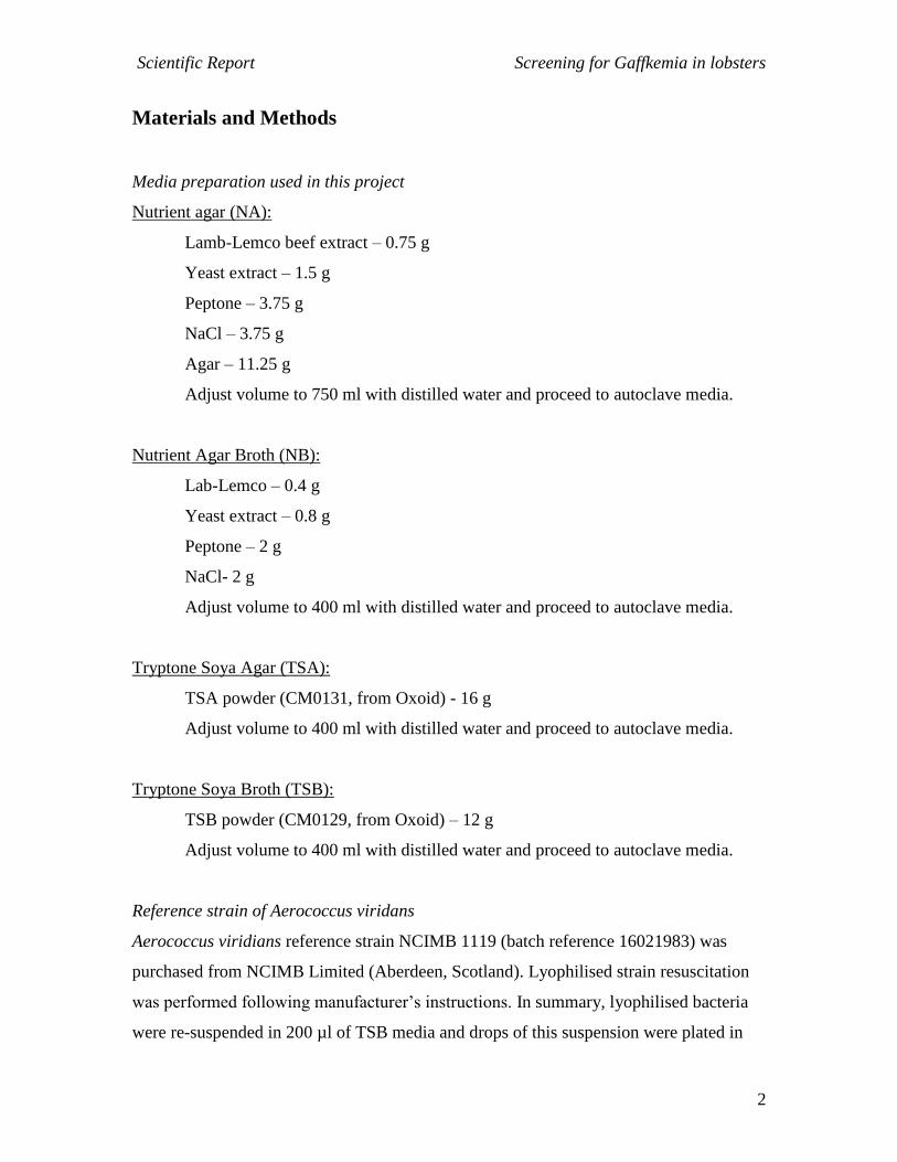
Biochemical test StrepQuick
StrepQuick was obtained from Hardy Diagnostis, California, USA. The catalogue
number was Z122. The test is intended to aid in the identification of gram-positive,
catalase-negative cocci based on the reactions pyroglutamate aminopeptidase (PYR),
leucine aminopeptidase (LAP) and esculin hydrolysis (ESC) activity. This test is to be
used only with cultures of isolated organisms. Test circles are slightly moisten by addid a
single drop of distilled water. Using a sterile plastic loop, we pick 2-3 isolated 18-24 h
colonies and rub them into a small area of the PYR reaction circle. This step is repeated
for the LAP and ESC test circles. After the test organism has been inoculated onto the
test circles we allow it to react for 10 min. After 10 min we add one drop of the
chromogenic developer to the PYR and LAP circles and immediately after addition of
this component we observe for the development of a bright pick or cherry red colour in
the PYR and LAP circles. Also observe for a light grey to grey colour to form in the ESC
circle. Interpretation of results is as follows:
Test Positive Reaction Negative reaction
PYR Cherry red or bright pink colour Orange, yellow or salmon colour or no change
LAP Cherry red or bright pink colour No colour development
ESC Light grey to grey colour Any colour other than grey
Expected results for A. viridans would be as follows:
PYR: Variable results
LAP: Negative, no colour change
ESC: Variable results

Scientific Report Screening for Gaffkemia in lobsters
4
Biochemical test Api20Strep
The Api20 STREP strip consist of 20 microtubes containing dehydrated substrates for the
demonstration of enzymatic activity or the fermentation of sugars. This enzymatic tests
have to be inoculated with a dense suspension of organisms coming from a pure culture,
which is used to rehydrate the enzymatic substrates. The metabolic end products
produced during the incubation period are either revealed through spontaneous colored
reactions or by the addition of reagents. The reactions are read according to the
Interpretation Reactions given by the manufacturer into positive or negative results. The
compiled results is transformed into a code numeric profile and the identification of the
organisms is done using the software apiwebTM
. The principle of the code profile is to
condense the binary information (+,-) into a numeric profile. Test on the strip are
separated into groups of 3 and to each reaction a value equal to 1, 2 or 4 is attributed
depending on the position of the test in tis group, 1sr, 2nd
or 3rd
respectively. The addition
of the 3 retained values (0 for negative reactions) gives a figure between 0 and 7 for each
group. The resulting profile is matched to a database. Based on the accuracy of the
identification the following identifications are possible:
- Excellent: % i.d. ≥ 99.9 % and T ≥ 0.75
- Very good: % i.d. ≥ 99.0 % and T ≥ 0.50
- Good: % i.d. ≥ 90.0 % and T ≥ 0.25
- Acceptable: % i.d. ≥ 80.0 % and T ≥ 0.0
- Unacceptable: when the profile is very far from the taxa of the database, all gross
frequencies being less than the threshold values.
Isolation of the bacterial micro-flora DNA for amplification and sequencing
Single bacterial colonies were picked randomly with sterile scrapers from TSA or PEA
plates. A fraction of the bacteria colony was dissolved in 100 µl of sterile-filtered
distilled H2O. The samples were heated at 100 ºC for 10 min and subseuqnently cooled
on ice. This step denatures all proteins within the sample and thereby releases genomic
DNA from the cells. Samples were either immediately processed for polychain reaction
(PCR) or frozen at -20 ºC until needed.
Scientific Report Screening for Gaffkemia in lobsters
5
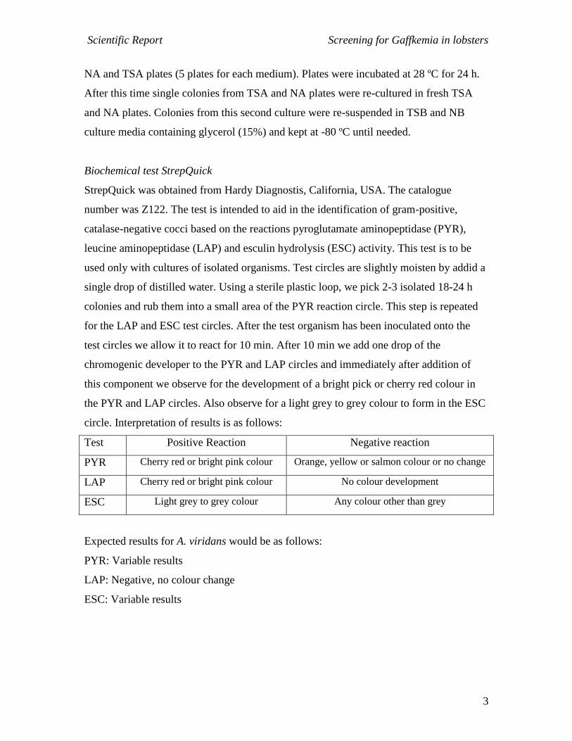
Amplification and sequencing of DNA
A 1 µl volume of crude DNA preparation was used per PCR reaction. The partial DNA
fragments of bacterial 16S rRNA genes were amplified by PCR using a forward primer
27F (equivalent to positions 8 to 27 in Escherichia coli 16S rRNA) and a reverse primer
1100R (equivalent to positions 1114 to 1110 in Escherichia coli 16S rRNA) (Table 1).
The PCR mixture (50 µl) was set up with 10 µl of 5x PCR-buffer containing 10 mM
MgCl2, 20 mM dNTPs (1 µl), 0.25 µl Taq-polymerase, 1.5 µl of each primer (stocks at 10
µM), 34.75 µl of distilled and sterile H2O and 1 µl of DNA template. The PCR
conditions were: 94 ºC for 5 min, followed by 35 cycles of 94 ºC for 1 min, 55 ºC for 1
min and 72 ºC for 2 min with a subsequent extension cycle at 72 ºC for 10 min. A 10 µl
aliquot of the amplified product was visualized on a 1.0 % agarose gel stained with
containing 10 µg/ml (w/v) of ethidium bromide. The remaining 40 µl volume of the PCR
reaction was purified using a QIAqucik PCR purification kit (QUIAGEN) following the
supplied protocol and send for sequencing.
Table 11. Primers used for 16S-rDNA amplification and sequencing.
Primer name Sequence
27-F 5’-AGA GTT TGA TCM TGG CTC AG-3’
685-R 5’-TCT ACG CAT TTC ACY GCT AC-3’
1100-R 5’-GGG TTG CGC TCG TTG-3’
Scientific Report Screening for Gaffkemia in lobsters
6
Results
Sampling of lobsters in Tarbert Fish farm
MARCH SAMPLING – On the 16/03/2010 4 lobsters with no signs of disease were
taken from Tarbert fish farm to the laboratory in Glasgow University by ice where they
were sampled. Furthermore, 2 samples of sediment were also taken:
- sediment A: bottom of the tank;
- sediment B: collected from boxes containing some lobsters
On arrival in Glasgow haemolymph from each animal was plated in MIA-media plates at
the following dilutions: 1/1, 1/10, 1/100, 1/1000 and hepatopancreas smears were also
plated in MIA-media plates. Sediments were re-suspended with 1 ml of Nephrops saline
solution and supernatants were plated at the dilutions of: 1/1, 1/10, 1/100, 1/1000,
1/10000. Volume plated in the case of haemolymph and sediment was 100 µl. Plates
were incubated at 18 °C for 48 h.
Results on the total bacteria counts in haemolymph and sediment supernatants obtained in
the plates are shown in table 1.
Table 1. TVC in haemolymph and sediment supernatants obtained in the March sampling. Each
haemolymph and sediment supernatant was plated in triplicates. Individual values and not
averages are shown in the table.
Lobster 1 Lobster 2 Lobster 3 Lobster 4 Sediment A Sediment B
Dilution 1/1 0/1/1 3/2/4 3/1/2 8/17/15
Dilution 1/10 0/0/1 1/0/0 0/0/0 4/2/1
Dilution
1/10000
37/57/45 63/79/85
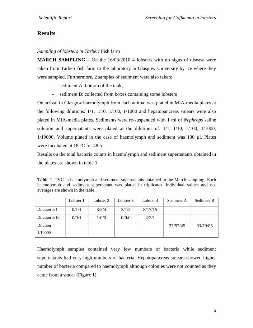
Haemolymph samples contained very few numbers of bacteria while sediment
supernatants had very high numbers of bacteria. Hepatopancreas smears showed higher
number of bacteria compared to haemolymph although colonies were not counted as they
came from a smear (Figure 1).
Scientific Report Screening for Gaffkemia in lobsters
7
Figure 1. Picture showing MIA plates with colonies obtained from sediment supernatants
(dilution 1/10000), haemolymph (dilution 1/1) and from a hepatopancreas smear
From all the colonies obtained in the plates a total of 13 different colonies of
haemolymph, 6 colonies from the hepatopancreas and 7 colonies from the sediments
supernatants were further studied by doing gram staining and results are summarized in
Table 2.
Table 2. Information and gram staining results from selected colonies (March sampling)
Num colony
Shape colony
Colour colony Size colony
Origin More info
Gram staining/shape bacteria
1 Round Orange M/B Haemolymph Lobs 2 Gram negative, rod. Similar to colony 36
2 Round Cream/trans M Haemolymph Lobs 2 Gram negative, short rod
3 Round Cream/white M/B Haemolymph Lobs 4 Gram negative, long filamentous rod
4 Round Yellow M/B Haemolymph Lobs 4 Gram negative, rod
5 Round/Irreg Orange/yell B Haemolymph Lobs 4 Gram negative, rod
6 Round/Irreg Trans lucid M Haemolymph Lobs 4 Gram negative, rod
7 Irregular White B Haemolymph Lobs 4 Gram negative
8 Round Trans /yellow B Haemolymph Lobs 4 Gram negative
9 Round Trans lucid S Haemolymph Lobs 4 Gram positive, rod
10 Round White/Trans S Haemolymph Lobs 1 Gram negative, rod
11 Round Cream/Trans M Haemolymph Lobs 2 Gram negative, short rod
Scientific Report Screening for Gaffkemia in lobsters
8
Num colony
Shape colony
Colour colony Size colony
Origin More info
Gram staining/shape bacteria
12 Round Orange S/M Haemolymph Lobs 4 Gram negative, short rod
13 Round/Irreg Yellow M Haemolymph Lobs 4 Gram negative. Short rod
21 Round Cream/black M Hepatopanc Lobs 2 Gram negative, short rod
22 Round Cream M Hepatopanc Lobs 3 Gram negative, ovoid. Similar to colonies 24, 26.
23 Round Trans lucid M Hepatopanc Lobs 4 Gram negative, rod
24 Round Cream S Hepatopanc Lobs 1 Gram negative, ovoid. Similar to colonies 22, 26.
25 Round Cream/Orange M Hepatopanc Lobs 2 Gran negative, rod. Similar to colony 23.
26 Round Cream M Hepatopanc Lobs 4 Gram negative, ovoid. Similar to colonies 22, 24.
31 Round Trans/black M Sediment A Gram negative, rod
32 Round Cream M/B Sediment A Gram negative, cocci
33 Round Yellow/Trans M/B Sediment A Gram negative, rod
34 Round/Irreg Orange B Sediment B Gram negative, filamentous
35 Round Pink middle M Sediment B Gram positive, rod. Possibly H2S producer
36 Round Orange M Sediment B Gram negative, rod
37 Round Yellow M/S Sediment A Gram negative, rod curved like kidney shape
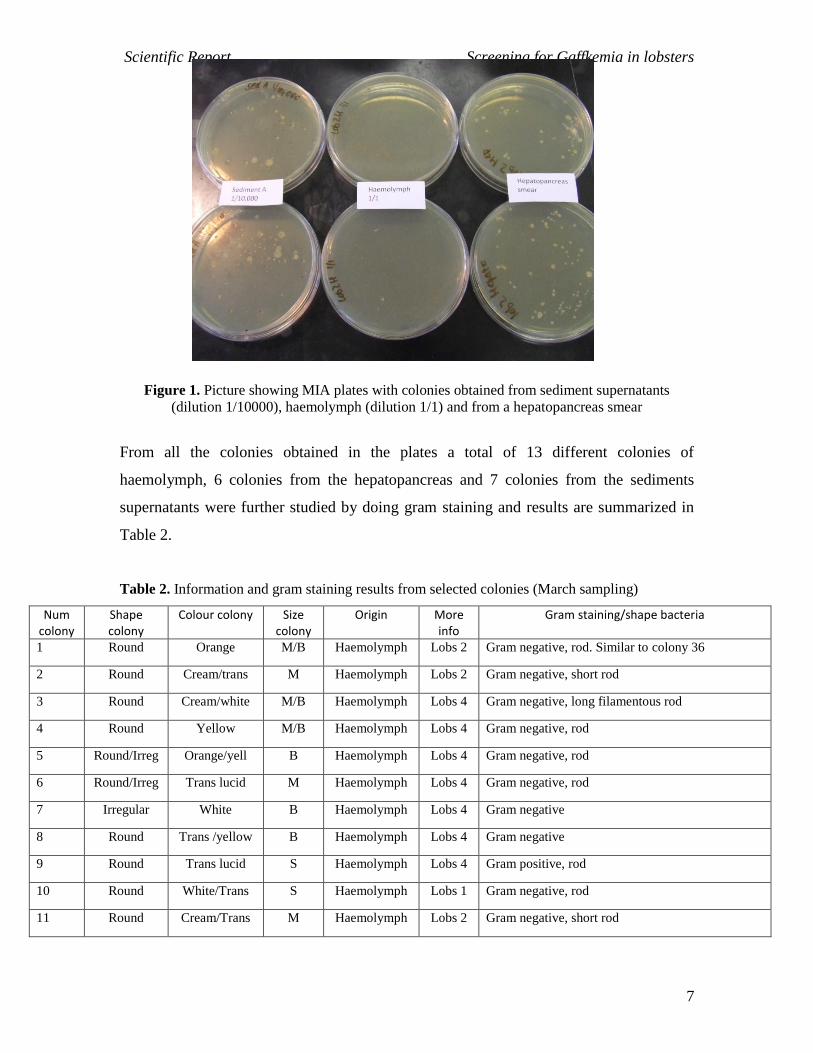
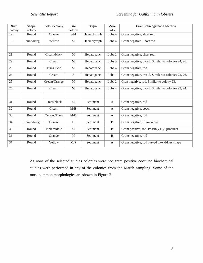
As none of the selected studies colonies were not gram positive cocci no biochemical
studies were performed in any of the colonies from the March sampling. Some of the
most common morphologies are shown in Figure 2.
Scientific Report Screening for Gaffkemia in lobsters
9
Colony 4 Colony 21
Colony 25 Colony 34
Figure 2. Gram staining slides of colonies 4, 21, 25 and 34 all obtained in the March sampling in
a magnification of x1000.
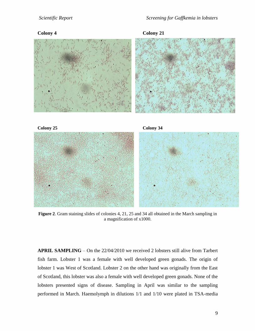
APRIL SAMPLING – On the 22/04/2010 we received 2 lobsters still alive from Tarbert
fish farm. Lobster 1 was a female with well developed green gonads. The origin of
lobster 1 was West of Scotland. Lobster 2 on the other hand was originally from the East
of Scotland, this lobster was also a female with well developed green gonads. None of the
lobsters presented signs of disease. Sampling in April was similar to the sampling
performed in March. Haemolymph in dilutions 1/1 and 1/10 were plated in TSA-media

Scientific Report Screening for Gaffkemia in lobsters
10
plates and we also performed smears of hepatopancreas that were also plated in TSA-
media plates. All plates were incubated at 28 °C for 48 h. Furthermore, in this sampling
we also performed gram staining of the haemolymph directly in order to establish if by
any chance the lobsters had an advance stage of infection that could be detected in this
way. Results as expected indicated that these lobsters were free of advance infection as
haemolymph gram staining did not show any aggregation of bacteria. Results from the
plates indicated that the levels of bacteria were low in haemolymph (Table 3) and also in
hepatopancreas.
Table 3. TVC in haemolymph plated samples obtained in the April sampling. Each haemolymph
was plated in triplicates. Individual values and not averages are shown in the table.
Haemolymph
dilution
Lobster 1-West of Scotland Lobster 2- East of Scotland
Dilution 1/1 2/1/1 0/0/0
Dilution 1/10 0/0/0 0/0/0
All the colonies obtained from lobster 1 haemolymph were further studied by performing
gram staining and results are shown in Table 4. Furthermore, as some more colonies were
obtained in the plates from hepatopancreas smears a total of 4 colonies of lobster 1 and 5
colonies of lobster 2 were also further studied.
Table 4. Information and gram staining results from selected colonies (March sampling). In black
we highlight colonies were re-cultured and maintained at -80 °C to perform biochemical tests.
Num colony
Shape colony
Colour colony Size colony
Origin More info
Gram staining/shape bacteria
40 Round Yellow M Hepatopancr Lobs 1 Gram positive, cocci. Some tetrads, re-culture
41 Round Cream/white M/B Hepatopancr Lobs 1 Gram negative, rod
42 Round Cream/white B Hepatopancr Lobs 1 Gram negative, rod. Similar to colonies 46 and 47.
43 Round White S Hepatopancr Lobs 1 Gram negative, cocci. Re-culture
44 Round Orange M Hepatopancr Lobs 2 Gram negative, rod
45 Round White M Hepatopancr Lobs 2 Gram negative, short rod

Scientific Report Screening for Gaffkemia in lobsters
11
46 Round Cream/white B Hepatopancr Lobs 2 Gram negative, rod. Similar to colonies 42 and 47.
47 Irregular Cream/white B Hepatopancr Lobs 2 Gram negative, rod. Similar to colonies 42 and 46.
48 Irregular Trans lucid M Hepatopancr Lobs 2 Gram positive, short rod
49 Round Yellow M Hepatopancr Lobs 2 Gram positive, cocci. Re-culture
54 Round Yellow/trans S Hepatopancr Lobs 1 Gram negative, short rod
50 Round Yellow M Haemolymph Lobs 1 Gram positive, cocci. Some tetrads. Re-culture
51 Round Yellow M Haemolymph Lobs 1 Gram positive, cocci. Some tetrads. Re-culture
52 Round White S Haemolymph Lobs 1 Gram positive, cocci. Re-culture
53 Round Cream/Trans M Haemolymph Lobs 1 Gram negative, short rod
In the April sampling interestingly we found 5 colonies that were gram positive cocci and
one more colony that was gram negative cocci. As gram staining sometimes can be
confusing we re-cultured all 6 colonies named: 40, 43, 49, 50, 51 and 52 in TSA-media
plates and they were stored at -80 °C until the biochemical tests were performed.
Colony 40 Colony 52
Figure 3. Gram staining slides of colonies 40 and 52 in a magnification of x1000.
Scientific Report Screening for Gaffkemia in lobsters
12
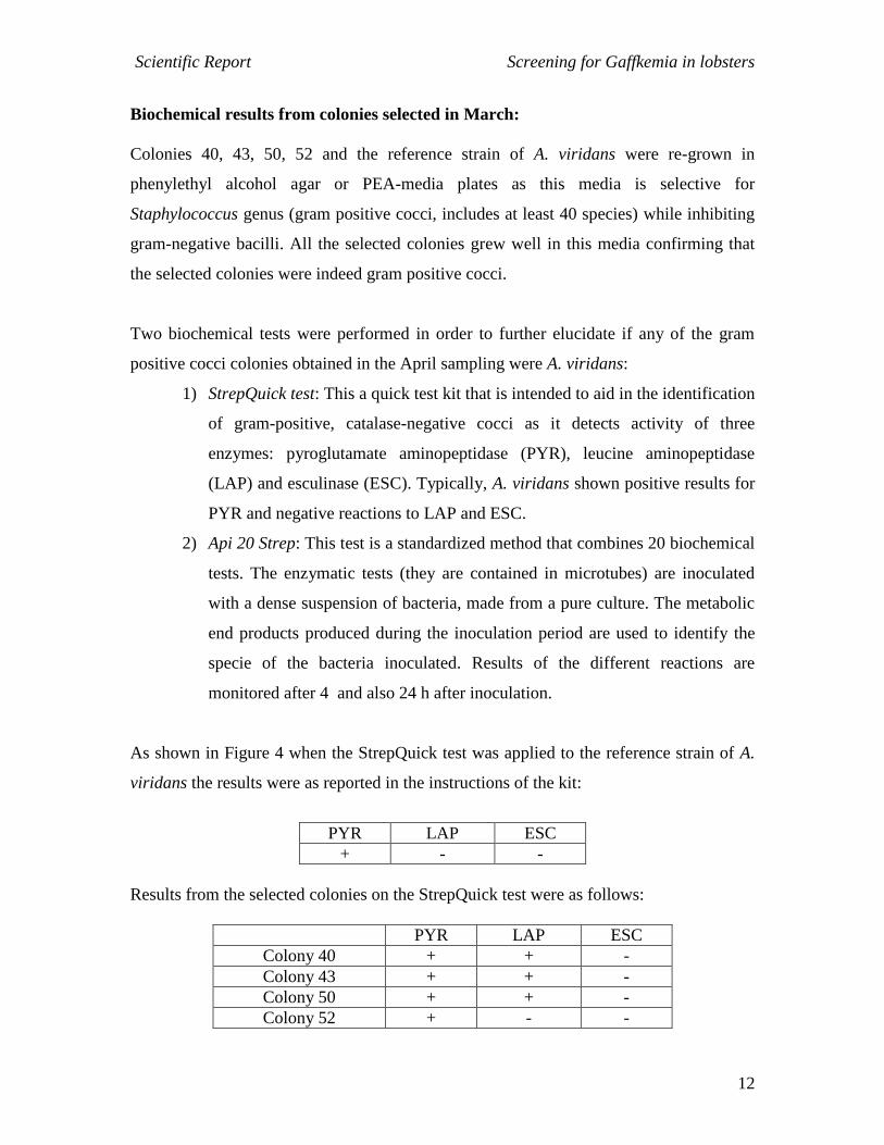
Biochemical results from colonies selected in March:
Colonies 40, 43, 50, 52 and the reference strain of A. viridans were re-grown in
phenylethyl alcohol agar or PEA-media plates as this media is selective for
Staphylococcus genus (gram positive cocci, includes at least 40 species) while inhibiting
gram-negative bacilli. All the selected colonies grew well in this media confirming that
the selected colonies were indeed gram positive cocci.
Two biochemical tests were performed in order to further elucidate if any of the gram
positive cocci colonies obtained in the April sampling were A. viridans:
1) StrepQuick test: This a quick test kit that is intended to aid in the identification
of gram-positive, catalase-negative cocci as it detects activity of three
enzymes: pyroglutamate aminopeptidase (PYR), leucine aminopeptidase
(LAP) and esculinase (ESC). Typically, A. viridans shown positive results for
PYR and negative reactions to LAP and ESC.
2) Api 20 Strep: This test is a standardized method that combines 20 biochemical
tests. The enzymatic tests (they are contained in microtubes) are inoculated
with a dense suspension of bacteria, made from a pure culture. The metabolic
end products produced during the inoculation period are used to identify the
specie of the bacteria inoculated. Results of the different reactions are
monitored after 4 and also 24 h after inoculation.
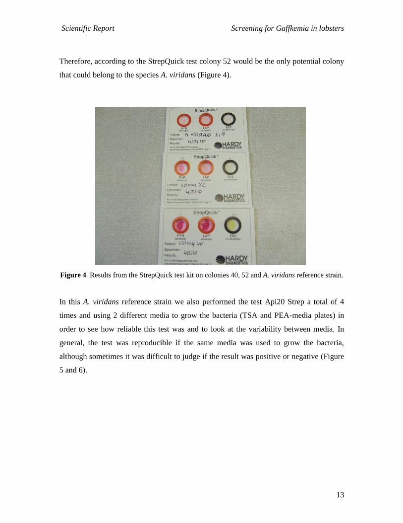
As shown in Figure 4 when the StrepQuick test was applied to the reference strain of A.
viridans the results were as reported in the instructions of the kit:
PYR LAP ESC
+ - -
Results from the selected colonies on the StrepQuick test were as follows:
PYR LAP ESC
Colony 40 + + -
Colony 43 + + -
Colony 50 + + -
Colony 52 + - -
Scientific Report Screening for Gaffkemia in lobsters
13
Therefore, according to the StrepQuick test colony 52 would be the only potential colony
that could belong to the species A. viridans (Figure 4).
Figure 4. Results from the StrepQuick test kit on colonies 40, 52 and A. viridans reference strain.
In this A. viridans reference strain we also performed the test Api20 Strep a total of 4
times and using 2 different media to grow the bacteria (TSA and PEA-media plates) in
order to see how reliable this test was and to look at the variability between media. In
general, the test was reproducible if the same media was used to grow the bacteria,
although sometimes it was difficult to judge if the result was positive or negative (Figure
5 and 6).

Scientific Report Screening for Gaffkemia in lobsters
14
A)
B)
Figure 5. Results from the Api 20 Strep test on A. viridans reference strain cultured in TSA
plated. A) Results after 4 h and B) Results after 24 h.

Scientific Report Screening for Gaffkemia in lobsters
15
A)
B)
Figure 6. Results from the Api 20 Strep test on A. viridans reference strain cultured in PEA
plated. A) Results after 4 h and B) Results after 24 h.
Scientific Report Screening for Gaffkemia in lobsters
16
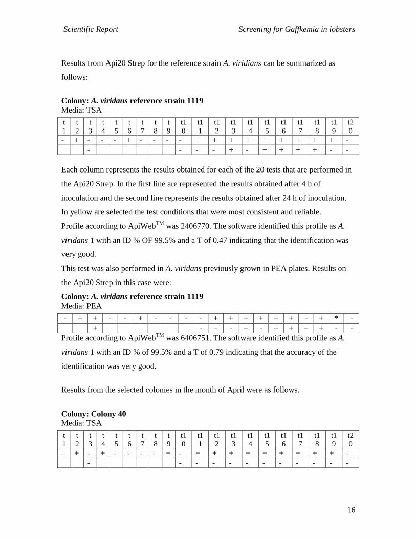
Results from Api20 Strep for the reference strain A. viridians can be summarized as
follows:
Colony: A. viridans reference strain 1119
Media: TSA
Each column represents the results obtained for each of the 20 tests that are performed in
the Api20 Strep. In the first line are represented the results obtained after 4 h of
inoculation and the second line represents the results obtained after 24 h of inoculation.
In yellow are selected the test conditions that were most consistent and reliable.
Profile according to ApiWebTM
was 2406770. The software identified this profile as A.
viridans 1 with an ID % OF 99.5% and a T of 0.47 indicating that the identification was
very good.
This test was also performed in A. viridans previously grown in PEA plates. Results on
the Api20 Strep in this case were:
Colony: A. viridans reference strain 1119
Media: PEA
Profile according to ApiWebTM
was 6406751. The software identified this profile as A.
viridans 1 with an ID % of 99.5% and a T of 0.79 indicating that the accuracy of the
identification was very good.
Results from the selected colonies in the month of April were as follows.
Colony: Colony 40
Media: TSA
t
1
t
2
t
3
t
4
t
5
t
6
t
7
t
8
t
9
t1
0
t1
1
t1
2
t1
3
t1
4
t1
5
t1
6
t1
7
t1
8
t1
9
t2
0
- + - - - + - - - - + + + + + + + + + -
- - - - + - + + + + - -
- + + - - + - - - - + + + + + + - + * -
+ - - - + - + + + + - -
t
1
t
2
t
3
t
4
t
5
t
6
t
7
t
8
t
9
t1
0
t1
1
t1
2
t1
3
t1
4
t1
5
t1
6
t1
7
t1
8
t1
9
t2
0
- + - + - - - - + - + + + + + + + + + -
- - - - - - - - - - - -
Scientific Report Screening for Gaffkemia in lobsters
17
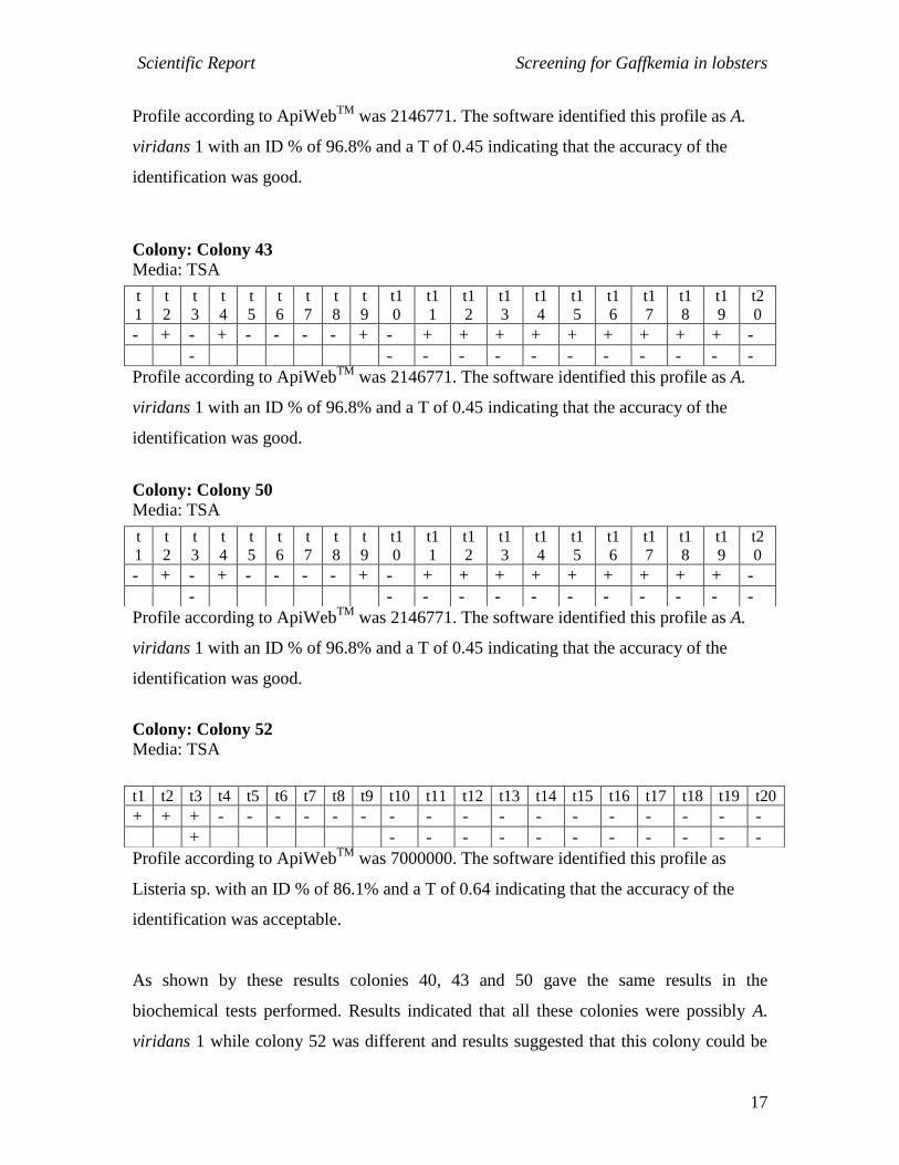
Profile according to ApiWebTM
was 2146771. The software identified this profile as A.
viridans 1 with an ID % of 96.8% and a T of 0.45 indicating that the accuracy of the
identification was good.
Colony: Colony 43
Media: TSA
Profile according to ApiWebTM
was 2146771. The software identified this profile as A.
viridans 1 with an ID % of 96.8% and a T of 0.45 indicating that the accuracy of the
identification was good.
Colony: Colony 50
Media: TSA
Profile according to ApiWebTM
was 2146771. The software identified this profile as A.
viridans 1 with an ID % of 96.8% and a T of 0.45 indicating that the accuracy of the
identification was good.
Colony: Colony 52
Media: TSA
Profile according to ApiWebTM
was 7000000. The software identified this profile as
Listeria sp. with an ID % of 86.1% and a T of 0.64 indicating that the accuracy of the
identification was acceptable.
As shown by these results colonies 40, 43 and 50 gave the same results in the
biochemical tests performed. Results indicated that all these colonies were possibly A.
viridans 1 while colony 52 was different and results suggested that this colony could be
t
1
t
2
t
3
t
4
t
5
t
6
t
7
t
8
t
9
t1
0
t1
1
t1
2
t1
3
t1
4
t1
5
t1
6
t1
7
t1
8
t1
9
t2
0
- + - + - - - - + - + + + + + + + + + -
- - - - - - - - - - - -
t
1
t
2
t
3
t
4
t
5
t
6
t
7
t
8
t
9
t1
0
t1
1
t1
2
t1
3
t1
4
t1
5
t1
6
t1
7
t1
8
t1
9
t2
0
- + - + - - - - + - + + + + + + + + + -
- - - - - - - - - - - -
t1 t2 t3 t4 t5 t6 t7 t8 t9 t10 t11 t12 t13 t14 t15 t16 t17 t18 t19 t20
+ + + - - - - - - - - - - - - - - - - -
+ - - - - - - - - - - -

Scientific Report Screening for Gaffkemia in lobsters
18
from the genus Listeria. However, none of the identifications gave an accuracy of very
good or excellent.
MAY SAMPLING – On the 13th
of May 2010 we received another 2 lobsters from
Tarbert fish farm. Lobster 1 was originally from the East Coast, male with a CL of 92.6
mm while lobster 2 was originally from the West Coast, male with a CL of 97.2 mm. In
this sampling haemolymph (un-diluted) and hepatopancreas smears were platted in TSA
and also in PEA-media plates. All plates were incubated at 28 °C for 48 h. Total viable
counts of bacteria are shown in Table 5 and 6.
Table 5. TVC in haemolymph plated samples obtained in the May sampling. Each haemolymph
was plated in tetraplicates. Individual values and not averages are shown in the table.
Haemolymph Lobster 1-East of Scotland Lobster 2- West of Scotland
Dilution 1/1 TSA-media 0/0/0/1 0/0/0/1
Dilution 1/1 PEA-media 0/0/0/0 3/0/0/0
Table 6. TVC in hepatopancreas smears plated samples obtained in the May sampling. Each plate
contained the bacteria from an individual hepatopancreas smear. Individual values and not
averages are shown in the table.
Hepatopancreas smears Lobster 1-East of Scotland Lobster 2- West of Scotland
TSA-media 1/2/1/6 12/3/7
PEA-media 0/0/0 2/0/0
Similarly, to previous samplings bacteria numbers were low especially in the
haemolymph suggesting that none of the lobsters had an advanced level of infection.
Table 4. Information and gram staining results from selected colonies (May sampling). In black
we highlight colonies were re-cultured and maintained at -80 °C to perform biochemical tests.
Num colony
Shape colony
Colour colony Size colony
Origin More info
Gram staining/shape bacteria
60 Irregular White S Hepatopancr Lobs 2 Gram staining not clear, small cocci
61 Irregular Cream S Hepatopancr Lobs 2 Gram staining not clear, small cocci
Scientific Report Screening for Gaffkemia in lobsters
19
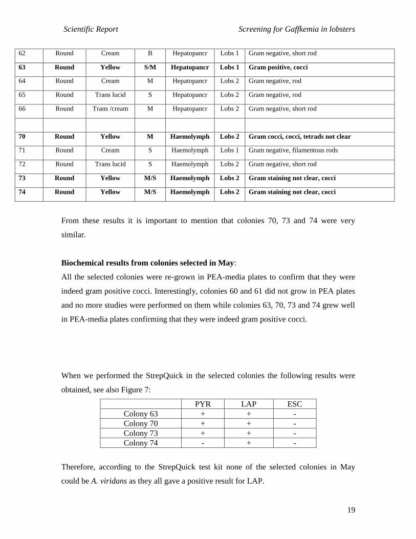
62 Round Cream B Hepatopancr Lobs 1 Gram negative, short rod
63 Round Yellow S/M Hepatopancr Lobs 1 Gram positive, cocci
64 Round Cream M Hepatopancr Lobs 2 Gram negative, rod
65 Round Trans lucid S Hepatopancr Lobs 2 Gram negative, rod
66 Round Trans /cream M Hepatopancr Lobs 2 Gram negative, short rod
70 Round Yellow M Haemolymph Lobs 2 Gram cocci, cocci, tetrads not clear
71 Round Cream S Haemolymph Lobs 1 Gram negative, filamentous rods
72 Round Trans lucid S Haemolymph Lobs 2 Gram negative, short rod
73 Round Yellow M/S Haemolymph Lobs 2 Gram staining not clear, cocci
74 Round Yellow M/S Haemolymph Lobs 2 Gram staining not clear, cocci
From these results it is important to mention that colonies 70, 73 and 74 were very
similar.
Biochemical results from colonies selected in May:
All the selected colonies were re-grown in PEA-media plates to confirm that they were
indeed gram positive cocci. Interestingly, colonies 60 and 61 did not grow in PEA plates
and no more studies were performed on them while colonies 63, 70, 73 and 74 grew well
in PEA-media plates confirming that they were indeed gram positive cocci.
When we performed the StrepQuick in the selected colonies the following results were
obtained, see also Figure 7:
PYR LAP ESC
Colony 63 + + -
Colony 70 + + -
Colony 73 + + -
Colony 74 - + -
Therefore, according to the StrepQuick test kit none of the selected colonies in May
could be A. viridans as they all gave a positive result for LAP.
Scientific Report Screening for Gaffkemia in lobsters
20
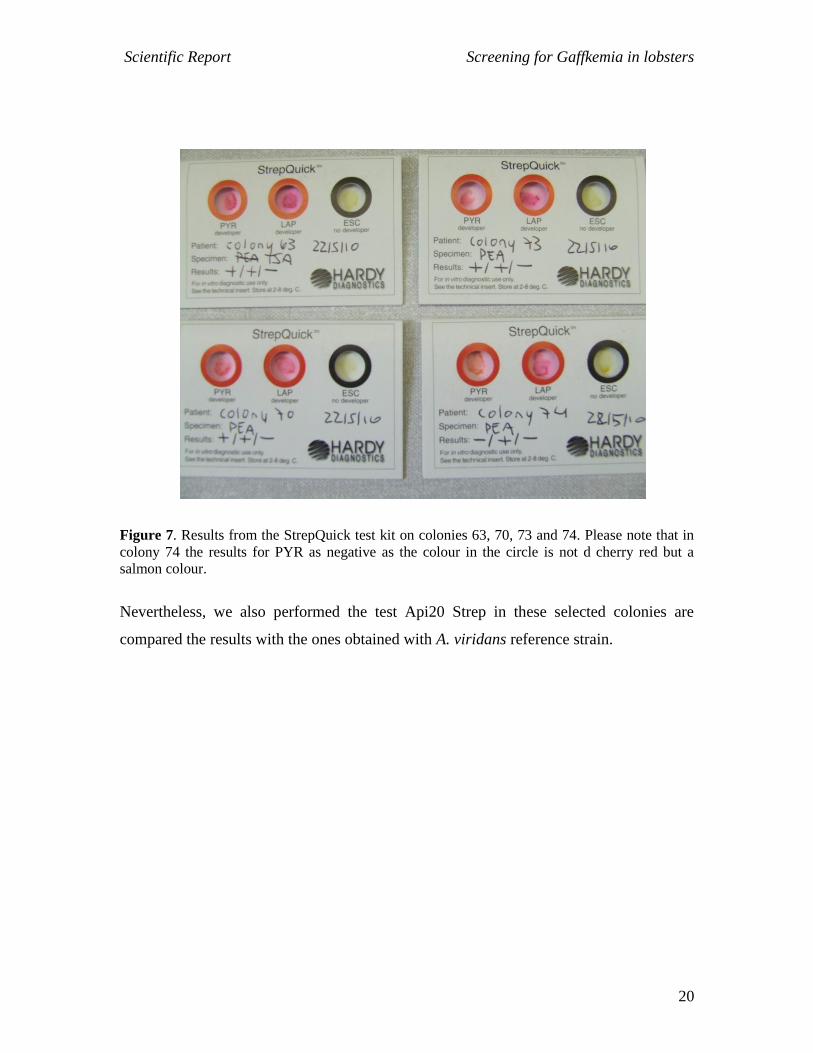
Figure 7. Results from the StrepQuick test kit on colonies 63, 70, 73 and 74. Please note that in
colony 74 the results for PYR as negative as the colour in the circle is not d cherry red but a
salmon colour.
Nevertheless, we also performed the test Api20 Strep in these selected colonies are
compared the results with the ones obtained with A. viridans reference strain.
Scientific Report Screening for Gaffkemia in lobsters
21
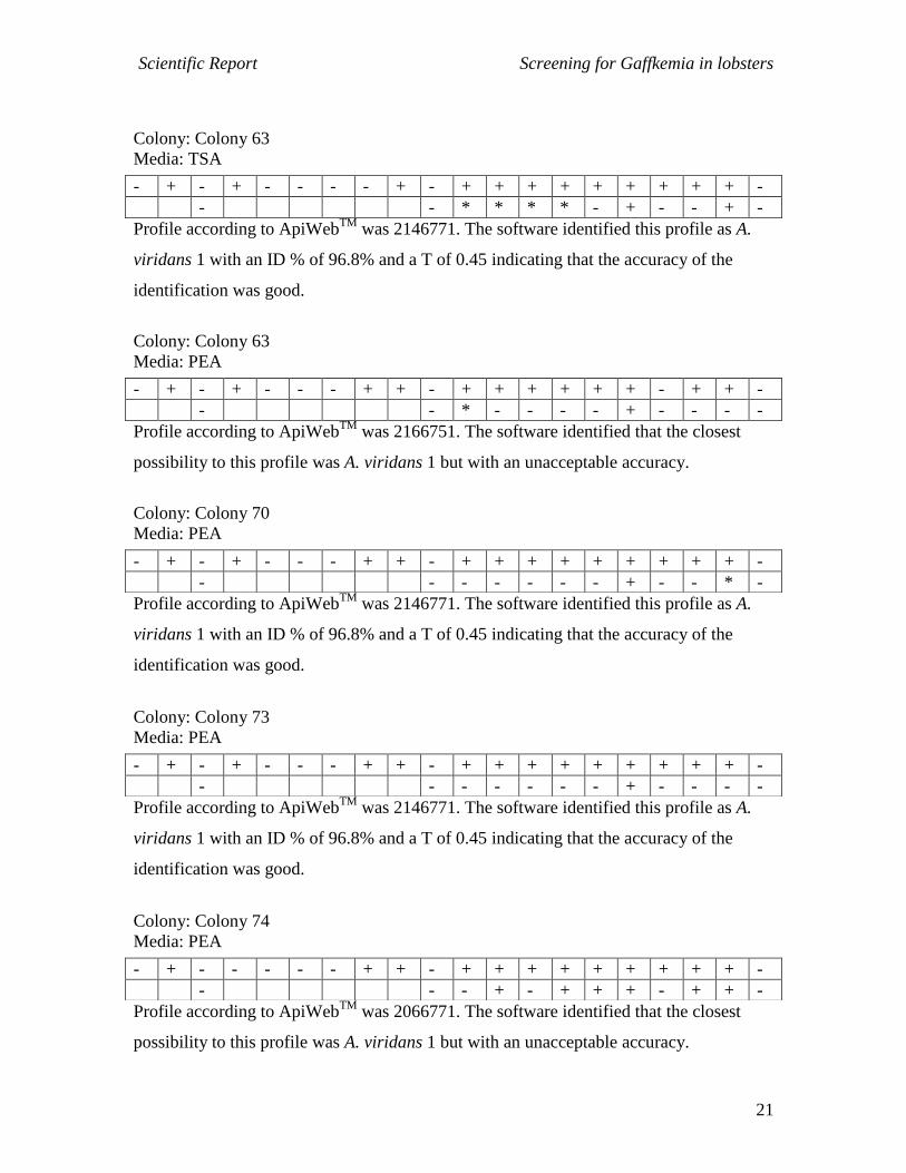
Colony: Colony 63
Media: TSA
Profile according to ApiWebTM
was 2146771. The software identified this profile as A.
viridans 1 with an ID % of 96.8% and a T of 0.45 indicating that the accuracy of the
identification was good.
Colony: Colony 63
Media: PEA
Profile according to ApiWebTM
was 2166751. The software identified that the closest
possibility to this profile was A. viridans 1 but with an unacceptable accuracy.
Colony: Colony 70
Media: PEA
Profile according to ApiWebTM
was 2146771. The software identified this profile as A.
viridans 1 with an ID % of 96.8% and a T of 0.45 indicating that the accuracy of the
identification was good.
Colony: Colony 73
Media: PEA
Profile according to ApiWebTM
was 2146771. The software identified this profile as A.
viridans 1 with an ID % of 96.8% and a T of 0.45 indicating that the accuracy of the
identification was good.
Colony: Colony 74
Media: PEA
Profile according to ApiWebTM
was 2066771. The software identified that the closest
possibility to this profile was A. viridans 1 but with an unacceptable accuracy.
- + - + - - - - + - + + + + + + + + + -
- - * * * * - + - - + -
- + - + - - - + + - + + + + + + - + + -
- - * - - - - + - - - -
- + - + - - - + + - + + + + + + + + + -
- - - - - - - + - - * -
- + - + - - - + + - + + + + + + + + + -
- - - - - - - + - - - -
- + - - - - - + + - + + + + + + + + + -
- - - + - + + + - + + -

Scientific Report Screening for Gaffkemia in lobsters
22
Similarly to the situation in the April sampling it was not possible to conclude that any of
the selected colonies were A. viridians although colonies 63, 70 and 73 had strong
possibilities to be A. viridans.
Scientific Report Screening for Gaffkemia in lobsters
23
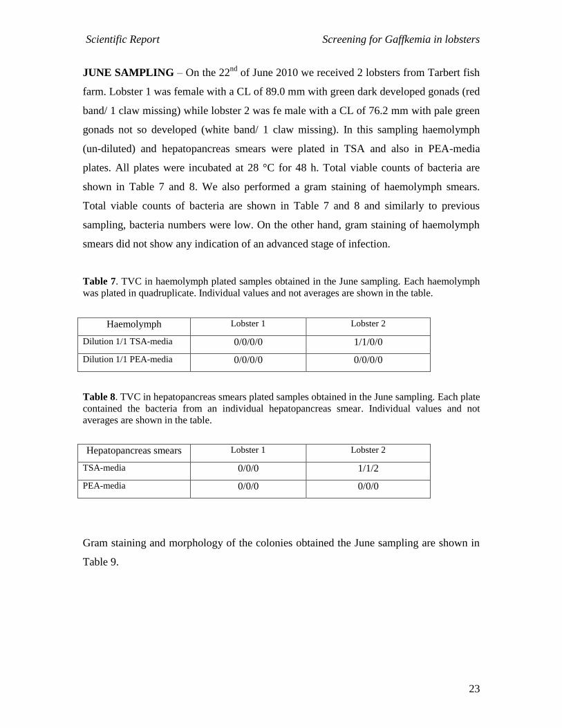
JUNE SAMPLING – On the 22nd
of June 2010 we received 2 lobsters from Tarbert fish
farm. Lobster 1 was female with a CL of 89.0 mm with green dark developed gonads (red
band/ 1 claw missing) while lobster 2 was fe male with a CL of 76.2 mm with pale green
gonads not so developed (white band/ 1 claw missing). In this sampling haemolymph
(un-diluted) and hepatopancreas smears were plated in TSA and also in PEA-media
plates. All plates were incubated at 28 °C for 48 h. Total viable counts of bacteria are
shown in Table 7 and 8. We also performed a gram staining of haemolymph smears.
Total viable counts of bacteria are shown in Table 7 and 8 and similarly to previous
sampling, bacteria numbers were low. On the other hand, gram staining of haemolymph
smears did not show any indication of an advanced stage of infection.
Table 7. TVC in haemolymph plated samples obtained in the June sampling. Each haemolymph
was plated in quadruplicate. Individual values and not averages are shown in the table.
Haemolymph Lobster 1 Lobster 2
Dilution 1/1 TSA-media 0/0/0/0 1/1/0/0
Dilution 1/1 PEA-media 0/0/0/0 0/0/0/0
Table 8. TVC in hepatopancreas smears plated samples obtained in the June sampling. Each plate
contained the bacteria from an individual hepatopancreas smear. Individual values and not
averages are shown in the table.
Hepatopancreas smears Lobster 1 Lobster 2
TSA-media 0/0/0 1/1/2
PEA-media 0/0/0 0/0/0
Gram staining and morphology of the colonies obtained the June sampling are shown in
Table 9.
Scientific Report Screening for Gaffkemia in lobsters
24
Table 9. Information and gram staining results from selected colonies (June sampling). In black
we highlight colonies were re-cultured and maintained at -80 °C to perform biochemical tests.
Num colony
Shape colony
Colour colony Size colony
Origin More info
Gram staining/shape bacteria
81 Round White M/S Haemolymph Lobs 1 From TSA plate/Gram negative, cocci, no tetrad
82 Round Orange B Haemolymph Lobs 2 From TSA plate/Gram positive, cocci, no tetrads
83 Round Cream B Hepatopancr Lobs 2 From TSA plate/Gram positive, cocci
84 Round Cream M Hepatopancr Lobs 2 Gram negative, short rods
85 Round Cream M Hepatopancr Lobs 2 Gram negative, rod
86 Round Trans lucid S Hepatopancr Lobs 2 Gram negative, short rod
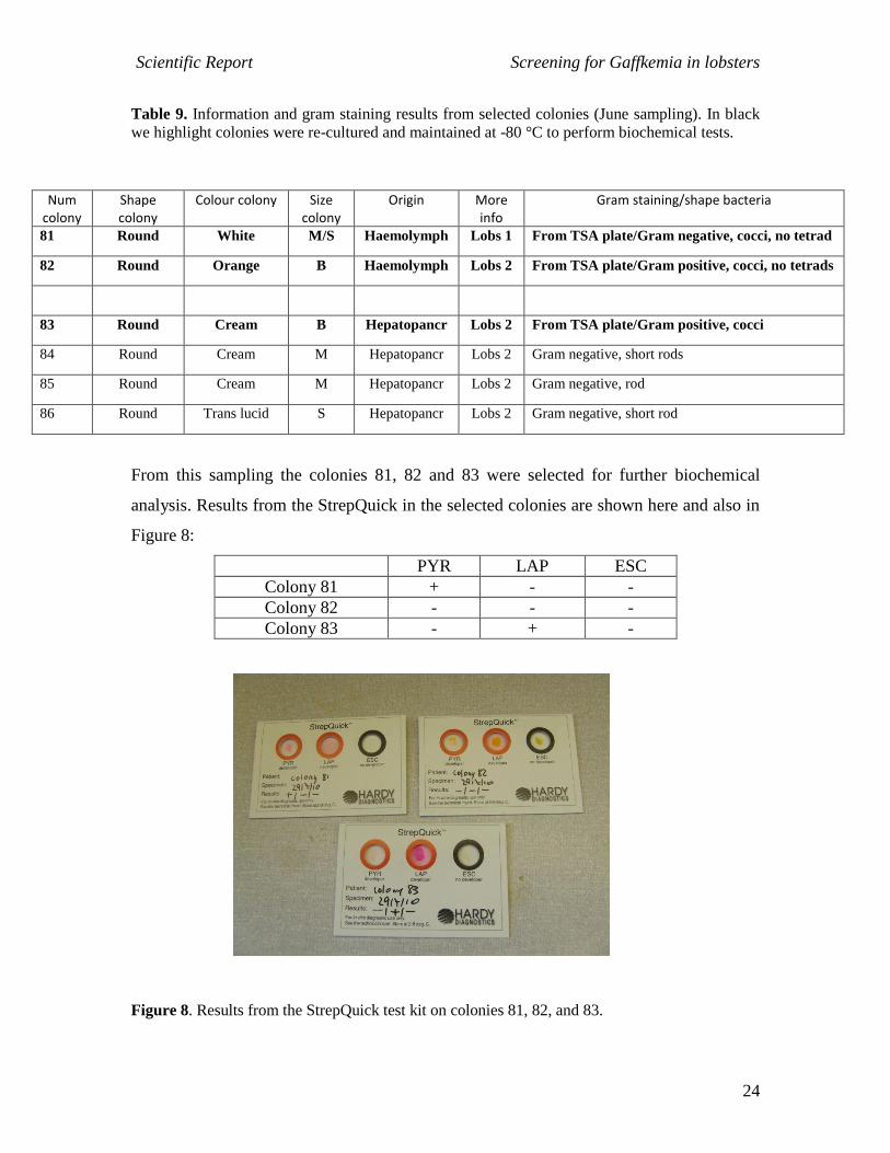
From this sampling the colonies 81, 82 and 83 were selected for further biochemical
analysis. Results from the StrepQuick in the selected colonies are shown here and also in
Figure 8:
PYR LAP ESC
Colony 81 + - -
Colony 82 - - -
Colony 83 - + -
Figure 8. Results from the StrepQuick test kit on colonies 81, 82, and 83.
Scientific Report Screening for Gaffkemia in lobsters
25
Therefore according to this test the only colony that could be A. viridians would be
colony 81. Results from the Api20 Strep are shown here:
Colony: Colony 81
Media: TSA
Profile according to ApiWebTM
was 3100000. The software identified this profile as A.
viridans 2 with an ID % of 94.4% and a T of 0.67 indicating that the accuracy of the
identification was good.
Colony: Colony 82
Media: TSA
Profile according to ApiWebTM
was 3446753. The software identified that the closest
possibility to this profile was A. viridans 1 but with an unacceptable accuracy.
Colony: Colony 83
Media: TSA
Profile according to ApiWebTM
was 0046751. The software identified this profile as A.
viridans 2 with an ID % of 93.0% and a T of 0.49 indicating that the accuracy of the
identification was good.
AUGUST SAMPLING – On the 8th of August 2010 we received 3 lobsters we proceed
with some in situ sampling in Tarbert fish farm. We collected 3 lobsters and also some
water samples from one of the 2 tanks were lobsters were kept. Lobster 1 was male with a
CL of 89.6 mm (1 red band; stocked 2-3 weeks ago and placed in a tray); lobster 2 was
male with a CL of 95.5 mm (2 red bands; stocked 1 week ago; right tank and not placed
in a tray) and finally lobster 3 was a dead male which was a new comer that had not
survived the transport. Furthermore, 4 water samples were collected from one of the
tanks (see Figure 9).
+ + - +/- - - - - - - - - - - - - - - - -
- + - - - - - - - - - -
+ + - - - + - - + - + + + + + + - + + +
+/- + + - + - - + - + - -
- - - - - - - - + - + + + + + + - + + -
- - - - - - - - - - - -

Scientific Report Screening for Gaffkemia in lobsters
26
Figure 9. Location of the water samples collected in one of the tanks. Note that inlet water was
clear while samples 1, 2 and 3 had scum.
In this sampling haemolymph (un-diluted), water un-diluted or diluted and
hepatopancreas smears were plated in TSA and also in PEA-media plates. All plates were
incubated at 28 °C for 48 h. Total viable counts of bacteria are shown in Table 10, 11 and
12. We also performed a gram staining of haemolymph smears. Total viable counts of
bacteria in haemolypmh and hepatopancreas from lobster 1 and 2 were low while higher
number were obtained in lobster 3 possibly due to the fact that the animal was dead. On
the other hand, gram staining of haemolymph smears did not show any indication of an
advanced stage of infection.
Table 10. TVC in haemolymph plated samples obtained in the August sampling. Each
haemolymph was plated in tetraplicates. Individual values and not averages are shown in the
table.
Haemolymph Lobster 1 Lobster 2 Lobster 3
Dilution 1/1 TSA-media 1/1/7 0/0/0 Dead, not available
Dilution 1/1 PEA-media 0/0/1 0/0/1 Dead, not available
Inlet
1 2
3
Circulation of water
Scientific Report Screening for Gaffkemia in lobsters
27
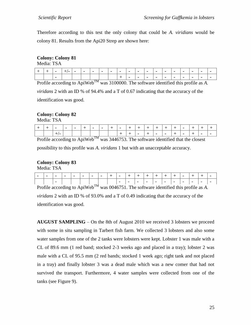
Table 11. TVC in hepatopancreas smears plated samples obtained in the August sampling. Each
plate contained the bacteria from an individual hepatopancreas smear. Individual values and not
averages are shown in the table.
Hepatopancreas smears Lobster 1 Lobster 2 Lobster 3
TSA-media 9/0/4 5/6/4 -
PEA-media 0/0/0 1/2/13 18/54/14
Table 12. TVC in water samples obtained in the August sampling. Each plate contained the
bacteria from an individual hepatopancreas smear. Individual values and not averages are shown
in the table.
Hepatopancreas smears Inlet 1 Water 1 Water 2 Water 3
TSA-media (1/1) 37/95
PEA-media (1/1) 4/4
TSA-media (1/10)
PEA-media (1/10) 13/5/1 13/15 13/10
TSA-media (1/100)
PEA-media (1/100) 1/2 2/3 2/0
TSA-media (1/1000) 53/43 10/15 35/21
PEA-media (1/1000)
Characteristics of the bacteria obtained are shown in Table 13.
Scientific Report Screening for Gaffkemia in lobsters
28
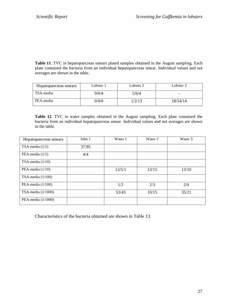
Table 9. Information and gram staining results from selected colonies (August sampling). In
black we highlight colonies were re-cultured and maintained at -80 °C to perform biochemical
tests.
Num colony
Shape colony
Colour colony Size colony
Origin More info
Gram staining/shape bacteria
90 Round Yellow M Haemolymph Lobs 1 From PEA plate/Gram positive, cocci
91 Round White M Haemolymph Lobs 2 From PEA plate/Gram positive, cocci, no tetrads
92 Round White M Haemolymph Lobs 1 From TSA plate/Gram positive, cocci, no tetrads
93 Round Yellow M/S Haemolymph Lobs 1 From TSA plate/Gram negative, short rods
94 Round Cream/Trans M Hepatopancr Lobs 3 From PEA plate/Filamentous bacteria
95 Round White M Hepatopancr Lobs 3 From PEA plate/Gram positive, cocci
96 Round Trans/white
centre
M Hepatopancr Lobs 3 From PEA plate/Staining not conclusive, rods
98 Round Cream M Hepatopancr Lobs 2 From PEA plate/Gram negative, rods
99 Round White M Inlet H2O Lobs 2 From PEA plate/Gram positive, cocci, no tetrads
100 Round Cream dark M Inlet H2O Lobs 2 From PEA plate/Gram positive, cocci, no tetrads
101 Round Cream M Inlet H2O Lobs 2 From PEA plate/Staining not conclusive, rods
102 Round Trans lucid M/S 1-H2O Lobs 2 From TSA plate/Gram negative, rods
103 Irregular White B 3- H2O Lobs 2 From PEA plate/Gram positive, cocci, no tetrads
Colonies selected for biochemical analysis were 90, 91, 92, 99 and 100. Results from the
StrepQuick in the selected colonies are shown here and also in Figure 10:
PYR LAP ESC
Colony 90 + + -
Colony 91 + - -
Colony 92 + - -
Colony 99 - - -
Colony 100 + - -
Scientific Report Screening for Gaffkemia in lobsters
29
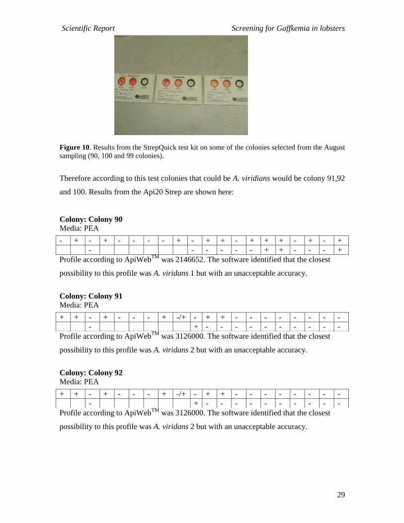
Figure 10. Results from the StrepQuick test kit on some of the colonies selected from the August
sampling (90, 100 and 99 colonies).
Therefore according to this test colonies that could be A. viridians would be colony 91,92
and 100. Results from the Api20 Strep are shown here:
Colony: Colony 90
Media: PEA
Profile according to ApiWebTM
was 2146652. The software identified that the closest
possibility to this profile was A. viridans 1 but with an unacceptable accuracy.
Colony: Colony 91
Media: PEA
Profile according to ApiWebTM
was 3126000. The software identified that the closest
possibility to this profile was A. viridans 2 but with an unacceptable accuracy.
Colony: Colony 92
Media: PEA
Profile according to ApiWebTM
was 3126000. The software identified that the closest
possibility to this profile was A. viridans 2 but with an unacceptable accuracy.
- + - + - - - - + - + + - + + + - + - +
- - - - - - + + - - - +
+ + - + - - - + -/+ - + + - - - - - - - -
- + - - - - - - - - - -
+ + - + - - - + -/+ - + + - - - - - - - -
- + - - - - - - - - - -
Scientific Report Screening for Gaffkemia in lobsters
30
Colony: Colony 99
Media: PEA
Profile according to ApiWebTM
was 306660. The software identified that the closest
possibility to this profile was A. viridans 1 but with an unacceptable accuracy.
Colony: Colony 100
Media: PEA
Profile according to ApiWebTM
was 6166651. The software identified that the closest
possibility to this profile was A. viridans 1 but with an unacceptable accuracy.
In summary, the colonies suspected to be A. viridans collected through this project were
as follows:
Table 10. Resume of the gram positive cocci bacterial colonies selected for further work. In black
we highlight colonies that were similar to A. viridians in the StrepQuick test.
Num colony
Shape /size colony
Colour colony Sample type
TSA growth
PEA growth StrepQuick Api20 Strep ID.
40 Round/M Yellow Hepato + + +/+/- A. viridans 1 good
43 Round/S White Hepato + + +/+/- A. viridans 1 good
50 Round/M Yellow Haemo + + +/+/- A. viridans 1 good
52 Round/S White Haemo + + +/-/- Listeria sp. good
63 Round/M Yellow Hepato + + +/+/- A. viridans 1 good
70 Round/M Yellow Haemo + + +/+/- A. viridans 1 good
73 Round/M-S Yellow Haemo + + +/+/- A. viridans 1 good
74 Round/M-S Yellow Haemo + + -/+/- A. viridans 1
unacceptable
81 Round/M-S White Haemo + + +/-/- A. viridans 2 good
82 Round/B Yellow Haemo + + -/-/- A. viridans 1
unacceptable 83 Round/B Cream Hepato + - -/+/- A. viridans 2 good
+ + - - - - - + -/+ - + + - + + - + + - -
- + - - - - - - - - - -
- + + + - - - + + - + + - + + + - + + +
+ - - - - - - + - - - -
Scientific Report Screening for Gaffkemia in lobsters
31
90 Round/M Yellow Haemo + + +/+/- A. viridans 1
unacceptable 91 Round/M White Haemo + + +/-/- A. viridans 2
unacceptable 92 Round/M White Haemo + + +/-/- A. viridans 2
unacceptable 99 Round/M White H2O + + -/-/- A. viridans 1
unacceptable 100 Round/M Cream H2O + + +/-/- A. viridans 1
unacceptable
Therefore, although some colonies had a high potential to be A. viridans none of the
selected colonies gave an ‘excellent’ or ‘very good’ accuracy according to the
biochemical tests. For this reason, it was important to confirm the identity of the different
colonies in order to identify which of the colonies were indeed A. viridans and therefore
to identify the most appropriate identification method to be used in the future. For this
reason the colonies shown in Table 10 were re-grown in PEA and TSA plates and DNA
was extracted to be able to do a molecular characterization and identification of the
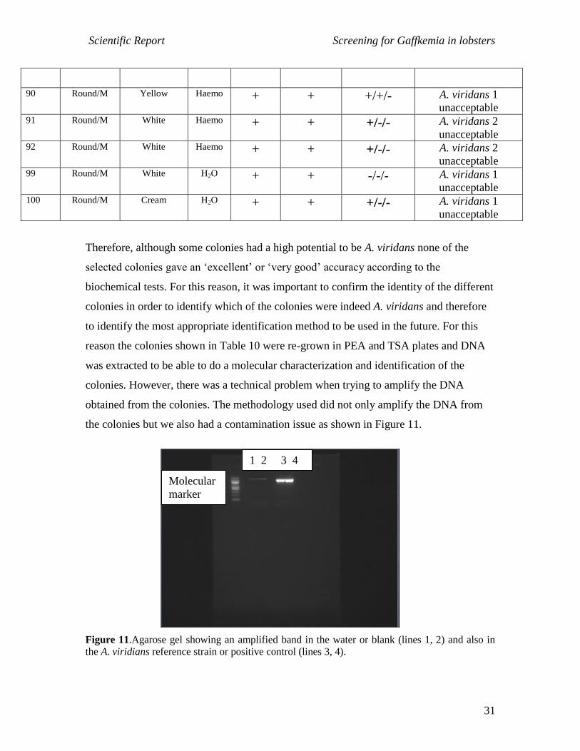
colonies. However, there was a technical problem when trying to amplify the DNA
obtained from the colonies. The methodology used did not only amplify the DNA from
the colonies but we also had a contamination issue as shown in Figure 11.
Figure 11.Agarose gel showing an amplified band in the water or blank (lines 1, 2) and also in
the A. viridians reference strain or positive control (lines 3, 4).
1 2 3 4
Molecular
marker
Scientific Report Screening for Gaffkemia in lobsters
32
Therefore, although it was possible to obtain and amplify the DNA from all the selected
colonies it was not possible to proceed with the sequencing as the negative control
indicated that following this protocol led to a contamination of the samples. This was the
case following the methodology indicated in Materials and Methods section and also after
treating the samples prior to the PCR amplification with ‘shrimp nuclease’.
Conclusions and Outlook
- During the period of this project there was no indication of a heavy infection
of A. viridans in Tarbert fish farm. This was supported by a low mortality
(data given by the company) and by the microbiological results of the present
work.
- The microbiological data indicated that although there was no outbreak during
this investigation it is possible that some of the sampled lobster had low
numbers of A. viridians.
- From a methodological point of view it would be recommended to do further
work in order to sequence and identify molecularly the selected colonies. This
step would allow to design and recommend the most suitable tests for future
determination of A. viridans.
- From the bibliographical data it would appear that the key parameter to
control A. viridians would be a reduced temperature in the tanks.
- Therefore, it is recommended to cool the water in the tanks.
Acknowledgements
This study was funded in part by an Innovation Voucher Award from the Scottish
Funding Council, administered by Interface Ltd.