copyright © 2009 pearson education, inc. human biology chapter 2 chemistry of living things ...
TRANSCRIPT

Copyright © 2009 Pearson Education, Inc.
Human Biology
Chapter 2 Chemistry of living things
Atoms/Elements
Bonds
Water
pH
Molecules of life
Carbohydrates *Proteins
Lipids *Nucleic Acids

Copyright © 2009 Pearson Education, Inc.
All Matter Consists of Elements Made of Atoms
Chemistry The study of matter
Atoms, the smallest functional unit, consist of Protons: positive charge, have mass
Neutrons: no charge, have mass
Electrons: negative charge, have no discernable mass

Copyright © 2009 Pearson Education, Inc.
Atoms Combine to Form Molecules
Joining atoms requires energy Energy is the capacity to do work
Stored energy: potential energy
Energy in motion, doing work: kinetic energy
Electrons have potential energy Shells: the energy levels of electrons
Orbitals describe the probable location of an electron

Copyright © 2009 Pearson Education, Inc.
Three Types of Chemical Bonds
Table 2.1
Copyright © 2009 Pearson Education, Inc.
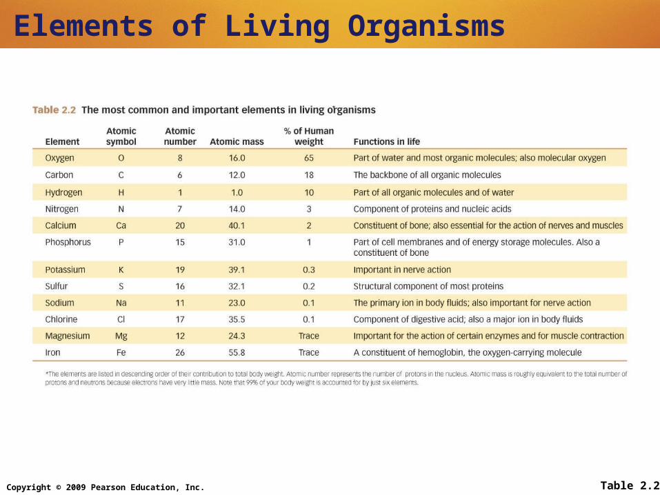
Elements of Living Organisms
Table 2.2

Copyright © 2009 Pearson Education, Inc.
Life Depends on Water
Water molecules are polar
Water is liquid at body temperature
Water can absorb and hold heat energy
Copyright © 2009 Pearson Education, Inc.
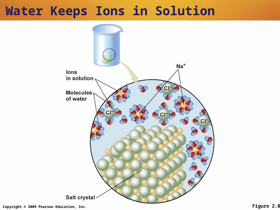
Water Keeps Ions in Solution
Figure 2.8

Copyright © 2009 Pearson Education, Inc.
The Importance of Hydrogen Ions
Acids are proton (hydrogen ion) donors
Bases accept hydrogen ions
pH Scale Hydrogen ion concentration
Buffers Minimize pH change
Carbonic acid and bicarbonate act as one of the body’s most important buffer pairs

Copyright © 2009 Pearson Education, Inc.
The pH Scale
Figure 2.10

Copyright © 2009 Pearson Education, Inc.
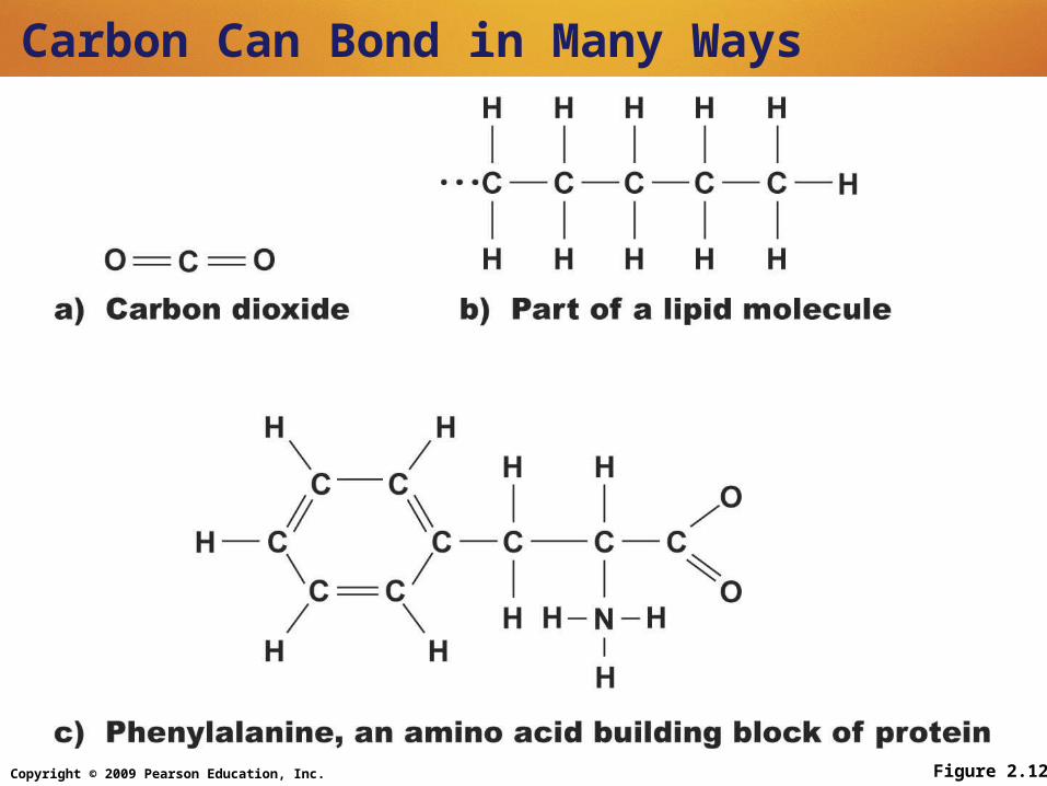
The Organic Molecules of Living Organisms
Carbon, the building block of living things Comprises 18% of the body by weight
Forms four covalent bonds
Can form single or double bonds
Can build micro- or macromolecules
Copyright © 2009 Pearson Education, Inc.
Carbon Can Bond in Many Ways
Figure 2.12

Copyright © 2009 Pearson Education, Inc.
Making and Breaking Biological Macromolecules: Dehydration Synthesis and Hydrolysis
Figure 2.13

Copyright © 2009 Pearson Education, Inc.
Dehydration Synthesis Is the Reverse of Hydrolysis
Dehydration synthesis Removes equivalent of a water molecule to
link molecular units Requires energy
Hydrolysis Adds the equivalent of a water molecule to
break apart macromolecules Releases energy

Copyright © 2009 Pearson Education, Inc. Figure 2.14
Carbohydrates are Composed of Monosaccharides

Copyright © 2009 Pearson Education, Inc.
Carbohydrates are Used for Energy and Structural Support
Oligosaccharides Short chains of monosaccharides
Disaccharides Sucrose, fructose, lactose

Copyright © 2009 Pearson Education, Inc.
Carbohydrates are Used for Energy and Structural Support
Polysaccharides: thousands of monosaccharides joined in chains and branches Starch: made in plants; stores energy
Glycogen: made in animals; stores energy
Cellulose: indigestible polysaccharide made in plants for structural support

Copyright © 2009 Pearson Education, Inc.
Animation—Lipid Structure and Function
Lipids: Insoluble in Water
Triglycerides: energy storage molecules Fatty acids: saturated and unsaturated
Phospholipids: cell membranes
Steroids: carbon-based ring structures Cholesterol: used in making estrogen and
testosterone
PLAY

Copyright © 2009 Pearson Education, Inc.
Proteins: Complex Structures Constructed of Amino Acids
Structure Primary: amino acid sequence
Secondary: describes chain’s orientation in space (e.g., alpha helix, beta sheet)

Copyright © 2009 Pearson Education, Inc.
Proteins: Complex Structures Constructed of Amino Acids
Tertiary: describes three-dimensional shape created by disulfide and hydrogen bonds Creates polar and nonpolar areas in
molecule
Quaternary: describes proteins in which two or more tertiary protein chains are associated

Copyright © 2009 Pearson Education, Inc.
Proteins: Complex Structures Constructed of Amino Acids
Denaturation Permanent disruption of protein structure
Can be damaged by temperature or changes in pH
Leads to loss of biological function
Copyright © 2009 Pearson Education, Inc.
Enzyme Function
Enzymes Are proteins
Function as catalysts
Speed up chemical reactions
Are not altered or consumed by the reaction

Copyright © 2009 Pearson Education, Inc.
Enzyme Function
The functional shape of an enzyme is dependent on Temperature of reaction medium
pH
Ion concentration
Presence of inhibitors

Copyright © 2009 Pearson Education, Inc.
Structure and Function of Nucleic Acids
Functions Store genetic information
Provide information used in making proteins
Structure Nucleotides consist of a phosphate group, a sugar,
and a nitrogenous base
DNA structure is a double helix: two associated strands of nucleic acids
RNA is a single-stranded molecule

Copyright © 2009 Pearson Education, Inc.
Structure of DNA
DNA Deoxyribonucleic acid
Double–stranded
Sugar
Deoxyribose

Copyright © 2009 Pearson Education, Inc.
Structure of DNA
DNA Nitrogenous bases
Adenine
Thymine
Cytosine
Guanine
Pairing
Adenine–thymine
Cytosine–guanine

Copyright © 2009 Pearson Education, Inc.
Structure of RNA
RNA Ribonucleic acid
Single–stranded
Sugar
Ribose
Copyright © 2009 Pearson Education, Inc.
Structure of RNA
RNA Nitrogenous bases
Adenine
Uracil
Cytosine
Guanine
Pairing
Adenine–uracil
Cytosine–guanine

Copyright © 2009 Pearson Education, Inc.
Structure and Function of Adenosine Triphosphate (ATP)
Universal energy source
Bonds between phosphate groups contain potential energy
Breaking the bonds releases energy ATP ADP + P1 + energy

Copyright © 2009 Pearson Education, Inc.
Structure and Function of Adenosine Triphosphate (ATP)
Figure 2.26