copd laily
TRANSCRIPT
CHRONIC OBSTRUCTIVE PULMONARY DISEASE
Laily Hidayati, S.Kep., Ns., M.Kep.

WHAT IS COPD?
Chronic Obstructive Pulmonary Disease (COPD) is a preventable and treatable disease with some significant extrapulmonary effects that may contribute to the severity in individual patients.
Its pulmonary component is characterized by airflow limitation that is not fully reversible.
The airflow limitation is usually progressive and associated with an abnormal inflammatory responseof the lung to noxious particles or gases.

The chronic airflow limitation characteristic of COPD is caused by a mixture of 1. small airway disease
(obstructive bronchiolitis) and
2. parenchymal destruction (emphysema),
the relative contributions of which vary from person to person.

Chronic inflammation causes structural changes and narrowing of the small airways.
Destruction of the lung parenchyma, also by inflammatory processes leads to the loss of alveolar attachments to the small airways decreases lung elastic recoil diminish the ability of the airways to remain open during expiration.
Airflow limitation is best measured by spirometry, as this is the most widely available, reproducible test of lung function.

In COPD, less air flows in and out of the airways because of one or more of the following:1. The airways and air sacs lose their elastic
quality.2. The walls between many of the air sacs are
destroyed.3. The walls of the airways become thick and
inflamed.4. The airways make more mucus than usual,
which can clog them.(NHLBI, 2012)

CLASSIFICATION
Many previous definitions of COPD have emphasized the terms :1. emphysema, and 2. chronic bronchitis
Emphysema, or destruction of the gas exchanging surfaces of the lung (alveoli)
Chronic bronchitis, or the presence of cough and sputum production for at least 3 months in each of two consecutive years.
And also other several structural abnormalities present in patients with COPD (except asthma)
ASTHMA VS COPD
COPD can coexist with asthma, the other major chronic obstructive airway disease characterized by an underlying airway inflammation.
The underlying chronic airway inflammation is very different in these two diseases.

ASTHMA VS COPD
Source: Global Initiative for Chronic Obstructive Lung Disease, 2006

SPIROMETRIC CLASSIFICATION OF SEVERITY

Stage I: Mild COPD – Characterized by mild airflow limitation (FEV1/FVC < 0.70; FEV1 ≥ 80% predicted).
1. Symptoms of chronic cough and sputum production may be present, but not always.
At this stage, the individual is usually unaware that his or her lung function is abnormal.
Stage II: Moderate COPD – Characterized by worsening airflow limitation (FEV1/FVC < 0.70; 50% ≤ FEV1 < 80%predicted),
1. shortness of breath typically developing on exertion, and 2. cough and sputum production sometimes also present.
This is the stage at which patients typically seek medical attention because of chronic respiratory symptoms or an exacerbation of their disease.

Stage III: Severe COPD – Characterized by further worsening of airflow limitation (FEV1/FVC < 0.70; 30% ≤ FEV1< 50% predicted), 1. greater shortness of breath, 2. Reduced exercise capacity, 3. fatigue, and 4. repeated exacerbations that almost always have an impact on patients’ quality
of life.
Stage IV: Very Severe COPD – Characterized by severe airflow limitation (FEV1/FVC < 0.70; FEV1 < 30% predicted or FEV1 < 50% predicted plus the presence of chronic respiratory failure).
1. Respiratory failure is defined as an arterial partial pressure of O2 (PaO2) less than 8.0 kPa (60 mm Hg), with or without arterial partial pressure of CO2 (PaCO2) greater than 6.7 kPa (50 mm Hg) while breathing air at sea level.
2. Respiratory failure may also lead to effects on the heart such as cor pulmonale (right heart failure). Clinical signs of cor pulmonale include elevation of the jugular venous pressure and pitting ankle edema.
Patients may have Stage IV: Very Severe COPD even if the FEV1 is > 30% predicted, whenever these complications are present. At this stage, quality of life is very appreciably impaired and exacerbations may be life threatening.


COPD AND COMORBIDITIES COPD it self also has significant extrapulmonary (systemic)
effects that lead to comorbid conditions. Data from the Netherlands show that up to 25% of the
population 65 years and older suffer from two comorbid conditions and up to 17% have three.
Well recognized comorbid conditions (extrapulmonary effects) are:
1. Weight loss, 2. nutritional abnormalities, and 3. skeletal muscle dysfunction
Other comorbid conditions:1. increased risk for myocardial infarction, angina, 2. osteoporosis, bone fractures, 3. respiratory infection, 4. depression, sleep-disorders, 5. diabetes, anemia, and 6. glaucoma.

COPD AND COMORBIDITIES
The existence of COPD may actually increase the risk for other diseases; this is particularly striking for COPD and lung cancer.
Causes of death in patients with COPD are mainly cardiovascular diseases, lung cancer, and, in those with advanced COPD, respiratory failure.
Morbidity due to COPD increases with age and is greater in men than in women
PREVALENCE
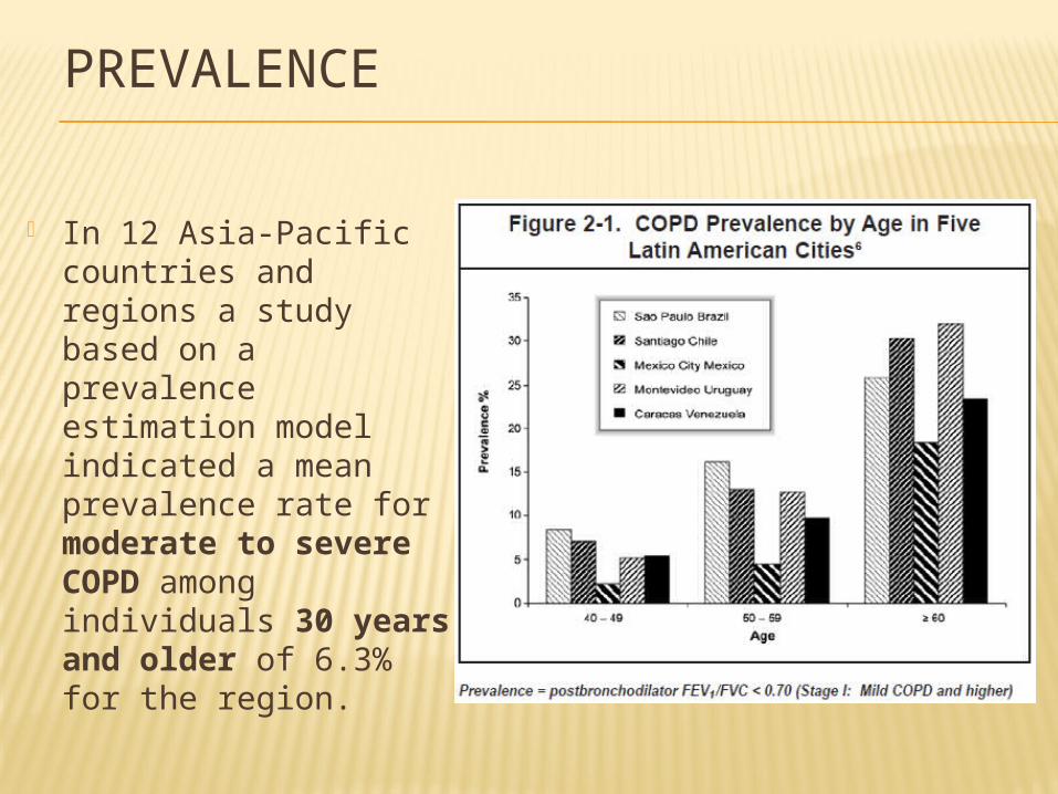
In 12 Asia-Pacific countries and regions a study based on a prevalence estimation model indicated a mean prevalence rate for moderate to severe COPD among individuals 30 years and older of 6.3% for the region.

RISK FACTORS

COPD Risk is related to The Total Burden of Inhaled Particles
Source: GOLD, 2006

PATHOLOGY, PATHOGENESIS, AND PATHOPHYSIOLOGY
Pathological changes characteristic of COPD are found in the proximal airways, peripheral airways, lung parenchyma, and pulmonary vasculature.
These changes include chronic inflammation, and structural changes resulting from repeated injury and repair.
There is a characteristic pattern of inflammation in the lungs of COPD patients, with increased numbers of neutrophils (in the airway lumen), macrophages (airway lumen, airway wall, and parenchyma), and CD8+ lymphocytes (airway wall and parenchyma).
Lung inflammation is further amplified by oxidative stress and an excess of proteases in the lung.
Physiological changes characteristic of the disease include mucus hypersecretion, airflow limitation and air trapping (leading to hyperinflation), gas exchange abnormalities, and cor pulmonale.

Systemic features of COPD, particularly in patients with severe disease, include cachexia, skeletal muscle wasting, increased risk of cardiovascular disease, anemia, osteoporosis, and depression.
Exacerbations represent a further amplification of the inflammatory response in the airways of patients with COPD, and may be triggered by infection with bacteria or viruses or by environmental pollutants.


PATHOGENESIS OF COPD

INFLAMMATORY CELLS IN COPD
Neutrophils: in sputum of normal smokers. Further in COPD and related to disease severity. Few neutrophils are seen in tissue. They may be important in mucus hypersecretion and through release of proteases.
Macrophages: Greatly numbers are seen in airway lumen, lung parenchyma, and bronchoalveolar lavage fluid. Derived from blood monocytes that differentiate within lung tissue. Produce increased inflammatory mediators and proteases in COPD patients in response to cigarette smoke and may show defective phagocytosis.
T lymphocytes: Both CD4+ and CD8+ cells are increased in the airway wall and lung parenchyma, with CD8+:CD4+ ratio. CD8+ T cells(Tc1) and Th1 cells which secrete interferon- and express the chemokine receptor CXCR3. CD8+ cells may be cytotoxic to alveolar cells, contributing to their destruction.
B lymphocytes: in peripheral airways and within lymphoid follicles, possibly as a response to chronic colonization and infection of the airways.
Eosinophils: eosinophil proteins in sputum and eosinophils in airway wall during exacerbations.
Epithelial cells: May be activated by cigarette smoke to produce inflammatory mediators.


EXACERBATIONS Exacerbations represent a further amplification of the inflammatory
response in the airways of COPD patients, and may be triggered by infection with bacteria or viruses or by environmental pollutants.
In mild and moderate exacerbations :1) there is an increase in neutrophils and in some studies also
eosinophils in sputum and the airway wall. 2) This is associated with increased concentrations of certain
mediators, including TNF-, LTB4 and IL-8, and an increase in biomarkers of oxidative stress.
In severe exacerbations: 1) showed a marked increase in neutrophils in the airway wall and
increased expression of chemokines. 2) During an exacerbation there is increased hyperinflation and air
trapping, with reduced expiratory flow, thus accounting for the increased dyspnea.
3) There is also worsening of VA/Q abnormalities resulting in severe hypoxemia.

COPD MANAGEMENT
Effective management should be aimed at the following goals:
1) Relieve symptoms2) Prevent disease progression3) Improve exercise tolerance4) Improve health status5) Prevent and treat complications6) Prevent and treat exacerbations7) Reduce mortality

COPD MANAGEMENT
Mild to Moderate COPD (Stages I and II): The avoidance of risk factors to prevent disease progression, pharmacotherapy as needed to control symptoms.
Severe (Stage III) and Very Severe (Stage IV) COPD require: the integration of several different disciplines, a variety of treatment approaches, and a commitment of the clinician to the continued support of the
patient as the illness progresses.
In addition to patient education, health advice, and pharmacotherapy, COPD patients may require specific counseling about smoking cessation, instruction in physical exercise, nutritional advice, and continued nursing support.
Not all approaches are needed for every patient
COPD MANAGEMENT
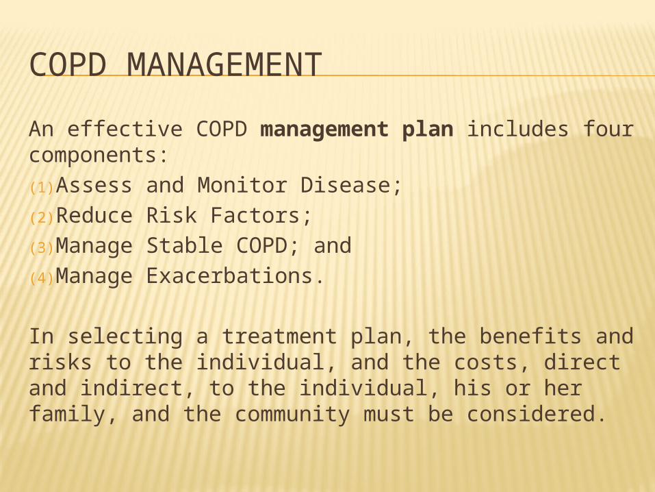
An effective COPD management plan includes four components: (1) Assess and Monitor Disease; (2) Reduce Risk Factors; (3) Manage Stable COPD; and (4) Manage Exacerbations.
In selecting a treatment plan, the benefits and risks to the individual, and the costs, direct and indirect, to the individual, his or her family, and the community must be considered.

1. ASSES AND MONITOR DISEASE

The impact of breathlessness on a patient’s health status (The British Medical Research Council (MRC) questionnaire)

PHYSICAL EXAMINATION
Inspection.1) Central cyanosis, or bluish discoloration of the mucosal membranes, may
be present but is difficult to detect in artificial light and in many racial groups.
2) Common chest wall abnormalities, which reflect the pulmonary hyperinflation seen in COPD, include relatively horizontal ribs, “barrel-shaped” chest, and protruding abdomen.
3) Flattening of the hemi-diaphragms may be associated with paradoxical in-drawing of the lower rib cage on inspiration, and widening of the xiphosternal angle.
4) Resting respiratory rate is often increased to more than 20 breaths per minute and breathing can be relatively shallow.
5) Patients commonly show pursed-lip breathing, which may serve to slow expiratory flow and permit more efficient lung emptying.
6) COPD patients often have resting muscle activation while lying supine. Use of the scalene and sternocleidomastoid muscles is a further indicator of respiratory distress.
7) Ankle or lower leg edema can be a sign of right heart failure.

Palpation and percussion.These are often unhelpful in COPD. Detection of the heart apex beat may be difficult due to pulmonary
hyperinflation. Hyperinflation also leads to downward displacement of the liver and
an increase in the ability to palpate this organ without it being enlarged.
Auscultation. Patients with COPD often have reduced breath sounds, but this
finding is not sufficiently characteristic to make the diagnosis. The presence of wheezing during quiet breathing is a useful pointer
to airflow limitation. However, wheezing heard only after forced expiration has not been validated as a diagnostic test for COPD.
Inspiratory crackles occur in some COPD patients but are of little help diagnostically.
Heart sounds are best heard over the xiphoid area.

MEASUREMENT OF AIRFLOW LIMITATION (SPIROMETRY)

Chest X-ray. Arterial blood gas measurement. Alpha-1 antitrypsin deficiency screening. Assessment of pulmonary hemodynamics
(JVP, pulmonary hypertension) Hematocrit. Respiratory muscle function Sleep studies. Exercise testing CT and ventilation-perfusion scanning

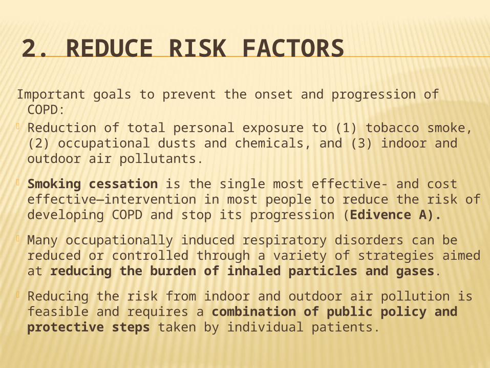
2. REDUCE RISK FACTORS
Important goals to prevent the onset and progression of COPD: Reduction of total personal exposure to (1) tobacco smoke,
(2) occupational dusts and chemicals, and (3) indoor and outdoor air pollutants.
Smoking cessation is the single most effective- and cost effective—intervention in most people to reduce the risk of developing COPD and stop its progression (Edivence A).
Many occupationally induced respiratory disorders can be reduced or controlled through a variety of strategies aimed at reducing the burden of inhaled particles and gases.
Reducing the risk from indoor and outdoor air pollution is feasible and requires a combination of public policy and protective steps taken by individual patients.
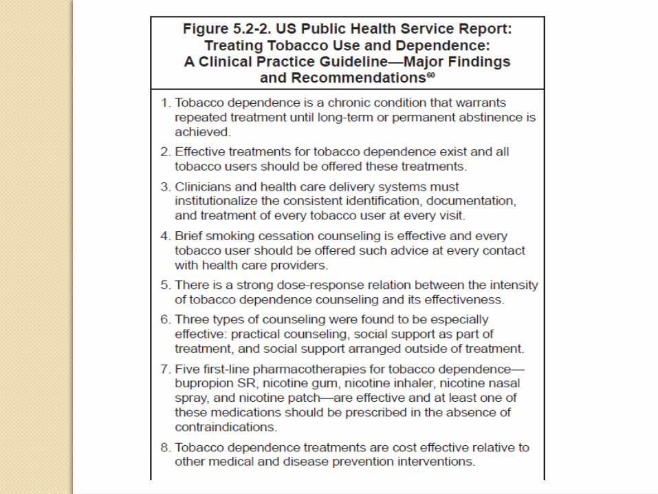


Smoking Cessation Counseling
The common subjects covered in successful problem solving/skillstraining programs include:1)Recognition of danger signals (the risk of relapse, such
as being around other smokers, psychosocial stress, being under time pressure, getting into an argument, drinking alcohol, and negative moods)
2)Enhancement of skills (to handle these situations, such as learning to anticipate and manage or avoid a particular stress)
3)Basic information about smoking and successful quitting (such as the nature and time course of withdrawal, the addictive nature of smoking, and the fact that any return to smoking, including even a single puff, increases the likelihood of a relapse)

Pharmacotherapy1) Nicotine replacement products (nicotine
gum, inhaler, nasal spray, transdermal patch, sublingual tablet, or lozenge)
2) Antidepressants bupropion73 and nortriptyline
3) Varenicline, (nicotinic acetylcholine receptor partial agonist) that aids smoking cessation by relieving nicotine withdrawal symptoms and reducing the rewarding properties of nicotine.

3. MANAGE STABLE COPD The overall approach to managing stable COPD
should be individualized to address symptoms and improve quality of life.
For patients with COPD, health education plays an important role in smoking cessation (Evidence A) and can also play a role in improving skills, ability to cope with illness and health status
None of the existing medications for COPD have been shown to modify the long-term decline in lung function that is the hallmark of this disease (Evidence A). Therefore, pharmacotherapy for COPD is used to decrease symptoms and/or complications.


OTHER PHARMACOLOGIC TREATMENTS Vaccines.
Influenza vaccines can reduce serious illness and death in COPD patients by about 50% (Evidence A).
Alpha-1 antitrypsin augmentation therapy. Young patients with severe hereditary alpha-1 antitrypsindeficiency and established emphysema may be candidates for alpha-1 antitrypsin augmentation therapy. However, this therapy is very expensive, is not available in most countries.
Immunoregulators (immunostimulators, immunomodulators). Studies using an immunoregulator in COPD show a decrease in the severity and frequency of exacerbations
Antioxidant agents. Mucolytic (mucokinetic, mucoregulator) agents (ambroxol, erdosteine,
carbocysteine, iodinated glycerol). Antibiotics (Prophylactic) Antitussives Vasodilators Narcotics (morphine).
Oral and parenteral opioids are effective for treating dyspnea in COPD patients withadvanced disease.

NON PHARMACOLOGIC TREATMENT
1. RehabilitationThe principal goals of pulmonary rehabilitation are to (1) reduce symptoms, (2) improve quality of life, and (3) increase physical and emotional participation in everyday activities.

NON PHARMACOLOGIC TREATMENT
Components of Pulmonary Rehabilitation Exercise training
Exercise training ranges in frequency from daily to weekly, in duration from 10 minutes to 45 minutes per session, and in intensity from 50% peak oxygen consumption (VO2 max) to maximum tolerated.
Nutritional CounselingA reduction in body mass index is an independent risk factor for mortality in COPD patients (Evidence A).
Education


2. Oxygen Therapy for patients with Stage IV: Very Severe COPD The primary goal of oxygen therapy is to increase the
baseline PaO2 to at least 8.0 kPa (60 mmHg) at sea level and rest, and/or produce an SaO2 at least 90%,
long-term administration of oxygen (> 15 hours per day)
3. Ventilatory SupportCombining noninvasive intermittent positive pressure ventilation (NIPPV) with long-term oxygen therapy
4. Surgical Treatments (Bullectomy, Lung volume reduction surgery (LVRS), Lung transplantation)

4. MANAGE EXACERBATIONS
An exacerbation of COPD is defined as an event in the natural course of the disease characterized by a change in the patient’s baseline dyspnea, cough, and/or sputum that is beyond normal day-to-day variations, is acute in onset, and may warrant a change in regular medication in a patient with underlying COPD.
The most common causes of an exacerbation are infection of the tracheobronchial tree and air pollution, but the cause of about one-third of severe exacerbations cannot be identified (Evidence B).

1) Inhaled bronchodilators (particularly inhaled 2-agonists with or without anticholinergics) and oral glucocorticosteroids are effective treatments for exacerbations of COPD (Evidence A).
2) Patients experiencing COPD exacerbations with clinical signs of airway infection (e.g., increased sputum purulence) may benefit from antibiotic treatment (Evidence B).
3) Noninvasive mechanical ventilation in exacerbations improves respiratory acidosis, increases pH, decreases the need for endotracheal intubation, and reduces PaCO2, respiratory rate, severity of breathlessness, the length of hospital stay, and mortality (Evidence A).