coordination of eye and head components of movements
TRANSCRIPT

RESEARCH ARTICLE
Coordination of eye and head components of movements evokedby stimulation of the paramedian pontine reticular formation
Neeraj J. Gandhi Æ Ellen J. Barton ÆDavid L. Sparks
Received: 14 March 2008 / Accepted: 19 April 2008
� Springer-Verlag 2008
Abstract Constant frequency microstimulation of the
paramedian pontine reticular formation (PPRF) in head-
restrained monkeys evokes a constant velocity eye move-
ment. Since the PPRF receives significant projections from
structures that control coordinated eye-head movements,
we asked whether stimulation of the pontine reticular
formation in the head-unrestrained animal generates a
combined eye-head movement or only an eye movement.
Microstimulation of most sites yielded a constant-velocity
gaze shift executed as a coordinated eye-head movement,
although eye-only movements were evoked from some
sites. The eye and head contributions to the stimulation-
evoked movements varied across stimulation sites and
were drastically different from the lawful relationship
observed for visually-guided gaze shifts. These results
indicate that the microstimulation activated elements that
issued movement commands to the extraocular and, for
most sites, neck motoneurons. In addition, the stimulation-
evoked changes in gaze were similar in the head-restrained
and head-unrestrained conditions despite the assortment of
eye and head contributions, suggesting that the vestibulo-
ocular reflex (VOR) gain must be near unity during the
coordinated eye-head movements evoked by stimulation of
the PPRF. These findings contrast the attenuation of VOR
gain associated with visually-guided gaze shifts and sug-
gest that the vestibulo-ocular pathway processes volitional
and PPRF stimulation-evoked gaze shifts differently.
Keywords Gaze shifts � PPRF � Saccade �Superior colliculus � Head movements � Spinal cord
Introduction
The generation of head-restrained saccades requires a
spatial to temporal transformation of the neural activity
along the oculomotor neuraxis (see Sparks and Gandhi
2003 for a review). Neurons in the cortical eye fields and
the superior colliculus are topographically organized, such
that the locus of the active population encodes the desired
change in gaze. The place code signal is transformed into a
temporal code downstream at the level of the paramedian
pontine reticular formation (PPRF), which produces the
horizontal component of saccades, and the midbrain/dien-
cephalon junction, which controls the vertical and torsional
components. Most burst neurons in the PPRF, notably the
putative excitatory burst neurons that project to the abdu-
cens motoneurons, discharge a high-frequency burst for all
saccades with an ipsiversive horizontal component. The
number of spikes in the burst dictates the saccade ampli-
tude (Keller 1974) and the discharge dynamics determine
the velocity profile (Van Gisbergen et al. 1981).
Microstimulation is also useful for distinguishing
between spatial and temporal encoding schemes. Current
injected into the superior colliculus of head-restrained
animals, for example, evokes a site-specific vector, pro-
vided that the stimulation duration is sufficiently prolonged
N. J. Gandhi � E. J. Barton � D. L. Sparks
Department of Neuroscience, Baylor College of Medicine,
Houston, TX 77030, USA
N. J. Gandhi
Departments of Otolaryngology, Bioengineering,
and Neuroscience, Center for the Neural Basis of Cognition,
University of Pittsburgh, Pittsburgh, PA 15213, USA
N. J. Gandhi (&)
Department of Otolaryngology,
Eye and Ear Institute, Room 108, University of Pittsburgh,
203 Lothrop Street, Pittsburgh, PA 15213, USA
e-mail: [email protected]
123
Exp Brain Res
DOI 10.1007/s00221-008-1401-1
(Robinson 1972; Schiller and Stryker 1972; Guitton et al.
1980). Manipulation of stimulation intensity and current
changes the speed of the eye movement, but its velocity
profile continues to resemble a saccade (Stanford et al.
1996). Constant frequency microstimulation of the PPRF,
in contrast, moves the eyes ipsilaterally at a constant
velocity for the duration of the stimulation (barring con-
straints placed by oculomotor range limits), and the speed
of the ramp-like movement scales with stimulation fre-
quency (Cohen and Komatsuzaki 1972).
When the head is unrestrained, the site-specific output of
frontal and supplementary eye fields, as well as the superior
colliculus, still encodes a desired change in gaze, but the
gaze shift can now be produced as a coordinated movement
of the eyes and the head (Roucoux et al. 1980; Munoz et al.
1991; Pare et al. 1994; Freedman et al. 1996; Freedman and
Sparks 1997a; Tu and Keating 2000; Klier et al. 2001;
Martinez-Trujillo et al. 2003; Chen and Walton 2005;
Knight and Fuchs 2007). Since the extraocular and neck
muscles provide independent means of producing move-
ments, the desired gaze displacement signal must be
processed separately for the extraocular and neck moto-
neurons. The downstream PPRF region is a potential site
where the separate eye and head control pathways exist
(Gandhi and Sparks 2007). Thus, the primary objectives of
this study were to characterize the eye and/or head move-
ments evoked by stimulation of the monkey PPRF; to
compare the coordination of eye and head movements,
should they be observed, with those recorded during
visually-guided gaze shifts; and to compare the movements
evoked using matched stimulation parameters in head-
restrained and head-unrestrained conditions. A secondary
goal, which also served as a control, was to perform the
microstimulation experiments on extraocular motoneuron
pools in order to confirm that movements were limited to
the eyes only, even when the head was unrestrained.
Preliminary versions of this study have been published
previously (Gandhi and Sparks 2000; Sparks et al. 2001).
Methods
Data were obtained from two juvenile rhesus monkeys
(Macaca mulatta). All experimental protocols were
approved by the Institutional Animal Care and Use Com-
mittee at the Baylor College of Medicine and complied with
the guidelines of the Public Health Service policy on
Humane Care and Use of Laboratory Animals. Surgical and
behavioral procedures, as well as the apparatus used for
these experiments, have been described previously (Gandhi
and Sparks 2001). Briefly, using the magnetic induction
technique, gaze (eye-in-space) and head (head-in-space)
positions were sampled from search coils attached to the
eye and head, respectively. For each experimental session,
the monkey sat in a primate chair and faced a tangent screen
consisting of an array of tri-state (red, green and yellow)
light emitting diodes (LEDs) in a dimly lit room. A mini-
ature laser module, mounted on the monkey’s head,
projected a red beam onto the visual display. At the start of
each trial, the laser was illuminated simultaneously with a
red LED, located between -20� and 20� along the hori-
zontal axis. The monkeys were granted 4,000 ms to direct
the laser beam within 4–6� of the red LED and maintain the
alignment for 700–1,000 ms. Next, a green LED was illu-
minated on the horizontal axis within 30� of the red target.
The monkeys were trained to direct their gaze within 2–4�of the green target in the next 500 ms, without significantly
changing their head position, and fixate for 700–1,000 ms.
Next, the laser and both targets were extinguished. A yellow
target was presented following a gap period of 100–600 ms,
and the monkeys were allowed 800 ms and any combina-
tion of eye-head movement to bring gaze position within
6–10� of the stimulus. Fixation of the yellow LED for
800–1,600 ms triggered a solenoid to administer a liquid
reward. When the head was restrained, the trial initiated
with illumination of the green stimulus and followed the
remaining steps as described above.
For each electrophysiology session, a microelectrode
was lowered through a 15-mm diameter chamber slanted
laterally at an angle of 26� in the frontal plane, an approach
that permits access to the PPRF region, including the
abducens nucleus (Gandhi and Sparks 2007). For an initial
series of tracks, the penetrations were performed in the
head-restrained animal; for later tracks, once the PPRF
region was identified, the microelectrode was usually
lowered while the head was unrestrained. The abducens
nuclei were identified by their burst-tonic activity associ-
ated with saccades (Schiller 1970) and by the effects of
stimulation (Reinhart and Zuber 1970). The PPRF was
identified by its location with respect to the abducens
nuclei (Strassman et al. 1986), the effects of stimulation
(Cohen and Komatsuzaki 1972), and the discharge char-
acteristics during head-restrained saccades (Keller 1974)
and head-unrestrained gaze shifts (Whittington et al. 1984;
Ling et al. 1999; Sylvestre and Cullen 2006). Stimulation
was applied on penetrations that spanned anterior from the
rostral pole of the abducens nucleus by 3.5 mm in one
animal and 1.0 mm in the second. The distances covered
along the medial-lateral extent spanned 1.25 mm in one
and 2.5 mm in the other, with the midline lying roughly in
the center of each range. Microstimulation was delivered
mostly to the burst neuron region of the PPRF, although
tracks penetrating through the abducens motoneurons were
stimulated on occasion also. Furthermore, for several sites,
stimulation of identical parameters was delivered in both
head-restrained and head-unrestrained preparations.
Exp Brain Res
123

On 20–33% of the trials, biphasic current pulses
(0.25 ms pulse duration, 20–40 lA, 200–500 Hz,
200–500 ms) were delivered 100–200 ms into the gap
period, and the yellow target was programmed to appear
after stimulation offset. Stimulation parameters were
selected on a site-by-site basis. We typically used the
weakest parameters that both yielded a visually appre-
ciable effect online and kept the animal engaged in the
behavioral tasks. In a few cases, stimulation was delivered
manually during the inter-trial interval. Furthermore, for
some sites, the animal often produced a saccade that was
superimposed on the stimulation-evoked movement. We
believe these saccades were not produced by the stimu-
lation because they did not occur on every trial, their
onset times were quite variable, and their amplitudes and
directions were random; such trials were removed from
the analyses. Given these constraints and the need to
preserve a sufficient number of trials to assess repeat-
ability, our database was trimmed to include just the
stimulation-evoked movements recorded from 21 sites.
For these trials, the eyes were within 12� of the center of
the orbits and the head was pointing within 10� of
straight-ahead location at stimulation onset.
Data were sampled at 500 Hz by an in-house acquisition
program and analyzed off-line with Matlab, Statistica and
customized software programs. Velocity criteria were used
to detect the onset and offset of gaze, eye and head
movements. For gaze and eye channels, the onset and
offset velocity thresholds were 8 and 6�/s, respectively. For
the head movement waveform, the onset and offset velocity
thresholds were 5 and 3�/s, respectively. Every measure-
ment was inspected visually and corrected manually if
necessary. Gaze, eye and head amplitudes were computed
as the displacements of position between the onset and
offset of the respective waveforms. For all movements,
head and eye contributions were defined as the respective
changes in head and eye positions during the gaze shift. For
visually-guided gaze shifts, the offset of the eye movement
typically corresponds to the end of the saccadic eye
movement. For PPRF stimulation-evoked movements, eye
offset determined on the basis of a velocity criterion need
not correspond to a meaningful event. Hence, the pertinent
analyses employed eye contribution as a parameter.
Results
The effects of stimulation of the burst neuron region of
the PPRF on eye and head signals were recorded from 21
sites (n = 16, monkey BE; n = 5, monkey CH). For 4 of
these sites (all from monkey BE) the data are limited to
the condition when microstimulation was delivered
during the inter-trial interval, when the animal was sitting
in the dark. The results in the two configurations were
similar and therefore pooled together. For 10 (n = 8,
monkey BE; n = 2, monkey CH) of the 21 sites, the
effects of matched stimulation parameters were investi-
gated in both head-unrestrained and head-restrained
conditions.
Microstimulation of PPRF
Amplitude and velocity traces of eye movements evoked
by stimulation of the PPRF region in the head-restrained
monkey are shown in Fig. 1. Each panel overlays several
trials from a given site, and each column illustrates data
from a different site. In agreement with previous reports
(Cohen and Komatsuzaki 1972), constant-frequency stim-
ulation evoked a relatively constant velocity eye movement
(Fig. 1a–c). At other sites (n = 5), the stimulation initially
produced a bell-shaped velocity profile that smoothly
transitioned into a steady-state, constant velocity pattern
(Fig. 1d). This waveform is distinct from that described
previously (Cohen and Komatsuzaki 1972) as well as from
that evoked by high-frequency and prolonged duration
stimulation of the superior colliculus, which produces
repeated patterns of the high velocity profiles interspersed
with pursuit-like movement in between (Breznen et al.
1996; Missal et al. 1996; Moschovakis et al. 1998). Note
that the changes in gaze and eye positions are equivalent
because the head was held stationary.
When the head was allowed to move, constant-fre-
quency stimulation produced nearly constant-velocity
changes in the gaze, but not the eye, component of the gaze
shift. In fact, an assortment of eye and head contribution
patterns was observed across the stimulation sites. Panels
e–h show amplitude and velocity profiles of gaze, head,
and eye components evoked by stimulation of the same
sites and with the same parameters as those highlighted in
panels a–d. For the site illustrated in Fig. 1e the head
contribution was significant, but it remained smaller than
the change in gaze. The initial effect was similar for the site
shown in Fig. 1f. However, with prolonged stimulation the
head accelerated dramatically, such that head position led
gaze position, and the eyes moved in the opposite direction
in the orbits. For the site represented in Fig. 1g, the eyes
and head began to rotate in opposite directions much earlier
in the stimulation train, but the head did not display any
high velocity movements. Stimulation of numerous sites
(n = 8) also produced a transient, high velocity change in
gaze, which was followed by a constant velocity movement
(Fig. 1h). Although the head movements associated with
this example site appear negligible, this was not the case
for all eight stimulation sites. Five of these 8 sites were also
tested in the head-restrained condition (e.g, Fig. 1d), and
similar velocity waveforms were observed for all sites.
Exp Brain Res
123

Overall, there was no systematic relationship between the
location of the track along the anterior-posterior and
medial-lateral axes, and the amount the head contributed to
the gaze shift or the magnitude of the gaze shift.
For two sites, stimulation duration was systematically
varied, because the animal continued to perform the
behavioral tasks. Figure 2 shows stimulation-evoked
movements obtained from one site for stimulation dura-
tions of 50, 100 and 200 ms; stimulation intensity (25 lA)
and frequency (300 Hz) were held constant. For both head-
unrestrained and head-restrained conditions, the gaze
velocity reached a steady-state value of *100�/s, and the
gaze amplitude scaled linearly with stimulation duration.
When the head was free to move, the change in gaze was
produced as a coordinated movement of the eyes and head.
Results from the second site were comparable.
Eye-head coordination
One way to quantify the eye-head coupling during the
stimulation-evoked movements is to measure the contri-
bution of each component associated with the stimulation-
evoked change in gaze. Eye and head contributions are
defined as the displacements in eye-in-head and head-in-
space positions, respectively, during the period of the gaze
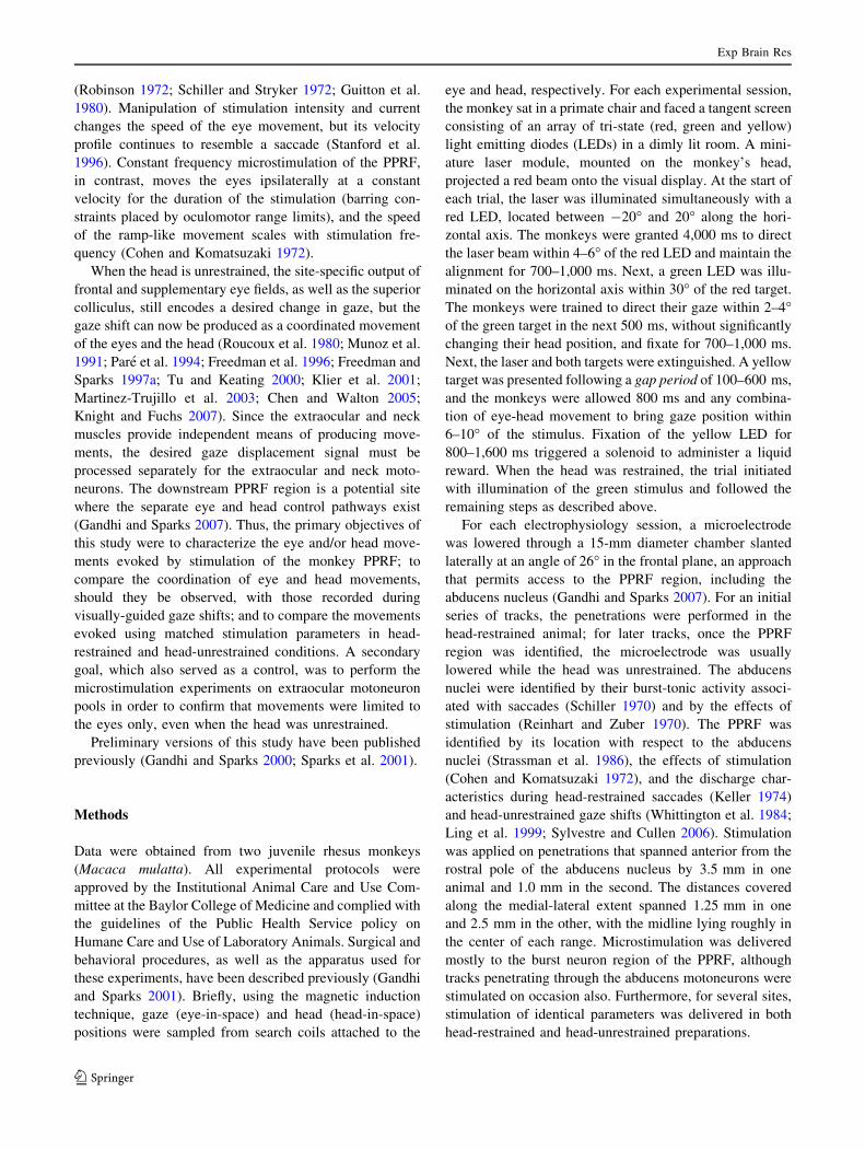
shift. Figure 3 plots the eye (filled circles) and head (filled
squares) contributions as a function of gaze shift amplitude
for movements evoked by stimulation of the PPRF; each
pair of symbols represents the average contribution for one
stimulation site (n = 21). For comparison, the eye and
head contributions associated with visually-guided gaze
shifts are shown in the open gray circles and squares,
respectively. As expected, for gaze shifts generated to
Hor
izon
tal
Am
plitu
de (
deg)
Hor
izon
tal
Vel
ocity
(de
g/se
c)
-10
0
10
20
0 200 400 600
-50
0
50
be06090300
C
0
10
20
0 100 200 300 400
-100
0
100
be03030200
A
0
10
20
0 200 400 600
0
50
100
be12080100
150
D
Time (msec)
0
10
20
-100
0
100
200
0 200 400 600
B
ch08230200
Hor
izon
tal
Am
plitu
de (
deg)
Hor
izon
tal
Vel
ocity
(de
g/se
c)
-10
0
10
20
-50
0
50
0 200 400 600
G
0 100 200 300 400
0
10
20
-100
0
100
E
0
10
20 H
0 200 400 600
0
50
100
Time (msec)
0
10
20
F
-100
0
100
200
0 200 400 600
GazeHeadEye
Fig. 1 Effects of microstimulation of the PPRF region in monkey.
a–d Stimulation delivered with the head immobilized. Each panel
superimposes several traces (n = 4–5) of horizontal amplitude (top)
and velocity (bottom) waveforms of stimulation-evoked movements
from four different sites. Stimulation parameters: a 25 lA, 200 ms,
300 Hz; b 20 lA, 400 ms, 300 Hz; c 25 lA, 400 ms, 300 Hz;
d 20 lA, 400 ms, 400 Hz. Vertical dashed lines indicate stimulation
onset and offset, and horizontal dashed lines mark zero displacement
and velocity. The initial position was subtracted from each trace to
align the movements. e–h Stimulation delivered with the head
unrestrained. Each panel superimposes representative stimulation-
evoked movements (n = 3–7) evoked from the same four sites and
using the same parameters as above. Top and bottom panels plot
horizontal components of representative stimulation-evoked changes
in amplitude and velocity, respectively, of gaze (green), head-in-
space (blue), and eye-in-head (red)
Exp Brain Res
123

orient to a visual target, eye and head contributions obey a
lawful relationship (Freedman and Sparks 1997b): eye
contribution increases linearly and then begins to saturate
for gaze shifts greater than *30�, whereas head contri-
bution is negligible for small changes in line of sight and
increases linearly for gaze shifts greater than *20�. In
contrast, the distributions of eye and head contributions
measured in the movements evoked by stimulation of the
PPRF do not obey this relationship. An obvious distinction
is that eye contribution can be negative, highlighting the
observation that the eyes and head can rotate in opposite
directions during PPRF stimulation, even as gaze position
shifts at a constant velocity in the ipsilateral direction.
Figure 3b provides another representation of the differ-
ent characteristics shown by visually guided gaze shifts and
PPRF stimulation-evoked movements. The illustration
plots head contribution as a function of eye contribution for
visually guided gaze shifts (s) and for movements evoked
by stimulation of the PPRF (j). The two distributions are
clearly different and nearly non-overlapping.
According to the data shown in Fig. 3, stimulation of
some sites (n = 5) evoked gaze amplitudes executed as eye
movements with minimal head contributions (\3�),
although the head contribution for these sites ranged from 5
to 25% of gaze amplitude. Others (n = 4) produced large
changes in head with a relatively small change in gaze
(\6�), because the eyes counter-rotated substantially in the
head; for these sites, head contribution ranged from 95 to
355% of the gaze shift. While we recognize that the exact
distributions of eye and head contributions depend on the
stimulation parameters, the fundamental difference in eye-
head coordination between visually-guided and PPRF
stimulation-evoked movements remains clear.
Figure 4a plots mean head latency as a function of mean
gaze latency for the 21 stimulation sites tested with the
head free to move. The mean ± SD gaze latency was
14.3 ± 7.9 ms (median: 13.3 ms; range: 3.6–37.7 ms)
(Note that the 3.6 ms is less than the 4.8 ± 0.5 ms reported
for midflight electrical stimulation of the PPRF (Miyashita
and Hikosaka 1996). This difference is attributed to our
0 100 200 300-20
-10
0
0 100 200 300
-100
-50
0
-10
0
-100
0
-10
-5
0
-100
-50
0
0 100 200 300
0 100 200 300
0 100 200 300
0 100 200 300
0 100 200 300
0 100 200 300
A
B
Horizontal Amplitude (deg) Horizontal Velocity (deg/sec)
Gaz
eE
yeH
ead
Gaz
e
Time (msec)
-50
-5
100
50
-100
-50
0
50
-10
-5
0
Head-Unrestrained Condition
Head-Restrained Condition
Time (msec)
Fig. 2 Effects of varying
stimulation duration on eye-
head movements evoked by
stimulation of the PPRF region.
a Head-unrestrained condition.
The green, blue, and magentatraces show movements when
stimulation duration was set to
50, 100, and 200 ms,
respectively. Stimulation
intensity (25 lA) and
frequency (300 Hz) were held
constant. The individual rows
plot horizontal gaze, eye-in-
head and head amplitude (leftcolumn) and velocity (rightcolumn) for representative,
individual trials. The first
vertical line marks stimulation
onset, and the remaining
vertical lines denote stimulation
offset for the three different
durations (indicated by the
different colors). b Head-
restrained condition. The same
format as above, but only the
gaze channel is shown as the
head was held stationary. Thus,
the eye component equals the
gaze component
Exp Brain Res
123
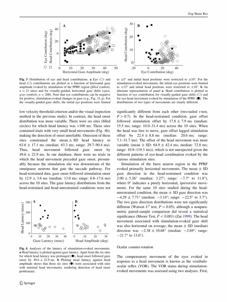
low velocity threshold criterion and/or the visual inspection
method in the previous study). In contrast, the head onset
distribution was more variable. There were six sites (filled
circles) for which head latency was [100 ms. These sites
contained trials with very small head movements (Fig. 4b),
making the detection of onset unreliable. Omission of these
sites constrained the mean ± SD head latency to
63.6 ± 17.1 ms (median: 63.3 ms; range: 29.7–90.4 ms).
Thus, head movement followed gaze onset by
49.6 ± 22.9 ms. In our database, there were no trials in
which the head movement preceded gaze onset, presum-
ably because the stimulation site was downstream of the
omnipause neurons that gate the saccade pathway. For
head-restrained data, gaze onset followed stimulation onset
by 12.9 ± 3.6 ms (median: 13.0 ms; range: 6.8–17.6 ms)
across the 10 sites. The gaze latency distributions from the
head-restrained and head-unrestrained conditions were not
significantly different from each other (two-tailed t-test,
P [ 0.7). In the head-restrained condition, gaze offset
followed stimulation offset by 17.8 ± 7.9 ms (median:
15.5 ms; range: 10.0–31.4 ms) across the 10 sites. When
the head was free to move, gaze offset lagged stimulation
offset by 22.4 ± 8.8 ms (median: 20.6 ms; range:
7.1–31.7 ms). The offset of the head movement was more
variable (mean ± SD: 64.9 ± 42.4 ms; median: 72.8 ms;
range: 10.9–119.3 ms)), which is not unexpected given the
different patterns of eye-head coordination evoked by the
various stimulation sites.
Stimulation of the burst neuron region in the PPRF
evoked primarily horizontal movements. The mean ± SD
gaze direction in the head-restrained condition was
2.00 ± 5.26� (median: 3.27�; range: -7.7� to 11.8�),
where 0� indicates a purely horizontal, ipsiversive move-
ments. For the same 10 sites studied during the head-
unrestrained condition, the mean ± SD gaze direction was
-4.29 ± 7.71� (median: -3.14�; range: -22.5� to 3.7�).
The two gaze direction distributions were not significantly
different (Watson U2 test, P [ 0.05), although a nonpara-
metric paired-sample comparison did reveal a statistical
significance (Moore Test, P \ 0.001) (Zar 1999). The head
movement associated with stimulation-evoked gaze shift
was also horizontal on average; the mean ± SD (median)
direction was -2.38 ± 10.68� (median: -2.69�; range:
-21.7� to 13.6�).
Ocular counter-rotation
The compensatory movement of the eyes evoked in
response to a head movement is known as the vestibulo-
ocular reflex (VOR). The VOR status during stimulation-
evoked movements was assessed using two analyses. First,
0 10 20 30 40 50
0
20
40
Horizontal Gaze Amplitude (deg)-20 0 20 40
0
10
20
30
Eye Contribution (deg)
Hea
d C
ontr
ibut
ion
(deg
)
Hea
d C
ontr
ibut
ion
(deg
)
Eye
Con
trib
utio
n (d
eg)
BA
Fig. 3 Distribution of eye and head contributions. a Eye (s) and
head (h) contributions are plotted as a function of horizontal gaze
amplitude evoked by stimulation of the PPRF region (filled symbols;
n = 21 sites) and for visually-guided, horizontal gaze shifts (open,gray symbols; n = 200). Note that eye contributions can be negative
for positive, stimulation-evoked changes in gaze (e.g., Fig. 1f, g). For
the visually-guided gaze shifts, the initial eye positions were limited
to ±5� and initial head positions were restricted to ±10�. For the
stimulation-evoked movements, the initial eye positions were limited
to ±12� and initial head positions were restricted to ±10�. b An
alternate representation of panel a. Head contribution is plotted as
function of eye contribution for visually-guided gaze shifts (s) and
for eye-head movement evoked by stimulation of the PPRF (j). The
distributions of two types of movements are clearly different
0 10 20 30 400
100
200
300
Gaze Latency (msec)
Hea
d L
aten
cy (
mse
c)
0 10 20 30
Head Amplitude (deg)
BA
Fig. 4 Analyses of the latency of stimulation-evoked movements.
a Head latency is plotted against gaze latency. Apart from the six sites
for which head latency was prolonged (d), head onset followed gaze
onset by 49.6 ± 22.9 ms. b Plotting head latency against head
amplitude shows that these six sites (d) were associated with sites
with minimal head movements, rendering detection of head onset
problematic
Exp Brain Res
123
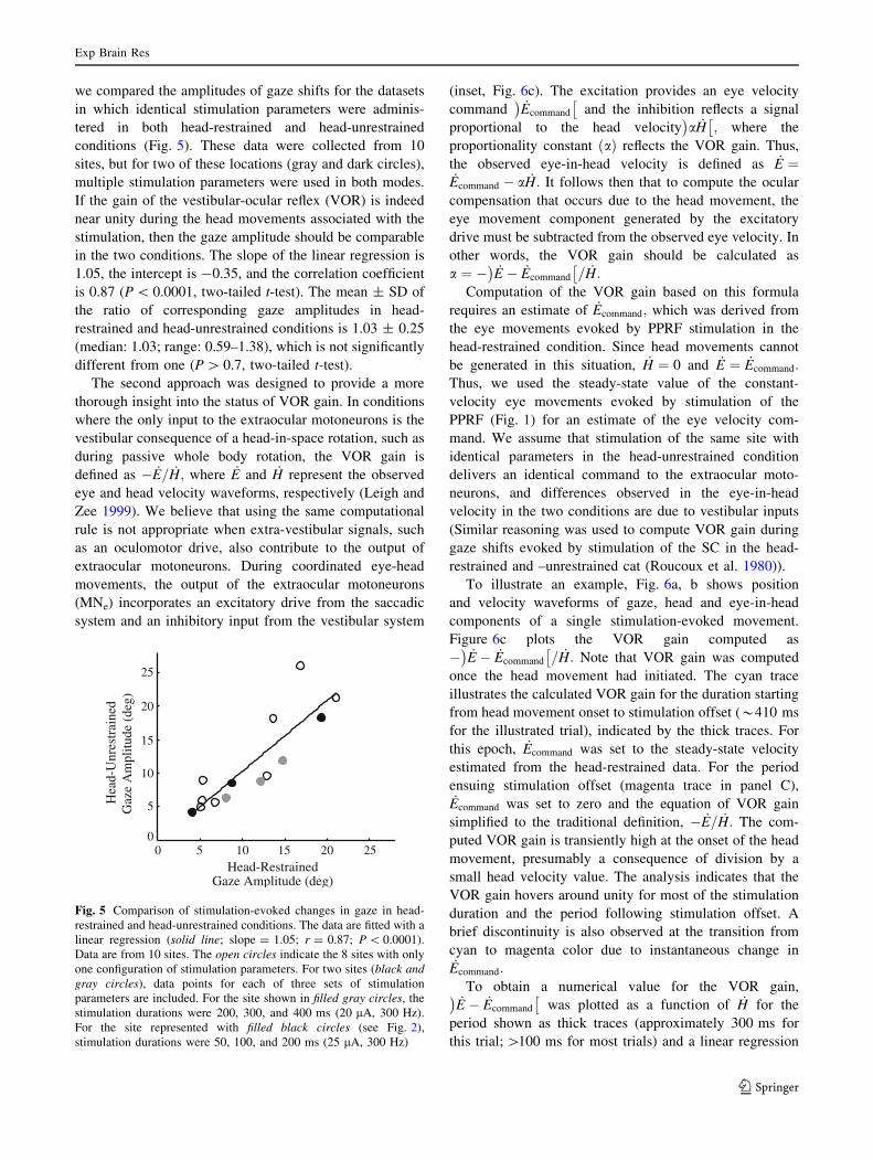
we compared the amplitudes of gaze shifts for the datasets
in which identical stimulation parameters were adminis-
tered in both head-restrained and head-unrestrained
conditions (Fig. 5). These data were collected from 10
sites, but for two of these locations (gray and dark circles),
multiple stimulation parameters were used in both modes.
If the gain of the vestibular-ocular reflex (VOR) is indeed
near unity during the head movements associated with the
stimulation, then the gaze amplitude should be comparable
in the two conditions. The slope of the linear regression is
1.05, the intercept is -0.35, and the correlation coefficient
is 0.87 (P \ 0.0001, two-tailed t-test). The mean ± SD of
the ratio of corresponding gaze amplitudes in head-
restrained and head-unrestrained conditions is 1.03 ± 0.25
(median: 1.03; range: 0.59–1.38), which is not significantly
different from one (P [ 0.7, two-tailed t-test).
The second approach was designed to provide a more
thorough insight into the status of VOR gain. In conditions
where the only input to the extraocular motoneurons is the
vestibular consequence of a head-in-space rotation, such as
during passive whole body rotation, the VOR gain is
defined as � _E= _H; where _E and _H represent the observed
eye and head velocity waveforms, respectively (Leigh and
Zee 1999). We believe that using the same computational
rule is not appropriate when extra-vestibular signals, such
as an oculomotor drive, also contribute to the output of
extraocular motoneurons. During coordinated eye-head
movements, the output of the extraocular motoneurons
(MNe) incorporates an excitatory drive from the saccadic
system and an inhibitory input from the vestibular system
(inset, Fig. 6c). The excitation provides an eye velocity
command _Ecommand
� �and the inhibition reflects a signal
proportional to the head velocity a _H� �
; where the
proportionality constant að Þ reflects the VOR gain. Thus,
the observed eye-in-head velocity is defined as _E ¼_Ecommand � a _H: It follows then that to compute the ocular
compensation that occurs due to the head movement, the
eye movement component generated by the excitatory
drive must be subtracted from the observed eye velocity. In
other words, the VOR gain should be calculated as
a ¼ � _E � _Ecommand
� �= _H:
Computation of the VOR gain based on this formula
requires an estimate of _Ecommand; which was derived from
the eye movements evoked by PPRF stimulation in the
head-restrained condition. Since head movements cannot
be generated in this situation, _H ¼ 0 and _E ¼ _Ecommand:
Thus, we used the steady-state value of the constant-
velocity eye movements evoked by stimulation of the
PPRF (Fig. 1) for an estimate of the eye velocity com-
mand. We assume that stimulation of the same site with
identical parameters in the head-unrestrained condition
delivers an identical command to the extraocular moto-
neurons, and differences observed in the eye-in-head
velocity in the two conditions are due to vestibular inputs
(Similar reasoning was used to compute VOR gain during
gaze shifts evoked by stimulation of the SC in the head-
restrained and –unrestrained cat (Roucoux et al. 1980)).
To illustrate an example, Fig. 6a, b shows position
and velocity waveforms of gaze, head and eye-in-head
components of a single stimulation-evoked movement.
Figure 6c plots the VOR gain computed as
� _E � _Ecommand
� �= _H: Note that VOR gain was computed
once the head movement had initiated. The cyan trace
illustrates the calculated VOR gain for the duration starting
from head movement onset to stimulation offset (*410 ms
for the illustrated trial), indicated by the thick traces. For
this epoch, _Ecommand was set to the steady-state velocity
estimated from the head-restrained data. For the period
ensuing stimulation offset (magenta trace in panel C),_Ecommand was set to zero and the equation of VOR gain
simplified to the traditional definition, � _E= _H: The com-
puted VOR gain is transiently high at the onset of the head
movement, presumably a consequence of division by a
small head velocity value. The analysis indicates that the
VOR gain hovers around unity for most of the stimulation
duration and the period following stimulation offset. A
brief discontinuity is also observed at the transition from
cyan to magenta color due to instantaneous change in_Ecommand:
To obtain a numerical value for the VOR gain,_E � _Ecommand
� �was plotted as a function of _H for the
period shown as thick traces (approximately 300 ms for
this trial; [100 ms for most trials) and a linear regression
0 5 10 15 20 250
5
10
15
20
25
Head-RestrainedGaze Amplitude (deg)
Hea
d-U
nres
trai
ned
Gaz
e A
mpl
itude
(de
g)
Fig. 5 Comparison of stimulation-evoked changes in gaze in head-
restrained and head-unrestrained conditions. The data are fitted with a
linear regression (solid line; slope = 1.05; r = 0.87; P \ 0.0001).
Data are from 10 sites. The open circles indicate the 8 sites with only
one configuration of stimulation parameters. For two sites (black andgray circles), data points for each of three sets of stimulation
parameters are included. For the site shown in filled gray circles, the
stimulation durations were 200, 300, and 400 ms (20 lA, 300 Hz).
For the site represented with filled black circles (see Fig. 2),
stimulation durations were 50, 100, and 200 ms (25 lA, 300 Hz)
Exp Brain Res
123

was applied to the data. Figure 7a (left) plots for all 125
trials from 10 stimulation sites a histogram of the VOR
gain, which equals the negative of the slope of the best fit
line. Table 1 summarizes this information by site. The
mean ± SD counter-rotation gain across these trials was
1.03 ± 1.00, which is not significantly different from one
(t-test, P [ 0.05). The large variance in the gain is partly
due to trials with small head contributions (Fig. 7a, right).
When only considering trials with head contribution [3�,
the mean ± SD and median counter-rotation gains were
1.14 ± 0.53 and 1.07, respectively (n = 69 trials). When
only considering trials with head contribution [5�, the
mean ± SD and median counter-rotation gains were
1.09 ± 0.42 and 1.07, respectively (n = 48 trials). In both
cases, the mean gain was not significantly different from
one (t-test, P [ 0.05).
Figure 7b shows the distribution of the correlation
coefficients of the individual linear regressions. Trials with
small head contributions were typically associated with
coefficients closer to zero, while trials with large head
contributions had correlation coefficient approaching -1.
Thus, when the PPRF stimulation evoked a sizeable head
movement, the VOR gain hovered around unity and this
was a robust observation. When the stimulation produced a
small head movement, the counter-rotation gain tended to
be more variable, as was our confidence in the statistical
significance of the fits.
Microstimulation of extraocular motoneurons
Stimulation of the ipsilateral abducens nucleus in the head-
unrestrained animal (Fig. 8a) guided the gaze position to an
eccentric, ipsilateral location with an exponential time
course. Once the stimulation was terminated, gaze
retreated, also exponentially, towards the initial position.
The change in gaze was produced almost exclusively by
the eye. The head contribution was negligible and, if
detected, its onset occurred late in the trial, typically after
the eyes reached eccentric locations in the orbits; thus, the
head movement could have occurred due to modulation of
neck muscle activity by eye positions in orbits (Vidal et al.
GazeHeadEye
0102030
-1000
100200
0 50 100 150 200 250 300 350 400 4500
1
2
3
Time re Stimulation Onset (msec)
VO
R g
ain
ch08230200
A
B
C ( ) HEE command&&& /−−VOR gain = α =
Posi
tion
(deg
)V
eloc
ity(d
eg/s
ec)
commandE&
E&
αH&
+ -MNe
Fig. 6 Example illustration of VOR gain computation during coor-
dinated eye-head movements generated by PPRF stimulation.
Temporal traces of the horizontal position (a) and velocity (b) of
gaze (green), head (blue) and eye-in-head (red) components are
shown for one trial. Plot starts at stimulation onset. The thick overlays
on the traces mark the period from head movement onset to the end of
the stimulation train. c The instantaneous VOR gain
(a ¼ � _E � _Ecommand
� �= _H; see text for details) is plotted as a function
of time. During the stimulation epoch (cyan trace), the velocity
command _Ecommand
� �equaled the constant velocity observed in the
head-restrained condition at the same site and with identical
stimulation parameters. It was set to zero after stimulation offset
(magenta trace). The inset shows a schematic in which the output of
the extraocular motoneurons incorporates an excitatory velocity input
from the PPRF (saccade pathway) and an inhibitory velocity input
due to the head movement (vestibular pathway)
Table 1 Linear regression parameters between _E � _Ecommand
� �and _H
Site No. No.
of
Trials
VOR gain Intercept Corr. coeff.
Mean ± SD Mean ± SD Mean ± SD
be03030200 21 1.12 ± 0.16 8.21 ± 11.79 -0.98 ± 0.02*
be03020400 9 0.81 ± 0.30 3.24 ± 5.56 -0.46 ± 0.13*
be06090300 8 1.18 ± 0.08 -7.50 ± 3.05 -0.97 ± 0.01*
be12070300 12 1.37 ± 0.86 -5.16 ± 16.46 -0.41 ± 0.15*
be11280200 14 0.72 ± 1.34 -0.01 ± 31.34 -0.40 ± 0.48*
be12140100 18 0.98 ± 1.46 -7.04 ± 5.10 -0.24 ± 0.34
be04280300 11 0.71 ± 0.15 -5.47 ± 3.04 -0.59 ± 0.07*
be12080100 11 0.83 ± 2.17 -22.67 ± 6.96 -0.13 ± 0.29
ch08220200 11 1.08 ± 0.16 -2.05 ± 1.67 -0.90 ± 0.08*
ch08230200 10 0.85 ± 0.08 2.09 ± 4.95 -0.99 ± 0.001*
The VOR gain equals the negative of slope of the linear regression fit
* Statistically significant correlation (t-test, P \ 0.05)
Exp Brain Res
123

1982; Andre-Deshays et al. 1988; Corneil et al. 2002).
When the head was restrained from moving (Fig. 8b), the
change in gaze strikingly resembled the eye-in-head posi-
tion waveforms observed in the head-unrestrained
condition. For 9 sites (n = 3, monkey BE; n = 6, monkey
CH) we compared the stimulation-evoked change in gaze
in the head-restrained and head-unrestrained conditions.
For identical stimulation parameters, the mean ± SD ratio
of gaze amplitudes (head-unrestrained/head-restrained)
was 1.09 ± 0.27 (median: 1.03), which was not signifi-
cantly different from one (two-tailed t-test, P [ 0.3). Thus
the primary effect of stimulation of extraocular motoneu-
rons was to exponentially drive the eye only to an eccentric
location in the orbit.
Discussion
The role of PPRF neurons in the control of coordinated
eye-head movements was addressed using microstimula-
tion techniques. Biphasic current pulses of constant
amplitude and frequency were delivered where neurons
that discharge high frequency bursts during gaze shifts
were encountered. In the head-unrestrained monkey, the
stimulation evoked a relatively constant velocity change in
gaze using combinations of eye and head movements.
Though the eye-head coordination was consistent within a
site, the pattern was unpredictable on a site-by-site basis
(Fig. 1). Unlike what is observed for gaze shifts oriented to
a visual target (Freedman and Sparks 1997b; Gandhi and
Sparks 2001), there was no systematic change in eye and
head contributions as a function of gaze amplitude (Fig. 3).
For a subset of sites, stimulation of identical parameters
was also delivered to the PPRF region in the head-
restrained condition. As reported previously (Cohen and
Komatsuzaki 1972), the stimulation evoked a constant
velocity change in eye position (Fig. 1a–d). Interestingly,
n = 125
02040 0 5 10 15 20 25 30-4
-2
0
2
4
VO
R G
ain
0 5 10 15 20 25 30
-1
-0.5
0
0.5
1
r-va
lue
Head Contribution (deg)
02040
# of Trials
A
B
Fig. 7 Results of the linear regression fits. a The estimated VOR gain
is plotted in histogram format (left) and as a function of head
contribution (right). The head contribution equals the displacement of
the head during the change in gaze evoked by PPRF stimulation. The
analysis was performed for the 125 trials across 10 stimulation sites.
The dashed horizontal line indicates unity VOR gain. b The
correlation coefficient or, the r value, of each individual linear fit is
shown in the same format as a
be05310100
Hor
izon
tal
Am
plitu
de (
deg)
Hor
izon
tal
Vel
ocity
(de
g/se
c)
0
10
20
-300
0
300
A
-600
600
30
Time (msec)0 100 200 300 400
Time (msec)
BGazeHeadEye
0 100 200 300 400
Fig. 8 Microstimulation of the abducens nucleus in a head-unre-
strained and b head-restrained conditions. a Top and bottom panelsplot horizontal components of stimulation-evoked changes in position
and velocity, respectively, in gaze (green), eye-in-head (red) and
head-in-space (blue). b Same format at a, but only gaze profiles
(green) are displayed; eye-in-head equals gaze because head-in-space
is held constant at zero. Stimulation parameters: 25 lA, 200 ms,
300 Hz. Vertical dashed lines indicate stimulation onset and offset.
These representative trials for both head-restrained (n = 5 trials) and
head-unrestrained (n = 7 trials) data were evoked from the same site
Exp Brain Res
123

the changes in gaze were comparable for the head-
restrained and head-unrestrained conditions, suggesting
that the VOR gain was near unity during the stimulation-
evoked eye-head movements (Figs. 5, 6, 7). For compari-
son, microstimulation was also delivered to the extraocular
motoneurons (Fig. 8). The stimulation induced an expo-
nential change in gaze produced predominantly by the eye
(Reinhart and Zuber 1970; Sklavos et al. 2002), and the
head movement was negligible when the head was unre-
strained. Following stimulation offset, gaze retreated
exponentially towards the initial position, although its
return was usually interrupted by a spontaneous gaze shift.
Effectors controlled by PPRF neurons
What do the data obtained when stimulating the PPRF
burst neuron region imply about the effectors controlled by
these cells? The participation of neurons with saccade-
related bursts in the control of eye movements is well
documented (see Sparks 2002 for a review). At least a
subset of these cells, excitatory burst neurons (EBNs),
projects directly to the ipsilateral abducens nucleus
(Strassman et al. 1986) and discharges a number of spikes
directly proportional to the amplitude of the horizontal
component of a saccade (Keller 1974). The eye movement
evoked by stimulation of the PPRF region in the head-
restrained monkey is consistent with EBN activity: for a
fixed current intensity and frequency, the amplitude of the
movement is proportional to the number of stimulation
pulses (Cohen and Komatsuzaki 1972). In contrast, the role
of pontine burst cells in the control of coordinated eye-head
movement is controversial (Sparks 1999; Sparks and
Gandhi 2003). When the head is unrestrained, the number
of spikes is directly proportional to gaze amplitude, not its
ocular component (Ling et al. 1999), and the temporal
waveform of the neural activity is best modeled as a
function of eye and head velocities (based on an extension
of the Cullen and Guitton (1997) study on IBNs, which are
the inhibitory counterpart of EBNs). However, if the VOR
gain during the gaze shift is greater than zero (it doesn’t
have to be one), the vestibular system imposes an inhibi-
tion command that counteracts a portion of the excitatory,
saccadic drive on the abducens motoneurons (see inset,
Fig. 6c). Consequently, the executed ocular component
will not correlate strongly with the pontine burst neuron
activity, and the observed eye movement will not match the
motor command that was issued for the eye component of
the gaze shift (Cullen and Guitton 1997; Ling et al. 1999;
Sparks 1999; Sparks and Gandhi 2003).
In the present study, a microstimulation approach was
used to gain additional insight regarding the role of the
PPRF region in the control of head movements. One con-
sistent finding was the similarity in gaze velocity profiles
for both head-restrained and head-unrestrained conditions
for any given site. We do not interpret this result to suggest
that the PPRF region encodes gaze movement commands
because the eye and head components of the change in gaze
were not systematic across sites; i.e., a variety of effects
were noted, ranging from eye-only to combined eye-head
to predominantly head-only movements (Fig. 1e–h). These
observations imply that stimulation of some sites activated
neurons that participate in eye movement control only,
whereas stimulation of other sites also activated cell bodies
and/or fibers of passage that contribute to head movement
control (see below). Unfortunately, confounds introduced
by the likely activation of passing fibers that relay infor-
mation to the neck motoneurons prevents any firm
conclusions. In fact, one interpretation that cannot be
rejected is the traditional and parsimonious view that a
subset of neurons, perhaps the EBNs, relays motor com-
mands to the extraocular motoneurons only. Results of
several previous studies are consistent with this hypothesis.
During large head-unrestrained gaze shifts, stimulation of
omnipause neurons, which inhibit EBNs, interrupts pri-
marily the ocular component of gaze shifts (Gandhi and
Sparks 2007). In fact, a head movement without an
accompanying gaze shift is typically initiated during pro-
longed stimulation of the omnipause neurons prior to gaze
onset (Gandhi and Sparks 2007). Also, intra-axonal injec-
tions of physiologically characterized EBNs in the squirrel
monkey did not reveal axons that extend beyond the pre-
positus (Strassman et al. 1986), although a poly-synaptic
connection cannot be excluded. Finally, some component
of the observed head movement could have resulted from
modulation of neck muscle activity as the eyes were driven
to increasingly ipsilateral positions in the orbits (Vidal
et al. 1982; Andre-Deshays et al. 1988; Corneil et al. 2002;
Wang et al. 2007).
If not the EBNs, then what neural elements mediate the
head movements evoked by stimulation of some PPRF
sites? One possibility is that stimulation recruited fibers
that traverse through the PPRF region and relay projections
to the neck motoneurons (e.g., May and Porter 1992;
Warren et al. 2008). Another candidate is a subset of
reticulospinal neurons that reside in the PPRF region
(Grantyn et al. 1987; Iwamoto and Sasaki 1990; Iwamoto
et al. 1990; Robinson et al. 1994). Scudder et al. (2002)
proposed that long-lead burst neurons (LLBNs), which are
intermingled with the EBNs, may be involved in control-
ling both eye and head movements. Intra-axonal injection
of LLBNs revealed projections toward the spinal cord in
the squirrel monkey (Scudder et al. 1996) and also to the
abducens nucleus in the cat (Grantyn and Berthoz 1987;
Grantyn et al. 1987). The discharge characteristics of
LLBNs is not temporally constrained to the duration of the
gaze shift. In fact, many LLBNs continue to discharge until
Exp Brain Res
123

the end of the head movement, well beyond the end of the
gaze shift (Phillips et al. 2001). Neurons in the medullary
reticular formation in the cat also exhibit similar discharge
characteristics (Isa and Naito 1995).
Status of the VOR gain: comparison with previous
studies
Two different analyses—comparison of gaze amplitude in
the head-restrained and head-unrestraind conditions
(Fig. 5) and accounting for the oculomotor input to the
abducens nucleus (Figs. 6, 7; Table 1)—were used to
evaluate the VOR gain. Both approaches suggest that the
gain is near unity during movements evoked by PPRF
stimulation. In contrast to the PPRF data, stimulation of
certain regions of the nucleus reticularis gigantocellularis
(NRG) produces movements (Sprague and Chambers
1954; Cowie and Robinson 1994; Quessy and Freedman
2004; Sasaki et al. 2004) with an attenuated VOR gain
(Quessy and Freedman 2004). The NRG resides caudal to
the abducens nucleus (Peterson et al. 1975; Tohyama
et al. 1979; Huerta and Harting 1982; Cowie et al. 1994;
Robinson et al. 1994) and, compared to the PPRF, has a
higher density of projections to the neck motoneurons
(Tohyama et al. 1979; Grantyn and Berthoz 1988).
Quessy and Freedman (2004) assumed that they activated
neurons within the pathway controlling only head move-
ments and, therefore, interpreted the observed eye
movement as VOR gain readout. They noted a counter-
rotation gain of \0.4, which contrasts with the near unity
gain we noted during PPRF stimulation. While we don’t
have a solid explanation for the discrepancy, we entertain
the possibility that stimulation of the NRG activated—
antidromically, orthodromically or via current spread—
neural circuits in the oculomotor pathway in addition to
recruiting elements in the head pathway.
Roucoux et al. (1980) stimulated the feline superior
colliculus in head-restrained and head-unrestrained condi-
tions. Using analysis like that represented in Fig. 7, they
showed that the VOR gain varies dynamically and is
dependent on the site of stimulation within the colliculus.
The gain was near unity for stimulation of the anterior zone
but substantially suppressed, even negative, when stimu-
lation was applied in the intermediate zone, which evoked
large amplitude gaze shifts. Their results are more in line
with the accepted notion that the VOR gain is dynamically
attenuated during visually-guided gaze shifts (Laurutis and
Robinson 1986; Pelisson and Prablanc 1986; Tomlinson
and Bahra 1986; Guitton and Volle 1987; Pelisson et al.
1988; Lefevre et al. 1992; Tabak et al. 1996; Roy and
Cullen 1998, 2002; Cullen et al. 2004). How can this dis-
crepancy be resolved? Noting the obvious differences in
the kinematics of the visually-guided and PPRF
stimulation-evoked movements, it is apparent that the
stimulation recruited only a subset of the neural circuitry
activated during large gaze shifts to eccentric targets. For
example, generation of a horizontal orienting movement to
an object presented in the visual periphery excites saccade-
related burst neurons in cortical and subcortical oculomotor
structures; requires a spatiotemporal transformation that
activates a population of neurons in the pontomedullary
reticular formation (including the EBNs and LLBNs),
cerebellum, and cervical spinal cord; performs a decom-
position computation of desired gaze shift into desired eye
and head components; and induces an inhibition of OPNs
in PPRF to trigger the gaze shift. Stimulation of the PPRF,
in contrast, may not activate cortical regions or the superior
colliculus in a normal manner, if at all, and therefore may
not necessitate a spatiotemporal transformation; the EBNs
and LLBNs activated by the stimulation are probably only
a fraction of the population recruited during visually-gui-
ded movements; and the OPNs may not be inhibited. These
factors are likely to yield movements with slower gaze
kinematics. Furthermore, the circuit that gates or attenuates
the VOR may not be activated during the PPRF stimula-
tion. In essence, the head movements produced by PPRF
stimulation may be treated as passive head movements and,
therefore, processed differently from active head move-
ments associated with gaze shifts (Cullen and Roy 2004).
Thus, the default VOR state (gain of unity) persists during
the eye-head movements evoked by PPRF stimulation.
Acknowledgments We thank Dennis Murray for surgical assistance
and animal care, and Kathy Pearson for software development. The
study was funded by NIH grants EY001189, EY007001, EY007009
and EY015485.
References
Andre-Deshays C, Berthoz A, Revel M (1988) Eye-head coupling in
humans. I. Simultaneous recording of isolated motor units in
dorsal neck muscles and horizontal eye movements. Exp Brain
Res 69:399–406
Breznen B, Lu SM, Gnadt JW (1996) Analysis of the step response of
the saccadic feedback: system behavior. Exp Brain Res 111:337–
344
Chen LL, Walton MM (2005) Head movement evoked by electrical
stimulation in the supplementary eye field of the rhesus monkey.
J Neurophysiol 94:4502–4519
Cohen B, Komatsuzaki A (1972) Eye movements induced by
stimulation of the pontine reticular formation: evidence for
integration in oculomotor pathways. Exp Neurol 36:101–117
Corneil BD, Olivier E, Munoz DP (2002) Neck muscle activity
evoked by stimulation of the monkey superior colliculus. I.
Topography and manipulation of stimulation parameters.
J Neurophysiol 88:1980–1999
Cowie RJ, Robinson DL (1994) Subcortical contributions to head
movements in macaques. I. Contrasting effects of electrical
stimulation of a medial pontomedullary region and the superior
colliculus. J Neurophysiol 72:2648–2664
Exp Brain Res
123

Cowie RJ, Smith MK, Robinson DL (1994) Subcortical contributions
to head movements in macaques. II. Connections of a medial
pontomedullary head-movement region. J Neurophysiol
72:2665–2682
Cullen KE, Guitton D (1997) Analysis of primate IBN spike trains
using system identification techniques. II. Relationship to gaze,
eye, and head movement dynamics during head-free gaze shifts.
J Neurophysiol 78:3283–3306
Cullen KE, Roy JE (2004) Signal processing in the vestibular system
during active versus passive head movements. J Neurophysiol
91:1919–1933
Cullen KE, Huterer M, Braidwood DA, Sylvestre PA (2004) Time
course of vestibuloocular reflex suppression during gaze shifts.
J Neurophysiol 92:3408–3422
Freedman EG, Sparks DL (1997a) Activity of cells in the deeper layers
of the superior colliculus of the rhesus monkey: evidence for a
gaze displacement command. J Neurophysiol 78:1669–1690
Freedman EG, Sparks DL (1997b) Eye-head coordination during
head-unrestrained gaze shifts in rhesus monkeys. J Neurophysiol
77:2328–2348
Freedman EG, Stanford TR, Sparks DL (1996) Combined eye-head
gaze shifts produced by electrical stimulation of the superior
colliculus in rhesus monkeys. J Neurophysiol 76:927–952
Gandhi NJ, Sparks DL (2000) Microstimulation of the pontine
reticular formation in monkey: effects on coordinated eye-head
movements. Soc Neurosci Abstr 109.8
Gandhi NJ, Sparks DL (2001) Experimental control of eye and head
positions prior to head-unrestrained gaze shifts in monkey.
Vision Res 41:3243–3254
Gandhi NJ, Sparks DL (2007) Dissociation of eye and head
components of gaze shifts by stimulation of the omnipause
neuron region. J Neurophysiol 98:360–373
Grantyn A, Berthoz A (1987) Reticulo-spinal neurons participating in
the control of synergic eye and head movements during orienting
in the cat. I. Behavioral properties. Exp Brain Res 66:339–354
Grantyn A, Berthoz A (1988) The role of the tectoreticulospinal
system in the control of head movement. In: Peterson BW,
Richmond FJ (eds) Control of head movement. Oxford Univer-
sity Press, New York, pp 224–244
Grantyn A, Ong-Meang Jacques V, Berthoz A (1987) Reticulo-spinal
neurons participating in the control of synergic eye and head
movements during orienting in the cat. II. Morphological
properties as revealed by intra-axonal injections of horseradish
peroxidase. Exp Brain Res 66:355–377
Guitton D, Volle M (1987) Gaze control in humans: eye-head
coordination during orienting movements to targets within and
beyond the oculomotor range. J Neurophysiol 58:427–459
Guitton D, Crommelinck M, Roucoux A (1980) Stimulation of the
superior colliculus in the alert cat. I. Eye movements and neck
EMG activity evoked when the head is restrained. Exp Brain Res
39:63–73
Huerta MF, Harting JK (1982) Tectal control of spinal cord activity:
neuroanatomical demonstration of pathways connecting the
superior colliculus with the cervical spinal cord grey. Prog
Brain Res 57:293–328
Isa T, Naito K (1995) Activity of neurons in the medial pontome-
dullary reticular formation during orienting movements in alert
head-free cats. J Neurophysiol 74:73–95
Iwamoto Y, Sasaki S (1990) Monosynaptic excitatory connections of
reticulospinal neurones in the nucleus reticularis pontis caudalis
with dorsal neck motoneurones in the cat. Exp Brain Res
80:277–289
Iwamoto Y, Sasaki S, Suzuki I (1990) Input-output organization of
reticulospinal neurones, with special reference to connections
with dorsal neck motoneurones in the cat. Exp Brain Res
80:260–276
Keller EL (1974) Participation of medial pontine reticular formation in
eye movement generation in monkey. J Neurophysiol 37:316–332
Klier EM, Wang H, Crawford JD (2001) The superior colliculus
encodes gaze commands in retinal coordinates. Nat Neurosci
4:627–632
Knight TA, Fuchs AF (2007) Contribution of the frontal eye field to
gaze shifts in the head-unrestrained monkey: effects of micr-
ostimulation. J Neurophysiol 97:618–634
Laurutis VP, Robinson DA (1986) The vestibulo-ocular reflex during
human saccadic eye movements. J Physiol Lond 373:209–233
Lefevre P, Bottemanne I, Roucoux A (1992) Experimental study and
modeling of vestibulo-ocular reflex modulation during large
shifts of gaze in humans. Exp Brain Res 91:496–508
Leigh RJ, Zee DS (1999) The neurology of eye movements. Oxford
University Press, New York
Ling L, Fuchs AF, Phillips JO, Freedman EG (1999) Apparent
dissociation between saccadic eye movements and the firing
patterns of premotor neurons and motoneurons. J Neurophysiol
82:2808–2811
Martinez-Trujillo JC, Wang H, Crawford DJ (2003) Electrical
stimulation of the supplementary eye fields in the head-free
macaque evokes kinematically normal gaze shifts. J Neurophys-
iol 89:2961–2974
May PJ, Porter JD (1992) The laminar distribution of macaque
tectobulbar and tectospinal neurons. Vis Neurosci 8:257–276
Missal M, Lefevre P, Delinte A, Crommelinck M, Roucoux A (1996)
Smooth eye movements evoked by electrical stimulation of the
cat’s superior colliculus. Exp Brain Res 107:382–390
Miyashita N, Hikosaka O (1996) Minimal synaptic delay in the
saccadic output pathway of the superior colliculus studied in
awake monkey. Exp Brain Res 112:187–196
Moschovakis AK, Dalezios Y, Petit J, Grantyn AA (1998) New
mechanism that accounts for position sensitivity of saccades
evoked in response to stimulation of superior colliculus.
J Neurophysiol 80:3373–3379
Munoz DP, Guitton D, Pelisson D (1991) Control of orienting gaze
shifts by the tectoreticulospinal system in the head-free cat. III.
Spatiotemporal characteristics of phasic motor discharges.
J Neurophysiol 66:1642–1666
Pare M, Crommelinck M, Guitton D (1994) Gaze shifts evoked by
stimulation of the superior colliculus in the head-free cat
conform to the motor map but also depend on stimulus strength
and fixation activity. Exp Brain Res 101:123–139
Pelisson D, Prablanc C (1986) Vestibulo-ocular reflex (VOR) induced
by passive head rotation and goal-directed saccadic eye move-
ments do not simply add in man. Brain Res 380:397–400
Pelisson D, Prablanc C, Urquizar C (1988) Vestibuloocular reflex
inhibition and gaze saccade control characteristics during eye-
head orientation in humans. J Neurophysiol 59:997–1013
Peterson BW, Maunz RA, Pitts NG, Mackel RG (1975) Patterns of
projection and branching of reticulospinal neurons. Exp Brain
Res 23:333–351
Phillips JO, Ling L, Fuchs AF (2001) A comparison of excitatory and
inhibitory burst neuron activity during active head-unrestrained
gaze shifts. Soc Neurosci Abstr 405.10
Quessy S, Freedman EG (2004) Electrical stimulation of rhesus
monkey nucleus reticularis gigantocellularis. I. Characteristics of
evoked head movements. Exp Brain Res 156:342–356
Reinhart RJ, Zuber BL (1970) Horizontal eye movements from
abducens nerve stimulation in the cat. IEEE Trans Biomed Eng
17:11–14
Robinson DA (1972) Eye movements evoked by collicular stimula-
tion in the alert monkey. Vision Res 12:1795–1808
Robinson FR, Phillips JO, Fuchs AF (1994) Coordination of gaze
shifts in primates: brainstem inputs to neck and extraocular
motoneuron pools. J Comp Neurol 346:43–62
Exp Brain Res
123
Roucoux A, Guitton D, Crommelinck M (1980) Stimulation of the
superior colliculus in the alert cat. II. Eye and head movements
evoked when the head is unrestrained. Exp Brain Res 39:75–85
Roy JE, Cullen KE (1998) A neural correlate for vestibulo-ocular
reflex suppression during voluntary eye-head gaze shifts. Nat
Neurosci 1:404–410
Roy JE, Cullen KE (2002) Vestibuloocular reflex signal modulation
during voluntary and passive head movements. J Neurophysiol
87:2337–2357
Sasaki S, Yoshimura K, Naito K (2004) The neural control of
orienting: role of multiple-branching reticulospinal neurons.
Prog Brain Res 143:383–389
Schiller PH (1970) The discharge characteristics of single units in the
oculomotor and abducens nuclei of the unanesthetized monkey.
Exp Brain Res 10:347–362
Schiller PH, Stryker M (1972) Single-unit recording and stimulation
in superior colliculus of the alert rhesus monkey. J Neurophysiol
35:915–924
Scudder CA, Moschovakis AK, Karabelas AB, Highstein SM (1996)
Anatomy and physiology of saccadic long-lead burst neurons
recorded in the alert squirrel monkey. II. Pontine neurons.
J Neurophysiol 76:353–370
Scudder CA, Kaneko CS, Fuchs AF (2002) The brainstem burst
generator for saccadic eye movements: A modern synthesis. Exp
Brain Res 142:439–462
Sklavos SG, Gandhi NJ, Sparks DL, Porrill J, Dean P (2002)
Mechanics of oculomotor plant estimated from effects of
abducens microstimulation. Soc Neurosci Abstr 463.5
Sparks DL (1999) Conceptual issues related to the role of the superior
colliculus in the control of gaze. Curr Opin Neurobiol 9:698–707
Sparks DL (2002) The brainstem control of saccadic eye movements.
Nat Rev Neurosci 3:952–964
Sparks DL, Gandhi NJ (2003) Single cell signals: an oculomotor
perspective. Prog Brain Res 142:35–53
Sparks DL, Freedman EG, Chen LL, Gandhi NJ (2001) Cortical and
subcortical contributions to coordinated eye and head move-
ments. Vision Res 41:3295–3305
Sprague JM, Chambers WW (1954) Control of posture by reticular
formation and cerebellum in the intact, anesthetized and
unanesthetized and in the decerebrated cat. Am J Physiol
176:52–64
Stanford TR, Freedman EG, Sparks DL (1996) Site and parameters of
microstimulation: evidence for independent effects on the
properties of saccades evoked from the primate superior
colliculus. J Neurophysiol 76:3360–3381
Strassman A, Highstein SM, McCrea RA (1986) Anatomy and
physiology of saccadic burst neurons in the alert squirrel
monkey. I. Excitatory burst neurons. J Comp Neurol 249:337–
357
Sylvestre PA, Cullen KE (2006) Premotor correlates of integrated
feedback control for eye-head gaze shifts. J Neurosci 26:4922–
4929
Tabak S, Smeets JB, Collewijn H (1996) Modulation of the human
vestibuloocular reflex during saccades: probing by high-fre-
quency oscillation and torque pulses of the head. J Neurophysiol
76:3249–3263
Tohyama M, Sakai K, Salvert D, Touret M, Jouvet M (1979) Spinal
projections from the lower brain stem in the cat as demonstrated
by the horseradish peroxidase technique. I. Origins of the
reticulospinal tracts and their funicular trajectories. Brain Res
173:383–403
Tomlinson RD, Bahra PS (1986) Combined eye-head gaze shifts in
the primate. II. Interactions between saccades and the vestibu-
loocular reflex. J Neurophysiol 56:1558–1570
Tu TA, Keating EG (2000) Electrical stimulation of the frontal eye
field in a monkey produces combined eye and head movements.
J Neurophysiol 84:1103–1106
Van Gisbergen JA, Robinson DA, Gielen S (1981) A quantitative
analysis of generation of saccadic eye movements by burst
neurons. J Neurophysiol 45:417–442
Vidal PP, Roucoux A, Berthoz A (1982) Horizontal eye position-
related activity in neck muscles of the alert cat. Exp Brain Res
46:448–453
Wang X, Zhang M, Cohen IS, Goldberg ME (2007) The propriocep-
tive representation of eye position in monkey primary
somatosensory cortex. Nat Neurosci 10:640–646
Warren S, Waitzman DM, May PJ (2008) Anatomical evidence for
interconnections between the central mesencephalic reticular
formation and cervical spinal cord in the cat and macaque. Anat
Rec 291:141–160
Whittington DA, Lestienne F, Bizzi E (1984) Behavior of preocu-
lomotor burst neurons during eye-head coordination. Exp Brain
Res 55:215–222
Zar JH (1999) Biostatistical analysis. Prentice-Hall, Upper Saddle
River, NJ
Exp Brain Res
123