contribution of oxidative enzymes to the degradation of...
TRANSCRIPT

Contribution of Oxidative Enzymes to the Degradation of Cellulose by Filamentous
Fungi
by
William Templeton Beeson IV
A dissertation in submitted in partial satisfaction of the requirements for the degree
of
Doctor of Philosophy
in
Chemistry
in the
Graduate Division
of the
University of California, Berkeley
Committee in Charge:
Professors Jamie H. D. Cate & Michael A. Marletta, Co-Chairs
Professor Michelle C. Y. Chang
Professor Russell E. Vance
Fall 2011


1
Abstract
Contribution of Oxidative Enzymes to the Degradation of Cellulose by Filamentous
Fungi
by
William Templeton Beeson IV
Doctor of Philosophy in Chemistry
University of California, Berkeley
Professors Jamie H. D. Cate and Michael A. Marletta, Co-Chairs
The cost of cellulose degrading enzymes is still a major barrier to the economical
production of liquid fuels from lignocellulose. Fungi play a central role in the
degradation of plant biomass in terrestrial environments. They use a wide variety of
secreted enzymes to break down biopolymers present in the plant cell wall. Here I
report on the mechanisms of cellulose degradation used by Neurospora crassa, a
model genetic organism, and Myceliopthora thermophila, a thermophilic fungus.
Enzymes important for the degradation process were identified using a combination of
transcriptomics, proteomics, genetics, and biochemistry. A key result from this work is
that, in N. crassa and many other fungi, oxidative enzymes play a critical role in the
depolymerization of cellulose. In contrast to the accepted models of oxidative cellulose
degradation, via non-specific hydroxyl radical species, I found that in N. crassa, an
oxidoreductase and several copper oxidases bind to cellulose and hydroxylate the
substrate at specific positions leading to cleavage of the glycosidic bonds. This mode of
action is orthogonal to that of traditional hydrolases and if used optimally in conjunction
with other cellulases, could reduce the required enzyme loading for lignocellulose
saccharification by 2-4 fold.

i
Dedication
This dissertation is dedicated to my parents, Trey and Linda Beeson, who have always
supported and encouraged me in my studies. This dissertation is also dedicated to Dr.
James Horvath and Dr. Nigel Richards, of the University of Florida, who rekindled my
interest and love of science during my freshmen and sophomore years of college. This
dissertation is also dedicated to my wife, Kristin Mimbs, who has endured life as a “lab
widow” for the past four years while I performed the research described in the following
pages.

ii
Contents
1. Introduction to biofuels and enzymatic cellulose degradation
1.1 Biomass as a feedstock for renewable fuels production
1.2 Plant cell wall composition and structure
1.3 Organisms capable of cellulose degradation
1.4 Hydrolytic cellulose degradation
1.5 Oxidative cellulose degradation
2. Systems analysis of plant cell wall degradation by Neurospora crassa
2.1 Abstract
2.2 Introduction to Neurospora crassa as a model organism
2.3 Results
2.3.1 Transcriptome analysis of N. crassa grown on Miscanthus and Avicel
2.3.2 Secretome analysis of N. crassa grown on Miscanthus and Avicel
2.3.3 Characterization of extracellular proteins and cellulase activity in strains
containing deletions in genes identified in the overlap of the transcriptome and
secretome datasets.
2.4 Discussion
2.5 Materials and Methods
2.6 Acknowledgements
3. Extracellular aldonolactonase from Myceliophthora thermophila
3.1 Abstract
3.2 Introduction to lactonase enzymes
3.3 Results
3.3.1 Purification and properties of M. thermophila extracellular lactonase
3.3.2 Substrate specificity of M. thermophila extracellular lactonase
iii
3.3.3 Amino acid sequence of extracellular lactonase
3.3.4 Sequence alignments and phylogenetic relationships of the lactonase
3.3.5 Induction of extracellular lactonase by cellulose
3.4 Discussion
3.5 Materials and Methods
3.6 Acknowledgements
4. Contribution of cellobiose dehydrogenase to the degradation of cellulose
4.1 Abstract
4.2 Introduction to cellobiose dehydrogenase and oxidative cellulose degradation
4.3 Results
4.3.1 Production of a strain of N. crassa containing a deletion of cdh-1
4.3.2 Stimulation of cellulose degradation by CDH
4.3.3 Oxygen and metal ion dependence on the stimulation of cellulose
degradation by CDH.
4.4 Discussion
4.5 Materials and Methods
4.6 Acknowledgements
5. Identification of GH61 proteins as an integral part of the of the CDH-dependent
enhancement of cellulose degradation
5.1 Abstract
5.2 Introduction to GH61 proteins and CBP21
5.3 Results
5.3.1 Screening strategy to identify other proteins involved in CDH-dependent
enhancement of cellulose degradation
5.3.2 Native purification strategy to isolate GH61 proteins
5.4 Discussion

iv
5.5 Materials and Methods
5.6 Acknowledgements
6. Oxidative cleavage of cellulose by polysaccharide monooxygenases
6.1 Abstract
6.2 Introduction to GH61 proteins and CBP21
6.3 Results
6.3.1 GH61 proteins are copper metalloenzymes
6.3.2 GH61 proteins form oxidized cellodextrin products
6.3.3 Isotope labeling studies
6.4 Discussion
6.5 Materials and Methods
6.6 Acknowledgements
7. Conclusions and future directions
8. References

1
Chapter 1: Introduction to biofuels and enzymatic cellulose degradation
1.1 Biomass as a renewable feedstock for liquid fuel production
Increasing energy demand from developing nations and limited petroleum resources
in politically unstable parts of the world are driving a world-wide search for new sources
of liquid fuels. Biomass has long been regarded as a source of renewable sugars that
could be fermented by microorganisms to make biofuels (1). The key barrier to
adopting biomass derived fuels has been the high costs of conversion to
monosaccharides (2). Enzymatic depolymerization is too slow and chemical
depolymerization forms too many unwanted side products. The key applied goal of my
thesis research was to identify new ways to enzymatically depolymerize lignocellulosic
biomass so that low cost cellulosic feedstocks could be used for biofuel production.
Lignocellulosic biomass represents a large, untapped, and renewable resource for
the production of fuels and chemicals. In 2005, the US Department of Energy released
the Billion-Ton Study which reported on the potential biomass resources available in the
United States (3). The purpose of the analysis was to determine if 30% of liquid
transportation fuels could be displaced by fuels sourced from renewable biomass.
Using conservative estimates of biomass availability, the report concluded that with
minimal changes to agricultural practices a 1.3 billion ton annual supply of biomass
could be produced in the U.S.
There are three categories of biomass resources produced at large enough scale to
be considered for high volume fuel production: forestland, agricultural, and secondary
residues and waste. Each type of biomass is sourced from a different place and would
require substantially different conversion technologies for biofuel production. A large
amount of research and development has focused on the conversion of existing
agricultural wastes, including corn stover and sugarcane bagasse as well as potential
dedicated energy crops, like switchgrass, energy cane, and Miscanthus (4, 5).
Dedicated energy crops provide several environmental and potentially economic
advantages as compared to agricultural waste. The productivity of dedicated energy
crops already exceeds that of the major cereal crops which have been domesticated
and improved by humans for several thousand years. The inputs for the production of
cereal crops, including tillage, fertilizer, and irrigation are also very energy intensive.
Miscanthus X giganteus is a C4 perennial grass that is being developed as a
potential bioenergy crop (Figure 1) (4). A key advantage of Miscanthus compared to
other potential bioenergy crops like sugar cane and energy cane is cold tolerance (6).
Miscanthus can be grown throughout most of the Midwestern and Southeastern
portions of the U.S., while sugarcane and energy cane are limited to locations with near
tropical climates (Florida, Louisiana, Hawaii). After the initial establishment, Miscanthus

2
can be harvested for several years without replanting (7). At the end of the growing
season the plant senesces and most of the nutrients are transported to the root system
where they can be reused during the next year’s growth. In the U.S., 30 tons or more of
dry biomass from Miscanthus X giganteus has been produced per hectare on cropland
in field trials (8). Currently, the establishment costs for Miscanthus are high because it
has to be propagated as rhizomes and mechanized farm equipment has not yet been
developed for efficient planting. Recent estimates for the break even cost to produce
Miscanthus are as low as $46 per dry ton in Missouri (9). These costs are comparable
to energy cane production costs in Brazil, which are estimated to be as low as $34 per
dry ton. Future improvements in agronomical practices, traditional plant breeding, and
genetic tools will further lower the costs to produce biomass in the U.S.
Figure 1. Miscanthus X giganteus growing at the University of Illinois. Photo taken early in the growing
season. Will Beeson is pictured on the right (ht. = 6 ft. 5 in.).
First generation biofuels have been produced from starch, plant oil, or sucrose. In
2010, 13.2 billion gallons of ethanol were produced in the United States with most of the
production coming from the conversion of corn starch (9). Although the raw feedstock
costs for first generation biofuels are higher, the conversion cost is much lower than for
lignocellulosic material. Corn starch is composed principally of α-1-4 linked glucans.
Two main enzymes are used to depolymerize corn starch, α-amylase and
glucoamylase. The α-amylase works as an endoglucanase, rapidly decreasing the
viscosity and degree of polymerization of the starch. Amylases used for corn starch
hydrolysis have been engineered to work at temperatures above 90 °C where the corn
starch is gelatinized. Glucoamylase is then added and hydrolyzes the oligosaccharides
to glucose which is typically simultaneously fermented by yeast.

3
Second generation biofuels will be produced from lignocellulosic materials.
Lignocellulose is derived from the cell walls of plants and is principally composed of
cellulose, hemicellulose, and lignin (3). The relative amounts of the plant cell wall
constituents varies from plant to plant, but for perennial grasses, like Miscanthus,
cellulose constitutes 43 percent, hemicellulose 21 percent, and lignin 21 percent of the
of the total dry biomass (Bauer, S., unpublished). To understand the challenges
associated with depolymerization of the plant cell wall, a more detailed analysis of the
composition of the cell wall is necessary.
1.2 Plant cell wall composition and structure
Plant cell walls are complex structures containing cellulose, hemicellulose, lignin,
pectins, and proteins (10). The organization of plant cell walls can be thought of as a
multicomponent gel matrix consisting of heterogeneous non-cellulose polysaccharides
reinforced by crystalline cellulose microfibrils and structural proteins (Figure 2). The
various polymer components of the wall may be linked via covalent or non-covalent
interactions. The plant cell wall can be further divided into a primary cell wall and a
secondary cell wall. The primary cell wall is produced first and is composed mostly of
cellulose, hemicellulose, and pectins. The secondary cell wall is produced after the cell
has stopped expanding and contains more cellulose and lignin. The secondary cell wall
waterproofs the cell and provides extra rigidity. A key function of the cell wall is to
protect the plant cell from microbial and enzymatic assault. The lignin and cellulose
polymers are especially resistant to enzymatic degradation.
Figure 2. Schematic of the plant cell wall (11).
Cellulose is the most abundant polysaccharide on earth and an essential
structural component of the plant cell wall that must be utilized for economical biofuel
4
production. Cellulose is a linear homopolymer of β-1-4-linked β-D-glucopyranose units.
The pyranose rings are known to be in the chair conformation 4C1, with the hydroxyl
groups in equatorial orientation. Each glucopyranose unit is inverted 180 degrees
relative to the previous monomer unit facilitating inter and intra-chain hydrogen bonding.
The average degree of polymerization of native cellulose in primary cell walls is around
6,000 and up to 14,000 in secondary walls (12). In native cellulose, the chains are all
oriented in the same direction and form microfibrils, probably consisting of 24-36
individual chains (13). The microfibrils are thought to be formed by the action of large,
multi-subunit cellulose synthases present in the plasma membranes of plant cells that
coordinately secrete growing cellulose chains into the cell wall (14).
Hemicelluloses are an important group of polysaccharides present in the plant
cell wall that will also need to be utilized for next generation biofuel production. In
contrast to cellulose, hemicellulose is a general designation that refers to the
amorphous and branched polysaccharides of mostly arabinose and xylose, which are
linked through β-1-4 glycosidic bonds (15). Both arabinose and xylose are pentose
sugars lacking the C6 hydroxymethyl group and thus do not form the strong inter and
intra-chain hydrogen bonding networks that cellulose does. Covalent crosslinks
between hemicellulose and lignin are known (16). Hemicellulose is also thought to coat
the cellulose microfibrils and is probably within van der Waals distance of cellulose
chains. Hemicellulose can be completely hydrolyzed by dilute acid pretreatments;
however, it is resistant to base pretreatments (17).
Lignin is the third major component of the plant cell wall and gives the cell wall
strength and rigidity. Lignin is enriched in woody biomass and in contrast to cellulose
and hemicellulose is a polyphenolic material that contains no carbohydrate component
(15). The structure of lignin is poorly understood; however, it is known to be composed
principally of three monomer units with varying degrees of methylation: p-coumaryl
alcohol, coniferyl alcohol, and sinapyl alcohol (18). The monomer units are bonded to
one another through carbon-carbon and carbon-oxygen bonds, possibly due to
untemplated free radical polymerization (18); however, very little is known about lignin
formation in the cell wall. The average molecular weight of lignin is estimated to be
higher than 10,000 (19). The amount of lignin in the cell wall is thought to be strongly
correlated to the recalcitrance of the biomass to hydrolysis by enzymes (20). Removal
or modification of lignin in cellulosic biomass may reduce conversion costs.
1.3 Organisms capable of cellulose degradation
More than 1011 metric tons of cellulose are produced per year by plants (21).
Cellulose is an important source of carbon and energy for many organisms. The most
commonly known cellulose degraders are termites and ruminant animals. Despite the
common public association of cellulose degradation with animal species, both termites

5
and ruminant animals have large consortia of gut microorganisms that are responsible
for the hydrolysis of cellulose (22, 23). Indeed, the vast majority of cellulose utilization
is microbial (24).
Microbial cellulose degradation occurs both aerobically and anaerobically.
Anaerobic cellulose degradation has been estimated to account for approximately 10%
of total cellulose degradation (25). Cellulose-degrading anaerobes are found in the guts
of animals, bogs, waste water treatment facilities, and in hot springs. They include
many species of Clostridia, Fibrobacter, and poorly studied species of anaerobic fungi.
Anaerobic cellulose degrading organisms are thriftier than aerobes in their secretion of
hydrolytic enzymes, probably because much less energy can be extracted from glucose
in the absence of oxygen. Aerobic microorganisms are responsible for most of the
cellulose degradation in the environment (25). Aerobic cellulose degrading
microorganisms include fungi and bacteria which live in soils, leaf litters, and compost
piles.
In terrestrial environments fungi are major contributors to carbon cycling. Fungi
are a large group of eukaryotic organisms including molds, mushrooms, and yeasts
(26). Fungi are used to produce important food products like bread, beer, soy sauce,
wine, and truffles. Most fungi are filamentous multi-cellular organisms. All fungi are
heterotrophic, getting energy and carbon for growth from the consumption of organic
compounds in their environment. Since they lack digestive systems, fungi must secrete
enzymes to convert large insoluble nutrient sources into water soluble metabolites that
can be imported by transporters in the cell membrane. Hence, many fungi have been
exploited by the industrial biotechnology industry for the production of proteases,
cellulases, pectinases, hemicellulases, and several other classes of enzymes (27).
Fungi are one of the most promising sources of enzymes for production of cellulosic
biofuels because they have been engineered for many years to secrete very high
concentrations of protein. Several reports in the literature claim protein production
levels higher than 100 grams per liter from fungal fermentation broths (28).
1.4 Hydrolytic cellulose degradation
Cellulose is highly crystalline and completely insoluble in water and most organic
solvents (12). Both of these properties make cellulose resistant to enzymatic and
chemical hydrolysis. Enzymatic hydrolysis of cellulose is catalyzed by cellulases.
Cellulases are the third largest industrial enzyme product worldwide. They are used in
textile processing, laundry detergents, juice extraction, paper recycling, and as animal
feed additives (29). If a large cellulosic biofuels industry takes hold, they will become
the largest produced industrial enzyme. Most commercial cellulases are produced in
fungal hosts, like Trichoderma reesei, Humicola insolens, and Myceliophthora
thermophila (28). Even though the specific activity of cellulases is very slow when

6
compared to other enzymes, their overall rate enhancement is dramatic. The half-life of
crystalline cellulose in water at pH 7.0 is estimated to be approximately 100 million
years (15). In sharp contrast to native cellulose, regenerated or amorphous cellulose
can be hydrolyzed rapidly by cellulases, similar to the rates of starch hydrolysis by
amylases (30). Accessibility to the substrate is thus key for the enzymes’ hydrolytic
activity.
Figure 3. Structures of the open and closed form of D-glucose.
Before discussing mechanistic issues of cellulose depolymerization, a more
thorough discussion of the chemistry of carbohydrates is warranted. The simplest
repeating unit of cellulose is the monosaccharide glucose. Glucose can exist in an
open form with a terminal aldehyde functional group or in a cyclic 6-membered ring
hemiacetal form where the C5 hydroxyl group has added to the C1 aldehyde (Figure 3).
The cyclic six membered ring form of glucose is commonly referred to as a pyranose.
In aqueous solution the pyranose and open chain form of glucose are in dynamic
equilibrium, with the pyranose accounting for greater than 99% of species in solution
(31). The cyclic form of glucose can also exist in an alpha or beta form, depending on
the configuration of the C1 hydroxyl group. If the C1 hydroxyl group is axial, the
glucose is designated alpha, if equatorial it is beta. The conformation of the C1 position
has large implications for the properties of glucose polymers. Glucose linked through α-
1-4-glycosidic bonds forms the polymer amylose, while β-1-4-linked glucose polymers
are known as cellulose. The axial linkages in amylose do not allow for the strong inter
and intra-chain hydrogen bonding that makes cellulose so recalcitrant. Starches can be
partially solubilized in water by heating to 100 °C (32), while cellulose remains
completely insoluble and largely unchanged.
Figure 4. Acid catalyzed hydrolysis of cellulose.

7
The rate of chemical hydrolysis of glycosidic bonds is enhanced by general acid
catalysis (Figure 4). Protonation of the glycosidic oxygen makes the reducing end
carbohydrate moiety a better leaving group. The adjacent carbohydrate is eliminated
and a transient cyclic oxonium ion is formed. Water then adds to the oxonium ion
completing the hydrolysis reaction. Amorphous cellulose is hydrolyzed one to two
orders of magnitude more rapidly than crystalline cellulose (33). Dilute acid treatment of
cellulose increases the crystallinity over time because amorphous regions are more
susceptible to hydrolysis. A possible explanation for the difference in reactivity is that
hydrolysis of crystalline internal regions of cellulose will lead to “snap back” reactions
where the glycosidic bond is reformed rather than being hydrolyzed by free water.
As of 2011, there are ten well studied families of cellulases (15, 34). The family
designation is based on sequence homology, the fold of the protein, and the catalytic
mechanism. Crystal structures have been solved for several different cellulases and
there are least seven distinct protein folds known for cellulases (34). The diversity of
cellulases found in nature may be due to the variations in the natural substrate, which is
thought to be embedded in the complex milieu of the plant cell wall or because cellulose
hydrolysis is still under positive selection.
There are three functional classes of cellulases produced by filamentous fungi:
endoglucanases, reducing end exoglucanases, and non-reducing end exoglucanases
(Figure 5) (35). For complete hydrolysis of cellulose to glucose, an additional type of
enzyme, β-glucosidase may also be produced. Most fungi produce an intracellular and
extracellular form of β-glucosidase. A trend that has emerged from the many cellulase
crystal structures is that exoglucanases contain active sites present in tunnels (36),
while endoglucanase active sites are present in solvent exposed clefts (37). This
structural variation suggests that endoglucanases, as their name implies, access and
cleave disordered or solvent exposed internal regions of the cellulose chains.
Exoglucanases work from the ends of cellulose chains and thread the polysaccharide
through their active site tunnels. Once an exoglucanase has engaged a chain it is
hypothesized that it will process along it for several successive cuts.
Figure 5. Schematic showing endoglucanases (38) and exoglucanases (36) acting on cellulose.

8
Further experimental data to support these models have been provided over the
years. Endoglucanases rapidly decrease the viscosity of solutions of carboxymethyl
cellulose, a soluble cellulose derivative, while exoglucanases have little effect on the
viscosity (15). Viscosity is directly proportional the number average degree of
polymerization of the cellulose and the drastic reduction in viscosity is consistent with a
reduction in the length of the cellulose. When incubated with insoluble forms of
cellulose, endoglucanases produce many more insoluble reducing ends than do
exoglucanases. Processivity has been a more difficult property to study, but recent
work using single molecule imaging techniques have confirmed that exoglucanases will
process along an individual chain for many successive cuts (39, 40).
An additional feature of many cellulases is the presence of cellulose binding
modules (CBMs) (34). Typically, CBMs are present at the N- or C- terminus of a
cellulase, linked to the catalytic domain by a short flexible linker region. In fungi, the
CBM is a short, 30 amino acid domain containing three conserved aromatic residues
(41). The aromatic residues are spaced and oriented so they form a flat plane that can
hydrogen bond and form stacking interactions with the pyranose rings present on the
surface of crystalline cellulose. The presence of the CBM increases the local
concentration of the catalytic domain on the surface of the cellulose. Because catalytic
domains have only weak affinity for cellulose, the removal of the CBM will generally
drastically decrease cellulase activity on crystalline cellulose. Interestingly, the activity
on amorphous cellulose has been reported to increase when the CBM is removed (42).
There are about as many naturally occurring cellulases that do not contain a CBM as
those that do. Clearly there is a tradeoff associated with the enhanced binding that may
not always be beneficial. Often CBMs are also attached to catalytic domains that have
no hydrolytic activity on cellulose, including xylanases, xyloglucanases, cellobiose
dehydrogenases, and cutinases (43). The attachment of CBMs to these diverse
catalytic domains further supports the model of cellulose fibrils being embedded in a
complex matrix of other polysaccharides in the plant cell wall.
It was observed in the 1950s that no single cellulase could completely hydrolyze
crystalline cellulose (44). Since then synergy between cellulases has been studied
extensively and several important conclusions on the topic have been reached (35, 45).
The simplest model of synergy is based on the notion that endoglucanases will make
cuts in internal regions of cellulose chains which generates new chain ends for action of
exoglucanases. There is no evidence that endoglucanase-exoglucanase synergy
requires multi-protein complexation and in general any endoglucanase will show
synergy with any exoglucanase (15). Exoglucanases can also show synergy with one
another, so long as they target opposite ends of the cellulose chains. Synergy effects
are largest in the digestion of crystalline cellulose and most amorphous or soluble
substrates show only very small effects. As more genomes have been sequenced, an

9
emerging trend is that many species of fungi secrete several different endoglucanases.
Whether these endoglucanases have different substrate specificities or enhanced
synergism in the presence of each other is an area of active research.
1.5 Oxidative cellulose degradation
Hydrolytic cellulose degradation has been extensively studied over the last 60
years and is a relatively well understood biological process (46). Recent genome
sequencing projects of diverse species of fungi have suggested that some fungi may be
using other mechanisms to degrade cellulose. The genome sequence of Postia
placenta, a brown-rot fungus known to degrade cellulose, lacks genes encoding
exoglucanases, or any proteins with a cellulose binding module (47). The dogma in the
field is that for efficient crystalline cellulose degradation there must be both
endoglucanases and exoglucanases. During growth on cellulose Postia placenta
upregulates the expression of genes encoding iron reductases, quinone reductase, and
multiple diverse oxidases. It was speculated that Postia placenta might be using
oxidative mechanisms to degrade cellulose, although direct biochemical evidence in
support of this hypothesis has not been reported (47).
The most prominent model for oxidative degradation of cellulose is based on the
Fenton reaction (Figure 6) (48). The Fenton reaction was developed by Henry Fenton
in the late 1800s as a reagent to destroy tartaric acid (49). Later, the mechanism of
action was better understood by Haber and Weiss and the reaction was developed as a
way to oxidize toxic organic compounds (50). In the Fenton reaction, an aqueous
solution of hydrogen peroxide is mixed with ferrous iron leading to the formation of
hydroxyl radicals. More than 20 years ago it was reported that several species of fungi
produced hydrogen peroxide during growth on cellulosic substrates (51, 52). The
availability of hydrogen peroxide and iron complexes in wood led researchers to
propose an oxidative degradation pathway based on the Fenton reaction.
Figure 6. The Fenton reaction.
In the Fenton model, oxidases and reductases secreted by fungi during growth
on cellulose participate in redox cycling reactions with molecular oxygen and transition
metals which lead ultimately to the production of hydroxyl radicals which non-specifically
degrade plant cell wall polymers (53). Hydroxyl radicals have been reported to abstract
hydrogen atoms from cellulose and other polysaccharides with rate constants near 109

10
M-1s-1 (54). After hydrogen atom abstraction, carbon centered radicals will be formed
which can then rapidly react with molecular oxygen to give peroxyl radical species. If
the radical is formed on C1 or C4, an elimination reaction may take place which results
in glycosidic bond cleavage and ejection of superoxide (55). In the most well developed
Fenton model, an extracellular heme-flavoprotein, cellobiose dehydrogenase, which is
produced by many fungi during growth on cellulose, is thought to play a key role (48,
56).
Figure 7. Domain architecture and reaction catalyzed by cellobiose dehydrogenase.
Cellobiose dehydrogenase (CDH) is the only known example of an extracellular
heme-flavoprotein and is produced only by filamentous fungi during growth on cellulose.
CDH is a multi-domain enzyme consisting of an N-terminal cytochrome domain, a flavin
catalytic domain, and in some species a C-terminal cellulose binding module (Figure 7)
(57). It catalyzes the 2-electron oxidation of the reducing end of cellobiose and longer
chain cellodextrins to the corresponding aldonolactones. Glucose is a very poor
substrate for CDH; the apparent second order rate constant for cellobiose is 87,000
times higher than that for glucose (57). Substrate oxidation takes place in the flavin
domain and electrons are then transferred to a heme prosthetic group bound in the
cytochrome domain. The heme iron is complexed by absolutely conserved methionine
and histidine residues which cannot be displaced by diatomic ligands or azide,
suggesting a potential role in outer-sphere electron transfer reactions (58). To
regenerate the enzyme for subsequent turnover, the electrons would then need to be
passed on to an exogenous electron acceptor. CDH has very low reactivity with
molecular oxygen and it has been speculated that the natural electron acceptor might
be ferric-oxalate complexes present in wood or quinones secreted by the fungus (57). If
the electrons were transferred to ferric complexes, CDH would be playing a critical role
in the generation of ferrous iron for Fenton chemistry. Many experiments have been
performed that show CDH is able to transfer electrons to quinones and metal ions at
more than 20-fold the rate of reduction of molecular oxygen.

11
From a purely chemical standpoint, the scope of the reactions proposed in the
Fenton chemistry model of oxidative cellulose degradation is reasonable, but from a
biological perspective, there are potentially many pitfalls to uncontrolled generation of
hydroxyl radicals. Hydroxyl radicals are some of the most reactive chemical species
known, and it is unclear how they would be targeted to the plant cell wall and kept away
from the penetrating hyphal tips of the fungus. Furthermore, the presence of free metal
ions has been reported to be highly inhibitory to cellulase activity in vitro (59). Although
there are conflicting reports (60), under most reaction conditions the addition of CDH to
mixtures of free cellulases has no effect on cellulose hydrolysis or is inhibitory (Beeson
W.T., unpublished results).
Clearly, CDH is produced in the presence of many cellulases and it would be
counter-productive for the fungus to secrete an enzyme that reduces the activity of
cellulases it relies upon for food. The genome of the model filamentous fungus,
Neurospora crassa, contains two genes encoding predicted CDHs and many genes
encoding predicted cellulases and other carbohydrate active enzymes (61). A central
theme of the work presented in this thesis is aimed at using a combination of genetic
and biochemical experiments to understand how N. crassa degrades plant cell wall
material and what role cellobiose dehydrogenase plays in the process. Chapter one
describes a systems biology analysis of plant cell wall utilization in N. crassa where
many genes and proteins were identified as likely to be involved in cell wall degradation.
Chapter two reports on the first biochemical characterization of a fungal extracellular
aldonolactonase induced during growth on cellulose. These aldonolactonases are
highly conserved in cellulolytic fungi and may play a secondary role in oxidative
cellulose degradation pathways. The final four chapters describe genetic and
biochemical experiments performed to determine the contribution of oxidative enzymes
to cellulose degradation in N. crassa. These results set the foundation for future work to
develop more cost efficient enzyme systems for lignocellulosic biomass conversion.
The addition of oxidative enzymes to commercial cellulase mixtures could lower the
amount enzyme required for biofuel production by as much as 2-4 fold.
12
Chapter 2: Systems analysis of plant cell wall degradation by Neurospora crassa
2.1 Abstract
The filamentous fungus Neurospora crassa is a model laboratory organism, but in
nature is commonly found growing on dead plant material, particularly grasses. Using
functional genomics resources available for N. crassa, which include a near-full genome
deletion strain set and whole genome microarrays, we undertook a system-wide
analysis of plant cell wall and cellulose degradation. We identified approximately 770
genes that showed expression differences when N. crassa was cultured on
ground Miscanthus stems as a sole carbon source. An overlap set of 114 genes was
identified from expression analysis of N. crassa grown on pure cellulose. Functional
annotation of up-regulated genes showed enrichment for proteins predicted to be
involved in plant cell wall degradation, but also many genes encoding proteins of
unknown function. As a complement to expression data, the secretome associated
with N. crassa growth on Miscanthus and cellulose was determined using a shotgun
proteomics approach. Over 50 proteins were identified, including 10 of the 23
predicted N. crassa cellulases. Strains containing deletions in genes encoding 16
proteins detected in both the microarray and mass spectrometry experiments were
analyzed for phenotypic changes during growth on crystalline cellulose and for cellulase
activity. While growth of some of the deletion strains on cellulose was severely
diminished, other deletion strains produced higher levels of extracellular proteins that
showed increased cellulase activity. These results show that the powerful tools
available in N. crassa allow for a comprehensive system level understanding of plant
cell wall degradation mechanisms used by a ubiquitous filamentous fungus.

13
2.2 Introduction
Neurospora crassa is a well-known model organism that has been used for over
90 years to study genetics, biochemistry and fungal biology (62). Many N. crassa
isolates have been recovered from sugar cane, which is closely related to Miscanthus,
an attractive crop for biofuel production (63-65). Although it was shown to degrade
cellulose more than 30 years ago (66, 67), relatively little has been reported on plant
biomass utilization by N. crassa. The N. crassa genome is predicted to contain twice as
many cellulases as H. jecorina (68), as well as many hemicellulases and other enzymes
involved in plant biomass degradation. Genetic and molecular tools to manipulate N.
crassa are extensive (62) as are genomic resources, including whole genome
microarrays and a near full genome deletion strain set (69). N. crassa is the only
example of a model organism that also happens to be a proficient degrader of plant cell
walls.
In this study, we exploit functional genomic resources to perform a systems
analysis of the N. crassa transcriptome associated with complex plant biomass and
pure cellulose utilization. In addition, the secretome of N. crassa grown under identical
conditions was analyzed using a shotgun proteomics approach. We evaluated strains
containing deletions in genes encoding proteins identified from overlapping
transcriptome and secretome datasets for their ability to utilize cellulose and for
cellulase activity. From this analysis, we identified known proteins involved in plant cell
wall degradation, but also proteins of unknown function that affect cellulose degradation
and cellulase activity. Taken together, these data begin to unravel the functionally
distinct strategies used by N. crassa to degrade plant cell walls and highlight how a
systems biology approach using genomic resources is a powerful tool to identify novel
and industrially important components associated with plant cell wall degradation.
2.3 Results
2.3.1 Transcriptome analysis of N. crassa grown on Miscanthus and Avicel
Growth and cellulase activity of N. crassa (FGSC 2489) cultured on minimal
medium with crystalline cellulose (Avicel) as the sole carbon source was similar to that
of H. jecorina (QM9414) (Fig. 1); N. crassa completely degraded Avicel in ~4 days. N.
crassa also grew rapidly on ground Miscanthus stems, suggesting functional cellulase
and hemicellulase degradative capacity. To determine the transcriptome associated
with plant cell wall deconstruction, we used full genome microarrays (70-72) to monitor
gene expression profiles during growth of N. crassa on ground Miscanthus stems. RNA
sampled from N. crassa grown for 16 hours of growth on sucrose was compared to
RNA from N. crassa grown on Miscanthus medium at 16 hours, 40 hours, 5 days and
10 days (Fig. 2).
14
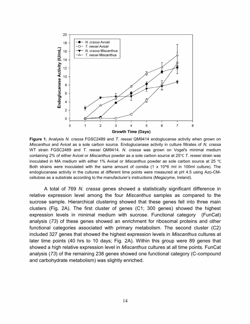
Figure 1. Analysis N. crassa FGSC2489 and T. reesei QM9414 endoglucanse activity when grown on
Miscanthus and Avicel as a sole carbon source. Endoglucanase activity in culture filtrates of N. crassa
WT strain FGSC2489 and T. reesei QM9414. N. crassa was grown on Vogel's minimal medium
containing 2% of either Avicel or Miscanthus powder as a sole carbon source at 25℃. T. reseei strain was
inoculated in MA medium with either 1% Avicel or Miscanthus powder as sole carbon source at 25 ℃.
Both strains were inoculated with the same amount of conidia (1 x 10^6 /ml in 100ml culture). The
endoglucanase activity in the cultures at different time points were measured at pH 4.5 using Azo-CM-
cellulose as a substrate according to the manufacturer’s instructions (Megazyme, Ireland).
A total of 769 N. crassa genes showed a statistically significant difference in
relative expression level among the four Miscanthus samples as compared to the
sucrose sample. Hierarchical clustering showed that these genes fell into three main
clusters (Fig. 2A). The first cluster of genes (C1; 300 genes) showed the highest
expression levels in minimal medium with sucrose. Functional category (FunCat)
analysis (73) of these genes showed an enrichment for ribosomal proteins and other
functional categories associated with primary metabolism. The second cluster (C2)
included 327 genes that showed the highest expression levels in Miscanthus cultures at
later time points (40 hrs to 10 days; Fig. 2A). Within this group were 89 genes that
showed a high relative expression level in Miscanthus cultures at all time points. FunCat
analysis (73) of the remaining 238 genes showed one functional category (C-compound
and carbohydrate metabolism) was slightly enriched.

15
Figure 2. Transcriptional profiling of N. crassa grown on Miscanthus and Avicel. (A) Hierarchical
clustering analysis of 769 genes showing expression differences in Miscanthus culture. Red indicates
higher relative expression and green indicates lower relative expression. Lane 1: A 16 h N. crassa culture
grown in sucrose minimal medium. Lane 2: A 16 h culture with Miscanthus as a sole carbon source.
Lanes 3–5: Expression profiles from cultures grown on Miscanthus for 40 h, 5 days, and 10 days. The C3
cluster showed increased expression levels of most of the cellulase and hemicellulase genes (boxed). (B)
Overlap in expression profiles between the N. crassa Miscanthus versus Avicel grown cultures (Top).
Overlap of proteins in culture filtrates detected by tandem mass spectrometry (Bottom). (C) Functional
category analysis (16) of the 231 genes that showed a significant enrichment (P < 0.001) in relative
expression levels in Miscanthus cultures.
A third cluster of 142 genes showed the highest relative expression level after 16
hours of growth of N. crassa on Miscanthus (C3, Fig. 2A). FunCat analysis (73) of these
142 genes plus the 89 genes that showed high expression levels in Miscanthus cultures
at all time points (C3+ cluster; total 231 genes) showed an enrichment for proteins
involved with carbon metabolism, including predicted cellulases and hemicellulases
(Fig. 2C). Of the 23 predicted cellulase genes in the N. crassa genome, 18 showed
significant increases in expression levels during growth on Miscanthus (Table 1),
particularly at the 16 hour time point (Fig. 3). Five genes showed an increase in
expression level over 200-fold (cbh-1 (CBH(I); NCU07340, gh6-2 (CBH(II)-like gene;

16
NCU09680), gh6-3 (NCU07190) and two GH61 genes (gh61-4; NCU01050 and
NCU07898)).
Figure 3. Fold change induction of cellulases during growth on Miscanthus relative to sucrose. Most
cellulases show highest induction at 16 hours, then a lower, but steady level of induction at later time
points.
Plant cell walls are complex structures composed of cellulose microfibrils,
hemicellulose, lignin, pectin, cutin, and protein. Thus, we compared expression profiles
of N. crassa grown on Miscanthus to expression profiles of N. crassa grown on Avicel, a
pure form of crystalline cellulose. Over 187 genes showed a significant increase in
relative expression level during growth of N. crassa on Avicel. Of these genes, 114
overlapped with the 231 genes in the C3+ cluster (Fig. 2B). FunCat analysis of the 114-
Table 1. Predicted cellulases genes in Neurospora crassa
Gene GH family CBM1 SP MS EL Miscanthus EL Avicel
NCU00762 5 Yes Yes Both 29.6 31.5
NCU03996 6 No No ND ND ND
NCU07190 6 No Yes Both 526.0 119
NCU09680 6 Yes Yes Both 230.9 251.3
NCU04854 7 No Yes ND 32.9 10.8
NCU05057 7 No Yes Both 8.7 7.4
NCU05104 7 No Yes ND 11.6 NC7
NCU07340 7 Yes Yes Both 426.4 382.2

17
Gene GH family CBM1 SP MS EL Miscanthus EL Avicel
NCU05121 45 Yes Yes avi 8.6 17.2
NCU00836 61 Yes Yes ND 91.2 31
NCU01050 61 No Yes Both 206.7 382.1
NCU01867 61 Yes Yes ND 2.2 NC
NCU02240 61 Yes Yes avi 193.5 84
NCU02344 61 No Yes ND 8.1 4.1
NCU02916 61 Yes Yes ND 85.2 17.7
NCU03000 61 No Yes ND NC ND
NCU03328 61 No Yes ND 26.4 23.8
NCU05969 61 No Yes ND ND 12.7
NCU07520 61 No Yes ND ND ND
NCU07760 61 Yes Yes ND 3.7 NC
NCU07898 61 No Yes Both 376.3 230
NCU07974 61 No Yes ND NC NC
NCU08760 61 Yes Yes Both 107.5 44.7
* GH, glycoside hydrolase; CBM1, carbohydrate binding module; SP, signal peptide prediction; MS, mass
spectrometry analysis; EL, relative expression level; ND, not detected; NC, no change.
overlap gene set showed a clear enrichment for genes predicted to be involved in
carbon metabolism. Within this gene set, there was a further enrichment for secreted
proteins (53 of the 114 gene products). Of the 53 genes, 32 encode predicted proteins
with annotation suggesting a role in plant cell wall degradation, while 16 encode
putative or hypothetical proteins. The remaining 61 genes encode predicted intracellular
proteins, including ten predicted major facilitator superfamily transporters (NCU00801,
NCU00988, NCU01231, NCU04963, NCU05519, NCU05853, NCU05897, NCU06138,
NCU08114 and NCU10021) and 23 putative or hypothetical proteins.
Of the 117 genes within the Miscanthus-specific cluster (Fig. 2B), 37 encoded
proteins predicted to be secreted. Nine predicted hemicellulases or enzymes related to
the degradation of hemicellulose were identified (NCU00710, NCU04265, NCU04870,
NCU05751, NCU05965, NCU09170, NCU09775, NCU09923 and NCU09976). The
remaining 80 Miscanthus-specific genes encode predicted intracellular proteins,
including genes involved in the metabolism of pentose sugars (for example, NCU00891,
xylitol dehydrogenase and NCU00643, a predicted arabinitol dehydrogenase), a
predicted sugar transporter (NCU01132), and 48 proteins of unknown function.

18
2.3.2 Secretome analysis of N. crassa grown on Miscanthus and Avicel
Lignocellulose degradation by fungi requires the secretion of proteins associated
with depolymerization of cell wall constituents (74). To compare with transcriptional
profiling data, we analyzed the secretome of N. crassa using a shotgun proteomics
approach (Fig. 1B). Supernatants from three and seven day old Miscanthus and Avicel
cultures were digested with trypsin and analyzed by liquid chromatography nano-
electrospray ionization tandem mass spectrometry (MS); datasets from the 3 and 7 day
samples showed no significant difference. Secreted proteins that bound to phosphoric
acid swollen cellulose (PASC) were enriched and also analyzed by MS.
A total of 50 proteins were identified with confidence by tandem MS (Table 2).
There were 34 proteins detected in the Miscanthus grown N. crassa cultures, while 38
proteins were identified from Avicel grown cultures; twenty-two proteins were detected
in both samples. Of these 22 proteins, 21 were predicted to be secreted based on
computational analyses and 19 showed increased expression levels in both the
Miscanthus and Avicel grown cultures. The overlap dataset included eight of the 23
predicted cellulases in N. crassa (Table 1). There were also five predicted
hemicellulases, a predicted -glucosidase (gh3-4; NCU04952), five proteins with
predicted activity on carbohydrates, and two proteins with unknown function
(NCU07143 and NCU05137) (Table 2).
There were 16 proteins identified with confidence in the Avicel culture only and
14 of these were predicted to be secreted (Table 2) including two predicted cellulases
(gh61-1; NCU02240 and gh45-1; NCU05121), one xylanase (gh11-1; NCU02855), one
predicted protease (NCU04205), three other proteins with predicted activity on
carbohydrates (NCU08909, NCU05974 and gh30-1 (NCU04395)), three Neurospora-
specific proteins of unknown function, and four conserved hypothetical proteins,
including one protein with a cellulose binding domain (NCU09764). Twelve proteins
were specific for culture filtrates of Miscanthus cultures and seven of these had
predicted secretion signals (Table 2). Three of the five predicted intracellular proteins
were conserved hypothetical proteins. The remaining two included a predicted glyoxal
oxidase (NCU09267, identified from the N. crassa Miscanthus transcriptome) and a
nucleoside diphosphate kinase (ndk-1; NCU04202, not identified in the N. crassa
transcriptome). The seven proteins predicted to be secreted included three predicted
esterases (NCU04870, NCU05159, and NCU08785), two predicted xylanases (GH51;
NCU02343 and GH54; NCU09775), a predicted -xylosidase (gh3-7; NCU09923) and a
conserved hypothetical protein (NCU05751).
Many plant cell wall degrading enzymes contain a cellulose-binding module
(CBM), which aids in attachment of the enzyme to the substrate (75). Within the N.
crassa genome, 19 genes are predicted to encode proteins with a CBM1 domain (76).
19
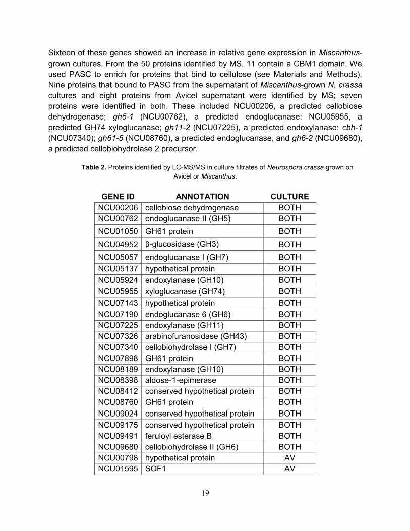
Sixteen of these genes showed an increase in relative gene expression in Miscanthus-
grown cultures. From the 50 proteins identified by MS, 11 contain a CBM1 domain. We
used PASC to enrich for proteins that bind to cellulose (see Materials and Methods).
Nine proteins that bound to PASC from the supernatant of Miscanthus-grown N. crassa
cultures and eight proteins from Avicel supernatant were identified by MS; seven
proteins were identified in both. These included NCU00206, a predicted cellobiose
dehydrogenase; gh5-1 (NCU00762), a predicted endoglucanase; NCU05955, a
predicted GH74 xyloglucanase; gh11-2 (NCU07225), a predicted endoxylanase; cbh-1
(NCU07340); gh61-5 (NCU08760), a predicted endoglucanase, and gh6-2 (NCU09680),
a predicted cellobiohydrolase 2 precursor.
Table 2. Proteins identified by LC-MS/MS in culture filtrates of Neurospora crassa grown on
Avicel or Miscanthus.
GENE ID ANNOTATION CULTURE
NCU00206 cellobiose dehydrogenase BOTH
NCU00762 endoglucanase II (GH5) BOTH
NCU01050 GH61 protein BOTH
NCU04952 β-glucosidase (GH3) BOTH
NCU05057 endoglucanase I (GH7) BOTH
NCU05137 hypothetical protein BOTH
NCU05924 endoxylanase (GH10) BOTH
NCU05955 xyloglucanase (GH74) BOTH
NCU07143 hypothetical protein BOTH
NCU07190 endoglucanase 6 (GH6) BOTH
NCU07225 endoxylanase (GH11) BOTH
NCU07326 arabinofuranosidase (GH43) BOTH
NCU07340 cellobiohydrolase I (GH7) BOTH
NCU07898 GH61 protein BOTH
NCU08189 endoxylanase (GH10) BOTH
NCU08398 aldose-1-epimerase BOTH
NCU08412 conserved hypothetical protein BOTH
NCU08760 GH61 protein BOTH
NCU09024 conserved hypothetical protein BOTH
NCU09175 conserved hypothetical protein BOTH
NCU09491 feruloyl esterase B BOTH
NCU09680 cellobiohydrolase II (GH6) BOTH
NCU00798 hypothetical protein AV
NCU01595 SOF1 AV

20
Table 2 continued
GENE ID ANNOTATION CULTURE
NCU02240 GH61 protein AV
NCU02696 DEAD DEAH box RNA helicase AV
NCU02855 endoxylanase (GH11) AV
NCU04205 peptidase A4 family AV
NCU04395 endo-1,6-b-d-glucanase (GH30) AV
NCU05121 endoglucanase V (GH45) AV
NCU05134 hypothetical protein AV
NCU05852 hypothetical protein AV
NCU05974 similar to Mwg1 AV
NCU08171 predicted protein AV
NCU08909 beta-1,3 glucanosyltransferase AV
NCU08936 hypothetical protein AV
NCU09046 hypothetical protein AV
NCU09764 hypothetical protein AV
NCU01651 hypothetical protein MIS
NCU02343 α-l-arabinofuranosidase MIS
NCU04202 nucleoside diphosphate kinase MIS
NCU04870 acetyl xylan esterase MIS
NCU05159 acetylxylan esterase MIS
NCU05751 hypothetical protein MIS
NCU06239 hypothetical protein MIS
NCU08785 hypothetical protein MIS
NCU09267 hypothetical protein MIS
NCU09708 hypothetical protein MIS
NCU09775 α-L-arabinofuranosidase MIS
NCU09923 β-xylosidase (GH3) MIS
2.3.3 Characterization of extracellular proteins and cellulase activity in strains
containing deletions in genes identified in the overlap of the transcriptome and
secretome datasets.
Of the 22 extracellular proteins detected in both the Miscanthus and Avicel grown
cultures, homokaryotic strains containing deletions in 16 genes are available (69). None
of these 16 deletion strains have been previously characterized with respect to plant cell
wall or cellulose degradation by N. crassa. The 16 deletion strains were grown in media
containing sucrose or Avicel as a carbon source. All strains showed a wild-type

21
phenotype on sucrose medium. The total secreted protein, endoglucanase activity, -
glucosidase activity, and aggregate Avicelase activity of Avicel-grown culture filtrates
was measured after seven days and compared to the wild-type strain from which all
mutants were derived (Fig. 4). SDS-PAGE of unconcentrated culture supernatants
assessed the relative abundance of secreted proteins.
There were growth deficiencies on Avicel for strains containing deletions of two
predicted exoglucanases, cbh-1 (NCU07340) and gh6-2 (NCU09680), plus a
predicted -glucosidase, gh3-4 (NCU04952). The phenotype of the cbh-1 mutant was
the most severe; after seven days in culture much of the Avicel remained, while in the
wild-type strain all of the Avicel was degraded. SDS-PAGE of extracellular proteins from
10 of the 16 deletion strains showed an altered protein profile with loss of a single band,
allowing assignment of a particular protein band to a predicted gene (Fig. 4A, boxes).
These included NCU00762 (gh5-1), NCU04952 (gh3-4), NCU05057 (gh7-1),
NCU05137, NCU05924 (gh10-1), NCU05955, NCU07190 (gh6-3), NCU07326,
NCU07340 (cbh-1) and NCU09680 (gh6-2).
For the majority of the deletion strains, the total secreted protein, endoglucanase,
-glucosidase, and Avicelase activities of the culture supernatants were similar to wild
type (WT) (Fig. 4B, C). Deviations from this trend were seen with gh5-1 (NCU00762),
gh3-4 (NCU04952), NCU05137, cbh-1 (NCU07340) and gh6-2 (NCU09680). In
gh5-1 (NCU00762), gh3-4 (NCU04952), and cbh-1 (NCU07340) Avicelase,
endoglucanase or -glucosidase activities were lower than the corresponding WT
activities. In particular, the deletion of NCU04952 eliminated all -glucosidase activity
from the culture supernatant, as evidenced by PNPGase activity and by higher levels of
cellobiose and lower levels of glucose in the Avicelase enzyme assays (Fig. 4B, C).
Unlike a WT strain, culture filtrates from NCU04952 were completely unable to
hydrolyze cellobiose to glucose, consistent with loss of -glucosidase activity. Despite
lowering endoglucanase activity, the culture filtrate from gh5-1 (NCU00762) showed
no significant deficiency in Avicelase activity relative to the WT strain (Fig. 4C). As
expected, mutations in cbh-1 (NCU07340) resulted in lower endoglucanase and
Avicelase activity. A strain containing a deletion of NCU09680, encoding a CBH(II)-like
protein (gh6-2), also showed reduced cellobiose accumulation (Fig. 4C).
Mutations in three strains resulted in an increased level of secreted proteins,
especially CBH(I) (Fig. 4A); gh3-4 (NCU04952), gh7-1 (NCU05057) and a
hypothetical protein gene, NCU05137. The NCU05137 mutant also showed
increased endoglucanase, -glucosidase and Avicelase activity (Fig. 4B, C). NCU05137
is highly conserved in the genomes of a number of filamentous ascomycete fungi,
including other cellulolytic fungi, but notably does not have an ortholog in H. jecorina. It
is possible that the increase in CBH(I) levels observed in gh3-4, gh7-1 and
NCU05137 could be due to either increased secretion, protein stability or feedback

22
Figure 4. Protein profile and enzymatic activity of culture supernatants from strains containing deletions
of genes encoding secreted proteins identified by MS. (A) SDS/PAGE of proteins present in the culture
filtrates of 16 deletion strains as compared to WT when grown on Avicel for 7 days. Missing protein bands
that correspond to the deleted genes are marked with boxes. (B) Total secreted protein, azo-CMCase,
and β-glucosidase activity assays performed on 16 deletion strains and the WT parental strain (FGSC
2489) using the same sample from A. (C) Cellulase activity of the culture filtrates from the 16 deletion
strains using the same samples as in A. Bars, standard deviation. Glucose (black) and cellobiose (white)
were measured after 8 h of incubation at 40 °C. Bars, standard deviation.

23
Figure 5. Time Course Analysis of Secreted Protein in WT and ΔNCU04952, ΔNCU05137.
(A.) SDS-PAGE of total secreted proteins in WT, ΔNCU04952, and ΔNCU05137. The cultures were
grown on Avicel and harvested at 30 hours, 48 hours, and 72 hours. Lanes 1-3, 20x concentrated culture
filtrates after 30 hours of growth on Avicel from WT, ∆NCU04952, and ∆NCU05137. Lanes 4-6,
unconcentrated culture filtrates after two days of growth from WT, ∆NCU04952, and ∆NCU05137. Lane
7-9, unconcentrated culture filtrates after three days of growth from WT and ∆NCU04952 and
∆NCU05137. (B) RT-PCR of CBHI and CBHII in the WT, ∆NCU04952, and ∆NCU05137 during growth on
Avicel (see materials and methods).
that results in increased expression of cbh-1. To differentiate these possibilities, we
compared the profile of extracellular proteins produced by NCU05137 and gh3-4
(NCU04952) with gene expression levels of cbh-1 (NCU07340) and gh6-2 (CBH(II);
NCU09680) as assayed by quantitative RT-PCR (Fig. 5). The NCU05137 and gh3-4
strains showed a higher level of CBH(I) protein as early as two days in an Avicel-grown
culture (Fig. 5a) and higher expression levels of both cbh-1 and gh6-2 at three days of
growth, while expression of both of these genes decreased significantly in the WT strain
(Fig. 5b). Sustained expression of cbh-1 and gh6-2 genes in the NCU05137 and gh3-
4 mutants could be responsible for the observed increase in CBH(I) and CBH(II) protein
levels.
2.4 Discussion
Degradation of plant biomass requires production of many different enzymes,
which are regulated by the type and complexity of the available plant material (Fig. 6)
(77). Here, we report on the systematic analyses of plant cell wall degradation by a
cellulolytic fungus, which includes transcriptome, secretome and mutant analyses. Our
profiling data shows that N. crassa coordinately expresses a host of extracellular and
intracellular proteins when challenged by growth on Miscanthus or Avicel (Fig. 6). The
most highly expressed genes encode proteins predicted to be involved in the
metabolism of plant cell wall polysaccharides, many of which were identified by MS
analyses. The genomes of filamentous fungi have a large number of predicted glycosyl

24
hydrolases (~200) with varying numbers of predicted cellulases, from 10 in H. jecorina
(68) to 60 in Podospora anserina (78). A comparison between our results and a cDNA
expression/Northern analysis of 8 endoglucanases and 7 -glucosidases in H. jecorina
(79) showed complete overlap with our profiling data, with the exception of one ortholog
of a -glucosidase (cel-3e=NCU05577). However, a recent transcriptome/secretome
study on the white rot basidiomycete fungus, Phanerochaete chrysosporium (80)
showed little overlap. These data suggest that different fungi may utilize different gene
sets for plant cell wall degradation. One aspect all of these studies have in common is
the high number of uncharacterized genes/proteins associated with cellulose
degradation. Using the functional genomic tools available with N. crassa, we can
address both function and redundancy of plant cell wall degrading enzyme systems to
create optimal enzyme mixtures for industrial production of liquid fuels from
lignocellulose biomass.
N. crassa cellobiohydrolase(I) (CBHI) is the most highly produced extracellular
protein during growth on Avicel or Miscanthus, and deletion of this gene causes the
most severe growth deficiencies on cellulosic substrates. By contrast, in H. jecorina,
deletion of cbhII caused the most severe phenotype (81-83). In N. crassa, deletion of
cellobiohydrolase(II) also causes growth deficiencies on cellulosic substrates, but to a
much lesser extent than CBH(I), suggesting that exoglucanase activity in N. crassa is
predominantly from CBH(I) and that cellulases and other CBHs do not compensate for
the loss of CBH(I). The three most highly produced endoglucanases are proteins
encoded by NCU05057, NCU00762, and NCU07190. These proteins have homology to
endoglucanases EG1, EG2, and EG6, respectively. Deletion of these genes did not
affect growth on Avicel, although differences in secreted protein levels and
endoglucanase activity were observed. Unexpectedly, in ΔNCU05057, extracellular
protein levels were much higher, especially CBH(I), suggesting increased production of
other cellulases or that the products of NCU05057 catalysis may repress cellulase
production to maintain the WT growth phenotype on crystalline cellulose. We conclude
that no one endoglucanase in N. crassa is required for growth on crystalline cellulose
and that different endoglucanases have overlapping substrate specificities.
The glycoside hydrolase family 61 enzymes are greatly expanded in N. crassa
compared to H. jecorina (68). These enzymes have poorly defined biological function,
but their general conservation and abundance in cellulolytic fungi suggests an important
role in plant cell wall metabolism. Genes for 10 of the 14 GH61 enzymes were identified
in the N. crassa transcriptome, suggesting that these enzymes are utilized during
growth on cellulosic biomass. The four GH61 deletion strains tested showed only small
differences compared to wild type in the secreted protein levels, endoglucanase, and
total cellulase activities. However, analyses of additional GH61 mutants and the
capacity to create strains containing multiple mutations in N. crassa via sexual crosses
will address redundancy and expedite functional analysis of this family.

25
Figure 6. Model of plant cell wall deconstruction in N. crassa. Induction: Extracellular enzymes expressed
at low levels generate metabolites that signal N. crassa to dramatically increase the expression level of
genes encoding plant cell wall degrading enzymes. Utilization: Extracellular enzymes and transporters
specific for translocation of cell wall degradation products enable N. crassa to use plant cell material for
growth. Some extracellular proteins may generate metabolites that modulate gene expression of
cellulases and hemicellulases during the utilization phase. Double red hexagon (cellobiose), double teal
pentagon (xylobiose), black hexagon (glucose), and blue pentagon (xylose). Blue, CBH(I); red, CBH(II);
purple, EG2; green, EG1; orange, EG6; and yellow, xylanase. Additional cellulolytic enzymes not shown.
Thickness of arrows indicates relative strength of response.

26
In addition to predicted cellulase genes, genes encoding hemicellulases,
carbohydrate esterases, β-glucosidases, β-xylosidases and other proteins predicted to
have activity on carbohydrates were identified in the N. crassa transcriptome from both
Miscanthus and Avicel. Expression of hemicellulase genes even when N. crassa was
grown on bacterial cellulose indicates that cellulose is the primary inducer of genes
encoding plant cell wall degrading enzymes. However, genes encoding some
hemicellulases and carbohydrate esterases were only expressed during growth on
Miscanthus. Similarly, in other cellulolytic fungi such as H. jecorina and Aspergillus
niger, genes encoding some cellulases and hemicellulases are coordinately regulated,
while others are differentially regulated (84). As expected, deletions of non-cellulase
genes had little effect on growth on Avicel or cellulase activity, with the exception of
NCU05137 and gh3-4. The ΔNCU05137 strain secreted more protein, had higher
cellulase activity and showed higher expresssion of cbh-1 (CBH(I)) and gh6-2 (CBH(II))
than WT. NCU05137 encodes a secreted protein that lacks homology to proteins of
known function, but is highly conserved in other cellulolytic fungi. NCU05137 also has
distant homologs of unknown function in a number of bacterial species. We hypothesize
that the NCU05137 protein may affect signaling processes associated with the
regulation of cellulase gene expression N. crassa (Fig. 6). Similarly, mutations in gh3-4
(NCU04952) also increased CBH(I) activity. Deletion of gh3-4 completely removed
PNPGase activity and resulted in cellobiose accumulation in in vitro cellulase assays.
These data suggest that NCU04952 encodes the primary extracellular β-glucosidase in
N. crassa.
Extracellular degradation of plant cell walls results in the formation of soluble
carbohydrates that are subsequently transported into the cell (Fig. 6). In this study, we
identified 10 genes encoding permeases/transporters whose expression increased
significantly when N. crassa was grown on Miscanthus or Avicel. The major degradation
products by cellulases and hemicellulases in vitro are cellobiose, glucose, xylobiose,
and xylose. Some of these transporters may be functionally redundant or capable of
transporting oligosaccharides. Construction of downstream processing strains capable
of transporting oligosaccharides by heterologous expression of N. crassa transporters
may improve industrial fermentation of biomass hydrolysis products. None of these
transporters or what they may transport has been characterized at the molecular or
functional level in any filamentous fungus.
Many genes that increased in expression level during growth on Miscanthus and
Avicel encode proteins of unknown function and are conserved in other cellulolytic fungi.
By assessing the phenotype of only 16 strains, we identified a mutation in a protein of
unknown function that significantly affected cellulase activity. The well understood
genetics and availability of functional genomic resources make N. crassa an ideal model
organism to assess biological function of these proteins, examine regulatory aspects of

27
cellulase and hemicellulase production, and dissect redundancies and synergies
between extracellular enzymes involved in the degradation of plant cell walls.
2.5 Materials and Methods
Strains. The sequenced N. crassa strain (FGSC 2489) was used for transcriptional
profiling. Gene deletion strains were generated from the N. crassa functional genomics
project (69); all mutations were verified by PCR.. H. jecorina QM9414 was a gift from
Dr. Monika Schmoll (Vienna University of Technology). Strains were grown on Vogel’s
salts (85) with 2% (w/v) carbon source (Miscanthus, sucrose or Avicel (Sigma)). Growth
of N. crassa on Avicel and Miscanthus was monitored by macroscopic and microscopic
visual assessment. Deletion strains were obtained from the Fungal Genetics Stock
Center (FGSC), including FGSC 16747 (NCU00762), FGSC 16543 (NCU01050),
FGSC 13732 (NCU04952), FGSC 13343 (NCU05057), FGSC 11682 (NCU05137),
FGSC 15626 (NCU05924), FGSC 13535 (NCU05955), FGSC 19315 (NCU07190),
FGSC 19534 (NCU07326), FGSC 15630 (NCU07340), FGSC 19600 (NCU07898),
FGSC 19861 (NCU08189), FGSC 20310 (NCU08398), FGSC 15664 (NCU08760),
FGSC 11750 (NCU09175), FGSC 15633 (NCU09680). Stem and leaf tissues from
Miscanthus gigantaeus were harvested from field-grown plants at the University of
Illinois, Urbana-Champaign (2007) at the end of the growing season, air dried, and
milled to a particle size of 100 m.
Enzyme activity measurements. Total extracellular protein content was determined by
a Bio-Rad DC Protein Assay kit (Bio-Rad). Endoglucanase activity in culture
supernatants was measured with an azo-CMC kit (Megazyme SCMCL). -glucosidase
activity was measured by mixing 10x diluted culture supernatant with 500 uM 4-
nitrophenyl β-D-glucopyranoside in 50 mM sodium acetate buffer, pH 5.0, for 10
minutes at 40C. The reaction was quenched with 5% w/v sodium carbonate and the
absorbance at 400 nm was measured. Avicelase activity was measured by mixing 2X-
diluted culture supernatant with 50 mM sodium acetate pH 5.0 and 5 mg/mL Avicel at
40C. Supernatants were analyzed for glucose content using a coupled enzyme assay
with glucose oxidase/peroxidase. 50 uL of the avicelase reaction was transferred to
150 uL of glucose detection reagent containing 100 mM sodium acetate pH 5.0, 10
U/mL horseradish peroxidase, 10 U/mL glucose oxidase, and 1 mM o-dianisidine. After
30 minutes absorption was measured at 540 nm. Cellobiose concentrations in the
avicelase reaction mixture were determined using a coupled enzyme assay with
cellobiose dehydrogenase (CDH) from Sporotrichum thermophile. CDH was isolated
from S. thermophile similar to previous reports (86). 50 uL of the Avicelase reaction
was transferred to 250 uL of cellobiose detection reagent containing 125 mM sodium
28
acetate pH 5.0, 250 uM dichlorophenol indophenol, and 0.03 mg/mL CDH. After 10
minutes absorption was measured at 530 nm.
Transcriptome analysis. N. crassa strain FGSC 2489 was inoculated onto slants of
Vogel’s minimal medium (MM) with 1.5% agar and 2% sucrose and grown for 10 days
at 25C. A conidial suspension was inoculated into 50 ml of Vogel’s liquid medium (85)
in 250 ml Pyrex flasks supplemented with either 2% Avicel, Miscanthus powder or
sucrose as sole carbon sources at a final concentration of 106 conidia per ml. Cultures
were grown under controlled temperature (25C) with constant shaking and light (300
lux). Mycelia was harvested by vacuum filtration using Whatman paper #1, frozen
immediately in liquid nitrogen and stored at 80C. Two independent biological
duplicates (flasks) were evaluated for each time point.
RNA isolation and cDNA labeling. Total RNA from frozen samples was isolated using
Zirconia/silica beads (0.2 g, 0.5-mm diameter; Biospec) and a MiniBeadBeater
(Biospec) with 1 ml of TRIzol reagent (Invitrogen Life Technologies) according to
manufacturer’s instructions. Total RNA (100 g) was further purified using RNAeasy kit
(Qiagen). RNA integrity was checked by Nanodrop and agarose gel electrophoresis.
cDNA synthesis and labeling followed the protocol of ChipShot indirect labeling kit from
Promega (Catalog #Z4000). The dyes Cy3 and Cy5 (Amersham; catalog no. RPN5661)
were incorporated into cDNA by adding Cy3 or Cy5 monofunctional N-
hydroxysuccinimide ester dye to the cDNA solution for 1 h at 22°C. The cDNA was
subsequently cleaned by using a ChipShot membrane column and dried under vacuum.
Hybridization and image acquisition. A set of 10,910 70-mers representing predicted
genes in N. crassa were printed onto gamma amino propyl silane slides (Corning) at the
University of California—San Francisco Center for Advanced Technology
(http://cat.UCSF.edu/). N. crassa microarray slides are available from the Fungal
Genetics Stock Center (http://www.fgsc.net/). Information on the oligonucleotide gene
set is available at the Neurospora functional genomics database
(http://www.yale.edu/townsend/Links/ffdatabase/introduction.htm). Pre-hybridization and
hybridization of slides used the Pronto kit (Corning; catalog no. 40076) according to
manufacturer’s instructions. Microarray slides were added to a pre-soak solution that
was pre-warmed to 42°C and incubated for 20 min. Slides were subsequently washed
(Pronto kit) and transferred to pre-warmed pre-hybridization solution (42°C) for 15 min,
and rewashed. Labeled cDNA was resuspended in 30 l of hybridization solution
(Pronto kit). The suspension was heated at 95°C for 5 min and then pipetted into the
space between the microarray slide and a LifterSlip cover glass (Erie Scientific,
Portsmouth, NH). Hybridization was performed at 42°C for 16 hr. Unbound DNA was
washed off according to manufacturer’s instructions (Pronto kit).
29
Hybridized slides were scanned using Genepix 4000B. Four independent
hybridizations were performed per sample. Genepix6 software was used for alignment
of hybridization spots and initial analysis (.GPR files). Hybridized spots with a mean
fluorescence intensity for at least one of the Cy3 and Cy5 dyes that was greater than
the mean background intensity plus three standard deviations were scored for further
analysis. For quality control, slides were used where the ratio of Cy5:Cy3 signal was
approximately one. Global normalization was performed to each GPR file using in-
house excel-based Macro (72). The Macro is available at
http://glasslab.weebly.com/links.html and Yale Microarray Experimental Design site.
Experimental design and data analysis. A closed-circuit design for microarray
comparisons was used because they are statistically robust and improve resolution in
identifying differentially regulated genes compared to designs for microarrays that use a
universal reference (87-89). The gene relative expression data was estimated by
BAGEL software (90). BAGEL explores the ratio for all samples using a Markov Chain
Monte Carlo (MCMC) approach in a Bayesian framework, and infers relative expression
levels and statistical significance from the parameter values it samples. BAGEL also
provides gene-by-gene 95% credible intervals, values in which 95% of the samples from
the chain are bounded. The 769 N. crassa genes indentified in this study that were
identified as showing a significant difference in expression between Miscanthus and
minimal sucrose medium passed two filters. The first filter was that the 95% credible
interval of a gene expressed under Miscanthus versus sucrose conditions showed no
overlap and the closest boundary value between the two samples showed at least 1.5
fold difference in expression level. The second filter used was the average expression
of a gene in Miscanthus versus sucrose grown cultures was required to have at least a
2-fold change at one of four time points. These data were obtained by in-house PERL
script ”strict-errobar.pl” which is available in http://glasslab.weebly.com/links.html.
Clustering analysis performed by HCE3 (Hierarchical Clustering Explorer 3.0), using
default parameter in which a Euclidean Distance model (91) is used to join genes and
clusters successively. Distances between clusters represent the average distances
between genes in the clusters.
The MIPs functional catalog (FunCat) was used to associate functional annotation (73)
with Neurospora genes. The overrepresentation of Neurospora gene groups in
functional categories relative to the whole genome was determined using
hypergeometric distribution for P value calculation.
Signal peptides were predicted using the N-terminal 70 amino acid region of
each predicted protein with the signalP3 program (
http://www.cbs.dtu.dk/services/SignalP-3.0/).

30
Protein gel electrophoresis. Except where otherwise noted, unconcentrated culture
supernatants were treated with 5x SDS loading dye and boiled for 5 minutes before
loading onto Criterion 4-15% Tris-HCl polyacrylamide gels. Coomassie dye was used
for staining.
Preparation of tryptic peptides for secretome analysis. Culture supernatants were
concentrated with 10 kDa MWCO PES spin concentrators. Cellulose binding proteins
were isolated from the culture supernatant by addition of phosphoric acid swollen
cellulose (PASC). 5 mL of a suspension of 10 mg/mL PASC was added to 10 mL of
culture supernatant. After incubation at 4C for 5 minutes, the mixture was centrifuged
and the pelleted PASC was then washed with 20 pellet volumes of 100 mM sodium
acetate pH 5.0. The supernatant after treatment with PASC was saved as the unbound
fraction and concentrated. 36 mg of urea, 5 uL of 1M Tris PH 8.5, and 5 uL of 100 mM
DTT were then added to 100 uL of concentrated culture supernatant or protein-bound
PASC and the mixture was heated at 60C for 1 hour. After heating 700 uL of 25 mM
ammonium bicarbonate and 140 uL of methanol were added to the solution followed by
treatement with 50 uL of 100 ug/mL trypsin in 50 mM sodium acetate pH 5.0. For the
PASC bound proteins, the PASC was removed by centrifugation after heating, and the
supernatant was then treated with trypsin. The trypsin was left to react overnight at
37C. After digestion the volume was reduced by speedvac and washed with MilliQ
water three times. Residual salts in the sample were removed by using OMIX
microextraction pipette tips according to the manufacturer’s instructions.
Liquid chromatography of tryptic peptides. Trypsin-digested proteins were analyzed
using a tandem mass spectrometer that was connected in-line with ultraperformance
liquid chromatography (UPLC). Peptides were separated using a nanoAcquity UPLC
(Waters, Milford, MA) equipped with C18 trapping (180 µm × 20 mm) and analytical (100
µm × 100 mm) columns and a 10 µL sample loop. Solvent A was 0.1% formic
acid/99.9% water and solvent B was 0.1% formic acid/99.9% acetonitrile (v/v). Sample
solutions contained in 0.3 mL polypropylene snap-top vials sealed with septa caps
(Wheaton Science, Millville, NJ) were loaded into the nanoAcquity autosampler prior to
analysis. Following sample injection (2 µL, partial loop), trapping was performed for 5
min with 100% A at a flow rate of 3 µL/min. The injection needle was washed with 750
µL each of solvents A and B after injection to avoid cross-contamination between
samples. The elution program consisted of a linear gradient from 25% to 30% B over
55 min, a linear gradient to 40% B over 20 min, a linear gradient to 95% B over 0.33
min, isocratic conditions at 95% B for 11.67 min, a linear gradient to 1% B over 0.33
min, and isocratic conditions at 1% B for 11.67 min, at a flow rate of 500 nL/min. The
analytical column and sample compartment were maintained at 35 ºC and 8 ºC,
respectively.

31
Mass spectrometry. The column was connected to a NanoEase nanoelectrospray
ionization (nanoESI) emitter mounted in the nanoflow ion source of a quadrupole time-
of-flight mass spectrometer (Q-Tof Premier, Waters). The nanoESI source parameters
were as follows: nanoESI capillary voltage 2.3 kV, nebulizing gas (nitrogen) pressure
0.15 mbar, sample cone voltage 30 V, extraction cone voltage 5 V, ion guide voltage 3
V, and source block temperature 80 ºC. No cone gas was used. The collision cell
contained argon gas at a pressure of 8 × 10⁻³ mbar. The Tof analyzer was operated in
“V” mode. Under these conditions, a mass resolving power1 of 1.0 × 10⁴ (measured at
m/z = 771) was routinely achieved, which is sufficient to resolve the isotopic
distributions of the singly and multiply charged peptide ions measured in this study.
Thus, an ion’s mass and charge could be determined independently, i.e., the ion charge
was determined from the reciprocal of the spacing between adjacent isotope peaks in
the m/z spectrum. External mass calibration was performed immediately prior to
analysis, using solutions of sodium formate. Survey scans were acquired in the positive
ion mode over the range m/z = 450-1800 using a 0.95 s scan integration and a 0.05 s
interscan delay. In the data-dependent mode, up to five precursor ions exceeding an
intensity threshold of 35 counts/second (cps) were selected from each survey scan for
tandem mass spectrometry (MS/MS) analysis. Real-time deisotoping and charge state
recognition were used to select 2+, 3+, 4+, 5+, and 6+ charge state precursor ions for
MS/MS. Collision energies for collisionally activated dissociation (CAD) were
automatically selected based on the mass and charge state of a given precursor ion.
MS/MS spectra were acquired over the range m/z = 50-2500 using a 0.95 s scan
integration and a 0.05 s interscan delay. Ions were fragmented to achieve a minimum
total ion current (TIC) of 30,000 cps in the cumulative MS/MS spectrum for a maximum
of 3 s. To avoid the occurrence of redundant MS/MS measurements, real time
exclusion was used to preclude re-selection of previously analyzed precursor ions over
an exclusion width of ± 0.25 m/z unit for a period of 180 s.
Mass spectrometry data analysis. The data resulting from LC-MS/MS analysis of
trypsin-digested proteins were processed using ProteinLynx Global Server software
(version 2.3, Waters), which performed background subtraction (threshold 35% and fifth
order polynomial), smoothing (Savitzky-Golay2 10 times, over three channels), and
centroiding (top 80% of each peak and minimum peak width at half height four
channels) of the mass spectra and MS/MS spectra. The processed data were searched
against the N. crassa database (Broad Institute)3 using the following criteria: tryptic
fragments with up to five missed cleavages, precursor ion mass tolerance 50 ppm,
fragment ion mass tolerance 0.1 Da, and the following variable post-translational
modifications: carbamylation of N-terminus and Lys side chains, Met oxidation, and
Ser/Thr dehydration. The identification of at least three consecutive fragment ions from

32
the same series, i.e., b or y-type fragment ions, was required for assignment of a
peptide to an MS/MS spectrum. The MS/MS spectra were manually inspected to verify
the presence of the fragment ions that uniquely identify the peptides.
Quantitative RT-PCR. The RT-PCR was performed in a ABI7300 with reagents from
Qiagen (SYBR-green RT-PCR kit (cat#204243)) or Invitrogen (EXPRESS One-step
SYBR GreenER Kits (cat#11790-200)). The primers for CBHI (NCU07340) were:
forward 5’-ATCTGGGAAGCGAACAAAG-3’ and reverse 5’-
TAGCGGTCGTCGGAATAG-3’. The primers for CBHII (NCU09680) were: forward 5’-
CCCATCACCACTACTACC-3’ and reverse 5’-CCAGCCCTGAACACCAAG-3’. The
primers for NCU02855 were: forward 5’-CTT CAA CAA CGG CTT CTA-3’ and reverse
5’-TGC CAG TTC ACA GAG TAA-3’. The primers for NCU05924 were: forward 5’-AGG
GGC CAA TAT AAC TTT-3’and reverse 5’-GAG TGC CAG AGG AAG GTA TG-3’. The
primers for NCU08189 were: forward 5’-AGC ATG TCG CTG CCA TTA-3’ and reverse
5’-GCC ATT CTT ATA CGC CTC CT-3’. Actin was used as a control for normalization.
The primers for actin were: forward 5’-TGA TCT TAC CGA CTA CCT -3’ and reverse 5’-
CAG AGC TTC TCC TTG ATG -3’). Quantitative R-PCR was performed according to
(92).
Complementation and segregation analysis. The genotype of all deletion strains was
confirmed by using a gene-specific primer and a common primer for the hygromycin
(hph) cassette. The forward primer for hph was 5’-TGC AAT AGG TCA GGC TCT-3.
Reverse primers were: NCU00762: 5’-GGA AGA ACA CTG TCT TGA GG-3’;
NCU01050: 5’-TTG CAC TCC ATC TCT ACA CC-3’; NCU04952: 5’-AAC ACA CAC
ACA CAC ACT GG-3’; NCU05057: 5’-GGA AGG AGT TCC TCA AGT GG-3’;
NCU05137: 5’-CTT ATC TCC AGT CAC GAT CC-3’; NCU05924: 5’-CTG ATC TAT
CGG TGT TCA GC-3’; NCU05955: 5’-CCT TCC AAT CTG TCT CTT GC-3’;
NCU07190: 5’-CTG GTA TGT AGT CCT GAA GC-3’; NCU07326: 5’-GTG GTG GTA
CAA GTA GAT GG-3’; NCU07340: 5’-CGT CTG ATT TGT CCA GTA CC-3’;
NCU07898: 5’- GCC CAC AGT ACC TAC CCT GT-3’; NCU08189: 5’-TCT ATT CTC
GAT TCC CGT CC-3’; NCU08398: 5’-TAC CAA GGT ACA CCC TAT CG-3’;
NCU08760: 5’-AAA CTG TCT CTG TGG TCT GC-3’; NCU09175: 5’-GGT AAG AGA
GAG ATC GAT GG-3’; NCU09680: 5’-AAC TTA CCA CTC ACT CCT CC-3’.
A his-3 plasmid (93) was used target NCU05137-GFP and NCU00762-GFP to the his-3
locus of FGSC 6103 (his-3) via transformation. Transformants that were His+ and GFP
positive were crossed to NCU05137 or NCU00762 and progeny were screened for
hph resistance and GFP fluorescence. Positive transformants were grown in Vogel’s
(85) plus 2% Avicel for 4 days; an analysis of total secreted protein, cellulase activity
and protein profile by SDS-PAGE of secreted proteins of complemented strains was
identical to wild-type. The phenotype of the remaining mutants was assessed by

33
segregation analysis. Crosses analyzed were NCU04952 a (FGSC 13731) X mus-
52::bar his-3 A; NCU09680 a (FGSC 13731) X mus-52::bar his-3 A; NCU07340 A
(FGSC 15633) X mus-52::bar his-3 a (FGSC 9539). Twenty-four germinated ascospore
progeny inoculated onto VMM slants with histidine for 5 days and then conidia from
each progeny were screened for hygromycin resistance, histidine auxotrophy and
cellulase phenotype via analysis of secreted proteins and cellulase activity using assay
described above. In all cases, hygromycin resistance co-segregated with cellulase
phenotype.
2.6 Acknowledgements
We thank Spencer Diamond for technical assistance and Dr. Monica Schmoll for gift of
Hypocrea jecorina. Miscanthus gigantaeus was a gift from Drs. Frank Dohleman and
Steve Long (UIUC). We thank Dr. Raphael Lamed and Chris Phillips for their comments
on the manuscript. WTB is a recipient of an NSF pre-doctoral fellowship. This work was
funded by a NIH program project grant (GM068087) to NLG and a grant from EBI to
NLG and Drs. John W. Taylor and Tom Bruns (UC-Berkeley). LC-MS instrumentation
was acquired with NIH support (1S10RR022393-01).

34
*Footnote to chapter 2
This chapter, presented here with few modifications from its original format, represents
a peer-reviewed paper published in The Proceedings of the National Academies of
Sciences of the United States of America (2009), 106, 22157-22162. The authors are
Chaoguang Tian, William T. Beeson, Anthony T. Iavarone, Jianping Sun, Michael A.
Marletta, Jamie H.D. Cate, and N. Louise Glass.

35
Chapter 3: Extracellular aldonolactonases from Myceliophthora thermophila
3.1 Abstract
Fungi secrete many different enzymes to deconstruct lignocellulosic biomass,
including several families of hydrolases, oxidative enzymes, and many uncharacterized
proteins. Here we describe the isolation, characterization, and primary sequence
analysis of an extracellular aldonolactonase from the thermophilic fungus
Myceliophthora thermophila (syn. Sporotrichum thermophile). The lactonase is a 48
kDa glycoprotein with a broad pH optimum. The enzyme catalyzes the hydrolysis of
glucono-δ-lactone and cellobiono-δ-lactone with an apparent second order rate
constant, kcat/KM, of ~ 1 106 M-1 s-1 at pH 5.0 and 25 ºC, but is unable to hydrolyze
xylono-γ-lactone or arabino-γ-lactone. Sequence analyses of the lactonase show that it
has distant homology to cis-carboxy-muconate lactonizing enzymes as well as 6-
phosphogluconolactonases present in some bacteria. The M. thermophila genome
contains two predicted extracellular lactonases and expression of both genes is induced
by the presence of pure cellulose. Homologues of the M. thermophila lactonase, which
are also predicted to be extracellular, are present in nearly all known cellulolytic
ascomycetes.

36
3.2 Introduction
The thermophilic fungus, Myceliophthora thermophila (syn. Sporotrichum
thermophile), very rapidly degrades cellulose and metabolizes powdered cellulose at
nearly the same rate as glucose (94). The thermostability of the hydrolytic enzymes
from this organism provides some practical advantages over those from the mesophilic
fungus, Hypocrea jecorina (syn. Trichoderma reesei), which has traditionally been used
for production of biomass degrading enzymes. During growth on cellulosic substrates,
M. thermophila secretes cellulases, hemicellulases (95), oxidative enzymes (96), and
many proteins of unknown function.
Figure 1. Extracellular sugar oxidation and lactone hydrolysis. Reducing sugars are initially converted to
sugar lactones by extracellular oxidizing enzymes, including glucose oxidase(GOX) and cellobiose
dehydrogenase(CDH). Conversion to the corresponding sugar acid is catalyzed by extracellular
lactonases.
Cellobiose dehydrogenase (CDH) is an extracellular hemo-flavoprotein that is
produced in large amounts by M. thermophila during growth on cellulose (96). CDH is
produced by many cellulolytic fungi, although the biological function of the enzyme is
still unclear (97). CDH oxidizes the reducing end of cellobiose and longer cellodextrins
to the corresponding aldonolactones (Figure 1). Sugar lactones have been shown to
inhibit many different types of glycosyl hydrolases (98). For example, glucono-δ-lactone
is a potent inhibitor of β-glucosidases (99). The product of CDH catalysis, cellobiono-δ-
lactone, has been shown to be a strong inducer of cellulase in the filamentous fungus
Hypocrea jecorina (syn. Trichoderma reesei) (100). However, the mechanism of
cellulase induction and the metabolism of cellobiono-δ-lactone by fungi have not been
studied in detail.
Sugar lactones are unstable in aqueous solution and undergo hydrolysis to form the
corresponding aldonic acids. The extent and rate of uncatalyzed hydrolysis are
dependent on the specific lactone, pH and temperature. The equilibrium constant
between glucono-δ-lactone and gluconic acid is 7.7, favoring gluconic acid, and the half-
life of glucono-δ-lactone in water at room temperature and pH 5.0 is approximately one
hour (101). Despite this lack of stability, enzymes have evolved to catalyze the
hydrolysis of sugar lactones.

37
Enzymatic hydrolysis of sugar lactones has been studied most thoroughly in the
context of the pentose phosphate pathway (102-105). In the pentose phosphate
pathway, glucose-6-phosphate is converted to 6-phosphogluconolactone by glucose-6-
phosphate dehydrogenase. The lactone is then hydrolyzed by 6-
phosphogluconolactonase (PGL) to generate 6-phosphogluconate and finally converted
to ribulose-5-phosphate. In glycolysis-deficient strains of Escherichia coli, pgl deletion
leads to severe inhibition of growth on glucose (106), clearly demonstrating that
spontaneous hydrolysis of 6-phosphogluconolactone is insufficient in vivo.
There have been reports of lactonase activity secreted into the culture filtrates of
diverse fungi (107, 108). A lactonase from Aspergillus niger was purified by Bruchmann
et al. (107) and was shown to be an important part of the fungal cellulolytic system.
They speculated that the principal function of the lactonase was to relieve inhibition of
cellulases and β-glucosidases by glucono-δ-lactone and cellobiono-δ-lactone.
However, the gene responsible for extracellular lactonase activity in fungi and details of
its kinetic properties remains unknown.
Here we report the purification, identification, and characterization, of an enzyme
responsible for extracellular lactonase activity in the thermophilic ascomycete,
Myceliophthora thermophila. Orthologues of the lactonase in M. thermophila are
widespread among ascomycetes, but may be absent in basidiomycetes.
3.3 Results
3.3.1 Purification and properties of M. thermophila lactonase.
Extracellular lactonase was purified from the culture broth of M. thermophila
grown on rich media containing Avicel as the carbon source in five steps (Table 1). The
lactonase was purified 25-fold with an overall yield of 25%. Two key purification steps
were the Q HP ion-exchange chromatography and the hydrophobic interaction
chromatography (Figure 2). SDS-PAGE of the purified lactonase suggested a
molecular mass of approximately 48 kDa. LC-MS analyses of the intact lactonase
showed several species centered around 44 kDa and differing in mass by approximately
162 daltons, which corresponds to the mass of a hexose subunit (C6H10O5; Figure 3).
This result suggested the presence of protein glycosylation, a common post-
translational modification in extracellular proteins secreted by fungi. Native protein LC-
MS analyses of the lactonase in 10 mM ammonium acetate suggested the protein is a
monomer in solution, consistent with gel filtration retention times (data not shown).

38
Table 1. Purification of M. thermophila extracellular lactonase.
Step Volume
(mL)
Protein
(mg)
Total
Activityα
(U)
Sp.
Act.
(U/mg)
Purification
(fold)
Recovery
(%)
Crude
Concentrate 175 162.1 101,140 620 1 100
Avicel Binding 120 68.2 60,060 880 1.4 60
Q HP 10 14.6 39,510 2,710 4.3 39
MonoQ 6 3.9 24,930 6,430 10.3 25
Resource PHE 9 1.6 25,160 15,360 24.6 25
α Activity was measured by adding diluted enzyme mixture with 50 mM glucono-δ-lactone and quenching the
reaction after 1 minute. One unit of activity is defined as the hydrolysis of 1 µmol of glucono-δ-lactone per
minute at 25 ºC.
All enzyme activity assays were conducted at 25 ºC due to the instability of the sugar
lactones being assayed. The enzyme is stable for several days at 48 ºC, as this is the
growth temperature for the organism. The apparent second-order rate constant, kcat/KM,
for the lactonase with glucono-δ-lactone as a substrate at pH 5.0 and 25 ºC is ~ 6 x 10⁵
M-1 s-1. This activity is approximately 20-fold higher than reported for lactonases
isolated from other filamentous fungi (107). The lactonase reaction velocity shows a
broad pH optimum, with only a 3-fold change in activity from pH 3.0 to pH 7.0 (data not
shown). Twelve hours of incubation with 5.0 mM EDTA at 4 ºC had no effect on
hydrolysis of glucono-δ-lactone by the lactonase.
3.3.2 Substrate specificity of M. thermophila extracellular lactonase.
Pentose and hexose sugar lactones were synthesized as described under Materials
and Methods. The glucono, cellobiono, and lactono δ-lactones were hydrolyzed
completely by the lactonase to the corresponding aldonic acids at pH 5.0. The initial
rates of hydrolysis

39
Figure 2. SDS-PAGE of fractions from the purification of M. thermophila extracellular lactonase.
Fractions were denatured by boiling in SDS buffer containing DTT. Lanes 1 and 7, Benchmark protein
ladder; lane 2, crude supernatant concentrate; lane 3, after removal of Avicel binding proteins; lane 4,
QHP ion-exchange; lane 5, MonoQ ion-exchange; lane 6, Resource PHE hydrophobic interaction
chromatography.
Figure 3. Deconvoluted mass spectrum of purified, intact M. thermophila extracellular lactonase. Mass
difference between glycoforms is approximately 162 daltons. Mass difference for double peaks is
approximately 20 daltons.

40
were also similar (Table 2). The kcat/KM for the three substrates varied by less than 3
fold, suggesting that the reducing-end glucose moiety of each substrate is what
principally interacts with the lactonase. Xylonolactone and arabinolactone, which are
present as γ-lactones in aqueous solution (98), were not hydrolyzed by the enzyme.
Glucono-δ-lactone can be partially converted to glucono-γ-lactone by heating in water
above 100 ºC. When a supersaturated aqueous solution of glucono-δ-lactone was
autoclaved for 15 minutes and then provided as a substrate, a portion of the lactone
was rapidly hydrolyzed, while the remaining lactone was hydrolyzed at a much slower
rate (data not shown). This result is consistent with rapid, enzyme catalyzed hydrolysis
of the δ-lactone ring, and slow, uncatalyzed hydrolysis of the γ-lactone ring.
Table 2. Steady-state kinetic constants for various aldonolactones.
Substrate KM (mM) kcat (s-1) kcat/KM (105 M-1s-1)
D-Glucono-δ-lactone 20.3 ± 4.1 11,700 ± 1000 5.8 ± 1.3
D-Cellobiono-δ-lactone 21.6 ± 1.8 29,100 ±1,100 13.5 ± 1.2
D-Lactono-δ-lactone 5.9 ± 2.4 7,400 ± 600 12.5 ± 5.2
3.3.3 Amino acid sequence of extracellular lactonase.
The purified lactonase was digested with trypsin and the tryptic peptides were
analyzed using liquid chromatography and tandem mass spectrometry. A database
search against all possible tryptic fragments in the M. thermophila proteome (Joint
Genome Institute, Myceliophthora thermophila v1.0) revealed seven peptides matching
Spoth1|109678, and one peptide matching Spoth1|103702 (Figure 4). The signal
intensity for the Spoth1|103702 peptide was 20- to 100-fold lower than the signal
intensity for peptides from Spoth1|109678, indicating that it is a trace impurity of the
protein preparation. Based on the number of peptides detected and relative signal
intensity for those peptides, the lactonase enzyme was identified as Spoth1|109678.
The gene encoding Spoth1|109678 is 1,491 base pairs and contains three introns.
The open reading frame is 1,206 base pairs encoding a 401 amino acid polypeptide
with a calculated mass of 42,509 Da. The protein is predicted to contain a 20 amino
acid N-terminal signal peptide (109), however Edman degradation sequencing of the
mature protein to confirm this prediction was unsuccessful. There are also three
predicted N-linked glycosylation sites (http://www.cbs.dtu.dk/services/NetNGlyc/) and no
peptides were detected by MS in regions with predicted glycosylation.

41
Figure 4. Amino acid sequence of the extracellular lactonase Spoth1|109678. Underlined; signal peptide,
Green; predicted N-linked glycosylation site, Salmon; peptide detected by LC-MS/MS.
3.3.4. Sequence alignments and phylogenetic relationships of the lactonase.
The lactonase amino acid sequence was analyzed for conserved domains using the
Pfam database (110). Immediately after the predicted signal peptide is a 370 amino
acid domain classified as a 3-carboxy-cis,cis-muconate lactonizing enzyme (CMLE).
The overall sequence identity of the M. thermophila lactonase with Neurospora crassa
cis-carboxy-muconate lactonizing enzyme (111, 112) is only 13%; however, all of the
predicted active site residues in N. crassa CMLE (111) are conserved in the lactonase
(Figure 5). The cDNA sequence for lactonase 1 has been deposited at Genbank
(HQ437658).
Predicted homologues of the lactonase were retrieved from NCBI and JGI based on
amino acid sequences showing significant similarity by BLAST. Homologues of the
extracellular lactonase are present in many filamentous ascomycetes, and most are
predicted to be secreted. The M. thermophila genome contains an additional protein
with sequence similarity to the lactonase characterized in this work. Some
basidiomycetes have distant homologues, which are not predicted to be secreted.
Distant homologues, showing 25 to 30% sequence identity, are present in many species
of bacteria. The closest biochemically characterized bacterial proteins with homology to
the M. thermophila lactonase were shown to have 6-phosphogluconolactonase activity
in Escherichia coli (104, 105) and multifunctional lactonase activity in Pseudomonas
aeruginosa (113). No homologues were found in plants or animals with sequenced
genomes.

42
Figure 5. Multiple sequence alignment of proteins showing sequence similarity to S.thermophile
lactonase 1. Full circle indicates a residue in the predicted active site of NC_cmle. MT_1;
Myceliophthora thermophila lactonase 1, PA; Podospora anserina lactonase 1, TR Trichoderma reesei,
AN; Aspergillus niger, MT_2; Myceliophthora thermophila lactonase 2, EC_pgl; Escherichia coli 6-
phosphogluconolactonase, NC_cmle; Neurospora crassa cis-carboxy-muconate-lactonizing enzyme

43
Figure 6. Phylogenetic tree of representative sequences showing sequence similarity with M. thermophila
lactonase 1. Bootstrap values from 100 iterations are listed. (*) M. thermophila lactonase 1, (■) protein
that has been characterized biochemically, (▲) protein for which an x-ray crystal structure has been
solved. Full amino acid sequences are available in supplemental materials.
A sequence alignment and maximum likelihood phylogeny of representative
homologues shows two distinct clades within the ascomycetes (Figure 6). Many
sordariomycetes have two proteins with homology to the M. thermophila lactonase,
represented as group 1 and group 2 lactonases in Figure 5. The lactonase
characterized in this study is part of the group 1 lactonases, and will be referred to as
lactonase 1. Group 1 lactonases are less conserved than group 2 lactonases, and the
sequence identity between the two groups is 25-30%. The aspergilli only possess a
single copy of a group 2 lactonase. Expression of the group 2 lactonase in N. crassa
was shown to be highly upregulated in response to growth on pure cellulose or ground
Miscanthus stems and was identified in the secretome under both conditions (114).
3.3.5 Induction of extracellular lactonases by cellulose.
Lactonase activity in the culture broth of M. thermophila was approximately 4-fold
higher on Avicel compared to glucose after four days of growth (Figure 7a). Knowing
the nucleotide sequence, we investigated the expression levels of Spoth1|109678

44
(lactonase 1) and Spoth1|89286 (lactonase 2), a group 2 lactonase, the two lactonases
present in the M. thermophila genome, using mRNA-Seq (115) under the same growth
conditions (Figure 7b). Lactonase 1 showed a relatively high basal expression level on
glucose, but was upregulated six fold during growth on Avicel. Lactonase 2 was
expressed at extremely low levels on glucose, and strongly induced by Avicel, although
the absolute level of expression was substantially lower than that of lactonase 1.
Figure 6. Induction of extracellular lactonase by M. thermophila in response to Avicel. (a) Specific activity
of culture broth after 4 days of growth on Vogel’s salts supplemented with 2% glucose or Avicel. Activity
represented in arbitrary units (a.u.). (b) RNA-seq expression profiling of lactonase 1 (Spoth1|109678) and
lactonase 2 (Spoth1|89286) after 20 hours of growth on glucose or Avicel. Expression is normalized
based on the number of mapped reads per kilobase transcript per million mapped reads (RPKM).
Absolute expression of lactonase 1 is approximately 20 fold higher than lactonase 2. Light gray;
lactonase 1, black; lactonase 2.
3.4 Discussion
Two evolutionarily distinct groups of enzymes have previously been described with 6-
phosphogluconolactonase (PGL) activity. PGL activity in eukaryotes and some bacteria
is catalyzed by enzymes with homology to the E.coli glucosamine-6-phosphate
isomerase, NagB (102). Recently, the gene responsible for PGL activity was determined
in E. coli (104, 105). E. coli PGL has no sequence similarity with the NagB-like PGLs,
and 13% sequence identity with N. crassa cis-carboxy-muconate-lactonizing enzyme
(CMLE), an enzyme involved in the β-ketoadipate pathway (112). Homologues of E.
coli PGL are ubiquitous throughout the bacteria and fungi. In M. thermophila and other
ascomycetes, the NagB-like PGL is probably responsible for PGL activity in the pentose
phosphate pathway because it is predicted to be intracellular and shows ~40%
sequence identity to human and yeast PGL. Interestingly, in addition to the NagB-like
PGL, many ascomycetes have distant homologues of E. coli PGL that are predicted to

45
be extracellular proteins and probably have functions unrelated to that of PGL in the
pentose phosphate pathway.
In this work we identified a gene, with low homology to E.coli PGL, that encodes an
enzyme responsible for extracellular lactonase activity in M. thermophila. Lactonase 1,
encoded by Spoth1|109678 is a glycoprotein and has a broad pH optimum, similar to
the pH profile reported for N. crassa CMLE. The enzyme displays high activity with
hexose δ-lactone substrates, but no detectable activity with pentose γ-lactones.
Notably, there is only a threefold difference in kcat/KM values for the δ-lactones of
glucose, cellobiose, and lactose, suggesting that the enzyme is only interacting with the
glucose moiety. This is in stark contrast to the substrate specificity of cellobiose
dehydrogenase and many cellulases, which often have much higher affinity for
cellobiose over glucose (97, 116). Structural studies on N. crassa CMLE and E. coli
PGL have shown that the active sites of these enzymes are highly conserved and
probably on or near the surface of the protein (111). A surface exposed active site is
consistent with our kinetic results and may explain the small differences in substrate
specificity for mono and disaccharides. The complete lack of activity on the pentose
lactones is probably due to the smaller 5-membered ring structure and different
geometry of the pentose γ-lactones. Cellobiose dehydrogenase and glucose oxidase
both have very low activities on pentose sugars, so pentose lactones may be
uncommon under physiological conditions.
During growth on lignocellulosic biomass, M. thermophila induces the expression of
two E.coli PGL-like lactonases. A recent, genome wide expression profiling study of N.
crassa identified NCU07143, a protein with sequence homology to the M. thermophila
lactonases, as being strongly upregulated during growth on lignocellulose and pure
cellulose (114) NCU07143 was also detected in the secretome of N. crassa while
growing on cellulose and lignocellulose, confirming that it is also an extracellular
enzyme. M. thermophila lactonase 1 and lactonase 2 share approximately 33%
sequence identity. Despite the low sequence identity, all of the predicted active site
residues are conserved in both lactonases and they are more closely related to one
another than any predicted bacterial lactonases. It is unclear what the functional
differences between the two M. thermophila lactonases may be, but orthologues of
lactonase 2 are more widespread and have a higher degree of sequence conservation
than lactonase 1, suggesting a potentially more essential biological role. The direct
orthologue of lactonase 1 in N. crassa, NCU07703, was not expressed under any
condition tested by Tian et al (114). The induction characteristics of lactonase 2, and its
orthologue in N. crassa, NCU07143, are very similar to the expression profile for
cellulases and other enzymes known to be essential for plant cell wall metabolism.
Genes with sequence similarity to the N. crassa and M. thermophila lactonases are
also present in Aspergillus niger and Trichoderma reesei, and are probably what was

46
isolated in Bruchmann’s studies on the role of lactonases in cellulose degradation (107).
Extracellular lactonase-like genes appear to be absent in yeasts, but widespread in
filamentous ascomycetes. All cellulolytic ascomycetes with sequenced genomes have
genes with homology to M. thermophila lactonase 1 or lactonase 2. BLAST queries
against the genomes of currently sequenced basidiomycetes only return proteins with
very low homology, none of which are predicted by SignalP to be extracellular. The
genome of the basidiomycete, Phanerochaete chrysosporium (117), contains two
proteins, Pchr1|3055 and Pchr1|7029, with 15-19% sequence identity to M. thermophila
lactonase 1, but neither are predicted to be secreted. These proteins may function
similarly to ascomycete lactonases, but experimental characterization of the localization
and substrate specificities are necessary. The ascomycete lactonases may be working
synergistically with cellobiose dehydrogenases (CDH) or glucose oxidases (GOX),
although the genes do not appear to be co-conserved since the filamentous
ascomycete, Trichoderma reesei, has a gene for a lactonase, but does not possess
genes for CDH or GOX.
A periplasmic gluconolactonase (PpgL) with low sequence homology to the M.
thermophila lactonases was recently characterized in the bacterium Pseudomonas
aeruginosa (113). Like in fungi, P. aeruginosa contains a NagB-like intracellular PGL
that provides lactonase activity in the pentose phosphate pathway (103). Deletion of
PpgL caused severe growth phenotypes on gluconate, 2-ketogluconate, and mannitol
as well as a decrease in pigment formation. P. aeruginosa is not cellulolytic, so the
PpgL is not involved in any cell wall degradation processes, highlighting the diverse role
of lactonases in metabolism. Further studies will need to be conducted in fungi,
particularly gene deletion experiments, to clarify the biological role of the extracellular
lactonases.
3.5 Materials and Methods
Chemicals. With the exception of the sugar lactones, all chemicals were of reagent
grade quality and purchased from commercial vendors. The cellulose used for growth
studies was Avicel PH-101 purchased from Fluka. The sugar lactones were
synthesized by a modified Frush and Isbell procedure (118). In 100 mL of ice-cold
water, 0.015 mole of sugar, 0.03 mole of calcium carbonate, and 0.02 mole of bromine
were mixed and stored in the dark for 24 hours at room temperature. Residual bromine
was removed by purging the solution with nitrogen gas for 1 hour. Excess silver
carbonate was then added to the solution and the precipitate removed by filtration. The
filtrate was then applied to an amberlite IR-120(H+) resin. The column was washed with
5 column volumes of water and the eluate was collected. The eluate was then
concentrated on a rotary evaporator at 55 ºC to dryness. Purity was confirmed by
HPLC.

47
Strains and growth conditions. M. thermophila ATCC strain 42464 was obtained
from the American Type Culture Collection. M. thermophila was maintained on Vogel’s
salts agar (119) containing 2% cellobiose. Conidia were isolated from agar plates
grown for 7 days at 48 ºC. For induction studies fresh M. thermophila conidia were
inoculated into liquid culture containing Vogel’s salts supplemented with either 2%
glucose or Avicel and grown at 48 ºC and 200 RPM shaking.
For lactonase purification, M. thermophila conidia were inoculated into a complex
media containing 1.0 g/L casamino acids, 1.0 g/L yeast extract, 0.5 g/L potassium
chloride, 0.2 g/L magnesium sulfate heptahydrate, 1.0 g/L potassium dihydrogen
phosphate, and trace elements solution. The culture was grown at 48 ºC with shaking
at 200 rpm for 24 hours and then the mycelia was harvested by filtration and used to
inoculate fresh complex media, containing 5 g/L Avicel to induce expression of the
lactonase.
Enzyme assays. Lactonase activity was measured as described by Hestrin (120).
Freshly dissolved sugar lactone (100 µL) was immediately mixed with 100 mM buffered
solutions containing enzyme (100 µL) for 1 minute at 25 ºC. The reaction was
quenched by the addition of alkaline hydroxylamine (2.0 M hydroxylamine
hydrochloride, 1.5 M sodium hydroxide, 40 µL) for 1 minute. Hydrochloric acid was then
added to acidify the solution and stabilize the hydroxamate (4.0 M HCl, 20 µL). Color
was developed by the addition of ferric chloride (FeCl3, 0.5 M, 20 µL) and absorbance at
540 nm was used for quantification. For all experiments, control measurements were
made where no enzyme was added. The background hydrolysis was subtracted from
the total hydrolysis in the presence of enzyme. This typically amounted to less than
10% of the total lactone.
Protein purification. After 48 hours of growth on complex media containing Avicel, M.
thermophila mycelia and residual Avicel were removed from the culture by filtering over
0.2 micron polyethersulfone (PES) filters. The filtered culture broth was then
concentrated and buffer exchanged using tangential flow filtration with a 10 kDa MWCO
PES membrane (Millipore, Billerica, MA) into 25 mM HEPES buffer pH 7.4. The
concentrated culture broth was then mixed with excess Avicel to remove cellulose-
binding proteins and the Avicel particles were separated by vacuum filtration over a
glass microfiber filter. The filtrate was then loaded onto a 5 mL QHP column (GE
Healthcare) at 5 mL/min. Lactonase was eluted from the Q HP column with a linear
gradient from 0 to 0.5 M NaCl over 15 column volumes. Fractions with the highest
lactonase activity were pooled and buffer exchanged into 25 mM HEPES buffer pH 7.4.
The protein was then loaded onto a MonoQ 10/100 GL column (GE Healthcare) and
eluted with a linear gradient from 0 to 0.5 M NaCl over 8 column volumes. Fractions
containing lactonase activity were pooled and adjusted to 1.5 M ammonium sulfate, 25
mM HEPES pH 7.4. The sample was then loaded onto a 1.0 mL Resource PHE

48
column. Lactonase does not bind the PHE column and flows through as a pure
species. The purified lactonase was concentrated and buffer exchanged with 10 kDa
MWCO spin concentrators (Millipore) and stored at -80 ºC. Lactonase activity is stable
for several months under these conditions.
Mass spectrometry peptide fingerprinting. Thirty-six milligrams of urea, 5 µL of 100
mM DTT, and 5 µL of 1.0 M Tris pH 8.5, were added to a 100 µL aqueous solution of 10
µM lactonase and heated at 70 °C for 1 hour. After heat denaturation, 700 µL of 25 mM
ammonium bicarbonate and 140 µL of methanol were added to the solution followed by
treatment with 50 µL of 100 µg/mL trypsin in 50 mM sodium acetate, pH 5.0. The
protein was digested with trypsin overnight at 37 ºC. After digestion the volume was
reduced by Speedvac and washed with Milli-Q water three times. Residual salts in the
sample were removed by OMIX microextraction pipette tips according to the
manufacturer’s instructions (Varian, Palo Alto, CA). Peptides from the trypsin digest
were analyzed using a tandem mass spectrometer that was connected in-line with
ultraperformance liquid chromatography as described elsewhere (114).
LC-MS analysis of proteins. Protein samples were analyzed using an Agilent 1200
series liquid chromatograph (LC; Santa Clara, CA) that was connected in-line with an
LTQ Orbitrap XL hybrid mass spectrometer equipped with an Ion Max electrospray
ionization source (ESI; Thermo Fisher Scientific, Waltham, MA).
The LC was equipped with C8 guard (Poroshell 300SB-C8, 5 µm, 12.5 × 2.1 mm,
Agilent) and analytical (75 × 0.5 mm) columns and a 100 µL sample loop. Solvent A
was 0.1% formic acid/99.9% water and solvent B was 0.1% formic acid/99.9%
acetonitrile (v/v). Sample solutions contained in autosampler vials sealed with rubber
septa caps were loaded into the Agilent 1200 autosampler compartment prior to
analysis. For each sample, approximately 100 picomoles of protein analyte was
injected onto the column. Following sample injection, analyte trapping was performed
for 5 min with 99.5% A at a flow rate of 90 µL/min. The elution program consisted of a
linear gradient from 30% to 95% B over 24.5 min, isocratic conditions at 95% B for 5
min, a linear gradient to 0.5% B over 0.5 min, and then isocratic conditions at 0.5% B for
9.5 min, at a flow rate of 90 µL/min. The column and sample compartments were
maintained at 35 ºC and 10 ºC, respectively. Solvent (Milli-Q water) blanks were run
between samples, and the autosampler injection needle was rinsed with Milli-Q water
after each sample injection, to avoid cross-contamination between samples.
The connections between the LC column exit and the ESI probe of the mass
spectrometer were made using PEEK tubing (0.005” i.d. × 1/16” o.d., Western
Analytical, Lake Elsinore, CA). External mass calibration was performed prior to
analysis using the standard LTQ calibration mixture containing caffeine, the peptide
MRFA, and Ultramark 1621 dissolved in 51% acetonitrile/25% methanol/23% water/1%
49
acetic acid solution (v/v). The ESI source parameters were as follows: ion transfer
capillary temperature 275 ºC, normalized sheath gas (nitrogen) flow rate 25%, ESI
voltage 2.5 kV, ion transfer capillary voltage 33 V, and tube lens voltage 125 V. Mass
spectra were recorded in the positive ion mode over the range m/z = 500 to 2000 using
the Orbitrap mass analyzer, in profile format, with a full MS automatic gain control target
setting of 5 × 10⁵ charges and a resolution setting of 6 × 10⁴ (at m/z = 400, FWHM).
Raw mass spectra were processed using Xcalibur software (version 4.1, Thermo) and
measured charge state distributions were deconvoluted using ProMass software
(version 2.5 SR-1, Novatia, Monmouth Junction, NJ), using default “large protein”
parameters and a background subtraction factor of 1.5.
Nanoelectrospray ionization mass spectrometry of native proteins. Mass spectra
of native proteins were acquired using a quadrupole time-of-flight (Q-Tof) mass
spectrometer equipped with a Z-spray electrospray ionization (ESI) source (Q-Tof
Premier, Waters, Milford, MA). Ions were formed from aqueous solutions containing 10
µM analyte protein and 10 mM ammonium acetate, using positive-ion nanoelectrospray
ionization (nanoESI). NanoESI emitters were made from borosilicate glass capillary
tubes (1.0 mm o.d./0.78 mm i.d, Sutter Instruments, Novato, CA) that were pulled to a
tip with an inner diameter of approximately 5 to 20 µm using a Flaming/Brown
micropipette puller (Model P-87, Sutter). Approximately 10 L of the sample solution
was added into a nanoESI emitter using a 10 L syringe (Hamilton, Reno, NV). The
electrospray was initiated by gradually increasing the DC potential applied to a platinum
wire (0.127 mm diameter, Aldrich, Milwaukee, WI), which was inserted into the nanoESI
emitter to within approximately 2 mm of the tip, until the onset of mass spectral signal.
No back pressure was used for nanoESI. Instrument parameters during data collection
were as follows: nanoESI voltage 1.8 kV, sampling cone voltage 30 V, extraction cone
and ion guide voltages both 4.0 V, source block temperature 80 ºC, accelerating voltage
into the argon-filled cell 2 V, ion transfer stage pressure 6 × 10⁻³ mbar, argon-filled cell
pressure 8 × 10⁻³ mbar, Tof analyzer pressure 8 × 10⁻⁷ mbar. The pressure in the first
pumping stage was increased to 7.4 mbar by adjusting a Speedivalve (BOC Edwards)
to favor the preservation of non-covalent complexes in gas phase. No cone gas flow
was used. The Tof analyzer was operated in “V” mode. External mass calibration of
the TOF analyzer was performed immediately prior to measurements. Mass spectra
were processed using MassLynx software (version 4.1, Waters).
Multiple sequence alignments and phylogenetics. Homologues of the lactonase
were found with a BLAST (121) query of the lactonase sequence against a database of
predicted fungal proteins from finished and ongoing fungal genome projects. Bacterial
sequences were obtained by BLAST against the NCBI database. Multiple sequence
alignments were done locally using T-COFFEE (122). A maximum likelihood phylogeny

50
was determined using PhyML version 3.0 with 100 bootstraps through the Phylogeny.fr
webserver (123).
mRNA-Seq expression profiling. Messenger RNA was isolated from M. thermophila
after 20 hours of growth and Illumina cDNA libraries were generated following standard
protocols according to the manufacturer’s instructions (124). Sequencing was
performed on the Illumina Genome Analyzer II. Read lengths were trimmed to 31 nt
and mapped against all predicted transcripts in the M. thermophila genome using MAQ
software (125). Expression was normalized by counting the number of reads mapped
per kilobase of exon model divided by the total number of mapped reads in the whole
dataset, RPKM (115).
3.6 Acknowledgements
This work was supported by a grant from the Energy Biosciences Institute. W.T.B. is
the recipient of a National Science Foundation Predoctoral Fellowship. A mass
spectrometer used in this study was acquired with National Institutes of Health support
(grant number 1S10RR022393-01). We thank the Marletta lab for critical reading of the
manuscript.

51
*Footnote to chapter 3
This chapter, presented here with few modifications from its original format, represents
a peer-reviewed paper published in Applied and Environmental Microbiology (2011), 77,
650-656. The authors are William T. Beeson, Anthony T. Iavarone, Corinne D.
Hausmann, Jamie H.D. Cate and Michael A. Marletta.
52
Chapter 4: Contribution of cellobiose dehydrogenase to the degradation of
cellulose
4.1 Abstract
Enzymatic hydrolysis of lignocellulose by cellulases is a slow and expensive
process which must be improved for the economical production of cellulosic biofuels. In
addition to cellulases, many fungi secrete cellobiose dehydrogenases during growth on
lignocellulose. Here we report on the deletion of cdh-1, the gene encoding the major
cellobiose dehydrogenase (CDH) of Neurospora crassa. Deletion of cdh-1 reduced
cellulase activity in the culture filtrate of the Δcdh-1 strain to levels 37-49% lower than in
the wild-type strain. Addition of purified CDHs to the Δcdh-1 strain culture filtrate
resulted in a 1.6 to 2.0-fold stimulation in cellulase activity, which restored activity to
wild-type levels, while addition of CDH to a mixture of purified cellulases showed no
stimulatory effect. Domain truncations of a full length CDH showed that the heme
domain is required for the observed stimulatory effects on cellulase activity. The
stimulatory effect of CDH requires the presence of molecular oxygen and likely other
metalloproteins. The discovery that cellobiose dehydrogenase plays an integral role in
degradation of pure cellulose is a significant shift from previous models centered on the
degradation of cellulose by mixtures of glycosyl hydrolases.

53
4.2 Introduction
Fungi play a central role in the degradation of plant biomass in terrestrial
environments (126). They use a wide variety of secreted enzymes to break down
biopolymers present in the plant cell wall, including cellulases, hemicellulases,
pectinases, proteases, and oxidative enzymes (79, 127-129). Many of these fungal
enzymes are produced industrially and used in diverse applications (130). Currently,
the cost of cellulose degrading enzymes is still a major barrier to the economical
production of liquid fuels from lignocellulose (131). Considerable effort has focused
recently on the discovery and development of cellulases with better properties, but
improvement of the catalytic activity of cellulases has proved to be a slow and
challenging process.
The filamentous ascomycete, Neurospora crassa, is a well-known genetically
tractable organism that proficiently degrades plant cell walls (128, 132). N. crassa
secretes more than 30 different enzymes during growth on pure cellulose. Many of
these enzymes are similar to cellulases and hemicellulases that have been extensively
studied in other organisms, but there are also a significant number of enzymes with
poorly defined or no known biological functions. In addition to hydrolytic enzymes, N.
crassa also produces large amounts of cellobiose dehydrogenase (CDH) (133-135).
CDH-1 is the major oxidoreductase secreted by N. crassa during growth on crystalline
cellulose, and accounts for approximately 1-3% of the total secreted protein, suggesting
a potential role in the degradation process.
CDH was first described over 35 years ago and since then has been identified in
phylogenetically diverse fungi (108), but is notably absent from the industrial workhorse
Hypocrea jecorina (anamorph Trichoderma reesei). The CDH from the white-rot
fungus, Phanerochaete chrysosporium, has been extensively studied biochemically and
by x-ray crystallography (58, 136, 137). CDH catalyzes the oxidation of cellobiose and
longer cellodextrins to the corresponding 1-5-δ-lactones. All CDH enzymes contain an
N-terminal heme domain and a C-terminal flavin domain. The flavin domain is part of
the larger glucose-methanol-choline (GMC) oxidoreductase superfamily (138), which is
widespread throughout all domains of life, while orthologs of the heme domain are only
found in fungi (139). Oxidation of cellobiose takes place in the flavin domain with
subsequent electron transfer to the heme domain. The biological electron acceptor for
CDH is unknown, but the reduced heme is able to reduce a wide variety of substrates
including quinones, metal ions, and organic dyes. Reduced CDH can also react with
molecular oxygen, but at a 10-20 fold slower rate than organic dyes and metal ions
(140).
Several hypotheses have been proposed for the function of CDH including the
oxidative degradation of cellulose or lignin via hydroxyl radical production (53, 137, 141,
142), reduction of feedback inhibition of cellulases by cellobiose (143) or support of
ligninolytic enzymes by quinone and/or manganese reduction (144). The most

54
prominent hypothesis is that involving the generation of hydroxyl radicals formed via
oxidation of cellobiose followed by heme domain reduction of an extracellular ferric
complex, most likely Fe(III)-oxalate (137). Ferrous iron can then take part in a Fenton
reaction in the presence of hydrogen peroxide produced by CDH or another oxidase.
While the scope of these reactions is possible, control of hydroxyl radical reactivity
would be particularly challenging.
In this study, we have determined the contribution of CDH to cellulose
degradation in N. crassa using a combination of genetic and biochemical techniques.
CDH enhances the degradation of cellulose by the cocktail of enzymes secreted by
Neurospora nearly 2-fold, but this enhancement is not dependent on low molecular
weight ferric ion complexes. This enhancement of activity is also not seen when CDH is
added to purified N. crassa cellulases. Taken together, our results show that in N.
crassa, CDH is a central component in cellulose degradation and works in conjunction
with other factors to facilitate the degradation of crystalline cellulose by cellulases.
4.3 Results
4.3.1 Production of a strain of N. crassa containing a deletion of NCU00206, cdh-1
The Neurospora functional genomics project has generated knockout strains for
most of the genes in the N. crassa genome using targeted gene replacement through
homologous recombination (145). A heterokaryon strain of Δcdh-1 is available through
the Fungal Genetic Stock Center (FGSC), but despite numerous attempts, a
homokaryon strain could not be generated due to an ascospore-lethal linked mutation.
To obtain a clean deletion of cdh-1, a N. crassa strain deficient in non-homologous end
joining recombination was transformed with a cassette provided by the Neurospora
functional genomics project (146). Heterokaryon transformants showing hygromycin
resistance were genotyped using PCR to confirm the deletion of cdh-1. Transformants
were crossed with wild-type N. crassa and several hygromycin resistant progeny were
then screened for the production of CDH during growth on cellulose. The strains that
showed the best growth on Avicel®, a microcrystalline cellulose, and that were also
deficient in CDH activity in the culture filtrate were genotyped. Multiple homokaryon
strains in which cdh-1 was deleted were confirmed by PCR. Growth of the Δcdh-1
strains in liquid culture on Vogel’s salts supplemented with 2% sucrose was identical to
that of wild-type. There was only a slight growth defect on cellulose. Both the wild-type
and Δcdh-1 strains completely degraded all the cellulose in the culture after 6-7 days of
growth, as determined by light microscopy. The proteins present in the culture filtrate
were analyzed by SDS-PAGE (Fig. 1A) and the extracellular proteins secreted by the
Δcdh-1 strains were very similar to those of the wild-type, with the exception of the loss
of the CDH-1 band between 100 and 120 kDa. The total secreted protein in the Δcdh-1
strains varied from ~40% lower than the wild-type strain to equal to the wild-type strain
for the different transformants. CDH activity in the culture filtrate of the Δcdh-1 strains

55
was 800 ± 300-fold lower than in the wild-type culture filtrates (Fig. 1B). Cellulase-
specific activities of the Δcdh-1 strains and the wild-type were then compared. The
endoglucanase activity and cellobiohydrolase activity, as measured by the azo-CMC
and MULAC assays, respectively, were similar for the wild-type and Δcdh-1 strains
when equal levels of total protein were added. Cellulase activity was 37-49% lower in
the Δcdh-1 strain culture filtrates than in those from wild-type when added on an equal
protein basis (Fig. 1C). HPLC analysis of hydrolysis products after 24 hours of reaction
time showed that in the Δcdh-1 strain culture filtrate, glucose (>90%) was the main
sugar produced followed by cellobiose. Glucose remained the dominant product
(>80%) in assays of wild-type culture filtrate, followed by cellobiose, cellobionic acid and
trace amounts of gluconic acid. No additional peaks were present in the
chromatograms.
Figure 1. Deletion of N. crassa CDH-1. (A) SDS-PAGE of proteins present in the culture filtrate of the
wild-type and the ∆cdh-1 strain of N. crassa after 7 days of growth on Avicel®. Missing protein band that
corresponds to CDH-1 is marked by a box. (B) CDH activity in the culture filtrate of the wild-type and
∆cdh-1 cultures as measured by the cellobiose-dependent reduction of DCPIP. Values are the mean of
three biological replicates. Error bars are the SD between these replicates. (C) Cellulase activity of the

56
wild-type (●) and ∆cdh-1 (○) culture filtrates. Values are the mean of three biological replicates performed
in technical triplicate. Error bars are the SD between these replicates.
Figure 2. Stimulation of cellulose degradation by the addition of partially purified N. crassa CDH-1 to the
∆cdh-1 culture filtrate. (A) SDS-PAGE of partially purified N. crassa CDH-1. (B) Cellulase activity of the
∆cdh-1 culture filtrate. (○) Represent experiments where 400 µg N. crassa CDH-1 per gram of cellulose
was added. (●) Represent experiments where no exogenous CDH was added. Values are the mean of
three replicates. Error bars are the SD between these replicates.
4.3.2 Stimulation of cellulose degradation by CDH
To more directly assess the contribution of CDH-1 to the degradation of
cellulose, in vitro complementation assays were undertaken using purified CDHs. CDH-
1 is difficult to purify from N. crassa culture filtrates, and only a partially purified
preparation of N. crassa CDH-1 could be isolated (Fig. 2). The orthologous protein in
the closely related thermophilic fungus, Myceliophthora thermophila (140), can be
purified to homogeneity and was used for most of the complementation assays (Fig. 3).
M. thermophila and N. crassa CDH-1 share 70% sequence identity, the same domain
architecture and a C-terminal fungal cellulose binding domain. CDH-1 from M.
thermophila had no activity on cellulose, while the partially purified N. crassa CDH-1
had a slight contaminating hydrolytic activity.

57
Figure 3. SDS-PAGE of purified proteins used throughout the study. All proteins were loaded at 5 µg
per lane in the following order: (1) M. thermophila CDH-1, (2) M. thermophila CDH-2, (3) M. thermophila
CDH-2 flavin, (4) N. crassa CBH-1, (5) N. crassa GH6-2, (6) N. crassa GH5-1, (7) N. crassa GH3-4.

58
Figure 4. Stimulation of cellulose degradation by the addition of M. thermophila CDH-1 to the ∆cdh-1
culture filtrate. (●) No exogenous CDH added, (○) 400 µg M. thermophila CDH-1 per gram of Avicel®
added. Cellulase assays with or without addition of M. thermophila CDH-1 to (A) ∆cdh-1 N. crassa culture
filtrate, (B) Wild-type N. crassa culture filtrate, or (C) a mixture of purified cellulases (CBH-1, GH6-2,
GH5-1, GH3-4) from N. crassa. Values are the mean of three replicates. Error bars are the SD between
these replicates.

59
Figure 5. Stimulation of cellulose degradation by different isoforms of CDH. (A) Domain architectures of
M. thermophila CDH-1 and CDH-2. Red C-terminal domain on CDH-1 is a fungal cellulose-binding
domain (CBM1). (B) Cellulose binding assay for M. thermophila CDH-1 and CDH-2. Lane 1 M.
thermophila CDH-1, Lane 2 M. thermophila CDH-2, Lane 3 CDH-1 bound to Avicel®, Lane 4 CDH-2
bound to Avicel®. (C) Stimulation of cellulose degrading capacity of the ∆cdh-1 culture filtrate (●) by
addition of CDH-1 (○), or CDH-2 (▼). (D) Effect of the concentration of M. thermophila CDH-1 (black)
and M. thermophila CDH-2 (gray) on cellulase activity of the ∆cdh-1 culture filtrates at 12 hours. Values
are the mean of three replicates. Error bars are the SD between these replicates.
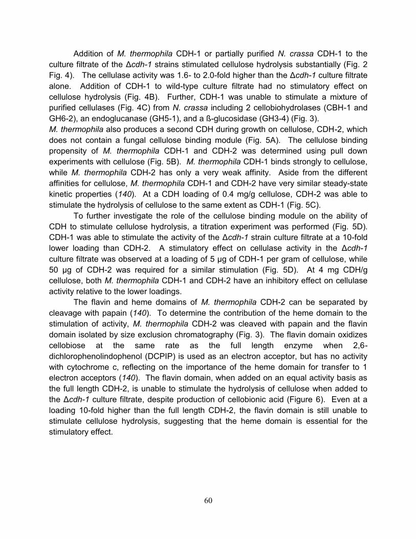
60
Addition of M. thermophila CDH-1 or partially purified N. crassa CDH-1 to the
culture filtrate of the Δcdh-1 strains stimulated cellulose hydrolysis substantially (Fig. 2
Fig. 4). The cellulase activity was 1.6- to 2.0-fold higher than the Δcdh-1 culture filtrate
alone. Addition of CDH-1 to wild-type culture filtrate had no stimulatory effect on
cellulose hydrolysis (Fig. 4B). Further, CDH-1 was unable to stimulate a mixture of
purified cellulases (Fig. 4C) from N. crassa including 2 cellobiohydrolases (CBH-1 and
GH6-2), an endoglucanase (GH5-1), and a ß-glucosidase (GH3-4) (Fig. 3).
M. thermophila also produces a second CDH during growth on cellulose, CDH-2, which
does not contain a fungal cellulose binding module (Fig. 5A). The cellulose binding
propensity of M. thermophila CDH-1 and CDH-2 was determined using pull down
experiments with cellulose (Fig. 5B). M. thermophila CDH-1 binds strongly to cellulose,
while M. thermophila CDH-2 has only a very weak affinity. Aside from the different
affinities for cellulose, M. thermophila CDH-1 and CDH-2 have very similar steady-state
kinetic properties (140). At a CDH loading of 0.4 mg/g cellulose, CDH-2 was able to
stimulate the hydrolysis of cellulose to the same extent as CDH-1 (Fig. 5C).
To further investigate the role of the cellulose binding module on the ability of
CDH to stimulate cellulose hydrolysis, a titration experiment was performed (Fig. 5D).
CDH-1 was able to stimulate the activity of the Δcdh-1 strain culture filtrate at a 10-fold
lower loading than CDH-2. A stimulatory effect on cellulase activity in the Δcdh-1
culture filtrate was observed at a loading of 5 µg of CDH-1 per gram of cellulose, while
50 µg of CDH-2 was required for a similar stimulation (Fig. 5D). At 4 mg CDH/g
cellulose, both M. thermophila CDH-1 and CDH-2 have an inhibitory effect on cellulase
activity relative to the lower loadings.
The flavin and heme domains of M. thermophila CDH-2 can be separated by
cleavage with papain (140). To determine the contribution of the heme domain to the
stimulation of activity, M. thermophila CDH-2 was cleaved with papain and the flavin
domain isolated by size exclusion chromatography (Fig. 3). The flavin domain oxidizes
cellobiose at the same rate as the full length enzyme when 2,6-
dichlorophenolindophenol (DCPIP) is used as an electron acceptor, but has no activity
with cytochrome c, reflecting on the importance of the heme domain for transfer to 1
electron acceptors (140). The flavin domain, when added on an equal activity basis as
the full length CDH-2, is unable to stimulate the hydrolysis of cellulose when added to
the Δcdh-1 culture filtrate, despite production of cellobionic acid (Figure 6). Even at a
loading 10-fold higher than the full length CDH-2, the flavin domain is still unable to
stimulate cellulose hydrolysis, suggesting that the heme domain is essential for the
stimulatory effect.
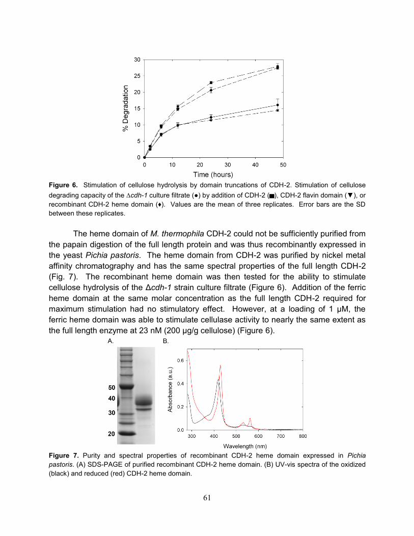
61
Figure 6. Stimulation of cellulose hydrolysis by domain truncations of CDH-2. Stimulation of cellulose
degrading capacity of the ∆cdh-1 culture filtrate (●) by addition of CDH-2 (▄), CDH-2 flavin domain (▼), or
recombinant CDH-2 heme domain (♦). Values are the mean of three replicates. Error bars are the SD
between these replicates.
The heme domain of M. thermophila CDH-2 could not be sufficiently purified from
the papain digestion of the full length protein and was thus recombinantly expressed in
the yeast Pichia pastoris. The heme domain from CDH-2 was purified by nickel metal
affinity chromatography and has the same spectral properties of the full length CDH-2
(Fig. 7). The recombinant heme domain was then tested for the ability to stimulate
cellulose hydrolysis of the Δcdh-1 strain culture filtrate (Figure 6). Addition of the ferric
heme domain at the same molar concentration as the full length CDH-2 required for
maximum stimulation had no stimulatory effect. However, at a loading of 1 µM, the
ferric heme domain was able to stimulate cellulase activity to nearly the same extent as
the full length enzyme at 23 nM (200 µg/g cellulose) (Figure 6).
Figure 7. Purity and spectral properties of recombinant CDH-2 heme domain expressed in Pichia
pastoris. (A) SDS-PAGE of purified recombinant CDH-2 heme domain. (B) UV-vis spectra of the oxidized
(black) and reduced (red) CDH-2 heme domain.

62
4.3.3 Oxygen and metal ion dependence on the stimulation of cellulose
degradation by CDH
A prevalent hypothesis for the biological function of CDH postulates that
electrons from the heme domain of CDH are transferred to ferric complexes, quinones,
molecular oxygen, or other redox mediators which lead to the production of radical
species that can non-specifically degrade cellulose or lignin. Experiments were carried
out to address if the stimulation of activity observed with CDH addition to the Δcdh-1
culture filtrate was due to a direct reaction with the cellulose or an indirect effect where
metals or small molecules became reduced by CDH and subsequently contributed to
the degradation.
To test for the effect of small molecules in the Δcdh-1 culture the culture filtrate
was buffer exchanged 10,000-fold using 10,000 MWCO spin concentrators. After this
treatment, CDH-1 was still able to stimulate the activity of the Δcdh-1 culture filtrate to
the same extent. To test if there was a metal dependence for the stimulation, buffer
exchanged culture filtrates from the Δcdh-1 cultures were incubated with 100 µM EDTA
Figure 8. Contribution of metals or small molecules to the CDH dependent stimulation. (A) Cellulase
activity of 10,000-fold buffer exchanged ∆cdh-1 secretome in the presence or absence of 0.004 mg/mL
CDH-1. For reactions containing EDTA, 100 µM EDTA was preincubated with the sample for 2 hours and
then assayed.
for 1 hour, and then assayed for cellulase activity. EDTA had no effect on the cellulase
activity of the Δcdh-1 culture filtrate; however, when M. thermophila CDH-1 was added
to the EDTA treated Δcdh-1 culture filtrate, no stimulatory effect was observed (Fig. 8).
Addition of EDTA to wild-type culture filtrate reduced cellulase activity by ~50% (data
not shown). Taken together, these results suggest that there is a protein bound metal
ion essential for the stimulation of cellulose degradation by CDH. Overnight incubation
of M. thermophila CDH-1 with 1.0 mM EDTA had no effect on the oxidation of cellobiose
with DCPIP or cytochrome c as electron acceptors.
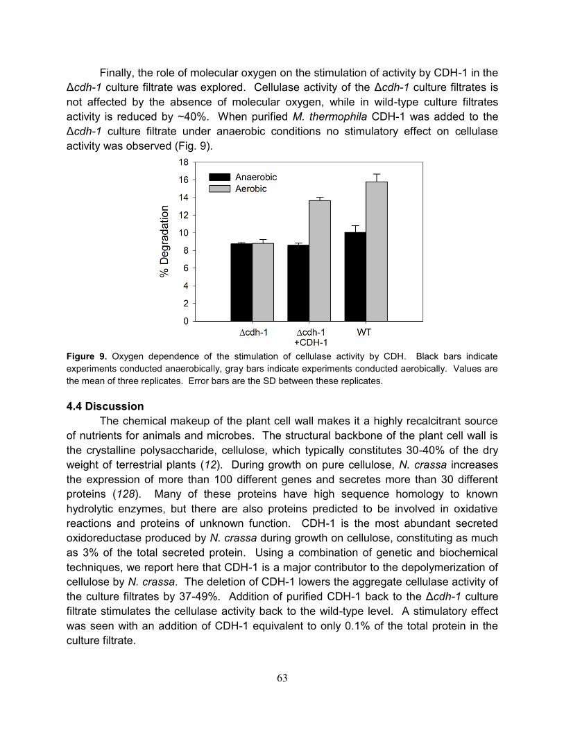
63
Finally, the role of molecular oxygen on the stimulation of activity by CDH-1 in the
Δcdh-1 culture filtrate was explored. Cellulase activity of the Δcdh-1 culture filtrates is
not affected by the absence of molecular oxygen, while in wild-type culture filtrates
activity is reduced by ~40%. When purified M. thermophila CDH-1 was added to the
Δcdh-1 culture filtrate under anaerobic conditions no stimulatory effect on cellulase
activity was observed (Fig. 9).
Figure 9. Oxygen dependence of the stimulation of cellulase activity by CDH. Black bars indicate
experiments conducted anaerobically, gray bars indicate experiments conducted aerobically. Values are
the mean of three replicates. Error bars are the SD between these replicates.
4.4 Discussion
The chemical makeup of the plant cell wall makes it a highly recalcitrant source
of nutrients for animals and microbes. The structural backbone of the plant cell wall is
the crystalline polysaccharide, cellulose, which typically constitutes 30-40% of the dry
weight of terrestrial plants (12). During growth on pure cellulose, N. crassa increases
the expression of more than 100 different genes and secretes more than 30 different
proteins (128). Many of these proteins have high sequence homology to known
hydrolytic enzymes, but there are also proteins predicted to be involved in oxidative
reactions and proteins of unknown function. CDH-1 is the most abundant secreted
oxidoreductase produced by N. crassa during growth on cellulose, constituting as much
as 3% of the total secreted protein. Using a combination of genetic and biochemical
techniques, we report here that CDH-1 is a major contributor to the depolymerization of
cellulose by N. crassa. The deletion of CDH-1 lowers the aggregate cellulase activity of
the culture filtrates by 37-49%. Addition of purified CDH-1 back to the Δcdh-1 culture
filtrate stimulates the cellulase activity back to the wild-type level. A stimulatory effect
was seen with an addition of CDH-1 equivalent to only 0.1% of the total protein in the
culture filtrate.

64
In the cellulolytic Sordariales sequenced thus far, all species have two copies of
CDH (134), one containing a C-terminal fungal cellulose binding domain (CBM), and
one without a CBM. In N. crassa, only the CBM-containing CDH-1 is produced in
significant amounts during growth on cellulose. However, in the closely related
thermophilic fungus, M. thermophila, both CDHs are produced at high levels during
growth on cellulose (140). Both CDHs from M. thermophila have been previously
shown to have very similar steady-state kinetic properties with soluble substrates (140),
and we now show that both enzymes can stimulate hydrolysis of cellulose to the same
extent. A key difference between the two CDHs is that CDH-1 was functional at a 10-
fold lower concentration than CDH-2. Cellulose binding modules are known to increase
the local concentration of cellulases on the surface of cellulose, and the results here are
consistent with the stimulatory effect being due to a reaction taking place on or near the
surface of the cellulose.
The isolated and catalytically active flavin domain of CDH-2 is unable to stimulate
hydrolysis of cellulose, whereas the CDH-2 heme domain was able to stimulate
cellulose hydrolysis to the same extent as full length CDH-2, at a 20 to 50-fold higher
concentration. The iron in the heme domain of CDH is axially ligated by absolutely
conserved methionine and histidine residues. These two residues cannot be displaced
by cyanide, azide when ferric, or carbon monoxide when ferrous (147), supporting the
hypothesis that the heme domain is likely involved in outer sphere electron transfer
reactions. The results presented here suggest that 1-electron transfer, mediated
through the heme domain, is essential for the stimulatory effect. The stimulatory effect
of the ferric heme domain was unexpected, as it would have no electron source.
However, the Δcdh-1 culture solution contains a trace CDH activity, due to a very low
level expression of CDH-2. Full length CDH-2 is able to reduce free CDH heme
domains in the presence of cellobiose and likely accounts for the stimulation in activity
by the free heme domain.
Proteins with sequence homology to the heme domain of CDH are widespread
throughout the fungal kingdom. As more fungal genome sequences have become
available, CDH-like heme domains have been found in proteins with diverse domain
architectures. In addition to a canonical CDH, the basidiomycete, Phanerochaete
chrysosporium also produces a secreted CDH-like heme domain with a C-terminal
fungal cellulose binding domain (148). In the N. crassa genome there are three genes
encoding proteins predicted to contain stand alone CDH-like heme domains with N-
terminal signal peptides; however, previous transcriptional profiling of N. crassa did not
show them upregulated during growth on cellulose. These free heme domains are
conserved throughout the Sordariales with sequenced genomes and all are predicted to
have the same methionine and histidine axial iron ligation.
The prevailing hypothesis for the biological function of CDH in the degradation of
lignocellulose involves flavin oxidation of cellobiose with sequential electron transfer to

65
the heme and then heme reduction of a one electron acceptor, speculated to be an
Fe(III)-oxalate complex (137). This ferrous iron is then proposed to take part in a
Fenton reaction with peroxide generated by CDH or other extracellular oxidases to
generate hydroxyl radicals which non-specifically degrade plant cell wall
polysaccharides. Fenton chemistry requires ferrous iron and peroxide, and has been
routinely used in DNA footprinting experiments (149). After a 10,000-fold buffer
exchange of the Δcdh-1 culture filtrate, the free iron in solution is less than 1 nM;
however, CDH was still able to fully stimulate cellulase activity. Treatment of the culture
filtrate with EDTA completely blocks the stimulation by CDH, although EDTA should not
prevent ferrous iron from participating in Fenton reactions. In fact, iron-EDTA chelates
are often used to generate hydroxyl radicals (149). Taken together, these results do not
support the involvement of Fenton chemistry in the stimulation of cellulase activity by
CDH in Neurospora. The requirement of molecular oxygen for the stimulation in activity;
however, suggests that oxidation reactions may play an important role.
Our results are consistent with the heme domain of CDH contributing to the
degradation of cellulose through interaction with other metalloproteins that are larger
than 10,000 daltons. Potential candidates include metal-dependent glycosyl hydrolase
family 61 proteins or other metal-dependent proteins of unknown function present in the
culture filtrate. GH61 family proteins were recently shown to stimulate the hydrolysis of
pretreated corn stover when added to a cocktail of industrial cellulases (150). The
stimulation by GH61 proteins is completely inhibited by addition of EDTA. This
inhibition of activity by EDTA has been ascribed to the removal of an essential surface
exposed divalent metal ion from the protein, and can be rescued by addition of divalent
metal ions to the reaction. Interestingly, no stimulation of activity by GH61 proteins was
observed on pure cellulosic substrates (150). CBP21, a protein structurally related to
GH61 proteins, from the chitinolytic bacterium Serratia marcescens, was recently shown
to stimulate the hydrolysis of crystalline chitin (151, 152), presumably by the same
mechanism as GH61 proteins. CBP21 requires divalent metal ions for activity, as well
as the presence of molecular oxygen (153). Addition of CBP21 to chitin in the presence
of molecular oxygen and ascorbic acid leads to the formation of oxidized chito-
oligosaccharides by an unknown mechanism. If fungal GH61 proteins stimulate
hydrolysis by the same mechanism as CBP21, it is possible that CDH is functioning as
an extracellular reductant. The N. crassa genome contains fourteen genes encoding
predicted extracellular GH61 proteins, of which, at least nine are upregulated during
growth on cellulose (128). There are also multiple hypothetical proteins secreted by
Neurospora which could be involved in the CDH stimulated degradation pathway.
The discovery that cellobiose dehydrogenase plays a major role in pure cellulose
degradation by some fungi is a significant shift from previous models centered on the
degradation of cellulose by a mixture of glycoside hydrolases. The well understood

66
genetics and resources available in Neurospora should facilitate the discovery of CDH’s
other partners so that the full system can be reconstituted in vitro.
4.5 Materials and Methods
Strains. All Neurospora crassa strains were obtained from the Fungal Genetics Stock
Center (FGSC) including wild-type N. crassa (FGSC 2489) and the ∆mus-51 (FGSC
9717) used for generating gene deletions. The ∆gh5-1 (FGSC 1647) was from the
Neurospora functional genomics project.
Preparation of ∆cdh-1 strains. The DNA cassette used to delete cdh-1 was provided
by the Neurospora functional genomics project. Details about how the cassette was
generated are available online at
(http://www.dartmouth.edu/~neurosporagenome/protocols.html). FGSC 9717 was
grown on Vogel’s minimal media (119) slants for 21 days at room temperature. Conidia
from the FGSC 9717 slant were transformed by electroporation with 1 µg of the ∆cdh-1
(∆NCU00206) cassette and plated onto media containing hygromycin. Positive
transformants were then crossed with wild-type N. crassa. Ascospores were
germinated on media containing hygromycin and several hygromycin resistant
transformants were harvested and screened for production of CDH. The genotypes of
three transformants that showed good growth on cellulose and lacked CDH activity
were confirmed by PCR using primers specific to cdh-1 and the hygromycin resistance
cassette.
Growth and harvest of N. crassa culture filtrates. Wild-type or ∆cdh-1 N. crassa was
inoculated onto slants of Vogel’s minimal media and grown for 3 days at 30 °C in the
dark followed by 7 days at room temperature. A conidial suspension was then
inoculated into 100 mL of Vogel’s salts supplemented with 2 % Avicel® PH101 in a 250
mL Erlenmeyer flask. After 7 days of growth on Avicel®, cultures were filtered over a
0.2 µm polyethersulfone (PES) filter.
Protein purification. For isolation of Myceliophthora thermophila (ATCC 46424) CDH-
1 and CDH-2, 10 day old conidia from cultures of M. thermophila were inoculated into
1.0 liter of Vogel’s salts supplemented with 40 g/L cotton balls in 2.8 liter Fernbach
flasks. After 6 days of growth at 48 °C and 200 RPM shaking, fungal biomass and
residual cotton was removed by filtration using 0.2 µm PES filters. The culture filtrate
was then passed over CaptoQ resin (GE Healthcare) at 20 mL/min. CDH-1 and CDH-2
were eluted from the CaptoQ resin with buffer containing 500 mM sodium chloride and
25 mM HEPES pH 7.4. The pooled CaptoQ concentrate was then desalted into 25 mM
HEPES pH 7.4 using a HiPrep 26/10 desalting column (GE Healthcare). The protein
was further purified on a 10/100 GL MonoQ column (GE Healthcare) and eluted with a

67
linear gradient from 0 to 500 mM sodium chloride. CDH-1 elutes from the column
between 180 and 220 mM sodium chloride; CDH-2 elutes between 280 and 320 mM
sodium chloride. CDH-1 was then further purified by binding to 1.0 grams of Avicel® in
50 mM sodium acetate buffer pH 5.0 for 5 minutes at 50 °C. The Avicel® was removed
by centrifugation in 50 mL conical tubes then washed with 50 mL of acetate buffer three
times. CDH-1 was eluted from the Avicel® by addition of 50 mL of 50 mM pH 11.3
phosphate buffer. The eluted CDH-1 was concentrated using 10,000 molecular weight
cutoff (MWCO) PES spin concentrators and stored at -80 °C. CDH-2 was further
purified by gel filtration chromatography using a 16/60 Sephacryl S200 column (GE
Healthcare) with a mobile phase of 25 mM HEPES pH 7.4 and 150 mM sodium
chloride. The flavin domain of CDH-2 was isolated by treatment of the full length
enzyme with papain (140). After papain cleavage, the fragments were separated by gel
filtration chromatography using a 16/60 Sephacryl S100 column (GE Healthcare) with a
mobile phase of 25 mM HEPES pH 7.4 and 150 mM sodium chloride. The fractions
enriched in flavin domain were then further purified using a 10/100 GL MonoQ column
and eluted with a linear gradient from 0 to 500 mM sodium chloride.
N. crassa CDH-1 (NCU00206) was partially purified from wild-type N. crassa.
Phosphoric acid swollen cellulose (PASC) was added (20 mL) to the culture filtrate at 4
°C and mixed for 10 minutes. The PASC was then removed from the culture filtrate by
centrifugation for 10 minutes at 4,000 RPM. Next, 724 g/L ammonium sulfate was
added to the culture filtrate and stirred at 4 °C for one hour. The precipitated proteins
were then isolated by centrifugation at 8,000 RPM for 25 minutes. The pellet was
resuspended in a minimal volume of water and then buffer exchanged into 25 mM Tris
pH 8.5 using a HiPrep 26/10 desalting column. The buffer exchanged proteins were
then loaded onto a 10/100 GL MonoQ column and eluted with a linear gradient from 0 to
500 mM sodium chloride. CDH-1 elutes from the column between 50 and 100 mM
sodium chloride and is ~80% pure by SDS-PAGE. N. crassa CBH-1 (NCU07340),
GH6-2 (NCU09680), GH5-1 (NCU00762), and GH3-4 (NCU04952) were purified as
previously described (154, 155).
Cellulase assays. All cellulase assays were performed in triplicate with 10 mg/mL
Avicel® PH101 (Sigma) in 50 mM sodium acetate pH 5.0 at 40 °C. Assays were
performed in 1.7 mL microcentrifuge tubes with 1.0 mL total volume and were inverted
20 times per minute. Each assay contained 0.05 mg/mL culture filtrate or 0.05 mg/mL
reconstituted cellulase mixture containing CBH-1, GH6-2, GH5-1, and GH3-4 present in
a ratio of 6:2.5:1:0.5. The concentration of heme domain used in stimulatory assays
was 1.0 µM as determined by absorption at 430 nm of the fully reduced protein.
Assays were centrifuged for two minutes at 4000 rpm to pellet the remaining
Avicel® and 20 µL of assay mix was removed per well. Samples were incubated with
100 µL of desalted, diluted Novozyme 188 (Sigma) at 40 °C for 20 minutes to hydrolyze

68
cellobiose and then 10-30 µL of the Novozyme 188 treated cellulase assay supernatant
was analyzed for glucose using the glucose oxidase/peroxidase assay as described
previously (128). Percent degradation was calculated based on the amount of glucose
measured relative to the maximum theoretical conversion of 10 mg/mL Avicel®.
Metal chelating reaction. To determine the requirement for small molecules and
metals, wild-type or ∆cdh-1 culture filtrate was buffer exchanged more than 10,000 fold
in a 10 kDa MWCO spin concentrator (Sartorius). The culture filtrate was then
incubated with 100 µM EDTA for 2 hours. EDTA or metal reconstituted culture filtrate
was then assayed as described above.
CDH binding to Avicel®. Purified CDH-1 or CDH-2 (20 µg) was added to 1.0 mL of 10
mg/mL Avicel® in 50 mM sodium acetate buffer pH 5.0. After incubation for 5 minutes at
room temperature, the Avicel® was spun down by centrifugation at 10,000 RPM. The
supernatant was removed and the Avicel® pellet was washed with 1.0 mL of fresh 50
mM sodium acetate buffer pH 5.0. The washing step was repeated three times and
then the Avicel® pellet was boiled in SDS loading buffer. After boiling, the Avicel® was
spun down and the supernatant loaded onto a 10% Tris-HCl polyacrylamide gel.
Anaerobic assays. Anaerobic cellulase assays were performed as above except all
assays were conducted in an anaerobic chamber (Coy) at room temperature. Buffers
were sparged with nitrogen for 1 hour and culture filtrates were concentrated more than
20-fold to volumes of less than 300 µL before introduction into the anaerobic chamber.
All solutions were left open in the anaerobic chamber, and buffer solutions were stirred
vigorously for 72 hours before use to fully remove dissolved oxygen. Aerobic reactions
were prepared in the anaerobic chamber in 3 mL reactivials and then removed from the
anaerobic chamber, exposed to air, sealed, and returned to the anaerobic chamber. At
specified timepoints, assays were centrifuged in the glove bag and 100 µL of assay mix
was removed and analyzed by the glucose-oxidase peroxidase assay as described
above.
4.6 Acknowledgements
WTB and CMP are recipients of NSF pre-doctoral fellowships. This work was funded by
a grant from the Energy Biosciences Institute to JDC and MAM.

69
*Footnote to chapter 4
This chapter, presented here with modifications from its original format, represents part
of a peer-reviewed paper published in ACS Chemical Biology (2011), In press. The
authors are Christopher M. Phillips, William T. Beeson, Jamie H.D. Cate and Michael A.
Marletta.
70
Chapter 5: Identification of GH61 proteins as an integral part of the of the CDH-
dependent enhancement of cellulose degradation
5.1 Abstract
Gene deletion and complementation studies demonstrated that cellobiose
dehydrogenase enhances the cellulose degrading capabilities of N. crassa by
approximately 1.6-2.0 folds. This stimulatory effect is dependent on molecular oxygen
and other, unknown factors. Here, using a combination of protein purification
techniques, mass spectrometry, and a novel activity screen we identify the other factors
involved in the stimulation of cellulose hydrolysis by CDH as glycosyl hydrolase family
61 proteins. Taking advantage of the metal binding properties of GH61 proteins, we
developed a purification strategy to isolate them free of cellulase contaminants. When
added to a defined mixture of cellulases from N. crassa, GH61 proteins were found to
enhance cellulose degradation.

71
5.2 Introduction
Recent transcriptomic and proteomic analyses of cellulolytic fungi have identified
oxidative enzymes involved in degradation of plant biomass (127, 156, 157). Despite
the widespread occurrence of these enzymes in fungi, the specific function of these
oxidative enzymes in cellulose degradation is unknown (Fig. 1). The filamentous
ascomycete, Neurospora crassa, is a proficient degrader of plant cell walls. During
growth on cellulose N. crassa produces cellobiose dehydrogenase (CDH) and many
other enzymes(43, 158, 159). CDH catalyzes the oxidation of cellobiose or longer
cellodextrins to the corresponding 1-5-δ-lactones (160).
Figure 1. Schematic of CDH catalyzed reaction.
During turnover electrons are transferred from the cellodextrin to the CDH flavin
domain and then a single electron is transferred to the heme domain (Figure 1). The
biologically relevant terminal electron acceptor for CDH is unknown. Molecular oxygen
is a poor substrate for CDH, however it reacts rapidly with quinones, metal ions, or
organic dyes. A prominent hypothesis for CDH involves the generation of hydroxyl
radicals through reduction of extracellular ferric complexes. Once reduced, the ferrous
iron takes part in Fenton chemistry to generate hydroxyl radicals that randomly
depolymerize plant cell wall polysaccharides.
The biological function of CDH is unknown; however, recent studies in N. crassa
suggest that it plays a role in cellulose degradation. CDH shows no beneficial effect
when added to a mixture of pure cellulases, but when added to a N. crassa secretome
deficient in CDH activity, enhances cellulase activity by 1.6-2.0 folds. The enhancement
of cellulose degradation is not due to Fenton chemistry, but does require other unknown
factors, hypothesized to be metalloproteins. In this report we use a CDH from M.
thermophila and a fractionation strategy to identify a novel family of copper dependent
enzymes in N. crassa that are necessary for CDH dependent enhancement of cellulose
degradation.

72
5.3 Results
5.3.1 Identification of GH61 proteins as part of the CDH dependent cellulose
degradation pathway
As reported in chapter 4, addition of CDH to a mixture of purified cellulases
showed no enhancement of activity, suggesting that additional factor(s) were required.
The strong inhibition by EDTA noted in chapter 4, is consistent with the hypothesis that
these factors were metalloproteins. Based on this information, an approach was
developed to identify the other factors in the N. crassa secretome involved in CDH
dependent enhancement of cellulose degradation. The secretome of the Δcdh-1 strain
grown on Avicel for 7 days was fractionated using a 10/100 GL MonoQ column. The
flow through, and fractions eluting from the column were then added to a core mixture of
N. crassa cellulases, including CBH-1 (NCU07340), GH6-2, a CBH-2; GH5-1, an
endoglucanase; and GH3-4, a β-glucosidase. If the factor was present in the fraction,
then addition of CDH would increase Avicel hydrolysis, and addition of EDTA would
block the stimulation. A typical fractionation is summarized in Figure 2.
Figure 2. Fractionation of the ∆cdh-1 secretome to identify other factors involved in CDH dependent
enhancement of cellulase activity. SDS-PAGE analysis of fractions from MonoQ separation of the ∆cdh-1
secretome. Above each lane is the fold stimulation of the fraction when added to a mixture of purified N.
crassa cellulases in the presence or absence of CDH-1. Fractions highlighted in red were analyzed by
mass spectrometry and showed the presence of four glycosyl hydrolase family 61 proteins (indicated by
black boxes).

73
From the initial fractionation, it was clear that the flow-through fractions and
fractions eluting from the column between 3-8 mS/cm contained the factors required for
stimulation of hydrolysis by CDH. Since the stimulatory activity was present in multiple
fractions, it was likely that multiple proteins could potentiate the CDH-dependent
stimulation of cellulose hydrolysis. The proteins present in all of the fractions from the
MonoQ column were identified by in-gel and solution tryptic digests coupled with LC-
MS/MS. A clear trend that emerged from this analysis was that in fractions where there
was CDH-dependent stimulation of cellulase activity, there were also GH61 proteins
(Figure 2). The GH61 proteins identified in the fractions showing stimulatory activity
were NCU01050, NCU02240, NCU07898, and NCU08760. Each of the fractions with
stimulatory activity; however, contained at least two GH61 proteins, so further
purification was required to confirm if a single GH61 protein could potentiate cellulose
degradation.
5.3.2 Purification strategy to isolate high purity native GH61 proteins from N.
crassa
After the initial MonoQ fractionation, the protein was less than 10% pure as
judged by SDS-PAGE. Previous reports on the x-ray crystal structure of GH61 proteins
had revealed an absolutely conserved divalent metal binding site on the surface of the
protein. We hypothesized that the affinity of GH61 proteins for the MonoQ column,
which binds proteins based on the amount of surface negative charge, would change if
the protein was in apo or fully metal bound forms. To test this hypothesis, the MonoQ
flow-through fraction, which contained NCU08760 and NCU01050, was treated with 1.0
mM EDTA for 12 hours and then reloaded onto the column. After EDTA treatment, the
majority of the proteins still flowed through the column; however, on the application of a
slight salt gradient two well defined peaks eluted between 1 and 3 mS/cm (Figure 3).

74
Figure 3. Fractionation of EDTA treated N. crassa MonoQ flow-through. Peaks indicated with arrows
contain NCU08760 and NCU01050.
Figure 4. SDS-PAGE and lane guide for MonoQ fractionation of EDTA treated original MonoQ flow
through. Comparing the load to the new flow through, it is clear that the bands between 25-30 kDa and
between 40-50 kDa are dramatically decreased or absent in the new flow through and then elute with a
shallow salt gradient. NCU01050 is predominantly in fraction A4 and NCU08760 is in fraction A5.

75
SDS-PAGE analysis of the fractions is shown in Figure 4. After EDTA treatment,
the MonoQ flow-through is clearly deficient in two proteins. One migrates between 40-
50 kDa and the other between 25-30 kDa. The 25-30 kDa protein elutes from the
column in the first sharp peak, and the 40-50 kDa protein in the second sharp peak.
Tryptic digests and LC-MS/MS analysis of the proteins in these two fractions confirmed
the presence of NCU01050 and NCU08760. These results are consistent with removal
of positive charge from the surface of the protein causing them to bind the column more
tightly. The same approach was used to separate NCU02240 and NCU07898 from
other proteins in the secretome (see Materials and Methods). After that native metal is
stripped from GH61 proteins, the metal site can be reloaded with exogenous divalent
metal ions including zinc and cobalt. The reconstituted protein can again be
fractionated on a MonoQ column to increase purity. After the GH61 proteins are
separated from all the other proteins in the secretome, a gel filtration column can be
used to separate GH61 proteins with cellulose binding modules from GH61 proteins
without cellulose binding modules. The four N. crassa GH61 proteins are more than
98% pure based on SDS-PAGE analysis (Figure 5)
Figure 5. SDS-PAGE of natively purified GH61 proteins.
5.3.3 Metal analysis of the secretome and natively purified GH61 proteins
Previous studies have reported that GH61 proteins can bind to a variety of
divalent metal ions. We wanted to determine the native metal present in GH61 proteins
produced by N. crassa. After extensive dialysis, the metal content of the Δcdh-1
secretome was analyzed using inductively coupled plasma atomic emission
spectroscopy (ICP-AES). Only copper and zinc were present in the secretome, with
copper 10-fold more abundant than zinc (Figure 6). However, no N. crassa proteins

76
secreted during growth on cellulose are predicted to bind copper. Metal analysis of the
fractions from the 10/100 GL MonoQ column showed that fractions containing GH61
proteins were also enriched in copper (Figure 7). Three of the natively purified GH61
proteins were analyzed by ICP-AES and found to bind copper with a 1:1 stoichiometry
(Figure 8). NCU02240 was not present in sufficient purity or yield for further
characterization.
5.3.4 Stimulation of Avicel and pretreated corn stover hydrolysis by purified GH61
proteins.
Next, we studied the ability of the natively purified GH61 proteins to stimulate the
hydrolysis of Avicel or pretreated corn stover by a defined mixture of N. crassa
cellulases (Figure 9). In the absence of CDH, there was only a 1.1-1.2 fold
enhancement of cellulase activity on Avicel by each of the GH61 proteins tested. When
CDH was added to the core cellulases and NCU01050 or NCU02240 there was a
substantial increase in hydrolytic activity. However, for NCU07898 and NCU08760,
there was no stimulatory effect on cellulase activity.
Degradation of pretreated corn stover was enhanced by the GH61 proteins to a
greater extent in the absence of CDH (1.3 to 1.5 fold) than Avicel hydrolysis was (Figure
9b). When CDH was added to the core cellulase plus GH61 mixture, a further
enhancement in cellulase activity was observed. On pretreated corn stover, a
stimulatory effect on hydrolysis was observed for all four GH61 proteins tested.
NCU08760 was especially stimulatory, with an overall stimulation in the presence of
CDH of more than 2 folds.

77
Figure 9. Stimulation of (A) Avicel or (B) pretreated corn stover hydrolysis by natively purified GH61
proteins. GH61 proteins (0.015 mg/mL) were added to N. crassa cellulases (0.05 mg/mL CBH-1, GH6-2,
and the ß-glucosidase GH3-4 (0.005mg/mL) in the presence (black bars) or absence (gray bars) of
MtCDH-1 (0.004 mg/mL).

78
5.4 Discussion
During growth on cellulose many different proteins are secreted by N. crassa.
CDH-1 is the major known oxidoreductase secreted by N. crassa and constitutes
approximately 2-3% of the secretome by weight. Despite only making up 2-3% of the
secretome, deletion of CDH-1 reduced aggregate cellulase activity by approximately
50%. Here we show that CDH-1 dependent enhancement of cellulose degradation also
requires the presence of GH61 proteins. GH61 proteins constitute 15-20% of the N.
crassa secretome and their biological function is unknown.
Figure 10. Comparison of the metal binding sites in CopC (left) and GH61E from Thielavia terrestris
(right). Copper is bound in the CopC structure and zinc is bound in GH61E.
GH61 proteins have been studied by x-ray crystallography and contain a highly
conserved metal binding site that includes two histidine residues. One of the histidines
is the N-terminal residue and functions as a bidentate ligand involving the amine and
ring N2. Crystal structures of GH61 proteins with nickel (161), magnesium, or zinc
(162), have been reported. Redox chemistry involving O2 generally requires a transition
metal. Copper was found to be the metal natively bound to GH61 proteins from N.
crassa and is found in many enzymes that activate molecular oxygen. These results
suggest that copper-bound GH61 proteins may accept electrons from the reduced CDH-
heme domain. Hence, the copper in GH61 proteins could function as redox couple with
CDH. Two other proteins that contain bidentate N-terminal histidine ligands were found
in the protein databank. One of them, CopC, is part of the cop operon involved in
copper resistance in bacteria and has been shown to bind Cu(II) with picomolar affinity
(Figure 10) (163). The other, particulate methane monooxygenase (pMMO), uses a
trinuclear copper site to oxidize methane (164). Combining our experimental results
with the similar ligation in CopC and pMMO, we conclude that copper is the native metal
in GH61 proteins.

79
[836] [1050] [2240] [2344] [2916] [3328] [5969] [7760] [7898] [8760]
[836] 100 30 42 38 41 39 26 26 31 38
[1050] 30 100 57 39 46 34 25 28 34 40
[2240] 42 57 100 37 48 33 26 25 30 41
[2344] 38 39 37 100 36 50 21 27 33 30
[2916] 41 46 48 36 100 27 32 35 34 40
[3328] 39 34 33 50 27 100 26 25 28 36
[5969] 26 25 26 21 32 26 100 64 38 25
[7760] 26 28 25 27 35 25 64 100 38 29
[7898] 31 34 30 33 34 28 38 38 100 34
[8760] 38 40 41 30 40 36 25 29 34 100
Figure 11. Identity matrix for the ten GH61 proteins upregulated two folds or more during growth on
cellulose or lignocellulose.
The N. crassa genome contains fourteen genes encoding proteins predicted to
contain GH61 domains. Of these fourteen genes, ten of them were upregulated 2 fold
or more during growth on cellulose or lignocellulose. Six of these predicted GH61
proteins also contain C-terminal fungal cellulose binding domains (CBM1), suggesting
that they directly interact with crystalline cellulose. There is a high amount of sequence
diversity in the N. crassa GH61 proteins, with most sharing approximately 30% pairwise
sequence identity. When incubated with CDH and a mixture of core cellulases, Avicel
hydrolysis was enhanced by GH61 proteins NCU01050 and NCU02240, while
NCU07898 and NCU08760 showed little or no beneficial effects. Both NCU07898 and
NCU08760 are probably functional, because they did show a stimulatory effect on
pretreated corn stover hydrolysis. Differences in the activities of GH61 proteins on
different cellulosic materials might be due to sequence variation on the surface of the
protein.
5.5 Materials and Methods
Growth of N. crassa. The ∆cdh-1 strain of N. crassa was inoculated onto slants of
Vogel’s minimal media and grown for 3 days at 30 °C in the dark followed by 7 days at
room temperature with ambient lighting. A conidial suspension was then inoculated into
100 mL of Vogel’s salts supplemented with 2% Avicel® PH101 (Sigma) in a 250 mL
Erlenmeyer flask. After 7 days of growth on Avicel®, cultures were filtered over 0.2 µm
polyethersulfone (PES) filters.
Purification of GH61 proteins. N. crassa GH61 proteins were partially purified from
the N. crassa ∆cdh-1 strain. Culture filtrates from multiple flasks were pooled and
concentrated 100-fold using a tangential flow filtration system with a 5,000 MWCO PES
membrane (Millipore, Billerica, MA). The concentrated culture filtrate was then buffer
80
exchanged into 10 mM Tris pH 8.5 using a HiPrep 26/10 desalting column. The
concentrated and buffer exchanged protein was then fractionated using an
AKTAexplorer FPLC system (GE Healthcare) and a 10/100 GL MonoQ column. The
mobile phases for the anion-exchange fractionation were buffer A: 10 mM Tris pH 8.5
and buffer B: 10 mM Tris pH 8.5 with 1.0 M sodium chloride. For each run on the
MonoQ column approximately 100 mg of total secretome protein was loaded and eluted
from the column with a linear gradient from 0 to 50% buffer B over six column volumes.
NCU08760 and NCU01050 do not bind the column under these conditions and are
present in the flow through. NCU02240 and NCU07898 elute from the column between
4-9 mS/cm. Fractions containing the target proteins were pooled and treated with 1.0
mM EDTA overnight to strip bound metals. The EDTA treated samples were then
concentrated using 3,000 MWCO PES spin concentrators and desalted into 10 mM Tris
pH 8.5 using a 26/10 desalting column. Removal of the bound metal ion increases the
affinity of the GH61 proteins for the anion-exchange resin and causes them to elute at
higher salt concentrations. For NCU08760 and NCU01050, removal of bound metal ion
causes the proteins to bind to the MonoQ column. The apo NCU01050 and NCU08760
are then eluted from the column using the same buffers as above with a linear gradient
from 0 to 3.5% buffer B over 3 column volumes. NCU01050 elutes at ~1.6 mS/cm and
NCU08760 at ~2.2 mS/cm conductivity. The apo forms of NCU02240 and NCU07898
elute from the MonoQ column between 7-9 mS/cm. The purity of the PMOs can be
further improved by reconstituting the apo GH61 proteins with Zn(II) and repeating the
anion-exchange fractionation. Other proteins in the secretome do not change their
affinity for the resin in the presence or absence of metal ions and can be effectively
removed from GH61 proteins by cycles of stripping the metal, fractionating,
reconstituting, and fractionating again. NCU01050 and NCU08760 can then be
separated from one another by size exclusion chromatography using a Sephacryl S100
column with a mobile phase of 10 mM Tris pH 8.5 with 150 mM sodium chloride.
NCU02240 and NCU07898 are separated from one another using the same method as
used for NCU01050 and NCU08760. The four natively purified PMOs from N. crassa
are stable for several months at 4 °C.
Analysis of copper and zinc in the secretome by ICP-AES. The N. crassa Δcdh-1
strain was grown on 2% w/v Avicel® as previously described with an additional 5 µM
copper sulfate, and 30 µM zinc sulfate supplemented in the minimal media. The culture
filtrate was concentrated using tangential flow filtration and buffer exchanged into 10
mM Tris pH 8.5. The concentrated and buffer exchanged culture filtrate was loaded
onto a 10/100 GL MonoQ column and separated into 5 fractions with a linear salt
gradient. Each fraction was then analyzed for the presence of copper or zinc. Metal
analysis was performed using a Perkin Elmer inductively coupled plasma atomic
emission spectrometer (ICP-AES). and nm were The wavelengths used for

81
quantification for copper were 327.393 or 324.752 nm and for zinc, 213.857 or 206.200
nm. Add a statement here saying what other metals we looked for and that they were
present at less than 1 uM in our mixture of concentrated proteins.
Copper stoichiometry of apo-PMOs. Apo-PMO stocks of NCU01050, NCU07898,
and NCU08760 were diluted to a final concentration of 1.0 mg/mL in 10 mM Tris pH 8.5
buffer and incubated with 200 µM copper sulfate at room temperature for 16 hours.
After reconstitution, the protein was diluted 5-fold into 10 mM Tris pH 8.5 and desalted
using a 26/10 desalting column to remove unbound copper. The desalted protein was
concentrated to a final volume of 2.5 mL using 3,000 MWCO PES spin concentrators.
The absorption at 280 nm was recorded for each sample and used to determine the
concentration of the protein. The concentration of copper in the sample was measured
using a Perkin Elmer 7000 series ICP-AES. The wavelengths used for copper
quantification were 327.393 and 324.752 nm.
5.6 Acknowledgements
S. Bauer for advice and technical assistance with LC-MS. W. Beeson and C. Phillips
are recipients of NSF pre-doctoral fellowships. This work was funded by a grant from
the Energy Biosciences Institute to J. Cate and M. Marletta.

82
*Footnote to chapter 5
This chapter, presented here with modifications from its original format, represents part
of a peer-reviewed paper published in ACS Chemical Biology (2011), In press. The
authors are Christopher M. Phillips, William T. Beeson, Jamie H.D. Cate and Michael A.
Marletta.

83
Chapter 6: Oxidative cleavage of cellulose by polysaccharide monooxygenases
6.1 Abstract
Glycosyl hydrolase family 61 (GH61) proteins were identified as essential to the
cellobiose dehydrogenase (CDH) dependent stimulation of cellulase activity. Here, the
mechanism of action of these proteins is elucidated. GH61 proteins were found to
oxidatively cleave cellulose in the presence of oxygen and CDH or chemical reductants.
The products of the reaction included oligosaccharides oxidized at the C1 or C4
position, depending on the GH61 examined. Isotope labeling experiments showed that
an oxygen atom from molecular oxygen was inserted into the oxidized oligosaccharides.
Based on these results a potential chemical mechanism for the enzymes is proposed.
Our data supports that GH61 proteins should be renamed polysaccharide
monoyxgenases.

84
6.2 Introduction
In chapter 5, GH61 proteins were found to be copper metalloproteins that were
necessary for CDH dependent enhancement of cellulase activity. The mechanism by
which the GH61 protein was enhancing cellulose degradation is unknown. Many
copper metalloproteins are involved in the binding or activation of molecular oxygen.
The CDH dependent enhancement of cellulase activity was found in chapter 3 to be
oxygen dependent. Taken together, it is likely that the GH61 protein is binding to
molecular oxygen and using it to enhance cellulose degradation.
Recently, CBP21, a protein with structural homology to GH61 proteins (less than
10% sequence identity), was identified as an oxidative enzyme important for chitin
degradation in the bacterium Serratia marcescens. When CBP21 was incubated with
crystalline chitin and a reductant, a series of oxidized chito-oligosaccharides were
released from the chitin. These oligosaccharides were found to be oxidized at the C1
position, forming aldonic acids. A metal and oxygen dependence was also reported for
the formation of oxidized products. All of these properties are similar to what we had
observed for the CDH dependent enhancement of cellulose degradation in N. crassa.
In the N. crassa system, CDH could be acting as the reductant, substituting for ascorbic
acid in the bacterial chitin degrading system. If GH61 proteins were making
cellodextrins oxidized at the C1 position, they may have been missed because CDH
generates the same product.
Here we use a combination of analytical techniques to better understand the
mechanism by which GH61 proteins and CDH enhance fungal cellulose degradation. In
contrast to the widely accepted Fenton model, which involves random attack of hydroxyl
radicals on the surface of cellulose, we find that CDH and GH61 proteins work together
to directly oxidize internal regions of the cellulose chains. This direct oxidative cleavage
probably enhances cellulose degradation by making new sites for exoglucanases to act.
6.3 Results
6.3.1 Product analysis of GH61 proteins.
The phylogenetic diversity of 10 N. crassa GH61s whose transcripts are
upregulated during growth on cellulose (43) suggests that these enzymes may target a
wide array of substrates in lignocellulose, or generate different products (Fig. 2). To
investigate the reaction products of the purified GH61s, assays were performed on
phosphoric acid swollen cellulose (PASC). When PASC was treated with GH61 and
CDH, a series of aldonic acids two to nine glucose residues in length (A2-A9) were
identified by high performance anion exchange chromatography (HPAEC). In addition
to aldonic acids, the combination of CDH and GH61s NCU01050 or NCU07898
produced peaks at a later retention time (Fig. 3A). Product analysis by liquid
chromatography-mass spectrometry confirmed the presence of aldonic acids (Gx+15

85
a.m.u.), as well as masses of Gx + 13 a.m.u. and Gx + 31 a.m.u (Fig. 3B). The Gx +13
mass is consistent with a doubly oxidized cellodextrin. Cellulose cleavage by these
GH61s likely results in oxidation at the non-reducing end followed by oxidation at the
reducing end by CDH. Given the necessity to cleave a 1,4-glycosidic bond, these
products are likely oligosaccharides with a 4-keto sugar at the non-reducing end. The
Gx + 31 mass is consistent with the hydrate of this product, a ketal. Ketoaldoses are
unstable in aqueous solution and are known to decompose spontaneously into many
different species (165). The third purified GH61, NCU08760, did not form the late
eluting peak on the HPAEC, or Gx + 13 and Gx + 31 species (Fig. 3A and 3B),
consistent with oxidation exclusively at the reducing end on C1 to form aldonic acids.
Incubation of PASC with GH61 alone led to the formation of low amounts of hydrolytic
products. The formation of hydrolytic products could be due to low levels of cellulase
contamination.
Figure 2. Phylogeny of GH61 proteins in N. crassa. Shown are the upregulated GH61 proteins. Of the 14 GH61 proteins in the N. crassa genome, 10 are upregulated >2-fold in response to growth on cellulose relative to sucrose (43). Proteins identified via proteomics and purified here are marked (●).
Since CDH is known to oxidize the C1 position of cellodextrins, a reaction with
NCU08760 was carried out with ascorbic acid substituted for CDH. In the presence of
ascorbic acid and copper, NCU08760 produced a ladder of aldonic acids (Fig. 4).
Under identical conditions, NCU01050 produced a ladder of products with a later
retention time when analyzed by HPAEC and 100-fold less aldonic acid than NCU08760
(Fig. 4b). The formation of oxidized sugars by the GH61s was also oxygen dependent,
suggesting that GH61s are oxidases (data not shown).

86
Figure 3. Reaction products of cellulose cleavage by CDH and GH61. GH61 (5.0 µM ) and 0.5 µM CDH-2 were incubated with 5 mg/mL phosphoric acid swollen cellulose for 2 hours in 10 mM ammonium acetate pH 5.0 at 40°C. Products of CDH-2 in combination with each GH61 protein (NCU01050-top, NCU07898-middle), or NCU08760-bottom)) were analyzed by HPAEC (A) and LC-MS (B). HPAEC standards were used to determine retention times of cellodextrins (G1-G6) and the respective aldonic acid derivatives (A2-A9). LC-MS of reaction mixtures in negative ion mode shows reaction mixtures comprised of masses consistent with a ladder of aldonic acids (Gx + 15 a.m.u.) separated by an anhydroglucose unit. Inset is a zoom around the G3 series. NCU01050 and NCU07898 show additional masses consistent with a keto-aldonic acid (Gx + 13 a.m.u.) or the hydrate of that product, a ketal-aldonic acid (Gx + 31 a.m.u.).
Figure 4. (A) Incubation of 5.0 µM apo-, Zn-bound, or Cu-bound NCU08760 with 2 mM ascorbic acid confirms that Cu-bound GH61 is required for generation of oxidative products. Error bars represent standard deviation of assays performed in triplicate (B) Ascorbic acid (2 mM) and 5.0 µM GH61 were assayed on PASC and analyzed by HPAEC. Products of NCU01050 include a ladder of cello-oligosaccharides (G2-G7) and a ladder of later eluting products that are likely oxidized at the non-reducing end to generate 4-keto sugars (K2-8). Products of NCU08760 include a ladder of cello-oligosaccharides (G3-G6) and aldonic acids (A2-A7). Trace amounts of aldonic acids were produced by NCU01050 but these are 100-fold less abundant than those produced by NCU08760 suggesting different regiospecificities for the 2 proteins. AA designates the peak due to ascorbic acid.

87
Next experiments were designed to determine if oxygen from molecular oxygen
was incorporated into the oxidized cellodextrins. To show oxygen insertion from O2,
reactions were prepared anaerobically that contained NCU08760, ascorbic acid, and
5.0 mg/mL phosphoric acid swollen cellulose (PASC). The solutions were sealed in 1.0
mL vials and then the headspace was replaced with 18O2 on a Schlenk line or opened to
atmospheric oxygen. Cellulase and our previous GH61 reactions were typically
performed at pH 5.0. A more basic pH was chosen here in order to limit the exchange
of bulk water into the products. Figure 5 shows the mass spectra confirming
incorporation of 1 atom of 18O into the aldonic acid products, providing evidence that the
enzyme functions as a monooxygenase. Similar experiments were performed where
the ascorbic acid was substituted with CDH (Figure 6). 18O incorporation into the
products was also observed at both pH 5.0 and pH 7.8. NCU01050 did not produce
products labeled with 18O; however, the predicted 4-keto-aldose products are present as
hydrates in aqueous solutions and would rapidly exchange with bulk water.
The identity of the non-reducing end product generated by the NCU01050 was
then investigated. Direct chemical hydrolysis, using trifluoro-acetic acid (TFA), of the
NCU01050 and CDH reaction products generated three peaks on a Dionex PA-200
column (Figure 7). As expected, glucose and gluconic acid are formed from the internal
carbohydrates and the oxidation of the reducing end by CDH. The third peak, eluting at
6.4 minutes, is absent in control reactions with NCU08760 and CDH and probably is the
GH61-oxidized non-reducing end carbohydrate. If a ketone functional group was
introduced in the ring, reduction with sodium borohydride would convert it back to a
racemic mixture of alcohols at that position (Figure 8a). Galactose is the C4 epimer of
glucose and can be readily separated from glucose using a Dionex PA-20 column.
Initial reaction products from the NCU01050 reaction were treated with sodium
borohydride. Analysis of the monosaccharide products on a Dionex HPAEC confirmed
the formation of galactose (Figure 8b). In control experiments where NCU08760 was
substituted for NCU01050, 10-fold less galactose was formed (data not shown).

88
Figure 5. Mass spectra illustrating the incorporation of
18O from
18O2 into the aldonic acid products of
NCU08760 with ascorbic acid as the reductant. Assays were performed with 16
O2 (A) or 18
O2 (B) and the products were analyzed by LC-MS. Shown is a ladder of aldonic acid products (DP 3-7). The inset is an expanded region around the mass of the cellotrionic acid ion.
Figure 6. Mass spectra illustrating the incorporation of
18O from
18O2 into the aldonic acid products of
NCU08760 with CDH as the reductant. Assays were performed with NCU08760 and CDH in 18
O2 at pH 7.8 (A) or pH 5.0 (B) and the products were analyzed by LC-MS.
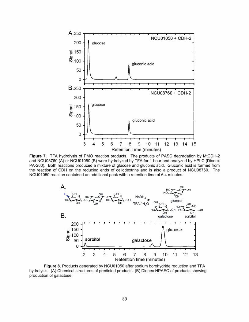
89
Figure 7. TFA hydrolysis of PMO reaction products. The products of PASC degradation by MtCDH-2 and NCU08760 (A) or NCU01050 (B) were hydrolyzed by TFA for 1 hour and analyzed by HPLC (Dionex PA-200). Both reactions produced a mixture of glucose and gluconic acid. Gluconic acid is formed from the reaction of CDH on the reducing ends of cellodextrins and is also a product of NCU08760. The NCU01050 reaction contained an additional peak with a retention time of 6.4 minutes.
Figure 8. Products generated by NCU01050 after sodium borohydride reduction and TFA
hydrolysis. (A) Chemical structures of predicted products. (B) Dionex HPAEC of products showing production of galactose.

90
6.4 Discussion
Evidence that GH61s are copper enzymes requiring electron transfer from CDH
to cleave cellulose in an oxygen dependent manner provides the basis to propose a
chemical mechanism for a new group of enzymes acting as polysaccharide
monooxygenases (PMOs) (Fig. 9). Precedent is drawn from the well-studied copper
monooxygenases (166, 167). The work reported here supports one electron reduction
of PMO-Cu(II) to PMO-Cu(I) by the CDH heme domain followed by oxygen binding and
internal electron transfer to form a copper superoxo intermediate. Hydrogen atom
abstraction by the copper superoxo at the 1-position (by NCU08760) or the 4-position
(by NCU01050) of an internal carbohydrate then takes place, generating a copper
hydroperoxo intermediate and a substrate radical. The 2nd electron from CDH then
facilitates O-O bond cleavage releasing water and generating a copper oxo radical that
couples with the substrate radical, thereby hydroxylating the polysaccharide at the 1- or
4-position. The additional oxygen atom destabilizes the glycosidic bond leading to
elimination of the adjacent glucan and formation of a sugar lactone or ketoaldose. This
elimination would be facilitated by a general acid, possibly a third absolutely conserved
histidine that is located on the surface of all fungal PMO proteins near the metal binding
site (161). It is possible that a 2-electron reduction of oxygen to a Cu-OOH intermediate
could abstract the hydrogen. However, peroxide is not able to shunt the reaction in the
absence of CDH, and catalase showed no inhibitory effect (data not shown).

91
Figure 9. PMO reactions and proposed mechanism. (Top) Type 1 PMOs abstract a hydrogen atom from the 4 position leading to formation of 4-ketoaldose products. Type 2 PMOs catalyze hydrogen atom abstraction from the 1 position leading to formation of aldonic acids. (Bottom) PMO mechanism: An electron from the heme domain of CDH reduces the PMO Cu(II) to Cu(I) and then O2 binds. Internal electron transfer takes place to form a copper superoxo intermediate, which then abstracts a H• from the 1 or 4 position on the carbohydrate. A second electron from CDH leads to homolytic cleavage of the Cu-bound hydroperoxide. The copper oxo species (Cu-O•) then couples with the substrate radical, hydroxylating the substrate. Addition of the oxygen atom destabilizes the glycosidic bond and leads to elimination of the adjacent glucan.

92
6.5 Materials and methods
Assays on PASC Phosphoric acid swollen cellulose (PASC) was prepared by addition
of 10g Avicel® to 500 mL 85% phosphoric acid and blended for 30 minutes. The
cellulose was then precipitated by the addition of 4L of ice cold water and washed with
water multiple times until the pH was >4. The concentration of PASC was determined
by the phenol-sulfuric acid assay. Assays contained 5.0 µM PMO, 0.5 µM CDH-2, and
9 mg/mL PASC in 10 mM ammonium acetate pH 5.0 and were performed at 40 °C
unless otherwise noted. In some assays, 2 mM ascorbic acid was used in place of
CDH-2.
Product analysis by HPAEC. Cellulase assays were mixed with 1 volume of 0.1 M
NaOH to quench the reaction and then centrifuged to remove the supernatant.
Samples were analyzed on a Dionex ICS-3000 HPAEC. Products were separated on a
PA-200 HPAEC column using 0.1 M NaOH in the mobile phase with the concentration
of sodium acetate increasing from 0 to 140 mM (14 min), 140-300 mM (8 min), 300 to
400 mM (4 min) and then held constant at 500 mM (3 min) before re-equilibration in 0.1
M NaOH (4 min). The flow rate was set to 0.4 mL/min, the column was maintained at a
temperature of 30 °C, and samples were detected on an electrochemical detector.
Authentic standards of glucose, cellodextrins, glucono-δ-lactone and cellobiono-δ-
lactone were used to determine retention times and for quantification. Cellobiono-δ-
lactone was synthesized as previously described (168).
Product analysis by LC-MS. Samples were analyzed by an Agilent HPLC (1200
series) connected to an electrospray ionization emitter in a linear ion trap mass
spectrometer (LTQ XL, Thermo Scientific). Carbohydrates were separated using a
SeQuant ZIC®-HILIC column (150 x 2.1 mm, 3.5 µM 100Å) with a SeQuant ZIC®-HILIC
guard column (20 x 2.1 mm, 5 µm). Solvent A was 5 mM ammonium acetate pH 7.2
and solvent B was 90% acetonitrile and 10 mM ammonium acetate pH 6.5. Samples
were prepared by centrifugation of the assay mixture followed by the addition of 1
volume of 100% acetonitrile and 1% formic acid to the supernatant. Sample injection
was set to 5 µL. The elution program consisted of a linear gradient from 80% B to 20%
B over 14 minutes followed by 5 minutes at 20% B then re-equilibration for 2 minutes at
80% B. The column temperature was maintained at 25 °C and the flow rate was 0.2
mL/minute. Mass spectra were acquired in negative ion mode over the range m/z =
310-2000. Data processing was performed using Xcalibur software (version 2.2,
Thermo Scientific).
Metal dependence of PMO activity. Apo-PMO was prepared by treatment of as
purified PMO with 10 mM EDTA for 24 hours. Protein was then concentrated in a 3 kDa

93
spin concentrator and loaded onto a Sephacryl S100 column with 10 mM Tris pH 8.0
and 100 µM EDTA in the mobile phase. Following elution, Sigma TraceSELECT grade
buffers, metals, and water were used for all assays and only extensively washed and
rinsed plastics were used due to problems with copper contamination. The protein was
buffer exchanged > 100-fold into 10 mM sodium acetate (Sigma Cat # 59929 and
#07692) in water (Sigma Cat # 14211) to a final concentration of >40 µM PMO. Cu- or
Zn-bound PMO was then produced by reconstitution with a 2-fold molar excess of
CuSO4 (Sigma Cat #: 203165) or ZnSO4 (Sigma Cat #: 204986).
PASC Assays to determine the metal dependence of NCU08760 activity were
performed as described above except TraceSelect grade buffer and water were used
with equimolar amounts of apo-, Zn-, or Cu-bound PMO. Before the assay, PASC was
mixed in a large volumetric excess of 100 uM EDTA for 48 hours. The PASC was then
washed multiple times with Traceselect water until the concentration of residual EDTA
was < 1nM.
Reactions in 18O or 16O gas. 18O labeled molecular oxygen was purchased from
Sigma (99 atom % 18O). For experiments with 18O2, all reagents were made anaerobic
by incubation in an anaerobic glove bag for at least 48 hours. Reactions were setup in
the glove bag (Coy) in oxygen impermeable vials with double faced PTFE/silicone
septa. The sample was then attached to a Schlenk line, then vacuum applied followed
by 18O2 delivery to the headspace of the sample.
Reduction with sodium borohydride. PMO reaction supernatant (100 µL) was mixed
with freshly prepared 20 mg/mL sodium borohydride in 1.0 M ammonium hydroxide
(100 µL). The reaction was allowed to proceed for 2 hours at room temperature with
occasional mixing. Glacial acetic acid was added until bubbling ceased (25 µL). Next,
200 µL of a 9:1 methanol:acetic acid mixture was added to the reaction and the samples
were then dried under a stream of nitrogen at 40 C. The resulting sample was
hydrolyzed by TFA as described below.
TFA hydrolysis. Samples were incubated with 2.0 M TFA at 121 C on a heating block
for 1 hour. The samples were then removed from the heat block, cooled on ice,
centrifuged briefly, and dried under a stream of nitrogen at 40 C. After drying the
sample was washed with 300 µL of isopropanol and dried under a stream of nitrogen
twice. Dried samples were then dissolved in 1.0 mL of water for analysis on a PA-20
column (Dionex) or 1.0 mL 0.1 M sodium hydroxide for analysis on a PA-200 column
(Dionex).
Monosaccharide analysis on Dionex HPAEC. The products of sodium borohydride
reduction and TFA hydrolysis were separated isocratically on a PA-20 column.
94
Products were eluted with 10 mM potassium hydroxide for 20 minutes followed by a 5
minute wash with 100 mM potassium hydroxide and a 5 minute equilibration at 10 mM
potassium hydroxide. Standards for glucose, galactose, sorbitol, gluconic acid, and
glucuronic acid were purchased from Sigma.
Isotope labeled oxygen incorporation analysis by LC-MS. Samples were analyzed
on an Agilent HPLC (1200 series) connected to an electrospray ionization emitter in a
linear ion trap mass spectrometer (LTQ XL, Thermo Scientific) as described
previously(169) with the following modifications. To limit the exchange of bulk water
into the oxidized products, the mobile phases were changed to contain 10 mM
ammonium bicarbonate pH 8.0 in both solvent A and solvent B. Following
centrifugation of the assays and removal of the supernatant, 1 volume of 100%
acetonitrile was added to the samples. Samples were immediately injected into the LC-
MS and the injection was set to 5 µL. The elution program consisted of a linear gradient
from 80% B to 20% B over 7 minutes followed by 5 minutes at 20% B then re-
equilibration for 2 minutes at 80% B.
6.6 Acknowledgements
S. Bauer for advice and technical assistance with LC-MS. W. Beeson and C. Phillips
are recipients of NSF pre-doctoral fellowships. This work was funded by a grant from
the Energy Biosciences Institute to J. Cate and M. Marletta.

95
*Footnote to chapter 6
This chapter, presented here with modifications from its original format, represents part
of a peer-reviewed paper published in ACS Chemical Biology (2011), In press. The
authors are Christopher M. Phillips, William T. Beeson, Jamie H.D. Cate and Michael A.
Marletta. This chapter also represents part of a paper under peer-review. The authors
are William T. Beeson, Christopher M. Phillips, Jamie H.D. Cate, and Michael A.
Marletta.

96
7. Conclusions and Outlook.
In closing, the data reported in this thesis shows that oxidative enzymes are key
components of the enzyme cocktails secreted by fungi for plant cell wall degradation.
Based on the results from numerous biochemical experiments we have proposed a
chemical mechanism for the action of a new family of metalloenzymes, the
polysaccharide monooxygenases. In contrast to the widely discussed Fenton model,
our data supports a direct enzymatic oxidation of cellulose leading to glycosidic bond
cleavage. The genetic and biochemical experiments reported here with CDH show that
in N. crassa, CDH-1 functions as the sole extracellular reductase and that the heme
domain of CDH is required for enhancement of cellulose degradation. In many highly
cellulolytic ascomycetes, CDH contains a C-terminal cellulose-binding module which
targets the enzyme to the cellulose surface, probably to facilitate electron transfer to
PMOs. Basidiomycete CDHs do not contain CBMs; however, many basidiomycetes
secrete high levels of a protein with a CDH heme domain fused to a CBM (170). These
“free” heme domains would require electrons from another, unidentified reductase, to
potentiate the action of PMOs. Reductases functionally similar to CDH presumably
exist in bacteria, but to our knowledge none have been identified.
Bacterial proteins with structural homology to fungal PMOs have recently been
biochemically characterized and shown to oxidize cellulose (171) and chitin (153).
These bacterial proteins have the same conserved metal ligands as fungal PMOs and
are likely copper metalloenzymes employing a similar mechanism. Further work is
needed to confirm that the activity of these proteins is dependent on bound copper.
PMOs are conserved in every cellulolytic fungus studied to date, and are generally
expressed at very high levels during growth on cellulose. In the white rot fungus, P.
chrysosporium, and the brown rot fungus, S. lacrymans expression profiling
experiments revealed that a PMO is the most highly upregulated protein in response to
growth on biomass (156, 172).
N. crassa has a potent genome defense mechanism, RIP, which prevents nearly
all gene duplications (61). Surprisingly, in the N. crassa genome, there are 14 genes
encoding predicted PMOs. Previous expression profiling studies showed that
expression of at least 10 of the PMOs was induced during growth on cellulose. The
average pairwise sequence identity between PMOs in N. crassa is only 33%,
suggesting that these proteins may have diverse functions. The biochemical
characterization of three members of the PMO superfamily reported here showed that
different PMOs catalyze reactions with different regiospecificity. At this point,
phylogenetic inference in uncharacterized clades does not allow for the prediction of
regioselectivity. Further work exploring the activity of the PMOs encoded by
NCU00836, NCU03328, and NCU07760 may reveal new reactions or phylogenetic
trends that allow for functional prediction.
97
In addition to their prevalence in nature, supplementation of cellulase cocktails
with PMOs can significantly reduce the enzyme loading required for saccharification of
lignocellulose. Because several different PMOs are produced by fungi, and PMOs work
through a mechanism completely orthogonal to that of cellulases, it is likely that addition
of multiple PMOs to cellulase cocktails will further reduce enzyme loadings. Additional
mechanistic insights into this large family of enzymes may facilitate their development
for commercial applications.

98
8 References
1. Reese, E., Mandels, M., and Weiss, A. (1972) Cellulose as a novel energy source
Advances in Biochemical Engineering, Volume 2, pp 181-200, Springer Berlin /
Heidelberg.
2. Himmel, M. E., Ding, S. Y., Johnson, D. K., Adney, W. S., Nimlos, M. R., Brady, J. W.,
and Foust, T. D. (2007) Biomass recalcitrance: engineering plants and enzymes for
biofuels production, Science 315, 804-807.
3. Perlack, R. D., Wright, L. L., Turhollow, A. F., Graham, R. L., Stokes, B. J., and Erbach,
D. C. (2005) Biomass as feedstock for a bioenergy and bioproducts industry: the
technical feasibility of a billion-ton annual supply., In DOE/GO-102005-2135, Oak
Ridge National Laboratory Oak Ridge, TN.
4. Somerville, C., Youngs, H., Taylor, C., Davis, S. C., and Long, S. P. (2010) Feedstocks
for lignocellulosic biofuels, Science 329, 790-792.
5. Aden, A. (2008) Biochemical production of ethanol from corn stover: 2007 state of
technology model., In NREL/TP-510-43205, National Renewable Energy Laboratory.
6. Wang, D., Portis, A. R., Jr., Moose, S. P., and Long, S. P. (2008) Cool C4
photosynthesis: pyruvate Pi dikinase expression and activity corresponds to the
exceptional cold tolerance of carbon assimilation in Miscanthus x giganteus, Plant
Physiol 148, 557-567.
7. Dohleman, F. G., Heaton, E. A., Leakey, A. D. B., and Long, S. P. (2009) Does greater
leaf-level photosynthesis explain the larger solar energy conversion efficiency of
Miscanthus relative to switchgrass?, Plant Cell Environ 32, 1525-1537.
8. Beale, C. V., and Long, S. P. (1995) Can perennial C4 grasses attain high efficiencies of
radiant energy conversion in cool climates?, Plant, Cell & Environment 18, 641-650.
9. Perlack, R. D., and Stokes, B. J. (2011) U.S. Billion-Ton Update: Biomass Supply for a
Bioenergy and Bioproducts Industry., (Energy, U. S. D. o., Ed.), p 227, Oak Ridge, TN.
10. Somerville, C., Bauer, S., Brininstool, G., Facette, M., Hamann, T., Milne, J., Osborne,
E., Paredez, A., Persson, S., Raab, T., Vorwerk, S., and Youngs, H. (2004) Toward a
systems approach to understanding plant-cell walls, Science 306, 2206-2211.
11. http://upload.wikimedia.org/wikipedia/commons/5/53/Plant_cell_wall_diagram.svg.
12. OSullivan, A. C. (1997) Cellulose: the structure slowly unravels, Cellulose 4, 173-207.
13. Fernandes, A. N., Thomas, L. H., Altaner, C. M., Callow, P., Forsyth, V. T., Apperley, D.
C., Kennedy, C. J., and Jarvis, M. C. (2011) Nanostructure of cellulose microfibrils in
spruce wood, P Natl Acad Sci USA 108, E1195-1203.
14. Somerville, C. (2006) Cellulose synthesis in higher plants, Annu Rev Cell Dev Bi 22, 53-
78.
15. Himmel, M. E. (2008) Biomass recalcitrance : deconstructing the plant cell wall for
bioenergy, Blackwell Pub., Oxford.
16. Balakshin, M. Y., Capanema, E. A., and Chang, H. M. (2007) MWL fraction with a high
concentration of lignin-carbohydrate linkages: Isolation and 2D NMR spectroscopic
analysis, Holzforschung 61, 1-7.
17. Lloyd, T. A., and Wyman, C. E. (2005) Combined sugar yields for dilute sulfuric acid
pretreatment of corn stover followed by enzymatic hydrolysis of the remaining solids,
Bioresour Technol 96, 1967-1977.

99
18. Freudenberg, K., and Neish, A. C. (1968) Constitution and biosynthesis of lignin,
Springer, Berlin, Heidelberg, New York,.
19. Domalski, E. S., Jobe, T. L., Milne, T. A., and American Society of Mechanical
Engineers. Research Committee on Industrial and Municipal Wastes. (1987)
Thermodynamic data for biomass materials and waste components, American Society of
Mechanical Engineers, New York, N.Y. (United Engineering Ctr., 345 E. 47th St., New
York 10017).
20. Yoshida, M., Liu, Y., Uchida, S., Kawarada, K., Ukagami, Y., Ichinose, H., Kaneko, S.,
and Fukuda, K. (2008) Effects of cellulose crystallinity, hemicellulose, and lignin on the
enzymatic hydrolysis of Miscanthus sinensis to monosaccharides, Biosci Biotechnol
Biochem 72, 805-810.
21. Malhi, Y. (2002) Carbon in the atmosphere and terrestrial biosphere in the 21st century,
Philos T Roy Soc A 360, 2925-2945.
22. Hess, M., Sczyrba, A., Egan, R., Kim, T. W., Chokhawala, H., Schroth, G., Luo, S.,
Clark, D. S., Chen, F., Zhang, T., Mackie, R. I., Pennacchio, L. A., Tringe, S. G., Visel,
A., Woyke, T., Wang, Z., and Rubin, E. M. (2011) Metagenomic discovery of biomass-
degrading genes and genomes from cow rumen, Science 331, 463-467.
23. Warnecke, F., Luginbuhl, P., Ivanova, N., Ghassemian, M., Richardson, T. H., Stege, J.
T., Cayouette, M., McHardy, A. C., Djordjevic, G., Aboushadi, N., Sorek, R., Tringe, S.
G., Podar, M., Martin, H. G., Kunin, V., Dalevi, D., Madejska, J., Kirton, E., Platt, D.,
Szeto, E., Salamov, A., Barry, K., Mikhailova, N., Kyrpides, N. C., Matson, E. G.,
Ottesen, E. A., Zhang, X., Hernandez, M., Murillo, C., Acosta, L. G., Rigoutsos, I.,
Tamayo, G., Green, B. D., Chang, C., Rubin, E. M., Mathur, E. J., Robertson, D. E.,
Hugenholtz, P., and Leadbetter, J. R. (2007) Metagenomic and functional analysis of
hindgut microbiota of a wood-feeding higher termite, Nature 450, 560-565.
24. Falkowski, P., Scholes, R. J., Boyle, E., Canadell, J., Canfield, D., Elser, J., Gruber, N.,
Hibbard, K., Hogberg, P., Linder, S., Mackenzie, F. T., Moore, B., Pedersen, T.,
Rosenthal, Y., Seitzinger, S., Smetacek, V., and Steffen, W. (2000) The global carbon
cycle: A test of our knowledge of earth as a system, Science 290, 291-296.
25. Wei, H., Xu, Q., Taylor, L. E., Baker, J. O., Tucker, M. P., and Ding, S. Y. (2009)
Natural paradigms of plant cell wall degradation, Curr Opin Biotechnol 20, 330-338.
26. Stajich, J. E., Berbee, M. L., Blackwell, M., Hibbett, D. S., James, T. Y., Spatafora, J.
W., and Taylor, J. W. (2009) The Fungi, Curr Biol 19, R840-R845.
27. Adrio, J. L., and Demain, A. L. (2003) Fungal biotechnology, Int Microbiol 6, 191-199.
28. Gusakov, A. V. (2011) Alternatives to Trichoderma reesei in biofuel production, Trends
Biotechnol 29, 419-425.
29. Bhat, M. K. (2000) Cellulases and related enzymes in biotechnology, Biotechnol Adv 18,
355-383.
30. Dadi, A. P., Varanasi, S., and Schall, C. A. (2006) Enhancement of cellulose
saccharification kinetics using an ionic liquid pretreatment step, Biotechnology and
Bioengineering 95, 904-910.
31. Garg, H. G., Cowman, M. K., and Hales, C. A. (2008) Carbohydrate chemistry, biology
and medical applications, 1st ed., Elsevier, Amsterdam ; Boston.
32. Belitz, H. D., Grosch, W., and Schieberle, P. (2004) Food chemistry, 3rd rev. ed.,
Springer, Berlin ; New York.

100
33. Xiang, Q., Lee, Y. Y., Pettersson, P. O., and Torget, R. (2003) Heterogeneous aspects of
acid hydrolysis of alpha-cellulose, Appl Biochem Biotechnol 105, 505-514.
34. Cantarel, B. L., Coutinho, P. M., Rancurel, C., Bernard, T., Lombard, V., and Henrissat,
B. (2009) The Carbohydrate-Active EnZymes database (CAZy): an expert resource for
Glycogenomics, Nucleic Acids Res 37, D233-D238.
35. Henrissat, B., Driguez, H., Viet, C., and Schulein, M. (1985) Synergism of Cellulases
from Trichoderma-Reesei in the Degradation of Cellulose, Bio-Technol 3, 722-726.
36. Divne, C., Stahlberg, J., Reinikainen, T., Ruohonen, L., Pettersson, G., Knowles, J. K.,
Teeri, T. T., and Jones, T. A. (1994) The three-dimensional crystal structure of the
catalytic core of cellobiohydrolase I from Trichoderma reesei, Science 265, 524-528.
37. MacKenzie, L. F., Sulzenbacher, G., Divne, C., Jones, T. A., Woldike, H. F., Schulein,
M., Withers, S. G., and Davies, G. J. (1998) Crystal structure of the family 7
endoglucanase I (Cel7B) from Humicola insolens at 2.2 A resolution and identification of
the catalytic nucleophile by trapping of the covalent glycosyl-enzyme intermediate,
Biochem J 335 ( Pt 2), 409-416.
38. Kleywegt, G. J., Zou, J. Y., Divne, C., Davies, G. J., Sinning, I., Stahlberg, J.,
Reinikainen, T., Srisodsuk, M., Teeri, T. T., and Jones, T. A. (1997) The crystal structure
of the catalytic core domain of endoglucanase I from Trichoderma reesei at 3.6 A
resolution, and a comparison with related enzymes, Journal of molecular biology 272,
383-397.
39. Igarashi, K., Uchihashi, T., Koivula, A., Wada, M., Kimura, S., Okamoto, T., Penttila,
M., Ando, T., and Samejima, M. (2011) Traffic jams reduce hydrolytic efficiency of
cellulase on cellulose surface, Science 333, 1279-1282.
40. Igarashi, K., Koivula, A., Wada, M., Kimura, S., Penttila, M., and Samejima, M. (2009)
High speed atomic force microscopy visualizes processive movement of Trichoderma
reesei cellobiohydrolase I on crystalline cellulose, The Journal of biological chemistry
284, 36186-36190.
41. Lehtio, J., Sugiyama, J., Gustavsson, M., Fransson, L., Linder, M., and Teeri, T. T.
(2003) The binding specificity and affinity determinants of family 1 and family 3
cellulose binding modules, P Natl Acad Sci USA 100, 484-489.
42. Shoseyov, O., Shani, Z., and Levy, I. (2006) Carbohydrate binding modules: biochemical
properties and novel applications, Microbiology and molecular biology reviews : MMBR
70, 283-295.
43. Tian, C., Beeson, W. T., Iavarone, A. T., Sun, J., Marletta, M. A., Cate, J. H., and Glass,
N. L. (2009) Systems analysis of plant cell wall degradation by the model filamentous
fungus Neurospora crassa, Proc Natl Acad Sci U S A 106, 22157-22162.
44. Reese, E. T., Siu, R. G., and Levinson, H. S. (1950) The biological degradation of soluble
cellulose derivatives and its relationship to the mechanism of cellulose hydrolysis, J
Bacteriol 59, 485-497.
45. Medve, J., Karlsson, J., Lee, D., and Tjerneld, F. (1998) Hydrolysis of microcrystalline
cellulose by cellobiohydrolase I and endoglucanase II from Trichoderma reesei:
adsorption, sugar production pattern, and synergism of the enzymes, Biotechnology and
bioengineering 59, 621-634.
46. Lynd, L. R., Weimer, P. J., van Zyl, W. H., and Pretorius, I. S. (2002) Microbial cellulose
utilization: fundamentals and biotechnology, Microbiol Mol Biol Rev 66 506-577.

101
47. Martinez, D., Challacombe, J., Morgenstern, I., Hibbett, D., Schmoll, M., Kubicek, C. P.,
Ferreira, P., Ruiz-Duenas, F. J., Martinez, A. T., Kersten, P., Hammel, K. E., Vanden
Wymelenberg, A., Gaskell, J., Lindquist, E., Sabat, G., Bondurant, S. S., Larrondo, L. F.,
Canessa, P., Vicuna, R., Yadav, J., Doddapaneni, H., Subramanian, V., Pisabarro, A. G.,
Lavin, J. L., Oguiza, J. A., Master, E., Henrissat, B., Coutinho, P. M., Harris, P.,
Magnuson, J. K., Baker, S. E., Bruno, K., Kenealy, W., Hoegger, P. J., Kues, U.,
Ramaiya, P., Lucas, S., Salamov, A., Shapiro, H., Tu, H., Chee, C. L., Misra, M., Xie, G.,
Teter, S., Yaver, D., James, T., Mokrejs, M., Pospisek, M., Grigoriev, I. V., Brettin, T.,
Rokhsar, D., Berka, R., and Cullen, D. (2009) Genome, transcriptome, and secretome
analysis of wood decay fungus Postia placenta supports unique mechanisms of
lignocellulose conversion, Proc Natl Acad Sci U S A 106 (6), 1954-1959.
48. Mason, M. G., Nicholls, P., and Wilson, M. T. (2003) Rotting by radicals--the role of
cellobiose oxidoreductase?, Biochem Soc Trans 31, 1335-1336.
49. Fenton, H. J. H. (1894) LXXIII.-Oxidation of tartaric acid in presence of iron, Journal of
the Chemical Society, Transactions 65, 899-910.
50. Haber, F., and Weiss, J. (1934) The Catalytic Decomposition of Hydrogen Peroxide by
Iron Salts, Proceedings of the Royal Society of London. Series A - Mathematical and
Physical Sciences 147, 332-351.
51. Kirk, T. K., Ibach, R., Mozuch, M. D., Conner, A. H., and Highley, T. L. (1991)
Characteristics of Cotton Cellulose Depolymerized by a Brown-Rot Fungus, by Acid, or
by Chemical Oxidants, Holzforschung 45, 239-244.
52. Kersten, P. J., and Kirk, T. K. (1987) Involvement of a New Enzyme, Glyoxal Oxidase,
in Extracellular H2o2 Production by Phanerochaete-Chrysosporium, J Bacteriol 169,
2195-2201.
53. Baldrian, P., and Valaskova, V. (2008) Degradation of cellulose by basidiomycetous
fungi, Fems Microbiol Rev 32, 501-521.
54. Ek, M., Gierer, J., and Jansbo, K. (1989) Study on the Selectivity of Bleaching with
Oxygen-Containing Species, Holzforschung 43, 391-396.
55. Henriksson, G., Johansson, G., and Pettersson, G. (2000) A critical review of cellobiose
dehydrogenases, J Biotechnol 78, 93-113.
56. Kremer, S. M., and Wood, P. M. (1992) Production of Fenton's reagent by cellobiose
oxidase from cellulolytic cultures of Phanerochaete chrysosporium, European journal of
biochemistry / FEBS 208, 807-814.
57. Zamocky, M., Ludwig, R., Peterbauer, C., Hallberg, B. M., Divne, C., Nicholls, P., and
Haltrich, D. (2006) Cellobiose dehydrogenase--a flavocytochrome from wood-degrading,
phytopathogenic and saprotropic fungi, Curr Protein Pept Sci 7, 255-280.
58. Hallberg, B. M., Bergfors, T., Backbro, K., Pettersson, G., Henriksson, G., and Divne, C.
(2000) A new scaffold for binding haem in the cytochrome domain of the extracellular
flavocytochrome cellobiose dehydrogenase, Structure 8, 79-88.
59. Tejirian, A., and Xu, F. (2010) Inhibition of cellulase-catalyzed lignocellulosic hydrolysis
by iron and oxidative metal ions and complexes, Applied and environmental
microbiology 76, 7673-7682.
60. Bao, W., and Renganathan, V. (1992) Cellobiose oxidase of Phanerochaete
chrysosporium enhances crystalline cellulose degradation by cellulases, FEBS Lett 302,
77-80.

102
61. Galagan, J. E., Calvo, S. E., Borkovich, K. A., Selker, E. U., Read, N. D., Jaffe, D.,
FitzHugh, W., Ma, L. J., Smirnov, S., Purcell, S., Rehman, B., Elkins, T., Engels, R.,
Wang, S., Nielsen, C. B., Butler, J., Endrizzi, M., Qui, D., Ianakiev, P., Bell-Pedersen,
D., Nelson, M. A., Werner-Washburne, M., Selitrennikoff, C. P., Kinsey, J. A., Braun, E.
L., Zelter, A., Schulte, U., Kothe, G. O., Jedd, G., Mewes, W., Staben, C., Marcotte, E.,
Greenberg, D., Roy, A., Foley, K., Naylor, J., Stange-Thomann, N., Barrett, R., Gnerre,
S., Kamal, M., Kamvysselis, M., Mauceli, E., Bielke, C., Rudd, S., Frishman, D.,
Krystofova, S., Rasmussen, C., Metzenberg, R. L., Perkins, D. D., Kroken, S., Cogoni,
C., Macino, G., Catcheside, D., Li, W., Pratt, R. J., Osmani, S. A., DeSouza, C. P., Glass,
L., Orbach, M. J., Berglund, J. A., Voelker, R., Yarden, O., Plamann, M., Seiler, S.,
Dunlap, J., Radford, A., Aramayo, R., Natvig, D. O., Alex, L. A., Mannhaupt, G.,
Ebbole, D. J., Freitag, M., Paulsen, I., Sachs, M. S., Lander, E. S., Nusbaum, C., and
Birren, B. (2003) The genome sequence of the filamentous fungus Neurospora crassa,
Nature 422, 859-868.
62. Davis, R. H., and Perkins, D. D. (2002) Neurospora: a model of model microbes
[Review], Nature Reviews Genetics 3, 397-403.
63. Perkins, D. D., Turner, B. C., and Barry, E. G. (1976) Strains of Neurospora collected
from nature, Evolution 30, 281-313.
64. Pandit, A., and Maheshwari, R. (1996) Life-history of Neurospora intermedia in a sugar
cane field, Journal of Biosciences (Bangalore) 21, 57-79.
65. Smith, M. L., Micali, O. C., Hubbard, S. P., Mir-Rashed, N., Jacobson, D. J., and Glass,
N. L. (2000) Vegetative incompatibility in the het-6 region of Neurospora crassa is
mediated by two linked genes, Genetics 155, 1095-1104.
66. Eberhart, B. M., Beck, R. S., and Goolsby, K. M. (1977) Cellulase of Neurospora crassa,
J. Bacteriol. 130, 181-186.
67. Romero, M. D., Aguado, J., L., G., and Ladero, M. (1999) Cellulase production by
Neurospora crassa on wheat straw, Enzyme Microbial Technol 25, 244-250.
68. Martinez, D., Berka, R. M., Henrissat, B., Saloheimo, M., Arvas, M., Baker, S. E.,
Chapman, J., Chertkov, O., Coutinho, P. M., Cullen, D., Danchin, E. G., Grigoriev, I. V.,
Harris, P., Jackson, M., Kubicek, C. P., Han, C. S., Ho, I., Larrondo, L. F., de Leon, A.
L., Magnuson, J. K., Merino, S., Misra, M., Nelson, B., Putnam, N., Robbertse, B.,
Salamov, A. A., Schmoll, M., Terry, A., Thayer, N., Westerholm-Parvinen, A., Schoch,
C. L., Yao, J., Barabote, R., Nelson, M. A., Detter, C., Bruce, D., Kuske, C. R., Xie, G.,
Richardson, P., Rokhsar, D. S., Lucas, S. M., Rubin, E. M., Dunn-Coleman, N., Ward,
M., and Brettin, T. S. (2008) Genome sequencing and analysis of the biomass-degrading
fungus Trichoderma reesei (syn. Hypocrea jecorina), Nat Biotechnol 26, 553-560.
69. Dunlap, J. C., Borkovich, K. A., Henn, M. R., Turner, G. E., Sachs, M. S., Glass, N. L.,
McCluskey, K., Plamann, M., Galagan, J. E., Birren, B. W., Weiss, R. L., Townsend, J.
P., Loros, J. J., Nelson, M. A., Lambreghts, R., Colot, H. V., Park, G., Collopy, P.,
Ringelberg, C., Crew, C., Litvinkova, L., DeCaprio, D., Hood, H. M., Curilla, S., Shi, M.,
Crawford, M., Koerhsen, M., Montgomery, P., Larson, L., Pearson, M., Kasuga, T., Tian,
C., Basturkmen, M., Altamirano, L., Xu, J., and Jay, C. D. (2007) Enabling a Community
to Dissect an Organism: Overview of the Neurospora Functional Genomics Project, In
Advances in Genetics, pp 49-96, Academic Press.

103
70. Kasuga, T., and Glass, N. L. (2008) Dissecting colony development of Neurospora crassa
using mRNA profiling and comparative genomics approaches, Eukaryot Cell 7, 1549-
1564.
71. Tian, C., Kasuga, T., Sachs, M. S., and Glass, N. L. (2007) Transcriptional profiling of
cross pathway control in Neurospora crassa and comparative analysis of the Gcn4 and
CPC1 regulons, Eukaryotic Cell 6, 1018-1029.
72. Kasuga, T., Townsend, J. P., Tian, C., Gilbert, L. B., Mannhaupt, G., Taylor, J. W., and
Glass, N. L. (2005) Long-oligomer microarray profiling in Neurospora crassa reveals the
transcriptional program underlying biochemical and physiological events of conidial
germination, Nucleic Acids Res 33, 6469-6485.
73. Ruepp, A., Zollner, A., Maier, D., Albermann, K., Hani, J., Mokrejs, M., Tetko, I., Guldener, U.,
Mannhaupt, G., Munsterkotter, M., and Mewes, H. W. (2004) The FunCat, a functional
annotation scheme for systematic classification of proteins from whole genomes., Nucleic
Acids Res 32, 5539-5545.
74. Lynd, L. R., Weimer, P. J., van Zyl, W. H., and Pretorius, I. S. (2002) Microbial cellulose
utilization: fundamentals and biotechnology, Microbiol Mol Biol Rev 66, 506-577, table
of contents.
75. Linder, M., and Teeri, T. T. (1996) The cellulose-binding domain of the major
cellobiohydrolase of Trichoderma reesei exhibits true reversibility and a high exchange
rate on crystalline cellulose, Proc Natl Acad Sci U S A 93, 12251-12255.
76. Cantarel, B. L., Coutinho, P. M., Rancurel, C., Bernard, T., Lombard, V., and Henrissat,
B. (2009) The Carbohydrate-Active EnZymes database (CAZy): an expert resource for
Glycogenomics, Nucleic Acids Res 37, D233-238.
77. Bouws, H., Wattenberg, A., and Zorn, H. (2008) Fungal secretomes--nature's toolbox for
white biotechnology, Appl Microbiol Biotechnol 80, 381-388.
78. Espagne, E., Lespinet, O., Malagnac, F., Da Silva, C., Jaillon, O., Porcel, B. M., Couloux,
A., Aury, J. M., Segurens, B., Poulain, J., Anthouard, V., Grossetete, S., Khalili, H.,
Coppin, E., Dequard-Chablat, M., Picard, M., Contamine, V., Arnaise, S., Bourdais, A.,
Berteaux-Lecellier, V., Gautheret, D., de Vries, R. P., Battaglia, E., Coutinho, P. M.,
Danchin, E. G., Henrissat, B., Khoury, R. E., Sainsard-Chanet, A., Boivin, A., Pinan-
Lucarre, B., Sellem, C. H., Debuchy, R., Wincker, P., Weissenbach, J., and Silar, P.
(2008) The genome sequence of the model ascomycete fungus Podospora anserina,
Genome Biol 9, R77.
79. Foreman, P. K., Brown, D., Dankmeyer, L., Dean, R., Diener, S., Dunn-Coleman, N. S.,
Goedegebuur, F., Houfek, T. D., England, G. J., Kelley, A. S., Meerman, H. J., Mitchell,
T., Mitchinson, C., Olivares, H. A., Teunissen, P. J., Yao, J., and Ward, M. (2003)
Transcriptional regulation of biomass-degrading enzymes in the filamentous fungus
Trichoderma reesei, J Biol Chem 278, 31988-31997.
80. Vanden Wymelenberg, A., Gaskell, J., Mozuch, M., Kersten, P., Sabat, G., Martinez, D.,
and Cullen, D. (2009) Transcriptome and secretome analyses of Phanerochaete
chrysosporium reveal complex patterns of gene expression, Appl Environ Microbiol 75,
4058-4068.
81. Seiboth, B., Messner, R., Gruber, F., and Kubicek, C. P. (1992) Disruption of the
Trichoderma reesei cbh2 gene coding for cellobiohydrolase II leads to a delay in the
triggering of cellulase formation by cellulose, J Gen Microbiol. 138, 1259-1264.
104
82. Suominen, P. L., Mantyla, A. L., Karhunen, T., Hakola, S., and Nevalainen, H. (1993)
High frequency one-step gene replacement in Trichoderma reesei. II. Effects of deletions
of individual cellulase genes, Mol Gen Genet 241, 523-530.
83. Seiboth, B., Hakola, S., Mach, R. L., Suominen, P. L., and Kubicek, C. P. (1997) Role of
four major cellulases in triggering of cellulase gene expression by cellulose in
Trichoderma reesei, J Bacteriol 179, 5318-5320.
84. Stricker, A. R., Mach, R. L., and de Graaff, L. H. (2008) Regulation of transcription of
cellulases- and hemicellulases-encoding genes in Aspergillus niger and Hypocrea
jecorina (Trichoderma reesei), Appl Microbiol Biotechnol 78, 211-220.
85. Vogel, H. J. (1956) A convenient growth medium for Neurospora, Microbiol. Genet.
Bull. 13, 42-46.
86. Canevascini, G. (1988) Cellobiose Dehydrogenase from Sporotrichum-Thermophile,
Method Enzymol 160, 443-448.
87. Townsend, J. P. (2004) Resolution of large and small differences in gene expression
using models for the Bayesian analysis of gene expression levels and spotted DNA
microarrays, BMC Bioinformatics 5, 54.
88. Yang, Y. H., and Speed, T. (2002) Design issues for cDNA microarray experiments, Nat
Rev Genet 3, 579-588.
89. Vinciotti, V., Khanin, R., D'Alimonte, D., Liu, X., Cattini, N., Hotchkiss, G., Bucca, G.,
de Jesus, O., Rasaiyaah, J., Smith, C. P., Kellam, P., and Wit, E. (2005) An experimental
evaluation of a loop versus a reference design for two-channel microarrays,
Bioinformatics 21, 492-501.
90. Meiklejohn, C. D., and Townsend, J. P. (2005) A Bayesian method for analysing spotted
microarray data, Brief Bioinform 6, 318-330.
91. Seo, J., Bakay, M., Chen, Y. W., Hilmer, S., Shneiderman, B., and Hoffman, E. P. (2004)
Interactively optimizing signal-to-noise ratios in expression profiling: project-specific
algorithm selection and detection p-value weighting in Affymetrix microarrays,
Bioinformatics 20, 2534-2544.
92. Dementhon, K., Iyer, G., and Glass, N. L. (2006) VIB-1 is required for expression of
genes necessary for programmed cell death in Neurospora crassa, Eukaryot Cell 5, 2161-
2173.
93. Margolin, B. S., Freitag, M., and Selker, E. U. (1997) Improved plasmids for gene
targeting at the his-3 locus of Neurospora crassa by electroporation, Fungal Genet
Newslett 44, 34-36.
94. Bhat, K. M., and Maheshwari, R. (1987) Sporotrichum thermophile growth, cellulose
degradation, and cellulase activity, Appl. Environ. Microbiol. 53, 2175-2182.
95. Katapodis, P., Vrsanska, M., Kekos, D., Nerinckx, W., Biely, P., Claeyssens, M., Macris,
B. J., and Christakopoulos, P. (2003) Biochemical and catalytic properties of an
endoxylanase purified from the culture filtrate of Sporotrichum thermophile, Carbohydr.
Res. 338, 1881-1890.
96. Canevascini, G., Borer, P., and Dreyer, J. L. (1991) Cellobiose dehydrogenases of
Sporotrichum-(chrysosporium) thermophile Eur. J. Biochem. 198, 43-52.
97. Zamocky, M., Ludwig, R., Peterbauer, C., Hallberg, B. M., Divne, C., Nicholls, P., and
Haltrich, D. (2006) Cellobiose dehydrogenase - A flavocytochrome from wood-
degrading, phytopathogenic and saprotropic fungi, Curr. Protein Peptide Sci. 7, 255-280.
105
98. Conchie, J., and Levvy, G. A. (1957) Inhibition of glycosidases by aldonolactones of
corresponding configuration, Biochem. J. 65, 389-395.
99. Parry, N. J., Beever, D. E., Owen, E., Vandenberghe, I., Van Beeumen, J., and Bhat, M.
K. (2001) Biochemical characterization and mechanism of action of a thermostable beta-
glucosidase purified from Thermoascus aurantiacus, Biochem. J. 353, 117-127.
100. Iyayi, C. B., Bruchmann, E. E., and Kubicek, C. P. (1989) Induction of cellulase
formation in Trichoderma-reesei by cellobiono-1,5-lactone, Arch. Microbiol. 151, 326-
330.
101. Sawyer, D. T., and Bagger, J. B. (1959) The lactone acid salt equilibria for D-glucono-
delta-lactone and the hydrolysis kinetics for this lactone, J. Am. Chem. Soc. 81, 5302-
5306.
102. Collard, F., Collet, J. F., Gerin, I., Veiga-da-Cunha, M., and Van Schaftingen, E. (1999)
Identification of the cDNA en coding human 6-phosphogluconolactonase, the enzyme
catalyzing the second step of the pentose phosphate pathway, FEBS Lett. 459, 223-226.
103. Hager, P. W., Calfee, M. W., and Phibbs, P. V. (2000) The Pseudomonas aeruginosa
devB/SOL homolog, pgl, is a member of the hex regulon and encodes 6-
phosphogluconolactonase, J. Bacteriol. 182, 3934-3941.
104. Zimenkov, D., Gulevich, A., Skorokhodova, A., Biriukova, I., Kozlov, Y., and Mashko,
S. (2005) Escherichia coli ORF ybhE is pgl gene encoding 6-phosphogluconolactonase
(EC 3.1.1.31) that has no homology with known 6PGLs from other organisms, FEMS
Microbiol. Lett. 244, 275-280.
105. Thomason, L. C., Court, D. L., Datta, A. R., Khanna, R., and Rosner, J. L. (2004)
Identification of the Escherichia coli K-12 ybhE gene as pgl, encoding 6-
phosphogluconolactonase, J. Bacteriol. 186, 8248-8253.
106. Kupor, S. R., and Fraenkel, D. G. (1969) 6-Phosphogluconolactonase mutants of
Escherichia coli and a maltose blue gene, J. Bacteriol. 100, 1296-1301.
107. Bruchmann, E. E., Schach, H., and Graf, H. (1987) Role and properties of lactonase in a
cellulase system, Biotechnol. Appl. Biochem. 9, 146-159.
108. Westermark, U., and Eriksson, K. E. (1974) Cellobiose-quinone oxidoreductase, a new
wood-degrading enzyme from white-rot fungi, Acta Chemica Scandinavica Series B-
Organic Chemistry and Biochemistry B 28, 209-214.
109. Bendtsen, J. D., Nielsen, H., von Heijne, G., and Brunak, S. (2004) Improved prediction
of signal peptides: SignalP 3.0, J. Mol. Biol. 340, 783-795.
110. Finn, R. D., Mistry, J., Tate, J., Coggill, P., Heger, A., Pollington, J. E., Gavin, O. L.,
Gunasekaran, P., Ceric, G., Forslund, K., Holm, L., Sonnhammer, E. L. L., Eddy, S. R.,
and Bateman, A. (2010) The Pfam protein families database, Nucleic Acids Res. 38,
D211-D222.
111. Kajander, T., Merckel, M. C., Thompson, A., Deacon, A. M., Mazur, P., Kozarich, J. W.,
and Goldman, A. (2002) The structure of Neurospora crassa 3-carboxy-cis,cis-muconate
lactonizing enzyme, a beta propeller cycloisomerase, Structure 10, 483-492.
112. Mazur, P., Henzel, W. J., Mattoo, S., and Kozarich, J. W. (1994) 3-carboxy-cis,cis-
muconate lactonizing enzyme from Neurospora crassa - An alternate cycloisomerase
motif, J. Bacteriol. 176, 1718-1728.
113. Tarighi, S., Wei, Q., Camara, M., Williams, P., Fletcher, M. P., Kajander, T., and
Cornelis, P. (2008) The PA4204 gene encodes a periplasmic gluconolactonase (PpgL)

106
which is important for fitness of Pseudomonas aeruginosa, Microbiology-Sgm 154, 2979-
2990.
114. Tian, C. G., Beeson, W. T., Iavarone, A. T., Sun, J. P., Marletta, M. A., Cate, J. H. D.,
and Glass, N. L. (2009) Systems analysis of plant cell wall degradation by the model
filamentous fungus Neurospora crassa, Proceedings of the National Academy of Sciences
of the United States of America 106, 22157-22162.
115. Mortazavi, A., Williams, B. A., McCue, K., Schaeffer, L., and Wold, B. (2008) Mapping
and quantifying mammalian transcriptomes by RNA-Seq, Nat. Methods 5, 621-628.
116. Claeyssens, M., Vantilbeurgh, H., Tomme, P., Wood, T. M., and McRae, S. I. (1989)
Fungal cellulase systems - Comparison of the specificities of the cellobiohydrolases
isolated from Penicillium pinophilum and Trichoderma reesei, Biochem. J. 261, 819-825.
117. Martinez, D., Larrondo, L. F., Putnam, N., Gelpke, M. D., Huang, K., Chapman, J.,
Helfenbein, K. G., Ramaiya, P., Detter, J. C., Larimer, F., Coutinho, P. M., Henrissat, B.,
Berka, R., Cullen, D., and Rokhsar, D. (2004) Genome sequence of the lignocellulose
degrading fungus Phanerochaete chrysosporium strain RP78, Nat Biotechnol 22, 695-
700.
118. Frush HL, I. H. (1963) Aldonic acids, lactonization of aldonic acids., In Methods in
Carbohydrate Chemistry (Whistler, R. L., and Wolfrom, M. L., Eds.), pp 13-14,
Academic Press, New York.
119. Vogel, H. (1956) A convenient growth medium for Neurospora, Microbiology Genetics
Bulletin 13, 42-43.
120. Hestrin, S. (1949) The reaction of acetylcholine and other carboxylic acid derivatives
with hydroxylamine, and its analytical application, J. Biol. Chem. 180, 249-261.
121. Altschul, S. F., Madden, T. L., Schaffer, A. A., Zhang, J. H., Zhang, Z., Miller, W., and
Lipman, D. J. (1997) Gapped BLAST and PSI-BLAST: a new generation of protein
database search programs, Nucleic Acids Res. 25, 3389-3402.
122. Notredame, C., Higgins, D. G., and Heringa, J. (2000) T-Coffee: A novel method for fast
and accurate multiple sequence alignment, Journal of Molecular Biology 302, 205-217.
123. Dereeper, A., Guignon, V., Blanc, G., Audic, S., Buffet, S., Chevenet, F., Dufayard, J. F.,
Guindon, S., Lefort, V., Lescot, M., Claverie, J. M., and Gascuel, O. (2008)
Phylogeny.fr: robust phylogenetic analysis for the non-specialist, Nucleic Acids Res. 36,
W465-W469.
124. Tian, C. G., Kasuga, T., Sachs, M. S., and Glass, N. L. (2007) Transcriptional profiling of
cross pathway control in Neurospora crassa and comparative analysis of the Gcn4 and
CPC1 regulons, Eukaryot. Cell 6, 1018-1029.
125. Li, H., Ruan, J., and Durbin, R. (2008) Mapping short DNA sequencing reads and calling
variants using mapping quality scores, Genome Res. 18, 1851-1858.
126. Wei, H., Xu, Q., Taylor, L. E., 2nd, Baker, J. O., Tucker, M. P., and Ding, S. Y. (2009)
Natural paradigms of plant cell wall degradation, Curr Opin Biotechnol 20, 330-338.
127. Vanden Wymelenberg, A., Gaskell, J., Mozuch, M., Sabat, G., Ralph, J., Skyba, O.,
Mansfield, S. D., Blanchette, R. A., Martinez, D., Grigoriev, I., Kersten, P. J., and Cullen,
D. (2010) Comparative transcriptome and secretome analysis of wood decay fungi Postia
placenta and Phanerochaete chrysosporium, Appl Environ Microbiol 76, 3599-3610.
128. Tian, C., Beeson, W. T., Iavarone, A. T., Sun, J., Marletta, M. A., Cate, J. H., and Glass,
N. L. (2009) Systems analysis of plant cell wall degradation by the model filamentous
fungus Neurospora crassa, Proc Natl Acad Sci U S A 106, 22157-22162.

107
129. Bouws, H., Wattenberg, A., and Zorn, H. (2008) Fungal secretomes--nature's toolbox for
white biotechnology, Appl Microbiol Biotechnol 80, 381-388.
130. Adrio, J. L., and Demain, A. L. (2003) Fungal biotechnology, Int Microbiol 6, 191-199.
131. Aden, A., and Foust, T. (2009) Technoeconomic analysis of the dilute sulfuric acid and
enzymatic hydrolysis process for the conversion of corn stover to ethanol, Cellulose 16,
535-545.
132. Dunlap, J. C., Borkovich, K. A., Henn, M. R., Turner, G. E., Sachs, M. S., Glass, N. L.,
McCluskey, K., Plamann, M., Galagan, J. E., Birren, B. W., Weiss, R. L., Townsend, J.
P., Loros, J. J., Nelson, M. A., Lambreghts, R., Colot, H. V., Park, G., Collopy, P.,
Ringelberg, C., Crew, C., Litvinkova, L., DeCaprio, D., Hood, H. M., Curilla, S., Shi, M.,
Crawford, M., Koerhsen, M., Montgomery, P., Larson, L., Pearson, M., Kasuga, T., Tian,
C., Basturkmen, M., Altamirano, L., and Xu, J. (2007) Enabling a community to dissect
an organism: overview of the Neurospora functional genomics project, Adv Genet 57, 49-
96.
133. Zhang, R., Fan, Z., and Kasuga, T. (2011) Expression of cellobiose dehydrogenase from
Neurospora crassa in Pichia pastoris and its purification and characterization, Protein
Expr Purif 75, 63-69.
134. Harreither, W., Sygmund, C., Augustin, M., Narciso, M., Rabinovich, M. L., Gorton, L.,
Haltrich, D., and Ludwig, R. (2011) Cellobiose dehydrogenases from ascomycetes:
Catalytic properties and classification, Appl Environ Microbiol.
135. Fang, J., Qu, Y. B., and Gao, P. J. (1997) Wide distribution of cellobiose-oxidizing
enzymes in wood-rot fungus indicates a physiological importance in lignocellulosics
degradation, Biotechnol Tech 11, 195-197.
136. Hallberg, B. M., Henriksson, G., Pettersson, G., and Divne, C. (2002) Crystal structure of
the flavoprotein domain of the extracellular flavocytochrome cellobiose dehydrogenase, J
Mol Biol 315, 421-434.
137. Zamocky, M., Ludwig, R., Peterbauer, C., Hallberg, B. M., Divne, C., Nicholls, P., and
Haltrich, D. (2006) Cellobiose dehydrogenase -- A flavocytochrome from wood-
degrading, phytopathogenic and saprotropic fungi, Curr. Protein Pept. Sci. 7, 255-280.
138. Cavener, D. R. (1992) GMC oxidoreductases. A newly defined family of homologous
proteins with diverse catalytic activities, J Mol Biol 223, 811-814.
139. Zamocky, M., Hallberg, M., Ludwig, R., Divne, C., and Haltrich, D. (2004) Ancestral
gene fusion in cellobiose dehydrogenases reflects a specific evolution of GMC
oxidoreductases in fungi, Gene 338, 1-14.
140. Canevascini, G., Borer, P., and Dreyer, J. L. (1991) Cellobiose dehydrogenases of
Sporotrichum (Chrysosporium) thermophile, Eur J Biochem 198, 43-52.
141. Kremer, S. M., and Wood, P. M. (1992) Production of Fenton reagent by cellobiose
oxidase from cellulolytic cultures of Phanerochaete Chrysosporium, Eur. J. Biochem.
208, 807-814.
142. Mason, M. G., Nicholls, P., and Wilson, M. T. (2003) Rotting by radicals--the role of
cellobiose oxidoreductase?, Biochem Soc Trans 31, 1335-1336.
143. Ayers, A. R., Ayers, S. B., and Eriksson, K. E. (1978) Cellobiose oxidase, purification
and partial characterization of a hemoprotein from Sporotrichum Pulverulentum, Eur. J.
Biochem. 90, 171-181.
144. Roy, B. P., Paice, M. G., Archibald, F. S., Misra, S. K., and Misiak, L. E. (1994) Creation
of metal-complexing agents, reduction of manganese-dioxide, and promotion of

108
manganese peroxidase-mediated Mn(III) production by cellobiose-quinone
oxidoreductase from Trametes Versicolor, J. Biol. Chem. 269, 19745-19750.
145. Colot, H. V., Park, G., Turner, G. E., Ringelberg, C., Crew, C. M., Litvinkova, L., Weiss,
R. L., Borkovich, K. A., and Dunlap, J. C. (2006) A high-throughput gene knockout
procedure for Neurospora reveals functions for multiple transcription factors, Proc Natl
Acad Sci U S A 103, 10352-10357.
146. Ninomiya, Y., Suzuki, K., Ishii, C., and Inoue, H. (2004) Highly efficient gene
replacements in Neurospora strains deficient for nonhomologous end-joining, Proc Natl
Acad Sci U S A 101, 12248-12253.
147. Bao, W., Usha, S. N., and Renganathan, V. (1993) Purification and characterization of
cellobiose dehydrogenase, a novel extracellular hemoflavoenzyme from the white-rot
fungus Phanerochaete chrysosporium, Arch Biochem Biophys 300, 705-713.
148. Yoshida, M., Igarashi, K., Wada, M., Kaneko, S., Suzuki, N., Matsumura, H., Nakamura,
N., Ohno, H., and Samejima, M. (2005) Characterization of carbohydrate-binding
cytochrome b(562) from the white-rot fungus Phanerochaete chrysosporium, Appl.
Environ. Microbiol. 71, 4548-4555.
149. Tullius, T. D. (1988) DNA footprinting with hydroxyl radical, Nature 332, 663-664.
150. Harris, P. V., Welner, D., McFarland, K. C., Re, E., Poulsen, J. C. N., Brown, K., Salbo,
R., Ding, H. S., Vlasenko, E., Merino, S., Xu, F., Cherry, J., Larsen, S., and Lo Leggio,
L. (2010) Stimulation of lignocellulosic biomass hydrolysis by proteins of glycoside
hydrolase family 61: Structure and function of a large, enigmatic family, Biochemistry
49, 3305-3316.
151. Vaaje-Kolstad, G., Horn, S. J., van Aalten, D. M. F., Synstad, B., and Eijsink, V. G. H.
(2005) The non-catalytic chitin-binding protein CBP21 from Serratia marcescens is
essential for chitin degradation, J. Biol. Chem. 280, 28492-28497.
152. Vaaje-Kolstad, G., Houston, D. R., Riemen, A. H. K., Eijsink, V. G. H., and van Aalten,
D. M. F. (2005) Crystal structure and binding properties of the Serratia marcescens
chitin-binding protein CBP21, J. Biol. Chem. 280, 11313-11319.
153. Vaaje-Kolstad, G., Westereng, B., Horn, S. J., Liu, Z., Zhai, H., Sorlie, M., and Eijsink,
V. G. (2010) An oxidative enzyme boosting the enzymatic conversion of recalcitrant
polysaccharides, Science 330, 219-222.
154. Sun, J., Phillips, C. M., Anderson, C. T., Beeson, W. T., Marletta, M. A., and Glass, N. L.
(2011) Expression and characterization of the Neurospora crassa endoglucanase GH5-1,
Protein Expr Purif 75, 147-154.
155. Phillips, C. M., Iavarone, A. T., and Marletta, M. A. (2011) Quantitative Proteomic
Approach for Cellulose Degradation by Neurospora crassa, J Proteome Res.
156. Eastwood, D. C., Floudas, D., Binder, M., Majcherczyk, A., Schneider, P., Aerts, A.,
Asiegbu, F. O., Baker, S. E., Barry, K., Bendiksby, M., Blumentritt, M., Coutinho, P. M.,
Cullen, D., de Vries, R. P., Gathman, A., Goodell, B., Henrissat, B., Ihrmark, K.,
Kauserud, H., Kohler, A., LaButti, K., Lapidus, A., Lavin, J. L., Lee, Y. H., Lindquist, E.,
Lilly, W., Lucas, S., Morin, E., Murat, C., Oguiza, J. A., Park, J., Pisabarro, A. G., Riley,
R., Rosling, A., Salamov, A., Schmidt, O., Schmutz, J., Skrede, I., Stenlid, J., Wiebenga,
A., Xie, X., Kues, U., Hibbett, D. S., Hoffmeister, D., Hogberg, N., Martin, F., Grigoriev,
I. V., and Watkinson, S. C. (2011) The plant cell wall-decomposing machinery underlies
the functional diversity of forest fungi, Science 333, 762-765.
109
157. MacDonald, J., Doering, M., Canam, T., Gong, Y., Guttman, D. S., Campbell, M. M.,
and Master, E. R. (2011) Transcriptomic responses of the softwood-degrading white-rot
fungus Phanerochaete carnosa during growth on coniferous and deciduous wood, Appl
Environ Microbiol 77, 3211-3218.
158. Phillips, C. M., Iavarone, A. T., and Marletta, M. A. (2011) Quantitative Proteomic
Approach for Cellulose Degradation by Neurospora crassa, J Proteome Res, In press.
159. Harreither, W., Sygmund, C., Augustin, M., Narciso, M., Rabinovich, M. L., Gorton, L.,
Haltrich, D., and Ludwig, R. (2011) Catalytic properties and classification of cellobiose
dehydrogenases from ascomycetes, Appl Environ Microbiol 77, 1804-1815.
160. Westermark, U., and Eriksson, K. E. (1975) Purification and properties of cellobiose:
quinone oxidoreductase from Sporotrichum pulverulentum, Acta Chem Scand B 29, 419-
424.
161. Karkehabadi, S., Hansson, H., Kim, S., Piens, K., Mitchinson, C., and Sandgren, M.
(2008) The first structure of a glycoside hydrolase family 61 member, Cel61B from
Hypocrea jecorina, at 1.6 A resolution, J Mol Biol 383, 144-154.
162. Harris, P. V., Welner, D., McFarland, K. C., Re, E., Navarro Poulsen, J. C., Brown, K.,
Salbo, R., Ding, H., Vlasenko, E., Merino, S., Xu, F., Cherry, J., Larsen, S., and Lo
Leggio, L. (2010) Stimulation of lignocellulosic biomass hydrolysis by proteins of
glycoside hydrolase family 61: structure and function of a large, enigmatic family,
Biochemistry 49, 3305-3316.
163. Zhang, L., Koay, M., Maher, M. J., Xiao, Z., and Wedd, A. G. (2006) Intermolecular
transfer of copper ions from the CopC protein of Pseudomonas syringae. Crystal
structures of fully loaded Cu(I)Cu(II) forms, J Am Chem Soc 128, 5834-5850.
164. Himes, R. A., and Karlin, K. D. (2009) Copper-dioxygen complex mediated C-H bond
oxygenation: relevance for particulate methane monooxygenase (pMMO), Curr Opin
Chem Biol 13, 119-131.
165. Freimund, S., and Kopper, S. (2004) The composition of 2-keto aldoses in organic
solvents as determined by NMR spectroscopy, Carbohydr Res 339, 217-220.
166. Klinman, J. P. (2006) The copper-enzyme family of dopamine beta-monooxygenase and
peptidylglycine alpha-hydroxylating monooxygenase: resolving the chemical pathway for
substrate hydroxylation, J Biol Chem 281, 3013-3016.
167. Solomon, E. I., Ginsbach, J. W., Heppner, D. E., Kieber-Emmons, M. T., Kjaergaard, C.
H., Smeets, P. J., Tian, L., and Woertink, J. S. (2011) Copper dioxygen (bio)inorganic
chemistry, Faraday Discuss 148, 11-39.
168. Beeson, W. T., Iavarone, A. T., Hausmann, C. D., Cate, J. H., and Marletta, M. A. (2011)
Extracellular Aldonolactonase from Myceliophthora thermophila, Appl Environ
Microbiol 77, 650-656.
169. Phillips, C. M., Beeson, W. T., Cate, J. H., and Marletta, M. A. (2011) Cellobiose
Dehydrogenase and a Copper-Dependent Polysaccharide Monooxygenase Potentiate
Cellulose Degradation by Neurospora crassa, ACS Chem Biol.
170. Yoshida, M., Igarashi, K., Wada, M., Kaneko, S., Suzuki, N., Matsumura, H., Nakamura,
N., Ohno, H., and Samejima, M. (2005) Characterization of carbohydrate-binding
cytochrome b562 from the white-rot fungus Phanerochaete chrysosporium, Applied and
environmental microbiology 71, 4548-4555.

110
171. Forsberg, Z., Vaaje-Kolstad, G., Westereng, B., Bunaes, A. C., Stenstrom, Y.,
Mackenzie, A., Sorlie, M., Horn, S. J., and Eijsink, V. G. (2011) Cleavage of cellulose by
a CBM33 protein, Protein Sci 20, 1479-1483.
172. Vanden Wymelenberg, A., Gaskell, J., Mozuch, M., Sabat, G., Ralph, J., Skyba, O.,
Mansfield, S. D., Blanchette, R. A., Martinez, D., Grigoriev, I., Kersten, P. J., and Cullen,
D. (2010) Comparative transcriptome and secretome analysis of wood decay fungi Postia
placenta and Phanerochaete chrysosporium, Applied and environmental microbiology 76,
3599-3610.