contribution of force feedback to ankle extensor activity in
TRANSCRIPT

Contribution of Force Feedback to Ankle Extensor Activity in DecerebrateWalking Cats
J. M. Donelan and K. G. PearsonDepartment of Physiology, University of Alberta, Edmonton T6G 2H7, Canada
Submitted 30 March 2004; accepted in final form 5 May 2004
Donelan, J. M. and K. G. Pearson. Contribution of force feedbackto ankle extensor activity in decerebrate walking cats. J Neurophysiol92: 2093–2104, 2004; 10.1152/jn.00325.2004. Previous investiga-tions have demonstrated that feedback from ankle extensor group Ibafferents, arising from force-sensitive Golgi tendon organs, contrib-utes to ankle extensor activity during the stance phase of walking inthe cat. The objective of this investigation was to gain insight into themagnitude of this contribution by determining the loop gain of thepositive force feedback pathway. Loop gain is the relative contribu-tion of force feedback to total muscle activity and force. In decere-brate cats, the isolated medial gastrocnemius muscle (MG) was heldat different lengths during sequences of rhythmic contractions asso-ciated with walking in the other three legs. We found that MG muscleactivity and force increased at longer muscle lengths. A number ofobservations indicated that this length dependence was not due tofeedback from muscle spindles. In particular, activity in group Iaafferents was insensitive to changes in muscle length during the MGbursts, and electrical stimulation of group II afferents had no influenceon the magnitude of burst activity in other ankle extensors. Weconcluded that the homonymous positive force feedback pathway wasisolated from other afferent pathways, allowing the use of a simplemodel of the neuromuscular system to estimate the pathway loop gain.This gain ranged from 0.2 at short muscle lengths to 0.5 at longermuscle lengths, demonstrating that force feedback was of modestimportance at short muscle lengths, accounting for 20% of totalactivity and force, and of substantial importance at long musclelengths, accounting for 50%. This length dependence was due to theintrinsic force-length property of muscle. The gain of the pathway thatconverts muscle force to motoneuron depolarization was independentof length. We discuss the relevance of this conclusion to the genera-tion of ankle extensor activity in intact walking cats. These findingsemphasize the general importance of feedback in generating ankleextensor activity during walking in the cat.
I N T R O D U C T I O N
Muscle proprioceptors, receptors that sense the biomechani-cal state of muscles, are an essential source of feedback duringwalking. One important function of these receptors is to controlthe timing of muscle activity. In cats, for example, sensoryfeedback from hip flexor and ankle extensor muscles controlsthe timing of a limb’s transition from stance to swing, andstepping of a hind leg can be stopped altogether by preventingleg extension or electrically stimulating group I afferents fromthe ankle extensor muscles (Grillner and Rossignol 1978;Hiebert et al. 1996; Whelan et al. 1995). Sensory feedback isalso involved in regulating the magnitude of muscle activityduring the stance phase of walking. A clear example of thisinfluence in intact, decerebrate, and spinal cats is the largereduction in the magnitude of ankle extensor activity when a
hind leg unexpectedly steps into a hole (Gorassini et al. 1994;Hiebert and Pearson 1999; Hiebert et al. 1994). This reductionin activity is most likely due to the loss of feedback fromproprioceptors in the ankle extensor muscles because loadingthe muscles as the foot enters the hole can restore muscleactivity to normal values (Hiebert and Pearson 1999). Inaddition, applying length changes that mimic those occurringin walking to the isolated ankle extensor muscles in decere-brate walking cats increases the average muscle force by�30% (Stein et al. 2000). Similarly, in spinal cats, feedbackrelated to length change accounts for about one-quarter of theongoing force in ankle extensor muscles (Bennett et al. 1996).In humans, there is also strong evidence that sensory feedbackfrom the ankle extensor muscles contributes substantially to theactivation of the soleus muscle during stance. Sinkjaer et al.(2000) have reported that an imposed rapid shortening of theankle extensors during the stance phase produced a 50%reduction in soleus muscle activity, thus indicating that sensoryfeedback contributes at least half of the suprathreshold input tosoleus motoneurons. This measure is consistent with a previousestimate derived from the analysis of reflex responses evokedin soleus by an imposed rapid lengthening of the ankle exten-sors early in the stance phase (Yang et al. 1991). Collectively,these studies clearly demonstrate that proprioceptive feedbackhas an important contribution to generating ongoing ankleextensor activity.
Given the substantial role of afferent feedback to the gen-eration of ankle extensor activity, it is important to identify theindividual contributions from each proprioceptive pathway.There is a growing body of data suggesting that group Ibafferents, arising from force-sensitive Golgi tendon organs(GTOs), contribute substantially to ongoing muscle activity.The most direct demonstration of an excitatory action of groupIb afferents is that electrical stimulation at an intensity thatrecruits both group Ia (from primary muscle spindle endings)and group Ib afferents has a larger excitatory action on ankleextensor activity than activation by vibration of the group Iaafferents alone (Pearson and Collins 1993). Guertin et al.(1995) have also reported that stimulation of ankle extensornerves at group I strength produces a widespread increase inipsilateral extensor activity that cannot be attributed solely toactivation of group Ia afferents. Intracellular recordings indi-cate that this excitatory action is mediated in part by a disyn-aptic excitatory pathway that is open only during the extensorphase of the locomotor cycle (Angel et al. 1996; McCrea et al.1995). Another pathway may involve interneurons in the cen-
Address reprint requests and other correspondence to: J. M. Donelan(E-mail: [email protected]).
The costs of publication of this article were defrayed in part by the paymentof page charges. The article must therefore be hereby marked “advertisement”in accordance with 18 U.S.C. Section 1734 solely to indicate this fact.
J Neurophysiol 92: 2093–2104, 2004;10.1152/jn.00325.2004.
20930022-3077/04 $5.00 Copyright © 2004 The American Physiological Societywww.jn.org
tral-pattern-generating network (Gossard et al. 1994). Ensem-ble averaging of activity in group Ib afferents from ankleextensors in normal walking cats shows that these afferentshave a high level of activity during the stance phase and astrong covariation with ongoing muscle activity (Prochazkaand Gorassini 1998). In humans, there is also a suggestion thatfeedback from Golgi tendon organs contributes to the activa-tion of the soleus muscle. Ischemic block on the thigh to reducefeedback from group Ia afferents, injection of lidocaine into thecommon peroneal nerve to reduce reciprocal inhibition andingestion of tizanidine to selectively depress transmission ingroup II pathways, all had little effect on the reduction insoleus activity produced by forcibly extending the ankle (Sink-jaer et al. 2000). In support of these empirical findings, mod-eling studies indicate that positive force feedback is an effec-tive method for controlling limb muscles to provide loadcompensation during walking (Prochazka et al. 1997) and forsustaining rhythmic movements (Geyer et al. 2003). While thecollective insight gained from these studies suggests that pos-itive force feedback via group Ib pathways may play animportant role in the modulation of ongoing ankle extensoractivity, the contribution of this pathway, relative to that ofother contributors to motoneuron activity, is not yet known.
Our objective in the present study was to gain insight intothe relative importance of force feedback from GTOs to on-going ankle extensor activity during walking in the cat. Tomeet this objective, we used a preparation that allowed us toisolate the influences of feedback from homonymous GTOs onmedial gastrocnemius (MG) muscle activity in decerebratewalking cats. We then determined the loop gain of this forcefeedback pathway using a simple model of the neuromuscularsystem and the measured relationship between MG activity andforce. From the loop gain estimates, we calculated the frac-tional contribution of force feedback to MG muscle activity inour preparation. Combining our measures of loop gain withknowledge of ankle extensor length in intact walking cats(Carlson-Kuhta et al. 1998; Goslow et al. 1973) providedinsight in to the normal contribution of positive force feedbackto ankle extensor activity.
M E T H O D S
Procedures common to all experiments
All procedures were approved by the University of Alberta HealthSciences Animal Policy and Welfare Committee. A total of 22 adultcats weighing between 2.5 and 5 kg were anesthetized with halothane.A tracheal cannula was then inserted to deliver anesthetic throughoutthe surgical procedures. Both carotid arteries were ligated, and acannula was inserted into one artery to monitor blood pressure. To
administer fluids and drugs, a cannula was placed in one jugular vein.The iliac crests were exposed, and a sturdy steel wire was insertedthrough each crest to support the hindquarters during walking. Afterfurther dissection (as described in subsequent sections), the animalwas transferred to a stereotaxic frame mounted over a treadmill anddecerebrated. The level of the decerebration cut was anterior to theedge of the superior colliculus at a 50o angle to the horizontal. If theblood pressure dropped �60 mmHg, we administered a bolus (2–5ml) of 5% dextrose solution volume expander (Dextran). Animalstypically began walking spontaneously within 1 h. Otherwise, weinduced walking by stimulating the mesencephalic locomotion region[MLR; 15 Hz, 0.5-ms duration, 100–200 �A; coordinates P2, L4, H6;(Shik et al. 1966)]. No consistent differences were observed in theresponses of animals walking spontaneously and animals walking inresponse to MLR stimulation.
Influence of feedback from the MG muscle
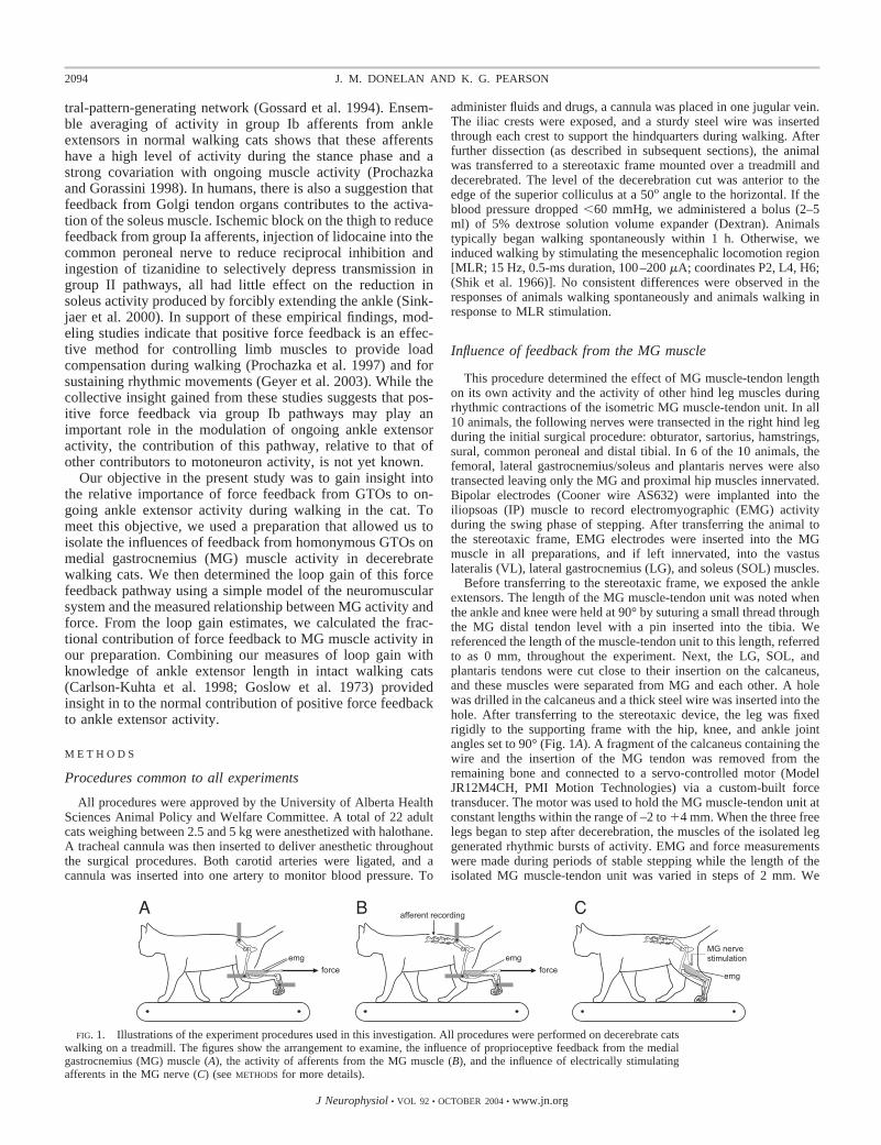
This procedure determined the effect of MG muscle-tendon lengthon its own activity and the activity of other hind leg muscles duringrhythmic contractions of the isometric MG muscle-tendon unit. In all10 animals, the following nerves were transected in the right hind legduring the initial surgical procedure: obturator, sartorius, hamstrings,sural, common peroneal and distal tibial. In 6 of the 10 animals, thefemoral, lateral gastrocnemius/soleus and plantaris nerves were alsotransected leaving only the MG and proximal hip muscles innervated.Bipolar electrodes (Cooner wire AS632) were implanted into theiliopsoas (IP) muscle to record electromyographic (EMG) activityduring the swing phase of stepping. After transferring the animal tothe stereotaxic frame, EMG electrodes were inserted into the MGmuscle in all preparations, and if left innervated, into the vastuslateralis (VL), lateral gastrocnemius (LG), and soleus (SOL) muscles.
Before transferring to the stereotaxic frame, we exposed the ankleextensors. The length of the MG muscle-tendon unit was noted whenthe ankle and knee were held at 90° by suturing a small thread throughthe MG distal tendon level with a pin inserted into the tibia. Wereferenced the length of the muscle-tendon unit to this length, referredto as 0 mm, throughout the experiment. Next, the LG, SOL, andplantaris tendons were cut close to their insertion on the calcaneus,and these muscles were separated from MG and each other. A holewas drilled in the calcaneus and a thick steel wire was inserted into thehole. After transferring to the stereotaxic device, the leg was fixedrigidly to the supporting frame with the hip, knee, and ankle jointangles set to 90° (Fig. 1A). A fragment of the calcaneus containing thewire and the insertion of the MG tendon was removed from theremaining bone and connected to a servo-controlled motor (ModelJR12M4CH, PMI Motion Technologies) via a custom-built forcetransducer. The motor was used to hold the MG muscle-tendon unit atconstant lengths within the range of –2 to �4 mm. When the three freelegs began to step after decerebration, the muscles of the isolated leggenerated rhythmic bursts of activity. EMG and force measurementswere made during periods of stable stepping while the length of theisolated MG muscle-tendon unit was varied in steps of 2 mm. We
FIG. 1. Illustrations of the experiment procedures used in this investigation. All procedures were performed on decerebrate catswalking on a treadmill. The figures show the arrangement to examine, the influence of proprioceptive feedback from the medialgastrocnemius (MG) muscle (A), the activity of afferents from the MG muscle (B), and the influence of electrically stimulatingafferents in the MG nerve (C) (see METHODS for more details).
2094 J. M. DONELAN AND K. G. PEARSON
J Neurophysiol • VOL 92 • OCTOBER 2004 • www.jn.org

allowed the muscle to remain at each length for 4–20 stride cycles.The EMGs were amplified and stored with force and length signals onDAT tape (Model VDAT8, Vetron Technology).
After the experiment, we digitized the data at 1.5 kHz with 12-bitresolution (Axotape, Axon Instruments) and stored it on computerdisk. The digitized EMG data were rectified and filtered using alow-pass, first-order, one-way, digital Butterworth filter with a cut-offfrequency of 30 Hz. The force signals were filtered using a two-waydigital filter of the same design. Sequences of consistent steppinglasting �3 min that included a range of MG lengths were selected foranalysis. Restricting each set of analyzed data to a relatively shortperiod of time reduced the possibility of a change in the excitability ofthe animal, independent of muscle length, confounding our results.
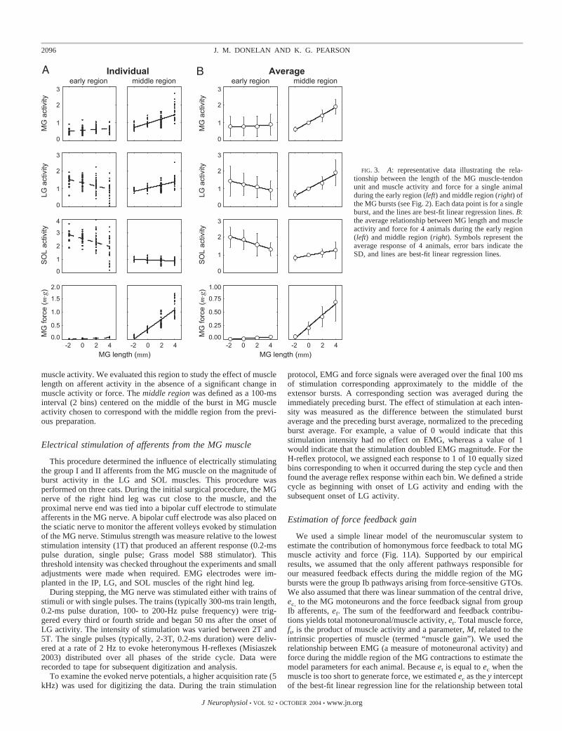
We computed the average EMG and force magnitudes within tworegions of MG bursts (Fig. 2). The early region was defined as a30-ms interval beginning with the onset of MG activity. We evaluatedthis region to study the effect of length on muscle activity in theabsence of a significant change in force (Fig. 3). The middle regionwas defined as a 100-ms interval centered on the middle of the MGburst chosen to correspond with high levels of muscle activity andforce. To make comparisons of feedback effects across muscles andbetween animals, we normalized the value of the EMG in each regionto each muscle’s average middle region value at a length of 0 mm.
Afferent recordings from the MG muscle
This procedure determined the effect of MG muscle-tendon lengthon the activity of single afferents from the MG muscle duringrhythmic contractions of the isometric MG muscle-tendon unit (Fig.1B). Unless otherwise described, this preparation was identical to theprevious preparation. The right hind leg and tail were extensivelydenervated by transecting the following nerves: femoral, obturator,
common peroneal, distal tibial proximal to the plantaris nerve, lateralgastrocnemius/soleus, hamstrings, gluteus, and all nerves to the rightside of the tail. A stimulating bipolar cuff electrode was placed aroundthe intact MG nerve to identify the afferents. A laminectomy exposedthe L7 and S1 dorsal roots. After transferring the animal to thestereotaxic frame, small filaments containing single afferents from theMG muscle were dissected from the L7 and S1 roots on the right side.Within a paraffin bath, the filaments were lifted onto monopolar silverwire recording electrodes. Afferent type was identified on the basis ofconduction velocity, and responses to muscle vibration, ramp-and-hold stretches, and muscle contractions (Matthews 1972). We mini-mized the number of dissected filaments to maximize the afferentinput to the spinal cord from the MG muscle. In the absence ofafferent input, rhythmic activity in the MG muscle usually does notoccur during walking (unpublished observations). Two to four affer-ents were isolated in each animal, but some did not remain viable afterdecerebration and the initiation of walking. Nine animals yieldedmeasurements from 10 Ia afferents, 5 Ib afferents, and 4 group IIafferents. When the intact legs began to step and the isolated MGgenerated rhythmic bursts of activity, the activity in the survivingafferents was recorded at different lengths of the isolated MG muscle-tendon unit. As described in the previous section, afferent activity,EMG and force signals were amplified and stored on tape.
After the experiment, data were digitized at 7 kHz to captureaccurately unit activity. Spikes were identified by determining wheneach unit signal exceeded a set threshold. Data were accumulated intoan event histogram (50-ms bin width) relative to the onset of MGburst activity. The average discharge rate (in Hz) for each bin wascomputed from the number of events in each bin divided by thenumber of strides and the bin width. We evaluated unit activity overtwo regions of the stride cycle (Fig. 6). The prior region was definedas a 100-ms interval (2 bins) immediately prior to the onset of MG
FIG. 2. Longer MG muscle-tendon lengths during a sequence of rhythmic contractions resulted in greater MG and lateralgastrocnemius (LG) muscle activity, and greater MG force. In this example, length did not influence the magnitude of burst activityin the soleus (SOL) muscle. These effects are further illustrated by the averages of 7 stride cycles at each length (right). Thick andthin lines, lengths of 4 and 2 mm, respectively; shaded vertical bars, the early and middle regions used in subsequent analyses.
2095FORCE FEEDBACK IN WALKING CATS
J Neurophysiol • VOL 92 • OCTOBER 2004 • www.jn.org
muscle activity. We evaluated this region to study the effect of musclelength on afferent activity in the absence of a significant change inmuscle activity or force. The middle region was defined as a 100-msinterval (2 bins) centered on the middle of the burst in MG muscleactivity chosen to correspond with the middle region from the previ-ous preparation.
Electrical stimulation of afferents from the MG muscle
This procedure determined the influence of electrically stimulatingthe group I and II afferents from the MG muscle on the magnitude ofburst activity in the LG and SOL muscles. This procedure wasperformed on three cats. During the initial surgical procedure, the MGnerve of the right hind leg was cut close to the muscle, and theproximal nerve end was tied into a bipolar cuff electrode to stimulateafferents in the MG nerve. A bipolar cuff electrode was also placed onthe sciatic nerve to monitor the afferent volleys evoked by stimulationof the MG nerve. Stimulus strength was measure relative to the loweststimulation intensity (1T) that produced an afferent response (0.2-mspulse duration, single pulse; Grass model S88 stimulator). Thisthreshold intensity was checked throughout the experiments and smalladjustments were made when required. EMG electrodes were im-planted in the IP, LG, and SOL muscles of the right hind leg.
During stepping, the MG nerve was stimulated either with trains ofstimuli or with single pulses. The trains (typically 300-ms train length,0.2-ms pulse duration, 100- to 200-Hz pulse frequency) were trig-gered every third or fourth stride and began 50 ms after the onset ofLG activity. The intensity of stimulation was varied between 2T and5T. The single pulses (typically, 2-3T, 0.2-ms duration) were deliv-ered at a rate of 2 Hz to evoke heteronymous H-reflexes (Misiaszek2003) distributed over all phases of the stride cycle. Data wererecorded to tape for subsequent digitization and analysis.
To examine the evoked nerve potentials, a higher acquisition rate (5kHz) was used for digitizing the data. During the train stimulation
protocol, EMG and force signals were averaged over the final 100 msof stimulation corresponding approximately to the middle of theextensor bursts. A corresponding section was averaged during theimmediately preceding burst. The effect of stimulation at each inten-sity was measured as the difference between the stimulated burstaverage and the preceding burst average, normalized to the precedingburst average. For example, a value of 0 would indicate that thisstimulation intensity had no effect on EMG, whereas a value of 1would indicate that the stimulation doubled EMG magnitude. For theH-reflex protocol, we assigned each response to 1 of 10 equally sizedbins corresponding to when it occurred during the step cycle and thenfound the average reflex response within each bin. We defined a stridecycle as beginning with onset of LG activity and ending with thesubsequent onset of LG activity.
Estimation of force feedback gain
We used a simple linear model of the neuromuscular system toestimate the contribution of homonymous force feedback to total MGmuscle activity and force (Fig. 11A). Supported by our empiricalresults, we assumed that the only afferent pathways responsible forour measured feedback effects during the middle region of the MGbursts were the group Ib pathways arising from force-sensitive GTOs.We also assumed that there was linear summation of the central drive,ec, to the MG motoneurons and the force feedback signal from groupIb afferents, ef. The sum of the feedforward and feedback contribu-tions yields total motoneuronal/muscle activity, et. Total muscle force,ft, is the product of muscle activity and a parameter, M, related to theintrinsic properties of muscle (termed “muscle gain”). We used therelationship between EMG (a measure of motoneuronal activity) andforce during the middle region of the MG contractions to estimate themodel parameters for each animal. Because et is equal to ec when themuscle is too short to generate force, we estimated ec as the y interceptof the best-fit linear regression line for the relationship between total
FIG. 3. A: representative data illustrating the rela-tionship between the length of the MG muscle-tendonunit and muscle activity and force for a single animalduring the early region (left) and middle region (right) ofthe MG bursts (see Fig. 2). Each data point is for a singleburst, and the lines are best-fit linear regression lines. B:the average relationship between MG length and muscleactivity and force for 4 animals during the early region(left) and middle region (right). Symbols represent theaverage response of 4 animals, error bars indicate theSD, and lines are best-fit linear regression lines.
2096 J. M. DONELAN AND K. G. PEARSON
J Neurophysiol • VOL 92 • OCTOBER 2004 • www.jn.org

muscle activity and force (Fig. 11B). We then normalized totalactivity by ec and total force by body weight, m �g. This normalizationmakes our subsequent calculations independent of size and nonphysi-ological factors like electrode placement allowing for more meaning-ful comparisons between animals. This estimate of ec, accompaniedby our measurements of et and ft, allow for the algebraic solution ofpathway gains. For each stride, we estimated force feedback gain, K,as
K �ef
ft
�et � ec
ft
(1)
The muscle gain, M, was defined as
M �ft
et
(2)
The dimensionless product of the muscle and force feedback gains,termed “loop gain” (Prochazka et al. 1997), is of particular impor-tance. First, it gives the relative contribution of force feedback to totalmuscle activity and force
ef
et
�ff
ft
� K � M (3)
where ff is the contribution of force feedback to ft. Second, forsteady-state muscle activity and force, the average loop gain in thispositive feedback system must be less than unity (Prochazka et al.1997).
These calculations were performed using the middle region datafrom our experiments studying the influence of feedback from the MGmuscle. At each muscle length, we estimated gains by first calculatingK, M, and K �M for the middle region of each stride and then averagingthese values at each length within each animal. Their dependence onMG length was determined using best-fit least-squares linear regres-sion and testing for a slope statistically different from zero.
Statistical analysis
The significance of measured relationships was generally testedusing linear regression. In addition to determining the slope and yintercept of the line that best fit the data, this procedure yielded ameasure of the probability that these coefficients were zero (i.e., Pvalue). If the probability was �5% (P � 0.05), we accepted therelationship as significant. To compare the effect of stimulationintensity, rather than use linear regression, we used one-tailed pairedt-test with the same level of significance.
R E S U L T S
Influence of feedback from the MG muscle
The purpose of the first set of experiments was to establishthe effect of proprioceptive feedback originating from MG onits own muscle activity and on the activity of other musclesduring walking in decerebrate cats (Fig. 1A). The primaryobservation was that activity and force in the MG muscleduring isometric contractions of the MG muscle-tendon unitwas greater at longer lengths. This is illustrated in Fig. 2 bytypical muscle activity and force patterns when the length ofthe MG muscle-tendon unit was reduced from 4 to 2 mm. LGactivity was also elevated at longer muscle lengths, whereasthe magnitude of bursts in SOL was relatively unaffected. Toquantify these changes in EMG magnitude and force at differ-ent lengths and in different animals, we averaged these signalsover two regions (gray vertical bars; Fig. 2). We choose anearly region to study the effect of length on muscle activity
before MG developed substantial force. The middle regioncorresponded with high levels of both muscle activity andforce.
Figure 3A illustrates the effect of length on the magnitude ofthe early and middle regions in a representative animal. Tomake comparisons of feedback effects across muscles andbetween animals, we normalized the value of the EMG in eachregion to each muscle’s average middle region value at a lengthof 0 mm. Figure 3A demonstrates that although there is vari-ability in burst magnitude and force, consistent effects oflength are present. During the early region, LG and SOLactivity decreased at longer lengths while MG activity in-creased slightly (P � 1.8e-6, 6.4e-6 and 0.01, respectively).During the middle region, MG and LG activity increased atlonger lengths while SOL activity decreased slightly (P �2.1e-12, 1.5e-8 and 0.01, respectively). We observed thesegeneral patterns in the four animals in which we studiedheteronymous effects, and the magnitude did not differ sub-stantially between animals. The notable exception was SOLmiddle region activity. It increased slightly in two animals,decreased slightly in one animal, and remained relativelyunchanged in one animal.
Figure 3B plots the average results from the four animals inwhich we recorded heteronymous influences on LG and SOL.During the early region, before the MG muscle could developsubstantial force, MG length did not have a significant effecton MG activity (P � 0.77). While LG and SOL activityappeared to decrease at longer muscle lengths, these trendswere not statistically significant (P � 0.24 and P � 0.06,respectively). Middle region results were quite different fromearly region responses. MG activity and force were signifi-cantly elevated at longer muscle lengths (P � 4.4e-6 and P �2.5e-4, respectively). Comparing changes between �2 and 4mm, the average increase in MG activity was 203%. We didnot study lengths shorter than �2 mm because MG did notgenerate substantial force at short lengths in this preparation.While LG activity was similarly elevated (194% increase; P �4.4e-4), SOL activity was less dependent on MG muscle length(50% increase; P � 0.03). The difference in response betweenLG and SOL indicates that proprioceptive feedback originatingfrom MG was directed predominantly to MG and LG.
We also observed that increasing MG length did not have ageneralized excitatory action on locomotor activity. Holdingthe MG muscle at increasingly longer lengths had no effect onthe ipsilateral IP muscle activity (P � 0.12; Fig. 4), and noobvious changes were noted in the stepping of the contralateralleg and the forelegs. In addition, a doubling of MG burstactivity was related to only a 4% increase in VL burst activity(P � 2.6e-4; Fig. 4).
Afferent recordings from the MG muscle
The purpose of this set of experiments was to measureactivity in identified muscle afferents to gain insight into whichafferent pathways were responsible for the increase in MGmuscle activity at longer lengths of the isometric muscle-tendon unit (Fig. 1B). Figure 5 illustrates typical activity of agroup Ia afferent from a primary spindle ending, a group IIafferent from a secondary spindle ending, and a group Ibafferent from a GTO during rhythmic isometric contractions ofthe MG muscle. The characteristic feature of activity in all
2097FORCE FEEDBACK IN WALKING CATS
J Neurophysiol • VOL 92 • OCTOBER 2004 • www.jn.org

group Ia and II afferents we recorded was that their activitywas reduced during the contractions with a minimum near themiddle of the contraction. This effect is likely a result of theextrafusal fibers unloading the intrafusal fibers. In contrast, allgroup Ib afferents increased their activity during the contrac-tions and were silent between the contractions. Figure 6 illus-trates the effect of changing MG length on activity in theserepresentative afferents. While the group II afferents and the Ibafferents showed substantial increases in middle region activityat longer lengths, increasing muscle length had little effect onthe Ia afferent activity. Figure 7, left, further illustrates thispattern by plotting the effect of MG length on the magnitude ofprior and middle region activity in these representative affer-ents. Although group Ia activity increased with length duringthe prior region (P � 6.1e-3; Fig. 7A), the effect of length onactivity in the middle region was not significant (P � 0.44; Fig.7A). In contrast, group II activity increased during both theprior and middle regions (P � 0.03 and 0.01, respectively; Fig.7B). Group Ib afferent middle region activity also increasedwith length (P � 7.6e-5; Fig. 7C). There was no activity duringthe prior region because the muscle was not yet generatingforce. The slopes of the linear regression lines shown in theseplots estimate the sensitivity of each afferent to length changewithin each region. Averaging sensitivities across the mea-sured afferents demonstrates that group Ia afferents were rel-atively insensitive to length change during the middle region[1.9 � 1.9 (SD) �Hz/�mm; n � 10; Fig. 7A, right]. In contrast,group II afferents (6.4 � 4.6 �Hz/�mm; n � 4; Fig. 7B, right)and Ib afferents (8.8 � 5.2 �Hz/�mm; n � 5; Fig. 7C, right)were, on average, quite sensitive to length change during themiddle region.
Electrical stimulation of afferents from the MG muscle
The purpose here was to examine the influence of stimulat-ing afferents in the MG nerve to establish whether group IIafferents have the appropriate action to contribute to the
elevation of ankle extensor activity occurring at longer MGlengths. The effect of electrical stimulation on the magnitudeof bursts in the close synergists LG and SOL was examinedusing two stimulation intensities; one intensity (2T) was at alevel that just begins to recruit group II afferents and the other(5T) was high enough to recruit most group II afferents (Jack
FIG. 5. Representative data illustrating the activity in afferents from aprimary spindle ending (A), a secondary spindle ending (B), and a Golgi tendonorgan (GTO; C), from the MG muscle and tendon during isometric contrac-tions of the muscle-tendon unit. Top: the rectified and filtered electromyograms(EMGs) from the MG muscle. Middle top: the forces in the MG muscle.Middle, bottom: the spikes recorded from isolated afferents. Bottom: instanta-neous firing frequency of the afferents. Note the activity in both spindleendings were reduced during the contractions with a minimum near the middleof the contractions while the GTO activity increased during the contraction.
FIG. 4. The magnitude of burst activity in ipsilateral vastus lateralis (VL),a knee extensor, and iliopsoas (IP), a hip flexor, was not substantiallyinfluenced by the magnitude of burst activity in MG. IP activity was averagedover a 100-ms interval centered on the middle of its own period of activitywhile VL was averaged over the previously defined middle region. Each datapoint is from a single burst. Lines are best-fit linear regression lines.
2098 J. M. DONELAN AND K. G. PEARSON
J Neurophysiol • VOL 92 • OCTOBER 2004 • www.jn.org

1978). It was necessary in these experiments to cut the MGnerve distal to the stimulating electrodes to prevent contrac-tions of the MG muscle, thus we were not able to examine thehomonymous actions of the MG group II afferents.
Figure 8 illustrates the typical effect of MG nerve stimula-tion on LG and SOL muscle activity, and Fig. 9 presents theaverage effect for three animals. Comparing the stimulatedcycle to the stride immediately prior to stimulation demon-strates that stimulation had a clear excitatory effect on both LGactivity (P � 0.02) and SOL activity (P � 3.4e-4) at 2T (Fig.8B and 9), thus demonstrating a strong heteronymous action ofMG group I afferents. However, stimulating at 5T (Figs. 8Cand 9) did not further increase activity over the increaseobserved at 2T for either LG (P � 0.22) or SOL (P � 0.88).Because this intensity range represents the range over whichmost of the group II afferents are normally recruited, it appearsthat transmission in pathways from MG group II afferents tomotoneurons innervating other ankle extensor muscles waseither very weak or functionally absent.
We also determined whether monosynaptic pathways fromgroup Ia afferents were functional in our preparation. Repre-sentative heteronymous H-reflex responses from a single ani-mal illustrates that heteronymous connections from MG groupIa afferents to LG and SOL motoneurons were open (Fig. 10).
Measurements in the two other animals yielded qualitativelysimilar results.
Estimation of force feedback gain
The purpose of this procedure was to estimate the contribu-tion of homonymous force feedback to MG activity and force.The basic assumption of this analysis is that the increase inmagnitude of the MG bursts at longer muscle lengths (Figs. 2and 3) is entirely due to feedback from force-sensitive afferentsin the MG muscle. In the DISCUSSION, we justify this assumptionbased on the empirical results of this investigation. Using alinear model for positive force feedback (Fig. 11A) and empir-ical data for the relationship between the middle region of MGEMG activity and the MG force (Fig. 11B), we could estimatethe contribution of force feedback to MG activity (see METH-ODS). The relationship between muscle force and EMG activitywas similar in all 10 animals in that the level of EMG activityalways increased substantially at longer muscle lengths. Weused the equations outlined in METHODS to estimate pathwaygains and their dependence on muscle length.
Figure 11C plots the estimated force feedback gain, K (Eq.1), as a function of muscle length. The magnitude of K variedfrom animal to animal. At 0 mm, for example, K ranged from0.69 to 3.91 and averaged 1.90 � 0.97 [in units of ec �
FIG. 7. Left: plots illustrating the influence of MG length on the activity ofa single primary spindle ending (A), secondary spindle endings (B), and GTO(C) during the prior region (- - -) and middle region (—) of the contractions inthe MG muscle. Lines are best-fit linear regression lines. Their slopes estimatethe sensitivity of each afferent to length change during each region. Right: barplots illustrating the average sensitivities for all afferents in each group. Errorbars denote the SD. Note that primary spindle endings were insensitive tolength during the middle region.
FIG. 6. Histograms illustrating the effect of length on activity in a primaryspindle ending (A), a secondary spindle ending (B), and a GTO (C) from theMG muscle during isometric contractions of the muscle-tendon unit at 4 mm(thick traces) and 0 mm (thin traces). Force traces are shown above each set ofhistograms. Note that during the middle region of the contractions (shadedvertical bar) the activity in the primary spindle ending was only slightlyincreased at the longer length.
2099FORCE FEEDBACK IN WALKING CATS
J Neurophysiol • VOL 92 • OCTOBER 2004 • www.jn.org

(m �g)�1]. Interestingly, K was independent of muscle length inall animals. Linear regression on each animal’s data yielded Pvalues that ranged from 0.06 to 0.89 with a median value of0.47. To determine the average force feedback gain and itsdependence on length, we combined our measurements fromall animals and found the best-fit linear regression line (Fig.11C). On average, K was also independent of muscle length(P � 0.39; slope � 0.05 � 0.12; y intercept � 1.72 � 0.26).
Figure 11D plots the estimated muscle gain, M (Eq. 2), as afunction of muscle length. Similar to force feedback gain, Mvaried from animal to animal. At 0 mm, for example, M ranged
from 0.06 to 0.33 and averaged 0.19 � 0.09 [in units ofm �g � (ec)
�1]. In contrast to force feedback gain, M generallyincreased with muscle length. Linear regression on each ani-mal’s data yielded P values that ranged from 2.1e-5 to 0.08with a median value of 5.7e-3. Two animals did not demon-strate significant effects of length on M (P � 0.08 and 0.05).While muscle gain in these animals showed a similar depen-dence on length as the other animals, the relationship was notsignificant because we were only able to make measurementsat three muscle lengths before the animals stopped walking.Combining our measurements from all animals and determin-ing the best-fit linear regression line demonstrated that, onaverage, M increased with muscle length (P � 1.3e-7; slope �0.04 � 0.01; y intercept � 0.21 � 0.03; Fig. 11D). Weexpected this increase due to the well known force-lengthproperties of muscle (Gordon et al. 1966).
Figure 11E plots the estimated loop gain, K �M (Eq. 3), as afunction of muscle length. This dimensionless number alsovaried from animal to animal. At 0 mm, for example, K �Mranged from 0.11 to 0.44 and averaged 0.30 � 0.10. WhileK �M generally increased with muscle length, there was con-siderable variability between animals. Linear regression oneach animal’s data yielded P values that ranged from 0.01 to0.20 with a median value of 0.04. Although four animals hadP values greater than our level of significance (0.20, 0.18, 0.06,and 0.06), all animals demonstrated an increase in loop gainwith length. Our statistical power was limited for some animalsbecause we were only able to make measurements at a subsetof muscle lengths before they stopped walking. Combining ourmeasurements from all animals and determining the best-fitlinear regression line demonstrated that, on average, K �M
FIG. 8. A: representative data illustrates that MG nerve elec-trical stimulation (2T) during a sequence of stepping resulted ingreater LG and SOL muscle activity. We stimulated the nerveevery 3rd stride with trains of stimuli, 300 ms in duration (top).B and C: the magnitude of the effect of 2T and 5T stimulationon LG and SOL activity was similar. The thick line and thinlines denote the stimulated stride and the stride immediatelyprior to stimulation, respectively, averaged for 10 consecutivestrides. The dotted vertical lines represent the period of stimu-lation. The shaded vertical bars denote the regions used insubsequent analyses.
FIG. 9. Electrical stimulation of group II afferents in the MG nerve does notinfluence the magnitude of burst activity in the LG and SOL muscles. The bargraphs compare the average relative effect of stimulation at 2T (�) and 5T (■ )during a 100-ms period (shaded bars in Fig. 8) for 3 cats. This range ofintensities represents the range over which most of the group II afferents arerecruited (Jack 1978). Error bars denote the SD.
2100 J. M. DONELAN AND K. G. PEARSON
J Neurophysiol • VOL 92 • OCTOBER 2004 • www.jn.org

increased with muscle length (P � 3.8e-7; slope � 0.05 �0.02; y intercept � 0.31 � 0.04; Fig. 11E). This increase wasa result of the loop gain being the product of force feedbackgain that, on average, was constant and muscle gain that, onaverage, monotonically increased with muscle length. Loopgain increased from 0.20 at �2 mm to 0.52 at 4 mm. Becauseloop gain is the relative contribution of force feedback to totalmuscle activity and force (Eq. 3), these results indicate thatforce feedback was of modest importance at short musclelengths, accounting for �20% of total activity and force, and ofsubstantial importance at long muscle lengths, accounting for50%. Loop gain was always less than unity, indicating that thispositive feedback system was stable (Prochazka et al. 1997).
D I S C U S S I O N
Numerous investigations over the past decade have demon-strated that sensory feedback contributes substantially to ankleextensor activity during the stance phase of walking in humans
and cats (Dietz and Duysens 2000; Donelan and Pearson2004). Currently the origin of the reinforcing sensory signals isuncertain, but it seems likely that they arise from multiplegroups of sensory receptors including ankle extensor Golgitendon organs, ankle extensor muscle spindles, and cutaneousreceptors in the feet. An important issue, therefore is toestablish the contribution of each class of receptors to theactivity in the ankle extensor muscles and the conditions underwhich each class exerts its influence. In this study, we deter-mined the contribution of feedback originating from MG GTOsto MG muscle activity by estimating the loop gain of thepositive force feedback pathway during walking in the cat.
A fundamental observation in our investigation was that themagnitude of MG muscle activity was elevated at longermuscle lengths (Figs. 2 and 3). It was quite clear that this wascaused by changes in sensory signals from receptors in the MGmuscle or tendon. Under the experimental conditions, the hindleg was extensively denervated, and MG was isolated from theother ankle extensor muscles (Fig. 1). This denervation in-cluded nerves innervating the knee joint thus removing feed-back from joint receptors. The enhancement of MG activity atlonger lengths was not due to a generalized increase in excit-ability because there was limited influence of MG lengthchanges on activity in some of the other muscles crossing thehip, knee, and ankle (Figs. 2–4) and on muscles of thecontralateral leg (unpublished observation). However, the ac-tivity in LG, a close synergist of MG, was enhanced when MGwas lengthened (Figs. 2 and 3). These observations demon-strate that the important sources of feedback in our investiga-tion are afferent signals arising from the MG muscle-tendonunit that have an excitatory action on homonymous motoneu-rons as well as motoneurons innervating specific ankle exten-sors.
Which receptors in the MG muscle are responsible forenhancing the MG burst activity at longer lengths? It isimpossible to distinguish the relative contribution of differentproprioceptors knowing only the relationship between musclelength and EMG activity. This is because force also increasedat longer lengths (Fig. 3). This increase in force was due toboth the increase in muscle activity and the intrinsic force-length properties of the MG muscle (Gordon et al. 1966). Inaddition, although the muscle-tendon length was isometric,there was internal shortening of muscle fibers (as indicated bythe decrease in activity of group Ia and II afferents) that mayhave been dependent on length. Thus sensory signals related tolength, velocity, and force all have the potential for modifyingthe MG activity in a length-dependent manner.
A number of observations allow us to exclude a majorcontribution of muscle spindles to the enhancement of MGmiddle region activity. First, the activity in group Ia MGafferents (arising from primary spindle endings) was relativelyinsensitive to changes in muscle length (Figs. 7). Second, burstactivity in SOL was sometimes completely uninfluenced bychanges in MG length, yet heteronymous connections fromMG group Ia afferents to SOL motoneurons were open asdemonstrated by the capacity to elicit an H-reflex in SOL (Fig.10). This supports our observation that group Ia afferentactivity was not significantly elevated during the middle of themuscle contraction. This insensitivity may have been com-pounded by inhibition of group Ia pathways at the level of thespinal cord; Gosgnach et al. (2000) have reported that group Ia
FIG. 10. Representative data illustrates the H-reflexes evoked in the LG(top) and SOL (bottom) muscles by electrical stimulation of the MG nerve. Thestep cycle was divided into 10 equal bins. Right: traces show average EMGresponses to single stimuli (3 T, 0.2-ms duration) for each bin (on average 43stimuli per bin). The traces begin when the MG nerve was stimulated. Left: theaverage EMG in the LG and SOL muscles is oriented vertically to indicate thecorresponding time within the step cycle of each average.
2101FORCE FEEDBACK IN WALKING CATS
J Neurophysiol • VOL 92 • OCTOBER 2004 • www.jn.org

pathways from the ankle extensor muscles are presynapticallyinhibited during locomotion in decerebrate cats. However, thefact that we could evoke H-reflexes demonstrates that thesepathways were not completely inhibited. Third, although groupII MG afferents (arising from secondary spindle endings) weresensitive to length (Fig. 7), electrical stimulation of theseafferents had negligible influence on the magnitude of LGactivity (Fig. 9). The degree to which this observation reflectson the homonymous action of these afferents depends on ourassumption that the same afferents were responsible for theelevation of both MG and LG muscle activity with length. Thisassumption is supported by our observation that middle regionLG and MG activity increased by similar amounts with in-creased length (Fig. 3). Although an alternative explanation forthese results is that stimulation at group I strength saturatedinterneurons common to both group I and II afferent pathways,previous investigations from other laboratories have also failedto find any excitatory action of extensor group II afferents oncat ankle extensor motoneurons (Gossard et al. 1994; Perreaultet al. 1995). Finally, the early region of the MG muscle activitywas uninfluenced by changes in muscle length (Fig. 3) despitea strong influence of muscle length on activity in both group Iaand II afferents during the same region (Fig. 7). All theseobservations allow us to conclude that, under the conditions ofour experiment, MG muscle spindles were not the major sourceof feedback responsible for elevating MG muscle activity atlonger muscle lengths. If muscle spindles are excluded, thenwe are left with the Golgi tendon organs and free nerve endingsresponsive to length and force as potential sources of thesensory signals enhancing MG activity. It is unlikely that free
nerve endings are responsible for the measured feedback effectbecause currently there is no evidence that group III and IVmuscle afferents have an excitatory action on triceps suraemotoneurons during walking (Schomburg et al. 2001).
By exclusion, the results indicate that MG GTOs were themain source of sensory signals acting to elevate MG muscleactivity in our preparation. This is a reasonable conclusionbecause previous studies have demonstrated that group Ibpathways from ankle extensors have an excitatory action onextensor activity during walking in spinal and decerebrate cats(Pearson and Collins 1993; Whelan et al. 1995) as well asfictive locomotion in spinal and decerebrate cats (Angel et al.1996; Conway et al. 1987; Gossard et al. 1994). In addition,this conclusion is consistent with our observation that GTOactivity is elevated during the region of interest (Figs. 6 and 7).
This isolation of the contribution of GTOs allowed us to usea simple neuromuscular model to estimate the loop gain of thispositive force feedback pathway (Fig. 11; outlined in METHODS).Average loop gain, a quantitative measure of the relativecontribution of MG GTO force feedback to MG muscle activ-ity, ranged from 0.2 at short muscle lengths to 0.5 at longmuscle lengths. While loop gain was greater at long lengthsthan short lengths in all animals, the magnitude of loop gain ata given length varied considerably from animal to animal (Fig.11E). While we endeavored to control differences betweenexperiments, some were unavoidable. To understand the sourceof this variability, it is useful to consider potential contributorsto differences in the constituents of loop gain: force feedbackgain and muscle gain. Force feedback gain is a property of thenervous system and can be extensively modified. Indeed, the
FIG. 11. Plots showing values of force feedback gain, muscle gain, and loop gain (C–E, respectively) that were derived usinga simple linear model of the neuromuscular system (A) and the relationship between MG activity and MG force (B). A: for moremodel details, refer to METHODS. B: representative data from a single animal. Each symbol is a single burst’s middle regionmagnitude. The line is a best-fit linear regression line. Its y-axis intercept estimates the feedforward contribution to muscle activity,ec (denoted by the horizontal shaded area). C: force feedback gain, K, was independent of muscle length. D: muscle gain, M,increased with muscle length due to the intrinsic properties of muscle. E: the product of force feedback gain and muscle gain yieldedloop gain, K � M. It also increased with length. Each symbol in C–E represents measurements from a different animal (n � 10).
2102 J. M. DONELAN AND K. G. PEARSON
J Neurophysiol • VOL 92 • OCTOBER 2004 • www.jn.org

nervous system modifies the gain of the force feedback path-way so that it is negative during standing and positive duringlocomotion (Pearson and Collins 1993). Thus variability in ourestimates of force feedback gain may reflect differences in thestate of each animal’s nervous system due to differences infactors like core temperature, time under anesthetic, and dura-tion of experiment. Muscle gain, an intrinsic property ofmuscle, is the amount of force generated per unit of muscleactivity (Eq. 2). This relationship depends on the condition ofthe muscle, which may have been slightly compromised insome preparations, and muscle fatigue, which tends to increasewith the duration of the experiment. In addition, it is importantrecognize that some of the variability is a consequence of theintrinsic differences between animals. While we endeavored toisolate the force feedback pathway, it is possible that otherafferents made small, length-dependent, contributions to mus-cle activity. Thus our values are likely slight overestimates. Itis important to note that this loop gain does not estimate thetotal contribution of force feedback because force feedbackfrom heteronymous sources may also be present in the intactwalking cat (Nichols 1999).
Our estimates of gain were made in a very artificial situation,so it is necessary to consider the relevance to conditions in anintact walking animal. During normal walking, MG muscle-tendon length during stance ranges from –1 to –9 mm withlength measured in the same manner as in our experiment(Goslow et al. 1973). This upper end of this range just overlapsthe low end of the range of lengths we examined in our currentinvestigation because MG did not generate substantial force atlengths shorter than �2 mm (Fig. 3B). The range of lengthsused in normal walking corresponds to lengths at which theestimated gain of the force-feedback pathway is low, suggest-ing that the contribution of force feedback to MG activationduring walking on a level surface is modest. When walking upslopes, however, the MG muscle can be 7 mm longer duringthe early part of the stance phase (our calculations usingCarlson-Kuhta et al. 1998). This is in the range where the gainof the force feedback pathway is high. Thus force feedbackmay make a major contribution to the elevation in the magni-tude of MG burst activity that is known to occur during uphillwalking (Carlson-Kuhta et al. 1998). The observation that MGforce increased substantially during uphill walking (Kaya et al.2003) is consistent with this prediction.
Another important fact to take into account when consider-ing the functional relevance of our data is that the length of theMG muscle is continuously changing during the stance phase:lengthening in early stance and shortening during mid- to latestance. The intrinsic force-velocity properties of the MG mus-cle (Hill 1938) will increase muscle gain, and thus loop gain,during early stance, yielding a force-feedback contributionhigher than that predicted based only on muscle length. On theother hand, the force-velocity properties will tend to reduceloop gain during late stance, decreasing the contribution offorce feedback below that estimated from length alone. Thesevelocity-dependent modifications in muscle force are probablyenhanced by increases and decreases in the velocity signalsfrom group Ia afferents during early and late stance, respec-tively.
The measured loop gain does not estimate the total contri-bution of all homonymous and heteronymous feedback to MGactivity during normal walking. It is likely that other afferent
pathways contribute to MG motoneuron depolarization undernormal walking conditions. Assuming an excitatory action ofadditional afferent feedback, then our loop gain values are bestviewed as estimates of the lower bound of the total contributionof feedback to ongoing ankle extensor activity. For example,because homonymous force feedback is responsible for 50% ofMG activity at long muscle lengths, then the remaining 50% isdue to feedforward sources as well as other sources of afferentfeedback. Under these circumstances, the maximum contribu-tion of feedforward control is 50% but possibly much less dueto the contribution of other afferent signals.
In summary, our results indicate that homonymous forcefeedback from GTOs has a substantial contribution to ongoingankle extensor activity during walking in the cat. Duringisometric contractions of the MG muscle-tendon unit, thiscontribution of feedback from GTOs is modest when themuscle is short but doubles MG muscle activity and force whenthe muscle is long. The length dependence is due to theintrinsic force-length property of muscle. The gain of thepathway that converts muscle force to motoneuron depolariza-tion, K, is independent of length. This positive feedback systemis quite stable as the loop gain is less than unity at all measuredlengths. In the intact cat, it is likely that homonymous forcefeedback is not the only important source of feedback as otherafferent pathways may make significant contributions whenmuscle length is allowed to change. This emphasizes thegeneral importance of afferent feedback for generating ongoingankle extensor activity during walking.
A C K N O W L E D G M E N T S
We thank D. McVea, J. Misiaszek, and J. Yang for helpful comments on adraft of this paper and R. Gramlich for excellent technical assistance.
G R A N T S
This research was supported by the Canadian Institutes for Health Research,the National Science and Engineering Council, and the Alberta HeritageFoundation for Medical Research.
R E F E R E N C E S
Angel MJ, Guertin P, Jimenez T, and McCrea DA. Group I extensorafferents evoke disynaptic EPSPs in cat hindlimb extensor motorneuronesduring fictive locomotion. J Physiol 494: 851–861, 1996.
Bennett DJ, De Serres SJ, and Stein RB. Gain of the triceps surae stretchreflex in decerebrate and spinal cats during postural and locomotor activities.J Physiol 496: 837–850, 1996.
Carlson-Kuhta P, Trank TV, and Smith JL. Forms of forward quadrupedallocomotion. II. A comparison of posture, hindlimb kinematics, and motorpatterns for upslope and level walking. J Neurophysiol 79: 1687–1701,1998.
Conway BA, Hultborn H, and Kiehn O. Proprioceptive input resets centrallocomotor rhythm in the spinal cat. Exp Brain Res 68: 643–656, 1987.
Dietz V and Duysens J. Significance of load receptor input during locomo-tion: a review. Gait Posture 11: 102–110, 2000.
Donelan JM and Pearson KG. Contribution of sensory feedback to ongoingankle extensor activity during the stance phase of walking. Can J PhysiolPharmacol 2004.
Geyer H, Seyfarth A, and Blickhan R. Positive force feedback in bouncinggaits? Proc R Soc Lond B Biol Sci 270: 2173–2183, 2003.
Gorassini MA, Prochazka A, Hiebert GW, and Gauthier MJ. Correctiveresponses to loss of ground support during walking. I. Intact cats. J Neuro-physiol 71: 603–610, 1994.
Gordon AM, Huxley AF, and Julian FJ. Variation in isometric tensionwith sarcomere length in vertebrate muscle fibers. J Physiol 184: 170 –192, 1966.
Gosgnach S, Quevedo J, Fedirchuk B, and McCrea DA. Depression ofgroup Ia monosynaptic EPSPs in cat hindlimb motoneurones during fictivelocomotion. J Physiol 526: 639–652, 2000.
2103FORCE FEEDBACK IN WALKING CATS
J Neurophysiol • VOL 92 • OCTOBER 2004 • www.jn.org

Goslow GE Jr, Reinking RM, and Stuart DG. The cat step cycle: hind limbjoint angles and muscle lengths during unrestrained locomotion. J Morphol141: 1–41, 1973.
Gossard JP, Brownstone RM, Barajon I, and Hultborn H. Transmission ina locomotor-related group Ib pathway from hindlimb extensor muscles inthe cat. Exp Brain Res 98: 213–228, 1994.
Grillner S and Rossignol S. On the initiation of the swing phase of locomo-tion in chronic spinal cats. Brain Res 146: 269–277, 1978.
Guertin P, Angel MJ, Perreault MC, and McCrea DA. Ankle extensorgroup I afferents excite extensors throughout the hindlimb during fictivelocomotion in the cat. J Physiol 487: 197–209, 1995.
Hiebert GW, Gorassini MA, Jiang W, Prochazka A, and Pearson KG.Corrective responses to loss of ground support during walking. II.Comparison of intact and chronic spinal cats. J Neurophysiol 71: 611–622, 1994.
Hiebert GW and Pearson KG. Contribution of sensory feedback to thegeneration of extensor activity during walking in the decerebrate cat.J Neurophysiol 81: 758–770, 1999.
Hiebert GW, Whelan PJ, Prochazka A, and Pearson KG. Contribution ofhind limb flexor muscle afferents to the timing of phase transitions in the catstep cycle. J Neurophysiol 75: 1126–1137, 1996.
Hill AV. The heat of shortening and the dynamic constants of muscle. P RoySoc Lond B Biol Sci 126: 136–195, 1938.
Jack JJB. Some methods for selective activation of muscle afferent fibres. In:Studies in Neurophysiology, edited by Porter R. Cambridge, MA: Cam-bridge Univ. Press, 1978, p. 155–176.
Kaya M, Leonard T, and Herzog W. Coordination of medial gastrocnemiusand soleus forces during cat locomotion. J Exp Biol 206: 3645–3655, 2003.
Matthews P. Muscle Receptors. London: Arnold, 1972.McCrea DA, Shefchyk SJ, Stephens MJ, and Pearson KG. Disynaptic
group I excitation of synergist ankle extensor motoneurones during fictivelocomotion in the cat. J Physiol 487: 527–539, 1995.
Misiaszek JE. The H-reflex as a tool in neurophysiology: its limitations anduses in understanding nervous system function. Muscle Nerve 28: 144–160,2003.
Nichols TR. Receptor mechanisms underlying heterogenic reflexesamong the triceps surae muscles of the cat. J Neurophysiol 81: 467– 478,1999.
Pearson KG and Collins DF. Reversal of the influence of group Ib afferentsfrom plantaris on activity in medial gastrocnemius muscle during locomotoractivity. J Neurophysiol 70: 1009–1017, 1993.
Perreault MC, Angel MJ, Guertin P, and McCrea DA. Effects of stimula-tion of hindlimb flexor group II afferents during fictive locomotion in thecat. J Physiol 487: 211–220, 1995.
Prochazka A, Gillard D, and Bennett DJ. Positive force feedback control ofmuscles. J Neurophysiol 77: 3226–3236, 1997.
Prochazka A and Gorassini M. Ensemble firing of muscle afferents recordedduring normal locomotion in cats. J Physiol 507: 293–304, 1998.
Schomburg ED, Steffens H, and Wada N. Parallel nociceptive reflex path-ways with negative and positive feedback functions to foot extensors in thecat. J Physiol 536: 605–613, 2001.
Shik ML, Severin FV, and Orlovskii GN. Control of walking and running bymeans of electric stimulation of the midbrain. Biofizika 11: 659–666, 1966.
Sinkjaer T, Andersen JB, Ladouceur M, Christensen LO, and Nielsen JB.Major role for sensory feedback in soleus EMG activity in the stance phaseof walking in man. J Physiol 523: 817–827, 2000.
Stein RB, Misiaszek JE, and Pearson KG. Functional role of muscle reflexes forforce generation in the decerebrate walking cat. J Physiol 525 Pt 3: 781–791, 2000.
Whelan PJ, Hiebert GW, and Pearson KG. Stimulation of the group Iextensor afferents prolongs the stance phase in walking cats. Exp Brain Res103: 20–30, 1995.
Yang JF, Stein RB, and James KB. Contribution of peripheral afferents tothe activation of the soleus muscle during walking in humans. Exp Brain Res87: 679–687, 1991.
2104 J. M. DONELAN AND K. G. PEARSON
J Neurophysiol • VOL 92 • OCTOBER 2004 • www.jn.org