contrasted innate responses to two viruses in zebrafish ... · insights into the ancestral...
TRANSCRIPT

of June 16, 2018.This information is current as
GenesRepertoire of Vertebrate IFN-Stimulatedin Zebrafish: Insights into the Ancestral Contrasted Innate Responses to Two Viruses
Herman P. Spaink, Jean-Pierre Levraud and Pierre BoudinotChristelle Langevin, Philippe Herbomel, Olivier Schwartz, Valérie Briolat, Luc Jouneau, Ralph Carvalho, Nuno Palha,
ol.1302611http://www.jimmunol.org/content/early/2014/03/28/jimmun
published online 28 March 2014J Immunol
MaterialSupplementary
1.DCSupplementalhttp://www.jimmunol.org/content/suppl/2014/03/30/jimmunol.130261
average*
4 weeks from acceptance to publicationFast Publication! •
Every submission reviewed by practicing scientistsNo Triage! •
from submission to initial decisionRapid Reviews! 30 days* •
Submit online. ?The JIWhy
Subscriptionhttp://jimmunol.org/subscription
is online at: The Journal of ImmunologyInformation about subscribing to
Permissionshttp://www.aai.org/About/Publications/JI/copyright.htmlSubmit copyright permission requests at:
Email Alertshttp://jimmunol.org/alertsReceive free email-alerts when new articles cite this article. Sign up at:
Print ISSN: 0022-1767 Online ISSN: 1550-6606. Immunologists, Inc. All rights reserved.Copyright © 2014 by The American Association of1451 Rockville Pike, Suite 650, Rockville, MD 20852The American Association of Immunologists, Inc.,
is published twice each month byThe Journal of Immunology
by guest on June 16, 2018http://w
ww
.jimm
unol.org/D
ownloaded from
by guest on June 16, 2018
http://ww
w.jim
munol.org/
Dow
nloaded from

The Journal of Immunology
Contrasted Innate Responses to Two Viruses in Zebrafish:Insights into the Ancestral Repertoire of VertebrateIFN-Stimulated Genes
Valerie Briolat,*,† Luc Jouneau,‡ Ralph Carvalho,x,1 Nuno Palha,*,†,{ Christelle Langevin,‡
Philippe Herbomel,*,† Olivier Schwartz,‖,# Herman P. Spaink,** Jean-Pierre Levraud,*,†,2
and Pierre Boudinot‡,2
Ease of imaging and abundance of genetic tools make the zebrafish an attractive model host to understand host–pathogen
interactions. However, basic knowledge regarding the identity of genes involved in antiviral immune responses is still lagging
in this species. We conducted a microarray analysis of the larval zebrafish response to two models of RNA virus infections with
very different outcomes. Chikungunya virus (CHIKV) induces a rapid and protective IFN response. Infection with infectious
hematopoietic necrosis virus is lethal and is associated with a delayed and inefficient IFN response. A typical signature of IFN-
stimulated genes (ISGs) was observed with both viruses, but was stronger for CHIKV. We further compared the zebrafish and
human ISG repertoires and made a genomic and phylogenic characterization of the main gene families. We describe a core set of
well-induced ISGs conserved across vertebrates, as well as multigenic families diversified independently in each taxon. The
conservation of ISGs involved in antiviral signaling indicates conservation of the main feedback loops in these pathways.
Whole-mount in situ hybridization of selected transcripts in infected larvae revealed a typical pattern of expression for ISGs
in the liver, gut, and blood vessels with both viruses. We further show that some inflammatory genes were additionally induced
through IFN-independent pathways by infectious hematopoietic necrosis virus and not by CHIKV. This study provides a useful
reference set for the analysis of host–virus interactions in zebrafish and highlights the differences between protective and non-
protective antiviral innate responses. The Journal of Immunology, 2014, 192: 000–000.
Viruses are ubiquitous pathogens, and all living organismspossess antiviral defenses. The evolution of thesemechanisms, mirroring that of viral strategies, is fast and
complex. Amongmetazoans, invertebrates appear to rely mainly onRNA interference–based mechanisms. In contrast, the innate an-tiviral defenses of vertebrates seem largely mediated by proteins,the expression of which is orchestrated by a group of cytokinesknown as IFNs. In mammals, type I IFNs directly induce theexpression of a few hundred genes collectively known as IFN-stimulated genes (ISGs) (1, 2). ISGs encode a variety of pro-teins with diverse biochemical properties and functions (3, 4).Some have been shown to exert a direct antiviral role (e.g., MXproteins, PKR, TRIM5), which may act at any stage of the viralcycle. Others contribute to antiviral signaling, generating positive(e.g., STAT1, IFN regulatory factor [IRF] 7, TRIM25) or negative
(e.g., SOCS proteins) feedback loops. A few ISGs also attractor activate cells with specialized roles in antiviral defense (e.g.,CXCL9–11). The function of most, however, remains elusive.Recent approaches based on loss- and gain-of-function screens,performed on immortalized cell lines, allowed rapid progress ofthis field (2, 5). Nevertheless, the use of model organisms will becritical to unravel the role of these proteins, especially as manyISGs may be predicted to exert complex activity involving cell–cell cooperation or regulatory functions that cannot be detected byin vitro screening approaches. The quick evolution of most ofthese genes may complicate the transposition from model speciesto humans. However, the patterns of evolution of ISG families alsoconvey functional information, as they bear witness to the evo-lutionary pressures in the arms race between host and pathogens.For example, host factors that directly interact with viral proteins
*Macrophages et Developpement de l’Immunite, Institut Pasteur, 75015 Paris,France; †Centre National de la Recherche Scientifique, Unite de Recherche Associee2578, 75015 Paris, France; ‡Virologie et Immunologie Moleculaire, Institut Nationalde la Recherche Agronomique, 78352 Jouy-en-Josas, France; xZF-screens, 2333 CHLeiden, The Netherlands; {Universite Pierre et Marie Curie, 75005 Paris,France; ‖Unite Virus et Immunite, Institut Pasteur, 75724 Paris, France; #CentreNational de la Recherche Scientifique, Unite de Recherche Associee 3015, 75015Paris, France; and **University of Leiden, 2311 EZ Leiden, The Netherlands
1Current address: Institute for Environmental Studies, Vrije University, Amsterdam,The Netherlands.
2P.B. and J.-P.L. contributed equally to this work.
Received for publication September 30, 2013. Accepted for publication February 13,2014.
This work was supported by French Agence Nationale de la Recherche Grant ANR-10-MIDI-009 (ZebraFlam and CHIK-HOST-PATH2 programs), by Fundacao paraa Ciencia e a Tecnologia Grant SFRH/BD/60678/2009 (to N.P.), and by institutionalgrants from the Institut Pasteur, Centre National de la Recherche Scientifique, andInstitut National de la Recherche Agronomique.
The microarray design and dataset presented in this article have been submitted to theGene Expression Omnibus database (http://www.ncbi.nlm.nih.gov/geo) under acces-sion numbers GPL7735 and GSE47057.
Address correspondence and reprint requests to Dr. Pierre Boudinot or Dr. Jean-PierreLevraud, Virologie et Immunologie Moleculaire, Institut National de la RechercheAgronomique, 78352 Jouy-en-Josas, France (P.B.) or Macrophages et Developpementde l’Immunite, Institut Pasteur, 75015 Paris, France (J.-P.L.). E-mail addresses: [email protected] (P.B.) or [email protected] (J.-P.L.)
The online version of this article contains supplemental material.
Abbreviations used in this article: CHIKV, Chikungunya virus; dpf, days postfertili-zation; FC, fold change; hpi, hour(s) postinfection; IFITM, IFN-inducible transmem-brane protein; IHNV, infectious hematopoietic necrosis virus; IPA, IngenuityPathway Analysis; IRF, IFN regulatory factor; ISG, IFN-stimulated gene; RLR,RIG-I–like receptor; WISH, whole-mount in situ hybridization.
Copyright� 2014 by TheAmerican Association of Immunologists, Inc. 0022-1767/14/$16.00
www.jimmunol.org/cgi/doi/10.4049/jimmunol.1302611
Published March 28, 2014, doi:10.4049/jimmunol.1302611 by guest on June 16, 2018
http://ww
w.jim
munol.org/
Dow
nloaded from
may be predicted to evolve under positive selection, and muchfaster than others implicated in a cascade where they contact onlyother host proteins (6). Family expansions may also be expectedto arise from such evolutionary pressures. Importantly, to drawfunctionally meaningful conclusions, it is necessary to determinewhether ISG orthologs present in distant genomes are indeed IFNand/or virus-inducible in the corresponding species.Several features of the zebrafish (Danio rerio) make this model
attractive for functional characterization of ISGs. The relativelybasal position of the tetrapod–teleost split in the vertebrate treemakes the phylogenetic comparisons highly relevant, and func-tional testing is facilitated by the optical and genetic tractability ofthe zebrafish larva. The zebrafish has therefore recently gainedacceptance as an immunological model and has been used tostudy host–pathogen interactions in a variety of bacterial or viralinfections (7–9). In recent studies, we have conducted detailedanalyses of two experimental viral infections in zebrafish larvae.The fish novirhabdovirus infectious hematopoietic necrosis virus(IHNV), a negative strand RNA virus that belongs to the Rhab-doviridae family, is systematically lethal in 3–4 d after inocula-tion, and primarily targets vascular endothelial cells; the host’sIFN response occurs too late to protect the larvae (10). In contrast,the human alphavirus Chikungunya virus (CHIKV), a positivestrand RNA virus that belongs to the Togaviridae family, inducesa powerful IFN response, which allows larvae to recover fromthe infection despite a strong early replication (11). A number ofvirus-induced genes have been identified in zebrafish in ourand other’s work, but mostly based on homology approaches; nosystematic study has been published so far in this species. Al-though IHNV and CHIKV are not natural viruses of zebrafish,none of which has been characterized so far, the comparison of thehighly contrasted host responses to these two pathogens shouldprovide useful insight as to the prototypical antiviral response ofzebrafish to viruses. In particular, because CHIKV induces a verystrong host IFN response, we expect a rather complete set of ISGsto be induced during this infection; the functional relevance ofthis response is demonstrated by the fact that knocking downIFN receptors results in death of CHIKV-infected larvae (11). Incontrast, the inefficient host response to IHNV may arise froma generally lower IFN response, or from a selective blockade ofa subset of ISGs; comparing the two responses will allow us to testthis hypothesis. A global analysis of the host response to bothviruses would also reveal possible additional response pathways tothese viruses, which may interfere or synergize with the IFN re-sponse.Large-scale analyses of transcriptome response to viral in-
fection have been carried out in a number of other fish species,including Atlantic salmon head kidney cell response to SAV-1(12), Atlantic salmon heart response to piscine myocarditis vi-rus (13), orange-spotted grouper spleen response to Singaporegrouper iridovirus (14), response of the grouper kidney cell lineinfected by betanodavirus (15), and Atlantic cod brain responseto nervous necrosis virus (16). These studies generally addressedthe transcriptional response of a particular tissue after infectionwith a given virus, and resulted in overlapping but largely dis-tinct sets of modulated genes. To target the largest number ofISGs, these analyses were often performed with lymphoid tissues(i.e., spleen or kidney), thus revealing transcriptome modifications,which reflect mixed innate and adaptive responses in complexcellular contexts. Additionally, other studies have compared dif-ferent states of transcriptome modulation between different con-ditions: Purcell et al. (17) studied the impact of infection with highand low virulence IHNVon kidney cells, whereas Martin et al. (18)compared the response of the salmon macrophage-like cell line
SHK-1 to IFN type I (aka IFN-w) and type II in one of the firstcomprehensive microarray-based ISG screenings in fish (18).To produce a comprehensive description of the genes modulated
by the virus infection in the whole zebrafish larva, we chose tofollow the modifications of the transcriptome of the whole larva,using the validatedmicroarray platform available in this species (19).At the selected developmental stage (5 d postferilization [dpf]),cells of the adaptive immunity are still absent and cannot skew thegeneral description of the response. We carried out a comparativetranscriptome analysis of the two highly contrasted infectionmodels, IHNV and CHIKV. We then analyzed the kinetics andspatial expression of a series of genes, identifying a conservedpattern of expression for ISGs at the scale of the whole organism.Finally, we performed a genome-wide analysis of ISG-containinggene families.
Materials and MethodsEthics statement
All animal experiments described in the present study were conducted atthe Institut Pasteur according to European Union guidelines for handlingof laboratory animals (http://ec.europa.eu/environment/chemicals/lab_animals/home_en.htm) and were approved by the Direction Sanitaireet Veterinaire de Paris under permit B-75-1061.
Zebrafish husbandry
Wild-type AB zebrafish, initially obtained from the Zebrafish Interna-tional Resource Center (Eugene, OR), were raised according to standardprocedures (20). Eggs obtained by natural spawning were bleached andraised at 28˚C in Volvic source water. When eggs were used for imagingpurposes, 1-phenyl-2-thiourea (Sigma-Aldrich, 0.003% final) was addedat 24 h postferilization to prevent melanin pigment formation. At 3 dpf,just before infections, larvae that had not hatched spontaneously weremanually dechorionated.
Viruses and experimental infections
IHNV strain 25.70 and CHIKV strain 115 were produced as describedpreviously (10, 11), and injections and handling of larvae were performedas described in Levraud et al. (20). Doses of ∼100 PFU/50% tissue cul-ture–infective dose were injected in a volume of 1 nl, either i.v. in thecaudal vein or aorta, posterior to the urogenital opening, or in the center ofthe yolk syncytial cell. Control larvae were injected with PBS. Larvae werethen incubated at 24˚C (for IHNV) or 28˚C (for CHIKV).
Microarrays design
The microarray slides were custom-designed by Agilent Technologies.They contained in total 43,371 probes (60-bp oligonucleotides). Of theseprobes, a total of 21,496 probes were identical to the probes present on theAgilent probe set that is commercially available (Agilent Technologies,catalog no. 013223_D). Most of the additional probes were designedusing the eArray software from Agilent Technologies (http://earray.chem.agilent.com/earray). Settings used were based on the following settings:base composition methodology, best probe methodology, and design with39 bias. The Agilent D. rerio transcriptome was used as a reference. Asmall number of probes were manually designed based on knowledge ofparticular polymorphisms for genes encoding protein families such as14-3-3 proteins, chitinase-like proteins, and TLRs to obtain gene-specificprobes. The microarray design has been submitted to the Gene Expres-sion Omnibus database (http://www.ncbi.nlm.nih.gov/geo) under acces-sion no. GPL7735. The following procedure was followed to get anupdated/refined annotation: 1) Ensembl biomart web interface (http://www.ensembl.org/biomart) was used to extract the relationship be-tween oligonucleotide identifiers and genes defined in Zv9 assembly(Ensembl gene identifier; gene symbol information). The extraction wasrestricted to oligonucleotides spotted on either the “Agilent G2518A,”“Spaink leiden 2,” or “Spaink leiden 3” microarray platform. 2) Oligo-nucleotide sequences were mapped on cDNA sequences correspondingto these genes using the alignment software bowtie (21), allowing fewerthan three mismatches. All alignments were taken into account (usingbowtie options “-a –best”). 3) We built a “zebrafish transcriptome”reference database with D. rerio RefSeq sequences. Oligonucleotide
2 ISG EVOLUTION FROM ZEBRAFISH TO HUMANS
by guest on June 16, 2018http://w
ww
.jimm
unol.org/D
ownloaded from
sequences were again mapped on this sequence database using bowtiealignment software as above. Finally, all relationships between oligo-nucleotide probes and Ensembl genes were gathered in a unique anno-tation file, aggregating results from BioMart extraction, mapping ofprobes on Ensembl transcripts, and mapping of probes against RefSeqsequences.
RNA isolation, labeling, and hybridization
RNAwas isolated from pooled larvae using TRIzol (Invitrogen), following themanufacturer’s protocol. The integrity of the RNA was confirmed bylaboratory-on-chip analysis using the 2100 Bioanalyzer (Agilent Technolo-gies), using only samples with an RNA integrity number of at least 8. La-beling and hybridization were performed as described in van Soest et al. (22).
Microarrays analysis
Statistical analysis was performed using R software (http://www.r-project.org/). For probes linked to the same Ensembl gene, the average of signalswas considered. A lowess normalization was operated for each micro-array using Limma R package. Limma functions were used to performthe differential expression analysis. Raw p values have been adjustedusing the Benjamini–Hochberg procedure. The entire dataset is availableat the National Center for Biotechnology Information Gene ExpressionOmnibus (http://www.ncbi.nlm.nih.gov/geo/info/linking.html; accessionno. GSE47057).
cDNA synthesis and quantitative real-time PCR
cDNA was synthesized using Moloney murine leukemia virus reversetranscriptase (Promega) and dT17 primer. Real-time quantitative PCR wasthen performed using the SYBR Green system (Applied Biosystems) intriplicate wells on an ABI3700 system (Applied Biosystems). Amplifica-tion yield was assessed in a titration assay (23) and used to deduce relativetranscript amounts. Absolute quantification was performed by comparinglevels in a reference cDNA mix with purified DNA fragments containingthe target sequence quantified by UV spectrophotometry; this referencecDNA was then systematically included in quantification assays along thetested samples. EF1a was used as a normalization housekeeping transcript.Quantitative PCR primer pairs are described in Table I.
Whole-mount in situ hybridization
Whole-mount in situ hybridization (WISH) has been performed as in Thisseand Thisse (25) (with a hybridization temperature of 65˚C). The mmp9
probe was transcribed using SP6 polymerase from the pTOPOmmp9plasmid provided by H.E. Volkman. To generate the other antisenseprobes, we RT-PCR amplified cDNA from infected larvae using an anti-sense primer with a 59 terminal T3 (except for E1chikV: T7) promotersequence added, a standard sense primer. PCR products were purified witha QIAquick PCR purification kit (Qiagen) and the probe was transcribedin vitro with polymerase (Promega). Unincorporated nucleotides wereremoved by purification on NucAway spin columns (Ambion). Sequencesof primers used to generate the probes are provided in Table I.
Morpholino knockdown experiments
Morpholino antisense oligonucleotides (Gene Tools) were injected at theone to two cells stage as described (20). crfb1 splice morpholino (2 ng) wasinjected together with crfb2 splice morpholino (2 ng), knocking down alltype I IFN receptors (26). Control morphants were injected with 4 ngcontrol morpholino, with no known target. Sequences of morpholinos aregiven in Table I.
Transcriptome data and phylogenetic analysis
Initial search for ISG orthologs was performed using the Ensembl database(release v72). Orthology relationships were analyzed by inspection of thephylogenetic trees provided in this database. For some families, familymembers were further investigated by performing tblasn searches onzebrafish genome assembly Zv9 and RefSeq sequences at the NationalCenter for Biotechnology Information, and further phylogenetic analysiswhen necessary.
Phylogenetic analyses
The evolutionary history was inferred using the maximum likelihoodmethod with the JTT matrix–based model, pairwise deletion, and defaultparameters of Mega5 (27). The bootstrap consensus trees computed from1000 replicates are shown with branches to scale, and the percentage oftrees in which the associated taxa clustered together in the bootstrap testare indicated close to the branches.
ResultsMicroarray analysis of the zebrafish larva response to IHNVand CHIKV
To compare responses to the highly IFN-inductive, nonlethalCHIKV and to the lethal fish rhabdovirus IHNV, we performed
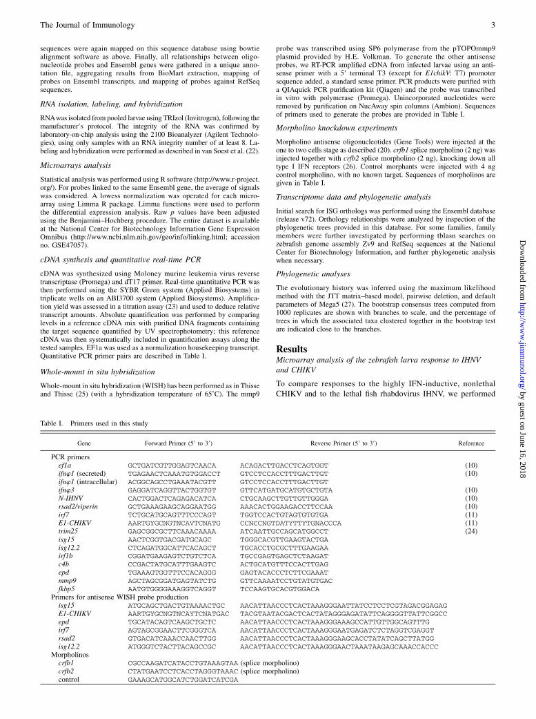
Table I. Primers used in this study
Gene Forward Primer (59 to 39) Reverse Primer (59 to 39) Reference
PCR primersef1a GCTGATCGTTGGAGTCAACA ACAGACTTGACCTCAGTGGT (10)ifnw1 (secreted) TGAGAACTCAAATGTGGACCT GTCCTCCACCTTTGACTTGT (10)ifnw1 (intracellular) ACGGCAGCCTGAAATACGTT GTCCTCCACCTTTGACTTGTifnw3 GAGGATCAGGTTACTGGTGT GTTCATGATGCATGTGCTGTA (10)N-IHNV CACTGGACTCAGAGACATCA CTGCAAGCTTGTTGTTGGGA (10)rsad2/viperin GCTGAAAGAAGCAGGAATGG AAACACTGGAAGACCTTCCAA (10)irf7 TCTGCATGCAGTTTCCCAGT TGGTCCACTGTAGTGTGTGA (11)E1-CHIKV AARTGYGCNGTNCAVTCNATG CCNCCNGTDATYTTYTGNACCCA (11)trim25 GAGCGGCGCTTCAAACAAAA ATCAATTGCCAGCATGGCCT (24)isg15 AACTCGGTGACGATGCAGC TGGGCACGTTGAAGTACTGAisg12.2 CTCAGATGGCATTCACAGCT TGCACCTGCGCTTTGAAGAAirf1b CGGATGAAGAGTCTGTCTCA TGCCGAGTGAGCTCTAAGATc4b CCGACTATGCATTTGAAGTC ACTGCATGTTTCCACTTGAGepd TGAAAGTGGTTTCCACAGGG GAGTACACCCTCTTCGAAATmmp9 AGCTAGCGGATGAGTATCTG GTTCAAAATCCTGTATGTGACfkbp5 AATGTGGGGAAAGGTCAGGT TCCAAGTGCACGTGGACA
Primers for antisense WISH probe productionisg15 ATGCAGCTGACTGTAAAACTGC AACATTAACCCTCACTAAAGGGAATTATCCTCCTCGTAGACGGAGAGE1-CHIKV AARTGYGCNGTNCAYTCNATGAC TACGTAATACGACTCACTATAGGGAGATATTCAGGGGTTATTCGGCCepd TGCATACAGTCAAGCTGCTC AACATTAACCCTCACTAAAGGGAAAGCCATTGTTGGCAGTTTGirf7 AGTAGCGGAACTTCGGGTCA AACATTAACCCTCACTAAAGGGAATGAGATCTCTAGGTCGAGGTrsad2 GTGACATCAAACCAACTTGG AACATTAACCCTCACTAAAGGGAAGCACCTATATCAGCTTATGGisg12.2 ATGGGTCTACTTACAGCCGC AACATTAACCCTCACTAAAGGGAACTAAATAAGAGCAAACCACCC
Morpholinoscrfb1 CGCCAAGATCATACCTGTAAAGTAA (splice morpholino)crfb2 CTATGAATCCTCACCTAGGGTAAAC (splice morpholino)control GAAAGCATGGCATCTGGATCATCGA
The Journal of Immunology 3
by guest on June 16, 2018http://w
ww
.jimm
unol.org/D
ownloaded from

Table II. Selection of up- and downregulated genes after CHIKV and /or INHV infection
Symbol Description Ensembl Gene IDCHIKV FC andAdjusted p Value
IHNV FC andAdjusted p Value
Upregulated genesIFN response
isg12-2 IFN-stimulated gene 12-2 ENSDARG00000039243 143.9*** 6.7*isg12-1 IFN-stimulated gene 12-1 ENSDARG00000017489 83.0*** 4.8*isg15 IFN-stimulated gene 15 ENSDARG00000086374 40.2*** 5.4*rsad2 Radical S-adenosyl methionine domain
containing 2; viperinENSDARG00000004952 9.8** 3.8*
IFITd3 IFN-induced (tetratricopeptide) ENSDARG00000071012 5.3** 2.7*IFITc IFN-induced (tetratricopeptide) ENSDARG00000057173 6.5* 1.6dram1 Damage-regulated autophagy; vig6 ENSDARG00000045561 5.0** 0.6*ifi35 IFN-induced protein 35 ENSDARG00000075643 3.6** 1.1isg12-4 IFN-stimulated gene 12-4 ENSDARG00000078389 7.3** 2.0isg12-3 IFN-stimulated gene 12-3 ENSDARG00000074217 13.7*** 1.639UTR isg12-7 Similar to IFN-stimulated gene 12-7 A_15_P118496 4.1** 1.9*Ifi44l IFN-induced protein 44-like ENSDARG00000010729 9.1** 1.7*mxa IFN-induced GTP-binding protein MxA ENSDARG00000021688 9.8** 2.1*mxe IFN-induced GTP-binding protein MxE ENSDARG00000014427 8.2** 2.5*
ComplementC4b Complement component 4B ENSDARG00000038424 5.1*** 3.0*
MMP and signalingmmp9 Matrix metalloproteinase 9 ENSDARG00000042816 2.7* 7.6**CD9c CD9 ENSDARG00000044990 34.3*** 4.0*junBb junB proto-oncogene ENSDARG00000088371 1.7* 3.2**Unc93b1 Unc-93 homolog B1 ENSDARG00000069114 0.56* 4.9*
Transcription factorsirf1b IFN regulatory factor 1 (annotated irf11) ENSDARG00000032768 7.1*** 1.8*irf7 IFN regulatory factor 7 ENSDARG00000045661 11.0*** 2.0*Stat1b STAT1b ENSDARG00000076182 5.9** 1.7Socs1 Suppressor of cytokine signaling 1 ENSDARG00000038095 3.6* 1.2cebpb CCAAT/enhancer binding protein ENSDARG00000042725 1.3* 5.9**
Oxidative response and mitochondriandufv1 NADH deH ubiquinone flavoprotein 1 ENSDARG00000036438 0.37*** 4.4*ndufv3 NADH deH ubiquinone flavoprotein 3 ENSDART00000113587 0.38*** 2.6*etfa Electron transfer flavoprotein a ENSDARG00000043944 0.48*** 3.0*mt-tRNA tRNA on mitochondrial genome ENSDARG00000083519 1.5** 3.7**mt-tRNA tRNA on mitochondrial genome ENSDARG00000082716 0.9 3.3**mt-atp8 ATP synthase protein 8 ENSDARG00000063910 0.6* 4.2**mt-co3 Cytochrome c oxidase subunit 3 ENSDARG00000063912 1.4* 4.0**mt-co1 Cytochrome c oxidase subunit 1 ENSDARG00000063905 1.6** 3.2**mt-cyb Cytochrome b ENSDARG00000063924 0.6** 2.9**
Secreted factors and binding proteinslgals3bpb-l Similar to galectin-3–binding protein ENSDARG00000040528 3.7** 0.8lgals3bpb Galectin 3–binding protein ENSDART00000101829 4.0** 0.68*lgals9l2 Galectin 9–like protein ENSDARG00000041060 3.0** 1.3lgals9l1 Galectin 9–like protein ENSDARG00000025903 2.7** 1.2lect2l Leukocyte cell–derived chemotaxin 2 ENSDARG00000033227 3.4*** 1.1CXCL-C1c CXC chemokine ENSDARG00000075045 2.6** 1.3*CXCL-C13b CXC chemokine ENSDARG00000074487 3.1** 1.3*CCL-C5a CC chemokine ENSDARG00000058389 3.0** 1.4
MHC and proteasomeb2m b2-microglobulin ENSDARG00000053136 7.7*** 1.1Tpsn Tapasin ENSDARG00000079402 3.4** 0.9Tpsn Tapasin ENSDARG00000045011 2.8** 1.04psme2 Proteasome activator subunit 2 ENSDARG00000033144 2.5** 1psma6b Proteasome subunit, a type, 6b ENSDARG00000043899 2.5** 1.4*psmb10 Proteasome subunit, b type, 10 ENSDARG00000001656 2.9** 1.1psmb11 Proteasome b 11 subunit ENSDARG00000031885 3.8** 1psmf1 Proteasome inhibitor subunit 1 ENSDARG00000022652 3.0*** 0.9
Trimtrim25 Tripartite motif-containing 25 ENSDARG00000015865 3.1*** 1.1ftr-like finTRIM-related protein; ftr80 ENSDARG00000067661 3.0** 1.1
Translationrpl23a Ribosomal protein L23a ENSDARG00000006316 0.9 2.9**rpl32 Ribosomal protein L32 ENSDARG00000054818 1.4** 2.6**rpl34 Ribosomal protein L34 ENSDARG00000029500 1.1 2.5**rpl35a Ribosomal protein L35a A_15_P115161 1.5* 2.6**rpl37 Ribosomal protein L37 LOLS06369 1.0 2.9**rpl39 Ribosomal protein L39 ENSDARG00000036316 1.0 2.6*rpl9 Rribosomal protein L9 ENSDARG00000037350 1.1 2.6**rps8 Ribosomal protein S8 ENSDARG00000055996 1.0 2.8**rps5 Ribosomal protein S5 ENSDARG00000079026 1.1 2.5**rps12 Ribosomal protein S12 ENSDARG00000036875 1.0 3.8**
(Table continues)
4 ISG EVOLUTION FROM ZEBRAFISH TO HUMANS
by guest on June 16, 2018http://w
ww
.jimm
unol.org/D
ownloaded from

a transcriptome analysis of infected zebrafish larvae. Based on ourprevious data (10, 11), we chose 48 h postinfection (hpi) as thetime point that would allow us to detect a well-developed responseto both viruses at a comparable stage of development. At thisstage, IHNV-infected larvae are not moribund yet, so that thetranscriptome analysis should not mainly reflect the general dis-tress leading to death.These experiments required infecting a large number of larvae,
which was performed by i.v. microinjections in our previous
studies (10, 11). Because this is challenging and time-consuming,we tested alternative infection methods that would still lead to theexpected outcome in a reproductive fashion. Immersion of 3 dpflarvae in water containing either virus did not yield infection.However, in the case of CHIKV, we knew that i.v. inoculationresults in infection of the yolk syncytial cell along various other celltypes (11). This prompted us to test direct injection of CHIKV inthe yolk, which is easier and faster than i.v. injection. We observedthat both routes of inoculation led to similar patterns, as shown in
Table II. (Continued )
Symbol Description Ensembl Gene IDCHIKV FC andAdjusted p Value
IHNV FC andAdjusted p Value
rps15 39UTR of ribosomal protein gene A_15_P109626 0.4*** 7.7*rps24 Ribosomal protein S24 isoform (intron) A_15_P120710 0.7* 2.6**rpsa Ribosomal protein S40 ENSDARG00000019181 2.6** 1.6*elf1g Elongation factor 1-g A_15_P110585 0.48** 2.7**sars Seryl-tRNA synthetase ENSDARG00000008237 0.7** 5.1**
Enzymesmov10b.1 Putative helicase mov-10 ENSDARG00000061177 6.6*** 1.5zgc:154040 Putative phospholipase A2 ENSDARG00000071326 11.4** 1.4ctssb1 Cathepsin S.b.1 ENSDARG00000075176 4.3** 0.5*gpx1b Glutathione peroxidase 1b ENSDARG00000006207 3.6*** 0.8cmpk2 Thymidylate kinase LPS-inducible (tyki) ENSDARG00000031359 3.5** 1.7rnasel3 RNase-like 3 ENSDARG00000036171 3.3*** 1.1adam8a Metalloproteinase domain 8a ENSDARG00000001452 2.6** 0.5**atp2a1 ATPase, Ca2+ transporting, fast twitch 1 ENSDARG00000020574 0.8* 5.4**tgm1 Transglutaminase 1 ENSDARG00000019024 1.0 3.0**nat8l N-acetyltransferase 8-like (EC 2.3.1.-) ENSDARG00000077256 0.5*** 2.9*pfkfb3 6-Phosphofructo-2-kinase ENSDARG00000001953 1.2 2.5*
ApolipoproteinsapoC1l Apolipoprotein C1–like A_15_P115491 0.2*** 8.0*zgc:193613 Apolipoprotein like ENSDARG00000015866 0.8 3.6**apoa1 Apolipoprotein A-I precursor ENSDARG00000012076 0.4** 2.6**
Dowregulated genesIFN responseprkrir Protein kinase, IFN-inducible
dsRNA-dependent inhibitor, repressorENSDARG00000042489 0.8** 0.3**
bril Bone-restricted IFITM-like protein(aka IFITM5)
ENSDARG00000091273 0.3*** 0.4*
Oxydative and inflammatory responsefkbp5 FK506 binding protein 5 ENSDARG00000028396 1.0 0.06**dhrs1 Dehydrogenase/reductase 1 (SDR family)
member 1ENSDARG00000054300 0.3*** 1.8
pla2g12b Phospholipase A2, group XIIB ENSDARG00000015662 0.3*** 2.4*prss35 Protease, serine, 35 ENSDARG00000017213 0.3*** 1.1dhrs1 Dehydrogenase/reductase 1 ENSDARG00000054300 0.3*** 1.8zgc:64031 Abhydrolase domain–containing 14B ENSDARG00000023880 0.3* 1.6ucmab Unique cartilage matrix-associated protein ENSDARG00000005485 0.2*** 2.4cyp26b1 Cytochrome P450 26B1 (Cyp26B1) ENSDARG00000077121 1.0 0.3**hamp2 Hepcidin-2 precursor ENSDARG00000053227 1.9 0.3*
Othersepd Ependymin precursor ENSDARG00000014745 0.7 0.26**mcam Melanoma cell adhesion molecule ENSDARG00000005368 0.9 0.27**fbln5 Fibulin 5 ENSDARG00000039273 0.7** 0.29**vsg1 Vessel-specific 1 ENSDARG00000045003 1.7** 0.17**tppp3 Tubulin polymerization-promoting ENSDARG00000030463 0.7* 0.31**acta2 Actin, a2 ENSDARG00000045180 0.9 0.32**pppde2a PPPDE peptidase domain–containing 2a ENSDARG00000033140 1.0 0.32**meox1 Mesenchyme homeobox 1 ENSDARG00000007891 0.8* 0.32**rcn3 Reticulocalbin 3, EF-hand calcium binding ENSDARG00000037961 0.6** 0.34*mad2l1 MAD2 mitotic arrest deficient-like 1 ENSDARG00000004713 0.9* 0.33**zgc:113183 SWI/SNF-related containing DEAD/Hbox 1 ENSDARG00000014041 0.8 0.33**kctd12.2 Potassium channel tetramerization ENSDARG00000053542 1.1 0.34**taf6 TAF6 RNA polymerase II, TATA box BP ENSDARG00000029135 0.7* 0.34**il13ra2 IL-13R, a2 ENSDARG00000039436 0.8 0.35*col8a2 Collagen, type VIII, a2 ENSDARG00000060893 0.7* 0.35*slc25a27 Solute carrier family 25. member 27 ENSDARG00000042873 0.55*** 0.35**gli2a GLI-Kr€uppel family member GLI2a ENSDARG00000025641 0.91 0.36**mthfd2 Methylenetetrahydrofolate dehydrogenase 2 ENSDARG00000026666 0.7** 0.36**cx23 Connexin 23 ENSDARG00000054150 0.75 0.38*
FC , 0.5 and FC . 2 are in bold.Adjusted p values: *p , 0.01–0.05, **p , 0.005–0.01; ***p , 0.001–0.005.
The Journal of Immunology 5
by guest on June 16, 2018http://w
ww
.jimm
unol.org/D
ownloaded from
Supplemental Fig. 1A. We also found that yolk injection of CHIKVresulted in only slightly lower levels of expression of the CHIKVE1 transcript compared with i.v. injection, and to a slightly lower,but still very strong, upregulation of ifnw1 transcripts, with similarkinetics (Supplemental Fig. 1B), Thus, we chose to perform yolkinjections for the microarray experiments. In contrast, injection ofIHNV in the yolk did not result in infection, and we had to performi.v. injections.For each virus, three independent experiments were performed,
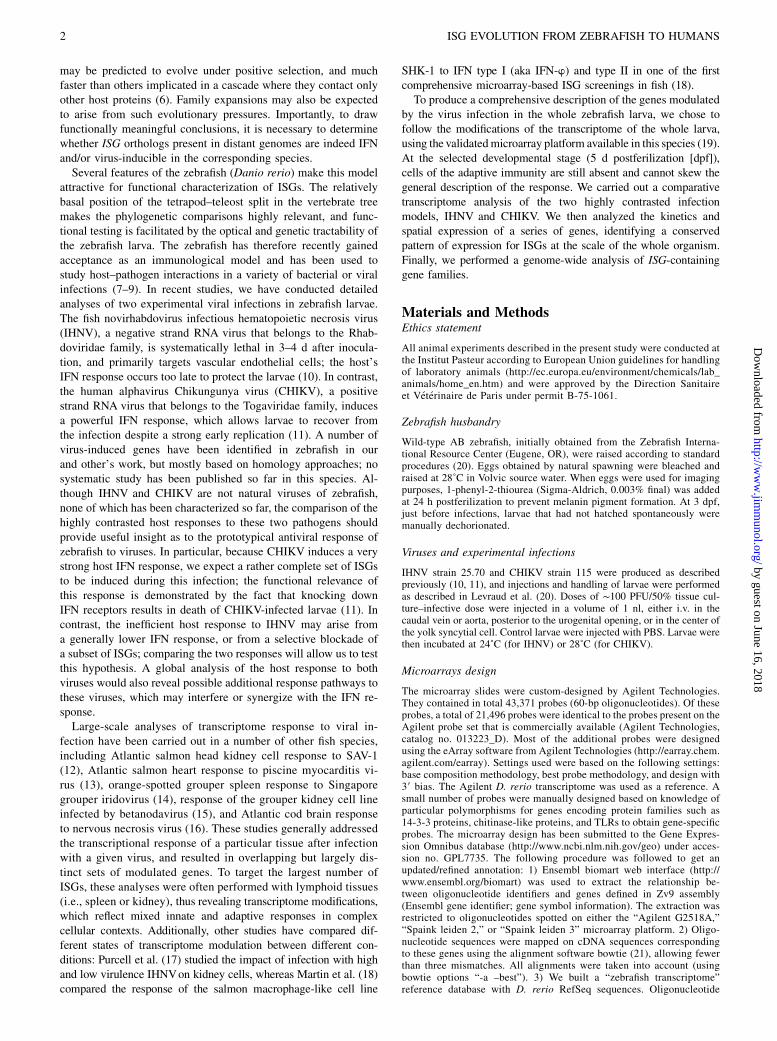
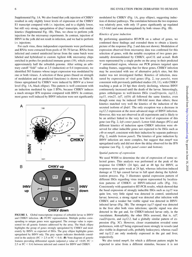
and RNAs were extracted from pools of 30–70 larvae. RNAs frominfected and control uninfected larvae from the same batch werelabeled and hybridized to custom Agilent 44K microarray chipsenriched in probes for predicted immune genes (19), which coversapproximately half the zebrafish genome. After setting an arbi-trary cutoff “fold” value at 2.5 (induction) or 0.4 (repression), weidentified 583 features whose target expression was modulated byone or both viruses. A selection of these genes (based on strengthof modulation and on predicted functions) is shown on Table II.Genes upregulated by CHIKV were induced by IHNV at a lowerlevel (Fig. 1A, black ellipse). This pattern is well consistent withan induction mediated by type I IFNs, because CHIKV inducesa much stronger IFN response compared with IHNV. In contrast,most genes well induced by IHNV infection were not significantly
modulated by CHIKV (Fig. 1A, gray ellipse), suggesting induc-tion of distinct pathways. The correlation between the two responseswas relatively poor, with only 15 genes significantly up regulated.2.5-fold (and none downregulated) by both viruses (Fig. 1B).
Kinetics of gene induction
By performing quantitative RT-PCR on a subset of genes, weconfirmed these findings and extended them to obtain a kineticpicture of the response (Fig. 2 and data not shown). Modulation ofexpression observed from microarray data was confirmed for thisselection of genes, with the notable exception of two genes pre-sumably downmodulated by CHIKV, apoC1L and ucmab. Bothwere represented by a single probe on the array in their predicted39 untranslated region, whereas our PCR primers targeted openreading frames, suggesting that for these genes, CHIKV infectionmay be associated with use of alternative polyA sites, but thismatter was not investigated further. Kinetics of infection, mea-sured by expression of viral genes (Fig. 2, top panels), wereconsistent with previously published studies (10, 11), with CHIKVreaching a peak at 24 hpi then slowly declining, whereas IHNVcontinuously increased until the death of the larvae. Interestingly,genes orthologous to well-known ISGs (rsad2/viperin, isg12.2,isg15, trim25, irf7, irf1b) all followed the same kinetics, eventhough some may be induced 100-fold more than others; thesekinetics matched very well the kinetics of the induction of thesecreted isoform of ifnf1. The only exception was a decrease inisg12.2 expression at the most advanced stage of IHNV infection.However, this was not observed in all experiments and is likely tobe an artifact linked to the very low level of expression of thisgene (see Fig. 2, left center panel). Lower fold changes (FCs) anddifferent kinetics of induction upon CHIKV infection were ob-served for other inducible genes not expected to be ISGs such asc4b or mmp9, consistent with their induction by separate pathways(Fig. 2, middle bottom panel). The kinetics of gene induction byIHNV infection are more complex, but suggest that mmp9 wasupregulated early and did not show the delay observed for the IFNresponse (see Fig. 2, right panel center and bottom).
Spatial patterns of expression
We used WISH to determine the site of expression of some se-lected genes. This analysis was performed at the peak of theresponse for CHIKV (24 hpi), and at 48 hpi for IHNV, asresponses were quite weak at 24 hpi, whereas infection-induceddamage at 72 hpi caused larvae to fall apart during the hybrid-ization process. Fig. 3 illustrates spatial expression pattern ofdifferent ISGs regarding virus tropism represented by localiza-tion patterns of CHIKV- or IHNV-infected cells (Fig. 3A).Consistently with quantitative RT-PCR results, which showed thatthe basal expression of strongly inducible ISGs such as isg15 wasquite low, very little signal was detected in control, uninfectedlarvae; however, a strong signal was detected after infection withCHIKV, and a weaker but visible signal was detected in IHNV-infected larvae (Fig. 3B). The strongest isg15 signal was detectedin the liver after both virus infections; specific signal was alsodetected in the gut and, for CHIKV-infected larvae only, in thevasculature. Remarkably, the other ISGs assessed, that is, irf7,rsad2/viperin, and isg12.2, had a globally similar pattern of ex-pression (Fig. 3C). However, closer examination revealed somepreferences, with irf7 being expressed more strongly in vessels (andalso visible in dispersed cells, probably leukocytes), whereas rsad2and isg12.2 are only modestly expressed in the gut and liver,respectively.We also tested mmp9, for which a different pattern might be
expected to arise from a different stimulus, because it is not
FIGURE 1. Global transcriptome response of zebrafish larvae to IHNV
and CHIKV infection. (A) FC/FC representation. Multiple probes corre-
sponding to unique genes were aggregated. The average value is repre-
sented for all genetic features addressed by the array. The black ellipse
highlights the group of genes strongly upregulated by CHIKV and mod-
erately by IHNV, as expected of ISGs. The gray ellipse highlights genes
upregulated by IHNV only. The gray square indicates thresholds consid-
ered in the analysis (FC . 2.5 or FC , 0.4). (B) Venn diagram of genetic
features providing differential signals (adjusted p value of ,0.05; FC .2.5 or FC , 0.4) between infected and control for IHNV and CHIKV.
6 ISG EVOLUTION FROM ZEBRAFISH TO HUMANS
by guest on June 16, 2018http://w
ww
.jimm
unol.org/D
ownloaded from

a typical ISG. Indeed, the pattern of mmp9 expression induced byIHNV was strikingly different from that of ISGs, with a staining ofsuperficial cells, probably in the epidermis (Fig. 3D). Finally, wealso tested a few downmodulated genes. Although no clear pattern ofexpression for fkbp5 could be observed (not shown), a striking patternwas observed in uninfected larvae for the epd gene encoding epen-dymin (Fig. 3E), in agreement with the pattern available on theZebrafish International Resource Center Web site (http://zfin.org/cgi-bin/webdriver?MIval=aa-fxfigureview.apg&OID=ZDB-FIG-070503-464) (28): a subset of cells in the head (and possibly in the endocrinepancreas) expresses epd. Despite the gene name, these cells do notline brain ventricles and are thus not ependymal; instead, they arelocated on the brain external surface, apparently ensheathing someblood vessels. IHNV infection induced a dramatic reduction in thenumber of epd-expressing cells, especially in the dorsal cranium.This, consistent with the infection patterns (see figure 2D of Ref. 10),suggests that epd-expressing cells are targeted and killed by the virus.Similarly, vsg-1, an endothelium-specific gene (29), is down-modulated by IHNV, and this expression was lost in many vesselsin IHNV-infected larvae (not shown), in accordance with ourprevious finding that vascular endothelium is a primary target ofIHNV (10).
Pathway analysis of the responses
IFN-related pathways. Analysis of the list of modulated genesby GenMAPP and Ingenuity Pathway Analysis (IPA) identifiedseveral biological pathways induced by the viral infections. IFNsignaling was found enriched by IPA for both viruses, althoughwith more genes involved and a lower p value for CHIKV; type I
IFNs were predicted as main upstream regulators in both cases.Many genes previously characterized as ISGs or homologous towell-known human ISGs were found to be highly upregulated byCHIKV, and to a lesser extent also by IHNV (Fig. 4, Table II).Analysis of genes presumably involved in signaling pathways up-stream of IFN induction was not as straightforward, probablyreflecting that activation of these pathways involves posttransla-tional modifications more often than transcriptional modulation.The detection of viruses may happen either in the cytosolic com-partment of cells via the RIG-I–like receptor (RLR) pathway, or inendosomal compartments via the TLR pathway. We have previ-ously implicated the RLR pathway in the zebrafish response toCHIKV, because knockdown of MAVS/IPS-1, a central adaptor ofthis pathway, was found to strongly reduce IFN induction (11). Nomodulation of the mavs transcript was seen with either virus, whichwas consistent with previous observations (30, 31); this was also thecase for most genes involved in the RLR pathway (SupplementalFig. 2), with the notable exceptions of trim25, isg15, and irf7, allorthologs of known ISGs, involved in signaling feedback loops. Asimilar picture emerged from the analysis of the TLR pathway(Supplemental Fig. 2); neither virus modulated most genes in thispathway. A few genes induced by IHNV included proteins typicallyinvolved in responses to bacteria rather than to viruses (e.g., mmp9).Again, this suggests that in addition to the typical virus-inducedIFN response, IHNV triggers other inflammatory pathways.Conspicuously absent from the list of IHNV- or CHIKV-
induced genes were the type I IFNs themselves, ifnw1–4. Themicroarray did not include probes for ifnw3 and ifnw4; ifnw4 doesnot appear to be involved in the antiviral response (32), and we
FIGURE 2. Kinetics of transcriptomic responses. Quantitative RT-PCR analysis; mean 6 SEM from three pools of three to five larvae per point; one
representative experiment out of two or more is shown. Left panels show basal expression in uninfected larvae raised at 28˚C; absolute quantifications were
normalized to ef1a expression. Center and right panels display expression measured in CHIK- and IHNV-infected larvae, respectively (i.v. infections in
both cases); in this case, results are reported as the ratio of expression in infected larvae to uninfected controls. The expression of viral genes (CHIKV-E1 or
N-IHNV), which are not detectable in uninfected larva, is shown on the top panels. In this case, normalization was made by dividing the measured copy
number arbitrarily by 1000 copies/106 ef1a copies.
The Journal of Immunology 7
by guest on June 16, 2018http://w
ww
.jimm
unol.org/D
ownloaded from

have found previously ifnw2 to be expressed by adult zebrafish,but not at the larval stage (26, 33). The apparent lack of induc-tion of ifnw1 is explained by the existence of two alternativetranscripts of this gene: a constitutively expressed transcriptencoding a putatively nonfunctional protein lacking a signalpeptide, and an inducible transcript encoding the active cytokine(32). Because the ifnw1-specific probes on the microarray do notdiscriminate between the two forms, the induction of the activecytokine transcript has been likely masked by the constitutiveexpression of the alternative transcript; indeed, as can be seen inFig. 2, the poorly inducible, nonsecreted isoform has an ∼30-foldhigher basal expression level than the secreted isoform.
Other pathways (e.g., redox, mmp, liver X receptor). Althoughtypical inflammatory pathways such as “airway pathology andpulmonary diseases” or “granzyme A” were found by IPA fromthe CHIKV response in addition to IFN signaling, additionalpathways were revealed as significantly enriched in the responseto IHNV: 1) the EIF2 signaling pathway, which refers to the up-regulation of a number of ribosomal protein genes (SupplementalFig. 3), many of which were upregulated by IHNV infection.Although it is generally accepted that virus infection inducesa shut-off of cell protein production, this pattern might eitherrepresent a compensatory transcriptional response or a virus-directed modification of cell syntheses beneficial to the patho-gen production (34). 2) The pathway “mitochondrial dysfunction”was also pointed out after IHNV infection; this is due to the up-regulation of eight genes encoded by the mitochondrial genomeand/or involved in redox regulation, and to the strong downregu-lation of fkbp5 in IHNV-infected larvae. Notably, two of them(ndufva and etfa) were downregulated in CHIKV-infected fish. 3)The liver X receptor/retinoid X receptor pathway was also high-
lighted after IHNV infection, comprising genes upregulatedby both viruses such as complement genes or mmp9, but alsothe apolipoproteins, which were modulated by IHNVonly, andphospholipase A2, which was induced by IHNV and repressedby CHIKV. Finally, “liver necrosis, cell death and inflammation”was among the top “tox lists” of IPA for both viruses, whichcould be related to the strong cytopathic effect of these viruses,as well as to the spatial expression pattern of modulated genes(see above).
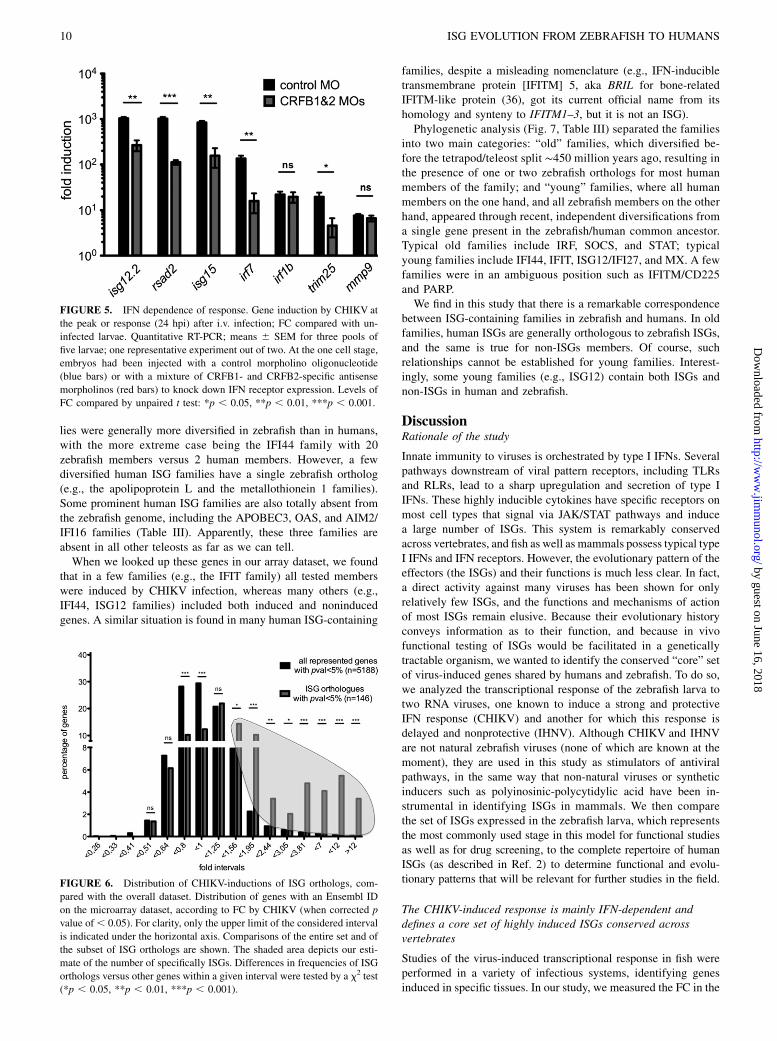
IFN signaling is involved in the upregulation of keyvirus-induced genes
Genes strongly induced by CHIKV, but less so by IHNV, arepresumably induced by the strong and protective IFN response inCHIKV-infected fish; indeed, most of these genes are orthologousto well-known mammalian ISGs. To check the IFN dependency ofthe upregulation observed during the infection for a selection ofgenes, we knocked down the two zebrafish type I IFN receptors (26)using antisense morpholinos, infected larvae with CHIKV, andmeasured gene expression by quantitative RT-PCR at the peak ofthe response. When compared with control morphants (Fig. 5),induction of genes such as rsad2/viperin, isg12.2, isg15, trim25,and irf7 was severely reduced, indicating that their induction wasIFN-dependent, as expected. In contrast, mmp9 and irf1b inductionappeared to be IFN-independent. This was expected for mmp9, butis surprising for irf1b, the human ortholog of which is an ISG.Therefore, we should be aware that induction by CHIKV does notnecessarily equate induction by type I IFN, even for ISG orthologs.Nevertheless, it does seem to be the case for most of the genes wetested, which prompted us to perform a genome-wide analysis ofthe orthologs of human ISGs.
FIGURE 3. Spatial expression patterns. WISH. Probes are indicated in bottom right of each panel; infection type is indicated in top right corner. For
CHIKV infections, analysis was performed at 24 hpi; for IHNV, at 48 hpi. (A) Viral genes, used as an example of the typical distribution of infected cells.
The uninfected control for the N-ihnv probe is not shown, but no signal is visible (as previously shown in Ref. 10). (B) Expression of isg15 chosen as
a typical ISG more strongly induced by both viruses, but more strongly by CHIKV. Uninfected control is shown at 4 dpf (5 dpf controls do not show any
signal either). (C) Expression of three other ISGs (irf7, rsad2, and isg12.2) in control (4 dpf) and CHIKV-infected larvae. (D) Expression of mmp9, a non-
ISG induced more strongly by IHNV than CHIKV. Middle panel, Close-up of the tail region. Control shown at 5 dpf. (E) Expression of epd, a gene
downregulated by IHNV. Head regions: lateral views on left, dorsal views on right. Uninfected control is shown at 5 dpf. Scale bars, 250 mm.
8 ISG EVOLUTION FROM ZEBRAFISH TO HUMANS
by guest on June 16, 2018http://w
ww
.jimm
unol.org/D
ownloaded from
Genomic and expression analysis of ISG orthologs
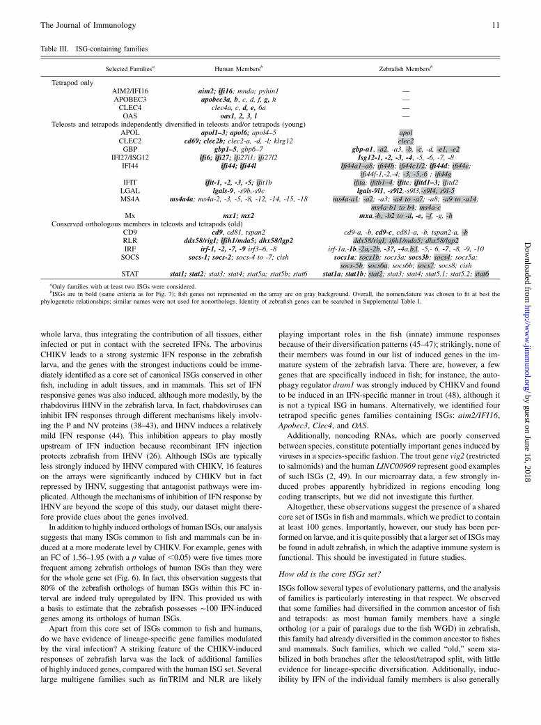
Are zebrafish genes orthologous to human ISGs generally IFN in-ducible? Although our initial GenMAPP and IPA analysis identifieda number of genes as being induced downstream of type I IFNs(displayed in Fig. 4), there are many other, less well-known ISGs.We took advantage of the exhaustive human ISG list compiled bySchoggins et al. (2) based on a compilation of a dozen humanmicroarray studies. We searched the zebrafish genome for theorthologs of these genes, with the help of the phylogeny tools atEnsembl.org (release 72); the results of this analysis are pro-vided as a searchable Excel file with links to gene pages(Supplemental Table I). This table includes 445 human genes forwhich a total of 593 zebrafish orthologs were found, of which 280were represented on the microarray.As expected, we found within this dataset the genes highly
induced by the viral infection. However, a significant enrichmentwas detected even among genes with modest levels of induction(Fig. 6). Also, the proportion of genes reproducibly modulatedbetween replicates (corrected p , 0.05, regardless of the actualFC) was higher within ISG orthologs (146 of 280 genes) com-pared with the whole dataset (5,188 of 12,524) (x2 test, p ,0.001). Thus, in addition to the well-known, strongly induced
ISGs, a large number of modestly inducible genes should also beincluded in the core vertebrate group of IFN (or rather, virus)-inducible genes. These observations led us to propose a tentativeestimation of the size of the core set of ISGs conserved betweenzebrafish and humans; subtracting the global distribution of in-duction of CHIKV-induced genes to the distribution of inductionof zebrafish orthologs of human ISGs, one obtains a roughassessment of 53 ISGs represented on the microarray (Fig. 6,highlighted area). Given that approximately half of genes arerepresented on the array, both for the entire dataset and for the ISGorthologs, the order of magnitude of the size of the core ISGsubset shared by fishes and mammals would be 100 genes.
Analysis of ISG-containing multigene families
A large number of mammalian ISGs constitute multigene families;Table III shows that it is also the case in the zebrafish. Phyloge-netic relationships between human ISGs and their zebrafishorthologs indicate that these genes are prone to extensive dupli-cation in each evolutionary branch: 35 examples of “many-to-many” orthology relationships can be observed in SupplementalTable I, which are ∼7-fold more frequent than for the overallgenome comparison (233 such relationships in total) (35). Fami-
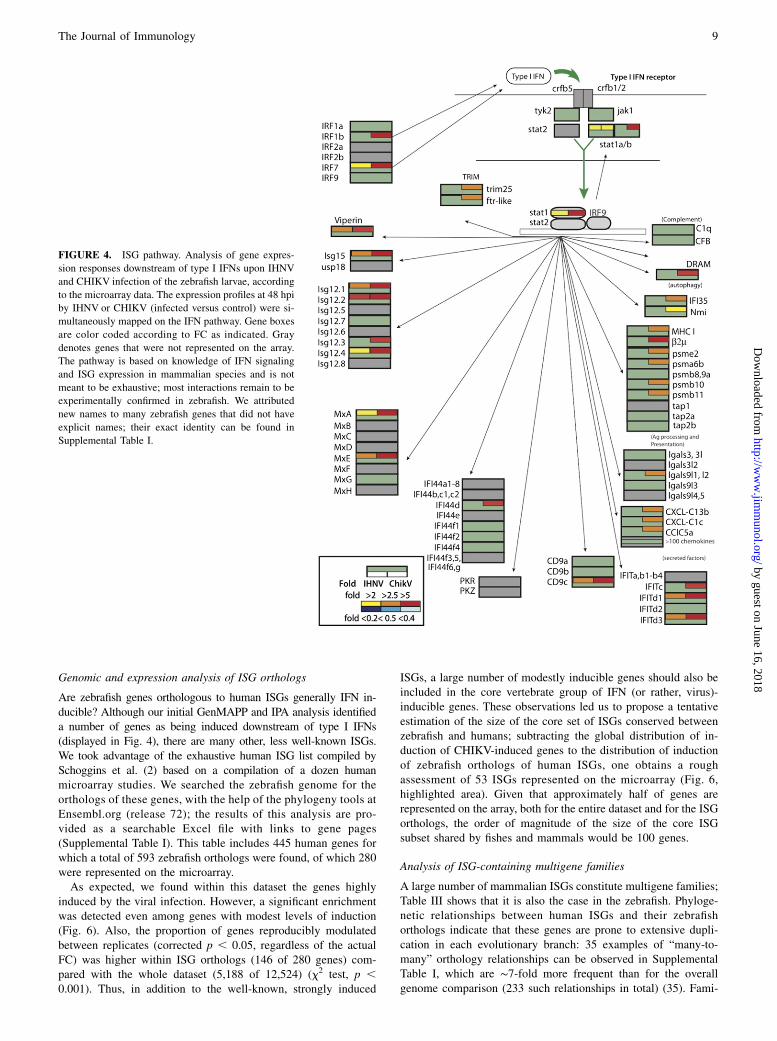
FIGURE 4. ISG pathway. Analysis of gene expres-
sion responses downstream of type I IFNs upon IHNV
and CHIKV infection of the zebrafish larvae, according
to the microarray data. The expression profiles at 48 hpi
by IHNV or CHIKV (infected versus control) were si-
multaneously mapped on the IFN pathway. Gene boxes
are color coded according to FC as indicated. Gray
denotes genes that were not represented on the array.
The pathway is based on knowledge of IFN signaling
and ISG expression in mammalian species and is not
meant to be exhaustive; most interactions remain to be
experimentally confirmed in zebrafish. We attributed
new names to many zebrafish genes that did not have
explicit names; their exact identity can be found in
Supplemental Table I.
The Journal of Immunology 9
by guest on June 16, 2018http://w
ww
.jimm
unol.org/D
ownloaded from
lies were generally more diversified in zebrafish than in humans,with the more extreme case being the IFI44 family with 20zebrafish members versus 2 human members. However, a fewdiversified human ISG families have a single zebrafish ortholog(e.g., the apolipoprotein L and the metallothionein 1 families).Some prominent human ISG families are also totally absent fromthe zebrafish genome, including the APOBEC3, OAS, and AIM2/IFI16 families (Table III). Apparently, these three families areabsent in all other teleosts as far as we can tell.When we looked up these genes in our array dataset, we found
that in a few families (e.g., the IFIT family) all tested memberswere induced by CHIKV infection, whereas many others (e.g.,IFI44, ISG12 families) included both induced and noninducedgenes. A similar situation is found in many human ISG-containing
families, despite a misleading nomenclature (e.g., IFN-inducibletransmembrane protein [IFITM] 5, aka BRIL for bone-relatedIFITM-like protein (36), got its current official name from itshomology and synteny to IFITM1–3, but it is not an ISG).Phylogenetic analysis (Fig. 7, Table III) separated the families
into two main categories: “old” families, which diversified be-fore the tetrapod/teleost split ∼450 million years ago, resulting inthe presence of one or two zebrafish orthologs for most humanmembers of the family; and “young” families, where all humanmembers on the one hand, and all zebrafish members on the otherhand, appeared through recent, independent diversifications froma single gene present in the zebrafish/human common ancestor.Typical old families include IRF, SOCS, and STAT; typicalyoung families include IFI44, IFIT, ISG12/IFI27, and MX. A fewfamilies were in an ambiguous position such as IFITM/CD225and PARP.We find in this study that there is a remarkable correspondence
between ISG-containing families in zebrafish and humans. In oldfamilies, human ISGs are generally orthologous to zebrafish ISGs,and the same is true for non-ISGs members. Of course, suchrelationships cannot be established for young families. Interest-ingly, some young families (e.g., ISG12) contain both ISGs andnon-ISGs in human and zebrafish.
DiscussionRationale of the study
Innate immunity to viruses is orchestrated by type I IFNs. Severalpathways downstream of viral pattern receptors, including TLRsand RLRs, lead to a sharp upregulation and secretion of type IIFNs. These highly inducible cytokines have specific receptors onmost cell types that signal via JAK/STAT pathways and inducea large number of ISGs. This system is remarkably conservedacross vertebrates, and fish as well as mammals possess typical typeI IFNs and IFN receptors. However, the evolutionary pattern of theeffectors (the ISGs) and their functions is much less clear. In fact,a direct activity against many viruses has been shown for onlyrelatively few ISGs, and the functions and mechanisms of actionof most ISGs remain elusive. Because their evolutionary historyconveys information as to their function, and because in vivofunctional testing of ISGs would be facilitated in a geneticallytractable organism, we wanted to identify the conserved “core” setof virus-induced genes shared by humans and zebrafish. To do so,we analyzed the transcriptional response of the zebrafish larva totwo RNA viruses, one known to induce a strong and protectiveIFN response (CHIKV) and another for which this response isdelayed and nonprotective (IHNV). Although CHIKV and IHNVare not natural zebrafish viruses (none of which are known at themoment), they are used in this study as stimulators of antiviralpathways, in the same way that non-natural viruses or syntheticinducers such as polyinosinic-polycytidylic acid have been in-strumental in identifying ISGs in mammals. We then comparethe set of ISGs expressed in the zebrafish larva, which representsthe most commonly used stage in this model for functional studiesas well as for drug screening, to the complete repertoire of humanISGs (as described in Ref. 2) to determine functional and evolu-tionary patterns that will be relevant for further studies in the field.
The CHIKV-induced response is mainly IFN-dependent anddefines a core set of highly induced ISGs conserved acrossvertebrates
Studies of the virus-induced transcriptional response in fish wereperformed in a variety of infectious systems, identifying genesinduced in specific tissues. In our study, we measured the FC in the
FIGURE 5. IFN dependence of response. Gene induction by CHIKV at
the peak or response (24 hpi) after i.v. infection; FC compared with un-
infected larvae. Quantitative RT-PCR; means 6 SEM for three pools of
five larvae; one representative experiment out of two. At the one cell stage,
embryos had been injected with a control morpholino oligonucleotide
(blue bars) or with a mixture of CRFB1- and CRFB2-specific antisense
morpholinos (red bars) to knock down IFN receptor expression. Levels of
FC compared by unpaired t test: *p , 0.05, **p , 0.01, ***p , 0.001.
FIGURE 6. Distribution of CHIKV-inductions of ISG orthologs, com-
pared with the overall dataset. Distribution of genes with an Ensembl ID
on the microarray dataset, according to FC by CHIKV (when corrected p
value of, 0.05). For clarity, only the upper limit of the considered interval
is indicated under the horizontal axis. Comparisons of the entire set and of
the subset of ISG orthologs are shown. The shaded area depicts our esti-
mate of the number of specifically ISGs. Differences in frequencies of ISG
orthologs versus other genes within a given interval were tested by a x2 test
(*p , 0.05, **p , 0.01, ***p , 0.001).
10 ISG EVOLUTION FROM ZEBRAFISH TO HUMANS
by guest on June 16, 2018http://w
ww
.jimm
unol.org/D
ownloaded from
whole larva, thus integrating the contribution of all tissues, eitherinfected or put in contact with the secreted IFNs. The arbovirusCHIKV leads to a strong systemic IFN response in the zebrafishlarva, and the genes with the strongest inductions could be imme-diately identified as a core set of canonical ISGs conserved in otherfish, including in adult tissues, and in mammals. This set of IFNresponsive genes was also induced, although more modestly, by therhabdovirus IHNV in the zebrafish larva. In fact, rhabdoviruses caninhibit IFN responses through different mechanisms likely involv-ing the P and NV proteins (38–43), and IHNV induces a relativelymild IFN response (44). This inhibition appears to play mostlyupstream of IFN induction because recombinant IFN injectionprotects zebrafish from IHNV (26). Although ISGs are typicallyless strongly induced by IHNV compared with CHIKV, 16 featureson the arrays were significantly induced by CHIKV but in factrepressed by IHNV, suggesting that antagonist pathways were im-plicated. Although the mechanisms of inhibition of IFN response byIHNV are beyond the scope of this study, our dataset might there-fore provide clues about the genes involved.In addition to highly induced orthologs of human ISGs, our analysis
suggests that many ISGs common to fish and mammals can be in-duced at a more moderate level by CHIKV. For example, genes withan FC of 1.56–1.95 (with a p value of ,0.05) were five times morefrequent among zebrafish orthologs of human ISGs than they werefor the whole gene set (Fig. 6). In fact, this observation suggests that80% of the zebrafish orthologs of human ISGs within this FC in-terval are indeed truly upregulated by IFN. This provided us witha basis to estimate that the zebrafish possesses ∼100 IFN-inducedgenes among its orthologs of human ISGs.Apart from this core set of ISGs common to fish and humans,
do we have evidence of lineage-specific gene families modulatedby the viral infection? A striking feature of the CHIKV-inducedresponses of zebrafish larva was the lack of additional familiesof highly induced genes, compared with the human ISG set. Severallarge multigene families such as finTRIM and NLR are likely
playing important roles in the fish (innate) immune responsesbecause of their diversification patterns (45–47); strikingly, none oftheir members was found in our list of induced genes in the im-mature system of the zebrafish larva. There are, however, a fewgenes that are specifically induced in fish; for instance, the auto-phagy regulator dram1 was strongly induced by CHIKVand foundto be induced in an IFN-specific manner in trout (48), although itis not a typical ISG in humans. Alternatively, we identified fourtetrapod specific genes families containing ISGs: aim2/IFI16,Apobec3, Clec4, and OAS.Additionally, noncoding RNAs, which are poorly conserved
between species, constitute potentially important genes induced byviruses in a species-specific fashion. The trout gene vig2 (restrictedto salmonids) and the human LINC00969 represent good examplesof such ISGs (2, 49). In our microarray data, a few strongly in-duced probes apparently hybridized in regions encoding longcoding transcripts, but we did not investigate this further.Altogether, these observations suggest the presence of a shared
core set of ISGs in fish and mammals, which we predict to containat least 100 genes. Importantly, however, our study has been per-formed on larvae, and it is quite possibly that a larger set of ISGsmaybe found in adult zebrafish, in which the adaptive immune system isfunctional. This should be investigated in future studies.
How old is the core ISGs set?
ISGs follow several types of evolutionary patterns, and the analysisof families is particularly interesting in that respect. We observedthat some families had diversified in the common ancestor of fishand tetrapods: as most human family members have a singleortholog (or a pair of paralogs due to the fish WGD) in zebrafish,this family had already diversified in the common ancestor to fishesand mammals. Such families, which we called “old,” seem sta-bilized in both branches after the teleost/tetrapod split, with littleevidence for lineage-specific diversification. Additionally, induc-ibility by IFN of the individual family members is also generally
Table III. ISG-containing families
Selected Familiesa Human Membersb Zebrafish Membersb
Tetrapod onlyAIM2/IFI16 aim2; ifi16; mnda; pyhin1 —APOBEC3 apobec3a, b, c, d, f, g, h —CLEC4 clec4a, c, d, e, 6a —OAS oas1, 2, 3, l —
Teleosts and tetrapods independently diversified in teleosts and/or tetrapods (young)APOL apol1–3; apol6; apol4–5 apolCLEC2 cd69; clec2b; clec2-a, -d, -l; klrg12 clec2GBP gbp1–5, gbp6–7 gbp-a1, -a2, -a3, -b, -c, -d, -e1, -e2
IFI27/ISG12 ifi6; ifi27; ifi27l1; ifi27l2 Isg12-1, -2, -3, -4, -5, -6, -7, -8IFI44 ifi44; ifi44l Ifi44a1–a8; ifi44b; ifi44c1/2; ifi44d; ifi44e;
ifi44f-1,-2,-4; -3, -5,-6 ; ifi44gIFIT ifit-1, -2, -3, -5; ifit1b ifita; ifitb1–4; ifitc; ifitd1–3; ifitd2LGAL lgals-9, -s9b,-s9c lgals-9l1, -s9l2,-s9l3,-s9l4, s9l-5MS4A ms4a4a; ms4a-2, -3, -5, -8, -12, -14, -15, -18 ms4a-a1; -a2; -a3; -a4 to -a7; -a8; -a9 to -a14;
ms4a-b1 to b4; ms4a-cMx mx1; mx2 mxa,-b, -b2 to -d, -e, –f, -g, -h
Conserved orthologous members in teleosts and tetrapods (old)CD9 cd9, cd81, tspan2 cd9-a, -b, cd9-c, cd81-a, -b, tspan2-a, -bRLR ddx58/rigI; ifih1/mda5; dhx58/lgp2 ddx58/rigI; ifih1/mda5; dhx58/lgp2IRF irf-1, -2, -7, -9 irf3–6, -8 irf-1a,-1b,-2a,-2b, -3?, -4a,b,l, -5,- 6, -7, -8, -9, -10SOCS socs-1; socs-2; socs-4 to -7; cish socs1a; socs1b; socs3a; socs3b; socs4; socs5a;
socs-5b; socs6a; socs6b; socs7; socs8; cishSTAT stat1; stat2; stat3; stat4; stat5a; stat5b; stat6 stat1a; stat1b; stat2; stat3; stat4; stat5.1; stat5.2; stat6
aOnly families with at least two ISGs were considered.bISGs are in bold (same criteria as for Fig. 7); fish genes not represented on the array are on gray background. Overall, the nomenclature was chosen to fit at best the
phylogenetic relationships; similar names were not used for nonorthologs. Identity of zebrafish genes can be searched in Supplemental Table I.
The Journal of Immunology 11
by guest on June 16, 2018http://w
ww
.jimm
unol.org/D
ownloaded from
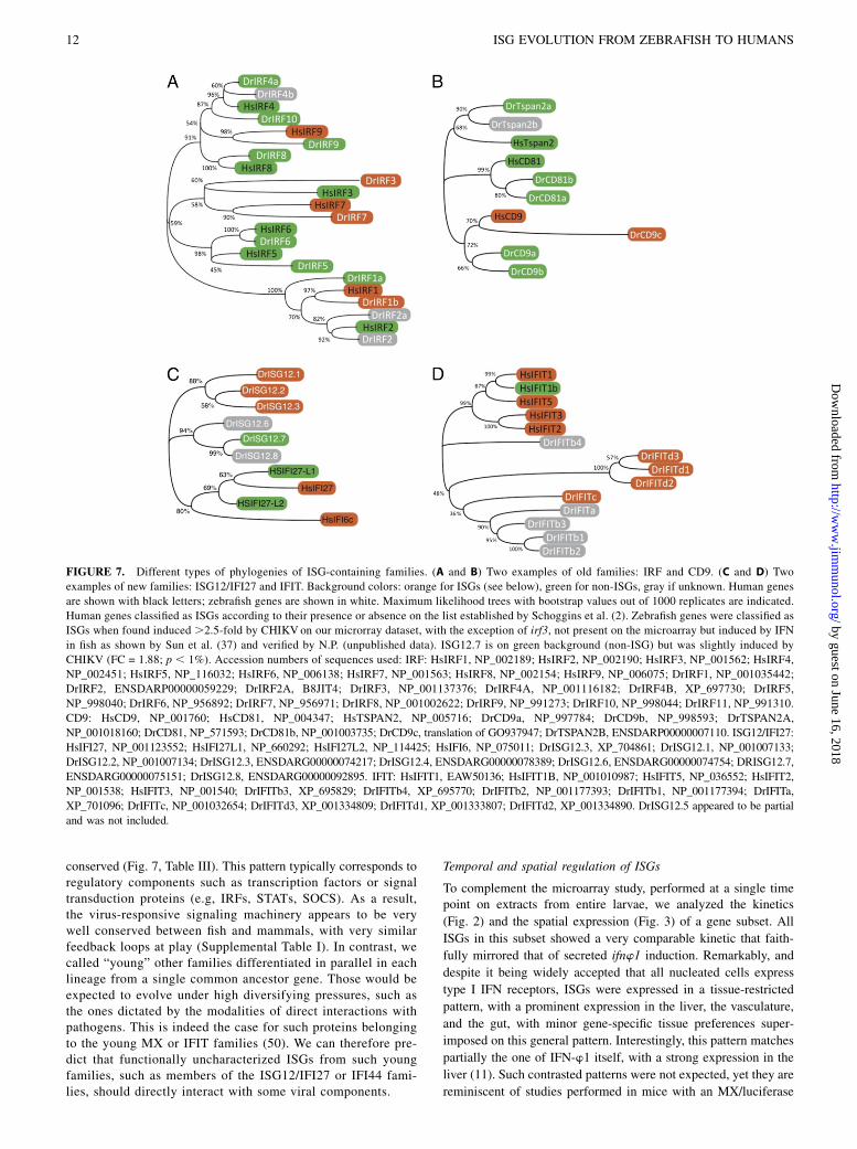
conserved (Fig. 7, Table III). This pattern typically corresponds toregulatory components such as transcription factors or signaltransduction proteins (e.g, IRFs, STATs, SOCS). As a result,the virus-responsive signaling machinery appears to be verywell conserved between fish and mammals, with very similarfeedback loops at play (Supplemental Table I). In contrast, wecalled “young” other families differentiated in parallel in eachlineage from a single common ancestor gene. Those would beexpected to evolve under high diversifying pressures, such asthe ones dictated by the modalities of direct interactions withpathogens. This is indeed the case for such proteins belongingto the young MX or IFIT families (50). We can therefore pre-dict that functionally uncharacterized ISGs from such youngfamilies, such as members of the ISG12/IFI27 or IFI44 fami-lies, should directly interact with some viral components.
Temporal and spatial regulation of ISGs
To complement the microarray study, performed at a single timepoint on extracts from entire larvae, we analyzed the kinetics
(Fig. 2) and the spatial expression (Fig. 3) of a gene subset. All
ISGs in this subset showed a very comparable kinetic that faith-
fully mirrored that of secreted ifnw1 induction. Remarkably, and
despite it being widely accepted that all nucleated cells express
type I IFN receptors, ISGs were expressed in a tissue-restricted
pattern, with a prominent expression in the liver, the vasculature,
and the gut, with minor gene-specific tissue preferences super-
imposed on this general pattern. Interestingly, this pattern matches
partially the one of IFN-w1 itself, with a strong expression in the
liver (11). Such contrasted patterns were not expected, yet they are
reminiscent of studies performed in mice with an MX/luciferase
FIGURE 7. Different types of phylogenies of ISG-containing families. (A and B) Two examples of old families: IRF and CD9. (C and D) Two
examples of new families: ISG12/IFI27 and IFIT. Background colors: orange for ISGs (see below), green for non-ISGs, gray if unknown. Human genes
are shown with black letters; zebrafish genes are shown in white. Maximum likelihood trees with bootstrap values out of 1000 replicates are indicated.
Human genes classified as ISGs according to their presence or absence on the list established by Schoggins et al. (2). Zebrafish genes were classified as
ISGs when found induced .2.5-fold by CHIKVon our microrray dataset, with the exception of irf3, not present on the microarray but induced by IFN
in fish as shown by Sun et al. (37) and verified by N.P. (unpublished data). ISG12.7 is on green background (non-ISG) but was slightly induced by
CHIKV (FC = 1.88; p , 1%). Accession numbers of sequences used: IRF: HsIRF1, NP_002189; HsIRF2, NP_002190; HsIRF3, NP_001562; HsIRF4,
NP_002451; HsIRF5, NP_116032; HsIRF6, NP_006138; HsIRF7, NP_001563; HsIRF8, NP_002154; HsIRF9, NP_006075; DrIRF1, NP_001035442;
DrIRF2, ENSDARP00000059229; DrIRF2A, B8JIT4; DrIRF3, NP_001137376; DrIRF4A, NP_001116182; DrIRF4B, XP_697730; DrIRF5,
NP_998040; DrIRF6, NP_956892; DrIRF7, NP_956971; DrIRF8, NP_001002622; DrIRF9, NP_991273; DrIRF10, NP_998044; DrIRF11, NP_991310.
CD9: HsCD9, NP_001760; HsCD81, NP_004347; HsTSPAN2, NP_005716; DrCD9a, NP_997784; DrCD9b, NP_998593; DrTSPAN2A,
NP_001018160; DrCD81, NP_571593; DrCD81b, NP_001003735; DrCD9c, translation of GO937947; DrTSPAN2B, ENSDARP00000007110. ISG12/IFI27:
HsIFI27, NP_001123552; HsIFI27L1, NP_660292; HsIFI27L2, NP_114425; HsIFI6, NP_075011; DrISG12.3, XP_704861; DrISG12.1, NP_001007133;
DrISG12.2, NP_001007134; DrISG12.3, ENSDARG00000074217; DrISG12.4, ENSDARG00000078389; DrISG12.6, ENSDARG00000074754; DRISG12.7,
ENSDARG00000075151; DrISG12.8, ENSDARG00000092895. IFIT: HsIFIT1, EAW50136; HsIFIT1B, NP_001010987; HsIFIT5, NP_036552; HsIFIT2,
NP_001538; HsIFIT3, NP_001540; DrIFITb3, XP_695829; DrIFITb4, XP_695770; DrIFITb2, NP_001177393; DrIFITb1, NP_001177394; DrIFITa,
XP_701096; DrIFITc, NP_001032654; DrIFITd3, XP_001334809; DrIFITd1, XP_001333807; DrIFITd2, XP_001334890. DrISG12.5 appeared to be partial
and was not included.
12 ISG EVOLUTION FROM ZEBRAFISH TO HUMANS
by guest on June 16, 2018http://w
ww
.jimm
unol.org/D
ownloaded from

reporter gene (51), where liver was observed to respond much morestrongly than other organs to injected IFN or to Thogoto virusinfection. Thus, strongly organ-biased expression of ISGs may wellbe the norm among vertebrates. It remains to be established whetherthis reflects local availability of IFNs or differential responsivenessof other organs.
Alternative pathways at play during IHNV infection
Whereas CHIKV infection induced a strong and protective IFNresponse, IHNV infection invariably leads to the death of thezebrafish larva. Many genes are induced by the INHV infection butpoorly or not induced by CHIKV. Their upregulation is likelylinked to the activation of IFN-independent pathways. This may belargely caused by extensive cell death due to viral cytopathicactivity and subsequent inflammation.Many mitochondrial genes were induced by IHNV, suggesting
that this virus induces a general mitochondrial response potentiallylinked to apoptosis and oxidative reaction. Interestingly, a cellularoxidative response can suppress the IFN-a–induced assembly ofSTAT factors at the ISG promoters, thus potentially contributing tothe knockdown of the IFN-induced response (52). Such an oxi-dative response may also be responsible for the induction of mmp9in epidermal cells that we observed by ISH, similar to what hasbeen observed during bacterial infections (53).A variety of ribosomal proteins are also modulated by IHNV,
possibly in response to translational shut-off of host proteinsachieved by rhabdoviruses. A downregulation of protein syn-thesis by IHNV infection have been reported before in rainbowtrout (44).Fkbp5 was strongly downregulated by the IHNV infection,
although it is a mammalian ISG, and it is induced in chicken bythe Newcastle disease virus (54). In fact, fkbp5 is probably notan ISG in zebrafish, because it is not induced by CHIKV (nor,for that matter, is fkbp4, another possible ortholog of humanFKBP5). FKBP5 links glucocorticoids to NF-kB signalingduring viral infection (55), but its role in antiviral responses isstill elusive. This immunophilin with peptidyl-prolyl-isomeraseactivity reminds one of cyclophilins, which are other targets ofcyclosporine A and may be either deleterious or beneficialfor virus infections (56). Whether FKBP5 downregulation has apositive or a negative impact on the response to IHNV remainsto be determined.Our study identified ependymin, a marker specific of brain
perivascular cells, as a gene repressed by the IHNV, but not byCHIKV. The progressive reduction of expression during infectionsuggested that peripheral brain cells in which it was expressedmight be targeted and killed by the virus. The WISH supportedthis notion, because the infected larvae lost completely ependyminexpression in the dorsal cranium. Thus, transcriptome survey maylead not only to the identification of responding cells throughlocalization of gene upregulation, but may also reveal virus cel-lular targets by extinction of specific markers.In conclusion, our study characterizes the contrasted tran-
scriptional responses of zebrafish to a fish rhabdovirus, IHNV, anda human arbovirus, CHIKV. The strong type I IFN response in-duced by CHIKV allowed us to greatly extend the list of zebrafishISGs, many of which belong to multigene families. Their differentevolutionary stories reflect their functional implication in antiviralpathways. Our transcriptome survey also led to the characteriza-tion of the spatial expression pattern of key up- or downregulatedmarkers, providing an important reference set for the study of host–virus interactions in zebrafish, a powerful model where one canperform gene silencing, mutagenesis, or drug screening.
AcknowledgmentsWe thank Hannah Volkman for the mmp9 probe and Abdenour Benmansour
for helpful discussions.
DisclosuresThe authors have no financial conflicts of interest.
References1. Der, S. D., A. Zhou, B. R. Williams, and R. H. Silverman. 1998. Identification of
genes differentially regulated by interferon a, b, or g using oligonucleotidearrays. Proc. Natl. Acad. Sci. USA 95: 15623–15628.
2. Schoggins, J. W., S. J. Wilson, M. Panis, M. Y. Murphy, C. T. Jones, P. Bieniasz,and C. M. Rice. 2011. A diverse range of gene products are effectors of the type Iinterferon antiviral response. Nature 472: 481–485.
3. Sadler, A. J., and B. R. G. Williams. 2008. Interferon-inducible antiviral effec-tors. Nat. Rev. Immunol. 8: 559–568.
4. Schoggins, J. W., and C. M. Rice. 2011. Interferon-stimulated genes and theirantiviral effector functions. Curr. Opin. Virol. 1: 519–525.
5. Brass, A. L., I.-C. Huang, Y. Benita, S. P. John, M. N. Krishnan, E. M. Feeley,B. J. Ryan, J. L. Weyer, L. van der Weyden, E. Fikrig, et al. 2009. The IFITMproteins mediate cellular resistance to influenza A H1N1 virus, West Nile virus,and dengue virus. Cell 139: 1243–1254.
6. Flajnik, M., and L. Du Pasquier. 2013. Evolution of the immune system. InFundamental Immunology, 7th Ed. W. Paul, ed. Wolters Kluwer and LippincottWillians & Wilkins, New York, p. 67–128.
7. Kanther, M., and J. F. Rawls. 2010. Host-microbe interactions in the developingzebrafish. Curr. Opin. Immunol. 22: 10–19.
8. Meijer, A. H., and H. P. Spaink. 2011. Host-pathogen interactions made trans-parent with the zebrafish model. Curr. Drug Targets 12: 1000–1017.
9. Tobin, D. M., F. J. Roca, S. F. Oh, R. McFarland, T. W. Vickery, J. P. Ray,D. C. Ko, Y. Zou, N. D. Bang, T. T. H. Chau, et al. 2012. Host genotype-specifictherapies can optimize the inflammatory response to mycobacterial infections.Cell 148: 434–446.
10. Ludwig, M., N. Palha, C. Torhy, V. Briolat, E. Colucci-Guyon, M. Bremont,P. Herbomel, P. Boudinot, and J.-P. Levraud. 2011. Whole-body analysis ofa viral infection: vascular endothelium is a primary target of infectious hema-topoietic necrosis virus in zebrafish larvae. PLoS Pathog. 7: e1001269.
11. Palha, N., F. Guivel-Benhassine, V. Briolat, G. Lutfalla, M. Sourisseau, F. Ellett,C.-H. Wang, G. J. Lieschke, P. Herbomel, O. Schwartz, and J.-P. Levraud. 2013.Real-time whole-body visualization of Chikungunya virus infection and hostinterferon response in zebrafish. PLoS Pathog. 9: e1003619.
12. Herath, T. K., J. E. Bron, K. D. Thompson, J. B. Taggart, A. Adams,J. H. Ireland, and R. H. Richards. 2012. Transcriptomic analysis of the hostresponse to early stage salmonid alphavirus (SAV-1) infection in Atlantic salmonSalmo salar L. Fish Shellfish Immunol. 32: 796–807.
13. Timmerhaus, G., A. Krasnov, P. Nilsen, M. Alarcon, S. Afanasyev, M. Rode,H. Takle, and S. M. Jørgensen. 2011. Transcriptome profiling of immuneresponses to cardiomyopathy syndrome (CMS) in Atlantic salmon. BMCGenomics 12: 459.
14. Huang, Y., X. Huang, Y. Yan, J. Cai, Z. Ouyang, H. Cui, P. Wang, and Q. Qin.2011. Transcriptome analysis of orange-spotted grouper (Epinephelus coioides)spleen in response to Singapore grouper iridovirus. BMC Genomics 12: 556.
15. Lu, M.-W., F.-H. Ngou, Y.-M. Chao, Y.-S. Lai, N.-Y. Chen, F.-Y. Lee, andP. P. Chiou. 2012. Transcriptome characterization and gene expression of Epi-nephelus spp in endoplasmic reticulum stress-related pathway during betano-davirus infection in vitro. BMC Genomics 13: 651.
16. Krasnov, A., Ø. Kileng, S. Skugor, S. M. Jørgensen, S. Afanasyev,G. Timmerhaus, A.-I. Sommer, and I. Jensen. 2013. Genomic analysis of the hostresponse to nervous necrosis virus in Atlantic cod (Gadus morhua) brain. Mol.Immunol. 54: 443–452.
17. Purcell, M. K., I. S. Marjara, W. Batts, G. Kurath, and J. D. Hansen. 2011. Tran-scriptome analysis of rainbow trout infected with high and low virulence strains ofinfectious hematopoietic necrosis virus. Fish Shellfish Immunol. 30: 84–93.
18. Martin, S. A. M., J. B. Taggart, P. Seear, J. E. Bron, R. Talbot, A. J. Teale,G. E. Sweeney, B. Høyheim, D. F. Houlihan, D. R. Tocher, et al. 2007. Interferontype I and type II responses in an Atlantic salmon (Salmo salar) SHK-1 cell lineby the salmon TRAITS/SGP microarray. Physiol. Genomics 32: 33–44.
19. Stockhammer, O. W., A. Zakrzewska, Z. Hegedus, H. P. Spaink, andA. H. Meijer. 2009. Transcriptome profiling and functional analyses of thezebrafish embryonic innate immune response to Salmonella infection. J.Immunol. 182: 5641–5653.
20. Levraud, J.-P., E. Colucci-Guyon, M. J. Redd, G. Lutfalla, and P. Herbomel. 2008.In vivo analysis of zebrafish innate immunity. Methods Mol. Biol. 415: 337–363.
21. Langmead, B., C. Trapnell, M. Pop, and S. L. Salzberg. 2009. Ultrafast andmemory-efficient alignment of short DNA sequences to the human genome.Genome Biol. 10: R25.
22. van Soest, J. J., O. W. Stockhammer, A. Ordas, G. V. Bloemberg, H. P. Spaink,and A. H. Meijer. 2011. Comparison of static immersion and intravenous in-jection systems for exposure of zebrafish embryos to the natural pathogenEdwardsiella tarda. BMC Immunol. 12: 58.
23. Lutfalla, G., and G. Uze. 2006. Performing quantitative reverse-transcribedpolymerase chain reaction experiments. Methods Enzymol. 410: 386–400.
The Journal of Immunology 13
by guest on June 16, 2018http://w
ww
.jimm
unol.org/D
ownloaded from
24. Boudinot, P., L. M. van der Aa, L. Jouneau, L. Du Pasquier, P. Pontarotti,V. Briolat, A. Benmansour, and J.-P. Levraud. 2011. Origin and evolution ofTRIM proteins: new insights from the complete TRIM repertoire of zebrafishand pufferfish. PLoS ONE 6: e22022.
25. Thisse, C., and B. Thisse. 2008. High-resolution in situ hybridization to whole-mount zebrafish embryos. Nat. Protoc. 3: 59–69.
26. Aggad, D., M. Mazel, P. Boudinot, K. E. Mogensen, O. J. Hamming,R. Hartmann, S. Kotenko, P. Herbomel, G. Lutfalla, and J.-P. Levraud. 2009. Thetwo groups of zebrafish virus-induced interferons signal via distinct receptorswith specific and shared chains. J. Immunol. 183: 3924–3931.
27. Tamura, K., D. Peterson, N. Peterson, G. Stecher, M. Nei, and S. Kumar. 2011.MEGA5: molecular evolutionary genetics analysis using maximum likelihood,evolutionary distance, and maximum parsimony methods. Mol. Biol. Evol. 28:2731–2739.
28. Thisse, B., V. Heyer, A. Lux, V. Alunni, A. Degrave, I. Seiliez, J. Kirchner,J.-P. Parkhill, and C. Thisse. 2004. Spatial and temporal expression of thezebrafish genome by large-scale in situ hybridization screening. MethodsCell Biol. 77: 505–519.
29. Covassin, L., J. D. Amigo, K. Suzuki, V. Teplyuk, J. Straubhaar, andN. D. Lawson. 2006. Global analysis of hematopoietic and vascular endothelialgene expression by tissue specific microarray profiling in zebrafish. Dev. Biol.299: 551–562.
30. Biacchesi, S., M. LeBerre, A. Lamoureux, Y. Louise, E. Lauret, P. Boudinot, andM. Bremont. 2009. Mitochondrial antiviral signaling protein plays a major rolein induction of the fish innate immune response against RNA and DNA viruses.J. Virol. 83: 7815–7827.
31. Lauksund, S., T. Svingerud, V. Bergan, and B. Robertsen. 2009. Atlantic salmonIPS-1 mediates induction of IFNa1 and activation of NF-kB and localizes tomitochondria. Dev. Comp. Immunol. 33: 1196–1204.
32. Levraud, J.-P., P. Boudinot, I. Colin, A. Benmansour, N. Peyrieras, P. Herbomel,and G. Lutfalla. 2007. Identification of the zebrafish IFN receptor: implicationsfor the origin of the vertebrate IFN system. J. Immunol. 178: 4385–4394.
33. Langevin, C., E. Aleksejeva, G. Passoni, N. Palha, J.-P. Levraud, andP. Boudinot. 2013. The antiviral innate immune response in fish: evolution andconservation of the IFN system. J. Mol. Biol. 425: 4904–4920.
34. Lyles, D. S. 2000. Cytopathogenesis and inhibition of host gene expression byRNA viruses. Microbiol. Mol. Biol. Rev. 64: 709–724.
35. Howe, K., M. D. Clark, C. F. Torroja, J. Torrance, C. Berthelot, M. Muffato,J. E. Collins, S. Humphray, K. McLaren, L. Matthews, et al. 2013. The zebrafishreference genome sequence and its relationship to the human genome. Nature496: 498–503.
36. Moffatt, P., M.-H. Gaumond, P. Salois, K. Sellin, M.-C. Bessette, E. Godin,P. T. de Oliveira, G. J. Atkins, A. Nanci, and G. Thomas. 2008. Bril: a novelbone-specific modulator of mineralization. J. Bone Miner. Res. 23: 1497–1508.
37. Sun, B., B. Robertsen, Z. Wang, and B. Liu. 2009. Identification of an Atlanticsalmon IFN multigene cluster encoding three IFN subtypes with very differentexpression properties. Dev. Comp. Immunol. 33: 547–558.
38. Thoulouze, M. I., E. Bouguyon, C. Carpentier, and M. Bremont. 2004. Essentialrole of the NV protein of Novirhabdovirus for pathogenicity in rainbow trout. J.Virol. 78: 4098–4107.
39. Vidy, A., M. Chelbi-Alix, and D. Blondel. 2005. Rabies virus P protein interacts withSTAT1 and inhibits interferon signal transduction pathways. J. Virol. 79: 14411–14420.
40. Brzozka, K., S. Finke, and K.-K. Conzelmann. 2006. Inhibition of interferonsignaling by rabies virus phosphoprotein P: activation-dependent binding ofSTAT1 and STAT2. J. Virol. 80: 2675–2683.
41. Chelbi-Alix, M. K., A. Vidy, J. El Bougrini, and D. Blondel. 2006. Rabies viralmechanisms to escape the IFN system: the viral protein P interferes with IRF-3,Stat1, and PML nuclear bodies. J. Interferon Cytokine Res. 26: 271–280.
42. Choi, M. K., C. H. Moon, M. S. Ko, U. H. Lee, W. J. Cho, S. J. Cha, J. W. Do,G. J. Heo, S. G. Jeong, Y. S. Hahm, et al. 2011. A nuclear localization of theinfectious haematopoietic necrosis virus NV protein is necessary for optimalviral growth. PLoS ONE 6: e22362.
43. Kim, M. S., and K. H. Kim. 2013. The role of viral hemorrhagic septicemia virus(VHSV) NV gene in TNF-a- and VHSV infection-mediated NF-kB activation.Fish Shellfish Immunol. 34: 1315–1319.
44. MacKenzie, S., J. C. Balasch, B. Novoa, L. Ribas, N. Roher, A. Krasnov, andA. Figueras. 2008. Comparative analysis of the acute response of the trout,O. mykiss, head kidney to in vivo challenge with virulent and attenuatedinfectious hematopoietic necrosis virus and LPS-induced inflammation. BMCGenomics 9: 141.
45. Laing, K. J., M. K. Purcell, J. R. Winton, and J. D. Hansen. 2008. A genomicview of the NOD-like receptor family in teleost fish: identification of a novelNLR subfamily in zebrafish. BMC Evol. Biol. 8: 42.
46. Stein, C., M. Caccamo, G. Laird, and M. Leptin. 2007. Conservation and di-vergence of gene families encoding components of innate immune responsesystems in zebrafish. Genome Biol. 8: R251.
47. van der Aa, L. M., J.-P. Levraud, M. Yahmi, E. Lauret, V. Briolat, P. Herbomel,A. Benmansour, and P. Boudinot. 2009. A large new subset of TRIM geneshighly diversified by duplication and positive selection in teleost fish. BMC Biol.7: 7.
48. O’Farrell, C., N. Vaghefi, M. Cantonnet, B. Buteau, P. Boudinot, andA. Benmansour. 2002. Survey of transcript expression in rainbow trout leuko-cytes reveals a major contribution of interferon-responsive genes in the earlyresponse to a rhabdovirus infection. J. Virol. 76: 8040–8049.
49. Boudinot, P., S. Salhi, M. Blanco, and A. Benmansour. 2001. Viral haemorrhagicsepticaemia virus induces vig-2, a new interferon-responsive gene in rainbowtrout. Fish Shellfish Immunol. 11: 383–397.
50. Kochs, G., and O. Haller. 1999. GTP-bound human MxA protein interacts withthe nucleocapsids of Thogoto virus (Orthomyxoviridae). J. Biol. Chem. 274:4370–4376.
51. Pulverer, J. E., U. Rand, S. Lienenklaus, D. Kugel, N. Zietara, G. Kochs,R. Naumann, S. Weiss, P. Staeheli, H. Hauser, and M. Koster. 2010. Temporaland spatial resolution of type I and III interferon responses in vivo. J. Virol. 84:8626–8638.
52. Di Bona, D., M. Cippitelli, C. Fionda, C. Camma, A. Licata, A. Santoni, andA. Craxı. 2006. Oxidative stress inhibits IFN-a-induced antiviral gene expres-sion by blocking the JAK-STAT pathway. J. Hepatol. 45: 271–279.
53. Hall, C. J., R. H. Boyle, J. W. Astin, M. V. Flores, S. H. Oehlers, L. E. Sanderson,F. Ellett, G. J. Lieschke, K. E. Crosier, and P. S. Crosier. 2013. Immunores-ponsive gene 1 augments bactericidal activity of macrophage-lineage cells byregulating b-oxidation-dependent mitochondrial ROS production. Cell Metab.18: 265–278.
54. Park, M.-S., J. Steel, A. Garcıa-Sastre, D. Swayne, and P. Palese. 2006. Engi-neered viral vaccine constructs with dual specificity: avian influenza and New-castle disease. Proc. Natl. Acad. Sci. USA 103: 8203–8208.
55. Karin, M., and Y. Ben-Neriah. 2000. Phosphorylation meets ubiquitination: thecontrol of NF-kB activity. Annu. Rev. Immunol. 18: 621–663.
56. He, H., D. Zhou, W. Fan, X. Fu, J. Zhang, Z. Shen, J. Li, J. Li, and Y. Wu. 2012.Cyclophilin A inhibits rotavirus replication by facilitating host IFN-I production.Biochem. Biophys. Res. Commun. 422: 664–669.
14 ISG EVOLUTION FROM ZEBRAFISH TO HUMANS
by guest on June 16, 2018http://w
ww
.jimm
unol.org/D
ownloaded from