contrast-enhanced ct- and mri-based perfusion … · contrast-enhanced ct- and mri-based perfusion...
TRANSCRIPT
Contrast-enhanced CT- and MRI-based perfusion assessment for pulmonary diseases: basics and clinical applications
Yoshiharu OhnoHisanobu KoyamaHo Yun LeeSachiko MiuraTakeshi YoshikawaKazuro Sugimura
Matched distribution of regional pulmonary blood flow (perfusion) and ventilation is required for pulmonary ventilation and perfusion assessment to proceed effi-ciently (1). A large amount of ventilation in lungs is matched by corresponding-
ly high perfusion. In addition, the balance between pulmonary perfusion and ventilation differs according to the physiopathology of various pulmonary diseases, and the normal pattern of pulmonary blood flow often changes, sometimes exacerbating the disturbance in gas exchange (2, 3). Therefore, assessment of these regional perfusion pattern changes is important to understand the pulmonary pathophysiology of various pulmonary diseases. Multiple methods are currently available to quantitatively and qualitatively evaluate pulmo-nary perfusion in patients with pulmonary diseases.
Currently, regional pulmonary perfusion assessment for various pulmonary diseases as well as nodule and tumor perfusion assessments are performed by means of nuclear med-icine studies (2–9), dual-energy (10–12) and dynamic first-pass contrast-enhanced perfu-sion computed tomography (CT) techniques (13–17), unenhanced and dynamic first-pass contrast-enhanced perfusion magnetic resonance imaging (MRI), as well as time-resolved three-dimensional (3D) or four-dimensional (4D) contrast-enhanced magnetic resonance angiography (MRA) (18–25). While perfusion scintigraphy, single-photon emission tomog-raphy (SPECT) and SPECT fused with CT (SPECT/CT) are established as clinically available scintigraphic methods, they are limited by factors such as perfusion information with poor spatial resolution. Moreover, absolute quantification of pulmonary perfusion by radionu-clide scanning requires arterial sampling and correction for tissue attenuation of gamma radiation emitted by technetium-99m. Although positron emission tomography (PET) with 15O water can measure absolute pulmonary perfusion (4), it requires a cyclotron for pro-duction of tracers with an extremely short half-life (2 min), and can currently be performed for limited academic and/or clinical purposes only. Clinicians are therefore concentrating
From the Division of Functional and Diagnostic Imaging Research, Department of Radiology and Advanced Biomedical Imaging Research Center (Y.O. [email protected], H.Y.L., T.Y.), , Kobe University Graduate School of Medicine, Kobe, Hyogo, Japan; the Division of Radiology (H.K., K.S.), Department of Radiology, Kobe University School of Medicine, Kobe, Hyogo, Japan; the Department of Radiology (H.Y.L.), Sungkyunkwan University School of Medicine, Irwon-Ro, Gangnam-Gu, Seoul, Korea; the Department of Radiology (H.Y.L.), Samsung Medical Center, Irwon-Ro, Gangnam-Gu, Seoul, Korea; the Department of Radiology (S.M.), Nara Medical University, Kashihara, Nara, Japan.
Received 29 February 2016; accepted 1 March 2016.
Published online 12 August 2016DOI 10.5152/dir.2016.16123
This is a preprint version of the article; the final version will appear in the September-October 2016 issue of the Diagnostic and Interventional Radiology.
Diagn Interv Radiol DOI 10.5152/dir.2016.16123
© Turkish Society of Radiology 2016
C H E S T I M AG I N GR E V I E W
ABSTRACT Assessment of regional pulmonary perfusion as well as nodule and tumor perfusions in various pulmonary diseases are currently performed by means of nuclear medicine studies requiring radioactive macroaggregates, dual-energy computed tomography (CT), and dynamic first-pass contrast-enhanced perfusion CT techniques and unenhanced and dynamic first-pass contrast enhanced perfusion magnetic resonance imaging (MRI), as well as time-resolved three-di-mensional (3D) or four-dimensional (4D) contrast-enhanced magnetic resonance angiography (MRA). Perfusion scintigraphy, single-photon emission tomography (SPECT) and SPECT fused with CT (SPECT/CT) have been established as clinically available scintigraphic methods; however, they are limited by perfusion information with poor spatial resolution and other shortcomings. Although positron emission tomography with 15O water can measure absolute pulmonary per-fusion, it requires a cyclotron for generation of a tracer with an extremely short half-life (2 min), and can only be performed for academic purposes. Therefore, clinicians are concentrating their efforts on the application of CT-based and MRI-based quantitative and qualitative perfusion as-sessment to various pulmonary diseases. This review article covers 1) the basics of dual-energy CT and dynamic first-pass contrast-enhanced perfusion CT techniques, 2) the basics of time-re-solved contrast-enhanced MRA and dynamic first-pass contrast-enhanced perfusion MRI, and 3) clinical applications of contrast-enhanced CT- and MRI-based perfusion assessment for patients with pulmonary nodule, lung cancer, and pulmonary vascular diseases. We believe that these new techniques can be useful in routine clinical practice for not only thoracic oncology patients, but also patients with different pulmonary vascular diseases.
Diagnostic and Interventional Radiology Ohno et al.
their efforts on the application of CT- and MRI-based quantitative and qualitative per-fusion assessment to various pulmonary diseases.
This review article covers 1) the basics of dual-energy CT and dynamic first-pass contrast-enhanced perfusion CT tech-niques, 2) the basics of time-resolved con-trast-enhanced MRA and dynamic first-pass contrast-enhanced perfusion MRI and 3) clinical applications of contrast-enhanced CT- and MRI-based perfusion assessment for patients with pulmonary nodule, lung cancer, and pulmonary vascular diseases.
Basics of CT-based imaging techniquesDual-energy CT
Dual-energy CT techniques using dual x-ray sources, dual-layer detectors, or fast kilovoltage-switching methods were in-troduced in the 1970s (24–26). However, several factors including insufficient tube technology and poor spatial and temporal resolutions have been identified as limita-tions of these techniques. In addition, mis-registration between the low-energy and high-energy datasets seemed unavoidable. However, improvements in the dual-energy CT technique have made it possible to si-multaneously obtain datasets for two differ-ent photon spectra with satisfactory image quality in a single CT acquisition. Moreover, recent technical advances in CT have result-ed in the development of CT scanners with a dual-source system that are equipped with two x-ray tubes and corresponding de-
tectors mounted with a particular angular offset (10, 11, 27, 28), a single-source CT sys-tem with ultrafast tube voltage switching at an x-ray tube and the corresponding detec-tor (10, 11), and a single-source CT system with a constant tube current and voltage setting and a corresponding detector that can compartmentalize detected x-ray pho-tons into energy bins (11, 28). The detector for these approaches comprises two layers, an upper layer that absorbs the lower-en-ergy photons and a lower layer registering the remaining higher-energy emissions; from these two datasets, two separate im-age series are reconstructed and analyzed (11, 28).
Each tube operates independently with regard to tube voltage and tube current on the dual-source CT platform, and makes it possible to obtain one consisting of simulta-neous registration of high- and low-energy x-ray spectra. On the other hand, ultrafast tube voltage switching on a single-source CT platform differs from dual-source CT-based dual-energy CT in terms of both im-age data acquisition and processing meth-ods but has similar applications. However, this method is characterized by a very small temporal difference between acquired CT data from low-energy and high-energy x-ray spectra, no matter which ultrafast tube voltage switching is performed during ultrafast gantry rotation (29). In contrast to the abovementioned two approaches, a third method, which has a single-source CT with two layered detectors, has been de-veloped, but is not yet available for routine clinical practice. Therefore, the dual-source CT system is most frequently used for du-al-energy CT-based perfusion assessment in routine clinical practice. Although their CT system and data acquisition methods are different, all three methods make it possible to demonstrate iodine distribution maps within the lungs in patients with pul-monary vascular diseases (Fig. 1) as well as nodules and/or masses. With dual-energy CT, the attenuation of iodine is far greater at 80 kVp than at 140 kVp, so this technique can be used for evaluation of pulmonary diseases by means of virtual unenhanced images (10, 30, 31), which may obviate ad-ditional acquisition of actual unenhanced CT scans. Thus, a single contrast-enhanced acquisition can yield both unenhanced and contrast-enhanced CT data (10, 30, 31). The basic principle of dual-energy CT involves material decomposition based on attenuation differences at different energy
levels (32). In the lung—as an example of the three-component system, consisting of air, soft tissue, and iodine—the algorithm assigns a ratio of air and soft tissue to the voxel. At the same time, CT data at both en-ergy levels are used to derive the additional iodine content.
Various image acquisition protocols for dual-energy CT by means of a few du-al-source CT systems have been proposed in the literature (11, 33–36), but currently only a few published protocols are avail-able for single-source CT systems (11, 33). Optimization of contrast medium injec-tion parameters, including the use of a saline chaser bolus, can reduce artifacts, improve image quality, and increase diag-nostic accuracy. High concentration of io-dine contrast media (i.e., >300 mgI/mL) is recommended for dual-energy CT studies to improve the differentiation of iodine by means of dual-energy postprocessing al-gorithms. To evaluate both anatomical and functional information about pulmonary circulation (i.e., pulmonary vasculatures and perfusion), the scanning delay should be slightly longer (e.g., 4–7 s) than that for regular pulmonary CT angiography (CTA) examinations to allow for distribution of the contrast material in the lung paren-chyma (33). Lu et al. (11) recommend bolus tracking for timing the injection with the
Main points
• Dual-energy contrast-enhanced CT can provide perfusion-based information as iodine distribution map in various pulmonary diseases, when appropriate bolus injection protocol as well as scan techniques are applied.
• Dynamic first-pass contrast-enhanced perfusion CT is able to evaluate quantitative perfusion parameters in patients with pulmonary nodule and lung cancer, and mathematical model is one of the key factors.
• Time-resolved contrast-enhanced MRA can qualitatively assess morphologic and functional information in patients with pulmonary vascular diseases.
• Dynamic contrast-enhanced perfusion MRI and quantitative perfusion indexes may be used as imaging-based biomarkers in patients with various pulmonary diseases.
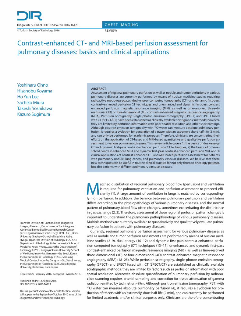
Figure 1. A 65-year-old female with pulmonary thromboembolism with right heart dysfunction. Color-coded lung perfused blood volume (Lung PBV) map and averaged CT image from 80 kVp and 140 kVp images of vasculature were fused by commercially available software. On fused image, thrombi in central zones (large arrow) and peripheral zones (small arrows) are clearly demonstrated as defects within pulmonary vasculatures. In addition, heterogeneously decreased perfusion within right upper and both lower lung fields are clearly demonstrated as Lung PBV map information. Dual energy CT can show not only morphologic, but also functional information on the same image.

detection of the region of interest in the pulmonary trunk. However, Geyer et al. (34) reported that there was no significant dif-ference in pulmonary artery enhancement when a timing bolus was used instead of automatic bolus tracking. Patients should hold their breath at a shallow inspiratory level during scan acquisition to avoid ex-cessive influx of unenhanced blood from the inferior vena cava, resulting from the Valsalva maneuver associated with deep in-spiration. In addition, dual-energy CT scans should be acquired in the caudal-cranial di-rection, so that the saline chaser bolus can reach the upper chest by the time this area is acquired, to avoid streak artifacts from highly concentrated contrast media in the subclavian vein or superior vena cava. On the other hand, Nance et al. (35) reported that a protocol using a high iodine concen-tration and a high injection delivery rate for contrast material delivery (iomeprol 400 at 4 mL/s, corresponding to an injection deliv-ery rate of 1.6 g I/s) resulted in the best im-age quality of both pulmonary multidetec-tor row CT angiography (MDCTA) images and perfusion map images of the lung. This is due to high attenuation in the pulmonary arteries and minimization of beam-harden-ing artifacts compared with the protocols involving a lower concentration or lower delivery rate. Kerl et al. (36) reported that a triphasic contrast medium injection proto-col (50 mL of undiluted contrast medium in the first phase, followed by a constant vol-ume of 30 mL of a 70%:30% saline and con-trast medium mixture, and 50 mL of pure saline in the third phase) could generally prevent streak artifacts from high-attenua-tion contrast material in the superior vena cava.
Dynamic first-pass contrast-enhanced perfusion CT
During the late 1990’s and in 2000, the use of quantitatively analyzed dynamic first-pass contrast-enhanced perfusion CT by means of electron-beam CT was report-ed in animals and also in normal individuals and/or patients with pulmonary thrombo-embolism (13). However, after the introduc-tion of multidetector row CT (MDCT) for clinical use, dynamic first-pass contrast-en-hanced perfusion CT examination shifted from electron-beam CT to MDCT, and a few investigators have reported on the latter’s potential for quantitative assessment of tumor or nodule perfusion assessment for diagnosis of pulmonary nodules or lung
cancer, or for therapeutic effect assessment of lung cancer patients undergoing con-servative therapy (14–16, 37–39). Although the number of detector rows has been in-creased by every vendor by 4 to 64-detec-tor row CT systems after clinical installation of MDCT, the limited scan range attainable with dynamic scanning at the same table position or the mix of a variety of perfusion data at different time points and positions within the scan range due to the helical scan method were major drawbacks of this technique until 2007 (14–16, 37–39).
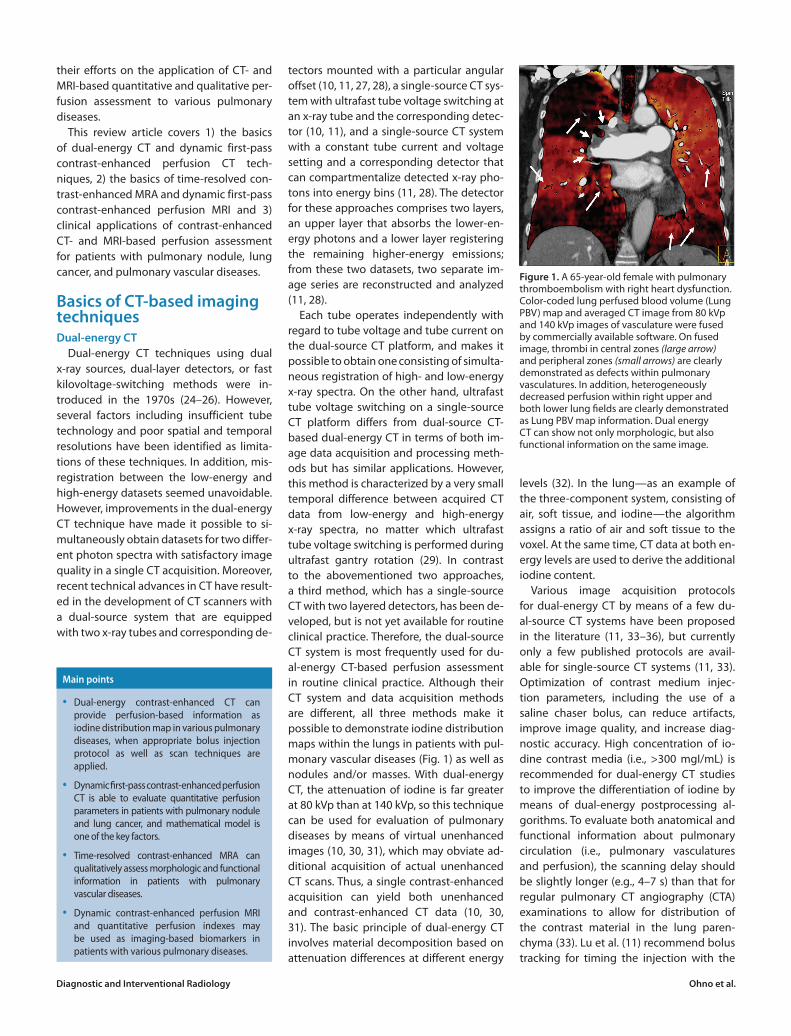
In 2007, Toshiba Medical Systems in-stalled a 320-detector row CT system with area detector CT (ADCT) for routine clinical practice. With ADCT, isotropic volume data of lung parenchyma and nodules or mass-es can be acquired simultaneously within a 160 mm area without helical scan. Thus, dy-namic first-pass contrast-enhanced perfu-sion ADCT data can be obtained by means of continuous dynamic scanning, allowing for qualitative and quantitative evaluation of perfusion of pulmonary nodules (40–42). For these reasons, ADCT systems are now being used for not only morphologic exam-inations, but also functional assessments, especially real first-pass evaluation of per-fusion a pulmonary nodule or mass per-fusion by means of the dynamic first-pass contrast-enhanced perfusion ADCT tech-nique using mathematical models (40–42). In addition, our proprietary software de-veloped with Toshiba causes dynamic first-pass contrast-enhanced perfusion ADCT at different table positions to generate whole-lung dynamic first-pass contrast-enhanced perfusion ADCT data, and quantitatively an-alyzes regional perfusion information using the mathematical models (Fig. 2). Following the introduction of Toshiba’s ADCT scanner, General Electronic Healthcare also intro-duced a similar and new ADCT system with a 256-detector row in 2014, and has started to test its potential.
In routine clinical practice, dynamic first-pass contrast-enhanced perfusion ADCT can be performed using a dynamic vol-umetric scan, which can obtain 160 mm volumetric thin-section CT data without helical scan. All dynamic first-pass con-trast-enhanced perfusion ADCT studies at our institution are currently performed with a 320-detector row CT scanner (40–42). Dy-namic first-pass ADCT is generally obtained through the nodule within a 16.0 cm area with the following parameters: 320×0.5 mm collimation, 80kVp, 120mA, 0.5 s gantry ro-
tation time, 512×512 matrix and 300–350 mm field of view (40–42). As contrast me-dia injection protocol for this setting, a du-al-head power injector is used for bolus ad-ministration of 20–45 mL (0.5 mL/kg body weight) of an iodinated contrast medium to all patients via a cubital vein at a rate of 5 mL/s, followed by 20 mL of saline solution at the same rate (40–42).
Although dynamic first-pass contrast-en-hanced perfusion ADCT data can be ob-tained in routine clinical practice, it is diffi-cult to qualitatively evaluate nodule and/or mass perfusion as well as lung paren-chyma differences on dynamic first-pass contrast-enhanced perfusion ADCT images. Therefore, dynamic first-pass contrast-en-hanced perfusion ADCT data for each subject is usually assessed in the form of quantitative perfusion parameter maps by means of mathematical models such as the single- and dual-input maximum slope and single-input Patlak plot models (41–43). The details of the mathematical model are not included in this paper, but several ex-ports have suggested that the Patlak plot method is not well suited for dynamic first-pass contrast-enhanced perfusion CT data assessment for diagnosis of pulmonary nodules, nor for therapeutic effect assess-ment in patients with lung cancer following conservative therapy (43). Although sev-eral vendors as well as academia provide software for quantitative assessment of dynamic first-pass contrast-enhanced per-fusion CT data, details of the software are usually of the black box variety. Therefore, clinicians should gain a clear understand-ing of the mathematical models involved in these software products before applying their clinical and academic purposes, when using dynamic first-pass contrast-enhanced perfusion CT examination in patients with pulmonary diseases.
Basics of MRI-based imaging techniques
Time-resolved contrast-enhanced MRASince the late 1990’s, 2D or 3D con-
trast-enhanced MRA has been widely uti-lized for pulmonary vasculature assess-ment and perfusion evaluation in routine clinical practice. In addition, high-gradi-ent-strength systems combined with the development of short TR 3D gradient-echo sequences made the development of single breath-hold 3D contrast-enhanced MRA possible (44–49). Depending on patients’ ability to hold their breath, either high-spa-
CT- and MRI-based perfusion assessment for pulmonary diseases
Diagnostic and Interventional Radiology Ohno et al.
tial resolution monophasic protocols with scan times of 20–30 s or time-resolved multiphasic imaging protocols with scan times of less than 10 s can be used (44–49). Thus, even patients with severe dyspnea can be imaged by means of time-resolved sequences.
In addition, after the introduction and clinical application of parallel imaging techniques such as sensitivity encoding (SENSE) and generalized autocalibrating partially parallel acquisitions (GRAPPA) in the early 2000’s (50–52), it became possi-ble to increase spatial and temporal res-olutions of 3D contrast-enhanced MRA for not only lung, but also various other organs. Parallel imaging uses the inherent geometry of surface coils and sensitivity maps to create k-space information from under-sampled scans. Although there is a trade-off between greater speed and a reduction in signal-to-noise ratio (SNR), parallel imaging offers greater flexibility for imaging in the difficult environment of pul-
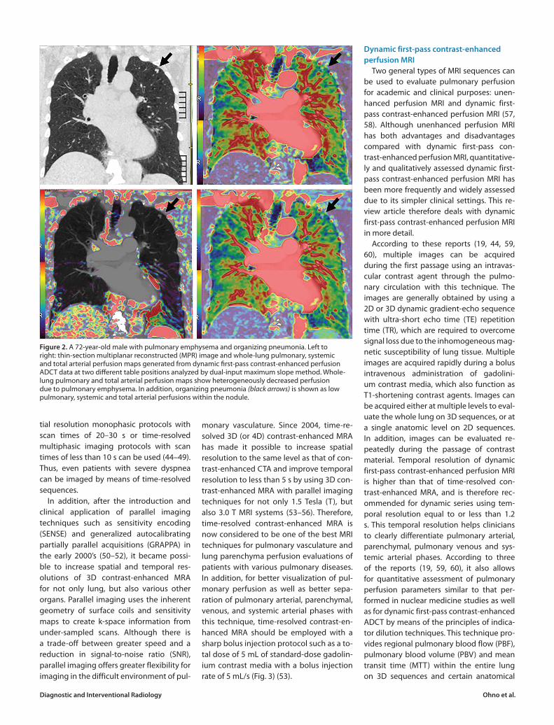
monary vasculature. Since 2004, time-re-solved 3D (or 4D) contrast-enhanced MRA has made it possible to increase spatial resolution to the same level as that of con-trast-enhanced CTA and improve temporal resolution to less than 5 s by using 3D con-trast-enhanced MRA with parallel imaging techniques for not only 1.5 Tesla (T), but also 3.0 T MRI systems (53–56). Therefore, time-resolved contrast-enhanced MRA is now considered to be one of the best MRI techniques for pulmonary vasculature and lung parenchyma perfusion evaluations of patients with various pulmonary diseases. In addition, for better visualization of pul-monary perfusion as well as better sepa-ration of pulmonary arterial, parenchymal, venous, and systemic arterial phases with this technique, time-resolved contrast-en-hanced MRA should be employed with a sharp bolus injection protocol such as a to-tal dose of 5 mL of standard-dose gadolin-ium contrast media with a bolus injection rate of 5 mL/s (Fig. 3) (53).
Dynamic first-pass contrast-enhanced perfusion MRI
Two general types of MRI sequences can be used to evaluate pulmonary perfusion for academic and clinical purposes: unen-hanced perfusion MRI and dynamic first-pass contrast-enhanced perfusion MRI (57, 58). Although unenhanced perfusion MRI has both advantages and disadvantages compared with dynamic first-pass con-trast-enhanced perfusion MRI, quantitative-ly and qualitatively assessed dynamic first-pass contrast-enhanced perfusion MRI has been more frequently and widely assessed due to its simpler clinical settings. This re-view article therefore deals with dynamic first-pass contrast-enhanced perfusion MRI in more detail.
According to these reports (19, 44, 59, 60), multiple images can be acquired during the first passage using an intravas-cular contrast agent through the pulmo-nary circulation with this technique. The images are generally obtained by using a 2D or 3D dynamic gradient-echo sequence with ultra-short echo time (TE) repetition time (TR), which are required to overcome signal loss due to the inhomogeneous mag-netic susceptibility of lung tissue. Multiple images are acquired rapidly during a bolus intravenous administration of gadolini-um contrast media, which also function as T1-shortening contrast agents. Images can be acquired either at multiple levels to eval-uate the whole lung on 3D sequences, or at a single anatomic level on 2D sequences. In addition, images can be evaluated re-peatedly during the passage of contrast material. Temporal resolution of dynamic first-pass contrast-enhanced perfusion MRI is higher than that of time-resolved con-trast-enhanced MRA, and is therefore rec-ommended for dynamic series using tem-poral resolution equal to or less than 1.2 s. This temporal resolution helps clinicians to clearly differentiate pulmonary arterial, parenchymal, pulmonary venous and sys-temic arterial phases. According to three of the reports (19, 59, 60), it also allows for quantitative assessment of pulmonary perfusion parameters similar to that per-formed in nuclear medicine studies as well as for dynamic first-pass contrast-enhanced ADCT by means of the principles of indica-tor dilution techniques. This technique pro-vides regional pulmonary blood flow (PBF), pulmonary blood volume (PBV) and mean transit time (MTT) within the entire lung on 3D sequences and certain anatomical
Figure 2. A 72-year-old male with pulmonary emphysema and organizing pneumonia. Left to right: thin-section multiplanar reconstructed (MPR) image and whole-lung pulmonary, systemic and total arterial perfusion maps generated from dynamic first-pass contrast-enhanced perfusion ADCT data at two different table positions analyzed by dual-input maximum slope method. Whole-lung pulmonary and total arterial perfusion maps show heterogeneously decreased perfusion due to pulmonary emphysema. In addition, organizing pneumonia (black arrows) is shown as low pulmonary, systemic and total arterial perfusions within the nodule.
positions within the lung on 2D sequenc-es by means of pixel-by-pixel analysis, and clearly shows regional differences for each perfusion parameter in gravitational and iso-gravitational directions for not only nor-mal subjects (Fig. 4), but also for patients with pulmonary diseases.
Clinical applications of imaging techniques
CT- and MRI-based perfusion assessment has been used for pulmonary diseases such as pulmonary nodules, lung cancer, chronic obstructive pulmonary diseases and pulmonary vascular diseases including pulmonary sequestration, pulmonary arte-
rial venous fistula or malformation (PAVF or PAVM), acute or chronic pulmonary throm-boembolism (PTE), and primary and sec-ondary pulmonary arterial hypertension (PH) (17, 19, 61–66). This article focuses on the clinical applications of these techniques for pulmonary nodules, lung cancer, and pulmonary vascular diseases.
Diagnosis of pulmonary nodules
Dual-energy contrast-enhanced CTIn a study to test the utility of dual-energy
CT for pulmonary nodule assessment, the detectability, number, and size of calcifica-tions on virtual unenhanced CT images were assessed in comparison with those obtained
on real unenhanced CT images. The results showed that 85% of calcifications within the nodule were detected on the virtual unen-hanced CT images (30), but the calcifications seen on the virtual unenhanced CT images were smaller than those on the actual unen-hanced CT images (30). This suggests that virtual unenhanced CT assessment of calci-fication within pulmonary nodules may not be satisfactory for the following reasons: greater image noise on the virtual than on the actual unenhanced CT images, compar-atively lower SNR on the surface of calcifica-tions, blurred edges resulting from the use of a low-pass filter, and low signal intensity of calcium removed from the iodine image during material decomposition (30). Since calcium detection within nodules implies a benign cause, the comparatively poorer depiction of calcification is one of the more serious limitations of virtual unenhanced CT.
The superior iodine distribution assess-ment with dual-energy CT may be even more important for routine clinical use. In contrast-enhanced CT with dual-energy CT, the material decomposition of iodine makes quantification theoretically possible by de-termining the CT number of pulmonary nod-ules on an iodine-enhanced image. Assess-ment of CT numbers on iodine-enhanced dual-energy CT images by subtracting the CT number on actual unenhanced CT from that on real contrast-enhanced CT images showed good agreement with the degree of enhancement determined by a convention-al method. In addition, when the diagnostic performance of this technique was com-pared with that of traditional contrast-en-hanced CT, iodine-enhanced imaging was more sensitive (23/25, 92% and accurate (37/45, 82.2%) than traditional contrast-en-hancement assessment or contrast-en-hanced CT (sensitivity: 18/25, 72%; accuracy: 32/45, 71.1%), whereas the specificity of the former was equal to that of the latter (14/20, 70%) (30). These results show that the iodine component is successfully decomposed from the dual-energy CT data and that virtu-al unenhanced CT is acceptable as a substi-tute for unenhanced CT. Furthermore, use of an iodine-enhanced image to measure the iodine component within a nodule can lead to a better assessment of the degree of con-trast enhancement (Fig. 5). Although further investigations may be needed to determine the true clinical significance of dual-energy CT for pulmonary nodule assessment, mea-suring iodine values on a single scan after contrast enhancement appears to be viable
CT- and MRI-based perfusion assessment for pulmonary diseases
Figure 3. a–c. A 37-year-old healthy volunteer. Source images of time-resolved 3D-contrast-enhanced MRA combined with SENSE (Top row from left to right; t = 0, 4, 8, and 12 s; bottom row from left to right; t = 16, 20, and 28 s) and SNR time-course curves (empty circle, PA; solid circle, lung parenchyma; square, PV; diamond, aorta) of each bolus injection protocol. Panels (a–c) correspond to protocols A–C, respectively. The separation of PA and PV using protocol C was clearer than that achieved with the other protocols. SNR of the pulmonary parenchyma using protocol A was significantly lower compared with the other protocols (P < 0.05). In this study, all patients were administered 3 mL (protocol A) or 6 mL (protocol B) of gadodiamide (Gd-DTPA BMA) at 3 mL/s, and 5 mL of Gd-DTPA BMA at 5 mL/s (protocol C), via a cubital vein using an automatic infusion system followed by 20 mL of saline solution at the same rate for each protocol. Figure was reproduced from Ohno et al. (53) with permission.
a
b
c
Diagnostic and Interventional Radiology Ohno et al.
in clinical practice, even though the iodine value does not represent the peak enhance-ment of the nodule.
Dynamic first-pass contrast-enhanced perfusion CT
The utility of dynamic contrast-enhanced CTs with a single-detector or MDCT scanner for differentiating malignant from benign nodules and tumors has been studied for the last few decades, and their respective sensi-tivities, specificities, and accuracies have been reported as 93%–100%, 52%–93%, and 77%–97% (17). Although several investi-gators have tested the capability of dynamic contrast-enhanced CT, one study assessed the utility of dynamic first-pass contrast-en-hanced perfusion CT using 64-MDCT and the slope method as nodule perfusion anal-ysis for differentiation of malignant from be-nign nodules, and reported that the sensitiv-ity, specificity, and accuracy were 91%–94%,
82%–86%, and 90%–93%, respectively (17). After clinical installation of ADCT in 2007,
dynamic first-pass contrast-enhanced per-fusion ADCT with mathematical models has been evaluated by investigators, who assessed the sensitivity, specificity, and accu-racy as 65%–98%, 26%–82%, and 65%–90%, respectively (40–42). In addition, studies have directly compared the diagnostic per-formance of quantitatively assessed dynam-ic first-pass contrast-enhanced perfusion ADCT with that of PET combined with CT (PET/CT) using 2-[fluorine-18]-fluoro-2-de-oxy-D-glucose (FDG) and/or dynamic first-pass contrast-enhanced MRI with ultra-short TE (40–42), and found that the diagnostic per-formance of dynamic first-pass contrast-en-hanced perfusion ADCT for distinguishing malignant from benign nodules was equal to or better than that of the other two systems (Fig. 6). Moreover, it may be even more im-portant to differentiate pulmonary nodules
requiring further intervention and treatment (malignant nodules and benign nodules with high biologic activity) from pulmonary nodules requiring no further evaluation (be-nign nodules with low biologic activity) than to differentiate malignant nodules from oth-er nodules. For this type of differentiation, quantitatively assessed dynamic first-pass contrast-enhanced ADCT has been found to be more specific and accurate for dividing all nodules into two categories (40–42). This means that quantitatively assessed dynamic first-pass contrast-enhanced ADCT should perhaps be used in routine clinical practice in a complementary role or as a substitute for dynamic contrast-enhanced CT, dynamic contrast-enhanced MRI, FDG-PET, or PET/CT to determine whether further intervention and treatment are indicated rather than to differentiate pulmonary nodules as malig-nant vs. benign.
Dynamic first-pass contrast-enhanced perfusion MRI
Several groups of investigators have test-ed dynamic contrast-enhanced MRI for its utility in differentiating malignant from be-nign nodules in both small and large patient populations. The findings of a meta-analy-sis indicated that there were no significant differences in diagnostic performance be-tween dynamic contrast-enhanced CT, dy-namic contrast-enhanced MRI with various sequences, FDG-PET, and 99mTc-depreotide SPECT (66). Therefore, dynamic contrast-en-hanced MRI can be considered at least as effective as other modalities. However, a study directly comparing the diagnostic performance of semiquantitatively analyzed dynamic contrast-enhanced MRI with ultra-short TE with that of dynamic contrast-en-hanced CT or PET/CT, also suggested the for-mer’s specificity and accuracy were superior to those reported for dynamic CT and almost equal to or superior to those of FDG-PET or PET/CT (67) (Fig. 7). Therefore, dynamic first-pass contrast-enhanced perfusion MRI should perhaps be used in a complemen-tary role or as a substitute for dynamic con-trast-enhanced CT and FDG-PET or PET/CT for diagnosis of pulmonary nodules in rou-tine clinical practice. Therapeutic effect assessment and pre-diction for non-small cell lung cancer
Dual-energy CTAfter dual-energy CT had been clinically
installed, one study found a moderate cor-relation between the maximum standard-ized uptake value (SUVmax) from FDG-PET/
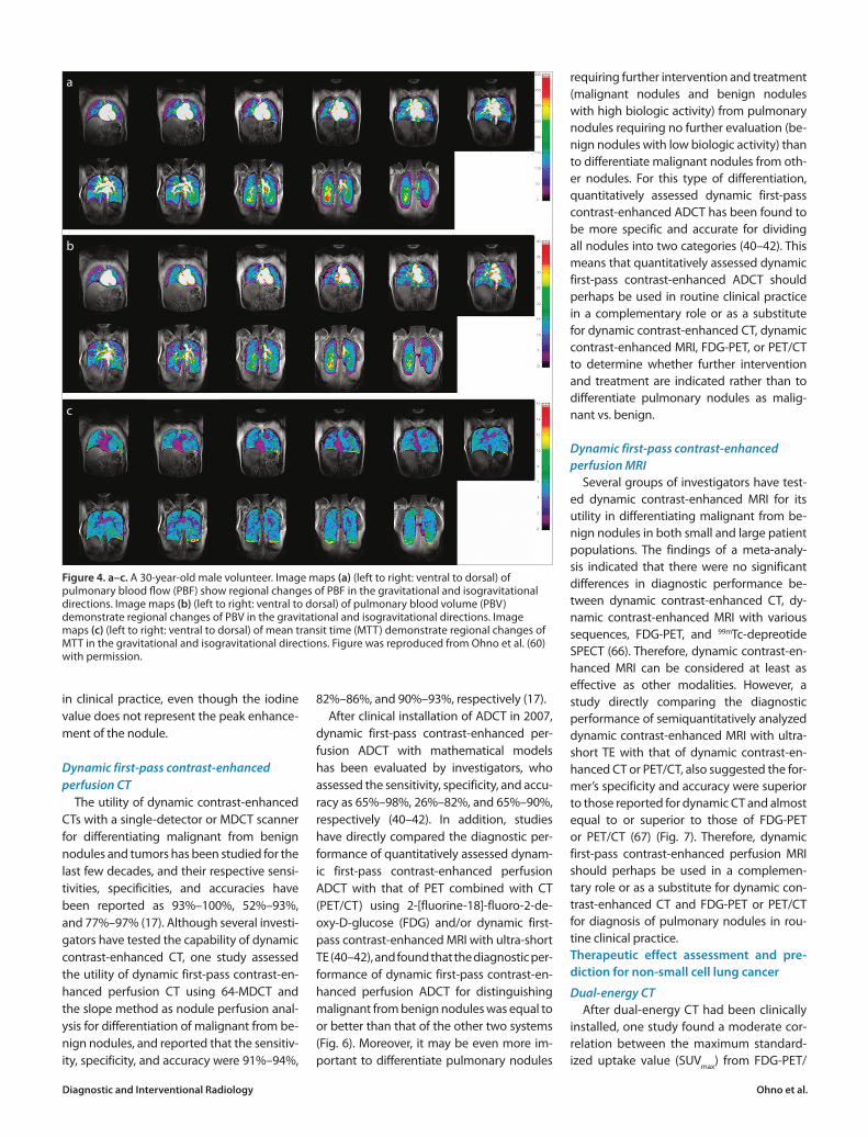
Figure 4. a–c. A 30-year-old male volunteer. Image maps (a) (left to right: ventral to dorsal) of pulmonary blood flow (PBF) show regional changes of PBF in the gravitational and isogravitational directions. Image maps (b) (left to right: ventral to dorsal) of pulmonary blood volume (PBV) demonstrate regional changes of PBV in the gravitational and isogravitational directions. Image maps (c) (left to right: ventral to dorsal) of mean transit time (MTT) demonstrate regional changes of MTT in the gravitational and isogravitational directions. Figure was reproduced from Ohno et al. (60) with permission.
a
b
c

CT and maximum iodine-related attenua-tion of dual-energy CT in lung cancers (68). Analyses of histologic subtypes of lung can-
cer showed a stronger correlation between SUVmax and maximum iodine-related atten-uation in non-small cell lung cancer (NS-
CLC) than in small cell lung cancer (SCLC) (68). This difference could be explained by differences in tumor biology such as those in angiogenetic features between NSCLC and SCLC. Therefore, measurements of the maximum iodine-related attenuation on dual-energy CT may be a useful surrogate parameter for the evaluation of therapy response of lung cancer. However, a lower correlation between SUVmax and the maxi-mum iodine-related attenuation in thoracic lymph nodes was noted, possibly because of differences in neo-angiogenesis be-tween intrapulmonary tumors and lymph node metastases (68).
In addition, dual-phase dual-energy CT was introduced as a new tool for thera-peutic effect assessment after conserva-tive therapy including anti-angiogenesis therapy for not only primary lesions, but also mediastinal lymph node metastases in NSCLC patients (69, 70). These studies found that dual-phase dual-energy CT with iodine uptake quantification in terms of io-dine uptake as well as arterial enhancement fraction is a feasible method with potential benefits for therapeutic effect prediction and/or assessment for NSCLC patients treated with conservative therapy (69, 70). In addition, this technique has the potential
CT- and MRI-based perfusion assessment for pulmonary diseases
Figure 5. a–d. A 76-year-old male with metastatic lung tumor in right lower lobe (arrows). Virtual unenhanced CT image (a) obtained from contrast-enhanced CT data is well matched with unenhanced CT image (b) and has similar image quality. Iodine map (c) generated from contrast-enhanced CT data depicts iodine distribution within nodule more clearly than contrast-enhanced CT image (d) generated from contrast-enhanced CT data obtained at 80 and 140 kVp does. Figure was reproduced from Ohno et al. (17) with permission.
c
a
d
b
Figure 6. a–d. A 72-year-old male with invasive adenocarcinoma in left lower lobe (arrows). Panel (a) shows dynamic first-pass contrast-enhanced perfusion CT data analyzed with single-input maximum slope method. Thin-section CT image (mediastinal window setting, left) shows a nodule with invasion of left hilum. Perfusion map (right) shows low perfusion of 23 mL/100 mL/min within the nodule. Extraction fraction (left) and blood volume (right) maps (b) from the same data as in panel (a) analyzed by Patlak plot method, indicating low extraction fraction (20 mL/100 mL/min) within the nodule (left) but high distribution volume (28 mL/100 mL) (right). Pulmonary perfusion (left), systemic perfusion (center), and total perfusion (right) maps (c) from the same data as in panel (a). Pulmonary and total perfusion maps show nodule perfusion is markedly lower than pulmonary parenchymal perfusion. Systemic perfusion map also shows low perfusion within the nodule. Pulmonary perfusion, systemic, and total nodule perfusions were calculated as 27, 23, and 50 mL/100 mL/min. FDG PET/CT image (d) shows high uptake of FDG within the nodule. Maximum standardized uptake value is 3.8. Figure was reproduced from Ohno et al. (17) with permission.
d
a b
c
Diagnostic and Interventional Radiology Ohno et al.
to identify a reduction in vascularization in the responding primary lesions or meta-static lymph nodes as well as nonsignificant variable development of vascularization in nonresponding lesions (69, 70).
Dynamic first-pass contrast-enhanced perfusion CT
The standard evaluation of treatment response in the oncologic field is based on the response evaluation criteria for solid tu-mors, which depend on the size of tumors observed on CT (71, 72). Currently, thera-peutic effect assessment and prediction may help physicians and patients to con-sider treatment options for personalized medicine, and have the potential to im-prove quality of life during and after treat-ment for NSCLC patients. During the past decade, several investigators have recom-mended two dynamic imaging techniques, dynamic first-pass contrast-enhanced perfusion MDCT and dynamic first-pass contrast-enhanced MRI using the dynamic
contrast-enhanced perfusion MRI method, as equally effective for treatment response assessment during or after chemotherapy, chemoradiotherapy and/or radiotherapy procedures such as FDG-PET or PET/CT (38, 73–79). The results reported by these in-vestigators suggest that tumor perfusion parameters can function as imaging-based biomarkers as effectively as glucose metab-olism evaluated by PET or PET/CT with FDG (38, 73–79). In addition, quantitatively ana-lyzed dynamic first-pass perfusion MDCT is a potential therapeutic effect assessment procedure based on information about angiogenetic status change for not only chemo- or chemoradiotherapy, but also an-tiangiogenetic therapy (38, 76–79). Howev-er, for these studies, MDCTs with a range of 4–64 detector rows were used with helical scanning employing various beam pitches and several software with mathematical models that have not been fully detailed because different versions were provided by different vendors (38, 76–79).
Since 2011, dynamic first-pass pulmo-nary contrast-enhanced perfusion ADCT has been in clinical use to obtain isotropic volume data within a 160 mm area with-out helical scan (40–42), and one study has tested its potential for quantitative thera-peutic effect assessment of dynamic first-pass contrast-enhanced perfusion ADCT examination with high spatial resolution (43). Moreover, this study has suggested that, when dynamic contrast-enhanced perfusion ADCT is used, mathematical models can perform a key function in the improvement of prediction performance of therapeutic effect on NSCLC patients (43). Although further investigations are required, this technique, using the dynamic first-pass contrast-enhanced perfusion MRI method, promises to be as effective a tool for therapeutic effect assessment or predic-tion NSCLC patients as is FDG-PET, PET/CT, or dynamic contrast-enhanced MRI.
Dynamic first-pass contrast-enhanced perfusion MRI with ultrashort TE
Several studies have suggested that contrast-enhanced T1-weighted MRI and dynamic contrast-enhanced MRI including the perfusion MRI technique are as effec-tive as FDG-PET or PET/CT for predicting as well as evaluating treatment response pre-diction or evaluation for thoracic oncology patients (17, 80). Another study has found that semiquantitatively assessed dynamic contrast-enhanced MRI using dynamic first-pass contrast-enhanced perfusion MRI can distinguish recurrence from nonrecurrence groups with a sensitivity of 55%–91%, spec-ificity of 91%, and accuracy of 84%–91% (74). Therefore, these promising findings for the use of dynamic contrast-enhanced MRI parameters indicate that these tech-niques can provide additional information based on biologic changes in the behavior of tumors in NSCLC patients treated with conservative therapy. Therefore, standard-ization of MRI sequences, image process-ing, and semiquantitative or quantitative analyses are needed to realize the real sig-nificance of these techniques in this setting, which may also be used for establishing more accurate response criteria in the not too distant future.
Pulmonary thromboembolism
Dual-energy CTConventional pulmonary MDCT angi-
ography can provide only morphologic information for patients with acute and
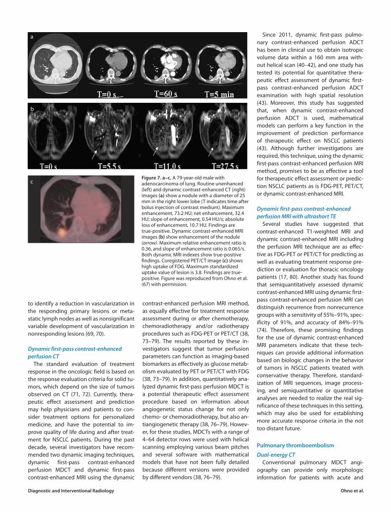
Figure 7. a–c. A 79-year-old male with adenocarcinoma of lung. Routine unenhanced (left) and dynamic contrast-enhanced CT (right) images (a) show a nodule with a diameter of 25 mm in the right lower lobe (T indicates time after bolus injection of contrast medium). Maximum enhancement, 73.2 HU; net enhancement, 32.4 HU; slope of enhancement, 0.54 HU/s; absolute loss of enhancement, 10.7 HU. Findings are true-positive. Dynamic contrast-enhanced MRI images (b) show enhancement of the nodule (arrow). Maximum relative enhancement ratio is 0.36, and slope of enhancement ratio is 0.065/s. Both dynamic MRI indexes show true-positive findings. Coregistered PET/CT image (c) shows high uptake of FDG. Maximum standardized uptake value of lesion is 3.8. Findings are true-positive. Figure was reproduced from Ohno et al. (67) with permission.
a
b
c
chronic PTE, but not directly assess lung pa-renchymal perfusion abnormalities due to thromboembolic clots. On the other hand, it has been suggested that time-resolved contrast-enhanced MRA as well as dynam-ic first-pass contrast-enhanced perfusion MRI can assess the latter aspect, which is important because of its implications for patient management, risk stratification, and prognostication (13, 19, 61, 62). Therefore, pulmonary MDCTA combined with an io-dine distribution map on dual-energy CT is considered as more useful than conven-tional MDCTA because it is able to simulta-neously provide pulmonary vasculature as well as lung perfusion-based information, and can evaluate functional information as a “one-stop shop” examination of patients with PTE.
Dual-energy CT for patients with PTE shows perfusion defects that are consis-tent with both acute and chronic PTE in-cluding those that are peripherally located, wedge-shaped, and segmentally or lobarly distributed. All other perfusion defects, such as patchy or band-like defects with-out segmental distribution or complete loss of color-coding are ordinarily consid-ered inconsistent with PTE. However, sev-eral investigators have suggested that pul-monary MDCTA and dual-energy CT-based perfusion imaging may help detect lung perfusion deficits that do not have an evi-dent morphologic thromboembolic surro-gate on conventional pulmonary MDCTA (13, 81–85). Their studies reported that sensitivity and specificity on dual-energy CT on a per-segment basis were 60%–89% and 92%–100%, respectively. In addition, the corresponding values on a per-patient basis were reported by the same studies as 75%–100% and 80%–100% (13, 81–85). Moreover, it was found that dual-energy CT-based perfusion imaging increases sensitivity for the detection of PTEs, partic-ularly for small peripheral PTEs (83). It can therefore be presumed that a simultane-ous detection of a clot in a pulmonary ar-tery on pulmonary MDCTA and of a corre-sponding perfusion defect on dual-energy CT-based perfusion imaging indicates oc-clusive PTE (13, 81–85). Pulmonary MDCTA and dual-energy CT-based perfusion im-aging techniques have therefore been rec-ommended as complementary procedures for the diagnosis of PTE in routine clinical practice.
In addition, Miura et al. (86) examined and reported that dual-energy CT may be
more effective than the right ventricle/left ventricle (RV/LV) diameter ratio for dis-ease severity assessment of patients with and without right heart dysfunction due to acute PTE. They found that the overall perfusion index, which was determined by placing the region of interest over the entire lung on a normalized lung perfused blood volume (nLung PBV) map, which was an iodine distribution map generated with commercially available software from Siemens Healthcare, was one of the predic-tors similar to RV/LV ratio (86). Therefore, when dual-energy CT is used for patients with suspected acute PTE, it was found that in the clinical setting it could not only diagnose acute PTE, but also more easily differentiate acute PTE patients with from those without right heart dysfunction (86). Therefore, dual-energy CT-based informa-tion may become one of the biomarkers for patients with acute PTE in routine clinical practice.
Time-resolved contrast-enhanced MRADuring the past decade, time-resolved
contrast-enhanced MRA as well as con-trast-enhanced MRA have been proposed as a new tool for diagnosis and patient man-agement of PTE patients (19, 61, 62). Several studies (54, 55, 87–91) have evaluated the diagnostic performance of both non-time-resolved and time-resolved contrast-en-hanced MRA for diagnosis of PTE (Table). In these studies, diagnostic performances of non-time-resolved and time-resolved contrast-enhanced MRA were evaluated as standard pulmonary digital subtraction an-giography (DSA) and/or contrast-enhanced MDCT angiography, and their sensitivity and specificity were determined as 75%–100% and 95%–100%, respectively.
Another study demonstrated that the sensitivity of time-resolved contrast-en-hanced MRA (83%) was significantly high-er than that of contrast-enhanced MDCT angiography (75%, P < 0.05) on a per-vas-cular-zone basis, and specificity and accu-racy of time-resolved contrast-enhanced MRA (specificity, 94%; accuracy, 94%) were significantly higher than those of ventila-tion-perfusion scan (specificity, 78% P < 0.05; accuracy, 75% P < 0.05) (54). Therefore, time-resolved contrast-enhanced MRA was found to be useful for the diagnosis of pul-monary embolism, and this technique may offer an alternative to ventilation-perfusion scintigraphy for imaging patients with sus-pected PTE (54).
The Prospective Investigation of Pulmo-nary Embolism Diagnosis III (PIOPED III) study (90), performed from 2006 to 2008, included 371 adults at seven centers, and the reference standard for this trial was determined by various tests including contrast-enhanced MDCTA and a nuclear medicine study. This study found that con-trast-enhanced MRA often resulted in tech-nically inadequate images, while the rate of such images varied considerably among centers. It was therefore concluded that the use of pulmonary contrast-enhanced MRA should be considered only at centers that routinely perform it well and only for patients for whom standard tests are con-traindicated.
Dynamic first-pass contrast-enhanced per-fusion MRI
Although the spatial resolution of dy-namic first-pass contrast-enhanced perfu-sion MRI is inferior to that of time-resolved contrast-enhanced MRA, it may be more sensitive for the detection of subsegmental PTE (55, 91). Although the feeding blood vessels cannot be directly visualized, the vi-sualization of characteristic wedge-shaped parenchymal perfusion defects allows for an indirect diagnosis of subsegmental pul-monary artery obstruction. This was also demonstrated in a recent study, where dynamic contrast-enhanced pulmonary perfusion MRI showed the highest sensi-tivity for assessment of PTE compared with real-time MRI with the True FISP sequence and time-resolved contrast-enhanced MRA (55, 91). In clinical practice, however, pul-monary perfusion MRI and time-resolved contrast-enhanced MRA will usually be performed as a combined protocol. More-over, a combined protocol of dynamic con-trast-enhanced perfusion MRI and time-re-solved contrast-enhanced MRA showed a sensitivity and specificity for the detection of PE of over 90%, which is nearly the same as what can be attained with contrast-en-hanced CTA (91).
Another study assessed the predictive value of quantitatively assessed dynamic first-pass contrast-enhanced perfusion MRI for the assessment of patient outcome for acute PTE (92). In this study, the acute PTE (APTE) index, which was defined as the ratio between the volume of perfusion defects and the total lung volume determined by means of dynamic first-pass contrast-en-hanced perfusion MRI, showed accuracy for the prediction of patient outcome sim-
CT- and MRI-based perfusion assessment for pulmonary diseases

Diagnostic and Interventional Radiology Ohno et al.
ilar to that of the well-investigated RV/LV diameter ratio (92). In addition, the speci-ficity and accuracy of RV/LV diameter ratio and the APTE index determined by means of dynamic first-pass contrast-enhanced perfusion MRI were significantly higher than those of APTE indexes obtained from embolic burdens and observed on con-trast-enhanced MDCTA and contrast-en-hanced MRA, although logistic regression analysis demonstrated that each index was a significant predictor (92).
Pulmonary hypertension
Dual-energy CTSince 2007 dual-energy CT has been used
for not only imaging of pulmonary vascular abnormality, but also of lung parenchyma perfusion abnormalities as a single exam-ination (13, 81, 93). It is well known that dual-energy CT has the capability to display pulmonary perfusion defects with results that are in good agreement with those obtained with perfusion scintigraphy with and without SPECT or SPECT fused with CT (SPECT/CT) (13, 81, 94, 95). Therefore, du-al-energy CT is currently being intensively tested to determine its utility for patients with pulmonary hypertension including chronic thromboembolic pulmonary hy-pertension (CTEPH) (13, 96).
The presence and significance of per-fusion abnormalities in patients with pul-monary hypertension have been most widely evaluated for patients with CTEPH (94, 97–100). The presence of consequent perfusion heterogeneity was first detect-ed with perfusion scintigraphy (101), and more recently observed on time-resolved MRA and/or dynamic first-pass contrast-en-hanced perfusion MRI (102). After installa-tion of dual-energy CT in routine clinical
practice, clinicians have been able to effec-tively identify this perfusion abnormality on iodine maps derived from dual-energy CT data. Furthermore, a meta-analysis report-ed on the capability of contrast-enhanced CTA with and without dual-energy CT or electrocardiogram-gated ADCT informa-tion for CTEPH patients (103). In this study, the patient-based analysis demonstrated a pooled sensitivity of 76%, a pooled speci-ficity of 96%, and a pooled diagnostic odds ratio of 191. In addition, the vessel-based analyses at three different levels showed a pooled sensitivity of 88%–95%, a pooled specificity of 89%–96%, and a pooled di-agnostic odds ratio of 76–751. The authors therefore concluded that CT is a suitable method for imaging proximal branches in order to differentiate between CTEPH and pulmonary endarterectomy patients. In addition, this study found that dual-ener-gy and electrocardiogram-gated ADCT can increase the sensitivity for subsegmental arterials, thus making them promising im-aging techniques for balloon pulmonary angioplasty. Further investigations are thus warranted to determine the clinical rele-vance of dual-energy CT for patients with CTEPH. Time-resolved contrast-enhanced MRA and dynamic first-pass contrast-enhanced perfusion MRI
Time-resolved contrast-enhanced MRA as well as contrast-enhanced MRA have been tested as qualitative methods for pa-tients with pulmonary hypertension due to not only CTEPH, but also other causes, while dynamic first-pass contrast-enhanced per-fusion MRI has been evaluated during the past decade as a new and more sophisticat-ed quantitative method (63, 64, 102–107) (Fig. 8). In addition, quantitatively assessed
dynamic first-pass contrast-enhanced per-fusion MRI has been tested to determine its utility as an imaging-based biomarker for various clinical purposes regarding patients with pulmonary hypertension (102).
Several studies have contributed to the evolution of the role of qualitative meth-ods using time-resolved contrast-enhanced MRA as well as dynamic first-pass con-trast-enhanced perfusion MRI in the work-up of CTEPH (63, 64, 103–107). One of the studies found that using contrast-enhanced MDCTA as the gold standard, combined un-enhanced MRI using the steady-state free precession (SSFP) sequence, contrast-en-hanced MRA and contrast-enhanced per-fusion MRI can improve the diagnostic performance of CTEPH as compared with contrast-enhanced MRA alone (106). In this study, the authors found that the SSFP se-quence was useful for visualization of the centrally based disease of chronic clot in the main pulmonary arteries. Moreover, both contrast-enhanced MRI techniques were considered useful for the detection of disease with greater frequency compared with CTA for stenosis, post-stenotic dilation, and occlusions, although these areas of better performance could not be confirmed when the statistical method was used that assumes contrast-enhanced MDCTA as the gold standard (106). It was therefore concluded that qualitatively assessed con-trast-enhanced MRA as well as dynamic first-pass contrast-enhanced perfusion MRI would appear to perform less satisfactorily than contrast-enhanced MDCTA from the point of view of simple sensitivity and spec-ificity.
In contrast to qualitative assessments such as time-resolved contrast-enhanced MRA, contrast-enhanced MRA and dynamic
Table. Summary of relevant studies for assessing diagnostic performance of non-time-resolved and time-resolved contrast-enhanced MRA in patients with pulmonary thromboembolism
References No. of patients Methods Gold standard Sensitivity (%) Specificity (%)
Meaney et al. (90) 30 3D contrast-enhanced MRA Pulmonary DSA 75–100 95–100
Gupta et al. (91) 36 3D contrast-enhanced MRA Pulmonary DSA 85 96
Oudkerk et al. (92) 141 3D contrast-enhanced MRA Pulmonary DSA 77 98
Ohno et al. (54) 48 Time-resolved contrast-enhanced MRA Pulmonary DSA 92 94
Kluge et al. (55) 62 Real-time MRI with True FISP, time-resolved 16-detector row CT angiography 81 100 contrast-enhanced MRA and dynamic contrast-enhanced perfusion MRI
Stein et al. (93) 371 3D Contrast-enhanced MRA Combination of various tests 78 99
3D, three dimensional; MRA, magnetic resonance angiography; DSA, digital subtraction angiography; MRI, magnetic resonance imaging; True FISP, true fast imaging with steady-state precession sequence; CT, computed tomography.

first-pass contrast-enhanced perfusion MRI, quantitatively assessed 3D dynamic first-pass contrast-enhanced perfusion MRI has been continuously tested since 2004 (60). By using indicator dilution theory as well as deconvolution analysis, this method can provide quantitatively analyzed PBF, PBV and MTT information for patients with pul-monary hypertension from various causes.
The therapeutic effect of dynamic first-pass contrast-enhanced perfusion MRI on CTEPH patients was assessed for a direct comparison with contrast-enhanced MDC-TA and time-resolved contrast-enhanced MRA (Fig. 9) (102). In this study, differenc-es in pre- and post-treatment PBF, PBV and MTT showed significant correlation with improvement of patients’ outcome determined by right heart catheterization and physiologic tests. In addition, appli-cation of the feasible threshold value for each index resulted in a significantly high-er specificity (91%) and accuracy (96%) for the determination of improvement in PBF than for that of assessment of improve-
ment of disease severity by means of con-trast-enhanced MDCTA (specificity: 36%, P = 0.03; accuracy: 71%; P = 0.03). It can therefore be concluded that the capability of quantitatively assessed dynamic first-pass contrast-enhanced perfusion MRI for assessment of therapeutic effect on CTEPH patients is as good as or better than that of contrast-enhanced MDCTA or time-re-solved contrast-enhanced MRA, and can be considered at least as effective as con-trast-enhanced MDCTA and time-resolved contrast-enhanced MRA.
On the other hand, one study reported that mean regional PBF and MTT of prima-ry pulmonary hypertension (PPH) patients were significantly different from those of healthy volunteers (108). In addition, PBF showed good negative correlation with pul-monary vascular resistance (PVR) (r=-0.79, P < 0.0001) and MTT and PVR moderately positive correlation (r=0.60, P = 0.022). Fur-ther, PBF showed moderately negative cor-relation with mean pulmonary arterial pres-sure (MPAP) (r=-0.70, P = 0.005), and MTT
and MPAP fairly positive correlation (r=0.54, P = 0.048). This indicates that 3D dynamic first-pass contrast-enhanced perfusion MRI can provide clinicians with a noninvasive assessment of disease severity as indicated by PVR and MPAP in patients with PPH.
As well as for PPH patients, this tech-nique was also tested for a direct compar-ison of disease severity assessment with thin-section CT for pulmonary hyperten-sion patients with connective tissue dis-ease (CTD) (109). In this study, systolic pul-monary arterial pressure and mean PBF of patients without pulmonary hypertension were significantly higher than of those with pulmonary hypertension (P < 0.05), and MTT of the former was significantly shorter than that of the latter (P < 0.05) (109). In addition, thin-section CT-based disease severity showed significantly good and negative correlation with mean PBF (r=-0.77, P < 0.01) and mean PBV (r=-0.59, P = 0.01), and significantly moderate and positive correlation with MTT (r=0.65, P < 0.01). Therefore, quantitatively assessed
CT- and MRI-based perfusion assessment for pulmonary diseases
Figure 8. a–c. A 67-year-old male with chronic thromboembolic pulmonary hypertension (CTEPH). Contrast-enhanced MDCT angiography (a) demonstrates dilatation of pulmonary artery due to CTEPH, although no thrombi are observed. Time-resolved contrast-enhanced MRA (b) shows heterogeneously decreased pulmonary perfusions with no depiction of thrombi in central and peripheral pulmonary arteries. Ventilation and perfusion SPECT (c) demonstrate no ventilation defects and heterogeneous perfusion defect in both lungs.
a b
c

Diagnostic and Interventional Radiology Ohno et al.
3D dynamic first-pass contrast-enhanced perfusion MRI appears to have good po-tential for assessment of disease severity and progression of pulmonary hyperten-sion in CTD patients. Moreover, this tech-nique, similar to Doppler echocardiogra-phy and the pulmonary function test, may be useful for noninvasive physiopatholog-ic assessment of CTD patients, and may be used as a substitute for right heart cathe-terization for CTD patients with pulmonary hypertension.
Although further investigations are war-ranted, quantitatively assessed dynamic first-pass contrast-enhanced perfusion MRI may in the near future be able to perform a complementary role in the management of patients with pulmonary hypertension from various causes in routine clinical prac-tice.
ConclusionNew contrast-enhanced imaging tech-
niques such as dual-energy CT, dynamic first-pass contrast-enhanced perfusion ADCT, time-resolved contrast-enhanced MRA and dynamic first-pass contrast-en-hanced perfusion MRI of the lung are use-ful for not only thoracic oncology patients, but also patients with various pulmonary vascular diseases in routine clinical practice. While the first-pass contrast agent tech-nique is minimally invasive but associated with risks and high costs for contrast ad-ministration, these new techniques are sim-ple and easily applied in the clinical setting. Moreover, some of these techniques can potentially be used for quantitative assess-ment of regional pulmonary perfusion, nod-ule or tumor parameters, and physiologic
and pathophysiologic analysis of various pulmonary diseases. Future developments in image acquisition and postprocessing for quantitative analyses can enhance the clinical application of these techniques for evaluation of pulmonary diseases, as well as expand their clinical relevance to other thoracic diseases.
AcknowledgementsAuthors thank Yuji Kishida, MD, Shinichiro Seki,
MD PhD (Department of Radiology, Kobe Universi-ty Graduate School of Medicine), Noriyuki Negi, RT, Katsusuke Kyotani, RT, Tohru Murakami, RT (Center for Radiology and Radiation Oncology, Kobe Univer-sity Hospital), Yoshimasa Maniwa, MD PhD (Division of Thoracic Surgery, Department of Surgery, Kobe University Graduate School of Medicine), Yoshi-hiro Nishimura, MD PhD (Division of Respiratory Medicine, Department of Internal Medicine, Kobe University Graduate School of Medicine), Kenichi Hi-rata, MD PhD (Division of Cardiovascular Medicine, Department of Internal Medicine, Kobe University
Figure 9. a–e. A 76-year-old male with CTEPH treated with conservative therapy and assessed as responder. Contrast-enhanced MDCT (a) (top to bottom: pretherapeutic MDCT to post-therapeutic MDCT; left to right: cranial to caudal) demonstrates presence of thrombi in subsegmental pulmonary arteries. Although some subsegmental thrombi disappeared, improvement in CTEPHCTA was 0%, but improvement in the RV/LV diameter ratio was -0.05. This case was identified as true positive on contrast-enhanced MDCTs. Source images of time-resolved contrast-enhanced MRA (b) (top to bottom: pretherapeutic time-resolved contrast-enhanced MRA to post-therapeutic time-resolved contrast-enhanced MRA; left to right: ventral to dorsal) shows pulmonary parenchymal enhancement in both lungs, while improvement in CTEPHMRA was -13.5%. This case was identified as true positive on time-resolved contrast-enhanced MRA. Quantitative PBF maps (c) (left to right: pretherapy to post-therapy) demonstrate improvement in PBF in both lungs to 57 mL/100 mL/min, respectively. This case was identified as true positive on PBF maps. Quantitative PBV maps (d) (left to right: pretherapy to post-therapy) demonstrate improvement in PBF in both lungs, while PBV improved by 12 mL/100 mL. This case was identified as true positive on PBV maps. Quantitative MTT maps (e) (left to right: pretherapy to post-therapy) demonstrate improved PBF in both lungs, while MTT improved by -2.3 s. This case was identified as true positive on MTT maps. Figure was reproduced from Ohno et al. (102) with permission.
a
b
c
d
e

Graduate School of Medicine), and Kimihiko Kichika-wa, MD (Department of Radiology, Nara Medical University) for their contribution to this work.
Financial disclosureThis work is financially and/ or technically sup-
ported by Toshiba Medical Systems, Philips Electron-ics Japan, Bayer Pharma, Guerbet, Daiichi-Sankyo, Eizai, Fuji RI Pharma, and Fuji Pharma.
Conflict of interest disclosureDrs. Ohno, Yoshikawa and Sugimura have re-
search grants from Toshiba Medical Systems, Philips Electronics Japan, Bayer Pharma, Guerbet, Daii-chi-Sankyo, Eizai, Fuji RI Pharma and Fuji Pharma.
References1. Wagner HN Jr. The use of radioisotope tech-
niques for the evaluation of patients with pulmonary disease. Am Rev Respir Dis 1976; 113:203–218.
2. Pistolesi M, Miniati M, Di Ricco G, Marini C, Gi-untini C. Perfusion lung imaging in the adult respiratory distress syndrome. J Thorac Imag-ing 1986; 1:11–24. [CrossRef]
3. Schuster DP. ARDS: clinical lessons from the oleic acid model of acute lung injury. Am J Respir Crit Care Med 1994; 149:245–260. [CrossRef]
4. Schuster DP, Kaplan JD, Gauvain K, Welch MJ, Markham J. Measurement of regional pulmo-nary blood flow with PET. J Nucl Med 1995; 36:371–377.
5. Kawakami K. Topics in pulmonary nuclear med-icine. Ann Nucl Med 1997; 11:67–73. [CrossRef]
6. Lewis DH, Kott B, Jacobson AF. Single-photon emission tomography imaging of the chest. Respir Care 2001; 46:940–945.
7. Musch G, Venegas JG. Positron emission to-mography imaging of regional pulmonary perfusion and ventilation. Proc Am Thorac Soc 2005; 2:522–527. [CrossRef]
8. Petersson J, Sánchez-Crespo A, Larsson SA, Mure M. Physiological imaging of the lung: single-pho-ton-emission computed tomography (SPECT). J Appl Physiol 2007; 102:468–476. [CrossRef]
9. Suga K. Pulmonary function-morphologic rela-tionships assessed by SPECT-CT fusion images. Ann Nucl Med 2012; 26:298–310. [CrossRef]
10. Chae EJ, Song JW, Krauss B, et al. Dual-energy computed tomography characterization of solitary pulmonary nodules. J Thorac Imaging 2010; 25:301–310. [CrossRef]
11. Lu GM, Zhao Y, Zhang LJ, Schoepf UJ. Dual-en-ergy CT of the lung. AJR Am J Roentgenol 2012; 199:S40–S53. [CrossRef]
12. Remy-Jardin M, Faivre JB, Pontana F, Molinari F, Tacelli N, Remy J. Thoracic applications of dual energy. Semin Respir Crit Care Med 2014; 35:64–73. [CrossRef]
13. Schoepf UJ, Bruening R, Konschitzky H, et al. Pulmonary embolism: comprehensive diagno-sis by using electron-beam CT for detection of emboli and assessment of pulmonary blood flow. Radiology 2000; 217:693–700. [CrossRef]
14. Herzog P, Wildberger JE, Niethammer M, Schaller S, Schoepf UJ. CT perfusion imaging of the lung in pulmonary embolism. Acad Radiol 2003; 10:1132–1146. [CrossRef]
15. Hoffman EA, Chon D. Computed tomography studies of lung ventilation and perfusion. Proc Am Thorac Soc 2005; 2:492–498. [CrossRef]
16. Ng QS, Goh V, Fichte H, et al. Lung cancer per-fusion at multi-detector row CT: reproducibili-ty of whole tumor quantitative measurements. Radiology 2006; 239:547–553. [CrossRef]
17. Ohno Y, Nishio M, Koyama H, et al. Dynamic contrast-enhanced CT and MRI for pulmonary nodule assessment. AJR Am J Roentgenol 2014; 202:515–529. [CrossRef]
18. Uematsu H, Ohno Y, Hatabu H. Recent advanc-es in magnetic resonance perfusion imaging of the lung. Top Magn Reson Imaging. 2003; 14: 245–251. [CrossRef]
19. Pedersen MR, Fisher MT, van Beek EJ. MR imag-ing of the pulmonary vasculature--an update. Eur Radiol 2006; 16:1374–1386. [CrossRef]
20. Matsuoka S, Hunsaker AR, Gill RR, et al. Func-tional MR imaging of the lung. Magn Reson Im-aging Clin N Am 2008; 16:275–289. [CrossRef]
21. Sieren JC, Ohno Y, Koyama H, Sugimura K, Mc-Lennan G. Recent technological and applica-tion developments in computed tomography and magnetic resonance imaging for improved pulmonary nodule detection and lung cancer staging. J Magn Reson Imaging 2010; 32:1353–1369. [CrossRef]
22. Ley S, Ley-Zaporozhan J. Pulmonary perfusion imaging using MRI: clinical application. In-sights Imaging 2012; 3:61–71. [CrossRef]
23. Ohno Y. New applications of magnetic resonance imaging for thoracic oncology. Semin Respir Crit Care Med 2014; 35:27–40. [CrossRef]
24. Genant HK, Boyd D. Quantitative bone mineral analysis using dual energy computed tomogra-phy. Invest Radiol 1977; 12:545–551. [CrossRef]
25. Chiro GD, Brooks RA, Kessler RM, et al. Tissue sig-natures with dual-energy computed tomogra-phy. Radiology 1979; 131:521–523. [CrossRef]
26. Millner MR, McDavid WD, Waggener RG, Den-nis MJ, Payne WH, Sank V. Extraction of infor-mation from CT scans at different energies. Med Phys 1979; 6:70–71. [CrossRef]
27. Flohr TG, McCollough CH, Bruder H, et al. First performance evaluation of a dual-source CT (DSCT) system. Eur Radiol 2006; 16:256–268. [CrossRef]
28. Ko JP, Brandman S, Stember J, Naidich DP. Du-al-energy computed tomography: concepts, performance, and thoracic applications. J Tho-rac Imaging 2012; 27:7–22. [CrossRef]
29. Kaza RK, Platt JF, Cohan RH, Caoili EM, Al-Ha-wary MM, Wasnik A. Dual-energy CT with sin-gle- and dual-source scanners: current appli-cations in evaluating the genitourinary tract. Radiographics 2012; 32:353–369. [CrossRef]
30. Chae EJ, Song JW, Seo JB, Krauss B, Jang YM, Song KS. Clinical utility of dual-energy CT in the evaluation of solitary pulmonary nodules: initial experience. Radiology 2008; 249:671–681. [CrossRef]
31. Kang MJ, Park CM, Lee CH, Goo JM, Lee HJ. Dual-energy CT: clinical applications in vari-ous pulmonary diseases. Radiographics 2010; 30:685–698. [CrossRef]
32. Johnson TR, Krauss B, Sedlmair M, et al. Material differentiation by dual energy CT: initial experi-ence. Eur Radiol 2007; 17:1510–1517. [CrossRef]
33. Thieme SF, Johnson TR, Lee C, et al. Dual-energy CT for the assessment of contrast material distri-bution in the pulmonary parenchyma. AJR Am J Roentgenol 2009; 193:144–149. [CrossRef]
34. Geyer LL, Scherr M, Körner M, et al. Imaging of acute pulmonary embolism using a dual energy CT system with rapid kVp switching: initial results. Eur J Radiol 2012; 81:3711–3718. [CrossRef]
35. Nance JW Jr, Henzler T, Meyer M, et al. Optimiza-tion of contrast material delivery for dual-energy computed tomography pulmonary angiography in patients with suspected pulmonary embolism. Invest Radiol 2012; 47:78–84. [CrossRef]
36. Kerl JM, Bauer RW, Renker M, et al. Triphasic con-trast injection improves evaluation of dual ener-gy lung perfusion in pulmonary CT angiography. Eur J Radiol 2011; 80:e483–e487. [CrossRef]
37. Sitartchouk I, Roberts HC, Pereira AM, Bayanati H, Waddell T, Roberts TP. Computed tomogra-phy perfusion using first pass methods for lung nodule characterization. Invest Radiol 2008; 43:349–358. [CrossRef]
38. Wang J, Wu N, Cham MD, Song Y. Tumor re-sponse in patients with advanced non-small cell lung cancer: perfusion CT evaluation of chemotherapy and radiation therapy. AJR Am J Roentgenol 2009; 193:1090–1096. [CrossRef]
39. Li Y, Yang ZG, Chen TW, Yu JQ, Sun JY, Chen HJ. First-pass perfusion imaging of solitary pulmo-nary nodules with 64-detector row CT: com-parison of perfusion parameters of malignant and benign lesions. Br J Radiol 2010; 83:785–790. [CrossRef]
40. Ohno Y, Koyama H, Matsumoto K, et al. Differ-entiation of malignant and benign pulmonary nodules with quantitative first-pass 320-de-tector row perfusion CT versus FDG PET/CT. Radiology 2011; 258:599–609. [CrossRef]
41. Ohno Y, Nishio M, Koyama H, et al. Comparison of quantitatively analyzed dynamic area-de-tector CT using various mathematic methods with FDG PET/CT in management of solitary pulmonary nodules. AJR Am J Roentgenol 2013; 200:W593–W602. [CrossRef]
42. Ohno Y, Nishio M, Koyama H, et al. Solitary pul-monary nodules: Comparison of dynamic first-pass contrast-enhanced perfusion area-detec-tor CT, dynamic first-pass contrast-enhanced MR imaging, and FDG PET/CT. Radiology 2015; 274:563–575. [CrossRef]
43. Ohno Y, Koyama H, Fujisawa Y, et al. Dynamic contrast-enhanced perfusion area detector ct for non-small cell lung cancer patients: influ-ence of mathematical models on early predic-tion capabilities for treatment response and re-currence after chemoradiotherapy. Eur J Radiol 2016; 85:176–186. [CrossRef]
44. Hatabu H, Gaa J, Kim D, Li W, Prasad PV, Edelman RR. Pulmonary perfusion and angiography: eval-uation with breath-hold enhanced three-dimen-sional fast imaging steady-state precession MR imaging with short TR and TE. AJR Am J Roent-genol 1996; 167:653–655. [CrossRef]
45. Leung DA, McKinnon GC, Davis CP, Pfammatter T, Krestin GP, Debatin JF. Breath-hold, contrast-en-hanced, three-dimensional MR angiography. Radiology 1996; 200:569–571. [CrossRef]
46. Kauczor HU. Contrast-enhanced magnetic resonance angiography of the pulmonary vas-culature. A review. Invest Radiol 1998; 33:606–617.[CrossRef]
47. Ohno Y, Adachi S, Motoyama A, et al. Multi-phase ECG-triggered 3D contrast-enhanced MR angiography: utility for evaluation of hilar and mediastinal invasion of bronchogenic car-cinoma. J Magn Reson Imaging 2001; 13:215–224. [CrossRef]
48. Goyen M, Ruehm SG, Jagenburg A, Barkhau-sen J, Kröger K, Debatin JF. Pulmonary arterio-venous malformation: Characterization with time-resolved ultrafast 3D MR angiography. J Magn Reson Imaging 2001; 13:458–460. [CrossRef]
49. Fink C, Bock M, Kiessling F, et al. Time-resolved contrast-enhanced three-dimensional pulmo-nary MR-angiography: 1.0 M gadobutrol vs. 0.5 M gadopentetate dimeglumine. J Magn Reson Imaging 2004; 19:202–208. [CrossRef]
CT- and MRI-based perfusion assessment for pulmonary diseases

Diagnostic and Interventional Radiology Ohno et al.
50. Pruessmann KP, Weiger M, Scheidegger MB, Boesiger P. SENSE: sensitivity encoding for fast MRI. Magn Reson Med 1999; 42:952–962. [CrossRef]
51. Griswold MA, Jakob PM, Heidemann RM, et al. Generalized autocalibrating partially parallel acquisitions (GRAPPA). Magn Reson Med 2002; 47:1202–1210. [CrossRef]
52. Blaimer M, Breuer F, Mueller M, Heidemann RM, Griswold MA, Jakob PM. SMASH, SENSE, PILS, GRAPPA: how to choose the optimal meth-od. Top Magn Reson Imaging 2004; 15:23–36.[CrossRef]
53. Ohno Y, Kawamitsu H, Higashino T, et al. Time-resolved contrast-enhanced pulmonary MR angiography using sensitivity encoding (SENSE). J Magn Reson Imaging 2003; 17:330–336. [CrossRef]
54. Ohno Y, Higashino T, Takenaka D, et al. MR angi-ography with sensitivity encoding (SENSE) for suspected pulmonary embolism: comparison with MDCT and ventilation-perfusion scintig-raphy. AJR Am J Roentgenol 2004; 183:91–98. [CrossRef]
55. Kluge A, Luboldt W, Bachmann G. Acute pul-monary embolism to the subsegmental level: diagnostic accuracy of three MRI techniques compared with 16-MDCT. AJR Am J Roentge-nol 2006; 187:W7–W14. [CrossRef]
56. Ohno Y, Nishio M, Koyama H, et al. Journal Club: Comparison of assessment of preoperative pulmonary vasculature in patients with non-small cell lung cancer by non-contrast- and 4D contrast-enhanced 3-T MR angiography and contrast-enhanced 64-MDCT. AJR Am J Roent-genol. 2014; 202:493–506. [CrossRef]
57. Hecht EM, Rosenkrantz A. Pulmonary MR angiog-raphy techniques and applications. Magn Reson Imaging Clin N Am 2009; 17:101–131. [CrossRef]
58. Miyazaki M, Akahane M. Non-contrast en-hanced MR angiography: established tech-niques. J Magn Reson Imaging 2012; 35:1–19. [CrossRef]
59. Levin DL, Chen Q, Zhang M, Edelman RR, Hatabu H. Evaluation of regional pulmonary perfusion using ultrafast magnetic resonance imaging. Magn Reson Med 2001; 46: 166–171. [CrossRef]
60. Ohno Y, Hatabu H, Murase K, et al. Quantitative assessment of regional pulmonary perfusion in the entire lung using three-dimensional ul-trafast dynamic contrast-enhanced magnetic resonance imaging: Preliminary experience in 40 subjects. J Magn Reson Imaging 2004; 20:353–365. [CrossRef]
61. Coulden R. State-of-the-art imaging tech-niques in chronic thromboembolic pulmo-nary hypertension. Proc Am Thorac Soc 2006; 3:577–583. [CrossRef]
62. Kuriakose J, Patel S. Acute pulmonary embo-lism. Radiol Clin North Am 2010; 48:31–50. [CrossRef]
63. Ley S, Grünig E, Kiely DG, van Beek E, Wild J. Com-puted tomography and magnetic resonance im-aging of pulmonary hypertension: Pulmonary vessels and right ventricle. J Magn Reson Imag-ing 2010; 32:1313–1324. [CrossRef]
64. Kreitner KF. Noninvasive imaging of pulmo-nary hypertension. Semin Respir Crit Care Med 2014; 35:99–111. [CrossRef]
65. Hoffman EA, Lynch DA, Barr RG, van Beek EJ, Parraga G; IWPFI Investigators. Pulmonary CT and MRI phenotypes that help explain chronic pulmonary obstruction disease pathophys-iology and outcomes. J Magn Reson Imaging 2016; 43:544–557. [CrossRef]
66. Cronin P, Dwamena BA, Kelly AM, Carlos RC. Solitary pulmonary nodules: meta-analytic comparison of cross-sectional imaging mo-dalities for diagnosis of malignancy. Radiology 2008; 246:772–782. [CrossRef]
67. Ohno Y, Koyama H, Takenaka D, et al. Dynam-ic MRI, dynamic multidetector-row computed tomography (MDCT), and coregistered 2-[flu-orine-18]-fluoro-2-deoxy-D-glucose-positron emission tomography (FDG-PET)/CT: compara-tive study of capability for management of pul-monary nodules. J Magn Reson Imaging 2008; 27:1284–1295. [CrossRef]
68. Schmid-Bindert G, Henzler T, Chu TQ, et al. Functional imaging of lung cancer using dual energy CT: how does iodine related attenua-tion correlate with standardized uptake value of 18FDG-PET-CT? Eur Radiol 2012; 22:93–103. [CrossRef]
69. Baxa J, Vondráková A, Matoušková T, et al. Du-al-phase dual-energy CT in patients with lung cancer: assessment of the additional value of iodine quantification in lymph node thera-py response. Eur Radiol 2014; 24:1981–1988. [CrossRef]
70. Baxa J, Matouskova T, Krakorova G, et al. Du-al-phase dual-energy CT in patients treated with erlotinib for advanced non-small cell lung cancer: possible benefits of iodine quantifica-tion in response assessment. Eur Radiol 2015 Nov 12. [Epub ahead of print]
71. Therasse P, Arbuck SG, Eisenhauer EA, et al. New guidelines to evaluate the response to treatment in solid tumors. European Organi-zation for Research and Treatment of Cancer, National Cancer Institute of the United States, National Cancer Institute of Canada. J Natl Can-cer Inst 2000; 92:205–216. [CrossRef]
72. Eisenhauer EA, Therasse P, Bogaerts J, et al. New response evaluation criteria in solid tu-mours: revised RECIST guideline (version 1.1). Eur J Cancer 2009; 45:228–247. [CrossRef]
73. Mac Manus MP, Hicks RJ, Matthews JP, et al. Positron emission tomography is superior to computed tomography scanning for re-sponse-assessment after radical radiother-apy or chemoradiotherapy in patients with non-small-cell lung cancer. J Clin Oncol 2003; 21:1285–1292. [CrossRef]
74. Ohno Y, Nogami M, Higashino T, et al. Prognos-tic value of dynamic MR imaging for non-small-cell lung cancer patients after chemoradiother-apy. J Magn Reson Imaging 2005; 21:775–783. [CrossRef]
75. Mac Manus M, Hicks RJ, Everitt S. Role of PET-CT in the optimization of thoracic radiothera-py. J Thorac Oncol 2006; 1:81–84. [CrossRef]
76. Ng QS, Goh V, Milner J, et al. Quantitative he-lical dynamic contrast enhanced computed tomography assessment of the spatial varia-tion in whole tumour blood volume with ra-diotherapy in lung cancer. Lung Cancer 2010; 69:71–76. [CrossRef]
77. Fraioli F, Anzidei M, Zaccagna F, et al. Whole-tu-mor perfusion CT in patients with advanced lung adenocarcinoma treated with conven-tional and antiangiogenetic chemotherapy: initial experience. Radiology 2011; 259:574–582. [CrossRef]
78. Tacelli N, Santangelo T, Scherpereel A, et al. Perfusion CT allows prediction of therapy re-sponse in non-small cell lung cancer treated with conventional and anti-angiogenic che-motherapy. Eur Radiol 2013; 23:2127–2136. [CrossRef]
79. Sudarski S, Shi J, Schmid-Bindert G, et al. Dynam-ic volume perfusion CT parameters versus RECIST for the prediction of outcome in lung cancer pa-tients treated with conventional chemotherapy. J Thorac Oncol 2015; 10:164–171. [CrossRef]
80. Nishino M, Hatabu H, Johnson BE, McLoud TC. State of the art: Response assessment in lung cancer in the era of genomic medicine. Radiol-ogy 2014; 271:6–27. [CrossRef]
81. Thieme SF, Becker CR, Hacker M, Nikolaou K, Reis-er MF, Johnson TR. Dual energy CT for the assess-ment of lung perfusion--correlation to scintigra-phy. Eur J Radiol. 2008; 68:369–374. [CrossRef]
82. Pontana F, Faivre JB, Remy-Jardin M, et al. Lung perfusion with dual-energy multidetector-row CT (MDCT): feasibility for the evaluation of acute pul-monary embolism in 117 consecutive patients. Acad Radiol 2008; 15:1494–1504. [CrossRef]
83. Lu GM, Wu SY, Yeh BM, Zhang LJ. Dual-energy computed tomography in pulmonary embo-lism. Br J Radiol 2010; 83:707–718. [CrossRef]
84. Wu HW, Cheng JJ, Li JY, Yin Y, Hua J, Xu JR. Pulmonary embolism detection and charac-terization through quantitative iodine-based material decomposition images with spectral computed tomography imaging. Invest Radiol 2012; 47:85–91. [CrossRef]
85. Zhang LJ, Wang ZJ, Zhou CS, Lu L, Luo S, Lu GM. Evaluation of pulmonary embolism in pediat-ric patients with nephrotic syndrome with dual energy CT pulmonary angiography. Acad Radi-ol 2012; 19:341–348. [CrossRef]
86. Miura S, Ohno Y, Kimura H, Kichikawa K. Quan-titative lung perfused blood volume imaging on dual-energy CT: capability for quantitative assessment of disease severity in patients with acute pulmonary thromboembolism. Acta Ra-diol 2015; 56:284–293. [CrossRef]
87. Meaney JF, Weg JG, Chenevert TL, Staf-ford-Johnson D, Hamilton BH, Prince MR. Diag-nosis of pulmonary embolism with magnetic resonance angiography. N Engl J Med 1997; 336:1422–1427. [CrossRef]
88. Gupta A, Frazer CK, Ferguson JM, et al. Acute pul-monary embolism: diagnosis with MR angiogra-phy. Radiology 1999; 210:353–359. [CrossRef]
89. Oudkerk M, van Beek EJ, Wielopolski P, et al. Comparison of contrast-enhanced magnetic resonance angiography and conventional pul-monary angiography for the diagnosis of pul-monary embolism: a prospective study. Lancet 2002; 359:1643–1647. [CrossRef]
90. Stein PD, Chenevert TL, Fowler SE, et al; PIOPED III (Prospective Investigation of Pulmonary Embolism Diagnosis III) Investigators. Gado-linium-enhanced magnetic resonance angiog-raphy for pulmonary embolism: a multicenter prospective study (PIOPED III). Ann Intern Med 2010; 152:434–443. [CrossRef]
91. Kluge A, Mueller C, Strunk J, Lange U, Bach-mann G. Experience in 207 combined MRI ex-aminations for acute pulmonary embolism and deep vein thrombosis. AJR Am J Roentgenol 2006; 186:1686–1696. [CrossRef]
92. Ohno Y, Koyama H, Matsumoto K, et al. Dy-namic MR perfusion imaging: capability for quantitative assessment of disease extent and prediction of outcome for patients with acute pulmonary thromboembolism. J Magn Reson Imaging 2010; 31:1081–1090. [CrossRef]
93. Remy-Jardin M, Pistolesi M, Goodman LR, et al. Management of suspected acute pulmonary embolism in the era of CT angiography: a state-ment from the Fleischner Society. Radiology 2007; 245:315–329. [CrossRef]

94. Nakazawa T, Watanabe Y, Hori Y, et al. Lung per-fused blood volume images with dual-energy computed tomography for chronic thrombo-embolic pulmonary hypertension: correlation to scintigraphy with single-photon emission computed tomography. J Comput Assist To-mogr 2011; 35:590–595. [CrossRef]
95. Thieme SF, Graute V, Nikolaou K, et al. Dual En-ergy CT lung perfusion imaging--correlation with SPECT/CT. Eur J Radiol 2012; 81:360–365. [CrossRef]
96. Junqueira FP, Lima CM, Coutinho AC Jr, et al. Pulmonary arterial hypertension: an imaging review comparing MR pulmonary angiography and perfusion with multidetector CT angiogra-phy. Br J Radiol 2012; 85:1446–1456. [CrossRef]
97. Hoey ET, Agrawal SK, Ganesh V, Gopalan D, Screaton NJ. Dual energy CT pulmonary an-giography: findings in a patient with chronic thromboembolic pulmonary hypertension. Thorax 2009; 64:1012. [CrossRef]
98. Hoey ET, Mirsadraee S, Pepke-Zaba J, Jenkins DP, Gopalan D, Screaton NJ. Dual-energy CT angiography for assessment of regional pul-monary perfusion in patients with chronic thromboembolic pulmonary hypertension: initial experience. AJR Am J Roentgenol 2011; 196:524–532. [CrossRef]
99. Renard B, Remy-Jardin M, Santangelo T, et al. Dual-energy CT angiography of chronic throm-boembolic disease: can it help recognize links between the severity of pulmonary arterial obstruction and perfusion defects? Eur J Radiol 2011; 79:467–472. [CrossRef]
100. Soler X, Kerr KM, Marsh JJ, et al. Pilot study comparing SPECT perfusion scintigraphy with CT pulmonary angiography in chronic throm-boembolic pulmonary hypertension. Respirol-ogy 2012; 17:180–184. [CrossRef]
101. Lisbona R, Kreisman H, Novales-Diaz J, Derbek-yan V. Perfusion lung scanning: differentiation of primary from thromboembolic pulmonary hypertension. AJR Am J Roentgenol 1985; 144:27–30. [CrossRef]
102. Ohno Y, Koyama H, Yoshikawa T, et al. Con-trast-enhanced multidetector-row computed tomography vs. Time-resolved magnetic reso-nance angiography vs. contrast-enhanced per-fusion MRI: assessment of treatment response by patients with inoperable chronic throm-boembolic pulmonary hypertension. J Magn Reson Imaging 2012; 36:612–623. [CrossRef]
103. Dong C, Zhou M, Liu D, Long X, Guo T, Kong X. Diagnostic accuracy of computed tomography for chronic thromboembolic pulmonary hyper-tension: a systematic review and meta-analysis. PLoS One 2015; 10:e0126985. [CrossRef]
104. Kreitner KF, Ley S, Kauczor HU, et al. Chronic thromboembolic pulmonary hypertension: pre- and postoperative assessment with breath-hold MR imaging techniques. Radiolo-gy 2004; 232:535–543. [CrossRef]
105. Nikolaou K, Schoenberg SO, Attenberger U, et al. Pulmonary arterial hypertension: diagnosis with fast perfusion MR imaging and high-spa-tial-resolution MR angiography--preliminary experience. Radiology 2005; 236:694–703. [CrossRef]
106. Rajaram S, Swift AJ, Capener D, et al. Diagnostic accuracy of contrast-enhanced MR angiography and unenhanced proton MR imaging compared with CT pulmonary angiography in chronic thromboembolic pulmonary hypertension. Eur Radiol 2012; 22:310–317. [CrossRef]
107. Ley S, Ley-Zaporozhan J, Pitton MB, et al. Diag-nostic performance of state-of-the-art imaging techniques for morphological assessment of vascular abnormalities in patients with chron-ic thromboembolic pulmonary hypertension (CTEPH). Eur Radiol 2012; 22:607–616. [CrossRef]
108. Ohno Y, Hatabu H, Murase K, et al. Primary pul-monary hypertension: 3D dynamic perfusion MRI for quantitative analysis of regional pul-monary perfusion. AJR Am J Roentgenol 2007; 188:48–56. [CrossRef]
109. Ohno Y, Koyama H, Nogami M, et al. Dynamic perfusion MRI: capability for evaluation of dis-ease severity and progression of pulmonary ar-terial hypertension in patients with connective tissue disease. J Magn Reson Imaging 2008; 8:887–899. [CrossRef]
CT- and MRI-based perfusion assessment for pulmonary diseases