continuous peripheral nerve blocks for pediatrics
TRANSCRIPT

Advances in Anesthesia 30 (2012) 61–73
ADVANCES IN ANESTHESIA
Continuous Peripheral Nerve Blocks forPediatrics
Ravi Shah, MD*, Amod Sawardekar, MD,Santhanam Suresh, MDDepartment of Pediatric Anesthesiology, Ann & Robert H. Lurie Children’s Hospital of Chicago,Feinberg School of Medicine, Northwestern University, 225 East Chicago Avenue, Box 19, Chicago,IL 60611, USA
Keywords� Continuous peripheral nerve blockade � Pediatric regional anesthesia� Peripheral nerve catheter � Nerve stimulation� Ultrasound-guided regional anesthesia
Key Points� Continuous peripheral nerve blocks (CPNBs) are gaining popularity in pediatricswith the advanced use of ultrasonography as well as accepted techniques ofnerve stimulation [1].
� As more appropriate equipment has become available, a growing number ofreports of their use for continuous postoperative pain management or therapeuticcare in children have been published [2]. Most indications for continuous nerveblocks are for children undergoing procedures that are associated with signifi-cant or prolonged postoperative pain [3].
� CPNBs have also been reported as effective in both controlling pain and facili-tating physical therapy in children with chronic regional pain syndromes [4].
� Despite such reports, limited data exist regarding the feasibility, safety, andefficacy of CPNB in children [5].
INDICATIONSIndications for continuous peripheral nerve block (CPNB) include surgery asso-ciated with significant or prolonged postoperative pain, including major ortho-pedic surgery such as congenital hand or foot malformation repair; osteotomiesof the humerus, femur, or tibia; limb-lengthening procedures; club foot repair;and femoral shaft fracture repair. In addition, postoperative pain managementfor several days after knee or ankle arthrolysis and knee ligamentoplasty maywarrant placement of CPNB [2].
*Corresponding author. E-mail address: [email protected]
0737-6146/12/$ – see front matterhttp://dx.doi.org/10.1016/j.aan.2012.09.001 Published by Elsevier Inc.

62 SHAH, SAWARDEKAR, & SURESH
TECHNIQUESVarious techniques are currently used to place perineural catheters in children.Because indwelling catheters are left in place for some time, aseptic technique ismandatory. Nonstimulating catheter techniques have been described by Steeleand colleagues [6] and a stimulating catheter technique has been described byBoezaart and colleagues [7]. The nonstimulating catheter technique involvesplacement of a conduit needle (ie, Tuohy needle or angiocatheter) near a nerveunder ultrasound guidance or nerve stimulation. Saline or local anestheticsolution is then injected to expand the perineural space and a standard epiduralcatheter is advanced through the conduit needle using a modified Seldingertechnique. The stimulating catheter technique uses an insulated conduit needlethat is inserted in a similar fashion. A catheter with an electrically conductiveconnection to its tip is inserted through the conduit needle while stimulatingthe nerve (via the catheter) to ensure accurate positioning. A paucity ofliterature comparing these techniques remains within the pediatric literature.We commonly use both of these techniques at our institution with goodresults.
COMPLICATIONS AND ADVERSE SIDE EFFECTSComplications of perineural catheters for continuous nerve blockade in adultsare rare and less likely than those for single-injection nerve blocks [8]. Largecomparative studies in this area have not been performed in children butseveral case reports suggest similar findings in the pediatric population [5].Common problems associated with continuous nerve blockade are technicalissues such as failed blocks, incomplete analgesia, catheter dislodgement, andleakage around the catheter entry site. More serious complications includeinfection, nerve injury, and systemic drug toxicity secondary to intravascularcatheter placement or overdosage.
INFUSION STRATEGIESThe growing use of continuous regional anesthetic techniques in adults has led toamore prevalent use of CPNBs in the pediatric population [9]. Nerve anatomy inchildren (smaller diameters, short distances between the nodes of Ranvier)warrant large volumes and low concentrations of local anesthetic solution[10,11]. Despite this, a higher risk of toxicity exists in infants because of immaturemetabolic systems and lower albumin and a1-glycoprotein levels. Use of low-concentration L-enantiomer solutions (ropivicaine or levobupivacation) havebeen recommended for children because they are accompanied by low plasmaconcentrations, limiting the risk of local anesthetic systemic toxicity.
CPNBS FOR UPPER LIMB SURGERY
Infraclavicular approach to the brachial plexusIndications CPNBs are particularly useful for surgical procedures of the elbow, forearm, andhand. Perineural catheter placement via the axillary approach has been described
63CONTINUOUS PERIPHERAL NERVE BLOCKS FOR PEDIATRICS
in childrenwith promising resultswith respect to pain control; however, difficultyin maintaining both immobilization and catheter sterility present significant limi-tations [12]. The infraclavicular approach to the brachial plexus does not presentthese limitations and is easier to perform for upper limb traumawhen arm abduc-tion is painful to perform. Continuous infraclavicular nerve blockade has beendescribed in children as a safe and effective means to control perioperative acutepain after simplified skin fixation and radial osteotomy [13].
Technique
The brachial plexus cords are situated just inferior to the coracoid process. Theaxillary artery and vein lie medial to the cords, whereas the pectoralis major andminor are superficial to the neurovascular bundle. The lateral cord is visualizedon ultrasound as a hyperechoic structure and the posterior cord is positioneddeep to the axillary artery. The medial cord may be difficult to identify becauseof its anatomic location between the axillary artery and vein (See Fig. 1).Marhofer and colleagues [14] described an approach to ultrasound-guided in-fraclavicular blockade in children using a lateral approach. The ultrasoundprobe is positioned in a transverse orientation below the clavicle to visualizethe brachial plexus. An out-of-plane technique is used to advance the needleafter it is inserted inferior to the probe. De Jose Maria and colleagues [15]placed the probe parallel to the clavicle in a parasagittal plane and directedthe needle in a cephalad direction toward the brachial plexus. Catheter place-ment may be performed by either a nonstimulating or stimulating techniques.The catheter is checked for inadvertent intravascular placement and secured tothe chest.
Fig. 1. Ultrasound image of the brachial plexus from the infraclavicular approach. The poste-rior, medial, and lateral cords surround the axillary artery.
64 SHAH, SAWARDEKAR, & SURESH
Complications
Complications include hematoma, infection, nerve injury, and intravascularinjection. The risk of a pneumothorax exists because of the proximity of thecervical pleura.Postoperative care
For moderately painful upper limb surgery, continuous infraclavicular nerveblocks have significantly reduced pain and opioid requirements and increasedpatient satisfaction [16]. In adults, the combination of a continuous local anes-thetic infusion with boluses is shown to provide optimal analgesia in this settingwhile minimizing oral analgesics compared with basal or bolus-only regimens[17]. However, this approach may not apply to younger children who are unableto appropriately administer demand doses.Supraclavicular approach to the brachial plexusIndications
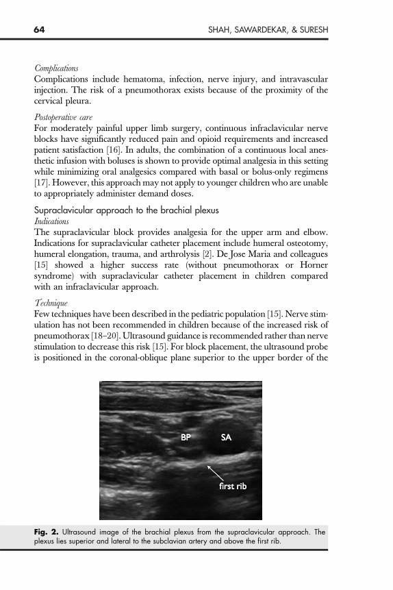
The supraclavicular block provides analgesia for the upper arm and elbow.Indications for supraclavicular catheter placement include humeral osteotomy,humeral elongation, trauma, and arthrolysis [2]. De Jose Maria and colleagues[15] showed a higher success rate (without pneumothorax or Hornersyndrome) with supraclavicular catheter placement in children comparedwith an infraclavicular approach.Technique
Few techniques have been described in the pediatric population [15]. Nerve stim-ulation has not been recommended in children because of the increased risk ofpneumothorax [18–20]. Ultrasound guidance is recommended rather than nervestimulation to decrease this risk [15]. For block placement, the ultrasound probeis positioned in the coronal-oblique plane superior to the upper border of theFig. 2. Ultrasound image of the brachial plexus from the supraclavicular approach. Theplexus lies superior and lateral to the subclavian artery and above the first rib.

65CONTINUOUS PERIPHERAL NERVE BLOCKS FOR PEDIATRICS
midclavicle. The subclavian artery is visualized as a hypoechoic and pulsatilestructure. An in-plane approach is used to direct the needle in a lateral-to-medial direction toward the brachial plexus, superior and lateral to the sub-clavian artery and above the first rib (See Fig. 2). Directing the needle ina lateral-to-medial direction avoids vascular structures and decreases the riskof intraneural injection. For continuous infusion, a 19-gauge or 20-gauge flexibleepidural catheter may be placed through an introducer needle and advanced 1 to3 cm beyond the needle tip between the brachial plexus and subclavian artery.The needle is removed over the catheter, the injection port is affixed, and thecatheter is secured and test dosed.
Complications
Complications include hematoma, infection, hematoma, and intravascular injec-tion. Furthermore, performing a supraclavicular block presents an increased riskof a pneumothorax because the lung parenchyma lies just medial to the first ribat the level where the block is completed. For this reason, visualization of the tipand shaft of the needle with ultrasonography may aid in its prevention.Postoperative care
Despite the known efficacy of single-injection supraclavicular nerve blocks,limited data exist regarding continuous supraclavicular blocks and infusionstrategies in children [21]. CPNB with supraclavicular catheters are typicallyused to provide 2 to 3 days of postoperative analgesia for pain related to upperextremity procedures.Interscalene approach to the brachial plexusIndications
The interscalene block provides analgesia to the shoulder and proximal arm viathe trunks and roots of the brachial plexus that lie deep to the sternocleidomas-toid (SCM) muscle and are surrounded by the anterior and middle scalenemuscles. The C5, C6, and C7 nerve roots are visualized between the anteriorand middle scalene muscles. This block is commonly performed for pediatricpatients undergoing surgical procedures involving the shoulder or proximalhumerus.Only a few case reports have been published on the safety and feasibility ofinterscalene brachial plexus catheter placement for shoulder surgery in childrenand adolescents. CPNB at the interscalene level has been successfully used inchildren to manage postoperative pain after tumor resection from the cephalicend of the humerus [22]. The interscalene block is known for the risk of pneu-mothorax, vascular punctures, or epidural blockade. Ultrasound-guided inter-scalene catheter placement has been described in children as an easier and saferalternative to using nerve stimulation, resulting in fewer complications whenperformed by experienced practitioners [23].
Technique
The ultrasound probe is placed at the level of the cricoid cartilage at theposterolateral aspect of the SCM in the transverse oblique plane. Deep to the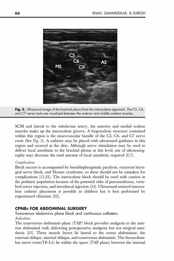
Fig. 3. Ultrasound image of the brachial plexus from the interscalene approach. The C5, C6,and C7 nerve roots are visualized between the anterior and middle scalene muscles.
66 SHAH, SAWARDEKAR, & SURESH
SCM and lateral to the subclavian artery, the anterior and medial scalenemuscles make up the interscalene groove. A hyperechoic structure containedwithin this region is the neurovascular bundle of the C5, C6, and C7 nerveroots (See Fig. 3). A catheter may be placed with ultrasound guidance in thisregion and secured at the skin. Although nerve stimulation may be used todeliver local anesthetic to the brachial plexus at this level, use of ultrasonog-raphy may decrease the total amount of local anesthetic required [6,7].
Complications
Block success is accompanied by hemidiaphragmatic paralysis, recurrent laryn-geal nerve block, and Horner syndrome, so these should not be mistaken forcomplications [15,22]. The interscalene block should be used with caution inthe pediatric population because of the potential risks of pneumothorax, verte-bral artery injection, and intrathecal injection [24]. Ultrasound-assisted intersca-lene catheter placement is possible in children but is best performed byexperienced clinicians [22].CPNBS FOR ABDOMINAL SURGERYTransversus abdominis plane block and continuous cathetersIndicationsThe transversus abdominis plane (TAP) block provides analgesia to the ante-rior abdominal wall, delivering postoperative analgesia but not surgical anes-thesia [25]. Three muscle layers lie lateral to the rectus abdominus: theexternal oblique, internal oblique, and transversus abdominis. The thoracolum-bar nerve roots(T8–L1) lie within the space (TAP plane) between the internal

67CONTINUOUS PERIPHERAL NERVE BLOCKS FOR PEDIATRICS
oblique and transversus abdominis muscle. These nerves provide sensoryinnervation to the muscles and skin of the anterior abdominal wall [8,26].
A single-shot TAP block with local anesthetic solution under ultrasoundguidance is frequently used for pain management after abdominal incisionsin combination with a systemic opioid regimen. The TAP block can be perpet-uated by use of a catheter and continuous analgesia can be achieved with a localanesthetic infusion or bolus doses, thereby decreasing supplemental systemicopioid consumption [25].
Despite the paucity of data on the use of continuous TAP catheters in pediatricpatients, this technique can provide a useful alternative to neuraxial analgesia.Unilateral TAP catheters have been successfully used in low-weight childrenfor analgesia following single-sided open lower abdominal surgery whenepidural analgesia was undesirable [27]. Similar reports have suggested thatTAP catheters can provide excellent postoperative analgesia in patients with pre-existing neurologic deficits and severe spinal deformities via the administrationof low-dose, low-concentration local anesthetic infusions [28].
Technique
Various techniques have been described that use an in-plane approach withultrasound guidance, allowing needle advancement and the placement of localanesthetic in the TAP plane [25,29]. The ultrasound probe is positioned lateralto the umbilicus and moved lateral to the rectus abdominus to visualize the 3muscle layers of the abdominal wall [30] (See Fig. 4). The needle is advancedusing an in-plane technique to the TAP plane. Injection of local anestheticcreates an elliptical pocket of local anesthetic that the nerves traverse. ForFig. 4. Sonoanatomy of the transversus abdominus plane. Muscle layers include externaloblique (EO), internal oblique (IO), and transversus abdominus (TA).

68 SHAH, SAWARDEKAR, & SURESH
TAP catheter placement, a Tuohy-tip needle may be passed under ultrasoundguidance using a similar in-plane approach and is placed between the transver-sus abdominis and internal oblique muscles. The tip of the catheter is localizedsonographically by injecting local anesthetic or saline solution through the nee-dle. The TAP plane is then expanded by injecting local anesthetic solution,which facilitates passage of an epidural catheter approximately 2 to 4 cmbeyond the tip of the needle. The bevel of the needle is placed laterally to directcatheter migration in a lateral fashion. After test dose administration, the cath-eter can be secured by subcutaneous tunneling.
Complications
Intravascular injection, peritoneal and/or bowel puncture, and infection arepotential complications of TAP catheter placement.CPNBS FOR LOWER LIMB SURGERYContinuous femoral nerve blockIndicationsThe femoral nerve block is primarily indicated for pain control in children forsurgery of the femur (femoral shaft fractures, femoral tumor excision, and limb-lengthening procedures) or for surgical procedures involving the knee. Thefemoral nerve originates from nerve roots L2, L3, and L4 and provides anal-gesia from the anterior thigh to the knee when blocked. The nerve is locatedlateral to the femoral artery and vein, and can be visualized when the ultra-sound probe is placed in the inguinal crease [31].
Continuous femoral nerve blocks have been used safely and effectively inthe pediatric population [32]. A wide range exists for applicability, includinguse in children with cancer-related pain, CRPS, and acute postoperative pain.
Fig. 5. Ultrasound image of the femoral nerve at the inguinal crease.

69CONTINUOUS PERIPHERAL NERVE BLOCKS FOR PEDIATRICS
Technique
The patient is placed in the supine position and the femoral artery is locatedwithin the inguinal crease. When using nerve stimulation, the needle isadvanced in a lateral-to-medial direction to elicit a quadriceps muscle twitchor patellar movement. Often there is thigh twitching indicating stimulation ofthe sartorius muscle, which should not misinterpreted as quadriceps stimulation.When using ultrasonography, the femoral vein, artery, and nerve can be visual-ized from medial to lateral (See Fig. 5). An in-plane or out-of-plane approach isused to direct needle placement to the femoral nerve to circumferentiallysurround it with local anesthetic [33,34].For ultrasound-guided perineural catheter placement, an insulated Tuohyneedle and a catheter with an inner steel spring that is capable of conductingelectrical impulses to its distal tip (stimulating catheter) may be used. The nee-dle tip is sonographically guided deep to the fascia iliaca and within thefemoral nerve sheath. Placement may be confirmed by injecting saline or localanesthetic solution. A nerve stimulator clip is then attached to the proximalend of the stimulating catheter and the catheter is inserted into the needle.The catheter is advanced beyond the tip of the needle while simultaneouslyappreciating patellar movement with nerve stimulation. The catheter may betunneled and secured, and should be test dosed before a bolus dose or infusionis started.
Complications
The femoral nerve’s proximity to the vein and artery make vessel puncture andhematoma formation possible complications. Risks of indwelling catheter place-ment include infection and prolonged sensory blockade.Continuous sciatic nerve blockIndications
Pain management for major foot and ankle surgery in children can be accom-plished by continuous sciatic nerve block. Common pediatric indications includepain control for club foot repair, trauma, ankle osteosynthesis and exostosis,tibial and fibular osteotomy, and foot or toe amputation [35]. Continuous sciaticnerve blockade has been shown to have fewer adverse effects compared withepidural anesthesia in children undergoing major foot and ankle surgery [36].The sciatic nerve is formed by nerve roots L4 to S3 and provides innervationto the posterior thigh and all but the medial aspect of the leg that lies distal tothe knee. The sciatic nerve exits the pelvis in the greater sciatic foramen andcourses inferior to the gluteus maximus muscle. The sciatic nerve continues tothe posterior popliteal fossa then bifurcates to form the tibial and common pero-neal nerves. The sciatic nerve can be blocked at the subgluteal, anterior thigh, orpopliteal approaches in children. In addition, the use of continuous sciatic nerveblockade has been successfully described to provide extended analgesia [37,38].Sciatic nerve catheter placement can be accurately and efficaciously facilitatedwith ultrasonography in children [39].
70 SHAH, SAWARDEKAR, & SURESH
Technique
The sciatic nerve catheter can be placed at different locations (ie, subgluteal,anterior, or distal approach).When approaching the sciatic nerve from the subgluteal area, the child isplaced in a lateral decubitus position with the hip and knee flexed. The use ofthe ultrasonography allows nerve visualization between the greater trochanterand the ischial tuberosity deep to the gluteus maximus muscle. Both in-planeand out-of-plane approaches have been described with success. Nerve stimula-tion may be used alone or in conjunction with ultrasonography. When used,hamstring, calf, foot, and toe twitches are anticipated. Successful continuousnerve blockade with catheter placement at this level has been described in chil-dren [40].
The anterior approach to the sciatic nerve is executed by placing the patientin the supine position and using ultrasonography and/or nerve stimulation[41,42]. After the child is positioned supine, the leg is abducted and rotatedlaterally and the knee is flexed to the frog-leg position as the ultrasound probeis positioned below the inguinal crease. The sciatic nerve is seen deep andmedial to the femur. This approach can be technically difficult in older childrenbecause of the increased depth to the sciatic nerve.
The distal approach in the popliteal fossa is the most frequently described inthe pediatric literature. This approach is easy to perform and shows excellentquality and duration of analgesia with low doses of local anesthetic [43]. Thepatient is placed prone and the ultrasound probe is placed above the popliteal
Fig. 6. Ultrasound image of the sciatic nerve from the distal approach in the popliteal fossa.

71CONTINUOUS PERIPHERAL NERVE BLOCKS FOR PEDIATRICS
crease. The sciatic nerve is seen adjacent to the easily visualized popliteal artery(See Fig. 6). Moving distally, the tibial and common peroneal nerves can be seenseparating from the sciatic nerve andmay be blocked specifically.With the use ofnerve stimulation, calf, foot, or toe twitches may be elicited at this location. As analternative, the patient may remain in the supine position with the hip and kneeflexed and the popliteal fossa approached using the same technique. Catheterinsertion may be performed with stimulating or nonstimulating catheterapproaches.
Complications
Complications include infection at the site of skin puncture and catheter place-ment, hematoma from vessel puncture, and local anesthetic toxicity. Comparedwith continuous epidural block in children undergoing major foot and anklesurgery, both techniques were associated with good postoperative analgesia,but children with continuous popliteal nerve catheters showed less urinaryretention, less discontinuation of local anesthetic infusion, and less nauseaand vomiting [2].SUMMARYCPNBs are emerging as an effective analgesic modality for children and cansignificantly contribute to an improved perioperative course by decreasingpostoperative pain duration and intensity. The safety and ease of these tech-niques will likely facilitate early ambulation with improved rehabilitation.
References
[1] Tsui B, Suresh S. Ultrasound imaging for regional anesthesia in infants, children, and adoles-cents: a review of current literature and its application to neuraxial blocks. Anesthesiology2010;112:719–28.
[2] Dadure C, Capdevila X. Peripheral catheter techniques. Paediatr Anaesth 2012;22:93–101.
[3] Diwan R, Lakshmi V, Shah T, et al. Continuous axillary block for upper limb surgery ina patient with epidermolysis bullosa simplex. Paediatr Anaesth 2001;11:603–6.
[4] Dadure C,Motais F, Ricard C, et al. Continuous peripheral nerve blocks at home in the treat-ment of complex regional pain syndrome in children. Anesthesiology 2005;102:387–91.
[5] Ganesh A, Rose J, Wells L, et al. Continuous peripheral nerve blockade for inpatient andoutpatient postoperative analgesia in children. Anesth Analg 2007;105(5):1234–42.
[6] Steele SM, Klein SM, D’Ercole FJ, et al. A new continuous catheter delivery system [letter].Anesth Analg 1988;87:28.
[7] Boezaart AP, de Beer JF. Accurate placement of a catheter for selective continuous intersca-lene brachial plexus nerve block [abstract V14]. Sydney, Australia: World Congress ofAnesthesiologists; 1996.
[8] ZaricD,BoysenK,Christiansen J, et al.Continuouspopliteal sciatic nerveblock foroutpatientfoot surgery: a randomized controlled trial. Acta Anaesthesiol Scand 2004;48:337–41.
[9] Ivani G, Tonetti F. Postoperative analgesia in infants and children: new developments.Minerva Anestesiol 2004;70:399–403.
[10] Bosenberg A, Ivani G. Regional anaesthesia: children are different [editorial]. PaediatrAnaesth 1998;8:447–50.
[11] Ilfeld BM, Morey TE,Wang RD, et al. Continuous popliteal sciatic nerve block for postoper-ative pain control at home. Anesthesiology 2002;97:959–65.

72 SHAH, SAWARDEKAR, & SURESH
[12] Diwan R, RauxO, Troncin R, et al. Continuous infraclavicular brachial plexus block for acutepain management in children. Anesth Analg 2003;97:691–3.
[13] Ponde VC. Continuous infraclavicular brachial plexus block: a modified technique to bettersecure catheter position in infants and children. Anesth Analg 2008;106:94–6.
[14] Marhofer P. Upper extremity peripheral blocks. Tech Reg Anesth Pain Manag 2007;11:215–21.
[15] De JoseMarıa B, Banus E,Navarro EM, et al. Ultrasound-guided supraclavicular vs infracla-vicular brachial plexus blocks in children. Paediatr Anaesth 2008;18:838–44.
[16] Ilfeld BM, Morey TE, Wright TW, et al. Interscalene perineural ropivicaine infusion:a comparison of two dosing regimens for postoperative analgesia. Reg Anesth Pain Med2004;29:9–16.
[17] Ilfeld BM, Morey TE, Enneking FK. Infraclavicular perineural local anesthetic infusion:a comparison of three dosing regimens for postoperative analgesia. Anesthesiology2004;100:395–402.
[18] Dalens B. Proximal blocks of the upper extremity: benefit/risk ratio. Regional anaes-thesia in infants, children and adolescents. Baltimore (MD): Williams & Wilkins;1995. p. 275–314.
[19] Tsui B, Suresh S. Ultrasound imaging for regional anesthesia in infants, children, and adoles-cents: a review of current literature and its application in the practice of extremity and trunkblocks. Anesthesiology 2010;112:473–92.
[20] Neal J, Brull R, Chan V. The ASRA evidence-based medicine assessment of ultrasound-guided regional anesthesia and pain medicine: executive summary. Reg Anesth PainMed 2010;35:S1–9.
[21] Mariano E, Sandhu NS, Loland V, et al. A randomized comparison of infraclavicular andsupraclavicular continuous peripheral nerve blocks for postoperative analgesia. RegAnesthPain Med 2011;36(1):26–31.
[22] Fredrickson MJ. Ultrasound-assisted interscalene catheter placement in a child. AnaesthIntensive Care 2007;35:807–8.
[23] Renes SH, Retting HC, Gielen MJ, et al. Ultrasound-guided low-dose interscalene brachialplexus block reduced the incidence of hemidiaphragmatic paresis. Reg Anesth Pain Med2009;34:498–502.
[24] Mariano ER, Ilfeld BM, Cheng GS, et al. Feasibility of ultrasound-guided peripheral nerveblock catheters for pain control on pediatric medical missions in developing countries.Paediatr Anaesth 2008;18:598–601.
[25] Suresh S, Chan VW. Ultrasound guided transversus abdominis plane block in infants, chil-dren and adolescents: a simple procedural guidance for their performance. PaediatrAnaesth 2009;19(1):296–9.
[26] McDonnell JG, O’Donnell B, Curley G, et al. The analgesic efficacy of transversus abdom-inis plane block after abdominal surgery: a prospective randomized controlled trial. AnesthAnalg 2007;104(1):193–7.
[27] Visoiu M, Boretsky KR, Goyal G, et al. Postoperative analgesia via transversus abdominisplane (TAP) catheter for small weight children - our initial experience. Paediatr Anaesth2012;22:281–4.
[28] Taylor L, Birmingham P, Yerkes E, et al. Children with spinal dysraphism: transversus abdom-inis plane (TAP) catheters to the rescue! Paediatr Anaesth 2010;20:951–4.
[29] Pak T,Mickelson J, Yerkes E, et al. Transverse abdominis plane block: a newapproach to themanagement of secondary hyperalgesia following major abdominal surgery. PaediatrAnaesth 2009;19(1):54–6.
[30] Fredrickson M, Seal P, Houghton J. Early experience with the transversus abdominis planeblock in children. Paediatr Anaesth 2008;18:891–2.
[31] Casati A, Baciarello M, Di Cianni S, et al. Effects of ultrasound guidance on the minimumeffective anaesthetic volume required to block the femoral nerve. Br J Anaesth 2007;98:823–7.

73CONTINUOUS PERIPHERAL NERVE BLOCKS FOR PEDIATRICS
[32] Johnson CM. Continuous femoral nerve blockade for analgesia in children with femoralfractures. Anaesth Intensive Care 1994;22:281–3.
[33] Oberndorfer U, Marhofer P, Bosenberg A, et al. Ultrasonographic guidance for sciatic andfemoral nerve blocks in children. Br J Anaesth 2007;98:797–801.
[34] Simion C, Suresh S. Lower extremity peripheral nerve blocks in children. Tech Reg AnesthPain Manag 2007;11:222–8.
[35] Chelly JE, Greger J, Casati A, et al. Continuous lateral sciatic blocks for acute postop-erative pain management after major ankle and foot surgery. Foot Ankle Int 2002;23:749–52.
[36] Dadure C, Bringuier S, Nicolas F, et al. Continuous epidural block versus continuous popli-teal nerve block for postoperative pain relief after major podiatric surgery in children:a prospective comparative randomized study. Anesth Analg 2006;102:744–9.
[37] van Geffen GJ, Scheuer M, Muller A, et al. Ultrasound-guided bilateral continuous sciaticnerve blocks with stimulating catheters for postoperative pain relief after bilateral lowerlimb amputations. Anaesthesia 2006;61:1204–7.
[38] vanGeffenGJ,GielenM.Ultrasound-guided subgluteal sciatic nerve blockswith stimulatingcatheters in children: a descriptive study. Anesth Analg 2006;103:328–33.
[39] Van Geffen GJ, Pirotte T, Gielen MJ, et al. Ultrasound-guided proximal and distal sciaticnerve blocks in children. J Clin Anesth 2010;22:241–5.
[40] VanGeffenGJ, Bruhn J, GielenM. Ultrasound-guided continuous sciatic nerve blocks in twochildren with venous malformations in the lower limb. Can J Anaesth 2007;54:952–3.
[41] Tsui BC, Ozelsel TJ. Ultrasound-guided anterior sciatic nerve block using a longitudinalapproach: ‘‘expanding the view.’’ Reg Anesth Pain Med 2008;33:275–6.
[42] Schwemmer U, Markus CK, Greim CA, et al. Sonographic imaging of the sciatic nerve andits division in the popliteal fossa in children. Paediatr Anaesth 2004;14:1005–8.
[43] Vas L. Continuous sciatic block for leg and foot surgery in 160 children. Paediatr Anaesth2005;15:971–8.