continuous conduction of impulses in peripheral€¦ · continuous conduction of impulses in...
TRANSCRIPT
CONTINUOUS CONDUCTION OF IMPULSES I N P E R I P H E R A L M Y E L I N A T E D NERVE FIBERS
BY ¥. LAPORTE*
(From the Laboratories of The Rockefeller Institute for Medical Research)
(Received for publication, May 31, 1951)
In the classical literature the propagation of the nerve impulse was im- plicitly accepted to be continuous. However, following a suggestion offered by Lillie (11) and experimental work by Erlanger and Blair (5, 2) and Tasaki and Takeuchi (21, 20) a number of observations have been reported in the literature which seem to indicate that the propagation of the nerve impulse is saltatory, the impulse progressing by jumps from one node of Ranvier to the next. The evidence for saltatory conduction has been recently reviewed by Huxley and St~tmpfli (8) and Schneider (19). Huxley and St~mpfli who have measured the external longitudinal action currents with single frog nerve fibers conclude that conduction is saltatory. Schneider, however, who has initiated impulses at the center of an internodal segment by cooling, and who has produced reversible blocks at the center of an internodal segment by pressure and by cooling concludes that his results are not in full agreement with the concept of saltatory conduction.
In appraising the evidence there is a detail to which considerable importance should be attached. The direct evidence for saltatory conduction has been obtained with single fibers dissected from a nerve trunk, which raises the question already mentioned by von Muralt (16) whether or not the results obtained also apply to the undissected fibers of a nerve trunk.
The experimental results to be presented in this paper cannot serve to decide whether the evidence for saltatory conduction obtained with dissected out single fibers is valid or not; they only lead to the conclusion that conduc- tion of the nerve impulse by undissected myelinated fibers occurs without appreciable discontinuity and therefore must be a continuous process.'
The use of carp nerves was suggested by a recent paper by Kynaston Thomas and Young (9) who have emphasized the theoretical significance of the fact, first described by Ranvier (18), that the distances between neighboring nodes in the large nerve fibers of the lateral line nerve are relatively very great, up to 8 ram. in some of their observations. Owing to the great length of the internodal
* Present address: Laboratoire de Physiologie, Facult6 de Medecine, Toulouse, France.
1 A preliminary note appeared in the Journa! de Physiolg.,~ie~ (10). 343
The Journal of General Physiology

344 CONTINUOUS NERVE CONDUCTION
segments the la teral line nerve is an excellent mater ia l for the analysis of the problem whether the conduction of the nerve impulse is sa l ta tory or contin- uous, since if the conduction were sal tatory, i t would occur with obvious discontinuities.
The lateral line nerve of carps about 55 cm. long contains three main sub- groups of A fibers; the fastest fibers of the first subgroup conduct impulses a t abou t 47 m./sec. (at 20-22 ° C.), and the fastest fibers of the second and th i rd subgroups at about 19 and 13 m./sec. , respectively. S tudy of nerves s tained with osmic acid and teased on the slide has shown tha t in the largest fibers (about 20/~ in diameter) the length of the internodal segments on the average is 4 mm. Consequently, by assuming tha t the speed of conduction is approxi- ma te ly proport ional to the diameter of the fiber it may be ascertained tha t t he act ion potent ia l of a single fiber which is conducting at maximal speed is the ac- t ion potent ia l of a fiber tha t has a node of Ranvier approximate ly every 4 mm.
In the frog the fastest fibers conduct a t about 30 m./sec. (at 20-22 ° C.) and in the bullfrog, a t about 40 m./sec, a difference which corresponds to a difference in the diameter of the fibers (Gasser and Erlanger (6)). In both frog and bullfrog the length of the internodal segments in the largest fibers varies from 1.5 mm. to, exceptionally, 3 mm.
The lateral line nerve is made up of a number of fascicles. A segment of the nerve 7 to 8 cm. long is excised. The peripheral segment in which the impulses are going to be initiated is left intact, but the diameter of the central segment is reduced by re- moving successively a number of fascicles until there remain only a few forming a bundle 4 to 5 cm. long with a uniform diameter of about 0.4 ram. After reduction of the diameter of the nerve the action potential of single nerve fibers can be recorded with sufficient amplitude. The stimulating electrodes are placed on the intact nerve (Fig. 2, c) and the recording electrodes on the dissected fascicle (Fig. 2, a); in addition, at the junction of segments c and a (Fig. 2, b) a number of fibers are crushed in order to insure that only a few fibers of low threshold can conduct impulses into segment a.
In the frog and bullfrog a nerve particularly suitable for the recording of the action potential of single myelinated fibers is the seventh spinal nerve (Lorente de N6 (13)). The seventh nerve is dissected together with one of its fine muscular branches, which is used to initiate the impulses. Often initation of impulses in only one fiber offers no difficulty. The diameter of the seventh nerve is uniform and in the majority of frogs it also is small enough to permit the recording of the action potential of single fibers with sufficient amplitude.
The nerves were mounted in an atmosphere of air in a humid chamber, in a horizontal position resting upon the electrodes of the stimulating and recording circuits. A chloridized silver wire 0.3 mm. in diameter was used as first recording electrode. This electrode had the shape of a hook and was mounted on a micrometric drive so that it could be displaced accurately by fractions of a millimeter along the nerve. Except in the experiments to be reported in section 3 the second electrode was always placed near the end of the nerve at a point which had been injured by sharp crushing.

Y. LAPORTE 345
Stimulation was effected by thyratron-controlled discharges of a small condenser through the primary of a transformer. Only those preparations were used in which a significant difference existed in the threshold of stimulation of the two most excitable fibers, so that a single fiber could be stimulated by a shock which was sufficiently above threshold to render constant the shock spike time. This precaution was imposed by the circumstance that a barely threshold shock often initiates impulses after a variable latency (Erlanger and Blair (4)), cf. Fig. 1. As a rule the nerve fibers were stimulated repetitively for approximately 1 second, with shocks at a frequency of 60 per second for the following reason: On a single sweep, the amplifier noise with the amplification used for recording single fiber action potentials on a trunk, usually makes uncertain the determination of the shape of the spike and of the conduction time. Since this noise produces perfectly random fluctuations of the base line, if a number of sweeps with a high negative bias on the grid of the oscillograph, i.e., with a faint spot on the screen, are photographed on a standing film, only those deflections that are constant (shock artifact and spike) will appear on the photograph, even though the width of the tracing will be determined by the amplification noise. As a rule the length of exposure of the film was { second so that each record was the result of the superposition of 12 successive sweeps. On the other hand, since with fibers of fast conduction stimulation at the frequency of 60 per second for brief intervals of time does not result in the de- velopment of cathodal depression the speed of conduction of impulses remains constant.
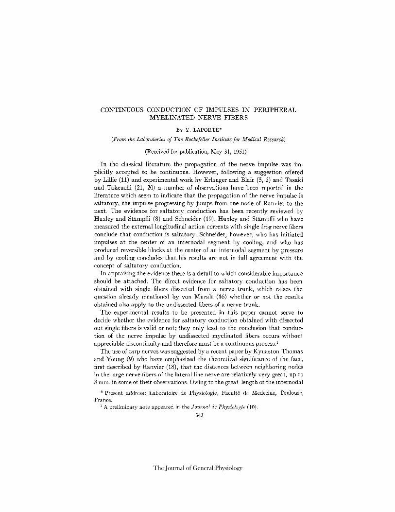
Fig. 1 illustrates the manner in which it can be ascertained tha t only one nerve fiber is being stimulated. The identification of single fiber responses is based upon the fact tha t the action potential is an all-or-none response (Blair and Erlanger (1), Monnier and Jasper (15)). The intensity of the stimulus is first chosen so that it elicits a nerve impulse only exceptionally (Fig. 1, 1); the intensity of the stimulus is then slightly increased, which results in an increase in the number of responses that the stimulus is able to elicit, and also in the appearance of responses after widely variable latencies; the shape and magnitude of the action potential, however, remain constant; i.e., no inter- mediate deflection is observed between the base line and the full spike (Fig. 1, 2, 3). If the stimulus is further increased the fiber will respond to each shock with smaller but still slightly variable latencies (Fig. 1, 4); finally, a further increase in the stimulus will make the latency at the cathode practically con- stant so that all the spikes are superposed (Fig. 1, 5). Should the stimulus be further increased, so that it would reach the threshold of a second fiber, the second action potential would reproduce the pictures given by the first fiber, but written upon the first action potential as base line. Thus, to ascertain whether the response belongs to one or to more than one fiber is a simple matter.
Since the action potential of single nerve fibers has been recorded from trunks about 0.4 ram. in diameter the question may be asked whether the action potential had the same shape that would have been recorded with the active fiber surrounded only by a thin layer of fluid. The question can be
346 CONTINUOUS NERVE CONDUCTION
answered readily by taking into account that a cylinder 0.4 ram. in diameter, i .e. a very thin cylinder, closely approaches a linear conductor, for which reason the action currents of the active fiber must flow in the nerve trunk in lines parallel to the cylindrical surface of the trunk. Therefore, the action potential must have practically identical shape whether the single fiber is surrounded by a thin layer of fluid or by a nerve trunk 0.4 ram. in diameter;
FIG. 1. Identification of the action potential of a single fiber by the all-or-none character of the response. The stimulus was successively increased from record 1 to record 5. Frog.
the presence of the trunk can result only in a decrease in the magnitude of the external action potential.
That the interstitial spaces of thin nerve trunks constitute a linear conductor can be readily demonstrated by mounting in the nerve chamber a uniform cylindrical conductor. Even when this conductor is 1.2 ram. in diameter it still approaches a linear conductor so closely that when a rectangular pulse of current is applied to it no measurable spread of current can be detected in the extrapolar segment at 0.5 to 0.75 ram. from the polarizing electrodes.

Y. LAPORTE 347
A further proof that action potentials of single fibers are recorded from thin nerve trunks without significant distortion is this: A frog nerve may be placed at the axis of a rectangular two dimensional conductor; for example, a slotted rectangle of blotting paper soaked with Ringer's solution. Even when the width of the rectangular conductor is 3 or 4 times the diameter of the nerve the action potential of volleys of impulses is recorded without significant distortion as long as the recording electrodes are at the margin of the rectangular conductor.
1. Conduction Time
Measurement of the conduction time of the nerve impulse at a number of points of the nerve is the easiest method to ascertain whether conduction is
FIG. 2. Above, diagram of the preparation. 1, 2, action potential of a single nerve fiber; 3, spikes of two fibers, the second one having a low speed of conduction. The stimulus was successively increased from record 1 to record 3. Lateral line nerve. Carp.
continuous or saltatory. If the conduction were continuous the conduction time would increase continuously and linearly with increasing distance of conduction. If the conduction were saltatory the conduction time would remain unchanged throughout each internodal segment and would increase suddenly each time that the recording electrode passes over one node of Ranvier. With the lateral line nerve of the carp the discontinuities in the conduction time should be particularly easy to detect because of the exceptional length of the internodal segments.
Figs. 2 and 3 illustrate the results of a typical experiment done with the lateral line nerve of a carp (Cyprinus carpio) of 55 cm. length. The three records reproduced in Fig. 2 prove that the action potential elicited by the stimulus used to obtain the records reproduced in Fig. 3 was the action po- tential of a single fiber. Record 1 (Fig. 2) was obtained with a shock of such a

348 CONTINUOUS NERVE CONDUCTION
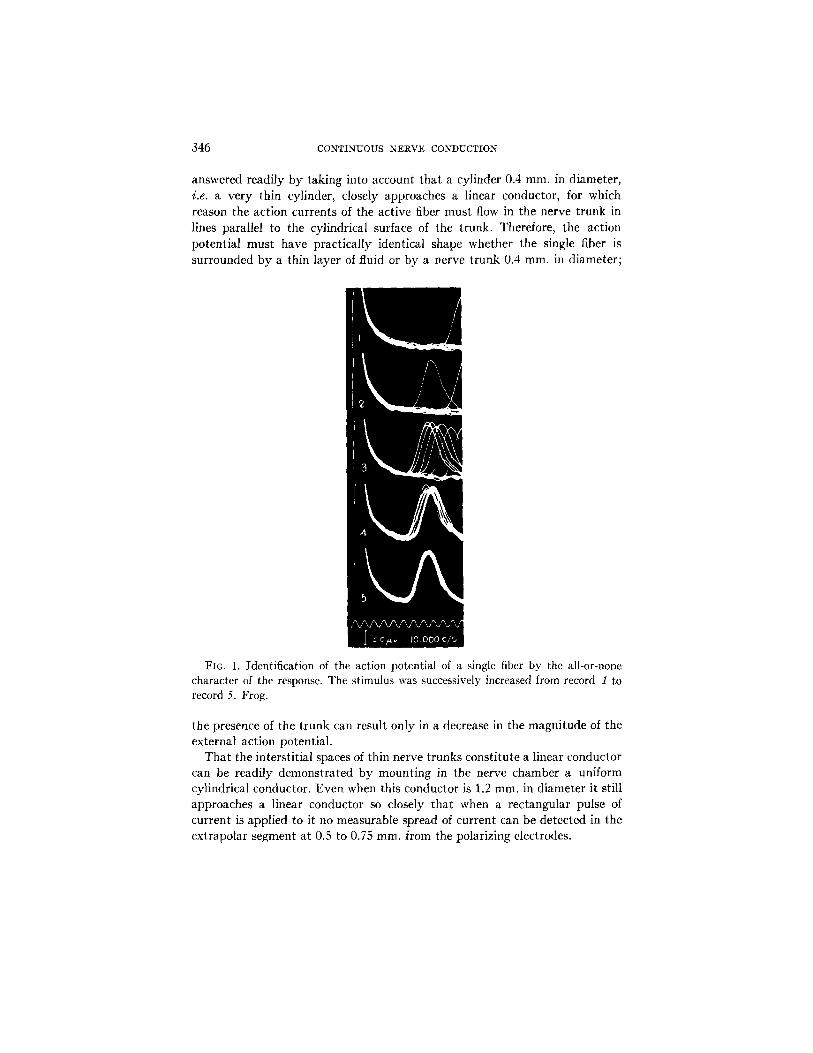
FIG. 3 (continuation of Fig. 2). Illustration of the continuous nature of the prop- agation of the nerve impulse. The numbers on the records indicate the distance between the stimulating cathode and the first recording electrode. The second re- cording electrode was located at point 39 mm., the nerve having been crushed at point 37 mm., for which reason the approach of the impulse to the second electrode resulted in a reduction of the height of the spikes recorded beyond point 25 mm.
s t rength tha t it ini t iated impulses only infrequently during 12 successive applicat ions; the record presents only the base line and the full sized spike; a slight increase in the s t imulat ing shock was sufficient to cause the nerve fiber to follow the st imulus rhythm, and to reduce sl ightly the la tency of the re-
Y. LAPORTE 349
sponse (Fig. 2, 2); but it was only after the shock had been considerably in- creased that a second fiber of relatively slow conduction was stimulated to conduct impulses (Fig. 2, 3).
Fig. 3 presents a series of 8 successive records that were obtained by dis- placing the first recording electrode by steps of 1 mm. It will be noted that the conduction time increased continuously and linerarly with increasing length of the conduction distance. The slope of the straight line drawn through the feet of the spikes indicates that the speed of conduction was approximately
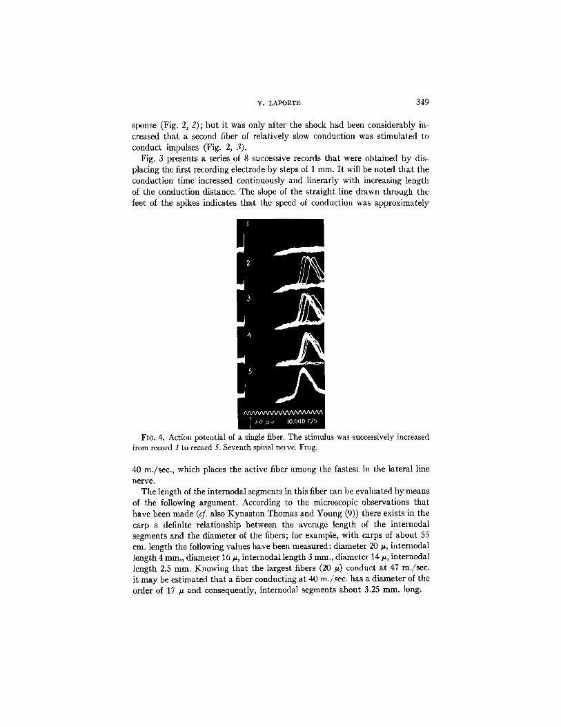
FIG. 4. Action potential of a single fiber. The stimulus was successively increased from record I to record 5. Seventh spinal nerve. Frog.
40 m./sec., which places the active fiber among the fastest in the lateral line nerve.
The length of the internodal segments in this fiber can be evaluated by means of the following argument. According to the microscopic observations that have been made (cf. also Kynaston Thomas and Young (9)) there exists in the carp a definite relationship between the average length of the internodal segments and the diameter of the fibers; for example, with carps of about 55 cm. length the following values have been measured: diameter 20/~, internodal length 4 mm., diameter 16 #, internodal length 3 mm., diameter 14/~, internodal length 2.5 mm. Knowing that the largest fibers (20/z) conduct at 47 m./sec. it may be estimated that a fiber conducting at 40 m./sec, has a diameter of the order of 17 /z and consequently, internodal segments about 3.25 mm. long.

350 CONTINUOUS NERVE CONDUCTION
22.5 ~
23.5U
~4 U
24.5 ~ . ~
. fX 25.5 ml~ mm,
T so ~ ,, Io.ooo c/s FIG. 5 (continuation of Fig. 4). Illustration of the continuous nature of the pr
agation of the nerve impulse. Second recording electrode at point 42 ram.

Y. LAPORTE 351
Since in order to obtain the series of records reproduced in Fig. 3 the first recording electrode was displaced through a 7 ram. long segment of nerve, the electrode must have passed over two successive nodes of Ranvier, and if the internodal segments happened to be exceptionally long in this fiber, the re- cording electrode must have passed at least over one node of Ranvier. There is no sign in Fig. 3 that the node or nodes had caused a discontinuity in the conduction of the nerve impulse.
On the other hand, at least several groups of three successive records (Fig. 3) must have been obtained within the same internodal segment. These groups of records show that the conduction time increased linearly with in- creasing conduction distance within each internodal segment. Therefore, con- duction of the nerve impulse must have been a continuous process.
Figs. 4 and 5 illustrate the results of a similar experiment done with the seventh spinal nerve of a frog (Rana sphenocephala). The records reproduced in Fig. 4 prove that only one nerve fiber was being stimulated to conduct impulses. In the case of record 1 the nerve fiber did not respond to any shock, and in that of record 2 the fiber responded only to 3 shocks of a train of approximately 12; the number of responses increased with increasing strength of stimulation (records 3 and 4), and finally the fiber responded to all the shocks after a constant latency (record 5).
The series of records reproduced in Fig. 5 were obtained by displacing the first recording electrode in steps of 0.5 mm. through a 3 mm. long segment of the nerve. The slope of the straight line drawn through the crests of the spikes indicates that the speed of conduction was approximately 31 m./sec, which shows that the fiber was among the fastest in the nerve. The length of the internodal segments varies in such fibers from 1.5 to exceptionally 3 mm. Therefore, the first recording electrode passed at least over one node of Ranvier; probably, it passed over two. Nevertheless, there is in the series of records no sign of a discontinuity in the conduction of the impulse. On the contrary, the conduction time increased linearly with increasing distance of conduction, in spite of the fact that at least several successive records must have been obtained within one internodal segment. Consequently, also in this case the conduction of impulses must have been a continuous process.
2. Shape and Magnitude of the Spike
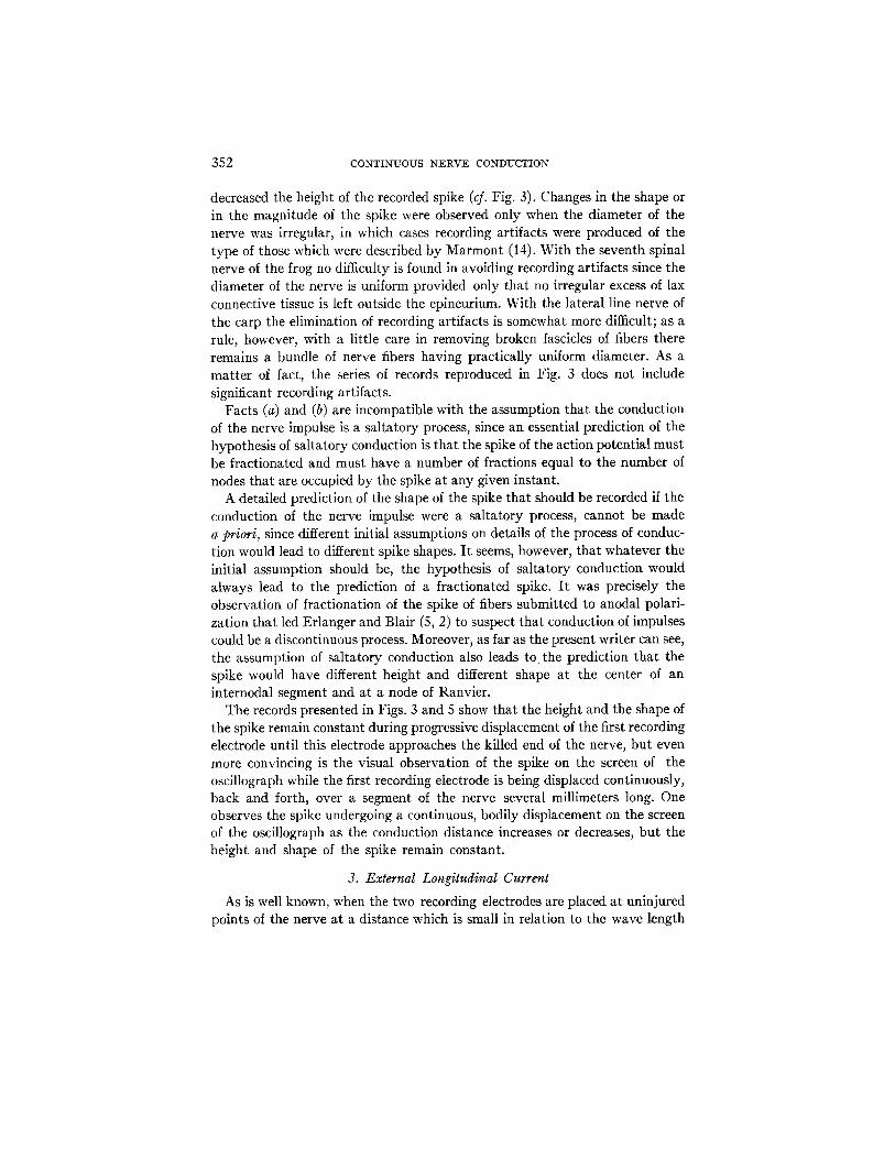
Two important facts can be observed in Figs. 1 to 5. (a) The ascending phase of the spike always had an S shape with an inflection point at about half the crest height. (b) The shape and magnitude of the spike remained constant during conduction.
A decrease in the spike height was never observed until the distance from the first recording electrode to the second became less than about 15 mm., in which case the diphasic artifact began before the spike had reached its crest and
352 CONTINUOUS NERVE CONDUCTION
decreased the height of the recorded spike (cf. Fig. 3). Changes in the shape or in the magnitude of the spike were observed only when the diameter of the nerve was irregular, in which cases recording artifacts were produced of the type of those which were described by Marmont (14). With the seventh spinal nerve of the frog no difficulty is found in avoiding recording artifacts since the diameter of the nerve is uniform provided only that no irregular excess of lax connective tissue is left outside the epineurium. With the lateral line nerve of the carp the elimination of recording artifacts is somewhat more difficult; as a rule, however, with a little care in removing broken fascicles of fibers there remains a bundle of nerve fibers having practically uniform diameter. As a matter of fact, the series of records reproduced in Fig. 3 does not include significant recording artifacts.
Facts (a) and (b) are incompatible with the assumption that the conduction of the nerve impulse is a saltatory process, since an essential prediction of the hypothesis of saltatory conduction is that the spike of the action potential must be fractionated and must have a number of fractions equal to the number of nodes that are occupied by the spike at any given instant.
A detailed prediction of the shape of the spike that should be recorded if the conduction of the nerve impulse were a saltatory process, cannot be made a priori, since different initial assumptions on details of the process of conduc- tion would lead to different spike shapes. I t seems, however, that whatever the initial assumption should be, the hypothesis of saltatory conduction would always lead to the prediction of a fractionated spike. I t was precisely the observation of fractionation of the spike of fibers submitted to anodal polari- zation that led Erlanger and Blair (5, 2) to suspect that conduction of impulses could be a discontinuous process. Moreover, as far as the present writer can see, the assumption of saltatory conduction also leads t o the prediction that the spike would have different height and different shape at the center of an internodal segment and at a node of Ranvier.
The records presented in Figs. 3 and 5 show that the height and the shape of the spike remain constant during progressive displacement of the first recording electrode until this electrode approaches the killed end of the nerve, but even more convincing is the visual observation of the spike on the screen of the oscillograph while the first recording electrode is being displaced continuously, back and forth, over a segment of the nerve several millimeters long. One observes the spike undergoing a continuous, bodily displacement on the screen of the oscillograph as the conduction distance increases or decreases, but the height and shape of the spike remain constant.
3. External Longitudinal Current
As is well known, when the two recording electrodes are placed at uninjured points of the nerve at a distance which is small in relation to the wave length

Y. LAPORTE 353
of the action potential the record obtained may be regarded as an accurate measure of the external logitudinal current that flows during the propagation of the impulse.
Fig. 6 presents records that were obtained with an interelectrode distance of 1.5 mm. The preparation was the sixth spinal nerve of a frog (R. sphenocephala).
FI6. 6. Records of the longitudinal action current of three different fibers. Re- cording electrodes 1.5 mm. apart. Sixth spinal nerve. Bullfrog.
They are very similar to records obtained with single frog nerve fibers by Lorente de N6 (13, Fig. V. 3).
Three fibers having only slightly different thresholds of stimulation often respond to the stimulating shock; nevertheless, by the use of repetitive stimu- lation with shocks of slightly different strengths it was possible, during the recovery periods, to stimulate in isolation each one of the three fibers. As measured by the duration of the shock spike time the three fibers were con- ducting impulses at the speeds of 32, 24, and 20 m./sec., respectively.
A great difference should be noted between the shapes of the records of the

354 CONTINUOUS NERVE CONDUCTION
longitudinal action current obtained with undissected nerve fibers (Fig. 6) and with dissected out nerve fibers (Huxley and St~tmpfli (8)).
4. Transmission of Impulses across a Conduclion Block
During the research that is being presented here nerve fibers have been encountered in which a conduction block existed, undoubtedly because the fibers had been injured during dissection of the nerve. I t is of interest to illus- trate some of the observations that have been made because they resemble observations made by Erlanger and Blair (5, 2) on fractionation of the spike during anodal polarization.
Fig. 7 reproduces a series of records that were obtained with the same fiber that was used to obtain the records reproduced in Fig. 3; however, the distances from the first recording electrode to the stimulating cathode were different in the two instances (23 to 30 ram., Fig. 3; 14 to 22 mm., Fig. 7).
Examination of Fig. 7 readily reveals that there existed a discontinuity in the conduction of the impulse in the segment extending between points 18 and 20 mm. The straight lines that have been drawn through the feet of the spikes (cf. also Fig. 3) show that the impulse propagated itself continuously and with the same speed between points 14 and 18 ram. and between points 20 and 30 ram., but the propagation of the impulse underwent a delay of approximately 0.2 msec. in passing through the segment 18 to 20 ram. 2 Moreover, the shape of the spikes recorded at points 18 to 22 ram. was distinctly abnormal; the spike was fractionated (cf. dots on records 18, 19, and 20 ram.), with the noteworthy peculiarity that the first part of the spike rapidly decremented in height with increasing distance from point 18.
In view of information on conduction blocks that is available in the literature (of. Werigo (22); Osterhout and Hill (17); Hodgkin (7); Lorente de N6 (12, 13)) the immediate assumption to be made is that a block of conduction existed somewhere between points 18 and 20 and that the propagation of the impulse beyond the block was due to restimulation by electrotonic spread of the action currents of the blocked impulse. The injury done to the nerve fiber was suf- ficient to prevent the production of the nerve impulse at the injured point but not sufficient to prevent electrotonic spread of the action currents to excitable points below the block. The delay in the propagation of the impulse across the block (about 0.2 msec.) was referable to the fact that restimulation beyond the block could take place only after the action potential at the upper margin of the block had reached its maximal value.
2 It hardly need be emphasized that the fractionation of the spike that appears in Fig. 7 was not a physiological phenomenon referable to the presence of a node of Ranvier. If that were the case the phenomenon should have been observed in Fig. 3, the records of which were obtained with the same fiber between points 23 and 30 mm.; i.e., in a segment which necessarily included a node. On the other hand, the phenomenon does not appear in all preparations.

Y. LAPORTE 355
m
?.1ram
. . 2Z
I.l~.v 5000 e/~ FIG. 7 (from the same experiment as Figs. 2 and 3). Discontinuity in conduction
resulting from the presence of a block in the neighborhood of point 19 ram. Lateral line nerve. Carp.
Tha t this in terpre ta t ion is correct was demonst ra ted by the results tha t were obtained when the fiber was submit ted to continuous tetanic s t imulat ion (Fig. 8).
356 CONTINUOUS N-ERVE CONDUCTION
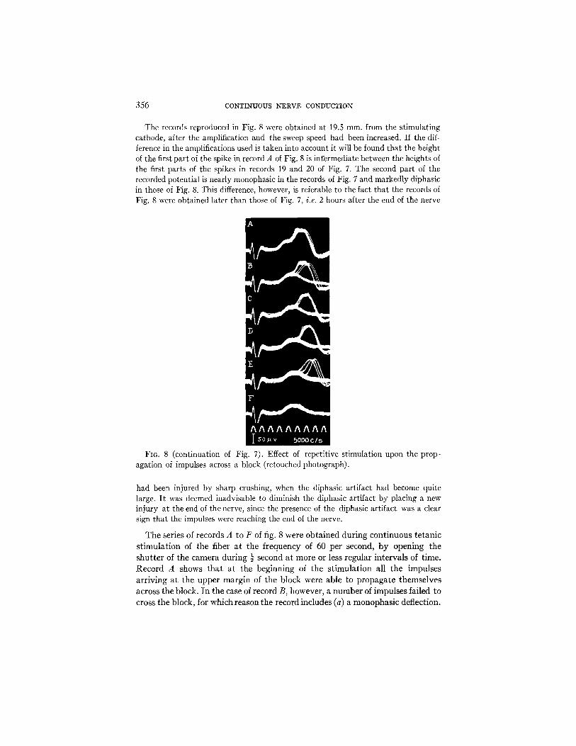
The records reproduced in Fig. 8 were obtained at 19.5 ram. from the stimulating cathode, after the amplification and the sweep speed had been increased. If the dif- ference in the amplifications used is taken into account it will be found that the height of the first part of the spike in record A of Fig. 8 is infermediate between the heights of the first parts of the spikes in records 19 and 20 of Fig. 7. The second part of the recorded potential is nearly monophasic in the records of Fig. 7 and markedly diphasic in those of Fig. 8. This difference, however, is referable to the fact that the records of Fig. 8 were obtained later than those of Fig. 7, i.e. 2 hours after the end of the nerve
FIG. 8 (continuation of Fig. 7). Effect of repetitive stimulation upon the prop- agation of impulses across a block (retouched photograph).
had been injured by sharp crushing, when the diphasic artifact had become quite large. I t was deemed inadvisable to diminish the diphasic artifact by placing a new injury at the end of the nerve, since the presence of the diphasic artifact was a clear sign that the impulses were reaching the end of the nerve.
The series of records A to F of fig. 8 were obta ined during continuous te tanic s t imulat ion of the fiber a t the frequency of 60 per second, b y opening the shut ter of the camera during ½ second at more or less regular intervals of time. Record A shows tha t a t the beginning of the s t imulat ion all the impulses arr iving a t the upper margin of the block were able to propagate themselves across the block. In the case of record B, however, a number of impulses failed to cross the block, for which reason the record includes (a) a monophasic deflection,

Y. LAPORTE 357
referable to the electrotonic spread of the action potential of blocked impulses, and (b) the spikes of propagated impulses superposed upon the monophasic deflection. In the case of the following records C and D a relatively large number of impulses was still able to cross the block, but propagation occurred after increased latencies. The number of propagated impulses decreased and the delay of propagation increased with advancing time (record E), until finally no impulse was able to propagate itself across the block and therefore the recorded potential (record F) appeared as a strictly monophasic deflection which un- doubtedly was referable to electrotonic spread of the action potential of blocked impulses.
Figs. 9 and 10 present results obtained in another experiment, which was also done with a single fiber of the lateral line nerve.
Fro. 9. Left, fractionation of the spike resulting from the presence of a conduction block. Right, effect of repetitive stimulation upon the propagation of impulses across the block. Lateral line nerve. Carp.
The records reproduced in Fig. 9 were obtained at 24.5 mm. from the stimu- lating cathode. Records 1 to 3 prove that only one nerve fiber was conducting impulses. Record 1 was obtained with a subliminal stimulus, it presents only the base line; record 2 was obtained with a stimulus that stimulated the fiber to conduct an impulse only occasionally, it presents the base line and three responses, two of which were initiated after a brief latency and the other after a long latency; finally, record 3, that was obtained with the use of a stronger shock presents a series of superposed responses; as can readily be noted, the recorded potential was fractionated into two parts.
Records A to D (Fig. 9) illustrate the changes in the response that were observed during continuous tetanic stimulation of the fiber, the changes were essentially those which have been described above in reference to Figure 8. In the case of records A and B (Fig. 9) all the impulses arriving at the upper margin of the block were able to initiate a new impulse below the block, even though in the case of record B the delay of propagation across the block was greater than in the case of record A. With advancing time many of the im- pulses arriving at the upper margin of the block failed to initiate impulses below the block, for which reason record C presents both a monophasic de-

358 CONTINUOUS N E R V E CONDUCTION
flection and the diphasic spikes of a number of impulses which were initiated below the block after a considerable delay. Finally, the reenforcement of the block resulted in the failure by all the impulses dying at the upper margin to initiate impulses below the block; at this stage only the electrotonic spread of the action potential of the blocked impulses could be observed below the block (record D).
FIG. 10 (from the same experiment as Fig. 9). Discontinuity in the conduction of the nerve impulse resulting from the presence of a conduction block. Note the de- cremental spread beyond the block of the first part of the fractionated spike.
Fig. 10 illustrates the longitudinal decrement of the first part of the frac- tionated action potential. In all probability the impulse was blocked between points 24.5 and 25 since the first part of the action potential was considerably smaller at point 25 than at point 24.5. The electrotonic spread of the blocked action potential could be followed, at the amplification that was being used, up to point 27.
DISCUSSION
The results presented in this paper lead to the conclusion that conduction of impulses by the undissected myelinated fibers of a nerve trunk is a continuous process, since (a) the conduction time increases continuously and linearly with

Y. LAPORTE 359
increasing conduction distance, (b) the spike displays constant magnitude and shape when the first recording electrode is displaced along the nerve, and (c) the spike presents no signs of fractionation, even though when a discontinuity in the conduction of the impulse is created, the fractionation of the spike is an apparent phenomenon (Figs. 7 to 10).
As already indicated in the introduction the results presented in this paper do not justify a discussion of the evidence obtained by previous investigators who have worked with dissected out single fibers. All that can be said is that dissected out single fibers and undissected nerve fibers seem to have different properties, which reveals the probable existence of a problem to be investi- gated.
The potential changes that have been observed during the propagation of impulses beyond a conduction block (Figs. 7 to 10) closely resemble certain potential changes observed by Erlanger and Blair with nerve fibers that were being submitted to anodal polarization by an externally appl:ed current. For example, records E and F in Fig. 2 of Blair and Erlanger (2) are similar to records B and C in Fig. 9, and record B in Fig. 3 of Erlanger and Blair (5) is comparable to record C in Fig. 8. Whether or not the conduction blocks were established at nodes of Ranvier is a question that cannot be answered solely on the basis of the available information. Since Bullock and Turner (3), how- ever, have observed similar blocks with invertebrate nerve fibers, the assump- tion that the blocks were established at nodes of Ranvier is not absolutely necessary.
CONCLUSIONS
1. Conduction of impulses in peripheral myelinated fibers of a nerve trunk is a continuous process, since with uninjured nerve fibers: (a) within each internodal segment the conduction time increases continuously and linearly with increasing conduction distance; (b) the presence of nodes of Ranvier does not result in any detectable discontinuity in the conduction of the impulse; (c) the ascending phase of the spike always has an S shape and never presents signs of fractionation; (d) the shape and magnitude of the spike are constant at all points of each internodal segment.
2. Records have been presented of the external logitudinal current that flows during propagation of an impulse in undissected single nerve fiber (Fig. 6).
3. Propagation of impulses across a conduction block occurs with a readily demonstrable discontinuity.
The author wishes to acknowledge his great indebtedness to Doctor Lorente de N6 for his advice throughout the course of this investigation.
L I T E R A T U R E CITED
1. Blair, E. A., and Erlanger, J., Responses of axons to brief shocks, Proc. Soc. Exp. Biol. and Med., 1932, 9.0, 926.

360 CONTINUOUS NERVE CONDUCTION
2. Blair, E. A., and Erlanger, J., Propagation and extension of excitatory effects of the nerve action potential across non-responding internodes, Am. r. Physiol., 1939, 126, 97.
3. Bullock, T. H., and Turner, R., Events associated with conduction failure in nerve fibers, J. Cell. and Comp. Physiol., 1950, 36, 59.
4. Erlanger, J., and Blair, E. A., A comparison of the characteristics of axons through their individual electrical responses, Am. J. Physiol., 1933, 106, 524.
5. Erlanger, J., and Blair, E. A., Manifestations of segmentation in myelinated axons, Am. J. Physiol., 1934, 110, 287.
6. Gasser, H. S., and Erlanger, J., The role played by the sizes of the constituent fibers of a nerve trunk in determining the form of its action potential wave, Am. J. Physiol., 1927, 80, 522.
7. Hodgkin, A. L., Evidence for electrical transmission in nerve, pt. 1, J. Physiol., 1937, 90, 183.
8. Huxley, A. F., and St~.mpfli, R., Evidence for saltatory conduction in peripheral myelinated nerve fibers, J. Physiol., 1949, 108, 315.
9. Kynaston Thomas, P., and Young, J. Z., Internode lengths in the nerves of fishes, ] . Anat., 1949, 83, 336.
10. Laporte, Y., De la conduction continue dans les fibres nerveuses my61inis6es p6riph6riques, J. de physiol., 1950, a~, 463.
11. Lillie, R. S., Factors affecting transmission and recovery in the passive iron nerve model, J. Gen. Physiol., 1925, 7, 473.
12. Lorente de N6, R., Transmission of impulses through cranial motor nuclei, J. Neurophysiol., 1939, 9., 404.
13. Lorente de N6, R., A Study of Nerve Physiology, Studies from The Rockefeller Institute/or Medical Research, 1947, 131.
14. Marmont, G., Action potential artifacts from single nerve fibers, Am. J. Physiol., 1940, 130, 392.
15. Monnier, A. M., and Jasper, H. H., Recherche de la relation entre les potentiels d'action ~16mentaires et la chronaxie de subordination. Nouvelle demonstration du fonctionnement par "tout ou rien" de la fibre nerveuse, Compt. rend. Soc. biol., 1932, 110, 547.
16. yon Muralt, A., Signal transmission in nerve, Harvey Lectures, 1948, 4,3, 230. 17. Osterhout, W. J., and Hill, S. E., Salt bridges and negative variations, J. Gen.
Physiol., 1930, 13, 547. 18. Ranvier, L., Des etranglements annulaires et des segments interannulaires chez
les Raies et les Torpilles, Compt. rend. Acad. sc., 1872, 78, 1129. 19. Schneider, D., Die lokale Reizung und Blockierungim Internodium der isolierten
markhaltigen Nervenfaser des Frosches, Z. vergleich. Physiol., 1950, 39., 34. 20. Takeuchi, T., and Tasaki, I., Ubertragung des Nervenimpulses in der polari-
sierten Nervenfaser, Arch. ges. Physiol., 1942, 9.46, 32. 21. Tasaki, I., and Takeuchi, T., Der am Ranvierschen Knoten entstehende Ak-
tionsstrom und seine Bedeuntung fur die Erregungsleitung, Arch. ges. Physiol., 1941, 244, 696.
22. Werigo, B., Zur Frage fiber die Beziehung zwischen Erregbarkeit und Leitungs- fahigkeit des Nerven, Arch. ges. Physiol., 1899, 76, 552.