contents wfsa’s sub-tenon’s block 3 complications of … · editorial dear readers, welcome to...
TRANSCRIPT

Update in
AnaesthesiaNumber 22, 2007
Editor: Bruce McCormick
ISSN 1353-4882
Sponsored by: World Federation of Societies of Anaesthesiologists, 21 Portland Place, London, W1B 1PY, United Kingdom. Tel: (+44) 20 7631 8880. Fax: (+44) 20 7631 8882. E-mail: [email protected]
Typeset by: Sumographics: www.sumographics.co.uk or e-mail: [email protected]
Printed by COS Printers
Correspondence to editor: Dr B McCormick, Anaesthetics Department, Royal Devon & Exeter NHS Foundation Trust, Barrack Road, Exeter, EX2 5DW, UK. E-mail: [email protected]
DisclaimerWorld Anaesthesia takes all reasonable care to ensure that the information contained in Update is accurate.
We cannot be held responsible for any errors or omissions and take no responsibility for the consequences of error or for any loss or damage which may arise from reliance on information contained.
WFSA’sContentsSub-Tenon’s Block ....................................................................................................... 3
Complications of Blood Transfusion ........................................................................... 8
Perioperative Neuropathies, Blindness and Positioning Problems ........................ 12
Acute Respiratory Distress Syndrome (ARDS) ........................................................ 19
Asessment of Spinal Anaesthetic Block ................................................................... 23
Meningococcal Disease in Children .......................................................................... 26
Resuscitation Update ................................................................................................. 31
Perioperative Management of Cardiovascular Drugs ............................................. 39
Management of Acute Liver Failure in ICU .............................................................. 46
Latex Allergy ............................................................................................................... 51
Cerebral Challenge ..................................................................................................... 53
UPDATE IN ANAESTHESIA
WA

EditorialDear Readers, welcome to Update 22. My first lines as editor of Update are to thank my predecessor, Iain Wilson, for fifteen years of outstanding dedication in producing this journal. It is only now, after editing my first ‘solo’ edition, that I realise the volume of work that Iain has put in. I hope I am able to send you articles of the same high quality, for which Update in Anaesthesia has become renowned since its launch in 1992.
I aim to continue to select articles that are of everyday relevance and use to working anaesthetists around the world. Unpredicted high spinal block continues to be cause of morbidity and mortality, particularly in obstetric practice. Two articles (one in this edition and one in the next) describe the factors that effect spinal anaesthetic spread and assessment of spinal block height. These articles should come as a timely refresher to help anaesthetists prevent unexpected high spinal blocks, and also to detect it rapidly and reliably if it does occur. Articles dealing with aspects of the care of critically ill patients reflect the rapid development of intensive care medicine around the world. Similarly, blood transfusion is increasingly widely available, so anaesthetists should be aware of the potential adverse effects of this life-saving treatment.
A new section, Cerebral Challenge, has been introduced to demonstrate interpretation of commonly performed investigations, such as chest Xray, ECG and CT scanning. I’d be grateful to receive similar examples from readers, demonstrating simple learning points.
Update in Anaesthesia is still available free on-line, however we have moved this access to our new website, www.worldanaesthesia.org. Anaesthesia Tutorial of the Week is also available from this website and several of our recent tutorials are reproduced in this edition of Update. If you rely on the printed version, please don’t forget to inform us if you change your address. Please note that our contact email is now [email protected].
As always the World Anaesthesia Society is indebted to the Publications Committee of the World Federation of Societies of Anaesthesiologists for their generous funding of this publication. I also thank Angie Frost for her typesetting skills and COS Printers for printing and distributing the journal.
Enjoy Update 22.
Best wishes,Bruce McCormick
Contents
Sub-Tenon’s Block .......................................3
Complications of Blood Transfusion ..........8
Perioperative Neuropathies, Blindness and Positioning Problems ................................12
Acute Respiratory Distress Syndrome (ARDS) ........................................................19
Asessment of Spinal Anaesthetic Block ..23
Meningococcal Disease in Children .........26
Resuscitation Update ................................31
Perioperative Management of Cardiovascular Drugs ................................39
Management of Acute Liver Failure in ICU ..........................................................46
Latex Allergy ..............................................51
Cerebral Challenge ....................................53
Contacts
Russian Edition:- Andrei Varvinski, Dept. of Anaesthesia, Torbay Hospital, Torquay, Devon, U.K. Email: [email protected] Website: www.ua.arh.ru
Spanish Edition:- Oscar Gonzales, Rio Parana 445, Bo Felicidad - Lambare, ParaquayEmail: [email protected]
French Edition:- Michel Pinaud. Service d’anaesthesia, Hotel Dieu, 44093 Nantes Cedex 1, France Website: www.sfar.org/update/updatechapo.htmlMailing list email: [email protected] Mandarin Edition:- Jing Zhao, Dept. of Anaesthesia, Peking Union Medical College Hospital, No. 1 Shuai Fu Yuan, Beijing 100730, Peoples Rep. of China
2

Patient comfort, safety and low complication rates are the essentials of any local anaesthetic technique. The anaesthetic requirements for ophthalmic surgery are dictated by the nature of the proposed surgery, the surgeon’s preference and the patient’s wishes. Cataract surgery is the commonest ophthalmic surgical procedure and local anaesthesia is the norm. Although akinesia (i.e. the extra-ocular muscles are paralysed) is not essential for modern cataract surgery some ophthalmic surgeons may prefer to operate on immobile eyes. The method of local anaesthesia for cataract surgery varies worldwide and both non-akinetic and akinetic methods are widely used1-3. Non-akinetic methods include topical, subconjunctival, deep fornix anaesthesia and lidocaine gel4. Akinetic blocks using needle techniques such as intraconal, extraconal or combined intraconal and extraconal blocks are common, although rare but serious complications have occurred following needle blocks4. This has led to the introduction of the newer sub-Tenon’s block as a safer alternative5.
In sub-Tenon’s block, local anaesthetic agent is injected under the Tenon’s capsule5. This block is also known as parabulbar block6, pinpoint anaesthesia7 and medial episcleral block8. A thorough knowledge of the anatomy of the orbit is a pre-requisite before embarking on a sub-Tenon’s block.
AnatomyThe orbit is an irregular four-sided pyramid with its apex pointing posteromedially and its base facing anteriorly. The annulus of Zinn, a fibrous ring arising from the superior orbital fissure, forms the apex. The base is formed by the surface of the cornea, the conjunctiva and the lids. Globe movements are controlled by the rectus muscles (inferior, lateral, medial and superior) and the oblique muscles (superior and inferior). The rectus muscles arise from the annulus of Zinn near the apex of the orbit and insert anterior to the equator of the globe thus forming an incomplete cone. Within the annulus and the muscle cone lie the optic nerve (IInd cranial nerve), the oculomotor nerve (III, containing both superior and inferior branches), the abducent nerve (VI), the nasociliary nerve (a branch of Vth nerve), the ciliary ganglion and blood vessels.
• The superior branch of the oculomotor nerve supplies the superior rectus and the levator palpebrae muscles.
• The inferior branch of oculomotor nerve supplies the medial rectus, the inferior rectus, and the inferior oblique muscles.
SUB-TENON’S BLOCK
• The abducens nerve supplies the lateral rectus.
• The trochlear nerve (IVth nerve) runs outside and above the annulus, and supplies the superior oblique muscle (retained activity of this muscle is frequently observed as anaesthetic agents often fail to block this nerve).
• Sensation of the cornea, perilimbal conjunctiva and superonasal quadrant of the peripheral conjunctiva is mediated through the nasociliary nerve. The remainder of the peripheral conjunctival sensation is supplied through the lacrimal, frontal, and infraorbital nerves coursing outside the muscle cone, hence intra-operative pain may be experienced if these nerves are not blocked.
Tenon’s capsule is a thin membrane that envelops the globe and separates it from the orbital fat9. The inner surface is smooth and shiny and is separated from the outer surface of the sclera by a potential space called sub-Tenon’s space. Crossing the space and attaching the fascial sheath to the sclera are numerous delicate bands of connective tissue (Figure 1).
3
Anteriorly the fascial sheath is firmly attached to the sclera, about 5 mm lateral to the corneoscleral junction. Posteriorly, the sheath fuses with the meninges around the optic nerve and with the sclera around the exit of the optic nerve. Injection of local anaesthetic agent under the Tenon’s capsule, blocks sensation from the eye by action on the short ciliary nerves as they pass
Figure 1: Sagittal section through globe. Arrow shows connective tissue bands between sclera and Tenon’s capsule (reproduced with kind permission from www.bartleby.com).
Professor Chandra M Kumar, Academic Department of Anaesthesia, The James Cook University Hospital, Middlesbrough, UK Email: [email protected]

through the Tenon’s capsule to the globe. Akinesia is obtained by direct blockade of anterior motor nerve fibres as they enter the extraocular muscles. Vision may be affected by direct action on the optic nerve as the anaesthetic solution diffuses along its anterior portion.
Assessment and preparationPreoperative preparation and assessment vary worldwide. In the UK, the Joint Colleges Working Party Report10 recommended that patients are not fasted, but fasting policies vary considerably. Complication rates as a result of aspiration under sub-Tenon’s block are unknown. Published guidelines and reports10, 11 suggest that routine investigations for patients undergoing cataract surgery do not alter the outcome of surgery.
Preoperative assessment should always include a specific enquiry about bleeding disorders and related drugs. There is an increased risk of subconjunctival haemorrhage during sub-Tenon’s block in patients receiving anticoagulants and this requires that a clotting profile is available (and recorded) prior to injection12,13. However, patients receiving anticoagulants are advised to continue their medication12, and clotting results should preferably be within the recommended therapeutic range: INR <3.5 is generally accepted in clinical practice. Currently there is no recommendation for patients receiving antiplatelet agents and an increased incidence of subconjunctival haemorrhage is reported13.
Pre-blockAnaesthetic procedure is explained to the patients. All monitoring and anaesthetic equipment in the operating environments should be fully functional10. Blood pressure, oxygen saturation and ECG leads are connected and baseline recordings are obtained10. A patent intravenous cannula is useful should the need arise.
TechniqueTechnique involves obtaining surface anaesthesia, instillation of antiseptic, surgical access to the sub-Tenon’s space, insertion of a blunt cannula and the subsequent administration of local anaesthetic agent into the sub-Tenon’s space14.
Surface anaesthesiaEffective surface anaesthesia is the key to the success of a sub-Tenon’s block. Surface anaesthesia can be achieved either by instilling topical agents such as amethocaine, proxymetacaine or benoxinate on the conjunctiva and cornea, or by the application of a cotton bud soaked with topical agent in the area of dissection14.
Antiseptic eye dropsThere is a UK recommendation that 5% povidone-iodine eye drops should be instilled before embarking on the block55. Importantly, 10% povidone-iodine has been shown to be toxic to the cornea15 and is not
recommended for instillation into the eye.
Surgical accessSub-Tenon’s space can be accessed from all 4 quadrants14 but the inferonasal quadrant is the most commonly accessed because placement of the cannula in this quadrant allows good fluid distribution superiorly, while avoiding the area of access for surgery and damage to the vortex veins. The patient is asked to look upwards and outwards (Figure 2). Under sterile conditions, the conjunctiva and Tenon’s capsule are gripped with non-toothed forceps 5mm to 10mm away from the limbus (Figure 3). A small incision is made through these layers with scissors to expose the white area and the sub-Tenon’s cannula is inserted following the globe.
4
Figure 2: Gaze of the globe during dissection (upward & outward position).
Figure 3: Dissection of sub-Tenon’s space using scissors and forceps while the eye is in upward and outward position.
Sub-Tenon’s cannulaeDifferent sub-Tenon’s cannulae are available and they are made of either metal or plastic. A typical, commonly used commercial cannulae (Figure 4) is made of metal, is 19G, 2.54cm long and curved with a blunt end14. There are other commercial and

non-commercial cannulae, which vary in lengths and gauges, and the choice of cannula depends on availability and the preference of the clinician.
Intra-operative care and monitoringThe patient must be comfortable with soft pads under pressure areas. All patients undergoing major eye surgery under local anaesthesia should be monitored with pulse oximetry, ECG and non-invasive blood pressure measurement. Once the patient is under the drapes, verbal and tactile contact must be maintained throughout the procedure10. Delivery of oxygen under the drapes produces an oxygen-enriched breathing atmosphere to prevent hypoxia and should be at a flow rate adequate to prevent re-breathing of CO2 under the drapes.
Uses of sub-Tenon’s blockSub-Tenon’s block is a versatile and effective technique14,16. Its use has been advocated primarily for cataract surgery but is also effective for viteroretinal surgery, panretinal photocoagulation, trabeculectomy, strabismus surgery, optic nerve sheath fenestration and the delivery of drugs16. This technique is also increasingly favoured in patients who are on anticoagulants, aspirin and non-steroidal anti-inflammatory drugs (NSAIDs)16.
Effectiveness of sub-Tenon’s blockThere are conflicting reports on the relative effectiveness of the different techniques for achieving an akinetic block. The evidence indicates that peribulbar and retrobulbar anaesthesia produce equally good akinesia and equivalent pain control during cataract surgery11. There is insufficient evidence in the literature to make a definite statement concerning the relative effectiveness of sub-Tenon’s block in producing akinesia when compared with peribulbar or retrobulbar block. Individual studies have revealed contradictory conclusions11. Overall there is moderate evidence that sub-Tenon’s block produced better pain control than retrobulbar and peribulbar block. Finally, there was weak evidence that sub-Tenon’s block produces better pain control than topical anaesthesia.
Limitations of sub-Tenon’s blockSubconjunctival haemorrhage and chemosis are common. Residual muscle movement or incomplete akinesia rarely causes intraoperative difficulties and is generally acceptable to surgeons. The block may be difficult to perform in patients who have had previous sub-Tenon’s block in the same quadrant, previous retinal detachment or strabismus surgery, eye trauma or orbital infection. Some glaucoma surgeons do not favour sub-Tenon’s block although this block has been used successfully for glaucoma surgery16.
ComplicationsComplications arising from sub-Tenon’s block may be limited to the orbit and its contents or may manifest systemically14,16,17,18. Some complications arise immediately while others are delayed. While some are minor, others are life and sight threatening. Complications may result from technique of block administration, local anaesthetic agent and adjuvant
5
Local anaesthetic agent and volumeAll the modern, high-potency local anaesthetic agents are suitable for ophthalmic blocks and numerous studies have shown little difference in the quality of anaesthesia, analgesia and akinesia14, however 2% lidocaine with or without epinephrine and hyaluronidase remains the author’s choice. The volume of local anaesthetic agent for sub-Tenon’s block varies from 1.5ml to 11ml, but 3 to 5ml is generally used14.
Figure 4: One inch curved metal posterior sub-Tenon’s cannula.
Figure 5: Horizontal view of right eye showing spread of 4ml anaesthetic (in black) injected into subTenon’s space (cannula shown). Note the episcleral space is almost totally filled. LA is seen in the medial (1) and lateral (2) rectus muscle sheaths, but also subconjunctivally (3) and in parts of the optic nerve sheath (4). Adapted from CT image in reference 28, by David Wilkinson.

drugs (if used). Other medical adverse events unrelated to the block have been reported.
Minor complications
Topical local anaesthetic agentAll local anaesthetic eye drops produce stinging on application, but tetracaine appears to produce more stings18. Some authorities are concerned that a significant increase in corneal thickness and opacification can result if local anaesthetics enter the anterior chamber of the eye.
Pain during injectionMinor to moderate pain during injection is reported in 46% of patients. The severity of pain is usually of VAS (visual analogue score) <3 but some patients complain of more pain and this is difficult to predict.
All injectable local anaesthetic agents produce a mild sting or burning sensation on injection. Introduction of the cannula through the potential space into the posterior sub-Tenon’s space may cause a feeling of pressure during injection, due to widening and stretching of the potential space.
Pain cannot be completely abolished but severity can be reduced by gentle insertion of the cannula, slow injection of warm local anaesthetic agent and reassurance.
ChemosisChemosis is swelling of conjunctiva and this occurs due to anterior spread of the local anaesthetic agent after injection. Mild to severe chemosis occurs after sub-Tenon’s block and the incidence varies between 25 to 100%, depending on the length of the sub-Tenon’s cannulae used.
Chemosis is unavoidable, but is more likely to occur if dissection of Tenon’s capsule is not adequate or a large volume of local anaesthetic is injected. This is usually limited to the site of injection but may spread to other quadrants of the globe.
Presence of chemosis does not usually interfere with cataract surgery but some glaucoma surgeons may not be satisfied. Simple measures such a gentle pressure on the globe limits its spread and may reduces the swelling.
Subconjunctival haemorrhageA red eye is a common occurrence following sub-Tenon’s block. Redness may due to handling of the conjunctiva causing hyperaemia or it may be real subconjunctival haemorrhage.
Subconjunctival haemorrhage is evitable as small blood vessels are severed during blunt dissection. The incidence of redness varies from 20-100% depending on the length of cannula used19. The assessment of conjunctival haemorrhage is subjective leading to under- or over-scoring. An objective method using comparison of photographs has been advocated20. The haemorrhage may be limited to the area of
dissection or spread to other quadrants. The incidence of conjunctival haemorrhage is higher in patients receiving anticoagulant, aspirin and clopidogrel13. Subconjunctival haemorrhage is not believed to compromise the outcome of glaucoma surgery.
Redness or subconjunctival haemorrhage can be minimised by careful dissection which minimises damage to fine vessels. Epinephrine containing local anaesthetic or application of vasoconstrictor using a soaked cotton bud may reduce the incidence of subconjunctival haemorrhage, but this remains unproven. Ophthalmologists can reduce the incidence of subconjunctival haemorrhage by applying diathermy using an operating microscope6, 21, but no such benefit was obtained when anaesthesia personnel22 used disposable diathermy. Application of gentle pressure on the globe may limit the spread of haemorrhage. Patients should be informed that the eye might look red in the immediate postoperative period.
Akinesia and eyelid movementsRectus muscle and eyelid movements are reduced following sub-Tenon’s block but this is variable and unpredictable14, 16.
Akinesia is volume dependent and if 4-5ml of local anaesthetic is injected, a large proportion of patients develop akinesia. Superior oblique muscle and lid movements may remain active in a significant number of patients.
Akinesia is not essential for modern phaco-emulsification surgery, however residual rectus muscle movements may cause inadequate operating conditions for certain procedures.
Major complicationsCase reports of sight- and life-threatening complications have been described14,16,17,18. Reports include orbital and retrobulbar haemorrhage due to trauma to blood vessels, rectus muscle paresis by direct trauma from the blunt cannula (ptosis and diplopia), orbital swelling resulting from inflammation, allergy and excessive growth of orbital tissue. Serious life-threatening complications such as central nervous system spread of local anaesthetic causing death have occurred. Sight-threatening complications such as globe perforation, retinal and choroidal vascular occlusion and optic nerve damage (dilated pupils, loss of accommodation, and optic neuropathy) are all reported. Other complications include conjunctival inclusion cyst, intractable glaucoma and cutaneous hypopigmentation.
Many of these complications may to be related to inadequate technique or deep insertion of long posterior sub-Tenon’s cannula, which enters the posterior part of the sub-Tenon’s space18, 20. Careful dissection and slow introduction of a posterior cannula without force is advised. If any resistance is met during insertion of a cannula, it should be
6

withdrawn, repositioned and reintroduced. The use of smaller and flexible cannulae may offer benefits but the incidence of chemosis and conjunctival haemorrhage increases23.
Intravascular injectionLocal anaesthetic toxicity may result from absorption, intravascular injection, allergic reaction. This may be difficult to differentiate from a vasovagal attack. These complications have been reported after peri- and retrobulbar block but fortunately no such complication has occurred following sub-Tenon’s block, presumably because the cannula is blunt. Utilization of a minimum effective dose, volume and concentration together with aspiration before injection and slow injection in fractional amounts, while maintaining verbal contact with the patient (for report of possible systemic symptoms) is considered safe practice.
Other complications
EpinephrineAdmixture with epinephrine is commonly used to prolong the block and reduce absorption of local anaesthetic agent. A concentration (1:200,000) has no systemic effects18. No adverse effects have been reported during sub-Tenon’s block. Epinephrine containing solution should be avoided in patients with severe cardiovascular disease.
HyaluronidaseHyaluronidase is used to improve onset, effectiveness and quality of sub-Tenon’s block but good anaesthesia and akinesia are possible without it24. The amount of hyaluronidase used during ophthalmic regional anaesthesia varies from 1 to 150 IU/ml. The British National Formulary recommends limiting the concentration to 15 IU/ml25. Orbital pseudotumour and orbital swelling after high dose hyaluronidase have been reported18. Rarely allergic reactions to hyaluronidase have been described during sub-Tenon’s block18. There is no evidence of muscle dysfunction if hyaluronidase is omitted.
Complications related to sedationSedation is appropriate in selected patients, in whom explanation and reassurance have proved inadequate. Routine use of sedation for orbital block is discouraged because of the increased risk of intra-operative events10, 26. When sedation is administered, a means of providing supplementation oxygen, equipment and personnel to manage any life-threatening events must be immediately accessible10.
Other adverse medical eventsA large prospective audit involving 6000 patients conducted in Auckland, New Zealand27 showed no serious complication related to sub-Tenon’s block, but some patients suffered cardiovascular complications, unrelated to the block.
ConclusionSub-Tenon’s block is a simple, effective and
relatively safe technique, but both minor and major complications including life- and sight-threatening complications have occurred. The exact incidence of these complications is not known. At present there is no absolutely safe technique for orbital block.
References1. Leaming DV. Practice styles and preferences of ASCRS members--2003 survey. J Cataract Refract Surg 2004; 30: 892-900.
2. Eke T, Thompson JR. The National Survey of Local Anaesthesia for Ocular Surgery. I. Survey methodology and current practice. Eye 1999; 13: 189-95.
3. Eke T, Thompson JR. The National Survey of Local Anaesthesia for Ocular Surgery. II. Safety profiles of local anaesthesia techniques. Eye 1999; 13: 196-204.
4. Kumar CM, Dodds C. Ophthalmic regional block. Ann Acad Med Singapore 2006; 35: 158-67.
5. Stevens JD. A new local anesthesia technique for cataract extraction by one quadrant sub-Tenon’s infiltration. Br J Ophthalmol 1992; 76: 670-4.
6. Greenbaum S. Parabulbar anesthesia. Am J Ophthalmol 1992; 114: 776.
7. Fukasaku H, Marron JA. Sub-Tenon’s pinpoint anesthesia. J Cataract Refract Surg 1994; 20: 673.
8. Ripart J, Lefrant JY, Vivien B, Charavel P, Fabbro-Peray P, Jaussaud A, Dupeyron G, Eledjam JJ. Ophthalmic regional anesthesia: medial canthus episcleral (sub-tenon) anesthesia is more efficient than peribulbar anesthesia: A double-blind randomized study. Anesthesiology 2000; 92: 1278-85.
9. Snell RS, Lemp MA, editors. Clinical Anatomy of the Eye. Boston: Blackwell Scientific Publications 1989.
10. Local Anaesthesia for Intraocular Surgery: The Royal College of Anaesthetists and The Royal College of Ophthalmologists 2001.
11. Agency for Healthcare Research and Quality. Evidence Report/Technology Assessment: Number 16: Anaesthesia Management During Cataract Surgery. Available at http://www.ahcpr.gov/clinic/epcsums/anestsum.htm accessed on 18th November 2004.
12. Konstantatos A. Anticoagulation and cataract surgery: A review of the current literature. Anaesth Intensive Care 2001; 29: 11-8.
13. Kumar N, Jivan S, Thomas P, McLure H, Sub-Tenon’s anesthesia with aspirin, warfarin, and chlopidogrel. J Cataract Refract Surgery 2006; 32: 1022-25.
14. Kumar CM, Williamson S, Manickam B. A review of sub-Tenon’s block: current practice and recent development. Eur J Anaesthesiol 2005; 22: 567-77.
15. Ta CN. Minimizing the risk of endophthalmitis following intravitreous injections. Retina 2004; 24: 699-705.
16. Kumar CM and Dodds TC. Unpublished data from ophthalmic clinic.
17. Kumar CM, Dowd TC. Complications of ophthalmic regional blocks: their treatment and prevention. Ophthalmologica 2006; 220: 3-82.
18. Kumar CM. Orbital regional anesthesia: complications and their prevention (Review). Indian J Ophthalmol 2006; 54: 77-84.
19. Kumar CM, Dodds C, McLure H, Chabria R. A
7

In 2004, 3.4 million blood components were issued in the UK and 539 events were voluntarily reported to the Serious Hazards of Transfusion Scheme (SHOT). This represents an increase of 19% over 2003. Data collected as reporting became compulsory are not yet available (www.transfusionguidelines.org.uk).1
Serious complications of blood transfusion are outlined in Table 1. Although immunologically mediated reactions to transfusion products are potentially serious, anaesthetists are most likely to encounter those relating to massive blood transfusion and transfusion related acute lung injury (TRALI). These adverse events are of most relevance to our profession and will be discussed first.
Massive transfusionA massive blood transfusion is defined as the replacement of a patient’s total blood volume in <24h.2 The abnormalities which result include effects upon coagulation status, serum biochemistry, acid–base balance and temperature homeostasis.
CoagulationA massive transfusion of red blood cells (RBCs) may lead to a dilutional coagulopathy, as plasma-reduced RBCs contain neither coagulation factors nor platelets. Secondly, haemorrhage, as a consequence of delayed or inadequate perfusion, can result in disseminated intravascular coagulation. This causes consumption of platelets and coagulation factors and may account for the numerical distortion of clotting
COMPLICATIONS OF BLOOD TRANSFUSION
Melanie J Maxwell, Queen’s Medical Centre, Nottingham and Matthew J A Wilson, Royal Hallamshire Hospital, Sheffield, Email: [email protected]
8
Reprinted from Continuing Education in Anaesthesia, Critical Care and Pain by kind permission of the British Journal of Anaesthesia.
Early Haemolytic reactions Immediate Delayed Non-haemolytic febrile reactions Allergic reactions to proteins, IgA Transfusion-related acute lung injury Reactions secondary to bacterial contamination Circulatory overload Air embolism Thrombophlebitis Hyperkalaemia Citrate toxicity Hypothermia Clotting abnormalities (after massive transfusion)
Late Transmission of infection Viral (hepatitis A, B, C, HIV, CMV) Bacterial (Treponema pallidum, Salmonella) Parasites (Malaria, Toxoplasma) Graft-vs-host disease Iron overload (after chronic transfusions) Immune sensitization (Rhesus D antigen)
Table 1: Complications of blood transfusion
comparison of three sub-Tenon’s cannulae. Eye 2004; 18: 873-6. Erratum in: Eye 2004; 18: 1279.
20. Kumar CM, Dowd TC, Adams WE, Puckering S. Methodology of evaluating conjunctival appearance following sub-Tenon’s block for phacoemulsification cataract surgery. Eye 2006; 20: 1110-1.
21. Gauba V, Saleh GM, Watson K, Chung A. Sub-Tenon anaesthesia: reduction in subconjunctival haemorrhage with controlled bipolar conjunctival cautery. Eye 2006; [Epub ahead of print].
22. Kumar CM, Williamson S. Diathermy does not reduce subconjunctival haemorrhage during sub-Tenon’s block. Br J Anaesth 2005; 95: 562.
23. Kumar CM, Dodds C. An anaesthetist evaluation of Greenbaum sub-Tenon’s block. Br J Anaesth 2001; 87: 631-3.
24. Alwitry A, Chaudhary S Gopee K, Butler TK, Holden R. Effect of hyaluronidase on ocular motility in sub-Tenon’s
anesthesia: randomized controlled trial. J Cataract Refract Surgery 2002; 28: 1420-3.
25. British National Formulary. A joint publication of the British Medical Association and the Royal Pharmaceutical Society of Great Britain: London 2002.
26. Katz J, Feldman MA, Bass EB, Lubomski LH, Tielsch JM, Petty BG et al. Study of Medical Testing for Cataract Surgery Stydy Team. Adverse intraoperative medical events and their association with anesthesia management strategies in cataract surgery. Ophthalmology 2001; 108: 1721-6.
27. Guise PA. Sub-Tenon anesthesia: a prospective study of 6,000 blocks. Anesthesiology 2003; 98: 964-8.
28. Ripart J, Metje L, Prat-Pradal D, Lopez F-M, Eledjam J-J. Medial canthus single-injection episcleral (subTenon) anaesthesia: computed tomography imaging. Anaesth Analg 1998; 87: 42-5.

HypothermiaRBCs are stored at 4oC. Rapid transfusion at this temperature will quickly lower the recipient’s core temperature and further impair haemostasis. Hypothermia reduces the metabolism of citrate and lactate and increases the likelihood of hypocalcaemia, metabolic acidosis and cardiac arrhythmias. A decrease in core temperature shifts the oxyhaemoglobin dissociation curve to the left, reducing tissue oxygen delivery at a time when it should be optimized. This reduction in temperature can be minimized by warming all IV fluids and by the use of forced air convection warming blankets to reduce radiant heat loss.2
Transfusion-related acute lung injuryTRALI is the most common cause of major morbidity and death after transfusion. It presents as an acute respiratory distress syndrome (ARDS) either during or within 6 hours of transfusion.3
Clinical featuresHypoxaemia, dyspnoea, cyanosis, fever, tachycardia and hypotension result from non-cardiogenic pulmonary oedema. Radiographic appearance is of bilateral pulmonary infiltration, characteristic of pulmonary oedema. It is important to differentiate TRALI from other causes of pulmonary oedema such as circulatory overload or myocardial disease, and other causes of ARDS, such as sepsis. Invasive monitoring in TRALI demonstrates normal intracardiac pressures.3
PathogenesisTwo different mechanisms for the pathogenesis of TRALI have been identified: immune (antibody mediated) and non-immune. Immune TRALI results from the presence of leucocyte antibodies in the plasma of donor blood, directed against human leucocyte antigens (HLA) and human neutrophil alloantigens (HNA) in the recipient. Antibodies present in the recipient only rarely cause TRALI. In up to 40% of patients, leucocyte antibodies cannot be detected in either donor or recipient. In these cases it is possible that reactive lipid products, released from the membranes of the donor blood cells act as the trigger. This is known as non-immune TRALI.3
The target cell in both forms of TRALI is the neutrophil granulocyte. On activation of their acute phase cycle, these cells migrate to the lungs where they become trapped within the pulmonary microvasculature. Oxygen free radicals and other proteolytic enzymes are then released, which destroy the endothelial cells of the lung capillaries. A pulmonary capillary leak syndrome develops with exudation of fluid and protein into the alveoli resulting in pulmonary oedema. The majority of reactions are severe, and often life- threatening; 70% require mechanical ventilation and 6–9% are fatal. A definitive diagnosis requires antibody detection. The mortality in non-immune
9
studies, appearing out of proportion to the volume of blood transfused.
Aggressive, expectant replacement of clotting factors with fresh frozen plasma (FFP), platelets and cryoprecipitate transfusions are required to prevent this coagulopathy becoming severe enough to makehaemorrhage worse.2
BiochemistryHypocalcaemiaRBCs in additive solution contain only traces of citrate, however, FFP and platelets contain much higher concentrations. Citrate binds calcium, thus lowering the ionized plasma calcium concentration. This is usually prevented by rapid hepatic metabolism unless the patient is hypothermic.2 Calcium is an important co-factor in coagulation, and has a key role in mediating the contractility of myocardial, skeletal and smooth muscles. Hypocalcaemia results in hypotension, reduced pulse pressure, flat ST-segments and prolonged QT intervals on the ECG. If there is clinical, biochemical or ECG evidence of hypocalcaemia, it should be treated with slow IV injection of calcium gluconate 10% (5ml).
HyperkalaemiaThe potassium concentration of blood increases during storage, by as much as 5–10mmol1. After transfusion, the RBC membrane Na+–K+ ATPase pumping mechanism is re-established and cellular potassium reuptake occurs rapidly. Hyperkalaemia rarely occurs during massive transfusions unless the patient is also hypothermic and acidotic.2
Acid–base abnormalitiesEach unit of RBCs contains 1–2mmol of acid. This is generated from the citric acid of the anticoagulant and from the lactic acid produced during storage; metabolism of this acid is usually very rapid. Citrate undergoes hepatic metabolism to bicarbonate and during a massive transfusion a metabolic alkalosis may occur. A patient’s acid–base status is also dependent on tissue perfusion, and acidosis often improves after adequate fluid resuscitation.2
Key points
l Complications of blood transfusion are rare but can be life-threatening.
lMost reported complications are due to transfusion of mismatched blood products and are avoidable through clinical vigilance.
lMassive blood transfusions result in abnormalities of coagulation status, serum biochemistry, acid–base balance and temperature homeostasis.
lTransfusion-related acute lung injury is the most common cause of major morbidity and death after transfusion.

TRALI is lower, and the syndrome is encountered predominantly in critically ill patients.3
IncidenceThe exact incidence is unknown. Immune TRALI is reported to occur with an overall frequency of 1 in 5000 transfused units and non-immune TRALI with a frequency of 1 in 1100.3 The 2004 SHOT report describes 13 reactions as follows: 6 to FFP, 4 to platelets, 2 to packed cells and 1 to whole blood. The preponderance of reactions with FFP and platelets is thought to result from their ‘high plasma component’, in comparison with packed cells and cryoprecipitate, which have a ‘low plasma component’. There is a 10-fold plasma difference between the two types of transfusion product; 300ml compared with 30ml.1 Measures taken to reduce the risk of TRALI include sourcing plasma for FFP and platelet suspension solely from male donors; HLA antibodies are more common in multiparous women as a result of transplacental passage during pregnancy. The incidence of immune TRALI has also been significantly reduced by the leucodepletion of transfused blood (www.blood co.uk).
Haemolytic transfusion reactionsThe most serious complications of blood transfusion result from interactions between antibodies in the recipient’s plasma and surface antigens on donor RBCs. Although more than 250 RBC group antigens have been described, they differ in their potential for causing immunization. The ABO and Rhesus D groups account for the majority of reactions of clinical significance.
Blood group antibodies are either naturally-occurring or immune in origin. Naturally-occurring antibodies are present in the plasma of individuals who lack the corresponding antigens. The most important are anti-A and anti-B, and they are usually of the IgM class. Immune antibodies develop after a subject’s exposure to RBCs expressing antigens which they lack. This results from previous blood transfusions or transplacental passage during pregnancy. They are commonly IgG in origin.
Haemolytic transfusion reactions may be either immediate or delayed
Immediate reactionsIncompatibility between donor RBC antigens and recipient plasma antibodies produces an antigen–antibody complex causing complement fixation, intravascular haemolysis and ultimately destruction of the transfused blood. The severity of the reaction depends upon the recipient’s antibody titre. Severe reactions are most often the result of ABO incompatibility and can be precipitated by transfused volumes of only a few millilitres.4 5
Symptoms manifest soon after starting the transfusion. In the conscious patient, they include head, chest
and flank pain, fever, chills, flushing, rigors, nausea and vomiting, urticaria, dyspnoea and hypotension. In anaesthetized patients, these features may be masked and the first signs may be hypotension and the features of increased blood destruction; namely, haemoglobinuria and disseminated intravascular coagulation.4 5
These reactions constitute medical emergencies. Consequently, management of the reaction precedes investigation into its cause. The transfusion should be stopped immediately, and attention directed towards cardiac and respiratory support and the maintenance of adequate renal perfusion. Microvascular thrombosis and deposition of haemoglobin in the distal renal tubule can result in acute renal failure. The extent of precipitation is inversely related to urine flow. IV fluids, vasopressors and diuretics should be given to maintain renal perfusion pressure, and to produce a diuresis. If acute renal failure develops, haemofiltration should be considered where available.4 5
Haemolytic transfusion reactions should be investigated as a matter of urgency. The transfusion products administered should be meticulously documented and returned to the laboratory, together with a post-transfusion blood sample. Repeat blood group analysis and compatibility testing will be performed. In cases of true haemolytic transfusion reaction, the direct antiglobulin test (Coombs’ test) will be positive, because donor RBCs are coated with recipient antibody. Haemoglobinaemia, haemoglobinuria and an increase in both serum unconjugated bilirubin and lactate dehydrogenase concentrations are useful in confirming the diagnosis.45
Delayed reactionsThe donor RBC antigen–plasma antibody interactions responsible for this subset of transfusion reaction more commonly result from incompatibility with minor blood groups such as Rhesus and Kidd. On pre-transfusion antibody screening, these patients commonly test negative because their antibody titres are too low to be detected. However, on further exposure to the antigen, their antibody production is greatly increased; this is known as an anamnestic response. Antibody–antigen interactions of this nature do not activate the complement system, so extravascular rather than intravascular haemolysis occurs. The RBCs become coated with IgG and are then removed by the reticuloendothelial system.4 5
The presence of a low concentration of antibody means that RBC destruction is delayed. Transfused cells are destroyed after a variable period of between 7 and 21 days. Indicators of a delayed haemolytic transfusion reaction are an unexpected reduction in haematocrit after transfusion, jaundice (unconjugated hyperbilirubinaemia) and a positive direct antiglobulin test.5
10

the same as for anaphylaxis from other causes, with IV fluid resuscitation, epinephrine administration (to reestablish vasomotor tone and reverse bronchospasm), antihistamines, corticosteroids and respiratory support. If subsequent transfusions are required in such patients, washed RBCs should be used (residual plasma and therefore IgA is removed).5
Transfusion-related infectionsBacterialBacterial contamination of blood components is an infrequent complication of transfusion. However, if it does occur, the potential for fulminant sepsis in the recipient is associated with high mortality. It can result from contamination during venepuncture or if an asymptomatic donor is bacteraemic at the time of donation. Symptoms occur during or shortly after transfusion of the contaminated unit and include high fever, rigors, erythema and cardiovascular collapse.6
RBCs are stored at 4oC, making contamination with Gram-negative bacteria such as Yersinia enterocolitica and Pseudomonas species more likely, as they proliferate rapidly at this temperature. Gram-positive bacteria such as Staphylococcus epidermidis, Staphylococcus aureus and Bacillus species proliferate more readily at room temperature and so are more commonly seen as platelet contaminants. There are no screening tests currently available for detection of bacterial contamination; therefore, visual inspection of the bag before transfusion is important.Contaminated bags may seem unusually dark in colour or contain gas bubbles. Diagnosis rests with culture of the same organism from both the patient and the implicated blood component.6
ViralThe incidence of transfusion-related viral infection has greatly reduced since the mid-1980s, when pre-donation questionnaires to identify groups with high-risk behaviour were implemented. There have also been improvements in pre-transfusion testing of donated blood. Currently, in the UK donor blood is screened for hepatitis B, hepatitis C, HIV 1 and 2, human T cell lymphotrophic virus, syphilis and cytomegalovirus. However, disease transmission may occur in the ‘window period’, that is, the time after infection when the donor is infectious but screening tests are negative.4
PrionVariant Creutzfeldt-Jakob disease (vCJD) is a human prion disease caused by infection with the bovine spongiform encephalopathy (BSE) agent. There is a theoretical risk that vCJD might be transmitted through blood transfusion. Therefore, the UK National Blood Service has undertaken precautionary measures. These include leucodepletion of blood, obtaining plasma for fractionation from countries other than the UK and exclusion of donors who themselves received transfusions before 1980. At present, no treatment or test for vCJD exists.4
11
Delayed transfusion reactions are difficult to prevent as very low titres of antibody in recipient’s plasma are not easily detected. Subsequent antibody production may complicate later transfusions.
Non-haemolytic febrile reactionsThese reactions are very common and are usually not life-threatening. Reactions result from donor leucocyte antigens reacting to antibodies present in the recipient’s plasma. These antibodies react with the leucocytes to form a leucocyte antigen–antibodycomplex that binds complement and results in the release of endogenous pyrogens—IL-1, IL-6 and TNFa. Non-haemolytic febrile reactions can also occur after platelet transfusions and are not caused by antibodies, but by cytokines derived from contaminating leucocytes, that have accumulated in the bag during storage.4 Since the introduction of universal leucodepletion in the UK in 1999, a noticeable reduction in febrile reactions to both RBCs and platelets has been observed.
Symptoms of non-haemolytic febrile reactions include fever, chills, headache, myalgia and general malaise. Rarely, they may progress to hypotension, vomiting and respiratory distress. Onset is during, or several hours after, transfusion and the severity of the reaction is dependent upon leucocyte load and the rate of transfusion. Fever is a feature of both non-haemolytic febrile and haemolytic transfusion reactions. Distinction may be drawn between these two diagnoses by performing a direct antiglobulin test. This will be negative with febrile reactions as there will be no attachment of plasma antibody to donor RBCs.4 5
Controversy exists in the current literature on whether the transfusion should be discontinued; however, there is consensus that the rate of transfusion should be reduced. Anti-pyretics such as acetaminophen should be administered.
Allergic reactionsAllergic reactions are common and usually mild. The majority are due to the presence of foreign proteins in donor plasma and are IgE-mediated. Pruritus and urticaria, with or without fever, are the most common features. The transfusion should be stopped and anti-histamines administered. If symptoms resolve in less than 30 min and there is no cardiovascular instability, the transfusion may be restarted. If the symptoms recur then administration of that particular unit of blood should be abandoned.5
Anaphylactic reactions are rare after transfusions. They occur most often in patients in whom a hereditary IgA deficiency and pre-existing anti-IgA antibodies, predisposes to an antibody–antigen interaction and subsequent anaphylaxis. This reaction occurs immediately after commencement of transfusion and is not dose-related. Clinical features include urticaria, dyspnoea, bronchospasm, laryngeal oedema and cardiovascular collapse. Treatment is
Transfusion-associated graft-vs-host diseaseTransfusion-associated graft-vs-host disease (GvHD) is a very rare complication of blood transfusion; there are no identifiable cases in the most recent SHOT report. This reduction in incidence has resulted from the implementation of universal leucodepletion. GvHD can complicate allogenic bone marrow transplants, but in those who are immunocompromised, it can occur after simple blood transfusion. Ninety per cent of cases are fatal. Donor derived immune cells, particularly T-lymphocytes, mount an immune response against host tissue. Clinical features include a maculopapular rash (typically affecting the face, palms and soles), abdominal pain, diarrhoea and abnormal liver function tests. Destruction of bone marrow stem cells by donor T-lymphocytes causes pancytopenia. Prevention is by irradiation of blood products, which inactivates any donor lymphocytes.4
ImmunomodulationThe potential to modulate the immune system of transfusion recipients remains an exciting but controversial area of transfusion medicine. The prolonged survival of renal allografts in patients who have received pre-transplantation blood transfusionsis evidence for this effect. Transfusion-related immune suppression is manifest as an increased risk of postoperative infections, increased tumour recurrence after surgical resection, activation of latent
viral infection, improvement in immune inflammatory disease and prevention of recurrent miscarriage. These effects are thought to be initiated by donor leucocytes and are related to the Class I and Class II HLA antigens which they express. It is possible that the aetiology of immunomodulation is multifactorial as laboratory studies have shown a reduction in natural killer cell activity, IL-2 production, CD4/CD8 ratios and macrophage function.7
References1. Serious Hazards of Transfusion Annual Report 2004. ISBN 0 9532 789 7 2. Available from www.shot-uk.org
2. Donaldson MDJ, Seaman MJ, Park GR. Massive blood transfusion. Br J Anaesth 1992; 69: 621–30
3. Bux J. Transfusion-related acute lung injury (TRALI): a serious adverse event of blood transfusion. Vox Sang 2005; 89: 1–10
4. Perrotta PL, Snyder EL. Blood transfusion. In: Warrell DA, Cox TM, Firth JD, Benz EJ. eds. Oxford Textbook of Medicine. Oxford: Oxford University Press, 2003; 791–800
5. Miller RD. Transfusion therapy. In: Miller RD. ed. Anaesthesia. Philadelphia, PA: Churchill Livingstone, 2000; 1613–44
6. Kopko PM, Holland PV. Mechanisms of severe transfusion reactions. Transfus Clin Biol 2001; 8: 278–81
7. Kirkley SA. Proposed mechanisms of transfusion-induced immunomodulation. Clin Diagn Lab Immnuol 1999; 6: 652–57
12
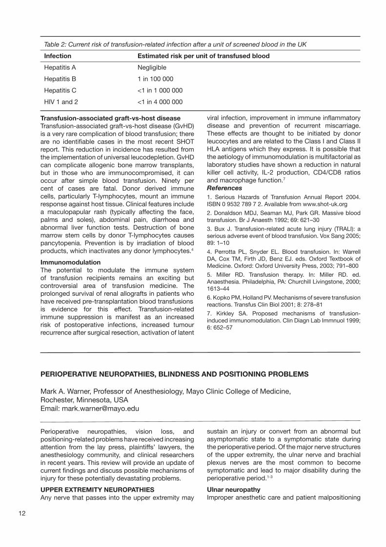
Table 2: Current risk of transfusion-related infection after a unit of screened blood in the UK
Infection Estimated risk per unit of transfused blood
Hepatitis A Negligible
Hepatitis B 1 in 100 000
Hepatitis C <1 in 1 000 000
HIV 1 and 2 <1 in 4 000 000
PERIOPERATIVE NEUROPATHIES, BLINDNESS AND POSITIONING PROBLEMS
Mark A. Warner, Professor of Anesthesiology, Mayo Clinic College of Medicine, Rochester, Minnesota, USA Email: [email protected]
Perioperative neuropathies, vision loss, and positioning-related problems have received increasing attention from the lay press, plaintiffs’ lawyers, the anesthesiology community, and clinical researchers in recent years. This review will provide an update of current findings and discuss possible mechanisms of injury for these potentially devastating problems.
UPPER EXTREMITY NEUROPATHIESAny nerve that passes into the upper extremity may
sustain an injury or convert from an abnormal but asymptomatic state to a symptomatic state during the perioperative period. Of the major nerve structures of the upper extremity, the ulnar nerve and brachial plexus nerves are the most common to become symptomatic and lead to major disability during the perioperative period.1-3
Ulnar neuropathyImproper anesthetic care and patient malpositioning

13
study of ulnar neuropathy in 1,502 surgical patients found that none of the patients developed symptoms of the neuropathy during the first two postoperative days.10
Currently available data suggest that perioperative ulnar neuropathy may be caused by factors other than improper patient positioning and padding of extremities during surgery. Elbow flexion, especially to greater than 100°, can elongate the ulnar nerve and tightening the cubital tunnel retinaculum, directly compressing the ulnar nerve (Figures 1-3).11-13 The
Figure 1: The proximal edge of the roof of the cubital tunnel is formed by a retinaculum that originates on the medial epicondyle and inserts on the olecranon. It is distinct from the aponeurosis of the flexor carpi ulnaris (FCU) with which its distal margin blends. From O’Driscoll SW, et al: J Bone Joint Surg 1991; 73-B:613-617, with permission.
Figure 3: In this medial-to-lateral view of the right elbow, the cubital tunnel retinaculum (CTR) is lax in extension (A) as it stretches from the medial epicondyle (ME) to the olecranon (Ol). The retinaculum tightens in flexion (B) and can compress the ulnar nerve (arrow). From O’Driscoll SW, et al: J Bone Joint Surg 1991; 73-B:613-617, with permission.
Figure 2: Intraneural and extraneural pressures for the ulnar nerve within the cubital tunnel increased dramatically with elbow flexion greater than 100°. From Gelberman RH, et al: J Bone Joint Surg 1998; 80:492-501, with permission.
have been implicated as causative factors in the development of ulnar neuropathies since reports by Budinger4 and Garriques5 in the 1890s. These factors are likely to play an aetiological role for this problem in some surgical patients. Other factors, however, may contribute to the development of postoperative ulnar neuropathies. In a series of twelve inpatients with newly acquired ulnar neuropathy, Wadsworth and Williams6 determined that external compression of an ulnar nerve during surgery was a factor in only two patients. A prospective study at the Mayo Clinic found that medical, as well as surgical, patients develop ulnar neuropathies during inpatient and outpatient care7. It is clear that both surgical and medical patients may develop ulnar neuropathies during or after an episode of care.
Typically, anesthesia-related ulnar nerve injury is thought to be associated with external nerve compression or stretch caused by malpositioning during the intraoperative period. While this implication may be true for some patients, three findings suggest that other factors may contribute. First, a retrospective study has found male gender, high body mass index (≥ 38) and prolonged bedrest postoperatively to be associated with these ulnar neuropathies8. Of these, male gender is the factor most commonly associated with perioperative ulnar neuropathy. Various reports suggest that 70-90% of patients who develop this problem are male.1,2,6,8-9 Second, many patients with perioperative ulnar neuropathies have a high frequency of contralateral ulnar nerve conduction dysfunction.9 This finding suggests that many of these patients are likely to have asymptomatic but abnormal ulnar nerves prior to their anaesthetics, and these abnormal nerves may become symptomatic during the perioperative period. Finally, many patients do not notice or complain of ulnar nerve symptoms until more than 48 hours after their surgical procedures.8,9 A prospective

clinical significance of this finding, however, is unclear. Morell et al14 found that elbow flexion did not inhibit ulnar nerve perception, while direct pressure on the ulnar nerve in the post-condylar groove did.
External compression of the ulnar nerve in the absence of elbow flexion also may damage the nerve. Compression within the bony groove posterior to the medial epicondyle may be possible. In a very innovative study Prielipp et al15 have shown that forearm rotation, especially pronation, can increase pressure in the postcondylar groove (Figure 4). Contreras et al16 have noted that the nerve may be more easily compressed by external forces distal to the medial epicondyle where the nerve and its associated artery are quite superficial than in the postcondylar groove (Figure 5).
14
Figure 4: In supination, the pressure over the ulnar nerve is uniformly low, and most of the data are clustered around the zero line. Prielipp RC, et al: Anesthesiology 1999; 91:345-354.
Figure 5: The ulnar nerve and its primary blood supply in the proximal forearm, the posterior ulnar recurrent artery, are very superficial and appear to be susceptible to compression from external pressure as they pass posteriomedially to the tubercle of the coronoid process. The tubercle is larger in men than women, and the adipose layer in this area is thinner in men.16
Brachial plexus neuropathyBrachial plexus neuropathies may masquerade as ulnar neuropathies or be associated with symptoms that suggest injuries to other nerve structures. In general, brachial plexus neuropathies are associated with median sternotomy.17-19 This neuropathy often involves stretch or compression of the brachial
plexus during sternal separation.18,19 Other potential mechanisms of injury include direct trauma from fractured first ribs. In general, brachial plexus neuropathy does not appear to be related to a patient’s arm position or padding during the sternotomy and related procedures.20
The brachial plexus is also vulnerable to stretch in a patient who is positioned prone (Figure 6).21 Stretch of the brachial plexus, especially its lower trunks, is most likely to occur when the head is turned to the contralateral side, the ipsilateral shoulder is abducted, and the ipsilateral elbow is bent. Other potential problems are noted in the legend for Figure 6. Although this position is commonly used during surgical procedures and the frequency of perioperative brachial plexus neuropathy is low, it would appear prudent to place the arms at the patient’s side whenever possible to decrease the risk of brachial plexus stretching. Kamel and colleagues have recently shown that the frequency of SSEP (somato-sensory evoked potential) abnormalities is 3-fold less with arms tucked at the side than elevated in a “surrender” position.22
LOWER EXTREMITY NEUROPATHIESAlthough neuropathies of the lower extremities may occur in a variety of patient postures, most of these occur in patients who are undergoing procedures while placed in a lithotomy position. These neuropathies have often been considered to be preventable and to occur because of poor intraoperative care (e.g. improper positioning or padding) or judgment (e.g. excessively prolonged use of lithotomy position).23 This perception has significant impact on the outcomes of medicolegal cases involving these types of problems.24 Interestingly, the majority of plaintiffs in medicolegal cases involving lower extremity neuropathies name anesthesiologists and surgeons in their complaints. In contrast, plaintiffs in cases involving upper extremity nerves often do not name surgeons.
A number of studies have suggested that there are many factors other than improper intraoperative care that may contribute to the risk of lower extremity nerve injury.25-27 A 1994 retrospective review of patients in lithotomy positions found that the most common lower extremity neuropathies were the common peroneal (81%), sciatic (15%), and femoral (4%).28 The authors found specific patient characteristics that contributed to the risk of neuropathy. A more recent prospective study found that the longer patients were in lithotomy, the greater their risk of developing a neuropathy.29 The obturator and lateral femoral cutaneous (LFC) nerve were most often involved in this study.
Obturator and Lateral Femoral Cutaneous Neuropathies Litwiller et al30 subsequently evaluated the strain of the obturator and LFC nerves associated with

Femoral Neuropathy Unlike most other neuropathies in which the anaesthesia provider is often considered to have acted improperly in order for the neuropathy to occur, those involving the femoral nerve and its cutaneous branches often are considered to result from improper placement of abdominal wall retractors and direct compression of the nerve. When related to retractors, the assumption is that retractors place continuous pressure on the iliopsoas muscle and either stretch the nerve or cause it to become ischemic by occluding the external iliac artery or penetrating vessels of the nerve as it passes through the muscle.31
PRACTICAL CONSIDERATIONS FOR NEUROPATHIESEfforts to prevent perioperative neuropathies are frequently debated, and there is often confusion on how to manage a neuropathy once it has occurred. In general, there are no data to support recommendations on any of these issues. Therefore, the following opinions have been formulated by personal experience, guided by advice from neurologists who primarily care for patients with peripheral neuropathies, and seasoned or supported by speculation derived from anecdotal case reports.
Padding exposed peripheral nerves Many types of padding materials are advocated to protect exposed peripheral nerves. They often consist of cloth (e.g. blankets and towels), foam sponges (e.g. “eggcrate” foam), and gel pads. There are no data to suggest that any of these materials is more effective than any other, or that any is better than no padding at all. A good rule-of-thumb would be to position and pad exposed peripheral nerves to (1) prevent their stretch beyond normally tolerated limits while awake,
15
lithotomy positions in fresh cadavers. They found that neither hip flexion nor abduction increased strain on the LFC nerve. However, abduction to >30o without concomitant hip flexion dramatically increased strain on the obturator nerve.
Common Peroneal Neuropathy The common peroneal nerve is very superficial as it wraps around the head of the fibula. Because it is quite exposed at this level, it may easily be compressed and injured. Although direct compression of the peroneal nerve by leg holders has commonly been considered the primary mechanism of injury in peroneal neuropathy, a recent study suggests that the superficial peroneal nerve may be affected distal to the fibular head.29 The authors speculated that compressive stockings or wraps may be aetiologic factors for this finding.
Sciatic NeuropathyThe same forces that contribute to stretch injuries of the hamstring group muscles (e.g. biceps femoris muscle) may stretch the sciatic nerve. Simultaneous hyperflexion of the hip and extension of the knee will stretch and possibly injure the sciatic nerve. This set of actions can occur during the establishment and maintenance of a lithotomy position. A patient in a lithotomy position may passively shift towards the caudal end of an operating table when placed in a head-up posture or be actively shifted caudally by a member of the operating team, in an attempt to obtain increased exposure of the perineum. This movement may increase the flexion of the hips and extension of the legs, if the legs are already fixated within leg holders. It would seem prudent to confirm that the flexor muscles of the knee (e.g. hamstring group) are not taut after placing a patient’s legs into any lithotomy position.
Figure 6: Sources of potential injury to the brachial plexus and its peripheral components in a prone patient. Head position stretching plexus against anchors in shoulder (A). Closure of retroclavicular space by chest support with arms at side; neurovascular bundle trapped against first rib (B). Head of humerus thrust into neurovascular bundle if arm and axilla are not relaxed (C). Compression of ulnar nerve in cubital tunnel (D). Area of vulnerability of radial nerve to compression above elbow (E).

(2) avoid their direct compression, if possible, and (3) distribute over as large an area as possible any compressive forces that must be placed on them.
What to do if your patient develops a neuropathy? Although each situation is unique and requires careful assessment, the following guidelines may suggest a basic course of action that will lead to appropriate care:
• Is the neuropathy sensory or motor? Sensory lesions are more frequently transient than motor lesions. If the symptoms are numbness and/or tingling only, it may be appropriate to inform the patient that many of these neuropathies will resolve during the first 5 days.10 The patient should be instructed to avoid postures that might compress or stretch the involved nerve. Arrangements should be made for frequent contact with the patient. A call to alert a neurologist would be appropriate, and if the symptoms still persist on postoperative day 5, the neurologist should be consulted.
• If the neuropathy has a motor component, a neurologist should be consulted immediately. Electromyographic studies may be needed to assess the location of any acute lesion. This knowledge may direct an appropriate treatment plan. The studies may also demonstrate chronic abnormalities of the nerve or, if applicable, the contralateral nerve.
BLINDNESSOver the past decade there has been speculation that the frequency of perioperative blindness has been increasing, especially in patients undergoing procedures while positioned prone for prolonged periods (e.g. major spine surgeries). Interestingly, there are few data to support this speculation. The rate of spinal fusion procedures has, however, increased in the past decade and may be a contributing factor.32 It appears that most non-surgically related postoperative vision loss occurs in patients undergoing cardiac procedures, followed in frequency by patients undergoing spine surgery.33
Potential Pathologies In the absence of surgical excision or trauma to visual tissues, most cases involve anterior or posterior ischemic optic neuropathy (AION and PION, respectively), central retinal artery occlusion, or undefined ischemia to the cerebral cortex. There are very few cases reported in the past 2 decades in which direct pressure to the globe is implicated in perioperative blindness. Blindness in cardiac patients is approximately balanced between AION and PION. In contrast, PION appears to be the predominant problem in prone-positioned patients.
The aetiology of PION is unknown. There is no doubt that prone-positioned, anesthetised patients develop an increase in intraocular pressure.34-36 This increase
appears related, in part, to the impact of gravity and increased central venous pressure in prone-positioned patients.34,37 Posture-induced changes in the anatomy and function of the iris and lens also may contribute.38 This potential contribution of intraocular anatomy in prone-positioned patients has been supported by the finding that timolol solution can attenuate the increase in intraocular pressure.39 Anemia and hypotension have been considered potential aetiologies, primarily based on information propagated by isolated case reports and small case series,40,41 but an exhaustive review on this topic, as it pertains to spine surgery patients, has found no evidence of an association between these factors and perioperative visual loss.42 Periorbital oedema may occur in prone-positioned patients, or vertically-inverted study subjects,43,44 but this oedema does not appear to be correlated with visual loss.42 There is speculation (without data) that engorgement of the veins in and around the optic nerve and its sheath may cause compartment compression of the optic nerve sheath, limiting arterial perfusion to its posterior extension. This posterior extension of the nerve just anterior to the optic chiasm, has few major arterial vessels and may have an increased risk of low perfusion.45
Risk Factors There are sufficient numbers of cases in cardiac surgical patients to retrospectively determine risk factors for this problem. Nuttall et al46 found in cardiac surgical patients that patient factors (advanced age and arteriosclerosis), procedure issues (prolonged pump perfusion and surgical disruption of particulate matter), and practice patterns (deliberate postoperative anemia and intraoperative hypotension) are associated with an increased frequency of vision loss. There are insufficient numbers of cases in any series to evaluate risk factors in non-cardiac surgical patients. However, a recent report from the ASA’s Closed Claims Postoperative Visual Loss Registry suggests that most cases of vision loss in spinal surgery occur in patients who are positioned prone, undergo procedures lasting more than 6 hours, and who experience substantial blood loss.47
General Guidelines The conclusions of the ASA Task Force on Perioperative Blindness are shown in Table 1.42
SEVERAL POTENTIAL CATASTROPHIC POSITIONING PROBLEMSSpinal cord ischemia or infarction from lumbar hyperextension Many patients who undergo pelvic procedures using an abdominal approach are positioned supine with their lumbar spines hyperextended in an attempt to increase surgeon visibility into the lower pelvis. This practice is reasonable as long as the mechanism for hyperextending the lumbar spine is limited to the maneuvers allowed by operating room tables (e.g. raising the kidney rest). Tables manufactured within
16

17
the U.S. do not allow hyperextension of the lumbar spine to great than 10°. When excessive padding is introduced under the lumbar spine to gain additional hyperextension, however, the degree of hyperextension may exceed 10°. The 10° angle is important because there are no reports of anterior spinal cord ischemia when patients are positioned using only the table mechanisms to induce lumbar hyperextension. When additional padding or other maneuvers are used to increase hyperextension, however, the spinal cord may become ischemic and infarct.48
Thoracic outlet obstruction Elevation of the arms at the shoulders to greater than 90° abduction may be associated with thoracic outlet obstruction in some patients. Patients positioned prone and who may have their shoulders abducted to greater than 90° (i.e. a “surrender” position) should be asked preoperatively if elevation of their arms causes cold, pain, or tingling. These symptoms suggest potential for thoracic outlet obstruction and should be considered when positioning patients. Most patients are most comfortable with their arms at their sides when positioned prone, and many procedures in prone-positioned patients can be performed when the arms are tucked at the sides.
References1. Kroll DA, Caplan RA, Posner K, Ward RJ, Cheney FW: Nerve injury associated with anesthesia. Anesthesiology 1990; 73: 202-7
2. Cheney FW, Domino KB, Caplan RA, Posner KL: Nerve injury associated with anesthesia: a closed claims analysis. Anesthesiology 1999; 90: 1062-9
3. Dawson DM, Krarup C: Perioperative nerve lesions. Arch Neurol 1989; 46: 1355-60
4. Büdinger K: Ueber Lähmungen nach chloroform-narkosen. Arch f Kin Chirc 1894; 47: 121-145
5. Garriques HJ: Anaesthesia-paralysis. Am J Med Sci 1897; 133: 81-89
Table 1
• There is a subset of patients who undergo spinal procedures, while they are positioned prone and receiving general anesthesia, that has an increased risk for development of perioperative visual loss. This subset includes patients who are anticipated preoperatively to undergo procedures that are prolonged, have substantial blood loss, or both (high-risk patients).
• Consider informing high-risk patients that there is a small, unpredictable risk of perioperative visual loss.
• The use of deliberate hypotensive techniques during spine surgery has not been shown to be associated with the development of perioperative visual loss.
• Colloids should be used along with crystalloids to maintain intravascular volume in patients who have substantial blood loss.
• At this time, there is no apparent transfusion threshold that would eliminate the risk of perioperative visual loss related to anemia.
• High-risk patients should be positioned so that their heads are level with or higher than the heart when possible. In addition, their heads should be maintained in a neutral forward position (i.e. without significant neck flexion, extension, lateral flexion, or rotation) when possible.
• Consideration should be given to the use of staged spine procedures in high-risk patients.
6. Wadsworth TG, Williams JR: Cubital tunnel external compression syndrome. Br Med J 1973; 1: 662-6
7. Warner MA, Warner DO, Harper CM, Schroeder DR, Maxson PM: Ulnar neuropathy in medical patients. Anesthesiology 2000; 92: 613-5
8. Warner MA, Warner ME, Martin JT: Ulnar neuropathy. Incidence, outcome, and risk factors in sedated or anesthetized patients. Anesthesiology 1994; 81: 1332-40
9. Alvine FG, Schurrer ME: Postoperative ulnar-nerve palsy. Are there predisposing factors? J Bone Joint Surg Am 1987; 69: 255-9
10. Warner MA, Warner DO, Matsumoto JY, Harper CM, Schroeder DR, Maxson PM: Ulnar neuropathy in surgical patients. Anesthesiology 1999; 90: 54-9
11. O’Driscoll SW, Horii E, Carmichael SW, Morrey BF: The cubital tunnel and ulnar neuropathy. J Bone Joint Surg Br 1991; 73: 613-7
12. Gelberman RH, Yamaguchi K, Hollstien SB, Winn SS, Heidenreich FP, Jr., Bindra RR, Hsieh P, Silva MJ: Changes in interstitial pressure and cross-sectional area of the cubital tunnel and of the ulnar nerve with flexion of the elbow. An experimental study in human cadavera. J Bone Joint Surg Am 1998; 80: 492-501
13. Grewal R, Varitimidis SE, Vardakas DG, Fu FH, Sotereanos DG: Ulnar nerve elongation and excursion in the cubital tunnel after decompression and anterior transposition. J Hand Surg [Br] 2000; 25: 457-60
14. Morell RC, Prielipp RC, Harwood TN, James RL, Butterworth JF: Men are more susceptible than women to direct pressure on unmyelinated ulnar nerve fibers. Anesth Analg 2003; 97: 1183-8, table of contents
15. Prielipp RC, Morell RC, Walker FO, Santos CC, Bennett J, Butterworth J: Ulnar nerve pressure: influence of arm position and relationship to somatosensory evoked potentials. Anesthesiology 1999; 91: 345-54
16. Contreras MG, Warner MA, Charboneau WJ, Cahill DR: Anatomy of the ulnar nerve at the elbow: potential relationship of acute ulnar neuropathy to gender differences. Clin Anat 1998; 11: 372-8

18
17. Hickey C, Gugino LD, Aglio LS, Mark JB, Son SL, Maddi R: Intraoperative somatosensory evoked potential monitoring predicts peripheral nerve injury during cardiac surgery. Anesthesiology 1993; 78: 29-35
18. Vahl CF, Carl I, Muller-Vahl H, Struck E: Brachial plexus injury after cardiac surgery. The role of internal mammary artery preparation: a prospective study on 1000 consecutive patients. J Thorac Cardiovasc Surg 1991; 102: 724-9
19. Roy RC, Stafford MA, Charlton JE: Nerve injury and musculoskeletal complaints after cardiac surgery: influence of internal mammary artery dissection and left arm position. Anesth Analg 1988; 67: 277-9
20. Jellish WS, Blakeman B, Warf P, Slogoff S: Hands-up positioning during asymmetric sternal retraction for internal mammary artery harvest: a possible method to reduce brachial plexus injury. Anesth Analg 1997; 84: 260-5
21. Martin JT: The ventral decubitus (prone) positions, Positioning in Anesthesia and Surgery, 3rd Edition. Edited by Martin JT, Warner MA. Philadelphia, W.B. Saunders Company, 1997, pp 155-195
22. Kamel IR, Drum ET, Koch SA, Whitten JA, Gaughan JP, Barnette RE, Wendling WW. Anesth Analg 2006;102:1538-42
23. Dornette WH: Compression neuropathies: medical aspects and legal implications. Int Anesthesiol Clin 1986; 24: 201-29
24. Dornette WHL: Identifying, moving, and positioning the patient, Legal Issues in Anesthesia Practice. Edited by Dornette WHL. Philadelphia, FA Davis, 1991, pp 120-123
25. Rose HA, Hood RW, Otis JC, Ranawat CS, Insall JN: Peroneal-nerve palsy following total knee arthroplasty. A review of The Hospital for Special Surgery experience. J Bone Joint Surg Am 1982; 64: 347-51
26. James SE, Wade PJ: Lateral popliteal nerve palsy as a complication of the use of a continuous passive motion knee machine--a case report. Injury 1987; 18: 72-3
27. Weber ER, Daube JR, Coventry MB: Peripheral neuropathies associated with total hip arthroplasty. J Bone Joint Surg Am 1976; 58: 66-9
28. Warner MA, Martin JT, Schroeder DR, Offord KP, Chute CG: Lower-extremity motor neuropathy associated with surgery performed on patients in a lithotomy position. Anesthesiology 1994; 81: 6-12
29. Warner MA, Warner DO, Harper CM, Schroeder DR, Maxson PM: Lower extremity neuropathies associated with lithotomy positions. Anesthesiology 2000; 93: 938-42
30. Litwiller JP, Wells RE, Jr., Halliwill JR, Carmichael SW, Warner MA: Effect of lithotomy positions on strain of the obturator and lateral femoral cutaneous nerves. Clin Anat 2004; 17: 45-9
31. Rosenblum J, Schwarz GA, Bendler E: Femoral neuropathy--a neurological complication of hysterectomy. Jama 1966; 195: 409-14
32. Deyo RA, Gray DT, Kreuter W, Mirza S, Martin BI. United States trends in lumbar fusion surgery for degenerative conditions. Spine 2005;30: 1441-5; discussion 1446-7
33. Warner MA. Postoperative visual loss: Experts, data, and practice. Anesthesiology 2006; 105:641-2
34. Lee LA, Vavilala MS, Sires BS, Chapman J, Lam AM: Intraocular pressure is partially dependent on central venous pressure during prone spine surgery. Anesthesiology 2003; 99: A289
35. Murphy DF: Anesthesia and intraocular pressure. Anesth Analg 1985; 64: 520-30
36. Cheng MA, Todorov A, Tempelhoff R, McHugh T, Crowder CM, Lauryssen C: The effect of prone positioning on intraocular pressure in anesthetized patients. Anesthesiology 2001; 95: 1351-5
37. Ozcan MS, Praetel C, Bhatti MT, Mahla ME, Seubert CN: The effect of body inclination during prone positioning on intraocular pressure in awake volunteers: Comparison of two operating tables. Anesthesiology 2003; 99: A363
38. Lam AK, Douthwaite WA: Does the change of anterior chamber depth or/and episcleral venous pressure cause intraocular pressure change in postural variation? Optom Vis Sci 1997; 74: 664-7
39. Caiati J, Eliazo R, Lien C, Mack PF: Timolol prevents the dramatic increase in intra-ocular pressure (IOP) associated with prone positioning for spine surgery. Anesthesiology 2003; 99: A309
40. Roth S, Barach P: Postoperative visual loss: still no answers--yet. Anesthesiology 2001; 95: 575-7
41. Brown RH, Schauble JF, Miller NR: Anemia and hypotension as contributors to perioperative loss of vision. Anesthesiology 1994; 80: 222-6
42. American Society of Anesthesiologists Task Force on Perioperative Blindness. Practice advisory for perioperative visual loss associated with spine surgry. Anesthesiology 2006; 104:1319-28
43. Draeger J, Hanke K: Postural variations of intraocular pressure--preflight experiments for the D1-mission. Ophthalmic Res 1986; 18: 55-60
44. Friberg TR, Weinreb RN: Ocular manifestations of gravity inversion. Jama 1985; 253: 1755-7
45. Hayreh SS. Prog Retin Eye Res 2001;20:563-93
46. Nuttall GA, Garrity JA, Dearani JA, Abel MD, Schroeder DR, Mullany CJ: Risk factors for ischemic optic neuropathy after cardiopulmonary bypass: a matched case/control study. Anesth Analg 2001; 93: 1410-6, table of contents
47. Lee LA, Roth S, Posner KL, Cheney FW, Caplan RA, Newman NJ, Domino KB. The American Society of Anesthesiologists postoperative visual loss registry: analysis of 93 spine surgery cases with postoperative visual loss. Anesthesiology 2006; 105: 652-9
48. Amoiridis G, Wohrle JC, Langkafel M, Maiwurm D, Przuntek H: Spinal cord infarction after surgery in a patient in the hyperlordotic position. Anesthesiology 1996; 84: 228-30
Please answer the following questions before reading the article. The answers are contained in the article.
1. A 70-year-old man with ischaemic heart disease has been in hospital for two days following a road traffic accident, in which he sustained injuries to his spleen, bowel and several long bones. He has become markedly short of breath. What further information would you seek to make a diagnosis of ARDS?
2. Describe the pathological processes occurring in the lungs of a patient with ARDS that can lead to
a. Shunt
b. Deadspace
c. Decreased compliance
3. ARDS is diagnosed in the patient in question 1. He has now been ventilated in the ICU for 3 days. You are called because the PaO2 has decreased and the airway pressures have increased. The nurse mentions that respiratory secretions have become purulent. Describe the procedures / investigations you might carry out.
4. A 70kg patient with ARDS is being ventilated with the following settings and arterial blood gases. Which if any would you consider changing, and why?
Tidal volume 650 ml
FiO2 0.7
Plateau pressure 35
PEEP 1 0 cmH2O
PaO2 9 kPa
PaCO2 8.8 kPa
pH 7.26
5. A 60kg patient with ARDS is receiving volume-controlled ventilation with the following settings:
Tidal volume 450 ml
FiO2 0.8
Plateau pressure 35 cmH2O
PEEP 15 cmH2O
PaO2 remains less than 8 kPa. What ventilatory strategies would you consider to improve the PaO2? Which if any have been shown to improve survival?
6. What are the disadvantages of using pressure-controlled inverse-ratio ventilation?
IntroductionFirst described in 1967, ARDS is a process of hypoxaemic respiratory failure associated with non-cardiogenic pulmonary oedema. It is the result of diffuse inflammatory damage to the alveoli and pulmonary capillaries from a range of local or systemic insults. ARDS is often associated with multiple organ dysfunction and carries a high mortality and financial cost.
DefinitionsARDS is diagnosed on clinical grounds. The most commonly used criteria were produced by the 1994 American-European Consensus Conference1.
• Acute onset
• Appropriate precipitating condition
• New bilateral fluffy infiltrates on chest x-ray (CXR)
• No clinical evidence of cardiac failure, fluid overload or chronic lung disease
• Pulmonary artery occlusion pressure of ≤ 18mmHg
• Pa02/Fi02 ratio of ≤ 200mmHg (27kPa) - note the FiO2 is expressed as a fraction (50% is 0.5)
Acute Lung Injury (ALI) is a less severe form of ARDS in which the Pa02/Fi02 ratio is ≤ 300mmHg (40kPa). Note that the Pa02/Fi02 ratio is independent of PEEP (positive end-expiratory pressure). As use of pulmonary artery catheters is becoming increasingly rare, the occlusion pressure ≤18 is often not sought and clinical grounds or echocardiography used to exclude cardiac failure.
EpidemiologyThe true incidence of ARDS is unknown; estimates vary depending on the definitions used, with values ranging from 1.5 per 100 000 population per year to 75 per 100 000 population per year. Recent data from an Australian study which used the 1994 consensus conference definition for ARDS would suggest that one in ten non-cardiothoracic ICU patients will develop ARDS.
Although ARDS may affect children it is more common in those over the age of 65, which may reflect a higher incidence of predisposing conditions. Gender has no effect.
In recent years mortality rates have decreased from about 60% to 30-40% but mortality is higher in the elderly and in patients with factors such as chronic liver disease. Most of those who die do so from sepsis or
ACUTE RESPIRATORY DISTRESS SYNDROME (ARDS)
19
David Lacquiere, Derriford Hospital, Plymouth, UK. Email: [email protected]

multiple organ failure and not from respiratory failure. Survivors usually have little in the way of pulmonary sequelae, although the severest cases may have restrictive lung disease.
Precipitating conditionsTheses can be classified as direct or indirect.
Direct Indirect
Pneumonia Multiple trauma
Lung contusion Massive transfusion
Aspiration of gastric Sepsis contents
Fat embolism Pancreatitis
Toxic inhalation Cardiopulmonary bypass
Near drowning Burns
Reperfusion injury Bone marrow transplant
Drugs and toxins
PathophysiologyIt is not understood why some individuals develop ARDS/ALI while others with the same pattern of predisposing injury do not. In those that do there are said to be three overlapping phases2: an inflammatory phase, a proliferative phase and a fibrotic phase, caused by the subsequent reparative response. Patients with ARDS/ALI do not have to progress through all three phases as resolution can occur at any point. However, the severest form of ARDS will progress to the fibrotic phase.
Inflammatory phaseThis lasts for one week after the onset of respiratory failure. Neutrophils accumulate in the capillaries, interstitial tissue and airspaces, and cause cell damage through the production of free radicals, inflammatory mediators and proteases. However, neutrophils are not the only cell type involved as ARDS does occur in neutropenic patients. Cytokines (most importantly TNF-a, IL-1 IL-6 and IL-8) are also released by endothelial and immune cells and promote similar microvascular damage. The result is leakage of fluid and plasma proteins into the alveoli and interstitial tissues (‘non-cardiogenic pulmonary oedema’), while at the same time the plasma proteins denature alveolar surfactant causing alveolar collapse. This creates hypoxia as the fluid-filled alveoli shunt blood. Shunt is created when areas of lung receive a blood supply but are unable to oxygenate it (in this case by creating a diffusion barrier).
To complicate matters further, vasoconstriction and occlusion of pulmonary capillaries by neutrophils, platelets and fibrin also occurs, leading to areas of lung that are ventilated but not perfused – deadspace.
The increase in total lung water also stiffens the lung (decrease in compliance) and this dramatically increases the work of breathing.
Proliferative phaseThis phase is characterised by proliferation of type II pneumocytes and fibroblasts, with the formation of hyaline membranes. However, these pneumocytes do not make any surfactant and total production of surfactant decreases, exacerbating the loss of surfactant caused by denaturing of protein.
Fibrotic phaseDisordered collagen deposition occurs, leading to extensive lung scarring. This makes the lung stiffer and further increases the work of breathing. This can be severe enough to make it impossible to wean the patient from a ventilator but normally it is a matter of restoring the patient’s muscle strength to the point where they are able to cope with the increased effort required.
PresentationThe timing of the onset of clinical features varies from a few hours to several days after the precipitating insult.
HistoryShortness of breath is universal, but other symptoms are related to the predisposing condition.
ExaminationFindings are similar to those of pulmonary oedema due to other causes:
lRespiratory – laboured breathing, tachypnoea, diffuse crackles, cyanosislCardiovascular – sweating, tachycardialCNS – agitation, leading to lethargy and decreased level of consciousness
In addition there may be features of the underlying condition.
InvestigationsArterial blood gases lPa02/Fi02 ratio of ≤ 200mmHg (27kPa) or ≤ 300mmHg (40kPa) for ALIlHypocarbia may be seen though hypercarbia develops later, as respiratory failure progresses
Radiology l CXR shows diffuse bilateral fluffy shadows (though initially they may be less widespread or unilateral), and may show other pulmonary signs if there is a direct pulmonary predisposing condition
Management (1) – supportive measuresThere are no specific treatments for ARDS but treating the underlying condition (for example eradicating infection with antibiotics or surgery), and providing support for each system are paramount.
20

Respiratory supportFrequent CXRs will help to detect pneumothorax, fluid overload and pneumonia, which may all complicate ARDS. Pneumothorax in particular should be sought if there is a sudden increase in ventilation pressures or deterioration in blood gases. CT scanning may help to show occult pneumothorax.
Frequent physiotherapy is also important, to prevent plugging of the airways by sputum. If plugging is suspected (for example by lobar collapse and deterioration in blood gases) bronchoscopy and lavage may help.
Cardiovascular supportThe aim is to maintain adequate oxygen delivery to the tissues.
In ARDS cardiac output can be decreased due to sepsis or due to medical treatments (high ventilation pressures, PEEP, or inversed inspiratory:expiratory ratios), thus monitoring of cardiac output and filling pressures are important. Where available this can be achieved using a pulmonary artery catheter, oesophageal Doppler, LidCO or PiCCO devices but clinical signs are also important (see Update 21). Fluid management is always difficult in these cases - excessive fluids will worsen lung function and inadequate fluids will exacerbate renal failure. With cardiac output monitoring the situation is easier - volume challenges of 250ml can be given to achieve the highest stroke volume the patient is capable of and if cardiac output is still inadequate then inotropes are indicated. If cardiac output monitoring is unavailable then systolic pressure variation can be helpful (although there are limitations such as the patient needing to be deeply sedated.) Measure the systolic blood pressure when the airway pressure is held at 30cm H20 and when it is held at 10cm H2O if the difference is greater than 10mmHg then fluid may help.
Appropriate targets are cardiac output 3.5 – 5 l/min/m2, Hb 7-9 g/dl (do not overtransfuse) and Sa02 ≥ 90% (see below).
Renal supportRenal failure is common, due to the underlying condition, low cardiac output and sepsis. Renal replacement therapy (for example with haemofiltration) may also improve gas exchange, by removing excess fluid.
NutritionEnteral nutrition should be established quickly, using nasogastric feed with prokinetics (such as metoclopramide) or nasojejunal feeding. TPN can be considered if all attempts at enteral feeding fail.
Managing SepsisSepsis may have precipitated the lung injury, or may develop during the course of ARDS. However, the
systemic inflammatory response syndrome is often associated with ARDS in the absence of infection, so detecting sepsis may be difficult.
Change in sputum colour and new shadows on the CXR may point to pulmonary infection. Other sources of sepsis should be frequently reviewed (line sites, urine, wounds).
If infection is suspected, appropriate samples should be sent for microscopy and culture. This may include bronchoscopy and lavage or removing and culturing invasive line tips, for example. Lavage is particularly useful in this setting. 20mls of normal saline are injected into the airway either through a bronchoscope or via a sterile suction catheter (placed blindly through the endotracheal tube until resistance is felt) and suctioned back into a culture pot. The likelihood of a significant positive result is higher with this technique and less tracheal contamination is encountered. Antimicrobial therapy should be guided by the results of these investigations, though ‘blind’ treatment may be reasonable if sepsis causes severe cardiovascular instability or impairment of gas exchange.
Management (2) – VentilationContinuous positive airways pressure (CPAP) may be of benefit in mild cases, however most patients will require early intubation and mechanical ventilation. Indications include hypoxaemic or hypercarbic respiratory failure, acidosis, exhaustion and reduced conscious level. Profound sedation is usually required for ventilation as struggling or coughing can cause loss of recruited lung and worse oxygenation. Paralysis may be necessary if sedation alone does not settle the patient.
The aim of ventilation is to improve oxygenation without causing further damage to the lungs. Difficulties arise as some alveoli are normal and open whilst other alveoli are stiff and collapsed. It is therefore necessary to try to open the collapsed alveoli without damaging the normal areas. The main causes of ventilator-induced lung damage are high Fi02 (increased free radical damage) and over-distension of alveoli. Ventilation reduces the work of breathing and reduces oxygen demand and this should help correct acidosis and improve cardiovascular stability.
With the exception of low tidal volumes (see below) there is little evidence of survival benefit for any particular ventilation strategy, however, volume-controlled ventilation is usually used initially, with the following targets:
• Fi02 0.5-0.6 to minimise oxygen toxicity.
• Pa02 ≥ 8kPa (Sa02 ≥ 90%) - do not attempt to achieve higher values.
• PaC02 < 10kPa as long as pH > 7.2. Do not attempt to achieve lower values if this requires excessively high tidal volumes (‘permissive hypercapnia’).
21

• Tidal volumes 6-8ml/kg body weight (to minimise alveolar distension and volutrauma), as suggested by the ARDS Network study3.
• Plateau pressures of 30-35 cmH20 to minimise alveolar distension and volutrauma.
• Positive end-expiratory pressure (PEEP) titrated to achieve best oxygen delivery – commonly 10-15cmH20. This increases functional residual capacity, recruits alveoli and puts the lung on the steeper part of the compliance curve. Higher levels of PEEP should be avoided, as they decrease venous return and thus cardiac output – PEEP should be set to maximise oxygen delivery rather than oxygenation alone.
• Recruitment manoeuvres. This is the use of a high level of CPAP (30-40cmH20) for 30 seconds in an apnoeic patient via a ventilator. The aim is to recruit collapsed alveoli, and its occasional use may lead to marked improvements in oxygenation.
Pressure-Controlled Inverse Ratio Ventilation (PC-IRV)When ventilation using the above targets fails to improve oxygenation, PC-IRV may be attempted. The key features are:
• The inspiratory time (I) is prolonged until it is equal to or greater than expiratory time (E), for example using an I:E ratio of 1:1, 2:1 or 3:1. This allows time for poorly compliant lung units to be ventilated and should improve oxygenation.
• The pressure-controlled nature of the breath allows a plateau pressure to be set, to prevent over-distension of compliant (less diseased) alveoli.
• Plateau pressures should not exceed 35 cmH20, and should be set to achieve tidal volumes of 6-8ml/kg body weight.
This technique has important side effects:
• Mean intra-thoracic pressures will be raised, thus decreasing venous return and cardiac output.
• The shortened expiratory time may not leave enough time for gas to escape from the lung, leading to high levels of ‘auto-PEEP’ (also called ‘intrinsic PEEP’). As well as further decreasing venous return, high auto-PEEP can impair ventilation, as the resting lung pressure becomes too high to allow expansion during inspiration. It is important therefore to periodically measure total PEEP (set PEEP plus auto-PEEP) and decrease set PEEP accordingly.
Auto-PEEP is measured by placing the ventilator into expiratory pause and measuring the highest airway pressure created. Airway pressure should be the same as PEEP but if gas trapping occurs airway pressure will rise as the alveoli empty - auto PEEP.
22
• The shortened expiratory time may also lead to hypercarbia – high respiratory frequency may be needed to avoid excessive respiratory acidosis.
• PC-IRV is also extremely uncomfortable for the patient, thus heavy sedation +/- paralysis are usually needed.
Ventilation in the prone positionDramatic improvements in oxygenation are often seen in patients who are turned into the prone position for several hours, and this improvement may be sustained when they are returned to the supine position4. The technique should be used for periods of 12 – 24hours. Prone ventilation is commonly used in patients with ARDS.
The mechanism probably involves better matching of lung perfusion with ventilation, and redistribution of dependent lung oedema. However, there are practical difficulties in turning the critically ill patient and in nursing the patient in the prone position. It should also be noted that there is no mortality benefit to be gained – at best it can buy time in cases of resistant hypoxaemia.
Management (3) – additional measures
A number of advanced techniques are available, but none have been proven to increase survival.
• Nebulised prostacyclin – this produces pulmonary vasodilation, dilating those vessels in well ventilated parts of the lung, thus improving ventilation perfusion matching. Because it is removed from the circulation rapidly it does not cause systemic hypotension. Prostacyclin should be continuously nebulised at a rate of 5-20ng/kg/min
• Inhaled nitric oxide – like prostacyclin this is a selective pulmonary vasodilator, and is used in doses of 1-40 parts per million. Neither agent has been shown to influence survival.
• Corticosteroids – there is some evidence from a small study of a reduction in mortality associated with the use of methylprednisolone to suppress ongoing inflammation during the fibroproliferative phase of ARDS. The initial regimen consists of methylprednisolone 2mg/kg daily. Any response should be seen in 3-5 days. In 1-2 weeks the dose can be tapered to methylprednisolone 0.5-1.0mg daily. In the absence of response, steroids should be discontinued.
• Surfactant therapy – aims to replace surfactant lost from the lung and thus improve compliance and alveolar stability, and decrease lung water. However, early results have been disappointing.
• High frequency jet ventilation – this can be used to raise mean airway pressure without dangerous increases in peak airway pressure, but is expensive and only available in specialist centres.

• Extracorporeal membrane oxygenation – a pump oxygenator performs gas exchange allows the lungs to be ‘rested’. Again, only available in specialist centres.
Summary
ARDS is diagnosed clinically on the basis of the acute development of hypoxaemic respiratory failure, CXR changes and non-cardiogenic pulmonary oedema, on the background of a pulmonary or non-pulmonary precipitating condition. ARDS may affect one in ten intensive care unit patients, and it carries a mortality of 30-40%.
Pathologically ARDS is characterised by an inflammatory phase involving neutrophils and cytokines, followed by a reparative process that may end in fibrosis.
Patients exhibit the signs and symptoms of pulmonary oedema, though features of the underlying condition may influence the picture.
Management consists of treating the underlying condition, providing support for failing systems and early invasive ventilation. Limiting the Fi02 may help
to prevent further lung damage, while limiting tidal volumes to 6-8ml/kg has been shown to reduce mortality. In cases of refractory hypoxaemia PC-IRV or ventilation in the prone position may improve blood gases, but have not been proven to influence survival. In addition there are many advanced techniques but many are only available in specialist centres, and none convincingly reduce mortality.
References
1. Bernard GR, Artigas A, Brigham KL, et al. The American-European Consensus Conference on ARDS: definitions, mechanisms, relevant outcomes, and clinical trial coordination. Am J Respir Crit Care Med 1994; 149: 818-24.
2. Luce JM. Acute lung injury and the acute respiratory distress syndrome. Crit Care Med 1998; 26(2): 369-76.
3. ARDSnet. Ventilation with lower tidal volumes as compared with traditional tidal volumes for acute lung injury and the acute respiratory distress syndrome. N Eng J Med 2000; 342: 1301-8.
4. Gattinoni L, Tognoni G, Pesenti A, et al. Effect of prone positioning on the survival of patients with acute respiratory failure. N Eng J Med 2001; 345: 568-73
23
ASSESSMENT OF SPINAL ANAESTHETIC BLOCK
Spinal anaesthesia has the advantage that profound nerve block can be produced in a large part of the body by the relatively simple injection of a small amount of local anaesthetic. The practical technique has been described previously so will not be covered again in this article [see Update 12]. However, the greatest challenge in spinal anaesthesia is to control the spread of local anaesthetic through the cerebrospinal fluid (CSF) to provide a block which is adequate for the proposed surgery without unnecessary extensive spread, and increased risk of complications. This article will cover the assessment of spinal anaesthetic block. In an article in Update 23 the factors that influence how the local anaesthetic spreads within the CSF, determining the extent of the block, will be covered. Learning will be improved if you try to answer the questions posed throughout the text before continuing on to the next section.
IntroductionStudies of drug distribution usually involve measurements of concentration in a relevant body fluid compartment over time. However, multiple sampling of CSF at one level, let alone at the several needed to build an image of drug distribution through
the theca, is impractical. Indirect indicators of spread are used based on tests of neurological response. Some indicator of the degree and extent of nerve block is needed before surgery can start.
Before moving on, think about what methods you know to test the extent of spinal anaesthetic block?
What types of nerves are you studying for each test you can think of?
Many methods may be used to test a block, but they fall broadly into one of two groups: assessment of either afferent (sensory), or efferent (motor or autonomic) function.
Afferent functionPinprick and cold are most commonly used, but mechanical stimuli such as touch, skin pinch, pressure and gas jets can be used. Generally, loss of sensation to cold occurs before pinprick, and both of these before touch, each stage correlating with inhibition of C, Aδ and Aβ fibres respectively. Thus, temperature perception is lost before pinprick, is
Graham Hocking, Sir Charles Gairdner Hospital, Perth, Western Australia. Email: [email protected]
24
generally at a higher level, and is usually assessed by the application of ‘cold’ using alcohol skin prep, ice, ethyl chloride, or a cold gel bag. Loss of vibration and proprioceptive sensation have also been used.
More definitive assessment of pain sensation has been attempted with tetanic stimulation using peripheral nerve stimulators, and transcutaneous electrical nerve stimulation, both of which correlate well with surgical incision.
Efferent functionAs a block extends cephalad, there is progressive impairment of motor as well as sensory function. The commonest method of assessment is the modified ‘Bromage scale’.
Grade Definition
0 No motor block
1 Inability to raise extended leg; able to move knees and feet
2 Inability to raise extended leg and move knee; able to move feet
3 Complete block of motor limb
This gives no more than a crude mix of information on both the spread and degree of motor block in the lumbo-sacral distribution. Complete inability to straight leg raise (Bromage grade 3) implies the spinal anaesthetic block has reached the high lumbar segments and any surgery on the leg below the groin should be able to proceed.
Thoracic nerve block paralyses the abdominal wall and intercostal muscles, and can be quantified using tests of pulmonary function. Although the effects are proportional to height of block, they are too difficult to test accurately to be used to identify the level accurately.
Sympathetic block leads to cardiovascular changes. Hypotension and bradycardia are related to block height, but again do not accurately indicate the extent of the block. Vasomotor responses can be used to detect neuronal integrity, and can be detected by colour and temperature changes in the affected area but are less reliable signs and occur at a higher level of block, than sensory changes.
sensation over the proposed site of surgery. This is usually reliable as anaesthetists gain confidence after repeated use of a technique.
Cold, most commonly applied as an ethyl chloride spray, is popular, but usually defines a level of block above the level of ‘surgical’ anaesthesia, and ethyl chloride is an atmospheric pollutant. Ice and alcoholic skin prep may be used as alternatives. Gentle pinprick has the advantages of being simple, repeatable, reproducible and applicable. It also allows discrimination between ‘sharp’ and ‘dull’ sensation and more closely indicates the level of ‘surgical’ anaesthesia. Pinprick testing should be performed using a sterile needle which does not need to pierce the skin and is compared to a non-anaesthetised part of the body (eg arm) so the patient can perceive the difference?
Inadequacy of the testAn apparently “adequate” spinal may fail because the block has been tested using a stimulus of significantly different modality or intensity than the planned surgery. Pain during surgery can occur despite altered sensation over the surgical site for a number of reasons. A simple, single stimulus (e.g. pinprick, cold) may be blocked, but only accurately tests responses to that stimulus in the local area. Surgery involves multiple forms of afferent stimulation and spinal cord mechanisms may result in repeated stimuli (temporal summation), or stimuli from adjacent regions (spatial summation), evoking pain and leading to a “failed block”. Intrathecal block is better than epidural at inhibiting spatial summation and this partly explains the more profound block produced. In addition, demonstration of the segmental extent of block of one modality does not enable accurate prediction of any other. In general, however, loss of cold sensation is observed at a higher dermatomal level than pinprick which in turn is higher than the level at which touch is lost, although there can be variation even in this observation.
Anatomical innervationWhen considering using regional anaesthesia for surgery it is important to remember that the skin, muscles, bones and organs all have different nerve supplies. Just because the area of skin a surgeon cuts through is numb, does not mean that everything underneath is anaesthetized. This arises due to the way the body develops and the spinal segmental level of innervation for dermatomes (skin innervation), myotomes (muscle innervation) and organ innervation do not necessarily coincide. These deep sensations are important to keep in mind. A good example to illustrate this is when the dentist is working on your teeth under local anaesthesia. You can often be numb
Which of the methods described do you think you might be able to use on a regular basis ?
Routine methodsExperienced clinicians may use very little formal testing, relying on early onset of lower limb weakness, expected cardiovascular changes and altered
I tested that the area of skin the surgeon planned to cut through was numb using cold and pinprick but the patient still got pain. Why did this happen?

often still feel something happening (sometimes felt as pressure) although it should not be painful. A gentle surgeon will be able to help minimize this.
Operations on the lower limbs do not tend to suffer from this problem in the clinical setting.
The diagram gives a guide to the level of spinal dermatomes and the level required for anaesthesia of some of the abdominal organs.
Summary
1. Decide on the highest level of innervation that will need to be blocked for the proposed surgery remembering that the underlying organs and peritoneum may come from higher spinal segments.
2. Check for lower limb weakness as an early indicator that the injection was correct. Inability to straight leg raise suggests the block will cover at least all the lumbar segments.
3. Cold can be used but will usually demonstrate blocked segments higher than those with surgical anaesthesia. Pinprick will generally be closer to the level of surgical anaesthesia.
4. To be completely happy that surgery can be performed painlessly it is wise to ensure that the level of testing to cold or pinprick is at least 2-3 segments higher than that needed. This will provide a margin for error and also ensure that the operative site does not regain sensation too quickly.
Further ReadingHocking G, Wildsmith JAW. Intrathecal drug spread. British Journal of Anaesthesia 2004;93:568-78
Ankorn C, Casey WF. Spinal Anaesthesia – a practical guide 2000; Update in Anaesthesia, 12: 21-34
Casey WF. Spinal Anaesthesia – a practical guide 1993; Update in Anaesthesia, 3: 2-15
Before leaving this article, make sure you can answer the questions below.
for pain but the sensation of deep pressure can still be very uncomfortable. This is often the sensation people get despite “good” superficial blocks. Either the spinal anaesthesia has to be extensive enough to block the sensations from deep structures or the patient warned they may experience some sensation of pressure during the procedure.
25
Can you think of an operation where this is very obvious?
When does checking the level of skin numbness mislead?
During caesarian section, although the skin cut usually occurs as a low midline incision (T12/L1), the structures underneath have spinal segmental innervations much higher. The uterus is innervated by T10 and the peritoneum has innervations as high as T4. This is why a patient should ideally be numb up as far as her nipple line (T4-5) if she is to not feel pain during this operation. The same principle is true of other operations within the abdomen. Since peritoneal innervation may not be completely blocked in some patients, it is important to warn them that they will
Questions1. What types of nerves are tested by cold, pinprick and touch?
2. Is the sensation of cold sensation lost above or below the level that pain could be felt on surgical incision?
3. What test is the best predictor of whether an area is numb enough to be cut?
4. Why is the anaesthesia from a spinal block more profound than with an epidural?
5. What level of spinal segmental innervation are the uterus, bladder, appendix and peritoneum?
The answers can all be found in the text of this article.
Diagram of Dermatones
Peritoneum
Uterus / Bladder
C2,3
S2,3
L5, S1,2
L5, S1
S1,2
L1
C3,4
L2,5
L3,4
L4,5
L1,2
TH2
TH3
TH4
TH5
TH7
TH8
TH9
TH10
TH11
TH12
TH6
26
MENINGOCOCCAL DISEASE IN CHILDREN
Case History 1
A three-year-old girl presents to her doctor with fever, lethargy and a rash. The rash is initially petechial but spreads rapidly. He makes a presumptive diagnosis of meningococcal disease and gives her intramuscular penicillin and refers her to hospital by ambulance. On arrival, she is confused, shocked and has widespread purpura. She receives appropriate resuscitation and emergency treatment in the emergency department and is transferred to the Intensive Care Unit. Meningococci are seen on microscopy of a skin scraping of a purpuric area. She develops multiple organ failure and requires inotropes and ventilation. Three fingers on her left hand become necrotic and require amputation, however after 5 days she has recovered and leaves ICU.
Case History 2
A fifteen-year-old boy presents to hospital with fever, vomiting and lethargy. He has no neck-stiffness, photophobia or rash and is admitted with a diagnosis of a viral infection. Over the next few hours he becomes irritable and drowsy and he is started on ceftriaxone and intravenous fluids, after a blood culture is taken. His level of consciousness continues to decline and he has a seizure on the ward. He is admitted to Intensive Care where he is ventilated but he later dies from raised intracranial pressure.
B, C, Y and W-135. They can be further serotyped and subtyped based on proteins in the outer membrane of the bacterium. More complex techniques of enzyme electrophoresis and DNA typing allow the accurate identification of individual strains of individual meningococci to be determined. This is important public health information.
EpidemiologyThe disease occurs worldwide but the incidence varies greatly. In the UK, the incidence is about 5 cases/100 000 population/year but in sub-Saharan Africa (the ‘meningitis belt’) epidemics occur every 5-10 years with rates of 500 cases/100 000 population/year. In the UK serogroups B and C, and worldwide serogroups A, B and C, are responsible for the majority of cases. Serogroup W-135 has been particularly associated with pilgrims attending the Haj religious festival in Saudia Arabia. The disease is characterised by local clusters or outbreaks and there is a winter predominance in the UK. Nasopharyngeal carriage of the organism occurs in about 10% of the population. Most of these strains are non-pathogenic and the factors associated with pathogenicity are not well understood at present. Age (< 1 year of age), overcrowding, poverty, smoking and complement deficiency are all risk factors for the disease. Although the relative risk of developing meningococcal disease following exposure to a case is high (500-1000 times the background rate in the population) only about 1 in 200 contacts will develop the disease.
The epidemiology of this disease may change due to vaccines being developed. Purified polysaccharide vaccines have been developed against serogroups A, C, Y and W-135 but they are poorly immunogenic in young children and the immunity is short-lived. This is because the immunological response is T-cell independent. These vaccines may be useful for controlling outbreaks and epidemics, but are not suitable for use as part of a primary vaccination program. A conjugated group C vaccine has been developed where the polysaccharide antigen is conjugated to a carrier protein. The immunological response to this is T-cell dependent, which overcomes the problems associated with the purified vaccines and makes it suitable for primary immunisation. In the UK all children receive conjugated meningococcal C vaccine at 2, 3 and 4 months of age. A conjugated group A vaccine has been developed but it has not yet been licensed due to problems with long-term immunity found in a study in the Gambia. Group B polysaccharide appears not to be immunogenic.
These two cases represent the opposite ends of the spectrum of meningococcal disease. The first case is an example of meningococcal septicaemia whilst case 2 is an example of meningococcal meningitis. A mixed picture is also very common. It is vital that all doctors that may treat sick children have a good understanding of how to diagnose and treat this condition, as it occurs worldwide and is currently the leading infective cause of death in children in the developed world.
This article will discuss the microbiology, epidemiology, pathophysiology, clinical features and treatment of this potentially devastating disease.
MicrobiologyNeisseria Meningitidis (meningococcus) is a capsulated gram-negative diplococcus. There are more than ten serogroups based on the polysaccharide that makes up their capsule. The commonest serogroups are A,
Rob Law, Shrewsbury, UK.Email: [email protected]

27
Pathophysiology The development of disease involves colonisation of the nasopharynx, invasion and multiplication. Both innate and acquired immune mechanisms are responsible for host protection. The resultant disease process may be focal infection (normally meningitis), septicaemia or both. About 60% of cases in Europe have evidence of meningitis and septicaemia while about 20% have meningitis only and 20% septicaemia only. Endotoxin and other bacterial factors cause a host response that results in much of the damage. This pattern of events is shown in Figure 1. In meningococcal septicaemic shock the endothelial changes cause capillary leak (leakage of fluid into the interstitial space and hypovolaemia) and pathological vasospasm and vasodilatation. Intravascular thrombosis causes organ ischaemia and consumptive coagulopathy. Generalised endothelial injury activates procoagulant pathways while anticoagulant pathways (protein-C and fibrinolytic) are down-regulated. Multiple organ failure is caused by cytokine production and ischaemia due to intravascular thrombosis and shock. Cardiac dysfunction is often an important feature of septic shock due to meningococci.
Clinical FeaturesPatients who present early may have very non-specific symptoms and signs. The disease may progress very rapidly however, and a high index of suspicion needs to be maintained if the diagnosis is to be made early enough for treatment to be effective. The classical feature of the disease is a petechial or purpuric rash (purple rash, which does not fade on pressure) but up to 20% of cases may have no rash or an atypical maculopapular rash. Other infections can rarely produce a similar rash and septicaemia. Symptoms of meningitis include headache, fever, vomiting, photophobia and lethargy or confusion. Some
patients may present with seizures. Neck stiffness, neurological signs and signs of raised intracranial pressure should be sought on examination. In infants, particularly, the features can be very non-specific and they frequently present with only irritability, refusal to eat, drowsiness or fever. Death is usually caused by refractory raised intracranial pressure.
Septicaemia is characterised by fever, rash, vomiting, headache, myalgia, abdominal pain, tachycardia, cool peripheries and hypotension. Typically the rash spreads rapidly and can lead to widespread necrosis and gangrene of skin and underlying tissues. The rash is a visible manifestation of the endothelial changes and coagulopathy, which is occurring throughout the body. Death due to septic shock will ensue rapidly if these patients are not resuscitated promptly.
DiagnosisBecause of the need for immediate treatment when the disease is suspected, laboratory results are not of use in making the initial diagnosis. If relied on, they may also offer false reassurance since in fulminant infections the white cell count, C-reactive protein and lumbar puncture may all be normal early in the disease. The initial diagnosis is based on clinical history and examination and, following the institution of treatment, the diagnosis can be confirmed later by microbiological culture (blood, CSF or skin), antigen detection (PCR, latex agglutination test) or serology. Blood cultures and CSF cultures are more likely to be positive if taken before antibiotics are given.
Following a number of reports suggesting that major morbidity (particularly death following cerebral herniation) was caused by performing lumbar punctures (LP) in patients with meningitis, and because cephalosporins are effective in treating all the common causes of bacterial meningitis, there
Figure 1: Pathophysiology of meningococcal disease.
EBP = Endotoxin binding protein
ENDOTOXIN
ENDOTHELIAL CHANGES
INTRAVENOUS THROMBOSISORGAN DYSFUNCTION
Cytotokines
Monocytes/macrophages
Endotoxin + EBP Compliment activation
Neutrophil activation
Proteases
Tissue factor
28
has been a trend not to perform LP on these patients. While it is vital to be aware of the contra-indications to lumbar puncture (GCS <13, focal neurological signs, raised intracranial pressure, recent or prolonged seizures, cardiorespiratory compromise, coagulopathy, infection at the site) some experts believe that too few lumbar punctures are done and this remains a controversial area.1
If a positive microbiological diagnosis can be made from a skin scraping, LP is unnecessary. However LP may be useful for the following reasons: first, a gram stain is frequently diagnostic and thus allows a definite diagnosis to be made early, second, it will detect resistance (in some areas pneumococci are resistant to penicillin and cephalosporins) and third, LP will identify unusual pathogens and allow a positive diagnosis of viral meningitis to be made (enteroviral meningitis can be diagnosed on PCR allowing hospital discharge on no antibiotics). LP also allows public health monitoring of the aetiology of meningitis, allows appropriate prophylaxis to be given to contacts and makes it possible to investigate vaccine failures. As a result, unless a contra-indication exists, patients with suspected meningitis should have a lumbar puncture, but it should be done promptly and should not delay giving the antibiotics by more than thirty minutes.
CT scanning is of no benefit in making the diagnosis of meningitis or in determining whether the intracranial pressure is raised in patients with known meningitis. It should only be used to exclude other causes for focal neurological signs or to investigate complications of meningitis.
Treatment - Initial Assessment and Resuscitation: Early recognition and prompt treatment is vital. If the diagnosis is suspected in the primary care setting the patient should be given intramuscular penicillin or a cephalosporin, if available, and referred immediately to hospital. In hospital, assessment and resuscitation of vital functions should occur together, with problems treated as they are found. Initial efforts should therefore be directed at maintaining a patent airway and supporting ventilation as necessary. All patients should receive a high concentration of inspired oxygen. Patients with a decreased level of consciousness due to meningitis or shock may need assistance in maintaining their airway. Ventilatory drive may be impaired due to raised intracranial pressure and hypoxia is common due to the capillary leak associated with shock (acute lung injury). Intubation and ventilation may be required soon after the patient reaches hospital because of these problems.
The next priority is the circulation. Shock is recognised by the presence of an increased heart and respiratory rate for age, a prolonged capillary refill time and cool skin and peripheries. Reduced end organ perfusion will cause a metabolic acidosis (Kussmaul breathing), oliguria (not a sign that can be elicited immediately)
and a decreased level of consciousness. Note that hypotension is often a very late clinical sign. The treatment of shock requires stabilisation of the airway and breathing, intravenous or intraosseous access and replacement of circulating blood volume. This should be achieved by giving 20ml/kg boluses of resuscitation fluid (crystalloid or colloid) and assessing the response to this treatment. As soon as intravenous access is obtained, blood should be taken for culture and biochemical (including glucose) and haematological tests and antibiotics given (see later). Large volumes of fluid may be required with 60ml/kg frequently required in the first hour. The increased vascular permeability that is associated with septic shock means that fluid will continue to extravasate and these patients may become very oedematous. If more than 40ml/kg of resuscitation fluid is required initially consider intubation and ventilation since pulmonary oedema is likely to develop. Use a tidal volume of 6-7ml/kg and add positive end-expiratory pressure (PEEP).
After the circulation has been assessed and resuscitation commenced, the next priority is to determine whether major neurological compromise exists. A rapid assessment of level of consciousness (‘AVPU’ - alert, responds to voice, responds only to pain or unresponsive), examination of the pupils, and observation for seizures or abnormal posturing should be done at this stage. Patients with meningitis rather than septicaemia may develop raised intracranial pressure and present with a fluctuating or decreasing level of consciousness, unequal, dilated or poorly-reacting pupils, focal neurological signs, abnormal posturing, seizures and hypertension accompanied by tachycardia or bardycardia. Papilloedema is sometimes seen. It may be difficult to distinguish the central nervous system effects of shock (caused by decreased cerebral perfusion) from those of raised intracranial pressure, especially since raised intracranial pressure, can sometimes be associated with abnormal vasoconstriction. Patients with raised intracranial pressure require treatment to optimise cerebral perfusion. They should have their airway protected by intubation and their breathing controlled by mechanical ventilation to a normal PaCO2. Shock, if present, should be treated aggressively. Patients with isolated meningitis (i.e. no shock) should receive dexamethasone (0.4 mg/kg bd for 2 days) either with or before the first dose of antibiotic (see later).2 Mannitol and frusemide may be useful if the intracranial pressure is raised. The patient should be examined for the typical rash but this may not always be present.
Antibiotic TherapyA third generation cephalosporin is the drug of choice for suspected meningococcal disease and should be given intravenously for 7 days. Cefotaxime 100mg/kg initially and then 200mg/kg/day in 3 divided

29
doses or ceftriaxone 80mg/kg/day as a single daily dose are appropriate. The advantage of these agents over penicillin is their broader spectrum (covering the other common causes of bacterial meningitis), their activity against meningococci that are less sensitive to penicillin (due to a different penicillin binding protein) or resistant to penicillin (rarely meningococci can produce β-lactamases), better CSF penetration and less CNS toxicity (especially important if renal failure). Cephalosporins also eliminate carriage of the organism which penicillin does not do.3
Ongoing Treatment and Intensive CareThe initial priority of management is the identification and treatment of immediately life threatening problems. These problems (e.g. airway obstruction or shock) should be treated as they are detected, even if the cause for them is not immediately obvious. After the resuscitation has commenced, a focussed medical history, fuller examination and the results of special investigations will either confirm the initial diagnosis of meningococcal disease, or allow a differential diagnosis to be made which will determine what further treatment is required. Other complications that may need treatment include hypoglycaemia, hypokalaemia, hypomagnesaemia, hypocalcaemia, anaemia and coagulopathy. Hypoglycaemia is particularly common, causes major morbidity if unrecognised, and is easy to treat. The blood glucose should be determined at the time that intravenous access is first obtained.
Many of these patients will require ongoing intensive care. Patients who remain hypotensive following intravenous fluid resuscitation need vasoactive drug administration to counter ventricular dysfunction and vasodilation. A central venous line should be inserted as a route for inotropic/vasopressor agents although their use as a guide fluid therapy is limited. In very young children a femoral line may be inserted as it is associated with less morbidity that jugular or subclavian lines. Where available an arterial line will be required for cardiovascular monitoring and to facilitate blood sampling. Ventilated patients should have a nasogastric tube and urinary catheter inserted.
The use of inotropes/vasopressors should be guided by clinical assessment and markers of ‘global metabolic status’ such as clinical signs, arterial blood pH, blood lactate, base deficit and mixed venous oxygen saturation.4 Choice of vasoactive drug should be guided by the clinical picture (warm shock and low BP, cold shock with low BP or cold shock with a normal BP) and titrated to achieve an acceptable cardiac output and systemic vascular resistance. The haemodynamic picture can change frequently during the first 48 hours and high doses of drugs may need to be given due to receptor down- regulation. Dopamine, dobutamine, epinephrine, norepinephrine, vasopressin and various vasodilators may all have a place in managing the haemodynamic changes associated with this condition. Because children with septic shock and particularly those with
Figure 2: Emergency Treatment
Intubate and ventilate, further fluids, inotropes, NG, catheter, lines
? SHOCK ? MENINGITIS
MONITORINGINTENSIVE CARE
? required >40ml/kg fluid
Fluids, antibiotics
? RAISED ICP
Intubate and ventilate, antibiotics, mannitol, frusemide, dexam-ethasone, cathter, NG, A-line, treat seizures
Antibiotics, dexamethasone
Anticpiate, montor and correct metablic abnormalities, anemia and coaguaopthy
YesYes
No No
30
meningococcal disease often die with a high systemic vascular resistance and low cardiac output (compared to adults that tend to have a low systemic vascular resistance that is refractory to therapy) the American Task Force on Paediatric Sepsis still recommends dopamine or dobutamine as the first line agents but high dose epinephrine or norepinephrine are usually required in severe cases.
The skin and limb involvement in meningococcal septicaemia distinguishes it from most other causes of sepsis and can be responsible for major morbidity. Widespread thrombosis and haemorrhagic necrosis of the skin and underlying tissues is called “purpura fulminans” and, when the thrombosis involves large vessels, infarction and gangrene of the limbs occurs. The combination of ischemia, necrosis and oedema can cause a compartment syndrome. The management of these problems is difficult but it has been suggested that fasciotomies are only indicated in the first 24 hours after the onset of purpura fulminans and only for compartment syndrome of the lower limb and where there is no major bleeding diathesis. A combination of clinical assessment, doppler flow studies and compartment pressures should be used to guide the decision to perform a fasciotomy. Gangrenous limbs should also be left to demarcate if possible and amputation should be done as an elective procedure.5
There is now evidence from randomised controlled trials of adults with septic shock that low dose hydrocortisone treatment (1mg/kg 6 hrly) in those patients with relativeadrenalinsufficiency (as shown by an ACTH challenge test) decreases mortality and the duration of shock6. In paediatric septic shock adrenal insufficiency has been shown to be associated with an increased vasopressor requirement and duration of shock. Also, in paediatric meningococcal septicaemia a low serum cortisol and a high ACTH has been shown to be associated with severe disease or death. As a result, many paediatric intensivists give hydrocortisone in a replacement dose (1 mg/kg 6 hrly) to patients with meningococcal septicaemia either on the basis of an ACTH stimulation test or to all those that have shock requiring high dose inotropic support. If the patient is already receiving dexamethasone, further steroid supplementation is not required.
CoagulopathyDeranged clotting is commonly seen as a part of the septic process and blood products are often required to correct this.
Protein C Protein C (an important natural anticoagulant and anti-inflammatory) activation is known to be impaired
in sepsis and low levels are associated with a risk of death. Following the PROWESS study7 activated protein C has now been licensed for use in adults with severe sepsis and a randomised controlled trial of its use in paediatric patients is in progress. It is very expensive and at present in UK it is only used in paediatric patients as part of a trial.
OutcomeThe mortality of all patients admitted to hospital is about 5-10%, but the mortality of those admitted to intensive care varies from 5-35%. The mortality is greater in those patients who have septicaemia. Approximately 10% of patients will have long-term morbidity due to neurological complications (especially deafness) or amputations. Long-term problems related to renal or myocardial function are less common.
Secondary PreventionPatients remain infectious for 24 hours after receiving a cephalosporin and should be isolated during this period. Household contacts and carers exposed to oropharyngeal secretions should receive chemoprophylaxis: either rifampicin twice daily for 2 days (< 1 year 5mg/kg/dose and >1 year 10mg/kg/dose up to 600mg) or a single IM injection of ceftriaxone (<12 years 125mg and > 12 years 250mg) or adults can take a single oral dose of ciprofloxacin 500mg. If the infection is due to serogroup C, contacts should also receive the conjugated group C vaccine.
Further information on meningitis and meningococcal disease is available at www.meningitis.org.
References
1. Kneen R et al. The role of lumbar puncture in suspected CNS infection-a disappearing skill? Arch Dis Child 2002; 87: 181-3.
2. McIntyre PB et al. Dexamethasone as adjunctive therapy in bacterial meningitis: A meta-analysis of randomised clinical trials since 1998. JAMA 1997; 278: 925-31.
3. Wall RA. Meningococcal disease: treatment and prevention. Annals of Medicine 2002; 34: 624-634.
4. Rivers E et al. Early goal-directed therapy in the treatment of severe sepsis and septic shock. NEJM 2001; 345: 1368-1377.
5. Davies MS et al. The orthopaedic management of peripheral ischemia in meningococcal septicaemia in children. J. of Bone and Joint Surgery 2000; 82B No.3: 383-86.
6. Annane D et al. Effect of treatment with low doses of hydrocortisone and fludrocortisone on mortality in patients with septic shock. JAMA 2002; 288: 862-871.
7. Bernard GR et al. Efficacy and safety of recombinant human activated protein-C for severe sepsis. NEJM 2001; 344: 699-709.

31
IntroductionResuscitation from Cardiac Arrest in Update 10 (1999) described the pathophysiology, aetiology and treatment of cardiac arrest, guided by the Resuscitation Council’s guidelines. These guidelines were updated in 2005 by a consensus meeting of the American Heart Association and the International Liaison Committee on Resuscitation. The updates have led to some important changes in patient management.
In addition to subtle differences in the treatment algorithms, several key areas have been identified as ‘linking victims with survival’:
In order to retain detail and ease teaching, the guidelines have been simplified by the generation of a universal algorithm adopted for adult or paediatric life support, at both basic and advanced level. An emphasis is now placed on effective chest compressions rather than rescue breaths at the start of resuscitation and also on a single defibrillation for a shockable rhythm regardless of resultant rhythm. The
RESUSCITATION FROM CARDIAC ARREST: UPDATED GUDIELINES FROM THE RESUSCITATION COUNCIL
introduction of a new compression:ventilation ratio aims to minimise interruptions and reduce ‘coronary no-flow time’.
1. A simplified approach within the guidelines.
2. Early notification of sick and pre-arrest patients to allow pre-emptive treatment.
3. Early chest compressions and a change in compression:ventilation ratio, during cardiopulmonary resuscitation.
4. The introduction of Automated External Defibrillators (AEDs).
5. Mild cooling in the unconscious patient after cardiac arrest, which may improve outcome.
Figure 1: The ‘Chain of Survival’ emphasises recognition of those at risk of cardiac arrest and calling for appropriate help (first link). The central links focus on cardiopulmonary resuscitation (CPR) and on early defibrillation. The final link is effective post resuscitation care with a particular emphasis placed on the protection of the brain and the heart.
Key changes
• Assume cardiac arrest if patient is unresponsive and apnoeic or has an abnormal breathing pattern. Give 30 chest compressions immediately, before any rescue breaths are attempted.
• For chest compressions, position hands at centre of chest (no longer measuring 2 finger breadths up from the xiphisternum).
• Rescue breaths have a shorter duration – deliver over 1 second (not 2 seconds).
Adult Basic Life Support (BLS)
© European Resuscitation Council
Basic life support is cardio-pulmonary resuscitation (CPR) using no equipment other than a protective oral device. The ratio of compressions to ventilations has changed in order to prevent pauses in the resuscitation process.
Additional changes to Adult BLS:
• Chest compression only CPR should be used where the rescuer is not prepared to perform mouth-to-mouth ventilation. In addition, this technique may reduce interruptions in CPR although it is not recommended for out-of-hospital CPR for more than 5 minutes.
• The central (carotid) pulse check has been found to be inaccurate in both untrained and healthcare worker rescuers and has been omitted. An absence of spontaneous ventilation, including agonal breaths is now taken as a sign of cardiac arrest.
Nick Wharton, Bristol, UK Email: [email protected]

32
• The person delivering chest compressions should change every 2 minutes with a minimum of delay, so that quality and performance of compressions is maintained.
• If it is obvious the victim has suffered drowning, outcome may be improved by delivering 5 rescue breaths followed by 1 minute of CPR, prior to getting help.
recovery. Up to a third of these patients subsequently die in hospital.
Early warning scores (EWS) involve observation of physiological signs that represent failure of the neurological, cardiovascular and respiratory systems (Early Warning Scores, Update 17, 2003). The recorded values are scored according to their deviation from the normal range and the totalled EWS can be used as a trigger to alert staff to deteriorating patients, who may benefit from more intensive medical and nursing therapy.
Some institutions use medical emergency teams (MET) to respond to EWS when alerted by ward staff, whereas others have developed outreach teams, a concept developed from intensive care nursing to provide management assistance for high-risk ward patients. Theoretically in-hospital mortality, ICU admissions and re-admissions will all be reduced.1 Outreach often involves simple manoeuvres such as prescribing fluids and oxygen, but also provides opportunities for education of ward staff, particularly emphasising the identification and management of the critically ill. ALERTTM (Acute Life Threatening Events - Recognition and Treatment) is an example of a stand-alone course aiming to provide this education to junior nurses and pre-registration doctors in the UK (information available at: www.port.ac.uk/special/alert/).
Similar systems relating physiological disturbance to a score are available for the recognition of sick children eg. Paediatric Early Warning Score (PEWS).
Key messages
• Match patients who have a critical illness with appropriate areas of care.
• Regularly record patient’s physiological variables on an appropriate record chart that allows frequent observations. Match the frequency of observations with the severity of illness.
• Encourage the recording of early warning scores for those patients with critical illness. Ensure that staff receive adequate training in the interpretation of the score and have an identifiable point of contact when help is needed (e.g. medical emergency team or outreach team.)
Prevention of In-Hospital Cardiac ArrestThe primary rhythm for an out-of-hospital cardiac arrest is usually ventricular fibrillation due to ischaemic heart disease, the treatment of which is early defibrillation. In contrast, hospital in-patients often have multi-system abnormalities that are non-cardiac in origin, commonly suffering a gradual deterioration in physiological state, with hypotension, hypoxaemia or both prior to cardiopulmonary arrest. The terminal event in these cases is usually pulseless electrical activity (PEA) or asystole, with less than 20% surviving to go home. Many in-patients suffer a warning ‘false cardiac arrest’ (a collapse or an apnoeic or unresponsive period), followed by spontaneous
Figure 3: Algorithm for treatment of in-hospital cardiac arrest 2005
In-Hospital Resuscitation
© European Resuscitation Council
Figure 2: Adult basic Life Support 2005
© European Resuscitation Council
Call Resuscitation Team
NOT BREATHING NORMALLY?
Open airway
Shout for HELP
UNRESPONSIVE
30 chest compressions
2 rescue breaths30 compressions

33
Adult Advanced Life Support (ALS)These updates are for healthcare workers trained in advanced life support (ALS).
• As with the previous guideline, the treatment pathway is divided into shockable rhythms (VF/VT) treated by defibrillation, and non-shockable rhythms (asystole and PEA). Other actions are common to both stems of the algorithm.
• The pre-cordial thump should only be administered by healthcare workers trained in the technique and following a witnessed, monitored cardiac arrest where a defibrillator is not immediately to hand. The technique is most effective for VT and should be delivered within 10 seconds.
Key changes to Adult ALSDefibrillation
• Delays in chest compressions (when analysing the rhythm between shocks or performing ventilations) reduce the chance of converting VF to another rhythm.
• First shock efficacy is much improved with biphasic defibrillators, reducing the need for subsequent shocks. Even if the output is restored post-shock, a period of CPR is believed to encourage myocardial perfusion.
• At an unwitnessed out-of-hospital cardiac arrest attended by healthcare workers with manual defibrillators, 2 minutes of CPR (approximately 5 cycles of 30:2) should be given
before defibrillation. However, defibrillation should not be delayed if the arrest is witnessed.
• Defibrillation should not be delayed for in-hospital cardiac arrest.
• Treat VF or pulseless VT with a single shock followed by resumption of CPR. Reassess rhythm after 2 minutes and give another shock if indicated.
• The initial shock for all biphasic defibrillators is 150J (this is a compromise between 120J for rectilinear biphasic waveforms and 150J for truncated exponential waveforms) with subsequent shocks at 200J. The initial and subsequent shocks for monophasic defib- rillators are now 360J.
• If there is difficulty differentiating between a rhythm of fine VF and asystole, the treatment should be as for asystole and no shock given. Defibrillation in these cases causes myocardial injury and chest compression is the preferred treatment.
• Safety aspects
- Remove any oxygen source away from the patient by more than 1m during defibrillation, or if the patient is intubated, leave the tracheal tube connected to the oxygen source and stand away.
Key messages
• Resuscitation in a healthcare setting should encompass both basic and advanced life support algorithms.
• Healthcare workers should all be able to identify a patient with cardiac arrest.
• There should be a common phone number to dial internally to immediately notify of cardiac arrest.
• Simple airway adjuncts (e.g. a pocket mask) and defibrillation should be available within 3 minutes.
• Basic life support with or without defibrillation should be continued until advanced life support providers arrive.
Figure 4: Adult Advanced Life Support 2005
© European Resuscitation Council

34
- Self-adhesive defibrillation pads may be safer than paddles, allowing the operator to stand clear without leaning over the patient. Since they are continuously attached the response time may be quicker.
• The electrode positioned in the axilla should be placed vertically under the clavicle, in the mid axillary line at the level of the female breast or the V6 ECG electrode, but antero-posterior pad placement may be more effective for cardioversion of atrial fibrillation.
• Implantable metal devices such as pacemakers or automated implantable cardiac defibrillators (AICDs) may be damaged in the defibrillation process. Care should be taken to avoid placing pads straight over such devices.
Ventilation
Airway and ventilation management has not changed from the previous resuscitation guidelines:
• In a witnessed cardiac arrest immediate defibrillation is the priority, followed by simple airway manoeuvres as part of BLS.
• Where available, supply oxygen-enriched air. Expired air ventilation only supplies 16-19% oxygen.
• Each breath should last about 1 second and be of sufficient pressure to allow normal chest excursion (and minimise the likelihood of insufflation).
• Alternative airway devices include the Laryngeal Mask Airway (LMA), the proseal LMA, the combitube and the tracheal tube.
- When using an LMA ventilations may have to be timed with compressions in order to minimise the leak around the cuff.
- A tracheal tube is the most efficient way to ventilate the lungs and protect against aspiration of gastric contents. However, intubation must be carried out swiftly, without the need to stop chest compressions. Confirmation of correct position of an endotracheal tube is imperative.
• Ventilations should continue at approximately 10 breaths/min. Avoid hyperventilation.
Adrenaline (Epinephrine)
• When commencing ALS for VF/VT, 1mg adrenaline should be given if the VF/VT persists beyond the second cycle of CPR and shock. It is then given every 3-5 minutes.
• For PEA and asystole, give 1mg adrenaline as soon as venous access is established and every 3-5 minutes thereafter (approximately every 2-3 CPR loops).
• Giving adrenaline in doses of 1mg causes massive peripheral vasoconstriction, thereby increasing
cerebral and myocardial perfusion. There is no clear evidence that adrenaline reduces mortality. Nevertheless the Consensus recommends that adrenaline is given immediately after confirmation of rhythm and before the shock. Hence the drug-shock-CPR-rhythm check sequence and the adrenaline will be circulated with the chest compressions (this sequence commences after the second shock in ALS for VF/VT).
• Vasopressin is under investigation as an alternative vasopressor.
Rhythm check
Following 2 minutes of CPR the electrical rhythm is assessed.
• If organised complexes are seen but there is no pulse palpable the PEA algorithm is followed.
• If organised complexes are seen during CPR, chest compressions are continued unless signs of life (suggesting a return of spontaneous circulation) are seen. The streamlining of CPR is emphasised to reduce time without chest compressions during shocks and rhythm checks.
• Operators performing chest compressions should be replaced every 2 minutes to maintain efficiency.
PEA and Asystole
Patients with electrical activity but no detectable pulse usually have myocardial contractions of low intensity. Survival is unlikely unless a reversible cause is diagnosed and treated: the four H’s and four T’s.
Table 1: Reversible causes of Pulseless Electrical Activity (PEA)
Four H’s Four T’s
Hypoxia Tension pneumothorax
Hypothermia Toxic disturbance
Hyperkalaemia, Thromboembolism
Hypokalaemia,
Hypocalcaemia, acidosis and other metabolic disturbances
• For asystole and slow PEA (<60bpm) 3mg of atropine is given to provide maximal vagal blockade.
Anti-arrhythmic drugs
• If VF/VT persists past 3 shocks, where available 300mg of amiodarone is given by bolus injection. A further 150mg can be given for persistent or
particularly pulmonary embolism – on-going CPR is not a contraindication to thrombolysis, which should be considered
Hypovolaemia Tamponade (cardiac)

35
Figure 5: Algorithm for treatment of peri-arrest bradycardia
refractory VF/VT, followed by 900mg amiodarone over 24 hours. There is no evidence that use of amiodarone improves survival to hospital discharge.
• If amiodarone is unavailable, lignocaine can be used at a dose of 1mg/kg. It should not be used with amiodarone.
Key messages: Peri-arrest arrhythmias
• The presence of adverse signs determines treat- ment options: clinical evidence of low cardiac output (eg BP<90mmHg), excessive tachycardia (>150bpm) or bradycardia (<40bpm), heart failure or chest pain.
• Treatment options are anti-arrhythmic drugs, electrical cardioversion and cardiac pacing.
• If adverse signs are present with bradycardia, give atropine 500mcg IV and repeat every 3-5min up to 3mg.
• If adverse signs are present with tachycardia, attempt synchronised cardioversion immediately. For broad-complex tachycardia or atrial fibrillation start at 120-150J biphasic shock (200J monophasic) and increase in increments if
Peri-Arrest Arrhythmias
The principles of treatment for peri-arrest arrhythmias remain similar to previous guidelines. The bradycardia algorithm is almost unchanged and the three separate algorithms for broad-complex, narrow-complex and atrial fibrillation have been streamlined into one algorithm sharing common treatment principles.
© European Resuscitation Council
unsuccessful. Narrow complex tachycardias and atrial flutter often revert at lower energy, start at 70- 120J biphasic (100J monophasic).
• A regular broad complex tachycardia in a stable patient should be treated with amiodarone 300mg intravenously over 20- 60min, followed by an infusion of 900mg over 24hr.
• A regular narrow complex tachycardia in a stable patient should be treated with vagal manoeuvres (this terminates up to a quarter of episodes of paroxysmal SVT). If the arrhythmia persists, treat with 6mg adenosine as a bolus. If there is no response, give 12mg of adenosine and one further 12mg bolus if needed. If this is unsuccessful, verapamil 2.5mg-5mg can be given over 2min.
• An irregular narrow complex tachycardia is likely to be AF. Patients who have been in AF less than 48hrs can be cardioverted (electrically or chemically). For those who have been in AF more than 48hrs, the risk of atrial thrombus is higher and they should not be treated by cardioversion until fully anti- coagulated for 3 weeks or mural thrombus within the heart has been excluded. Where available this requires transoesophageal echocardiography.

36
Figure 6: Algorithm for treatment of peri-arrest tachycardia
Figure 7: Algorithm for BLS with automated external defibrillators
Post-resuscitation care
• Evidence from two studies suggests that patients who suffer an out-of-hospital VF cardiac arrest, who have a return of circulation, but remain unconscious, should be cooled to 32-34OC (moderate hypothermia) for 12-24hrs.2,3
• Mild hypothermia may also benefit patients who have spontaneous return of circulation but remain unconscious after suffering an in-hospital cardiac arrest or after a non-VF arrest out-of-hospital.4
AutomatedExternalDefibrillators(AEDs)Although not available in all countries, the use of automated external defibrillators has been integrated into the basic life support guidelines. In many countries AEDs have become more widespread in public locations and healthcare institutions. AEDs function with a voice prompt to guide the user and this function can be programmable. All AEDs analyse the cardiac rhythm and the verbal prompts usually give advice for one of 4 options:
1. A shock for a shockable rhythm.
2. A reminder to omit the check pulse, breathing or rhythm after a shock.
3. Immediate resumption of CPR after a shock.
4. A voice prompt to assess rhythm and pulse after 2 minutes.
Fully automatic AEDs give the shocks automatically after a voice prompt, whereas semi-automatic AEDs require the operator to execute the shock after a voice prompt. Again, the emphasis is on streamlining the resuscitation process and minimising pauses between shocks.
The use of AEDs for cardiac arrest in the public domain (e.g. at airports) has been shown to increase survival when the response time for defibrillation is less than 6 minutes. In crowded places this is often achievable since the collapse is usually witnessed. Longer delays than this do not produce such good survival rates.
© European Resuscitation Council
© European Resuscitation Council

37
Most changes in the paediatric guidelines have been made to simplify and minimise differences between adult and paediatric protocols, to aid teaching and retention of knowledge. Slight modification in children includes:
• 5 rescue breaths prior to compressions.
• Continue CPR for 1 minute before going for help if you are a lone rescuer, unless the collapse was witnessed, in which case seek help immediately.
• Compress the chest by a third of the depth of the chest.
• 2 fingers are used for compressions in infants (less than 2yr) and 1 or 2 hands for older children as needed for adequate compression depth.
• The treatment of foreign body airway obstruction is unchanged from previous guidelines.
Paediatric Advanced Life SupportDrug Administration
• Drugs should be given intravascularly (intravenous or intraosseous) where possible. When administered by the tracheal route, the low plasma concentration of adrenaline may cause detrimental hypotension via transient beta-adrenergic effects.
• The dose of adrenaline is 10mcg/kg on each occasion in cardiac arrest. No benefit has been shown using higher dose adrenaline (100mcg/kg).
Ventilation
• Cuffed tracheal tubes are as safe as uncuffed tubes for infants and children but not newborns. Care must be taken to monitor inflation cuff pressure and verify tube position.
Defibrillation
• One defibrillating shock, as per adult algorithm, is recommended for VF/VT rather than 3 stacked shocks. There is a high rate of success for first shock conversion of VF and it is thought to be detrimental to interrupt cardiac compressions.
• The recommended shock energy is 4J/kg. Case reports suggest that shock levels of 2J/kg may be adequate but 4J/kg probably causes less myocardial damage than in adult hearts.
• AEDs - A standard AED can be used for children over 8yrs. An attenuated program or paediatric pads should be used for children under 8 years. If these are not available an unmodified AED may be used down to 1 year. Manual defibrillation must
Guidelines for AED use:
• The shock sequence is as per non-AED - there are no changes to the sequence of actions in operating the AED. The single shock should be immediately followed by CPR.
• CPR should be continued during application of pads.
• The axillary pad should be applied vertically to improve efficiency.
Issues concerning ongoing training, maintenance and audit need to be considered for AEDs used in public areas, since the average number of cardiac arrests is only 2 per year in each location. Most cardiac arrests occur in residential settings where the scenario is best serviced by fast response medical-trained personnel.
The algorithm emphasises early chest compressions until an AED arrives, and then defibrillation as soon as possible regardless of the place in the BLS algorithm. The chance of successful defibrillation decreases 7-10% with each minute of delay, but BLS does help to maintain a shockable rhythm in the short term.
AEDs are designed for use in adults and children above 8yrs, and are not recommended for use in children under 1 year. Paediatric pads are available for younger children with a specific paediatric mode.
Key change: Paediatric BLS
• Lay rescuers should use a ratio of 30 compressions to 2 ventilations. Two or more rescuers with specific specialist training in resuscitation should use a ratio of 15 compressions to 2 ventilations.
• Compression rate is 100 per minute.
Figure 8 Algorithm for Paediatric BLS
Paediatric Basic Life Support

be available for infants.
Initial Action
• Ensure the cord is clamped and the baby is covered with dry towels, this can often provide significant stimulation for spontaneous respiration. If preterm (<30weeks), the baby should be placed under a radiant heater or the body and head covered in food-grade plastic wrapping without
38
Key change: Newborn Life Support
• Attempts to aspirate meconium from the mouth and nose of the unborn baby, while the head is on the perineum, are no longer recommended.
• Initial ventilatory assistance may be with air. This should be supplemented with oxygen if there is no prompt improvement.
• Adrenaline should be given by intravenous or intraosseous route. Administration via an endo- tracheal tube is unlikely to be effective.
Figure 9: Algorithm for Paediatric Advanced Life Support
© European Resuscitation Council
Newborn Life Support
The full European Resuscitation Council guidelines can be found at:
www.erc.edu/index.php/guidelines_download_2005/en/
drying first.
• The heart rate is best assessed by auscultation with a stethoscope or palpating the umbilical cord. Normal heart beat of a new born healthy baby is 120-150 beats/min.
Airway and breathing
• The airway is best managed with the head in the neutral position. A chin lift or jaw thrust can help.
• Give 5inflationbreaths if there is no adequate breathing after 90 seconds. The baby’s lungs may be filled with fluid. Most babies needing help respond to successful lung inflation.
Chest compressions
• The most effective method is an encircling technique with the thumbs joining just below the inter-nipple line. Compress the chest by approximately one third the chest depth at a ratio of 3:1 (compressions:ventilations).
Drugs• If there is still no effective cardiac output adrenaline is given at a dose of 10mcg/kg (0.1ml/kg of 1:10 000 solution). A dose up to 30mcg/kg may be tried if this is ineffective (0.3ml/kg of 1:10 000 solution).
• Sodium bicarbonate at a dose of 2 to 4ml/kg of 4.2% bicarbonate solution can be given (1 to 2mmol of bicarbonate/kg). The recommended dose of glucose is 250 mg/kg (2.5ml/kg of 10% glucose).
References1. Ball C, Kirkby M, Williams S. Effect of the critical care outreach team on patient survival to discharge from hospital and readmission to critical care: non-randomised population based study. BMJ 2003; 327: 1014- 16
2. The Hypothermia after Cardiac Arrest Study Group. Mild Therapeutic Hypothermia to Improve the Neurological Outcome after Cardiac Arrest. New England Journal of Medicine 2002; 346: 549-556
3. Bernard SA, Gray TW, Buist MD et al. Treatment of comatose survivors of out-of-hospital cardiac arrest with induced hypothermia. New England Journal of Medicine 2002; 246: 557-63
4. Bernard SA, Jones BM, Horne MK. Clinical trial of induced hypothermia in comatose survivors of out of hospital cardiac arrest. Ann Emerg Med 1997:30:146-53
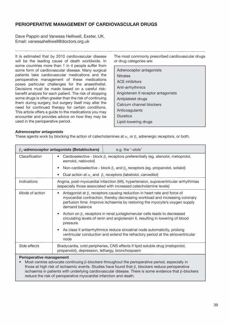
39
It is estimated that by 2010 cardiovascular disease will be the leading cause of death worldwide. In some countries more than 1 in 4 people suffer from some form of cardiovascular disease. Many surgical patients take cardiovascular medications and the perioperative management of these medications poses particular challenges for the anaesthetist. Decisions must be made based on a careful risk-benefit analysis for each patient. The risk of stopping some drugs is often greater than the risk of continuing them during surgery, but surgery itself may alter the need for continued therapy for certain conditions. This article offers a guide to the medications you may encounter and provides advice on how they may be used in the perioperative period.
PERIOPERATIVE MANAGEMENT OF CARDIOVASCULAR DRUGS
Adrenoceptor antagonists
Nitrates
ACE inhibitors
Anti-arrhythmics
Angiotensin II receptor antagonists
Antiplatelet drugs
Calcium channel blockers
Anticoagulants
Diuretics
Lipid-lowering drugs
The most commonly prescribed cardiovascular drugs or drug categories are:
Adrenoceptor antagonistsThese agents work by blocking the action of catecholamines at a1 or β1 adrenergic receptors, or both.
β1-adrenoceptor antagonists (Betablockers) e.g. the ‘-olols’
Classification • Cardioselective - block β1 receptors preferentially (eg. atenolol, metoprolol, esmolol, nebivolol)
• Non-cardioselective - block β1 and β2 receptors (eg. propanolol, sotalol)
• Dual action at a1 and β1 receptors (labetolol, carvedilol)
Indications Angina, post-myocardial infarction (MI), hypertension, supraventricular arrhythmias (especially those associated with increased catecholamine levels)
Mode of action • Antagonist at β1 receptors causing reduction in heart rate and force of myocardial contraction, thereby decreasing workload and increasing coronary perfusion time. Improve ischaemia by restoring the myocyte’s oxygen supply demand balance
• Action on β1 receptors in renal juxtaglomerular cells leads to decreased circulating levels of renin and angiotensin II, resulting in lowering of blood pressure
• As class II antiarrhythmics reduce sinoatrial node automaticity, prolong ventricular conduction and extend the refractory period at the atrioventricular node
Side effects Bradycardia, cold peripheries, CNS effects if lipid soluble drug (metoprolol, propanolol), depression, lethargy, bronchospasm
Perioperative management• Most centres advocate continuing β-blockers throughout the perioperative period, especially in those at high risk of ischaemic events. Studies have found that β1 blockers reduce perioperative ischaemia in patients with underlying cardiovascular disease. There is some evidence that β-blockers reduce the risk of perioperative myocardial infarction and death.
Dave Pappin and Vanessa Helliwell, Exeter, UK.Email: [email protected]

40
a1-adrenoceptor antagonists eg. indoramin and the ‘-azosins’ – doxazosin, prazosin, terazosin
Indications Hypertension, congestive heart failure, Raynaud’s syndrome, benign prostatic hypertrophy
Mode of action Prevents a1 - mediated vaso-constriction, reduces systemic vascular resistance and blood pressure
Side effects Nausea, postural hypotension, dizziness, headache
Perioperative management • Continue throughout perioperative period
• No intravenous formulations exist, so recommence once oral intake re-established
eg. the ‘-prils’ – captopril, lisinopril, enalapril, perindopril, ramipril, cilazapril, fosinopril
Indications Hypertension, left ventricular dysfunction, post-MI, delaying progression of proteinuria and renal impairment in diabetes
Mode of action Angiotensin converting enzyme (ACE) inhibition leads to decreased synthesis of angiotensin II. Angiotensin II normally causes peripheral vasoconstriction and stimulates aldosterone release, resulting in retention of Na+ and water, and excretion of K+. Lowering angiotensin II levels results in reduced vascular resistance and reduced fluid retention. Ventricular ejection and cardiac function are therefore improved. Remodelling of the ventricular muscle is also facilitated
Side effects Postural hypotension, dry cough (bradykinin is usually broken down by ACE and so levels rise), rash, angioedema (causing swollen tongue)
Perioperative management
• Often contributes to exaggerated of hypotension on induction and during maintenance of anaesthesia, particularly in presence of hypovolaemia
• Many anaesthetists omit on the morning of surgery particularly if performing neuroaxial blockade (epidural or spinal)
• Some centres recommend stopping ACEi on morning of surgery if part of therapy for LV dysfunction, but continue it if the indication is hypertension
Angiotensin II receptor antagonists (ARA’s) eg. the ‘-sartans’ - losartan, candesartan, irbesartan, valsartan
Indications Hypertension
Mode of action • Specific angiotensin II type-1 receptor (AT¬1) blocker
• Similar cardiovascular actions to ACE inhibitors
• Bradykinin-mediated side effects (eg. dry cough) are avoided since ACE is still active
Side effects Postural hypotension
Perioperative management
• As for ACEi
Angiotensin Converting Enzyme Inhibitors (ACEi)
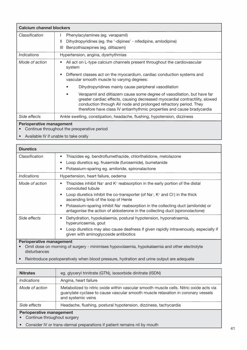
41
Calcium channel blockers
Classification I Phenylacylamines (eg. verapamil)
II Dihydropyridines (eg. the ‘-dipines’ - nifedipine, amlodipine)
III Benzothiazepines (eg. diltiazem)
Indications Hypertension, angina, dysrhythmias
Mode of action • All act on L-type calcium channels present throughout the cardiovascular system
• Different classes act on the myocardium, cardiac conduction systems and vascular smooth muscle to varying degrees:
• Dihydropyridines mainly cause peripheral vasodilation
• Verapamil and diltiazem cause some degree of vasodilation, but have far greater cardiac effects, causing decreased myocardial contractility, slowed conduction through AV node and prolonged refractory period. They therefore have class IV antiarrhythmic properties and cause bradycardia
Side effects Ankle swelling, constipation, headache, flushing, hypotension, dizziness
Perioperative management• Continue throughout the preoperative period
• Available IV if unable to take orally
Diuretics
Classification • Thiazides eg. bendroflumethazide, chlorthalidone, metolazone
• Loop diuretics eg. frusemide (furosemide), bumetanide
• Potassium-sparing eg. amiloride, spironalactone
Indications Hypertension, heart failure, oedema
Mode of action • Thiazides inhibit Na+ and K+ reabsorption in the early portion of the distal convoluted tubule
• Loop diuretics inhibit the co-transporter (of Na+, K+ and Cl-) in the thick ascending limb of the loop of Henle
• Potassium-sparing inhibit Na+ reabsorption in the collecting duct (amiloride) or antagonise the action of aldosterone in the collecting duct (spironolactone)
Side effects • Dehydration, hypokalaemia, postural hypotension, hyponatraemia, hyperuricaemia, gout
• Loop diuretics may also cause deafness if given rapidly intravenously, especially if given with aminoglycoside antibiotics
Perioperative management• Omit dose on morning of surgery - minimises hypovolaemia, hypokalaemia and other electrolyte disturbances
• Reintroduce postoperatively when blood pressure, hydration and urine output are adequate
Nitrates eg. glyceryl trinitrate (GTN), isosorbide dinitrate (ISDN)
Indications Angina, heart failure
Mode of action Metabolized to nitric oxide within vascular smooth muscle cells. Nitric oxide acts via guanylate cyclase to cause vascular smooth muscle relaxation in coronary vessels and systemic veins
Side effects Headache, flushing, postural hypotension, dizziness, tachycardia
Perioperative management• Continue throughout surgery
• Consider IV or trans-dermal preparations if patient remains nil by mouth

42
Visit the new World Anaesthesia Society website:
Free access to: Update in Anaesthesia, Anaesthesia Tutorial of the Week, Discussion forum, Future courses and meetings, Personal experiences and much more
www.worldanaesthesia.org
Anti-arrhythmics
The pharmacology of these agents is described in (Update 11, Cardiovascular pharmacology).
Pre-op Intra-op Post-op
Procainamide Give the night before Use IV procainamide Continue IV until able Disopyramide surgery or lidocaine (for VT/VF to take oral sips Quinidine prophylaxis)
Flecainide Give on morning of Give IV if needed Reinstate once stable surgery (may need levels)
β-blockers Give on morning of Continue IV if high risk Reinstate once stable surgery
Amiodarone Give the night before Give IV if needed Reinstate once stable surgery
Calcium Give on morning of IV verapamil can be Reinstate once stable channel blockers surgery used if needed
Digoxin Give normal dose Give IV if high risk Reinstate once stable (may need levels)

43
Drugs affecting haemostasis
The decision to continue or stop these drugs is particularly difficult because there are potentially devastating effects from both continuing (increased bleeding) and stopping the drug (cardiovascular events). The balance of risks versus the benefits guides management.
Antiplatelet drugs
Classification Cyclo-oxygenase inhibitors eg. aspirin
Phosphodiesterase inhibitor eg. dipyridamole
ADP binding inhibitors eg. clopidogrel, ticlodipine
Glycoprotein IIb/IIIa receptor antagonists eg. abciximab, eptifibatide, tirofiban
Indications • Act to reduce primary haemostasis by decreasing platelet aggregation and inhibiting thrombus formation. Widely used in primary and secondary prevention of thrombotic cerebrovascular or cardiovascular disease. They are effective in the arterial circulation, where anticoagulants have little effect.
• Aspirin is used in the prophylaxis of MI, ischaemic stroke, transient ischaemic attacks, intermittent claudication. Clopidogrel is an alternative
• Dipyridamole is given to some patients with prosthetic heart valves, in addition to anticoagulants. Also given with low-dose aspirin to reduce risk of recurrent stroke
• Clopidogrel often given as secondary prevention after an acute ischaemic event
• Abciximab is given as a bridging medical therapy to angioplasty or coronary artery bypass grafting, when patient has unstable angina or NSTEMI (non ST elevation MI) and is at high risk of further events
Mode of action • Aspirin irreversibly antagonises cyclo-oxygenase reducing thromboxane- mediated platelet aggregation
• Dypyridamole decreases platelet adenosine uptake, thereby inhibiting adhesion to damaged vessel walls
• Clopidogrel irreversibly prevents ADP from binding to its receptor on the platelet surface thereby stopping the glycoprotein IIb/IIIa receptor converting into its active form
• Abciximab (a monoclonal antibody) binds the glycoprotein IIb/IIIa receptor, impeding platelet aggregation
Side effects All can cause GI bleeding (particularly aspirin), hot flushes, tachycardia, headaches (dipyridamole), rarely neutropaenia, thrombocytopaenia (clopidogrel and glycoprotein IIb/IIIa blockers)
Perioperative management
Aspirin - stop for at least 7 days prior to surgery where the risks of perioperative bleeding are high (major surgery) or where the risks of even minor bleeding are significant (retinal or intracranial surgery). The risks of bleeding must be balanced against the risks of thromboembolic events, particularly in patients with unstable angina.
• It is safe to perform a subarachnoid block or insert an epidural catheter in the presence of aspirin
• Most recommend stopping other antiplatelet drugs 7-10 days preoperatively before major surgery or regional anaesthesia. If the risks of coronary thrombosis are high this must be balanced against the benefits of performing a regional block
• Dipyridamole should be stopped 7-10 days before surgery
• Abciximab is generally used as a rescue medical therapy before a more permanent re-vascularisation procedure may be performed and it is usually changed to clopidogrel postoperatively

Anticoagulants
• eg. warfarin, heparin, and the low molecular weight heparins – tinzaparin, enoxaparin, dalteparin
• Act to inhibit secondary haemostasis and formation of the fibrin clot by interfering directly with the blood coagulation cascade
Warfarin
Indications Thrombo-embolism prophylaxis – usually for atrial fibrillation, prosthetic heart valves, thrombophilia, previous thrombo-embolic disease
Mode of action • Vitamin K antagonist. Vitamin K is an essential co-factor for the synthesis of clotting factors II, VII, IX and X and also for proteins C and S
• Takes at least 48-72 hours to achieve its full anticoagulant effect (reflecting the half life of the clotting factors)
Side effects Haemorrhage. Metabolism of warfarin is affected by systemic illness or drug interactions
Perioperative management
• For patients at low risk of thromboembolism (eg. uncomplicated atrial fibrillation without prior history of thromboembolic events), warfarin should be discontinued 4-5 days before surgery. An INR (international normalized ratio) of 1.5 is generally considered safe for surgery
• Warfarin may be continued perioperatively in certain minor operations (eg. cataract and dental surgery)
• For other elective surgery weigh the risks and benefits of continuous anticoagulation for each patient. If anticoagulation is deemed necessary, warfarin is replaced intravenous heparin, from the time that the INR falls below the therapeuatic range for the underlying problem.
• An infusion of heparin is started at 1000units/hour and then adjusted to keep the APTT ratio between 1.5 and 2.5.
• The heparin infusion is stopped 6 hours prior to surgery and restarted 6-12 hours after surgery, if there is no clinical evidence of bleeding. This should be continued until warfarin therapy is restarted and the INR is greater than 2.0
• For emergency surgery, the effects of warfarin may be reversed by giving fresh frozen plasma (10-15ml/kg), or clotting factors. Intravenous vitamin K (1-2mg up to 10mg) may be used in life threatening haemorrhage, but the effects make re-anticoagulation difficult
44
Unfractionated Heparin
Indications Prophylaxis and treatment of DVT, PE, MI, unstable angina, vaso-occlusive disease
Mode of action Antithrombin III agonist, which binds to and potentiates antithrombin III action, causing inactivation of thrombin and other clotting factors (especially Xa)
Side effects Haemorrhage, heparin-induced thrombocytopaenia (HITS)
Perioperative management
• Heparin has a much shorter half-life (1 hour) than warfarin and is often used as a substitute bridging therapy for those patients at high risk of thromboembolism undergoing surgery (see above)
• If emergency reversal of heparin-induced haemorrhage is needed, protamine is used

Lipid-lowering drugs
Classification • HMGCoA reductase inhibitors eg. the ‘-statins’ - simvastatin, atorvastatin, fluvastatin
• Anion exchange resins eg. cholestyramine
• Fibric acid derivatives eg. the ‘-fibrates’ - clofibrate, bezafibrate, ciprofibrate
• Nicotinic acid derivative eg. acipimox
Indications Hyperlipidaemia, hypercholesterolaemia, patients with cardiac risk factors
Mode of action Statins reversibly inhibit HMGCoA reductase, the rate-limiting enzyme in cholesterol synthesis by the liver. The liver responds by increasing its expression of lower density lipoprotein (LDL) receptors, thereby increasing its uptake of LDLs from the circulation. Also thought to decrease cardiac disease via an unknown mechanism
Cholestyramine acts by sequestering bile acids in the intestine, preventing their reabsorption and enterohepatic circulation
Fibrates stimulate the enzyme lipoprotein lipase, converting triglycerides into fatty acids and glycerol
Acipimox causes a reduction in very low density lipoproteins (VLDL) and thus LDL
Side effects Generally well-tolerated, but reversible myositis, headache and GI disturbance can occur. Rhabdomyolysis is rare
Perioperative management
• Most lipid-lowering drugs can be continued throughout the perioperative period.
• Although manufacturers advise stopping ‘statins’ pre-operatively, it is now thought prudent to continue them in the perioperative period. Many myocardial infarcts are caused by coronary plaque rupture, thrombus formation and vessel occlusion. Statins stabilize plaques and therefore may be beneficial in preventing perioperative myocardial infarcts
In this review of perioperative prescription of cardiac medications, we have attempted to summarise the available information to provide a reference guide for daily use. The advice is not always clear-cut and the decision for each drug that a patient is taking must be considered carefully, and judged by weighing the relative risks of continuing or stopping the therapy.
45
Low Molecular Weight Heparins (LMWH) eg. tinzaparin, dalteparin, enoxaparin
Indications As for heparin
Mode of action More effective inhibition of factor Xa, but less effective inactivation of thrombin, compared to heparin
Side effects Haemorrhage, reduced risk of thrombocytopaenia
Perioperative management
• Used in place of heparin
• No monitoring necessary
• Last dose should be 12 hours before surgery, including those patients undergoing regional blocks (spinal or epidural)
• Protamine may be used to reverse the effects of LMWHs, although this may be less effective

46
Self assessment questionsScenario: A twenty-year-old female is brought into the Emergency Department (ED) having been found unconscious in her bedsit. There is no other recent history. She did not respond to a bolus of 50% dextrose in the ambulance, despite having an unrecordable blood glucose when tested by the paramedics. While she is being intubated on account of reduced level of consciousness, an arterial blood gas sample reveals profound lactic acidosis (pH 7.05, PaCO2 2.5 kPa, base deficit – 10, lactate 13mg/L). Blood pressure is 95/50mmHg.
1. What are the possible explanations for her presentation?
Laboratory tests demonstrate hepatocellular necrosis (AST 21,000 U/L) and coagulopathy (INR 9.1) with thrombocytopenia (platelet count 26 x 109/L). Acute liver failure appears the most likely diagnosis.
2. What are the most likely causes of acute liver failure (ALF) in this previously well patient?
Her mean arterial blood pressure remains low (50mmHg) after 3 litres of colloid and crystalloid. The ED nurse, who is doing half-hourly neurological observations, reports reduced pupillary response to light.
3. What severe complications of ALF may result in death within hours, and what are the immediate management priorities for this patient?
IntroductionSuccessful management of this rare but potentially devastating disorder relies on early recognition. The hallmark of acute liver failure (ALF) is encephalopathy (ranging from a subtle alterations in consciousness level to coma) in the context of an acute, severe liver injury. The presence of a liver injury is suggested by raised transaminase levels (in the thousands, indicating hepatocellular necrosis), impairment of synthetic function manifested by coagulopathy (INR>1.5), and metabolic derangements such as hypoglycaemia and lactic acidosis. Sometimes the history contains clues to the cause of the liver injury (eg. paracetamol ingestion), but frequently the cause is not apparent.
Types of liver failureThe speed of onset of encephalopathy after the onset of symptoms (usually jaundice) is important in both categorisation and prognosis. If this is less than seven days the term ‘hyperacute’ liver failure is used;
8-28 days indicates ‘acute’ liver failure and 5 to 26 weeks, is termed ‘subacute’ liver failure. However, ALF can be used to describe all these categories. The more slowly progressive types of ALF can result in an equally severe illness, but tend to be caused by idiosyncratic drug reactions, autoimmune hepatitis and ‘seronegative’ hepatitis (in which no specific cause is determined). Hyperacute types are caused by paracetamol and the viral hepatitides, and, because of the speed of onset, there is often not enough time for frank jaundice to develop before encephalopathy occurs. Table 1 summarizes the main causes of ALF in the United Kingdom. There is significant geographical variation; for example paracetamol accounts for only 2% in France, and viral hepatitis causes 55% of cases in Japan. In India Hepatitis E is responsible for 33% of cases.
This article will concentrate on the management of ‘hyperacute’ ALF, which is associated with the most dramatic presentations.
MANAGEMENT OF ACUTE LIVER FAILURE IN ICU
Table 1:
Causes of ALF in United KingdomParacetamol toxicity 54%
Seronegative 17%
Hepatitis A or B 14%
Drug reaction 7%
Other causes 8%
Drugs causing ALF include: isoniazid, rifampicin, NSAIDs, valproate, carbamezipine, allopurinol, phenytoin, gold, ketoconazole and trycyclic antidepressants among many others
Other causes of ALF:Viral: cytomegalovirus, herpes simplex virus, Epstein-Barr virus
Metabolic: Wilson’s disease, Reye’s syndrome
Vascular: Budd-Chiari syndrome, ischaemic hepatitis
Pregnancy related: acute fatty liver, HELLP/toxaemia of pregnancy
Neoplastic: lymphoma, gross metastatic infiltration
Toxic: aminata phalloides mushroom
Philip Berry, Institute of Liver Studies, Kings College Hospital, London, UK Email: [email protected]

47
ALF causes multi-system failure. The most dangerous consequences of ALF are cerebral oedema and the risk of tentorial herniation, severe vasodilatory shock, lactic acidosis and hypoglycaemia. Acute renal failure will almost always follow. Coagulopathy, although often profound, rarely results in catastrophic bleeding.
Assessment of encephalopathyGrading of encephalopathy in ALF borrows from features seen in cirrhosis. However, encephalopathy in ALF differs greatly from that seen in cirrhosis because cerebral oedema is a frequent complication in grade 3 and 4 (see section 2, Cerebral Protection).
Table 2: Grading of encephalopathy
Grade Clinical signs
1 Subtle changes in level of consciousness, reduced concentration span and ability to perform simple sums.
2 Obviously drowsy but remaining awake, disoriented, slow or slurred speech, ‘liver flap’ (asterixis).
3 Somnolent but responds to stimulation, very confused, occasionally aggressive or violent, hypertonic, hyperreflexic, ankle clonus.
4 Comatose. Physical signs as in grade 3, or evolving into decerebrate posturing. Signs of intracranial hypertension should be sought.
Investigations to determine the cause of ALF, although of paramount importance, are rarely informative in the early stages of care. For example, viral and autoimmune serology (where available) will not generally yield results within 12-24 hours. An ideal investigative screen is summarised in Table 3, but delay in receiving results should not hold up discussion with a liver unit. An ultrasound, if available, is always useful to exclude underlying cirrhosis (which will lead to different management), massive malignant infiltration (which may save the patient a disruptive transfer out of area) and an acute vascular pathology (eg. portal or hepatic vein thrombosis).
ManagementAcute management is focussed on:
1. Global organ support
2. Cerebral protection
3. Where available identification of patients likely to benefit from liver transplantation (LT), and liason with a specialist liver unit
4. Ensuring safe inter-hospital transfer
The role of specific antidotes to reverse the effect of the aetiological agent or pathogen (eg. antiviral therapy, copper chelation in Wilson’s disease, steroids in autoimmune disease) are very limited, however N-acetylcysteine should be administered if paracetamol toxicity is suspected, even if the conventional therapeutic window has passed. It may have a beneficial role in other types of ALF, but this remains unproven.
Table 3: Investigations in ALF
General INR/PT and FBC Urea & Electrolytes/Liver function tests Phosphate/Calcium/Magnesium Arterial blood gas with lactate Arterial ammonia (see below) Amylase (co-existing pancreatitis is not uncommon) Investigation • Paracetamol level • overdose • Toxicology screen • ecstasy, amphetamines, cocaine
• Hepatitis screen (anti-Hep A IgM, Hep B • acute viral hepatitis Sag, anti-Hep B Core IgM); anti-Hep E IgM/G if appropriate history
• Caeruloplasmin/urinary copper • Wilson’s disease excretion/slit lamp examination for Kaiser-Fleischer rings
• Autoantibodies (anti nuclear~, anti smooth • autoimmune hepatitis muscle~, anti liver kidney~) and immunoglobulins
Diagnostic [underlying disease]
Underlying disease

48
1. Global organ support
CirculationVasodilatory shock is common. Invasive monitoring (eg. with ‘pulse-induced contour cardiac output’ – PiCCO, see Update 21) will aid assessment of volume status and identify co-existing cardiac dysfunction. Aggressive fluid resuscitation is almost always required, with the fluid volume required often exceeding 3 litres. As in head injury, salt-containing fluids are appropriate, since hyponatraemia may exacerbate cerebral oedema. Vasopressor therapy should be instituted if mean arterial blood pressure does not respond to filling. Patients requiring inotropes may have relative adrenal deficiency and should have a short synacthen (ACTH stimulation) test and hydrocortisone 50mg 6 hourly commenced.
Renal/Acid-Base balanceWhere available continous veno-venous haemofiltration (CVVHF) will be required if the patient is anuric or acidotic. CVVHF is favoured since it is better tolerated by patients with haemodynamic compromise. A bicarbonate buffered replacement solution should be used (lactate will not be handled well by the liver), and epoprostanol (Flolan®) used to anticoagulate the circuit. High volume (90mls/kg/hr) can be used if the acid-base status is extremely deranged.
Respiratory functionAlthough ARDS can develop in ALF, specific ventilatory strategies are not usually recommended in the early phase. CO2 targets are covered below.
CoagulationThe extent of coagulopathy is critical in deciding if the patient will benefit from LT, and administration of FFP is not recommended unless there is clinically significant bleeding. Platelets can be supported freely. Coagulopathy should not necessarily preclude central line insertion (especially femoral) by an experienced operator. Dissemninated intravascular coagulation and fibrinogen defects can further complicate the coagulopathy. Gastric ulcer protection with IV or enteral proton pump inhibitors reduces the risk of gastrointestinal bleeding.
Protection against sepsisBacterial and fungal sepsis complicated a high proportion of ALF patients before anti-bacterial prophylaxis became standard. Recommended agents are Tazobactam-Piperacillin (Tazocin®) 4.5g/IV/tds and Fluconazole 200mg/iv daily.
Gastrointestinal tract and nutritionGastric ulcer protection with IV or enteral proton pump inhibitors reduces the risk of gastrointestinal bleeding. There are no contraindications to early enteral feeding, and standard amounts of nitrogen and carbohydrate can be administered.
2. Cerebral protectionCerebral oedema complicates severe encephalopathy, and may occur in 80% of patients in grade IV. Those most at risk are the young, the septic, the hyponatraemic and patients with significant elevations in blood ammonia (which appears to have an important role in both encephalopathy and oedema). Arterial ammonia concentration correlates with the degree of encephalopathy, and is useful in assessing the risk of developing intracranial hypertension. Experience at Kings College Hospital has shown that over 30% of patients with a level 100-200mcmol/l develop intracranial hypertension, and over 50% with levels exceeding 200mcmol/l. Venous ammonia levels are significantly lower than arterial levels due to the ability of skeletal muscle to metabolise ammonia.
Clinical signs of rising intracranial pressure (ICP) are hypertonia, clonus, pupillary abnormalities (dilatation, reduced responsiveness to light) and in the latter stages hypertension (bursts to > 200mmHg, or sustained above 150mmHg), bradycardia and cerebral posturing.
Such patients will have been intubated for airway protection. Recommended sedation is propofol (possible beneficial cerebral metabolic effects) and fentanyl. Additionally the following prophylactic measures are advised.
PosturePositioning the head in the midline, with the angle of the body 20° from horizontal will aid cerebral venous outflow.
Normocapnia (PaCO2 4.5-5 kPa) Hypercapnia should be avoided, however the ‘traditional’ approach of hyperventilation and low PaCO2, although effective in short term reduction of cerebral blood flow and ICP, can lead to cerebral vasospasm and increased risk of brain injury.
Body temperatureFever should be managed with cooling blankets, or if on CVVHF reduced thermal compensation.
Avoidance of stimulationSurges in ICP are seen in monitored patients who are exposed to stimuli such as loud noise, suctioning and excessive movement.
Specific treatments, if raised ICP is suspected, include the following. These would normally be undertaken only after discussion with a specialist centre.
Mannitol0.5g/kg boluses over 10 minutes may be administered and repeated, but serum osmolarity should be monitored so that 320mosm/l is not exceeded. To prevent fluid overload, a 500ml diuresis (or negative balance if being haemofilitered) should be obtained after each bolus.
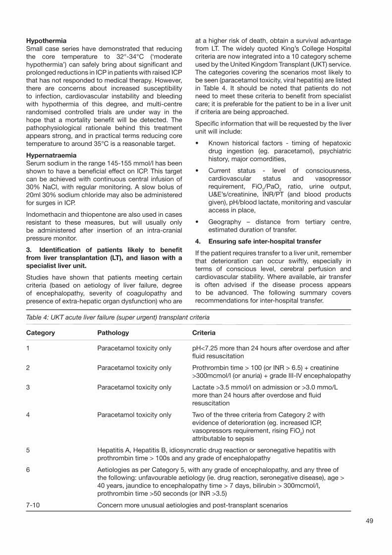
49
HypothermiaSmall case series have demonstrated that reducing the core temperature to 32°-34°C (‘moderate hypothermia’) can safely bring about significant and prolonged reductions in ICP in patients with raised ICP that has not responded to medical therapy. However, there are concerns about increased susceptibility to infection, cardiovascular instability and bleeding with hypothermia of this degree, and multi-centre randomised controlled trials are under way in the hope that a mortality benefit will be detected. The pathophysiological rationale behind this treatment appears strong, and in practical terms reducing core temperature to around 35°C is a reasonable target.
HypernatraemiaSerum sodium in the range 145-155 mmol/l has been shown to have a beneficial effect on ICP. This target can be achieved with continuous central infusion of 30% NaCl, with regular monitoring. A slow bolus of 20ml 30% sodium chloride may also be administered for surges in ICP.
Indomethacin and thiopentone are also used in cases resistant to these measures, but will usually only be administered after insertion of an intra-cranial pressure monitor.
3. Identification of patients likely to benefitfrom liver transplantation (LT), and liason with a specialist liver unit.
Studies have shown that patients meeting certain criteria (based on aetiology of liver failure, degree of encephalopathy, severity of coagulopathy and presence of extra-hepatic organ dysfunction) who are
at a higher risk of death, obtain a survival advantage from LT. The widely quoted King’s College Hospital criteria are now integrated into a 10 category scheme used by the United Kingdom Transplant (UKT) service. The categories covering the scenarios most likely to be seen (paracetamol toxicity, viral hepatitis) are listed in Table 4. It should be noted that patients do not need to meet these criteria to benefit from specialist care; it is preferable for the patient to be in a liver unit if criteria are being approached.
Specific information that will be requested by the liver unit will include:
• Known historical factors - timing of hepatoxic drug ingestion (eg. paracetamol), psychiatric history, major comordities,
• Current status - level of consciousness, cardiovascular status and vasopressor requirement, FiO2/PaO2 ratio, urine output, U&E’s/creatinine, INR/PT (and blood products given), pH/blood lactate, monitoring and vascular access in place,
• Geography – distance from tertiary centre, estimated duration of transfer.
4. Ensuring safe inter-hospital transfer
If the patient requires transfer to a liver unit, remember that deterioration can occur swiftly, especially in terms of conscious level, cerebral perfusion and cardiovascular stability. Where available, air transfer is often advised if the disease process appears to be advanced. The following summary covers recommendations for inter-hospital transfer.
Table 4: UKT acute liver failure (super urgent) transplant criteria
Category Pathology Criteria
1 Paracetamol toxicity only pH<7.25 more than 24 hours after overdose and after fluid resuscitation
2 Paracetamol toxicity only Prothrombin time > 100 (or INR > 6.5) + creatinine >300mcmol/l (or anuria) + grade III-IV encephalopathy
3 Paracetamol toxicity only Lactate >3.5 mmol/l on admission or >3.0 mmo/L more than 24 hours after overdose and fluid resuscitation
4 Paracetamol toxicity only Two of the three criteria from Category 2 with evidence of deterioration (eg. increased ICP, vasopressors requirement, rising FiO2) not attributable to sepsis
5 Hepatitis A, Hepatitis B, idiosyncratic drug reaction or seronegative hepatitis with prothrombin time > 100s and any grade of encephalopathy
6 Aetiologies as per Category 5, with any grade of encephalopathy, and any three of the following: unfavourable aetiology (ie. drug reaction, seronegative disease), age > 40 years, jaundice to encephalopathy time > 7 days, bilirubin > 300mcmol/l, prothrombin time >50 seconds (or INR >3.5)
7-10 Concern more unusual aetiologies and post-transplant scenarios

50
VentilationElective intubation is almost always required, even if a good conscious level has been maintained in the referring hospital, to avoid the risks of emergency intubation during transfer. Aim to achieve normocapnia.
CardiovascularAn arterial cannula with portable blood pressure monitoring is preferred. Wide bore peripheral venous access and central access, through the femoral vein if necessary, are usually advised in case of the need for large volume infusions and vasopressors. Ample reserves of colloid/crystalloid should be taken, as should norepinephrine (made up prior to departure, eg. 5mg/50mls). Adequate cerebral perfusion will depend on maintaining a mean arterial pressure of over 70mmHg.
NeurologicalMonitor pupil size and response. If there is a suspicion of rising ICP, mannitol 0.5g/kg should be given. For a 70kg man give 175ml of 20% mannitol or 350ml of 10% mannitol over 10 minutes.
MetabolismMeasure frequent blood sugars, with hypoglycaemia treated with 50% glucose. Sodium bicarbonate (50-100mls of 8.4%) can be given prior to departure if acidosis is profound.
by toxic drug effects (metformin, theophylline, ethylene glycol, methanol, cocaine, cyanide). A marked transaminitis (>40 times the upper limit of normal) will usually lead the clinician to suspect hepatocellular necrosis, although in non-hyperacute types AST/ALT may be ‘burning out’ at presentation due to the progressive loss of hepatocytes. Bilirubin may not be particularly high in rapid onset disease.
2. The most likely causes of ALF in a patient of this age are: paracetamol poisoning, acute hepatitis A or B infection, idiosyncratic drug reaction, seronegative hepatitis, Wilson’s disease and autoimmune hepatitis. Hepatitis E should be suspected if there is a history of recent travel to an area where the virus is endemic. Hepatitis E infection in pregnancy is associated with a 20% risk of death in some reports.
3. This patient may die due to raised intracranial pressure and tentorial herniation, progressive vasodilatory shock, unrecognised hypoglycaemia or severe acidosis and cardiac arrest. If ALF is strongly suspected the priorities are: airway protection, avoidance of hypoglycaemia, maintenance of cerebral perfusion pressure (fluid resuscitation +/- vasopressors), avoidance of hyponatraemia and, where available, liason with a liver transplant unit. If the patient is too unstable to be transferred (hypotension, profound acidosis resulting in suppression of cardiac function) further optimisation with guided volume resuscitation and continuous veno-venous haemofiltration may be required. See text for strategies to minimise the risk of raised intracranial hypertension.
Recommended reading and important papers in anaesthetic/intensive care management• O’Grady JG, Alexander GJM, Hayllar KM, Williams R. Early indicators of prognosis in fulminant hepatic failure. Gastroenterology 189;97:439-45• Bernal W, Donaldson N, Wyncoll D, Wendon J. Blood lactate as an early predictor of outcome in paracetamol-induced acute liver failure: a cohort study. Lancet. 2002; 359: 558-63.• Wijkicks EFM, Nyberg SL. Propofol to control intracranial pressure in fulminant hepatic failure. Transplant Proc 2002; 34: 1220-1222• Jalan R, Olde Daminsk SWM, Deutz NEP, et al. Moderate hypothermia in patients with acute liver failure and uncontrolled intracranial hypertension. Gastroenterology 2004; 127: 1338-1346• Murphy N, Auzinger G, Bernal W, Wendon J. The effect of hypertonic sodium chloride on intracranial in patients with acute liver failure. Hepatology 2002; 39: 464-470• Davenport A, Will EJ, Davidson AM. Improved cardiovascular stability during continuous modes of renal replacement therapy in crticially ill patients with acute hepatic and renal failure. Crit Care Med 1993; 21: 328-338• Harrison PM, Keays R, Bray GP, et al. Improved outcome of paracetamol-induced fulminant hepatic failure by late administration of acetylcysteine. Lancet. 1990; 30: 1572–1573
Key Points• ALF should be considered in the context of unexplained loss of consciousness, hypoglyaemia or coagulopathy.
• ALF causes death by cerebral oedema, vasodilatory shock and hypoglycaemia.
• Aggressive volume resuscitation is required. Saline and artificial colloids are safe, but 5% glucose may exacerbate cerebral oedema and should not be used.
• Vasopressors should be used if blood pressure remains low after fluid resuscitation – adequate cerebral perfusion pressure must be maintained.
• Encephalopathy in ALF is progressive - early elective intubation is preferable to emergent intubation.
Answers to self assessment questions1. ALF mimics other conditions that can present with obtundation or coma, shock, hypotension and acidosis, eg. septicaemia, hypoperfusion secondary to cardiac failure or volume depletion. Derangements in haematological and coagulation indices typical of ALF can also be seen in acute presentations of haematological malignancy or disseminated intravascular coagulation. Severe hyperlactataemia without liver failure or hypoperfusion may be induced

51
IntroductionLatex is a protein, processed from the sap of the rubber tree (Hevea brasiliensis). Not all products that are labelled as “latex” contain this product and therefore may not induce allergy in susceptible individuals. Susceptibility is determined by cumulative life exposure to one or more latex proteins or the chemicals used in its manufacture. Hospital workers and patients having frequent operations, for spina bifida for example, have high cumulative exposure and risk of allergy.
Latex allergy was first identified in the 1970s but, with more prevalent use of latex products in hospitals, the prevalence of latex allergy has increased. Adoption of universal precautions for prevention of blood-borne infections may have contributed to this increase. Prevalence rates as high as 17% are quoted amongst hospital workers and, although numerous papers indicate prevalence rates between 1 to 12% in the general population, the true prevalence is likely to be at the lower end of this range.
PathologyLatex extracts contain up to 14 antigenic proteins, which may be altered during processing of the latex for commercial use. In addition to direct contact, exposure to latex particles may occur by actions such as opening theatre packs or taking off a pair of gloves. Intravenous and mucosal routes are also important; particles released into the air may induce anaphylaxis or exacerbate pre-existing conditions such as asthma.
Common reactions to latex are type I and type IV hypersensitivity reactions. Type IV (T-cell mediated) hypersensitivity produces delayed contact dermatitis, 2-3 days after exposure. Type 1 reactions are more serious, are IgE-mediated, and are associated with immediate systemic release of histamine causing mast cell degranulation, release of tryptase, prostaglandins and leukotrienes, and systemic manifestations. Symptoms and signs include itching, sneezing, coryza, red itchy tearing eyes, urticaria, nausea, sore throat, bronchospasm, wheeze, shortness of breath or full-blown anaphylaxis. The extent of the reaction is unpredictable and early recognition of these symptoms is important. Anaphylactoid reactions are clinically indistinguishable from anaphylaxis, but are not mediated by sensitising IgE antibody. Whether a reaction is called anaphylactic or anaphylactoid may depend on whether it is investigated, the means by which it is investigated and how the results are interpreted.
ManagementManagement involves identification of at-risk patients, use of suitable latex-free equipment, vigilance for signs of anaphylaxis perioperatively and thorough investigation following suspected reactions.
PreventionThe main strategy for tackling latex allergy focuses on preventing exposure by using non-latex products. Taking and documenting a clear history is paramount; clinicians should ask about any previous reactions to latex, foods, and latex products such as washing up gloves. In addition to the multitude of hospital products (including stethoscopes, blood pressure cuffs, packs, gloves) latex is found in many commercially available products such as balloons, condoms, elastic and washing-up gloves.
The allergy is cross-reactive with many foods, in particular avocado, banana, kiwi fruit and chestnuts. Risk factors for latex allergy include:
• Occupational exposure (healthcare workers)
• Multiple operations (particularly laparotomy)
• Repeated bladder catheterisation (60% incidence in spina bifida)
• History of allergy to food with cross-reactivity with latex
• Women are at greater risk than men.
Patients with risk factors may be anaesthetised in a standard fashion but the anaesthetists should maintain a high index of suspicion for the development of anaphylaxis.
Latex-free equipmentLatex-sensitive patients should be managed in a latex-free environment and put first on elective lists, since there should be less airborne latex particles from other cases. Ward equipment (blood pressure cuffs, stethoscopes) should be latex-free.
Communication is important, with clear identification of the risk communicated between healthcare workers and clearly identified on the patient notes, the theatre list and using an alert bracelet, worn by the patient. Latex-free gloves should be worn by all staff and thorough handwashing is essential if latex containing gloves have been worn for previous cases. Some centres have used premedication with steroids and antihistamines, but anaphylaxis has still occurred and this practice is not recommended.
LATEX ALLERGY
Nick Preston and Bruce McCormick, Exeter, UK Email: [email protected]

Precautions in theatre include:• All latex containing equipment should be removed from theatre
• Bacterial and viral breathing circuit filters should be changed between cases (latex particles may be adsorbed to the filter)
• Laminar flow is desirable, if available
• Alert signs should be posted at the door of the theatre.
Use of latex-free equipment is facilitated by prior compilation of a list of safe equipment and consumables, or preparation of a latex-free trolley or box. Most equipment is now latex-free and readily identifiable as such, but it may be necessary to contact the manufacturer. Be aware that not all syringes have latex-free plungers, and some intravenous giving sets have latex-containing injection ports. In addition, the stoppers of some drug ampoules contain latex. The anaesthetist should also be vigilant that appropriate surgical equipment is used.
Early detection of anaphylaxisPatients should remain in recovery for at least one hour, since anaphylaxis is not always immediate and may take 20-60 minutes to develop. Slow-onset anaphylaxis has been described with onset several hours after exposure. The signs and symptoms are variable and may be attenuated by other anaesthetic drugs (e.g. epinephrine-containing local anaesthetic).
Management of anaphylaxisIf life-threatening exposure occurs, management is guided by standard protocols for the management of anaphylaxis. In the UK, the guidelines of the Association of Anaesthetists are followed, with removal of agent, administration of oxygen, early administration of epinephrine (adrenaline), fluids and steroids (guideline available at: www.aagbi.org/publications/guidelines/docs/anaphylaxis03.pdf). Latex constitutes a family of water-soluble proteins, so hand-washing and washing areas of exposure may be beneficial. It is also important to make sure provision is made on emergency crash trolleys for latex sensitive individuals, so that exposure is not compounded during an arrest.
Investigation of suspected anaphylaxisLatex sensitive individuals should be fully investigated after an event, counselled about their risks and advised to carry relevant information on their person at all times (e.g. alert bracelets). An internet link to a list of latex allergy links is included below.
It is vitally important that the sequence of events is clearly documented, including the timing of exposure to each possible allergen. Whilst further investigations are undertaken, this information should be made available to the patient’s usual doctor or general practitioner and to the patient themselves. It is the responsibility of the anaesthetist to ensure that all necessary information is made available and that referral for formal investigation proceeds.
Anaphylaxis is confirmed on clinical history and measurement of blood mast cell tryptase levels after the reaction. 10ml clotted blood should be sent as soon as possible after the reaction, one hour after the beginning of the reaction and 6 - 24 hours later. This will usually as soon as possible after the reaction go to a regional centre. Mast cell tryptase is the principal protein content of mast cell granules and is released, together with histamine and other amines, in anaphylactic and anaphylactoid reactions. Its concentration in the plasma or the serum is raised after reactions which involve mast cell degranulation. Approximately 99% of the body’s total enzyme is located within the mast cell. It is not present in red or white cells and therefore plasma concentrations are not affected by haemolysis. The basal tryptase concentration is 0.8 to 1.5ng/ml with the normal value usually <1 ng/ml. The half life is approximately 2.5 hours with maximum concentrations occurring rapidly.
Formal diagnosis and identification of the likely trigger agent requires immunological testing. This may involves skin-prick or patch testing with latex extracts, or in-vitro immunoglobulin-E testing with enzyme-linked immunosorbent assay (ELISA), the radioallergosorbent test (RAST) or ImmunoCAP systems. Unfortunately these tests have a high false-negative rate so the patient’s history is often the key factor. The Association of Anaesthetists of Great Britain & Ireland guidelines state: ‘There is no valid predictor of drug anaphylaxis at present. Claims that any form of screening will predict anaphylaxis are without foundation.’
Further readingArellano R, Bradley J, Sussman G. Prevalence of latex sensitization among hospital physicians occupationally exposed to latex gloves. Anesthesiology 1992; 77: 905-8.
Turjanmaa K. Ann Med. 1994 Aug;26(4):297-300
Dakin MJ, Yentis SM. Anaesthesia 1998; 53: 774-81. Latex allergy: a strategy for management.
Association of Anaesthetists of Great Britain and Northern Ireland. Anaphylactic reations associated with Anaesthesia 3 (2003). Available at: http://www.aagbi.org/publications/guidelines/docs/anaphylaxis03.pdf
http://www.latexallergylinks.org/prot.html
52

In each edition of Update, Cerebral Challenge will present examples of investigations demonstrating typical appearances of commonly encountered conditions.
Cerebral Challenge
What does the CXR show? How would you manage this patient? Discussion overleaf.
Figure 1: A 20-year-old male is found collapsed outside the gates of a football stadium, shortly after the home team have been defeated. On arrival in the Emergency Department he localises to pain with his left arm, but no movement is elicited from his right side. He groans and his eyes remain closed on painful stimulation. His left pupil is slightly dilated and the response to light is sluggish.
Case 1
Figure 2: A 34-year-old man suffered a spinal injury 24 hours ago. The sensory level is C4/5 and he has paradoxi-cal breathing. Until now he has maintained his oxygen saturations above 95% on air with a PaCO2 between 5 and 5.8 kPA (high-normal). Over the last 2 hours his respiratory rate has increased to 32/minute and he appears distressed. His SaO2 has fallen to 87% despite 10l/min oxygen by facemask. His chest xray (CXR), taken 20 minutes ago, is shown.
Case 2
What does the CT scan show? How would you manage this patient? Discussion overleaf.
What abnormalities are shown on this ECG and what are possible causes in this man? Discussion overleaf.
Compiled by Nicky Bosley and Bruce McCormick, Exeter, UK
Case 3
Figure 3: A 46-year-old man is booked for an elective hernia repair. He complains of chest pain that doesn’t sound typically cardiac and he has no risk factors for ischaemic heart disaese. He has had the pain for a week and describes it as sharp and worsened by moving. He has had a dry cough for 6 months, has lost weight and has frequently episodes of sweating. The ward nurse has done an ECG.
53

Discussion
Case 1CT scans are viewed as if we are looking up through the patient’s body from their feet, so as the patient lies, looking upwards, their right side is on the left as we look at the scan. Bone is bright white, fresh blood appears white, cerebrospinal fluid is black and brain parenchyma is varying shades of grey.
Figure 4: The CT scan shows a large left-sided, subdural haematoma (A). There is also left-to-right midline shift (B), due to the haematoma and oedema of the left cerebral hemisphere (C).
Figure 5: An extradural haematoma (A) on a CT scan
Figure 6: The appearance may be hard to interpret. This CT scan shows a haematoma that has an inner border that is both concave (A) and convex (B) towards the brain in dif-ferent areas. This CT shows an old subdural (the old blood is dark on the CT rather than white - C), but there has been a recent rebleed of fresh blood into the haematoma (D).
54
Our patient (Figures 1 and 4) requires an emergency burr hole or craniotomy for drainage of the haematoma. While this is arranged, give oxygen and address the airway, breathing and circulation. This man’s Glasgow Coma Score is 8 (eyes 1, speech 2, motor 5) and so, where possible, his airway should be secured by intubation. The priorities are to optimise his oxygenation and blood pressure. Anaesthesia for patients requiring neurosurgery will be covered by an article in Update 23.
Subdural haematomas lie under the dura mater and so tend to form an inner border that is concave (curved inwards) towards the brain. They may be dif-ferentiated from extradural haematomas, that track outside the dura mater and form an inner border that is convex (curved outwards) towards the brain (see Figure 5).
ABC
Right Left
A
D
C
B
A

This man has developed respiratory failure. His spinal injury has left his intercostal muscles paralysed and he is reliant on his diaphragm for ventilation. Diaphragmatic breathing manifests clinically as ‘paradoxical breathing’ – as the diaphragm contracts and moves down during inspiration, the abdomen moves out and the chest wall is sucked in. This is opposite (paradioxical) to conventional breathing, where the intercostal muscles are active.
His ability to cough is reduced since paralysis of his chest wall and accessory muscles means that forced expiration is markedly impaired. It is likely that retention of respiratory secretions has contributed to
the left lower lobe collapse. His ability to self ventilate was borderline (as indicated by the high/normal PaCO2 level), and lobar collapse has caused him to decompensate.
Where available he should be intubated and ventilated. Application of high PEEP (12 to 15cmH2O), physiotherapy, suction, fibrescope-guided bronchial toilet may all help to resolve the problem, but it is likely to recur. Weaning from the ventilator is likely to be prolonged and he may spend several weeks ventilated. Early tracheostomy should be considered.
Case 3The ECG shows sinus rhythm with a rate of 75 beats per minute (300 divided by the number of big squares between complexes).
There is ST segment elevation in leads II, III and aVF (reflecting the activity of the inferior part of the heart) and also in leads V5 and V6 (reflecting the activity of the lateral aspects of the heart). The ST segments are ‘saddle-shaped’.
This man is at low risk of ischaemic heart disease and gives a history that is more suggestive of musculoskeletal pain or inflammation of the pleura or pericardium.
The history and saddle-shaped ST elevation shown in this ECG is characteristic of percarditis. An additional point to note is that the ST changes associated with pericarditis are usually widespread across most of the ECG leads and cannot be readily explained by ischaemia of a single coronary artery.
Common causes of pericarditis are: TB, viral infection, connective tissue diseases, post-myocardial infarction. With this man’s history of a chronic cough, TB should be considered and a chest xray performed, with sputum sent for TB microscopy and culture.
His elective hernia repair should be delayed, pending the results of his investigations and commencement of treatment.
55
Figure 7: The CXR shows left lower lobe collapse. The left hemi-diaphragm cannot be seen. This implies that the lung overlying the left hemi-diaphragm is consolidated or collapsed. (Compare this to the right side where there is a clear line where the air-filled lungs meet the solid tissue – the diaphragm and liver). In this patient the CXR shows the ‘sail sign’ (A), since we are viewing the oblique fissure - B - (between the upper and lower lobes of the left lung) straight on, so the contrast between the inflated upper lobe and the collapsed lower lobe is accentuated.
Case 2
A
B