contamination of tissue cultures by mycoplasmas
TRANSCRIPT

Chapter 3
© 2012 Rottem et al., licensee InTech. This is an open access chapter distributed under the terms of the Creative Commons Attribution License (http://creativecommons.org/licenses/by/3.0), which permits unrestricted use, distribution, and reproduction in any medium, provided the original work is properly cited.
Contamination of Tissue Cultures by Mycoplasmas
Shlomo Rottem, Nechama S. Kosower and Jonathan D. Kornspan
Additional information is available at the end of the chapter
http://dx.doi.org/10.5772/51518
1. Introduction Mycoplasmas are the smallest and simplest self-replicating bacteria [1]. These microorganisms lack a peptidoglycan based rigid cell wall and thus are not susceptible to antibiotics, such as penicillin and its analogues, which are effective against most bacterial contaminants of cell cultures. The trivial name mycoplasma encompasses all species included in the class Mollicutes: i.e. the genera Mycoplasma, Acholeplasma, Spiroplasma, Anaeroplasma and Ureaplasma. Because mycoplasmas have an extremely small genome (0.58–2.20 Mb compared with the 4.64 Mb of Escherichia coli), these organisms have limited metabolic options for replication and survival. The smallest genome of a self-replicating organism known at present is the genome of Mycoplasma genitalium (0.58 Mb; Ref. 2). Comparative genomic studies suggested that the genome of this organism still carries almost double the number of genes included in the minimal gene set essential for cellular function [3]. Owing to their limited biosynthetic capabilities, most mycoplasmas are parasites, exhibiting strict host and tissue specificities [4]. The aim of this review is to collate present knowledge on the strategies employed by mycoplasmas while interacting with tissue culture cells. Prominent among these strategies is the adherence of mycoplasmas to host cells, the invasion of mycoplasmas into host cells and the fusion of mycoplasmas with host cells. We shall discuss the intriguing questions of how a mycoplasma infecting tissue culture cells subvert and damage the host cells by mediating transformation of the cells, affecting the signal-transduction pathways and the metabolism of immune and non-immune cells. We shall also present and discuss the common procedures for isolation, identification and eradication of a mycoplasma contamination of tissue cultures.
2. Mycoplasmas contaminating cultured cells
It is well established that stable cell cultures are frequently contaminated by mycoplasmas. In a study carried out in the USA at the Food and Drug Administration (FDA), over 20,000

Biomedical Tissue Culture 36
cell cultures were examined during a period of 30 years, 15% of which were found to be contaminated [5]. Higher incidences of contamination have also been reported. Three different surveys in Japan showed an incidence of mycoplasma contamination of 60-80%, and an incidence of 65% was reported in Argentina [5]. At least 20 distinct Mycoplasma or Acholeplasma species have been isolated from contaminated cell lines. Ninety-five percent of the contaminants were identified as either M. orale, M. arginini, M. hyorinis, M. fermentans or A. laidlawii [5]. although the frequency of isolation of a particular species varies with the particular study.
In general, primary cell cultures are less frequently contaminated than continuous cell lines. However, since many viral vaccines (such as those for measles, mumps, rubella, polio and rabies) are produced in primary cell cultures, many countries require such cultures to be screened carefully for mycoplasma contamination before approval can be given for release of the vaccine (or other biological products intended for human use) to the market-place.
All cell types, including virus-infected, transformed, or neoplastic cell cultures grown in monolayers and/or in suspension, derived from all host-types examined, are subject to contamination. Mammalian and avian cell lines were the most commonly contaminated although, on occasions, cell cultures derived from reptiles, fish, insects or plants were also contaminated. Most studies have examined fibroblast cell cultures, but epithelial, endothelial, lymphocytic and hybridoma cell-culture lines have also been found to be contaminated. Frequently, the number of mycoplasmas far exceeds (often by 1000-fold) the number of tissue-culture cells in an infected cell culture. The information available on the contamination of cultures of differentiated cell lines is limited, and more data are needed before a proper assessment can be made. Mycoplasma contamination of vaccines presents a potential health hazard; consequently, identifying the source(s) of contamination is a key concern. The probable source of most mycoplasma contaminants in primary cell culture is the original tissue used to develop the primary cell culture lot. Whereas lung, kidney, or liver tend to be mycoplasma-free, the foreskin, the lower female-urogenital tract, or tumor tissues, are subject to mycoplasma colonization, and generally show a higher rate of contamination [5]. Nonetheless, contamination from exogenous sources also occurs during cell propagation and continuous cell cultures are the most frequently contaminated. The main source of contamination is, in many cases, infection by previously-contaminated cell cultures that have been maintained and processed in the same laboratory [5]. Mycoplasmas are spread by using laboratory equipment, media, or reagents that have been contaminated by previous use in processing mycoplasma-infected cell cultures. New cell-culture acquisitions should be quarantined, tested and guaranteed mycoplasma-free before introduction into the tissue-culture laboratory. Common experimental stock materials, such as virus pools, or monoclonal antibody preparations, can also be a key source of mycoplasma contamination. As there is no legal requirement for suppliers to provide mycoplasma-free products, bovine serum should be considered as a possible source of contamination. Mycoplasma contaminants of bovine serum are primarily bovine species, with A. laidlawii and M. arginini being isolated most frequently [5].

Contamination of Tissue Cultures by Mycoplasmas 37
3. Mode of interaction with host cells
3.1. Adherence to host cells
Most mycoplasmas are typical extracellular microorganisms able to adhere to the surface of tissue culture cells. Many mycoplasmas exhibit the typical polymorphism of mycoplasmas, with the most common filamentous, flask shapes or ovoid structures (Figure 1, Ref. 6). The adherence of mycoplasmas to host cells is an initial and essential step in tissue colonization [4]. The lack of a cell wall has forced mycoplasmas to develop sophisticated molecular mechanisms to enable their prolonged adhesion. Adherence is associated with adhesins as well as host cell receptors that mediate interaction of the bacteria with the host cells [7].
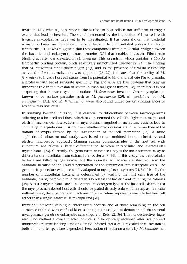
Figure 1. Transmission electron microscopy of M. hyorhinis (A) and of a melanoma cell culture infected by M. hyorhinis (B). Flask shaped bacteria in close proximity to melanoma cells are indicated by arrows.
A polar, tapered cell extension at one of the poles containing an electron-dense core in the cytoplasma was described in some mycoplasmas (Figure 2). This structure, termed the tip organelle, functions mainly as an attachment and motility organelle. A variety of surface proteins that participate in the adhesion process are densely clustered at the tip organelle [4]. The role of host cell surface sialoglycoconjugates as receptors for mycoplasmas has been suggested [8]. The carbohydrate moiety of the glycoprotein, which serves as a receptor for M. pneumoniae on human erythrocytes, has been identified as having a terminal NeuAc(α2–3)Gal(β1–4)GlcNAc sequence [9]. Nevertheless, neuraminidase treatment has frequently failed to abolish the ability of various eukaryotic cells to bind M. pneumoniae [10]. A sialic acid-free glycoprotein, isolated from cultured human lung fibroblasts, which serves as a receptor for M. pneumoniae, has been isolated by Geary et al. [11]. Sulfated glycolipids containing terminal Gal(3SO4)β1 residues were also found to function as receptors [12]. Clearly, there is more than one type of receptor for the various adhering mycoplasmas.

Biomedical Tissue Culture 38
Figure 2. A, scanning electron microscopy of filamentous M. pneumoniae. B, transmission electron microscopy of flask-shaped M. pneumoniae (M) attached by the terminal tip organelle (arrow) to ciliated mucosal cells. Magnification: A, x10,000; B, x36,000.
The attachment of mycoplasmas to the surface of host cells may interfere with membrane receptors or alter transport mechanisms of the host cell. The disruption of the K+ channels of ciliated bronchial epithelial cells by M. hyopneumoniae that resulted in ciliostasis has been described [13]. The host cell membrane is also vulnerable to toxic materials released by the adhering mycoplasmas. Although toxins have not been associated with mycoplasmas, the production of cytotoxic metabolites and the activity of cytolytic enzymes are well established. Oxidative damage to the host cell membrane by peroxide and superoxide radicals excreted by the adhering mycoplasmas appears to be experimentally well-substantiated [14]. The intimate contact of the mycoplasma with the host cell membrane may also result in the hydrolysis of host cell phospholipids catalyzed by the potent membrane-bound phospholipases present in many mycoplasma species [15]. This could trigger specific signal cascades [16] or release cytolytic lysophospholipids capable of disrupting the integrity of the host cell membrane [17, 18].
3.2. Invasion of host cells
It is generally accepted that mycoplasmas remain attached to the surface of host cells [1]. However, some mycoplasmas have evolved mechanisms for entering host cells that are not naturally phagocytic. The intracellular location is obviously a privileged niche, well protected from the action of many antibiotics. Mycoplasma invasion of host cells was intensively studied with M. penetrans, isolated from the urogenital tract of acquired immunodeficiency syndrome (AIDS) patients [19, 20]. It was shown that this microorganism has invasive properties and localizes in the cytoplasm and perinuclear regions [21, 22, 23]. Mycoplasmal invasion of host cells is a complex process that involves a variety of mycoplasmal and host cell factors. It is likely that surface molecules (proteins and lipids) that facilitate the adhesion process of mycoplasmas will have an effect on the
Contamination of Tissue Cultures by Mycoplasmas 39
invasion. Nevertheless, adherence to the surface of host cells is not sufficient to trigger events that lead to invasion. The signals generated by the interaction of host cells with invasive mycoplasmas have yet to be investigated. It has been shown that bacterial invasion is based on the ability of several bacteria to bind sulfated polysaccharides or fibronectin [24]. It was suggested that these compounds form a molecular bridge between the bacteria and eukaryotic surface proteins [25] that enables invasion. Fibronectin binding activity was detected in M. penetrans. This organism, which contains a 65-kDa fibronectin binding protein, binds selectively immobilized fibronectin [23]. The finding that M. fermentans binds plasminogen (Plg) and in the presence of urokinase-type Plg activated (uPA) internalization was apparent (26, 27), indicates that the ability of M. fermentans to invade host cell stems from its potential to bind and activate Plg to plasmin, a protease with broad substrate specificity. Plg and uPA are two proteins that play an important role in the invasion of several human malignant tumors [28], therefore it is not surprising that the same system stimulates M. fermentans invasion. Other mycoplasmas known to be surface parasites such as M. pneumoniae [29], M. genitalium [30], M. gallisepticum [31], and M. hyorhinis [6] were also found under certain circumstances to reside within host cells.
In studying bacterial invasion, it is essential to differentiate between microorganisms adhering to a host cell and those which have penetrated the cell. The light microscopic and electron microscopic observations of mycoplasmas engulfed in membrane vesicles lead to conflicting interpretations. It is not clear whether mycoplasmas are intra, or are they at the bottom of crypts formed by the invagination of the cell membrane [32]. A more sophisticated ultrastructural study was based on a combined immunochemistry and electron microscopy approach. Staining surface polysaccharides of the host cell with ruthenium red allows a better differentiation between intracellular and extracellular mycoplasmas [33]. Currently, the gentamicin resistance assay is the most common assay to differentiate intracellular from extracellular bacteria [7, 34]. In this assay, the extracellular bacteria are killed by gentamicin, but the intracellular bacteria are shielded from the antibiotic because of the limited penetration of the gentamicin into eukaryotic cells. The gentamicin procedure was successfully adapted to mycoplasma systems [21, 31]. Usually the number of intracellular bacteria is determined by washing the host cells free of the antibiotic, lysing them with mild detergents to release the bacteria and counting the colonies [35]. Because mycoplasmas are as susceptible to detergent lysis as the host cells, dilutions of the mycoplasma-infected host cells should be plated directly onto solid mycoplasma media without lysing them beforehand. Each mycoplasma colony represents one infected host cell rather than a single intracellular mycoplasma [34].
Immunofluorescent staining of internalized bacteria and of those remaining on the cell surface, combined with confocal laser scanning microscopy, has demonstrated that several mycoplasmas penetrate eukaryotic cells (Figure 3; Refs. 22, 36) This nondestructive, high-resolution method allowed infected host cells to be optically sectioned after fixation and immunofluorescent labeling. Imaging single infected HeLa cells revealed that invasion is both time and temperature dependent. Penetration of melanoma cells by M. hyorhinis has
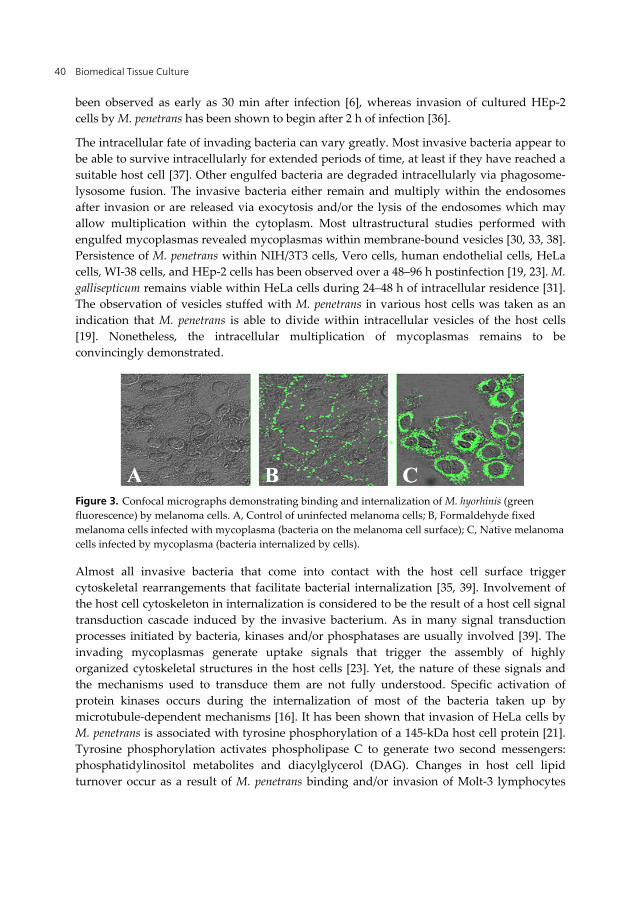
Biomedical Tissue Culture 40
been observed as early as 30 min after infection [6], whereas invasion of cultured HEp-2 cells by M. penetrans has been shown to begin after 2 h of infection [36].
The intracellular fate of invading bacteria can vary greatly. Most invasive bacteria appear to be able to survive intracellularly for extended periods of time, at least if they have reached a suitable host cell [37]. Other engulfed bacteria are degraded intracellularly via phagosome-lysosome fusion. The invasive bacteria either remain and multiply within the endosomes after invasion or are released via exocytosis and/or the lysis of the endosomes which may allow multiplication within the cytoplasm. Most ultrastructural studies performed with engulfed mycoplasmas revealed mycoplasmas within membrane-bound vesicles [30, 33, 38]. Persistence of M. penetrans within NIH/3T3 cells, Vero cells, human endothelial cells, HeLa cells, WI-38 cells, and HEp-2 cells has been observed over a 48–96 h postinfection [19, 23]. M. gallisepticum remains viable within HeLa cells during 24–48 h of intracellular residence [31]. The observation of vesicles stuffed with M. penetrans in various host cells was taken as an indication that M. penetrans is able to divide within intracellular vesicles of the host cells [19]. Nonetheless, the intracellular multiplication of mycoplasmas remains to be convincingly demonstrated.
Figure 3. Confocal micrographs demonstrating binding and internalization of M. hyorhinis (green fluorescence) by melanoma cells. A, Control of uninfected melanoma cells; B, Formaldehyde fixed melanoma cells infected with mycoplasma (bacteria on the melanoma cell surface); C, Native melanoma cells infected by mycoplasma (bacteria internalized by cells).
Almost all invasive bacteria that come into contact with the host cell surface trigger cytoskeletal rearrangements that facilitate bacterial internalization [35, 39]. Involvement of the host cell cytoskeleton in internalization is considered to be the result of a host cell signal transduction cascade induced by the invasive bacterium. As in many signal transduction processes initiated by bacteria, kinases and/or phosphatases are usually involved [39]. The invading mycoplasmas generate uptake signals that trigger the assembly of highly organized cytoskeletal structures in the host cells [23]. Yet, the nature of these signals and the mechanisms used to transduce them are not fully understood. Specific activation of protein kinases occurs during the internalization of most of the bacteria taken up by microtubule-dependent mechanisms [16]. It has been shown that invasion of HeLa cells by M. penetrans is associated with tyrosine phosphorylation of a 145-kDa host cell protein [21]. Tyrosine phosphorylation activates phospholipase C to generate two second messengers: phosphatidylinositol metabolites and diacylglycerol (DAG). Changes in host cell lipid turnover occur as a result of M. penetrans binding and/or invasion of Molt-3 lymphocytes

Contamination of Tissue Cultures by Mycoplasmas 41
[40]. These changes include the accumulation of DAG and the release of unsaturated fatty acids, predominantly long-chain polyunsaturated ones such as docosahexanoic acid (C22:6, 40]. Nonetheless, metabolites of phosphatidylinositol were not detected. These observations support the hypothesis that M. penetrans stimulates host phospholipases to cleave membrane phospholipids, thereby initiating the signal transduction cascade. Because in HeLa cells, which are invaded by M. penetrans, DAG is generated, it is likely that the protein kinase C is activated in the host cells. Indeed, transient protein kinase C activation was demonstrated in invaded HeLa cells by several methods, including translocation to the plasma membrane and enzymatic activity [22]. However, activation was weak and transient, peaking at 20 min postinfection. How any of these different signal transduction events lead to specific microtubule activity resulting in mycoplasmal internalization is unknown. The role of these signals in the penetration, survival, and proliferation of mycoplasmas within host cells, as well as the involvement of the lipid intermediates in the pathobiological alterations taking place in the host cells, merit further investigation.
3.3 Fusion with host cells
The lack of a rigid cell wall allows direct and intimate contact of the mycoplasma membrane with the cytoplasmic membrane of the eukaryotic cell. Under appropriate conditions, such contact may lead to cell fusion. Fusion of mycoplasmas with eukaryotic host cells has been first observed in electron microscopic studies [41]. The development of energy transfer and fluorescence methods has enabled investigation of the fusion process on a quantitative basis in an experimental cell culture-mycoplasma system and has also allowed the identification of fusogenic mycoplasmas. In all the fusogenic Mycoplasma species tested, fusogenicity is dependent on the unesterified cholesterol content of the cell membrane [42]. Fusogenic activity can be found only among mycoplasmas requiring unesterified cholesterol for growth, whereas species, which do not require cholesterol, are nonfusogenic. Among the Mycoplasma species, the human mycoplasma, M. fermentans, is highly fusogenic, capable of fusing with a variety of cells [2]. It is widely accepted that the reorganization of the membrane structure that occurs during fusion requires that the lipid bilayer is broken up and that other inverted configurations, such as reversed nonbilayer aggregates, are being formed [43, 44, 45]. It has been shown that the polar lipid fraction of M. fermentans is capable of enhancing the fusion of small, unilamellar phosphatidylcholine-cholesterol (1:1 molar ratio) vesicles with Molt-3 lymphocytes in a dose-dependent manner, suggesting that a lipid component acts as a fusogen [17, 46]. In an attempt to identify the fusogen, detailed lipid analyses of M. fermentans membranes were performed [17, 47, 48], revealing that the polar lipid fraction of this organism contains unusual choline-containing ether phosphoglycolipids, 1-O-alkyl/alkenyl-2-O-acyl-glycero-3-phosphocholine and its lyso-form 1-O-alkyl/alkenyl-glycero-3-phosphocholine [49]. The ether lipids, mainly the lyso-derivative has a marked effect on the fusion of M. fermentans with host eukaryotic cells [50]. Very little is known about the role of membrane proteins in the fusion process. The observation that fusion of M. fermentans with Molt-3 cells was inhibited by pretreatment of intact M. fermentans with proteolytic enzymes [51] implies that this organism possesses a

Biomedical Tissue Culture 42
proteinase-sensitive receptor(s) responsible for binding and/or the establishment of tight contact with the cell surface of the host cell involved in fusion. During the fusion process, mycoplasma components may be delivered into the host cell and affect the normal functions of the cell. A whole array of hydrolytic enzymes has been identified in mycoplasmas [1, 15, 52]. Most remarkable are the mycoplasmal nucleases [1] that may degrade host cell DNA. It has recently been shown that M. fermentans contains a potent phosphoprotein phosphatase [52]. The delivery of an active phosphoprotein phosphatase into the eukaryotic cell upon fusion may interfere with the normal signal transduction cascade of the host cell.
4. Effects of mycoplasmas on cell cultures
Effects on cell function and metabolism have long been recognized as common in mycoplasma contaminated cell cultures. The nature of the effects depends on the contaminating species and strain of mycoplasma, and on the type of cell infected. Frequently, the effects are due to nutrient deprivation, such as the depletion of amino acids, sugars, fatty acids, cholesterol or nucleic-acid precursors [5], the depletion of choline [4] or the activity of mycoplasmal endonucleases [53], mycoplasmal arginine deiminase [54] or mycoplasmal thymidine phosphorylase [55]. Some mycoplasmas have been shown to produce severe cytopathic effects (CPE) characterized by stunted, abnormal growth and rounded, degenerated cells, apparently due to the promotion or inhibition of apoptosis [56]. The promotion of apoptosis may be due to direct effects of mycoplasma components. Thus, M. bovis infection sensitizes some host cells to apoptosis through participation of mycoplasmal endonucleases [53]. Choline deficiency induced by M. fermentans enhances rat astrocyte apoptosis [4]. Some mycoplasmas promote host cell death via induction of pro-apoptotic genes [57, 58]. Pro-apoptotic and anti-apoptotic mycoplasmas appear to alter apoptosis regulatory genes differently [59].
4.1. Competition for precursors
Genomic analyses of mycoplasmas have revealed the limited biosynthetic capabilities of these microorganisms [60, 61]. Mycoplasmas apparently lost almost all the genes involved in the biosynthesis of amino acids, fatty acids, cofactors, and vitamins and therefore depend on the host microenvironment to supply the full spectrum of biochemical precursors required for the biosynthesis of macromolecules [1]. Competition for these biosynthetic precursors by mycoplasmas may disrupt host cell integrity and alter host cell function. Nonfermenting Mycoplasma spp. utilize the arginine dihydrolase pathway for generating ATP [62] and rapidly deplete the host's arginine reserves affecting protein synthesis, growth and host cell divisions. The effect on the cellular genome may be expressed in chromosomal breakage, multiple translocation events, reduction in chromosome number and the appearance of new and/or additional chromosome variants [63]. Since histones are arginine rich, it was suggested that mycoplasmas may exert their effects on cellular genomes by depleting arginine and thus inhibiting histone synthesis [62]. However, as fermenting mycoplasmas also induce chromosomal aberrations, other mechanisms, including competition for nucleic

Contamination of Tissue Cultures by Mycoplasmas 43
acid precursors, or degradation of host-cell DNA by mycoplasma nucleases, may be involved. M. fermentans infection of cell cultures has been shown to result in a choline-deficient environment and in the induction of apoptosis [64]. Choline is an essential dietary component that ensures the structural integrity and signaling functions of the cell membranes; it is the major source of methyl groups in the diet, and it directly affects lipid transport and metabolism and the cholinergic neurotransmission and transmembrane signaling of cells of the nervous system [65].
4.2. Cytopathic effects
Mycoplasmal attachment to eukaryotic cells may sometimes lead to a pronounced cytopathic effect. Attachment permits the mycoplasma contaminant to release noxious enzymatic and cytolytic metabolites directly onto the tissue cell membrane. Some mycoplasmas selectively colonize defined areas of the cell culture. This results in microcolony formation producing microlesions and small foci of necrosis, e.g., M. pulmonis, or form plaques, e.g., M. gallisepticum, in an agar overlay system [5]. Microcolonization suggests that mycoplasma-specific receptors are localized in defined areas of the cell monolayer. However, other fermenting mycoplasmas, e.g., M. hyorhinis, attach to every cell and destroy the entire monolayer, producing a generalized cytopathic effect. With HeLa cells infected by the invasive M. penetrans, the most pronounced effect was the vacuolation of the host cells [22]. The vacuoles appeared to be empty, differing from the described membrane-bound vesicles containing clusters of bacteria [19]. The number and size of the vacuoles depended on duration of infection. Because vacuolation is not obtained with M. penetrans cell fractions [22], it is unlikely that a necrotizing cytotoxin is involved in the generation of the cellular lesions. A possible mechanism that leads to vacuolation may be associated with the accumulation of organic peroxides upon invasion of HeLa cells by M. penetrans. Indeed, when HeLa cells were grown with the antioxidant α-tocopherol, the level of accumulated organic peroxides was extremely low, and vacuolation was almost completely abolished [22].
Being unable to synthesize nucleotides, mycoplasmas developed potent nucleases, either soluble ones secreted into the extracellular medium or membrane-bound nucleases [1, 66, 67] apparently as a means of producing nucleic acid precursors required for metabolism. It has been shown that, occasionally, secreted mycoplasmal nucleases are taken up by the host cells [68]. Thus, it was suggested that the cytotoxicity of M. penetrans is mediated at least in part by a secreted mycoplasmal endonuclease that is cleaving DNA and/or RNA of the host cells [66], and the endonuclease activity of M. bovis was implicated in the increased sensitivity of lymphocytic cell lines to various inducers of apoptosis [69].
4.3. Transformation of cells mediated by mycoplasmas
Cell culture contamination may go undetected because mycoplasma infections do not produce the overt turbid growth that is commonly associated with bacterial and fungal contamination. Mycoplasma growth can grow in close interaction with mammalian cells,
Biomedical Tissue Culture 44
often silently for a long period of time. However, prolonged interactions with mycoplasmas with seemingly low virulence could, through a gradual and progressive course, induce chromosomal instability as well as malignant transformation, promoting tumorous growth of mammalian cells [70, 71]. Mycoplasmal-induced malignant transformation is a multistage process [70] associated with increased or decreased expression of many genes, especially cancer-related genes [72]. Over expression of H-ras and c-myc oncogenes were found to be closely associated with both the initial reversible and the subsequent irreversible states of the mycoplasma-mediated transformation of cells [71]. In some cases, mycoplasmas have been shown to induce the production of proteins that play essential roles in the development of malignancy. Examples are the mycoplasmal-promoted production in diverse types of cultured cells of bone morphogenetic protein 2 (BMP2) that enhances tumor growth by increasing cell proliferation [73]; mycoplasma-induced diminished activation of the tumor suppression protein p53, and enhanced fibroblast transformation by the oncogenic H-ras [74] ; promotion of cancer cell motility and migration by P37, the major immunogen of M. hyorhinis, through activation of the matrix metalloproteinase-2 [75].
4.4. Modulation of immune and non-immune cell metabolism
The effects of mycoplasmas on the immune system are well established and include effects on differentiation and activation of innate immunity cells (macrophages, dentritic cells, neutrophils, NK) and on adaptive immunity cells (T and B cells). Mycoplasma and mycoplasmal components are potent macrophage activators, and stimulate the release of various proinflammatory cytokines, such as tumor necrosis factor α (TNFα), interleukin-1(IL-1), IL-6, NO [4, 76]. In turn, some cytokines participate in lymphocyte differentiation and maturation [4]. M. fermentans induces a partial differentiation of the human monocytic cell line THP-1 [77]. Mycoplasma-contaminated exosome fractions of dentritic cells are mitogens for naive B lymphocytes and promote immunoglobulin secretion [78].
Mycoplasmas and mycoplasmal components interact with diverse non-immune cells [56, 57, 58, 79], with some information available on the cellular proteins affected by them. M. salivarium and M. fermentans induce the cell surface expression of intercellular adhesion molecule 1 (ICAM-1) in human gingival fibroblasts [80]. Hyperammonia toxicity in irradiated hepatoma cells has been shown to be due to contamination by mycoplasma containing arginine deiminase, that converts arginine to citrulline and ammonia [54]. M. pneumoniae induces the expression of the major airway protein mucin (MUC5AC) in cultured airway epithelial cells isolated from asthmatic subjects, but not in cells isolated from normal subjects; the preferential expression of MUC5AC in cells isolated from asthmatic subjects suggests that asthmatic epithelial cells may be primed to respond to the mycoplasma [81], thus pointing to the importance of identifying consequences of mycoplasma contamination that may be observed only in certain specific types of cultured cells. Catabolic mycoplasmal enzymes may interfere with chemotherapy. This is illustrated by the finding that the antiviral and cytostatic activity of pyrimidine nucleoside analogues (used as chemotherapeutic agents) is markedly decreased in M. hyorhinis contaminated cells, due to the mycoplasmal thymidine phosphorylase that degrades pyrimidine nucleoside

Contamination of Tissue Cultures by Mycoplasmas 45
analogues [55]. Contamination of human cultured neuroblastoma SH-SY5Y and melanoma cell lines by M. hyorhinis results in increased levels of calpastatin (the endogenous inhibitor of the ubiquitous Ca2+-dependent protease calpain). The calpastatin upregulation resides in the M. hyorhinis lipoprotein fraction (LPP), via the IκB/NF-κB transcription pathway [79]. LPPs of several other mycoplasma species have also been found to upregulate calpastatin [J.D. Kornspan, T. vaisid, S. Rottem and N.S. Kosower, unpublished data]. Amyloid-β-peptide and Ca2+ (these are central to the pathogenesis of Alzheimer’s Disease) activate calpain and are toxic to neuroblastoma cultured cells. The increased calpastatin levels in the mycoplasma-infected cells attenuate the calpain-related amyloid-β-peptide and Ca2+-toxicity. Calpain and calpastatin are widely distributed in biological systems, with the ratio of calpastatin to calpain varying among cells. The calpain-calpastatin system has been implicated in a variety of cellular physiological and pathological processes [82]. Since calpastatin level is important in the control of calpain activity, mycoplasmas may play a role in a variety of metabolic and signal transduction pathways in some types of cultured cells. The mycoplasma-induced elevation of calpastatin provides an example of mycoplasmal effects on intracellular proteins in non-immune cells, resulting in important alterations in the host cell functions.
4.5. Effect on virus infection
Mycoplasmas may alter the progress of viral infections in cell cultures [83, 84]. As mycoplasmas may also cause virus-like CPE, many investigators have mistaken cytolytic mycoplasmas for viruses. Like viruses, mycoplasmas are filterable, hemadsorbant, hemagglutinant, resistant to certain antibiotics, able to induce chromosomal aberrations, and sensitive to detergents, ether and chloroform; thus the first established mycoplasma pathogens of humans (M. pneumoniae), animals (M. mycoides) or plants (Spiroplasma spp.) were believed to be viruses. Some mycoplasmas have no detectable effect on viral growth. Others can decrease, or even increase, virus yields in infected cell culture [85]. The effect depends on the strain or species of mycoplasma, the virus, and the cell culture used. At least two mechanisms responsible for decreasing viral yields in vitro have been identified. The cytolytic, fermenting mycoplasmas suppress metabolism and growth, resulting in a decrease in viral yields. Arginine-utilizing mycoplasmas decrease the titers of arginine-requiring DNA viruses by depleting arginine from the medium [62]. Mycoplasmas may render cell cultures less sensitive to exogenously supplied interferon and thus to increase virus yields [86]. Mycoplasmas may also inhibit viral transformation of cell cultures by known oncogenic viruses [5, 87].
4.6. Signal transduction pathways
Mycoplasmas and mycoplasmal membrane LPPs attach to certain Toll-like receptors (TLRs) of the host cell membrane. The main TLR involved appears to be TLR2, with participation of TLR6 as coreceptor. In some cases, TLR1 is also involved [88]. The interaction with the receptors triggers cascades of cellular signals within the cell, and the complex pathways

Biomedical Tissue Culture 46
culminate in a variety of host cell responses. Mycoplasmas and mycoplasmal LPP are known to activate the transcription factors NF- B [74, 79] and AP-1 [1 4], via TLR- downstream cascades involving kinases (MAPKKKs-IKKs and MAPKKKs-MAPKKs- MAPKs). Known mycoplasma-affected target genes are mainly those responsible for proinflammatory proteins [4], and those involved in malignant cell transformation [72], with little information available on genes responsible for other proteins [53, 79, 80, 81].
5. Detecting mycoplasmas in cell cultures
The ubiquitous nature of mycoplasma in man, animals and the environment increases the likelihood of the introduction of these organisms into cell cultures or a manufacturing process. Currently, the recommended test requirements for biologics are as follows: (1) The master- and working cell seed banks must be free of mycoplasmas. (2) The product-harvest concentrates must be free of mycoplasmas. (3) All products produced in cell cultures, a generic term used for all tissue cells grown in vitro, must be tested. This includes viral vaccines (such as poliovirus, adenovirus, measles, rubella, mumps and rabies), monoclonal antibodies, immunological modifiers and cell-culture-derived blood products, such as tissue-type plasminogen and erythropoietin. Guidelines for mycoplasma testing of cell cultures and biologics is addressed in several international pharmacopoeias e.g., United States Pharmacopoeia, (USP 33/NF 28 <63>and <1226>, Mycoplasma tests, 2010); European Pharmacopoeia (EP 2.6.7., Mycoplasmas, 7th ed.; 2012); Japanese Pharmacopoeia (JP); Section 21 of the Code of Federal Regulations (CFR), International Conference on Harmonisation (ICH), and FDA- Points to Consider (PTC) documents. Several different approaches are being used to detect mycoplasmas in contaminated tissue cultures including the culture procedures, a variety of nonspecific procedures and the polymerase chain reactions (PCR).
5.1. Standard culture procedures
The culture procedures require that the tested material will be inoculated onto solid and liquid growth media capable of growing a variety of mycoplasma including aerobic, microaerophilic and anaerobic strains. Broth cultures are incubated and sub passaged to plate agar. After the required incubation period, the agar plates are observed microscopically for the presence of mycoplasma colonies [5]. The variation inherent in the complex media usually used for in vitro culture of mycoplasmas is due to batch variation in compounds such as sera, or yeast extract. Such variation makes the development of defined media attractive. However, a key problem has been the supply of lipids in an available, but non-toxic form, hence, defined artificial media have been developed for only a few species [1]. Most mycoplasmas produce microscopic (100 - 400 µm in diameter) colonies with a characteristic 'fried-egg' appearance, growing embedded in the agar, although some (e.g. M. pulmonis) may not grow completely embedded, and some freshly-isolated pathogens (e.g. M. pneumoniae) produce a more granular, diffuse colony-type. Since they usually grow embedded, mycoplasma colonies can be distinguished from other bacteria by: (1) specific

Contamination of Tissue Cultures by Mycoplasmas 47
colony shape; (2) being difficult to scrape from the agar surface. Mycoplasmas growing on agar can be identified more specifically by immunofluorescent procedures, using fluorophores conjugated to species-specific antibodies [4]. The traditional culture-based techniques are relatively sensitive, capable of detecting as few as 1-10 colony forming units of mycoplasmas and therefore are required by pharmacopoeias and regulatory authorities worldwide. Nonetheless, this procedure is time consuming requiring a minimum of 28 days to complete, costly and not sensitive to non-cultivable strains, therefore, the development of more accurate and faster techniques are needed to facilitate faster detection of a contaminating mycoplasma and more rapid corrective action.
5.2. Polymerase chain reaction (PCR)
PCR methodology has existed for decades, however conventional PCR and real-time PCR assays have only recently been considered for mycoplasma detection in cell cultures and biological products. These assays are often based on the amplification of conserved regions of the 16S rDNA [89, 90] or the spacer region between the 16S and 23S rDNA [91, 92]. The PCR approach is rapid (1-2 days), inexpensive, and independent of culture conditions. Specific oligonucleotide primers capable of amplifying the conserved regions and thus detecting DNA of multiple Mollicutes species while excluding other contaminating DNA are used in the PCR assays. In comparison to conventional PCR methods, real-time PCR assays are quicker, simpler, and more suitable for handling a large number of samples [93]. Nonetheless, some of the primers used are not entirely specific for Mollicutes [94, 95]. Thus, sequence homologies between Mollicutes spp. and Chlamydia spp. led to false-positive results in Chlamydial cell cultures tested for mycoplasma contamination with a commercial PCR kit [96].
Throughout the last decade, new PCR assays for mycoplasma detection, which appeared to resolve these issues, were described, while being sufficiently simple and inexpensive for routine use. For example, a PCR assay which applied readily available techniques in DNA extraction together with a modified single-step PCR using a primer pair that was homologous to a broad spectrum of mycoplasma species was proposed [97]. A high sensitivity and specificity for mycoplasma detection in cell production cultures was made possible through the combination of three key techniques: 8-methoxypsoralen and UV light treatment to decontaminate PCR reagents of DNA; hot-start Taq DNA polymerase to reduce nonspecific priming events; and touchdown PCR to increase sensitivity while also reducing nonspecific priming events. Another proposed PCR assay for mycoplasma detection was a sensitive two-stage PCR procedure which detected 13 common mycoplasmal contaminants [92]. For primary amplification, the DNA regions encompassing the 16S and 23S rRNA genes of 13 species were targeted using general mycoplasma primers. The primary PCR products were then subjected to secondary nested PCR, using two different primer pair sets, designed via the multiple sequence alignment of nucleotide sequences obtained from the 13 mycoplasmal species. The nested PCR, which generated DNA fragments of 165-353 bp, was found to be able to detect 1-2 copies of the target DNA, and evidenced no cross-reactivity with the genomic DNA of related microorganisms or of human cell lines, thereby confirming the sensitivity and specificity of the primers used.
Biomedical Tissue Culture 48
Other studies showed that reverse transcription-PCR (RT-PCR) methods based on detection of the 16S rRNA, which is present in multiple (103–104) copies per bacterial cell [98, 99], are more sensitive than PCR detecting the 16S rDNA. Thus, a direct side-by-side comparison of RT-PCR and PCR targeting the 16S rRNA and the 16S rRNA gene, respectively, demonstrated that RT-PCR was able to provide up to a two-logarithm higher sensitivity of bacteria detection in comparison with the PCR-based assay [90, 100] and the sensitivity provided by RT-PCR is approaching the sensitivity of conventional microbiological culture methods [100]. Therefore, it was suggested that RT-PCR methods targeting the bacterial 16S or 23S rRNAs are having the real potential to provide the sensitivity of mycoplasma detection close to or even higher than that of conventional culture methods [101] .
Recently, the MycoTOOL PCR test kit from Roche (Roche, Diagnostic GmbH, Penzberg, Germany) was approved by the European Medicines Agency (EMEA) for release testing of pharmaceutical products. It is the first commercially available Mycoplasma PCR test that can replace traditional Mycoplasma tests (culture method as well as indicator cell culture method) during pharmaceutical production. In June 2009 the FDA approved the PCR concept of this test for seven commercial products from Genentech. Earlier, Bayer Health Care received approval for a pharmaceutical product from the EMEA and Japan’s Ministry of Health, Labour and Welfare (MHLW) using the same PCR-based test concept. Guidelines describing acceptable protocols for specific PCR methods are provided by the EP and JP. The pharmacopoeias, PTC, and CFR protocols vary with their recommendations on how to conduct the PCR assays.
5.3. Indirect non-specific procedures
Some 'non-cultivable' mycoplasma strains cannot readily be grown on standard agar or broth-culture media [5], and cell-assisted culture is required for their isolation. Cell-culture systems are therefore a valuable ancillary tool for the isolation and detection of mycoplasmas and 'indicator cell culture' procedures using either VERO (African green monkey kidney), or NIH 3T3 cell cultures have been developed [102]. These cell lines are susceptible to infection by the majority of mycoplasmas and are therefore a reliable 'indicator' system for detecting mycoplasma infection. These approaches are particularly useful for the identification and detection of mycoplasmas that adhere to host-cell surfaces.
The indirect non-specific procedures require that the tested material will be inoculated directly onto tissue culture cover slips or flasks containing a monolayer of the indicator cells. The indicator cell culture inoculated with the tested material are than fixed and stained with DNA-binding fluorochromes using bisbenzimidazole (such as Hoechst or DAPI stains) [103].
Identification of contaminating mycoplasma is by visual observation via fluorescent microscopy. Mycoplasmas are detected by their characteristic particulate or filamentous pattern of bright fluorescence on the cell surface (Figure 4) and, if contamination is heavy, in surrounding areas. These procedures are suitable for use with either non-specific DNA stains for detecting mycoplasmas, or in conjunction with mycoplasma-speciation methods,

Contamination of Tissue Cultures by Mycoplasmas 49
such as by immunofluorescence procedures using species-specific polyclonal antisera, or monoclonal antibodies, conjugated with fluorescein or peroxidase [104]. A wide variety of luminol-dependent chemiluminescence and bioluminescent methods were described [5, 63].
Figure 4. Mycoplasma contaminated eukaryotic cells stained with a fluorescent DNA stain.
Biochemical identification methods have also been in use [5, 78]. Procedures based on the comparative utilization of uridine versus uracil in contaminated versus mycoplasma-free cell cultures have been suggested [105]. Other methods are based on the detection of enzyme activity present in mycoplasmas, but absent, or minimal in uninfected cell cultures. The enzymic activities measured include: arginine deiminase [62]; thymidine, uridine, adenosine or pyrimidine nucleoside phosphorylase [102]; hypoxanthine or uracil phosphoribosyl transferase activities [106]. Positive results are based on arbitrary values, making low levels of mycoplasma contamination difficult to detect. Detection kit that provide a new, sensitive and rapid biochemical method was recently presented (Cambrex, Bio Science, Caravaggio, Bergamo, Italy). The test is based on a bioluminescent assay which can be assessed within 20 min for daily determination of the mycoplasma status of cell cultures. The performance sensitivity and specificity of the kit was evaluated and compared to the PCR/ELISA detection kit (Roche, Diagnostic GmbH, Penzberg, Germany) and the standard culture method [5]. Recently, a simple and inexpensive assay monitoring mycoplasma contamination, based on degradation of the Gaussia luciferase reporter in cell cultures was described [107]. This assay has been shown to be more sensitive for detecting mycoplasma contamination in seven different cell lines as compared to a commercially available bioluminescent assay [107]
6. Eliminating mycoplasmas from infected cultures
Ever since mycoplasma contamination of cell cultures was first reported, attempts have been made to develop methods for the elimination of mycoplasmas, including the use of antibiotics such as tetracycline, kanamycin, novobiocin, tylosin, gentamycin, doxycycline, thiayline and

Biomedical Tissue Culture 50
quinolones; surface-active agents; anti-mycoplasma antisera and prolonged heating treatments (40-42 °C) [63, 108]. Eliminating mycoplasmas by passage of a cell culture through nude mice [109] has been successful for some, but not all, mycoplasmas. An efficient procedure for eliminating mycoplasmas is based on the selective incorporation of 5-bromouracil (5-BrUra) into mycoplasmas, and the induction of breaks by light in the 5-BrUra-containing DNA [110]. The unusually high content of A+T makes the mycoplasma DNA an excellent candidate for the induction of breakage by the combined action of 5-BrUra, 33258-Hoechst and visible light [110]. Some of the elimination procedures may apply to some, but not all, mycoplasma species; some of them are laborious and/or time consuming. It was suggested, therefore, that whenever possible, the infected cell culture should be discarded and replaced with a mycoplasma-free culture [108]. When the cell culture is irreplaceable, the use of antibiotic mixtures, are the commonest approaches. One has to keep in mind that cell-culture contaminants that have been continuously exposed to antibiotics develop resistance to the drug, and antibiotic-resistant strains have been isolated for most Mycoplasma species tested. Treatment may also induce the selection of a subpopulation of cells and the treated cell culture may differ in its characteristics from the original culture.
Among the antibiotics that were shown to have strong anti-mycoplasma properties are different inhibitors of protein synthesis mainly tetracyclines or macrolides as well as quinolones [111]. The target enzymes of quinolones are considered to be DNA gyrase and topoisomerase IV which are essential enzymes for controlling the topological state of DNA in DNA replication and transcription . Most recently the quinolone garenoxacin was found to be a most valuable quinolone in the elimination M. pneumoniae [112].
The addition of antibiotics to the culture medium during a limited period of time (1-3 wk) is a simple, inexpensive, and very practical approach for decontaminating continuous cell lines. BM-cyclin (trade name of Roche, Mannheim, Germany), a combination of tiamulin and minocycline (both inhibiting protein synthesis), was introduced by Jung et al. [113] who show that three cycles of treatment of a contaminated cell culture with BM-cyclin I (containing the macrolide tiamulin) at a final concentration of 10 µg/ml for 3 days followed by BM-cyclin II (containing the tetracycline minocycline) at a concentration of 5 µg/ml for 4 days completely eradicated mycoplasmal infection from cultured cells [113].
Uphoff and Drexler [111, 114] examined the effectiveness of several quinolones and BM-cycline protocols. The contaminated cell cultures were exposed to one of the following five antibiotic regimens: mycoplasma removal agent (MRA, quinolone; a 1-wk treatment), enrofloxacin (quinolone; 1 wk), sparfloxacin (quinolone; 1 wk), ciprofloxacin (quinolone; 2 wk), and BM-Cyclin (alternating tiamulin and minocycline; 3 wk). The mycoplasma infection was permanently eliminated by the various antibiotics in 66-85% of the cultures treated. Mycoplasma resistance was seen in 7-21%, and loss of the culture as a result of cytotoxically caused cell death occurred in 3-11% of the cultures treated [111, 114].
Recently, MycoZap (trade name of Lonza, Verviers, Belgium) treatment has been introduced as a new therapeutic tool able to overcome the eukaryotic cytotoxicity of fluoroquinolones and BM-Cyclins [115]. MycoZap kit (Lonza, Verviers, Belgium) includes a combination of

Contamination of Tissue Cultures by Mycoplasmas 51
patented antibiotic and antimetabolic agents. An evaluation of the MycoZap kit performance was recently presented by Mariotti et al., [116] who exposed mycoplasma contaminated cells to the MycoZap protocol and compared the results obtained to the eradication efficiency of enrofloxacin (Fluka, Bio-Chemika, Missouri, USA), MRA (Euroclone, Lugano, Switzerland), ciprofloxacin and the BM-Cyclin protocol. Treatment of contaminated cell cultures by MycoZap, MRA, ciprofloxacin, enrofloxacin and BM-cycline, eliminated mycoplasma infection by 46%, 29%, 43%, 40% and 57% respectively. The use of an eradication mixture based on a combination of the antibiotics BM-Cyclins, ciprofloxacin, enrofloxacin and MRA was able to clean 88.6% of the infected cultures, whereas the addition of MycoZap to the eradication mixture resulted in the eradication of mycoplasmas from 100% of the contaminated cell cultures [116].
7. Conclusions
Mycoplasmas are shown to cause various alterations in cultured cells. As described above, some alterations are due to direct effects on the cells by mycoplasma components, and other alterations are due to indirect effects, via inducing the host cell to alter its gene and protein expression and activity. It is important to emphasize the fact that mycoplasmal-altered cell phenotype and function is often observed in specific types of cells under special conditions, e.g., when the cultured cells are exposed to certain agents. The detection of mycoplasma contamination, and the identification of the factors and pathways involved in the mycoplasmal effects are thus of utmost importance in handling cultured cells, including using stem cells for differentiation to specific tissues.
Author details
Shlomo Rottem and Jonathan D. Kornspan Department of Microbiology and Molecular Genetics, IMRIC, The Hebrew University-Hadassah Medical School, Jerusalem, Israel
Nechama S. Kosower Department of Human Molecular Genetics and Biochemistry, Sackler School of Medicine, Tel-Aviv University, Ramat-Aviv, Tel-Aviv, Israel
Acknowledgement
Figure 1A is a courtesy of S. Razin, The Hebrew University-Hadassah Medical School and Figure 1B is a courtesy of A. M. Collier, The University of North Carolina School of Medicine. We would like to thank M. Tarshis for the confocal microscopy.
8. References
[1] Razin S, Yogev D, Naot Y (1998) Molecular biology and pathogenicity of mycoplasmas. Microbiol. Rev. 63: 1094-1156.

Biomedical Tissue Culture 52
[2] Fraser CM, et. al. (1995) The minimal gene complement of Mycoplasma genitalium. Science 270: 397-403.
[3] Maniloff J (1996) The minimal gene genome: "on being the right size." Proc. Natl. Acad. Sci. USA 93: 10004-10006.
[4] Rottem S (2003) Interaction of mycoplasmas with host cells. Physiol. Rev. 83: 417-432. [5] Barile MF, Rottem S (1993) Mycoplasmas in cell cultures. In: Kahane I, Adoni A, editors.
Rapid diagnosis of mycoplasmas. New York: Plenum Press. pp. 155-193. [6] Kornspan JD, Tarshis M, Rottem S (2010) Invasion of melanoma cells by Mycoplasma
hyorhinis: enhancement by protease treatment. Infect. Immun. 78: 611-617. [7] Shaw JH, Falkow S (1988) Model for invasion of human tissue culture cells by Neisseria
gonorrhoeae. Infect. Immun. 56: 1625-1632. [8] Razin S, Jacobs E (1992) Mycoplasma adhesion. J. Gen. Microbiol. 138: 407-422. [9] Roberts DD, et.al. (1989) Sialic acid-dependent adhesion of Mycoplasma pneumoniae to
purified glycoproteins. J. Biol. Chem. 264: 9289-9293. [10] Geary SJ, Gabridge MG (1987) Characterization of a human lung fibroblast receptor site
for Mycoplasma pneumoniae. Isr. J. Med. Sci. 23: 462-468. [11] Geary SJ, et.al. (1990) Identification of mycoplasma binding proteins utilizing a 100
kilodalton lung fibroblast receptor. J. Rec. Res. 9: 465-478. [12] Krivan Hc, et. al. (1989) Adhesion of Mycoplasma pneumoniae to sulfated glycolipids and
inhibition by dextran sulfate. J. Biol. Chem. 264: 9283-9288. [13] Debey MC, Ross RF (1994) Ciliostasis and loss of cilia induced by Mycoplasma
hyopneumoniae in porcine tracheal organ cultures. Infect. Immun. 62: 5312-5318. [14] Almagor M, et. al. (1986) Protective effects of the glutathione redox cycle and vitamin E
on cultured fibroblasts infected by Mycoplasma pneumoniae. Infect. Immun. 52: 240-244. [15] Shibata KI, Sasaki T, Watanabe T (1995) AIDS-Associated mycoplasmas possess
phospholipases C in the membrane. Infect. Immun. 63: 4174-4177. [16] Rosenshine I, Finlay BB (1993) Exploitation of host signal transduction pathways and
cytoskeletal functions by invasive bacteria. BioEssays 15: 17-24. [17] Salman M, Rottem S (1995) The cell membrane of Mycoplasma penetrans: Lipid
composition and phospholipase A1 activity. Biochim. Biophys. Acta. 1235: 369-377. [18] Salman M, Borkovsky Z, Rottem S (1998) Mycoplasma penetrans invasion of Molt-3
lymphocytes induces changes in the lipid composition of host cells. Microbiology 144: 3447-3454.
[19] Lo SC (1992) Mycoplasmas in AIDS. In: Maniloff J, McElhaney RN, Finch LR, Baseman JB editors. Mycoplasmas: Molecular Biology and Pathogenesis. Washington, D.C.: American Society Microbiology. pp. 525-548.
[20] Lo SC, et. al. (1993) Adhesion onto and invasion into mammalian cells by Mycoplasma penetrans: a newly isolated mycoplasma from patients with AIDS. Mod. Pathol. 6: 276-280.
[21] Andreev J, et. al. (1995) Invasion of HeLa cells by Mycoplasma penetrans and the induction of tyrosine phosphorylation of a 145 kDa host cell protein. FEMS. Microbiol. Letts. 132: 189-194.

Contamination of Tissue Cultures by Mycoplasmas 53
[22] Borovsky Z, et. al. (1998). Mycoplasma penetrans invasion of HeLa cells induces protein kinase C activation and vacuolation in the host cells. J. Med. Microbiol. 47: 915-922.
[23] Giron JA, Lange M, Baseman JB (1996) Adherence, fibronectin binding, and induction of cytoskeleton reorganization in cultured human cells by Mycoplasma penetrans. Infect. Immun. 64: 197-208.
[24] Chausee MS, Cole R, Van Putten JPM (2000) Streptococcal erythrogenic toxin B abrogates fibronectin dependent internalization of Streptococcus pyogenes by cutured mammalian cells. Infect. Immun. 68: 3226-3232.
[25] Yavlovich A, Katzenell A, Rottem S (2007) Binding of host extracellular matrix proteins to Mycoplasma fermentans and its effect on adherence to, and invasion of HeLa cells. FEMS Microbiol. Letts. 266: 158-162.
[26] Yavlovich A, Higazi AR, Rottem S (2001) Plasminogen binding and activation by Mycoplasma fermentans. Infect. Immun. 69: 1977-1982.
[27] Yavlovich, A., et. al. (2004) Mycoplasma fermentans binds to and invades HeLa cells: Involvement of plasminogen and urokinase. Infect. and Immun. 72, 5004-5011.
[28] Rao JS (2003) Molecular mechanisms of glioma invasiveness: the role of proteases. Nat. Rev. Cancer 3:489-501.
[29] Balish MF, Krause DC (2002) Cytadherence and the cytoskeleton. In: Razin S, Herrmann R, editors. Molecular Biology and Pathogenicity of Mycoplasmas. New York: Kluwer Academic/Plenum. pp. 491-518.
[30] Jensen JG, Blom J, Lind K (1993) Intracellular location of Mycoplasma genitalium in cultured Vero cells as demonstrated by electron microscopy. Int. J. Path. 75: 91-98.
[31] Winner F, Rosengarten R, Citti C (2000) In vitro cell invasion of Mycoplasma gallisepticum. Infect. Immun. 68: 4238-4244.
[32] Zucker-Franklin D, Davidson M, Thomas L (1966) The interaction of mycoplasmas with mammalian cells, HeLa cells, neutrophiles, and eosinophils. J. Exp. Med. 124: 521-532.
[33] Taylor-Robinson D, et. al. (1991) Intracellular location of mycoplasmas in cultured cells demonstrated by immunocytochemistry and electron microscopy. Int. J. Exp. Pathol. 72: 705-714.
[34] Elsinghorst EA (1994) Measurement of invasion by gentamicin resistance. Methods Enzymol. 236: 405-420.
[35] Finlay BB, Ruschkowski S, Dedhar S (1991) Cytoskeletal rearrangements accompanying Salmonella entry into epithelial cells. J. Cell Sci. 99: 283-296.
[36] Baseman JB, Tully JG (1997) Mycoplasmas: sophisticated, reemerging, and burdened by their notoriety. Emerg. Infect. Dis. 3: 21-32.
[37] Feng SH, et. al. (1999) Mycoplasmal infections prevent apoptosis and induce malignant transformation of interleukin-3-dependent 32D hematopoietic cells. Mol. Cellular Biol. 19: 7995-8002.
[38] Stadtlander CT, et. al (1993) Cytopathogenicity of Mycoplasma fermentans (including strain incognitos). Clin. Infect. Dis. 17: 289-301.
[39] Oelschlaeger TA, Kopecko DJ (2000) Microtubule dependent invasion pathways to bacteria. In: Oelschlaeger TA, Hacker J, editors. Bacterial Invasion into Eukaryotic Cells.

Biomedical Tissue Culture 54
Subcellular Biochemistry, Volume 33. New York: Kluwer Academic/Plenum Publishers. pp. 3-19.
[40] Salman M, et. al. (1994) Membrane lipids of Mycoplasma fermentans. FEMS Microbiol. Lett. 123: 255-260.
[41] Rottem S, Naot Y (1998) Subversion and exploitation of host cells by mycoplasmas. Trends. Microbiol. 6: 436-440.
[42] Tarshis M, Salman M, Rottem S (1993) Cholesterol is required for the fusion of single unilamellar vesicles with M. capricolum. Biophys. J. 64: 709-715.
[43] Cullis PR, Hope MJ (1988) Lipid polymophism, lipid asymmetry and membrane fusion. In: Ohki S, Doyle D, Flanagan TD, Hui SW, Mayhew E, editors. Molecular Mechanisms of Membrane Fusion. New York: Plenum Press. pp. 37-51.
[44] Lucy J (1970) The fusion of biological membranes. Nature 227: 814-817. [45] Siegel DP (1999) Energetics of intermediates in membrane fusion: comparison of stalk
and inverted micellar intermediate structures. Biophys. J. 76: 291-313. [46] Ben-menachem G, Zähringer U, Rottem S (2001) The phosphocholine motif in
membranes of Mycoplasma fermentans strains. FEMS Microbiol. Letts 199: 137-141. [47] Deutsch J, Salman M, Rottem S (1995) An unusual polar lipid from the cell membrane of
Mycoplasma fermentans. Eur J Biochem 227: 897-902. [48] Zahringer U, et. al. (1997) Primary structure of a new phosphocholine-containing
glycoglycerolipid of Mycoplasma fermentans. J. Biol. Chem. 272: 26262-26270. [49] Wagner F, et. al. (2000) Ether lipids in the cell membrane of Mycoplasma fermentans.
Eur. J. Biochem. 267: 6276-6286. [50] Franzoso G, et. al. (1992) Fusion of Mycoplasma fermentans, strain incognitus, with T-
lymphocytes. FEBS Lett. 303: 251-254. [51] Dimitrov DS, et. al. (1993) Mycoplasma fermentans, incognitus strain, cells are able to fuse
with T-lymphocytes. Clin. Infect. Dis. 17: S305-S308. [52] Shibata KI, et. al. (1994) Acid phosphatase purified from Mycoplasma fermentans has
protein tyrosine phosphatase-like activity. Infect. Immun. 62: 313-315. [53] Sokolova IA, Vaughan AT, Khodarev NN (1998) Mycoplasma infection can sensitize
host cells to apoptosis through contribution of apoptotic-like endonuclease(s). Immunol. Cell. Biol. 76: 526-534.
[54] van Rijn J, et. al. (2004) Induction of hyperammonia in irradiated hepatoma cells: recapitulation and possible explanation of the phenomenon. Brit. J. Cancer 91: 150-152.
[55] van den Voorde J, et. al. (2012) Characterization of pyrimidine nucleoside phosphorylase of Mycoplasma hyorhinis: implications for the clinical efficacy of nucleoside analogues. Biochem. J. (In press).
[56] Gerlic M, et. al. (2007) The inhibitory effect of Mycoplasma fermentans on tumor necrosis factor (TNF)-alpha-induced apoptosis resides in the membrane lipoproteins. Cell Microbiol. 9: 142-153.
[57] Obara H, Harasawa R (2010) Nitric oxide causes anoikis through attenuation of E-cadherin and activation of caspase-3 in human gastric carcinoma AZ-521 cells infected with Mycoplasma hyorhinis. J. Vet. Med. Sci. 72: 869-874.

Contamination of Tissue Cultures by Mycoplasmas 55
[58] Dusanic D, et. al. (2012) Mycoplasma synoviae induces upregulation of apoptotic genes, secretion of nitric oxide and appearance of an apoptotic phenotype in infected chondrocytes. Veterinary Res. 43: 7-20.
[59] Zhang S, Lo SC (2007) Effect of mycoplasmas on apoptosis of 32D cells is species-dependent. Curr. Microbiol. 54: 388-395.
[60] Himmelreich R, et. al (1997) Comparative analysis of the genomes of the bacteria Mycoplasma pneumoniae and Mycoplasma genitalium. Nucleic Acids Res. 25: 701-712.
[61] Rechnitzer H., et. al. (2011) Genomic features and insights into the biology of Mycoplasma fermentans Microbiology 157: 760–773.
[62] Schimke RT, Barile MF (1963) Arginine breakdown in mammalian cell culture contaminated with pleuropneumonia-like organisms (PPLO). Exp. Cell Res. 30: 593-596.
[63] Rottem S, Barile MF (1993) Beware of mycoplasmas. Trends. Biotechnol. 11: 143-151. [64] Ben-menachem G, et. al. (2001) Choline-difficiency induced by Mycoplasma fermentans
enhances apoptosis of rat astroctes. FEMS Microbiol. Letts. 201: 157-162. [65] Zeisel SH, Blusztajn JK (1994) Choline and human nutrition. Annu. Rev. Nutr. 14: 269-
296. [66] Bendjennat M, et. al. (1999) Role of Mycoplasma penetrans endonuclase P40 as a potential
pathogenic determinant. Infect. Immun. 67: 4456-4462. [67] Minion FC, et. al. (1993) Membrane-associated nuclease activities in mycoplasmas. J.
Bacteriol. 175: 7842-7847. [68] Paddenberg R, et. al. (1996) Internucleosomal DNA fragmentation in cultured cells
under conditions reported to induce apoptosis may be caused by mycoplasma endonucleases. Eur. J. Cell Biol 71: 105-119.
[69] Sokolovai A, Vaughan AT, Khodarev NN (1998) Mycoplasma infection can sensitize host cells to apoptosis through contribution of apoptotic-like endonuclease(s). Immunol. Cell Biol. 76: 526-534.
[70] Tsai S, et. al. (1995) Mycoplasma and oncogenesis persistent infection and multistage malignant transformation. Proc. Natl. Acad. Sci. USA 92: 10197-10201.
[71] Zhang B, et. al. (1997) High-level expression of H-ras and c-myc onconogenes in mycoplasma-mediated malignant cell tranformation. Proc. Soc. Exp. Biol. Med. 214: 359-366.
[72] Zhang S, Tsai S, Lo SC (2006) Alteration of gene expression profiles during mycoplasma-induced malignant cell transformation. BMC Cancer 6: 116-126.
[73] Jiang S, et. al. (2008) Mycoplasma infection transforms normal lung cells and induces Bone Morphogenetic Protein 2 expression by post-transcriptional mechanisms. J. cellul. Biochem. 104: 580-594.
[74] Logunov DY, et. al. (2008) Mycoplasma infection suppresses p53, activates NF-κB and cooperates with oncogenic Ras in rodent fibroblast transformation. Oncogene 27: 4521- 4531.
[75] Gong M, et. al. (2008) p37 from Mycoplasma hyorhinis promotes cancer cell invasiveness and metastasis through activation of MMP-2 and followed by phosphorylation of EGFR. Mol. Cancer Ther. 7: 530-537.

Biomedical Tissue Culture 56
[76] Cole BC, et. al. (2005) Isolation and partial purification of macrophage-and dentritic cell-activating components from Mycoplasma arthritidis: association with organism virulence and involvement with Toll-like receptor 2. Infect. Immun. 73: 6039-6047.
[77] Reyes L, et. al. (1999) Effects of Mycoplasma fermentans incognitus on differentiation of THP-1 cells. Infect. Immun. 67: 3188-3192.
[78] Quah BJC, O’Neill HC (2007) Mycoplasma contaminants present in exosome preparations induce polyclonal B cell response. J. Leukoc. Biol. 82: 1070-1082.
[79] Elkind E, et. al. (2012) Calpastatin upregulation in Mycoplasma hyorhinis-infected cells is promoted by the mycoplasma lipoproteins via the NF-κB pathway. Cell. Microbiol. 14: 840-851.
[80] Dong L, et. al. (1999) Transcriptional activation of mRNA of intercellular Adhesion Molecule 1 and induction of its cell surface expression in normal human gingival fibroblasts by Mycoplasma salivarium and Mycoplasma fermentans. Infect. Immun. 67: 3061-3065.
[81] Kraft M, et al. (2008) Mycoplasma pneumoniae induces airway epithelial cell expression of MUC5AC in asthma. Eur. Respir. J. 31: 43-46.
[82] Goll DE, et. al. (2003) The calpain system. Physiol. Rev. 83: 731-801. [83] Manischewitz JE, Young BG, Barile MF (1975) The effect of mycoplasmas on replication
and plaquing ability of Herpes simplex virus. Proc. Soc. Exp. Biol. Med. 148: 859-863. [84] Singer SH, et. al. (1970) Effect of mycoplasmas on vaccinia virus growth: requirement
for arginine. Proc. Soc. Exp. Biol. Med. 133: 1439-1442. [85] Harper DR, et. al. (1988) Reduction in immunoreactivity of varicella-zoster virus
proteins induced by mycoplasma contamination. J. Virol. Methods 20: 65-72. [86] Beck J, et. al. (1982) Induction of interferon by mycoplasmas in mouse spleen cell
cultures. J. Interferon Res. 2: 31-36. [87] McGarrity GJ, Kotani H (1985) Cell Culture Mycoplasmas. In: Razin S, Barile MF,
editors. The Mycoplasmas, Vol. IV. New York & London: Academic Press. pp. 353-390. [88] Zuo LL, Wu YM, You XX (2009) Mycoplasma lipoproteins and Toll-like receptors J.
Zhejiang Univ. Sci. B 10: 67-76. [89] Jurstrand M, et. al. (2005) Detection of Mycoplasma genitalium in urogenital specimens by
real-time PCR and conventional PCR assay. J. Med. Microbiol. 54: 23–29. [90] van Kuppeveld FJ, et. al. (1993) Genus- and species-specific identification of
mycoplasmas by 16S rRNA amplification. Appl. Environ. Microbiol. 59: 655. [91] Harasawa R, et. al. (2005) Rapid detection and differentiation of the major mycoplasma
contaminants in cell cultures using real-time PCR with SYBR Green I and melting curve analysis. Microbiol. Immunol. 49: 859–863.
[92] Sung H, et. al. (2006) PCR-based detection of mycoplasma species. J. Microbiol. 44: 42–49.
[93] Störmer M, et. al. (2009) Broad-range real-time PCR assay for the rapid identification of cell-linecontaminants and clinically important mollicute species. Int. J. Med. Microbiol. 299: 291-300.
[94] Waites KB, Brenda K, Schelonka RL (2005) Mycoplasmas and ureaplasmas as neonatal pathogens. Clin. Microbiol. Rev. 18: 757–789.

Contamination of Tissue Cultures by Mycoplasmas 57
[95] Wang H, et. al. (2004) Simultaneous detection and identification of common cell culture contaminant and pathogenic Mollicutes strains by reverse line blot hybridization. Appl. Environ. Microbiol. 70: 1483–1486.
[96] Maass V, et. al. (2011) Sequence homologies between Mycoplasma and Chlamydia spp. lead to false-positive results in chlamydial cell cultures tested for mycoplasma contamination with a commercial PCR assay. J. Clin. Microbiol. 49: 3681-3682.
[97] Eldering JA, et. al. (2004) Development of a PCR method for mycoplasma testing of Chinese hamster ovary cell cultures used in the manufacture of recombinant therapeutic proteins. Biologicals 32: 183-193.
[98] Waters AP, McCuthan TF (1990) Ribosomal RNA: nature’s own polymerase-amplified target for diagnosis. Parasitol. Today 6: 56–59.
[99] Ortiz JO, et. al. (2006) Mapping 70S ribosomes in intact cells by cryoelectron tomography and pattern recognition. J. Struct. Biol. 156: 334–341.
[100] Matsuda K, et. al. (2007) Sensitive quantitative detection of commensal bacteria by rRNA-targeted reverse transcription-PCR. Appl. Environ. Microbiol. 7: 32–39.
[101] Peredeltchouk M, et. al. (2011) Detection of mycoplasma contamination in cell substrates using reversetranscription-PCR assays. J. Appl. Microbiol. 110: 54-60.
[102] Spierenburg GT, et. al. (1988) Indicator cell lines for the detection of hidden mycoplasma contamination, using an adenosine phosphorylase screening test. J. Immunol. Methods 114: 115-119.
[103] Chen TR (1977) In situ detection of mycoplasma contamination in cell cultures by fluorescent Hoechst 33258 stain. Exp. Cell Res. 104: 255-262
[104] Freiberg EF, Masover GK (1990) Mycoplasma detection in cell culture by concomitant use of bisbenzamide and fluoresceinated antibody. In vitro Cell Dev. Biol. 26: 585-588.
[105] Perez AG, et. al. (1972) Altered incorporation of nucleic acid precursors by mycoplasma-infected mammalian cells in culture. Exp. Cell Res. 70: 301-310.
[106] Mariotti E, et. al. (2008) Rapid detection of mycoplasma in continuous cell lines using a selective biochemical test. Leuk. Res. 32: 323-326.
[107] Degeling MH, et. al. (2012) Sensitive assay for mycoplasma detection in mammalian cell culture Anal. Chem. 84: 4227−4232.
[108] McGarrity GY, Kotani H, Bulter GH (1992) Mycoplasmas and tissue culture cells, In: Maniloff J, McElhaney RN, Finch LR, Baseman JB, editors. Mycoplasmas: Molecular Biology and Pathogenesis. Washington, D.C.: American Society Microbiology. pp. 445-454.
[109] van Diggelen OP, Shin SI, Phillips DM (1977) Reduction in cellular tumorigenicity after mycoplasma infection and elimination of mycoplasma from infected cultures by passage in nude mice. Cancer Res. 37: 2680-2687.
[110] Marcus M, et. al. (1980) Selective killing of mycoplasmas from contaminated mammalian cells in cell cultures. Nature 285: 659-661.
[111] Uphoff CC, Drexler HG (2005) Eradication of mycoplasma contaminations. Methods Mol. Biol. 290: 25-34.
[112] Nakatani M, et. al. (2012) Inhibitory activity of garenoxacin against DNA gyrase of Mycoplasma pneumoniae. J. Antimicrob. Chemother. (in press)

Biomedical Tissue Culture 58
[113] Jung H, et. al. (2003) Detection and treatment of mycoplasma contamination in cultured cells. Chang Gung Med. J. 26: 250-258.
[114] Uphoff CC, Drexler HG (2002) Comparative antibiotic eradication of mycoplasma infections from continuous cell lines. In Vitro Cell Dev. Biol. Anim. 38: 86-89.
[115] Uphoff CC, Drexler HG (2011) Elimination of mycoplasmas from infected cell lines using antibiotics. Methods Mol. Biol. 731: 105-114.
[116] Mariotti E, et. al. (2012) Mollicutes contamination: a new strategy for an effective rescue of cancer cell lines. Biologicals 40:88-91.