construction ofthe forthree loci in comparison
TRANSCRIPT

Proc. Natl. Acad. Sci. USAVol. 87, pp. 3415-3419, May 1990Genetics
Construction of the physical map for three loci in chromosome band13q14: Comparison to the genetic mapMICHAEL J. HIGGINS*t, CHANTAL TURMEL**, JAAN NOOLANDIt, PAUL E. NEUMANN§¶,AND MARC LALANDEII**ttt**National Research Council of Canada, Biotechnology Research Institute, 6100 Royalmount Avenue, Montreal, PQ, Canada H4P 2R2; *Xerox ResearchCentre of Canada, 2660 Speakman Drive, Mississauga, ON, Canada L5K 2L1; and Divisions of §Neurology and IlGenetics, The Children's Hospital,Departments of ¶Neurology and **Pediatrics, Harvard Medical School; and ttHoward Hughes Medical Institute, 300 Longwood Avenue,Boston, MA 02115
Communicated by George E. Pake, February 12, 1990
ABSTRACT Pulsed-field gel electrophoresis (PFGE) anddeletion mapping are being used to construct a physical map ofthe long arm of human chromosome 13. The present studyreports a 2700-kilobase (kb) Not I long-range restriction mapencompassing the 13q14-specific loci D13S10, D13S21, andD13S22, which are detected by the cloned DNA markers p7D2,pG24E2.4, and pG14E1.9, respectively. Analysis of a panel ofseven cell lines that showed differential methylation at a Not Isite between D13S10 and D13S21 proved physical linkage ofthetwo loci to the same 875-kb Not I fragment. D13S22 mapped toa different Not I fragment, precluding the possibility thatD13S22 is located between D13S10 and D13S21. PFGE analysisof Not I partial digests placed the 1850-kb Not I fragmentcontaining D13S22 immediately adjacent to the 875-kb frag-ment containing the other two loci. The proximal rearrange-ment breakpoint in a cell line carrying a dell3(q14.1q21.2) wasdetected by D13S21 but not by D13S10, demonstrating thatD13S21 lies proximal to D13S10. Quantitative analysis ofhybridization signals of the three DNA probes toDNA from thesame cell line indicated that only D13S10 was deleted, estab-lishing the order of these loci to be cen-D13S22-D13S21-D1310-tel. Surprisingly, this order was estimated to be 35,000times less likely than that favored by genetic linkage analysis.
The use of restriction fragment length polymorphisms(RFLPs) in human pedigree analysis is a powerful tool in thesearch for human disease loci (1). By following the inheri-tance ofRFLP genotypes in families in which a disease stateis segregating, the genes responsible for a large number ofgenetic disorders have been localized to specific chromo-somal subregions (2-6). In the absence of cytogenetic ab-normalities that may help to identify the position of theaffected gene (7-10), the limits of a disease locus can bedefined by RFLP markers on either side (11-13). This isgenerally carried out by multilocus linkage analysis (14),which predicts the most likely order of loci and estimates themap distances separating them. The nonuniformity ofrecom-bination frequency along a chromosome and between sexesnecessitates refinement ofgenetic maps by physical methodsbefore designing a rational approach to cloning a diseasegene. In this regard, pulsed-field gel electrophoresis (PFGE)(15) and long-range restriction mapping techniques (16) arenow being used extensively to generate physical maps ofvarious human chromosomes (17) or chromosome segmentsto which disease loci have been genetically mapped.We are constructing a physical map of the long arm of
human chromosome 13, which contains -100 megabase pairsof DNA. PFGE has been used previously to establish along-range restriction map surrounding the RBI locus and to
localize the breakpoints of three retinoblastoma-associatedtranslocations within the RBI gene (18). This map is beingextended in both directions by making use of cloned DNAprobes derived from several chromosome 13-specific librar-ies (19-21) and a panel of cell lines carrying overlappingdeletions of chromosome 13.
In addition to RBI, a number of other cloned DNAsegments have been mapped to chromosome band 13q14 bydeletion mapping or by in situ hybridization (22-24). Two ofthese loci, D13S21 and D13S22, have been shown to lieproximal to RBI following analysis, by in situ hybridization,of a retinoblastoma patient bearing a balanced chromosomaltranslocation (25) that disrupts the RBl gene (18, 26). In thisreport, we have used a combination of PFGE and deletionmapping to construct a physical map encompassing D13S21and D13S22 and a third 13q14 locus, D13S10. This hasallowed a comparison with the genetic linkage map of thisregion (27). We have found that the order established byphysical methods, cen-D13S22-D13S21-D13S10-tel, differsfrom that predicted by genetic linkage analysis (cen-D13S21-D13S22-D13S10-tel) despite a very large likelihoododds ratio supporting the latter order (27).
MATERIALS AND METHODSCell Lines. Lymphoblastoid cell lines from a normal male
(GM07048) and female (GM06991) and from two patients withsporadic bilateral retinoblastoma (GM01484, GM07312) wereobtained from the Human Genetic Mutant Cell Repository(Camden, NJ). The karyotypes of the patients showed dele-tions in one chromosome 13 at q14.1-q22.1 and q14.1-q21.2,respectively. Rbl31 is a fibroblast cell line derived from apatient with retinoblastoma and carries a balanced translo-cation described as t(13;18)(q14;q12) (47). Fibroblast celllines 414/73K, which has a normal karyotype except for ader(7)(7q32;13q14), and 502/86K, carrying a balancedt(4;13)(q21q14), are derived from phenotypically normal in-dividuals and were obtained from W. Cavenee (LudwigInstitute, Montreal). Lymphoblastoid cells were grown inRPMI 1640 medium, and fibroblast lines were grown inDulbecco's modified Eagle's medium. Both media weresupplemented with 17% fetal bovine serum.DNA Probes. The single copy cloned DNA fragments p7D2
(19), pG24E2.4 and pG14E1.9 (21), and H2-26 (20) have beengiven locus designations D13S10, D13S21, D13S22, andD13S28, respectively, by the Human Gene Mapping Com-mittee (28). Except for D13S28, which maps to 13ql2-q13(23), these loci have been mapped to 13q14 (19, 24). Human
Abbreviations: RFLP, restriction fragment length polymorphism;PFGE, pulsed-field gel electrophoresis.tPresent address: Department of Human Genetics, Roswell ParkMemorial Institute, 666 Elm Street, Buffalo, NY 14263.#VTo whom reprint requests should be addressed.
3415
The publication costs of this article were defrayed in part by page chargepayment. This article must therefore be hereby marked "advertisement"in accordance with 18 U.S.C. §1734 solely to indicate this fact.
Dow
nloa
ded
by g
uest
on
Feb
ruar
y 20
, 202
2
Proc. NaMl. Acad. Sci. USA 87 (1990)
1 2 3 4 5 6 7 1 2 3 4 5 6 7 1 2 3 4 5 6 7
kb kb
1850- *toala
A
875 **i ai
275
B
600
C
DNA inserts were isolated from plasmids in low-meltingpoint agarose and radiolabeled by the random primer method(29).Genomic DNA Preparation and Restriction Enzyme Diges-
tion. High molecular weight human DNA was prepared inagarose plugs from cells suspended in phosphate-bufferedsaline (107 cells per ml) as described (18, 30). Restrictionenzyme digests were carried out using the buffer conditionsrecommended by the supplier except that spermidine wasadded (to a final concentration of 5 mM) to buffers with >50mM NaCl. Complete restriction digests were incubated for 4hr with two additions of enzyme (20 units per plug). Partialdigests were carried out by combining all reaction components(including different amounts of restriction endonuclease) onice and maintaining the reaction mixtures overnight at 4°Cbefore incubation at the appropriate temperature for 90 min.Agarose plugs were loaded directly into the wells for pulsed-field gels but were melted and mixed with loading buffer priorto standard continuous electrophoresis for deletion mapping.PFGE and Southern Hybridization. The one-dimensional
PFGE system used was developed on the basis of a theoret-ical model of the electrophoretic process (30, 31) with alter-nating fields applied by a computer-controlled power supplydesigned and built at the Xerox Research Centre Canada. Allgels were 0.8% agarose and were run in a horizontal gelelectrophoresis chamber (model H1; Bethesda ResearchLaboratories) in either 1x TBE (89 mM Tris borate/89 mMboric acid/2 mM EDTA, pH 8.3) (Figs. 1 and 2A) or 1.5xTBE (Fig. 2B).
units Notl ug
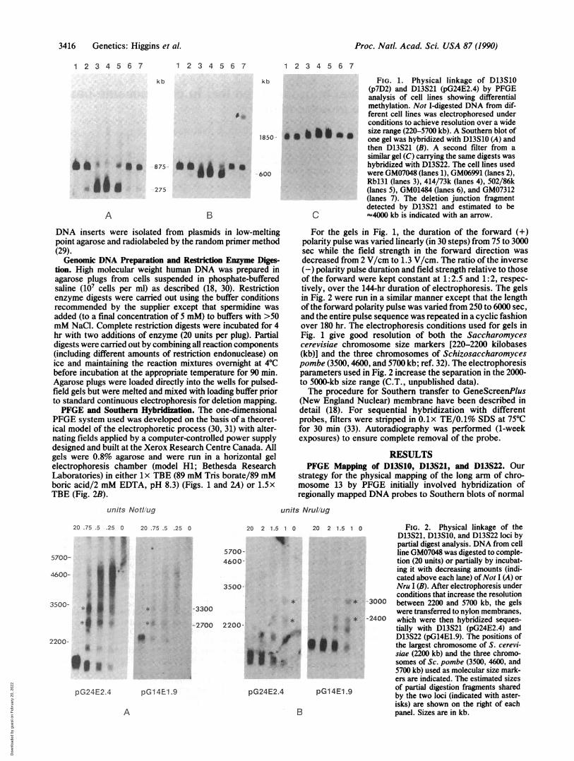
FIG. 1. Physical linkage of D13S10(p7D2) and D13S21 (pG24E2.4) by PFGEanalysis of cell lines showing differentialmethylation. Not I-digested DNA from dif-ferent cell lines was electrophoresed underconditions to achieve resolution over a widesize range (220-5700 kb). A Southern blot ofone gel was hybridized with D13S10 (A) andthen D13S21 (B). A second filter from asimilar gel (C) carrying the same digests washybridized with D13S22. The cell lines usedwere GM07048 (lanes 1), GM06991 (lanes 2),Rbl31 (lanes 3), 414/73k (lanes 4), 502/86k(lanes 5), GM01484 (lanes 6), and GM07312(lanes 7). The deletion junction fragmentdetected by D13S21 and estimated to be(4000 kb is indicated with an arrow.
For the gels in Fig. 1, the duration of the forward (+)polarity pulse was varied linearly (in 30 steps) from 75 to 3000sec while the field strength in the forward direction wasdecreased from 2 V/cm to 1.3 V/cm. The ratio of the inverse(-) polarity pulse duration and field strength relative to thoseof the forward were kept constant at 1: 2.5 and 1: 2, respec-tively, over the 144-hr duration of electrophoresis. The gelsin Fig. 2 were run in a similar manner except that the lengthof the forward polarity pulse was varied from 250 to 6000 sec,and the entire pulse sequence was repeated in a cyclic fashionover 180 hr. The electrophoresis conditions used for gels inFig. 1 give good resolution of both the Saccharomycescerevisiae chromosome size markers [220-2200 kilobases(kb)] and the three chromosomes of Schizosaccharomycespombe (3500, 4600, and 5700 kb; ref. 32). The electrophoresisparameters used in Fig. 2 increase the separation in the 2000-to 5000-kb size range (C.T., unpublished data).The procedure for Southern transfer to GeneScreenPlus
(New England Nuclear) membrane have been described indetail (18). For sequential hybridization with differentprobes, filters were stripped in 0.lx TE/0.1% SDS at 75°Cfor 30 min (33). Autoradiography was performed (1-weekexposures) to ensure complete removal of the probe.
RESULTSPFGE Mapping of D13S10, D13S21, and D13S22. Our
strategy for the physical mapping of the long arm of chro-mosome 13 by PFGE initially involved hybridization ofregionally mapped DNA probes to Southern blots of normal
units Nrul/ug
20 .75 .5 .25 0
*1
5700- ja. .tioo &I.
3500- *
2200-
*@ 1
pG24E2.4
20 .75 .5 .25 0 20 2 1.5 1 0 20 2 1.1
5700-4600-
3500-
-3300
* A
0
-2700 2200-
pG1 4E1.9
i~~~~
*fg
pG24E2.4 pG14El
A
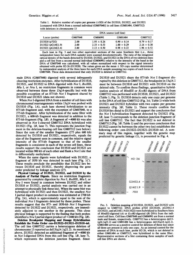
5 1 0 FIG. 2. Physical linkage of theD13S21, D13S10, and D13S22 loci bypartial digest analysis. DNA from cellline GM07048 was digested to comple-tion (20 units) or partially by incubat-ing it with decreasing amounts (indi-cated above each lane) ofNot I (A) orNru I (B). After electrophoresis underconditions that increase the resolution
x * -3000 between 2200 and 5700 kb, the gelswere transferred to nylon membranes,
X. * -2400 which were then hybridized sequen-* tially with D13S21 (pG24E2.4) and
D13S22 (pG14E1.9). The positions of* the largest chromosome of S. cerevi-
siae (2200 kb) and the three chromo-somes of Sc. pombe (3500, 4600, and5700 kb) used as molecular size mark-ers are indicated. The estimated sizes
1.9 of partial digestion fragments sharedby the two loci (indicated with aster-isks) are shown on the right of eachpanel. Sizes are in kb.
3416 Genetics: Higgins et al.
B
Dow
nloa
ded
by g
uest
on
Feb
ruar
y 20
, 202
2
Proc. Natl. Acad. Sci. USA 87 (1990) 3417
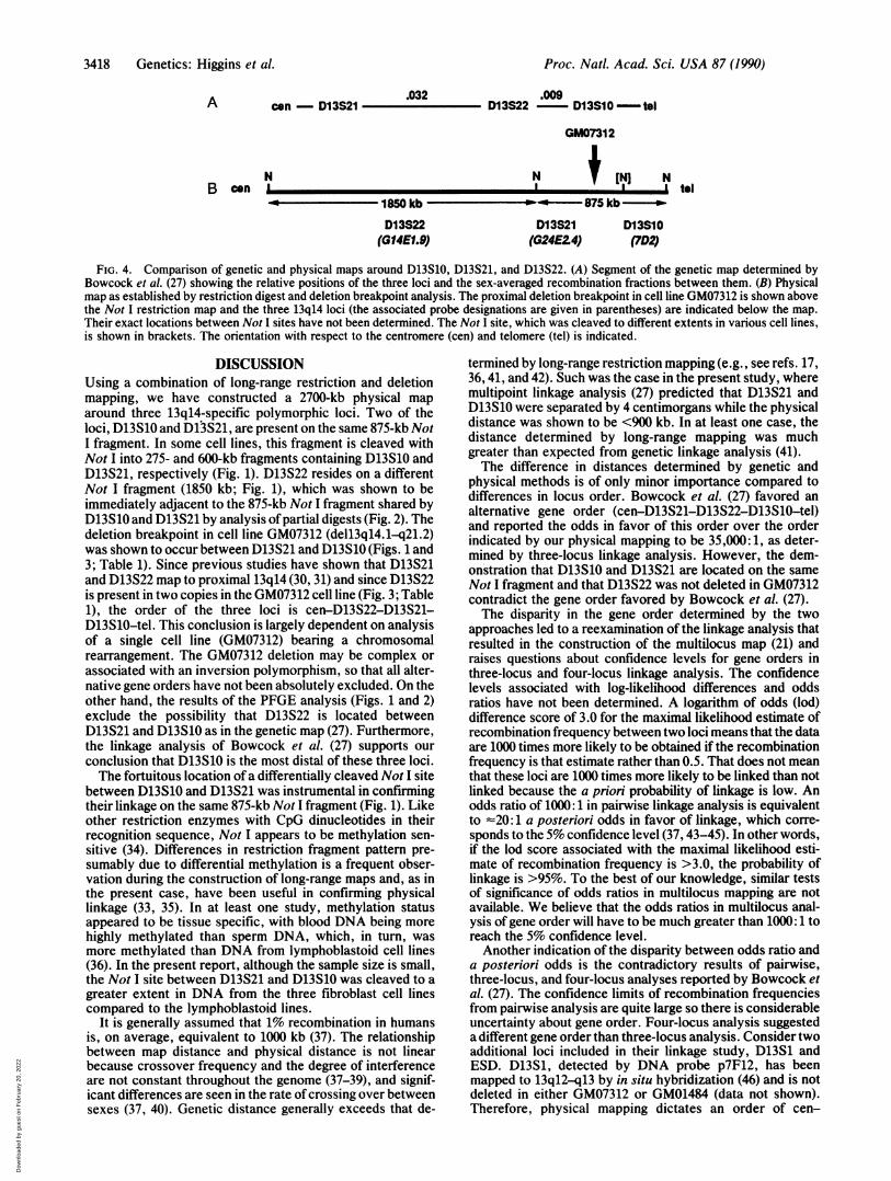
Table 1. Relative number of copies per genome (±SD) of the D13S10, D13S22, and D13S21[compared with DNA from a normal individual (GM07048)] in cell lines (GM01484, GM07312)with deletions in chromosome 13
DNA source (cell line)
Locus (probe) GM07048 GM06991 GM01484 GM07312
D13S10 (p7D2) 2.00 1.93 ± 0.26 0.90 ± 0.14 0.79 ± 0.17D13S22 (pG14E1.9) 2.00 2.33 ± 0.53 1.00 ± 0.29 2.16 ± 0.38D13S21 (pG24E2.4) 2.00 2.13 ± 0.50 0.98 ± 0.22 1.% ± 0.52
Each lane in Fig. 3 and two other equivalent sections of the same Southern blot (i.e., threeindependent lanes for each DNA sample) were scanned densitometrically. The ratio of the integratedoptical density of the bands detected by D13S10, D13S22, and D13S21 in the DNA oftwo deletion linesand a cell line from a second normal individual (GM06991) relative to the intensity of the band in theDNA of GM07048 was calculated, with all values normalized with respect to the signal intensityobtained with probe H2-26 (D13S28). The values given are the mean ± SD copy number determinedby independent analysis of the three lanes for each DNA sample assuming two copies of each locus inGM07048. These data demonstrated that only D13S10 is deleted in GM07312.
male DNA (GM07048) digested with several infrequentlycleaving restriction enzymes. After hybridization ofD13S10,D13S21, and D13S22 to DNA digested with Not I, BssHII,Mlu I, or Nru I, no restriction fragments in common wereobserved between these three 13q14-specific loci with thepossible exception of an 875-kb Not I fragment shared byD13S10 and D13S21 (data not shown).When Not I-digested DNA from a number of cell lines with
chromosomal rearrangements within 13q14 was probed withD13S10 (Fig. 1A), each lane showed hybridization to an875-kb fragment and, with the exception of GM07048, to a275-kb fragment. After hybridization of the same filter withD13S21, a 600-kb fragment was detected in addition to the875-kb fragment (Fig. 1B). A fragment of -4000 kb was alsoobserved in Not I-cleaved GM07312 DNA hybridized withD13S21 (Fig. 1B, lane 7). This represents the junction frag-ment in the deletion-bearing cell line GM07312 (see below).Since the sum of the smaller fragments (275 plus 600 kb)detected by D13S10 and D13S21, respectively, equals thesize of the fragment seen in common, and the relative signalintensity of the 875-kb fragment to that of the smallerfragments is consistent in each of the seven cell lines, theseresults support the conclusion that D13S10 and D13S21 arelocated within 900 kb of each other and flank a Not I site thatis differentially methylated.When the same digests were hybridized with D13S22, a
fragment of 1850 kb was detected in each lane (Fig. 1C).These results preclude the possibility that D13S22 lies be-tween D13S10 and D13S21, thereby disproving the geneorder predicted by linkage analysis (27).
Physical Linkage of D13S21, D13S22, and D13S10 by theAnalysis of Partial Digests. Since no restriction fragmentsgenerated by complete digestion by Not I, BssHII, Mlu I, orNru I were found in common between D13S22 and eitherD13S10 or D13S21, partial analysis was carried out in anattempt to physically link these loci. When the same blot washybridized with D13S21 and then D13S22, the smallest NotI partial digest product in common was estimated at 2700 kb(Fig. 2A), which is approximately equal to the sum of theindividual Not I fragments detected by these probes. Theseresults suggest that the 875- and 1850-kb Not I fragmentsdetected by D13S22 and D13S21, respectively, are immedi-ately adjacent to one another in the genome. This closephysical linkage is supported by the finding that both probesidentified a Nru I partial digest product of -2400 kb (Fig. 2B).
Deletion Breakpoint Mapping and Dosage HybridizationEstablish the Probe Order cen-D13S22-D13S21-D13S10-tel.The cell line GM07312 carries a heterozygous deletion ofchromosome 13 reported as dell3(q14.1q21.2). As mentionedabove, D13S21 detected an additional fragment of =4000 kbin Not I-digested DNA from this cell line (Fig. 1B, lane 7),which represents the deletion junction fragment. Since
D13S10 and D13S21 share the 875-kb Not I fragment dis-rupted by this deletion in GM07312, the breakpoint in 13q14.1must be between D13S10 and D13S21 with D13S10 on thedeleted side. To confirm these findings, quantitative hybrid-ization analysis of HindIII or EcoRI digests of DNA fromGM07312 was performed with D13S10, D13S21, and D13S22(Table 1; Fig. 3). D13S10 detects only one copy per genomein the DNA of cell line GM07312 (Fig. 3A; Table 1) while bothD13S21 and D13S22 hybridize with two copies per genomeintensity (Fig. 3B; Table 1). These results confirm thatD13S10 lies distal to D13S21 and support the conclusion thatthe Not I hybridization fragment detected by D13S21 (Fig.1B, lane 7) corresponds to the deletion junction fragment ofcell line GM07312. The fact that D13S22 is not deleted inGM07312 (Fig. 3B; Table 1) and the previous conclusion thatD13S22 cannot be between the other two loci establishes thefollowing order: cen-D13S22-D13S21-D13S1O-tel. A sum-mary map of this region, together with the genetic mappredicted by genetic linkage (1), is presented in Fig. 4.
-OZ c~ p do doIl
H2-26
c?
'-mm _ _ *
:9.
G24E2.4 - _ -
G 14 E 1.9 - :,
ol& .0 ':::,-: 7D2
A B
FIG. 3. Deletion mapping of D13S10, D13S21, and D13S22 withrespect to GM07312. DNA probes p7D2 (D13S10), pG24E2.4(D13S21), and pG14E1.9 (D13S22) were hybridized to Southern blotsof HindIl-digested (A) or EcoRI-digested (B) DNA from the indi-cated cell lines. Cell lines GM07048 and GM06991 are from a normalmale and female, respectively. GM07312 has a heterozygous dell3-(q14.1q21.2) and GM01484 has a heterozygous dell3(q14.1q22.1).GM01484 serves as a positive control for deletion of these loci sinceall three are present in only one copy. As an internal control for theamount of DNA in each lane, probe H2-26, which is not deleted ineither GM01484 or GM07312, was hybridized to the same filter.Representative sections of a blot containing three samples of eachcell line DNA are shown.
Genetics: Higgins et al.
Dow
nloa
ded
by g
uest
on
Feb
ruar
y 20
, 202
2
Proc. Natl. Acad. Sci. USA 87 (1990)
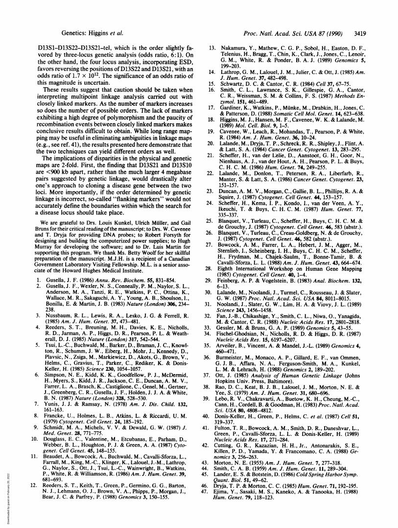
A con - D13S21 .032 .009D13S22 - D13SI0-tol
GM07312
B conNI
No 1850 kb -
D13S22(GI4E1.9)
Nt
D13S21(G24E2.4)
[N
D13S1O(7D2)
FIG. 4. Comparison of genetic and physical maps around D13S10, D13S21, and D13S22. (A) Segment of the genetic map determined byBowcock et al. (27) showing the relative positions of the three loci and the sex-averaged recombination fractions between them. (B) Physicalmap as established by restriction digest and deletion breakpoint analysis. The proximal deletion breakpoint in cell line GM07312 is shown abovethe Not I restriction map and the three 13q14 loci (the associated probe designations are given in parentheses) are indicated below the map.Their exact locations between Not I sites have not been determined. The Not I site, which was cleaved to different extents in various cell lines,is shown in brackets. The orientation with respect to the centromere (cen) and telomere (tel) is indicated.
DISCUSSIONUsing a combination of long-range restriction and deletionmapping, we have constructed a 2700-kb physical maparound three 13q14-specific polymorphic loci. Two of theloci, D13S10 and Dl3S21, are present on the same 875-kb NotI fragment. In some cell lines, this fragment is cleaved withNot I into 275- and 600-kb fragments containing D13S10 andD13S21, respectively (Fig. 1). D13S22 resides on a differentNot I fragment (1850 kb; Fig. 1), which was shown to beimmediately adjacent to the 875-kb Not I fragment shared byD13S1Oand D13S21 by analysis ofpartial digests (Fig. 2). Thedeletion breakpoint in cell line GM07312 (dell3ql4.1-q21.2)was shown to occur between D13S21 and D13S10 (Figs. 1 and3; Table 1). Since previous studies have shown that D13S21and D13S22 map to proximal 13q14 (30, 31) and since D13S22is present in two copies in the GM07312 cell line (Fig. 3; Table1), the order of the three loci is cen-D13S22-D13S21-D13S10-tel. This conclusion is largely dependent on analysisof a single cell line (GM07312) bearing a chromosomalrearrangement. The GM07312 deletion may be complex orassociated with an inversion polymorphism, so that all alter-native gene orders have not been absolutely excluded. On theother hand, the results of the PFGE analysis (Figs. 1 and 2)exclude the possibility that D13S22 is located betweenD13S21 and D13S10 as in the genetic map (27). Furthermore,the linkage analysis of Bowcock et al. (27) supports ourconclusion that D13S10 is the most distal of these three loci.The fortuitous location of a differentially cleaved Not I site
between D13S10 and D13S21 was instrumental in confirmingtheir linkage on the same 875-kb Not I fragment (Fig. 1). Likeother restriction enzymes with CpG dinucleotides in theirrecognition sequence, Not I appears to be methylation sen-sitive (34). Differences in restriction fragment pattern pre-sumably due to differential methylation is a frequent obser-vation during the construction of long-range maps and, as inthe present case, have been useful in confirming physicallinkage (33, 35). In at least one study, methylation statusappeared to be tissue specific, with blood DNA being morehighly methylated than sperm DNA, which, in turn, wasmore methylated than DNA from lymphoblastoid cell lines(36). In the present report, although the sample size is small,the Not I site between D13S21 and D13S10 was cleaved to agreater extent in DNA from the three fibroblast cell linescompared to the lymphoblastoid lines.
It is generally assumed that 1% recombination in humansis, on average, equivalent to 1000 kb (37). The relationshipbetween map distance and physical distance is not linearbecause crossover frequency and the degree of interferenceare not constant throughout the genome (37-39), and signif-icant differences are seen in the rate of crossing over betweensexes (37, 40). Genetic distance generally exceeds that de-
termined by long-range restriction mapping (e.g., see refs. 17,36, 41, and 42). Such was the case in the present study, wheremultipoint linkage analysis (27) predicted that D13S21 andD13S10 were separated by 4 centimorgans while the physicaldistance was shown to be <900 kb. In at least one case, thedistance determined by long-range mapping was muchgreater than expected from genetic linkage analysis (41).The difference in distances determined by genetic and
physical methods is of only minor importance compared todifferences in locus order. Bowcock et al. (27) favored analternative gene order (cen-D13S21-D13S22-D13S10-tel)and reported the odds in favor of this order over the orderindicated by our physical mapping to be 35,000: 1, as deter-mined by three-locus linkage analysis. However, the dem-onstration that D13S10 and D13S21 are located on the sameNot I fragment and that D13S22 was not deleted in GM07312contradict the gene order favored by Bowcock et al. (27).The disparity in the gene order determined by the two
approaches led to a reexamination of the linkage analysis thatresulted in the construction of the multilocus map (21) andraises questions about confidence levels for gene orders inthree-locus and four-locus linkage analysis. The confidencelevels associated with log-likelihood differences and oddsratios have not been determined. A logarithm of odds (lod)difference score of 3.0 for the maximal likelihood estimate ofrecombination frequency between two loci means that the dataare 1000 times more likely to be obtained if the recombinationfrequency is that estimate rather than 0.5. That does not meanthat these loci are 1000 times more likely to be linked than notlinked because the a priori probability of linkage is low. Anodds ratio of 1000: 1 in pairwise linkage analysis is equivalentto -20: 1 a posteriori odds in favor of linkage, which corre-sponds to the 5% confidence level (37, 43-45). In other words,if the lod score associated with the maximal likelihood esti-mate of recombination frequency is >3.0, the probability oflinkage is >95%. To the best of our knowledge, similar testsof significance of odds ratios in multilocus mapping are notavailable. We believe that the odds ratios in multilocus anal-ysis ofgene order will have to be much greater than 1000: 1 toreach the 5% confidence level.Another indication of the disparity between odds ratio and
a posteriori odds is the contradictory results of pairwise,three-locus, and four-locus analyses reported by Bowcock etal. (27). The confidence limits of recombination frequenciesfrom pairwise analysis are quite large so there is considerableuncertainty about gene order. Four-locus analysis suggesteda different gene order than three-locus analysis. Consider twoadditional loci included in their linkage study, D13S1 andESD. D13S1, detected by DNA probe p7F12, has beenmapped to 13ql2-ql3 by in situ hybridization (46) and is notdeleted in either GM07312 or GM01484 (data not shown).Therefore, physical mapping dictates an order of cen-
N1ltol
3418 Genetics: Higgins et al.
Dow
nloa
ded
by g
uest
on
Feb
ruar
y 20
, 202
2
Proc. Natl. Acad. Sci. USA 87 (1990) 3419
D13S1-D13S22-D13S21-tel, which is the order slightly fa-vored by three-locus genetic analysis (odds ratio, 6:1). Onthe other hand, the four locus analysis, incorporating ESD,favors reversing the positions ofD13S22 and D13S21, with anodds ratio of 1.7 x 10¹². The significance of an odds ratio ofthis magnitude is uncertain.These results suggest that caution should be taken when
interpreting multipoint linkage analysis carried out withclosely linked markers. As the number of markers increasesso does the number of possible orders. The lack of markersexhibiting a high degree of polymorphism and the paucity ofrecombination events between closely linked markers makesconclusive results difficult to obtain. While long range map-ping may be useful in eliminating ambiguities in linkage maps(e.g., see ref. 41), the results presented here demonstrate thatthe two techniques can yield different orders as well.The implications of disparities in the physical and genetic
maps are 2-fold. First, the finding that D13S21 and D13S10are <900 kb apart, rather than the much larger 4 megabasepairs suggested by genetic linkage, would drastically alterone's approach to cloning a disease gene between the twoloci. More importantly, if the order determined by geneticlinkage is incorrect, so-called "flanking markers" would notaccurately define the boundaries within which the search fora disease locus should take place.We are grateful to Drs. Louis Kunkel, Ulrich Muller, and Gail
Bruns for their critical reading ofthe manuscript; to Drs. W. Caveneeand T. Dryja for providing DNA probes; to Robert Forsyth fordesigning and building the computerized power supplies; to HughMurray for developing the software; and to Dr. Luis Martin forsupporting this program. We thank Ms. Betty Woolf for her skillfulpreparation of the manuscript. M.J.H. is a recipient of a CanadianGovernment Laboratory Visiting Fellowship. M.L. is a senior asso-ciate of the Howard Hughes Medical Institute.
1. Gusella, J. F. (1986) Annu. Rev. Biochem. 55, 831-854.2. Gusella, J. F., Wexler, N. S., Conneally, P. M., Naylor, S. L.,
Anderson, M. A., Tanzi, R. E., Watkins, P. C., Ottina, K.,Wallace, M. R., Sakaguchi, A. Y., Young, A. B., Shoulson, I.,Bonilla, E. & Martin, J. B. (1983) Nature (London) 306, 234-238.
3. Nussbaum, R. L., Lewis, R. A., Lesko, J. G. & Ferrell, R.(1985) Am. J. Hum. Genet. 37, 473-481.
4. Reeders, S. T., Breuning, M. H., Davies, K. E., Nicholls,R. D., Jarman, A. P., Higgs, D. R., Pearson, P. L. & Weath-erall, D. J. (1985) Nature (London) 317, 542-544.
5. Tsui, L.-C., Buchwald, M., Barker, D., Braman, J. C., Knowl-ton, R., Schumm, J. W., Eiberg, H., Mohr, J., Kennedy, D.,Plavsic, N., Zsiga, M., Markiewicz, D., Akots, G., Brown, V.,Helms, C., Gravius, T., Parker, C., Rediker, K. & Donis-Keller, H. (1985) Science 230, 1054-1057.
6. Simpson, N. E., Kidd, K. K., Goodfellow, P. J., McDermid,H., Myers, S., Kidd, J. R., Jackson, C. E., Duncan, A. M. V.,Farrer, L. A., Brasch, K., Castiglione, C., Genel, M., Gertner,J., Greenberg, C. R., Gusella, J. F., Holden, J. J. A. & White,B. N. (1987) Nature (London) 328, 528-530.
7. Yunis, J. J. & Ramsay, N. (1978) Am. J. Dis. Child. 132,161-163.
8. Francke, U., Holmes, L. B., Atkins, L. & Riccardi, U. M.(1979) Cytogenet. Cell Genet. 24, 185-192.
9. Schmidt, M. A., Michels, V. V. & Dewald, G. W. (1987) J.Med. Genet. 28, 771-775.
10. Douglass, E. C., Valentine, M., Etcubanas, E., Parham, D.,Webber, B. L., Houghton, P. J. & Green, A. A. (1987) Cyto-genet. Cell Genet. 45, 148-155.
11. Beaudet, A., Bowcock, A., Buchwald, M., Cavalli-Sforza, L.,Farrall, M., King, M.-C., Klinger, K., Lalouel, J.-M., Lathrop,G., Naylor, S., Ott, J., Tsui, L.-C., Wainwright, B., Watkins,P., White, R. & Williamson, R. (1986) Am. J. Hum. Genet. 39,681-693.
12. Reeders, S. T., Keith, T., Green, P., Germino, G. G., Barton,N. J., Lehmann, 0. J., Brown, V. A., Phipps, P., Morgan, J.,Bear, J. C. & Parfrey, P. (1988) Genomics 3, 150-155.
13. Nakamura, Y., Mathew, C. G. P., Sobol, H., Easton, D. F.,Telenius, H., Bragg, T., Chin, K., Clark, J., Jones, C., Lenoir,G. M., White, R. & Ponder, B. A. J. (1989) Genomics 5,199-203.
14. Lathrop, G. M., Lalouel, J. M., Julier, C. & Ott, J. (1985) Am.J. Hum. Genet. 37, 482-498.
15. Schwartz, D. C. & Cantor, C. R. (1984) Cell 37, 67-75.16. Smith, C. L., Lawrance, S. K., Gillespie, G. A., Cantor,
C. R., Weissman, S. M. & Collins, F. S. (1987) Methods En-zymol. 151, 461-489.
17. Gardiner, K., Watkins, P., Munke, M., Drabkin, H., Jones, C.& Patterson, D. (1988) Somatic Cell Mol. Genet. 14, 623-638.
18. Higgins, M. J., Hansen, M. F., Cavenee, W. K. & Lalande, M.(1989) Mol. Cell. Biol. 9, 1-5.
19. Cavenee, W., Leach, R., Mohandas, T., Pearson, P. & White,R. (1984) Am. J. Hum. Genet. 36, 10-24.
20. Lalande, M., Dryja, T. P., Schreck, R. R., Shipley, J., Flint, A.& Latt, S. A. (1984) Cancer Genet. Cytogenet. 13, 283-295.
21. Scheffer, H., van der Lelie, D., Aanstoot, G. H., Goor, N.,Nienhaus, A. J., van der Hout, A. H., Pearson, P. L. & Buys,C. H. C. M. (1986) Hum. Genet. 74, 249-255.
22. Lalande, M., Donlon, T., Petersen, R. A., Liberfarb, R.,Manter, S. & Latt, S. A. (1986) Cancer Genet. Cytogenet. 23,151-157.
23. Duncan, A. M. V., Morgan, C., Gallie, B. L., Phillips, R. A. &Squire, J. (1987) Cytogenet. Cell Genet. 44, 153-157.
24. Scheffer, H., Kema, I. P., Kondo, I., van der Veen, A. Y.,Ikeuchi, T. & Buys, C. H. C. M. (1987) Hum. Genet. 77,335-337.
25. Blanquet, V., Turleau, C., Scheffer, H., Buys, C. H. C. M. &de Grouchy, J. (1987) Cytogenet. Cell Genet. 46, 583 (abstr.).
26. Blanquet, V., Turleau, C., Creau-Goldberg, N. & de Grouchy,J. (1987) Cytogenet. Cell Genet. 46, 582 (abstr.).
27. Bowcock, A. M., Farrer, L. A., Hebert, J. M., Agger, M.,Sternlieb, I., Scheinberg, 1. H., Buys, C. H. C. M., Scheffer,H., Frydman, M., Chajek-Saulm, T., Bonne-Tamir, B. &Cavalli-Sforza, L. L. (1988) Am. J. Hum. Genet. 43, 664-674.
28. Eighth International Workshop on Human Gene Mapping(1985) Cytogenet. Cell Genet. 40, 1-4.
29. Feinberg, A. P. & Vogelstein, B. (1983) Anal. Biochem. 132,6-13.
30. Lalande, M., Noolandi, J., Turmel, C., Rousseau, J. & Slater,G. W. (1987) Proc. Natl. Acad. Sci. USA 84, 8011-8015.
31. Noolandi, J., Slater, G. W., Lim, H. A. & Viovy, J. L. (1989)Science 243, 1456-1458.
32. Fan, J.-B., Chikashige, Y., Smith, C. L., Niwa, O., Yanagida,M. & Cantor, C. R. (1988) Nucleic Acids Res. 17, 2801-2818.
33. Gessler, M. & Bruns, G. A. P. (1989) Genomics 5, 43-55.34. Fischel-Ghodsian, N., Nicholls, R. D. & Higgs, D. R. (1987)
Nucleic Acids Res. 15, 6197-6207.35. Arveiler, B., Vincent, A. & Mandel, J.-L. (1989) Genomics 4,
460-471.36. Burmeister, M., Monaco, A. P., Gillard, E. F., van Ommen,
G. J. B., Affara, N. A., Ferguson-Smith, M. A., Kunkel,L. M. & Lehrach, H. (1988) Genomics 2, 189-202.
37. Ott, J. (1985) Analysis of Human Genetic Linkage (JohnsHopkins Univ. Press, Baltimore).
38. Rao, D. C., Keat, B. J. B., Lalouel, J. M., Morton, N. E. &Yee, S. (1979) Am. J. Hum. Genet. 31, 680-696.
39. Lebo, R. V., Chakravarti, A., Buetow, K. H., Cheung, M.-C.,Cann, H., Cordell, B. & Goodman, H. (1983) Proc. Natl. Acad.Sci. USA 80, 4808-4812.
40. Donis-Keller, H., Green, P., Helms, C. et al. (1987) Cell 51,319-337.
41. Fulton, T. R., Bowcock, A. M., Smith, D. R., Daneshvar, L.,Green, P., Cavalli-Sforza, L. L. & Donis-Keller, H. (1989)Nucleic Acids Res. 17, 271-284.
42. Cutting, G. R., Kazazian, H. H., Jr., Antonarakis, S. E.,Killen, P. D., Yamada, Y. & Francomano, C. A. (1988) Ge-nomics 3, 256-263.
43. Morton, N. E. (1955) Am. J. Hum. Genet. 7, 277-318.44. Smith, C. A. B. (1959) Am. J. Hum. Genet. 11, 289-304.45. Lander, E. S. & Botstein, D. (1986) Cold Spring Harbor Symp.
Quant. Biol. 51, 49-62.46. Dryja, T. P. & Morton, C. C. (1985) Hum. Genet. 71, 192-195.47. Ejima, Y., Sasaki, M. S., Kaneko, A. & Tanooka, H. (1988)
Hum. Genet. 79, 118-123.
Genetics: Higgins et al.
Dow
nloa
ded
by g
uest
on
Feb
ruar
y 20
, 202
2