conodont affinity of the enigmatic carboniferous chordate ... · conodont affinity of the enigmatic...
TRANSCRIPT
DOI 10.1111/j.1502-3931.2008.00102.x © 2008 The Author, Journal compilation © 2008 The Lethaia Foundation
LETHAIA
Blackwell Publishing Ltd
Conodont affinity of the enigmatic Carboniferous chordate
Conopiscius
JERZY DZIK
Dzik, J. 2009: Conodont affinity of the enigmatic Carboniferous chordate
Conopiscius
.
Lethaia
, Vol. 42, pp. 31–38
Conopiscius
shares V-shaped myomeres with the co-occurring conodont
Clydagnathus
but instead of a complex oral apparatus it has only a single pair of conical elements,and structures resembling scales are associated with its myomeres. Moreover, thecoarsely crystalline crown tissue typical for conodonts has not been identified in the
Conopiscius
elements, which show only a finely lamellar skeletal tissue. The gapbetween conodonts and
Conopiscius
may be filled by isolated elements of similarmorphology and structure occurring in the Late Devonian. They reveal a very thinexternal layer developed mostly at the tooth tip and resembling conodont crown tissue.The pulp cavity is partially filled with layered or spherulitic phosphatic tissue of thekind known also in conodonts (basal filling tissue) and early vertebrates (lamellin).Conodont elements of similar morphology and representing uni-membrate oralapparatuses have not been previously reported from the Devonian or Carboniferousbut occur near the Cambrian–Ordovician transition (
Proconodontus
) and in the LatePermian (
Caenodontus
). It is proposed that
Conopiscius
represents a mostly crypticconodont lineage extending from the Early Ordovician to the Permian, instead of beingdirectly related to the agnathans.
�
Agnatha
,
Anaspida
,
conodonts
,
Early Carboniferous
,
evolution
,
histology
,
Late Devonian
,
microstructure
.
J. Dzik [[email protected]], Instytut Paleobiologii PAN, Twarda 51/55, 00-818Warszawa and Instytut Zoologii Uniwersytetu Warszawskiego, Banacha 2, 02-097Warszawa, Poland; manuscript received on 24/03/07; and manuscript accepted on09/01/08.
The early Carboniferous Granton Shrimp Bedexposed near Edinburgh, Scotland, is famous for thefirst discovery of conodont animals with soft tissuespreserved (Briggs
et al
. 1983; Aldridge
et al
. 1993).The anatomy of conodonts resembles that of Recentlamprey, with elongate body and narrow asymmetrictail fin but with
Branchiostoma
-like V-shaped myomeres.Such myomeres and general body appearancecharacterize also the second Granton chordate –
Conopiscius
. What makes
Conopiscius
rather basicallydifferent from conodonts is the presence of probableweakly mineralized scales associated with myomeresand its oral apparatus composed of only two elementsof simple morphology (Briggs & Clarkson 1987).
No Late Palaeozoic conodont element of similarmorphology has been known until recently, exceptfor the enigmatic Permian serrated conical elementsof
Caenodontus
(Behnken 1975). Typically, cono-donts bore a complex oral apparatus composed offifteen (sometimes even more) phosphatic elements.The apparatus was complex even in conodont groupscharacterized by a very simple, conical shape of theirelements (e.g. Sweet 1988; Dzik 1991; Donoghue &Purnell 1999). Such morphologically simple conodontsdid not survive the Frasnian–Famennian boundary(Dzik 2002), although some elements of the Famennianicriodontid conodonts developed secondarily simplified
morphologies resembling those known from theOrdovician (e.g. Dzik 2006).
Two new observations make affinities of
Conopiscius
to the conodonts more likely than originallyexpected: (i) isolated specimens similar to the elementspreserved
in situ
in the Granton material appear tobe common in the Late Devonian, and (ii) the internalstructure of these specimens is conodont-like, althoughmore similar to Cambrian than to other late Palaeozoicconodonts. In the present paper, these new data arepresented and discussed.
Materials and methods
The Devonian specimens come from limestone samplesdissolved in acetic (pure limestone) or formic (marls,dolomitized limestone) acids. Less acid was alwaysused than necessary to dissolve completely the sampleand this provided some buffering. Dried residueswere enriched in an electromagnetic mineralogicalseparator. Specimens studied for microstructure wereembedded in epoxy resin, ground and polished. Theywere etched with 0.5% orthophosphoric acid until arelief visible in reflected light under an opticalmicroscope developed. The time of etching variedsignificantly (from 1 to 5 min) depending on the

32
J. Dzik
LETHAIA 42 (2009)
specimen. The blocks with embedded specimenswere then attached to a stub and coated with platinumto photograph in a scanning electron microscope.
Specimens of
Conopiscius
are housed at the RoyalScottish Museum in Edinburgh, Scotland (abbreviatedRSM), Devonian isolated elements at the Institute ofPaleobiology of the Polish Academy of Sciences inWarsaw, Poland (ZPAL).
Morphology of oral denticles in
Conopiscius
The cone-shaped structures in the head region of
Conopiscius clarki
are preserved as flattened in bothspecimens found, but with some relief. This suggeststhat they were originally composed of a rigid organicmatter, if not mineralized. Their surface is slightlywrinkled (Briggs & Clarkson 1987, p. 111), whichsuggests growth by marginal increments. The conesare filled with sediment for most of their length, sothey had a deep pulp cavity and rather thin walls. Asnoticed by Briggs & Clarkson (1987, p. 111) at leastRSM GY 1986.25.5 ‘preserves tenuous evidence of atiny denticle near the proximal extremity of the outermargin’. Denticles are recognizable also in RSM GY1986.25.6 (Fig. 1). They are rather prominent, andtheir arrangement suggests that the whole concave
margin of the cone was originally armed with numerousserially distributed denticles, of which all except forthe proximal two became exfoliated when the slabwas split. This denticulated margin is thicker thanthe cone wall nearby, which means that it wasreinforced with a skeletal tissue, and sharp-edged.The cross-section of the cone was thus lenticular. Itscomplete length from the base to tip exceeded 2 mm.
The Late Devonian denticulated conical elements
Conical phosphatic (or perhaps secondarily phos-phatized) elements with a deep basal cavity anddenticulated concave margin occur widely, althoughsparsely, in pelagic sediments of the Late Devonian inthe Holy Cross Mountains, Poland. They are usuallyreferred to as
Caenodontus
, the type species of which,
C. serrulatus
Behnken, 1975 was originally describedas a conodont from the Permian of Texas. Like asso-ciated unquestionable conodonts, the Polish conicalelements are transparent, amber-coloured in sampleswith low diagenetic alteration (Kowala, Jab
l
onna)and dark, almost black in samples from strata whichexperienced significant burial heating (P
l
ucki,
L
agów).Some well-preserved specimens show folding andrupture of the wall proving their original flexibility,
Fig. 1. �A, B. Elements from the head region of Conopiscius clarki Briggs & Clarkson, 1987 from the Viséan (Early Carboniferous)Granton Shrimp Bed exposed near Edinburgh, Scotland; specimen RSM GY 1986.25.6; photograph (A) and interpretive drawing (B). Notethat phosphatic tissue of the elements is mostly exfoliated as a result of the slab splitting but a couple of preserved denticles are evident,presumably belonging to the series arming the cutting edge of the elements. �C. Composite drawings with the tail region of specimenRSM GY 1986.25.5 added to RSM GY 1986.25.6 (reversed) to show location of the mouth apparatus and inferred proportions of the body.
LETHAIA 42 (2009)
Conodont affinity of
Conopiscius 33
evidently owing to a high organic matter content.The element surface shows smooth transversewrinkles, which seem to have resulted from incrementalgrowth at the cone base. No true incrementaldiscontinuities (growth lines) are discernible, however,suggesting that the secretory tissue formed a con-tinuous cover from either the inside or the outside ofthe sclerite.
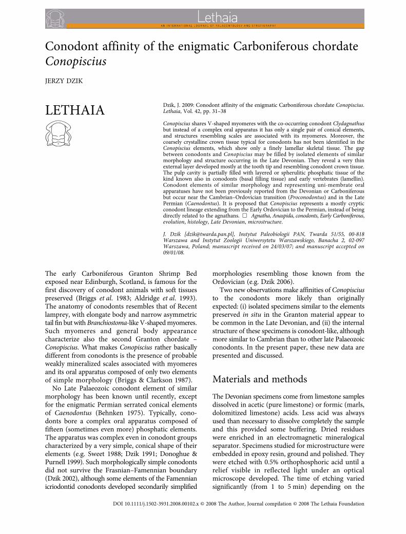
There are at least three distinct morphologicaltypes of conical element in the material studied,probably representing separate species. In all of themthe denticles are pointed and sharp-edged. Theprominence of denticles differs significantly,however. In the mid-Famennian specimens, the denticlesare almost twice as high as wide (Fig. 2A–E). Suchspecimens have been previously illustrated from thelate Famennian of Guizhou, south China (identifiedas
Belodella
sp. by Wang & Wang 1978, pl. 1:10–11),co-occurring with stratigraphically indicative
Pseu-dopolygnathus trigonicus
. Similar conical elements,
representing the same species, also occur in the earlyFamennian of the Montagne Noire in France (Fig. 2F–I).The geologically older late Frasnian specimens arecharacterized by the denticles being not longer thanwide and in some specimens being truly minute(Fig. 3). Rudimentary denticles characterize a coevalspecies with minute elements, almost round in cross-section (Fig. 4). It remains unclear whether thismorphological series of species corresponds to anyevolutionary succession but at least in the Frasniantwo sympatric species are represented by
Caenodontus
(and
Conopiscius
)-like elements.In the conical elements of the Viséan
Conopisciusclarki
only the proximal denticles are preserved andthey are somewhat more robust than in the Famennianforms. Each element is also wider in general outline,although this may be partially due to its compressionin the shale. The Permian
Caenodontus serrulatus
conical elements bear numerous minute denticlesalmost to the tip of the element (Behnken 1975). The
Fig. 2. Elements of the Famennian (Late Devonian) Conopiscius-like species A with prominent denticulation, as compared with otherDevonian species. �A–E. Well-preserved translucent specimens from the mid-Famennian Platyclymenia annulata Event (black shale) atKowala, Holy Cross Mountains, Poland; sample Ko-8a (A, B, D) and Ko-8 (C, E; for stratigraphy see Dzik 2006). Note variability indenticulation and curvature of the cusp. �F–I. Thermally altered black specimens from the early Famennian Cheiloceras Stufe limestoneat Soreille d’Izarre near Cabriéres, France, with rather uniform and robust appearance. Specimens ZPAL CVI/3108, 3113, 3112, 3109, 3111,and 3125–3128, respectively.

34
J. Dzik
LETHAIA 42 (2009)
cusp is needle-like and the cone expands stronglynear its base, more than in the Devonian specimens.Its general shape is thus somewhat closer to that ofthe Carboniferous
Conopiscius
.
Microstructure of the Famennian cones
The Famennian conical elements are laterally bentand twisted. This makes sectioning of them difficult,as there is no symmetry plane to follow. Among sev-eral elements sectioned by myself, in only a few caseshas the tip of the element been sectioned preciselyenough to recognize its internal organization. Each
element appears to be composed of two kinds ofphosphatic tissue. The boundary between these is dif-ficult to trace, but some specimens prove that it israther abrupt (Fig. 5). Acid-resistant minerals maydelimit the boundary in places, suggestive of a signif-icant structural discontinuity.
The external layer is composed of a somewhatmore regularly and coarsely crystalline tissue, withcrystal axes probably arranged parallel to the surface,although the available evidence is not completelyconvincing (Fig. 6C). The layer forms a regularlyconical unit, thickening distally, where it makes thesharp tip of the sclerite, but is extremely thin, lessthen 10
μ
m near the basal cavity tip. It thus roughlycorresponds in thickness to a single increment (prob-ably daily) in the platform margin of associated
Fig. 3. Elements of the terminal Frasnian (Late Devonian) Conopiscius-like species B with sharp-edged denticulation from the Kel-wasserkalk horizon at Plucki in the Holy Cross Mountains (for stratigraphy see Dzik 2002); specimens somewhat altered thermally (darkin coloration). �A–C. Broken apex exposing the crown (ct) and basal filling (bft) tissues, and the whole specimen in posterior and obliqueviews to show transverse wrinkles. �D. Specimen with partially exfoliated crown tissue. �E. Specimen with an abnormality in distributionof denticles indicating incremental growth at the base. �F–G. Unusually straight-cusped specimen and interior of its base with linearseries of voids probably representing odontoblasts incorporated in the basal cavity tissue (c). �H. Incomplete specimen of large sizecomparable with that of the Viséan Conopiscius clarki. Specimens ZPAL CVI/3114, 3118, 3122, 3117, and 3120, respectively.
LETHAIA 42 (2009)
Conodont affinity of
Conopiscius 35
conodont elements (Dzik 2006). Consequently, nointernal layering is recognizable within this unit,which makes it difficult to determine whether it wasdeposited from outside (like conodont crown tissue,ganoin or enamel) or from inside the basal cavity.The completely smooth surface of the elements andthe rather clear separation from the underlying tissuesuggest that this is a homologue (or at least an analogue)of conodont crown tissue.
The tissue forming most of the element volume isdistinctly lamellar, with crystal axes of each lamellaoriented perpendicular to the surface. It resemblesthe atubular dentine (lamellin) that occurs in scalesof Late Ordovician to Early Silurian agnathans(Karatajute-Talimaa & Smith 2004). The layers of thetissue close to the external layer run parallel to theboundary but material within this, secreted earlier orlater, is wavy and has a spherulitic pattern (Fig. 6).The spherulites are visible within the basal cavity inits deeper part. In the proximal part of the cone,where the layer is thinner, series of empty cavities arerecognizable in places (Fig. 3G, 4E). Their size, about3
μ
m wide, 10
μ
m long, suggests that these areimprints of secretory cells (odontoblasts?), partiallyincorporated into the phosphatic tissue.
In most samples containing the elements, phosphaticdetritus is abundant, including pieces of crustaceancuticle among taxonomically identifiable fossils (Rolfe& Dzik 2006). Originally, the cuticle was apparentlyorganic, so the taphonomic conditions duringformation of these fossiliferous strata were suitable
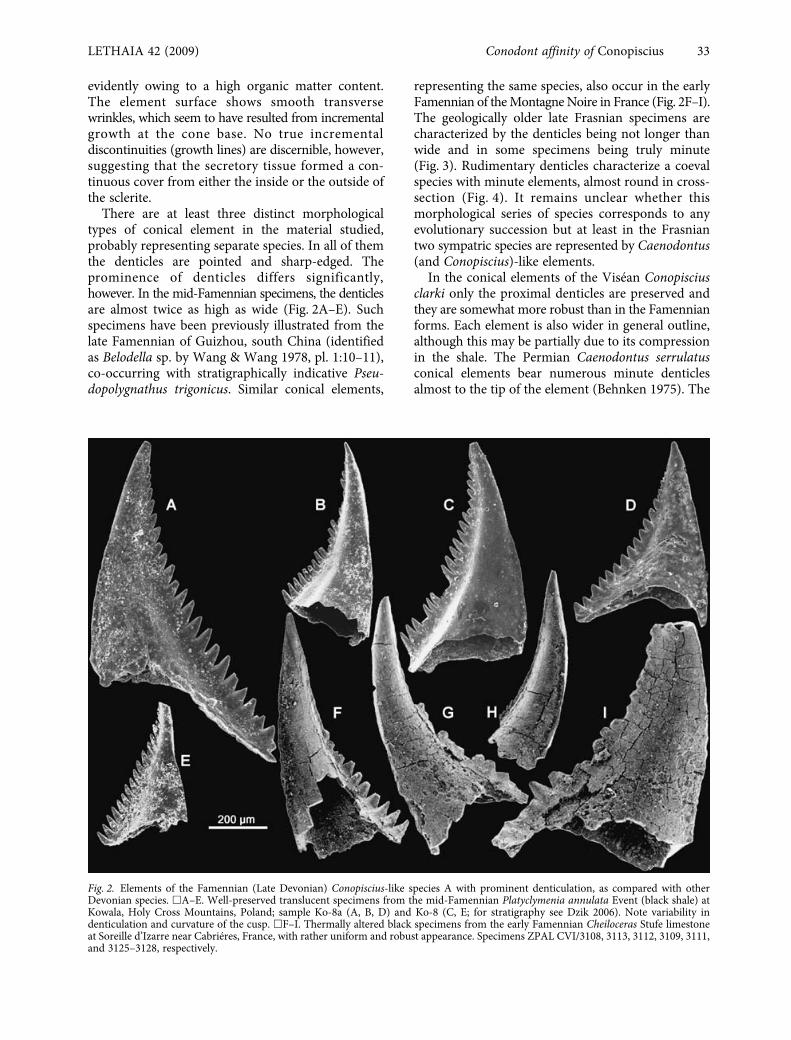
Fig. 4. Elements of the terminal Frasnian (Late Devonian) Conopiscius-like species C from Plucki. Note fine serration of the cutting edgeand robust appearance of specimens. �A, B. Posterior and oblique views of specimen ZPAL CVI/3115 showing transverse wrinkles anddisposition of minute denticles. �C. Specimen ZPAL CVI/3116 with partially exfoliated crown tissue. �D, E. Robust specimen ZPALCVI/3119 and interior of its basal cavity with probable odontoblasts and spherulitic mineralization of the basal filling tissue at later stagesof its secretion in the centre.
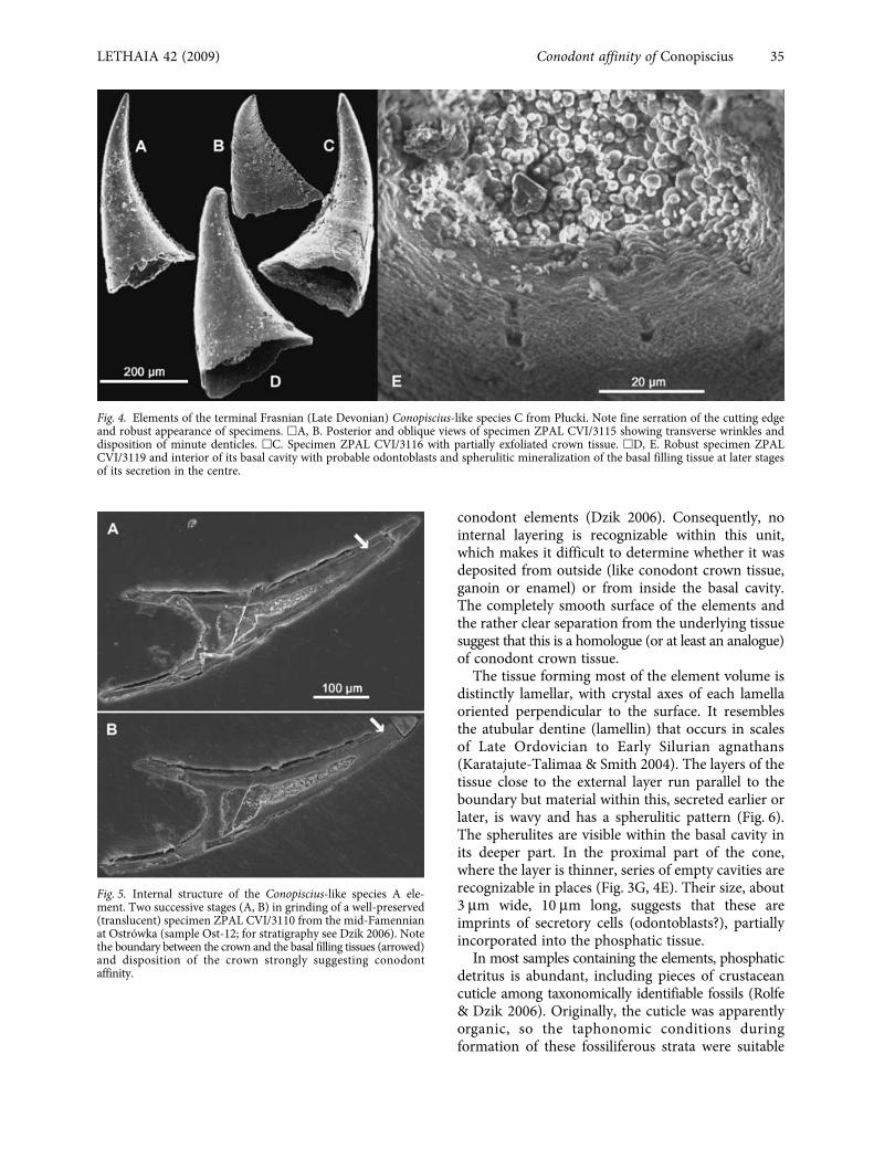
Fig. 5. Internal structure of the Conopiscius-like species A ele-ment. Two successive stages (A, B) in grinding of a well-preserved(translucent) specimen ZPAL CVI/3110 from the mid-Famennianat Ostrówka (sample Ost-12; for stratigraphy see Dzik 2006). Notethe boundary between the crown and the basal filling tissues (arrowed)and disposition of the crown strongly suggesting conodontaffinity.

36
J. Dzik
LETHAIA 42 (2009)
for secondary phosphatization. It is thus possible thatthe spherulitic apatite in the basal cavity of the Devonian
Conopiscius
-like elements developed partially post-mortem. This may explain uneven representation ofthese fossils in sampled sections, similar to that of themineralized basal tissue of associated conodonts.More likely, however, is that it represents a gradualmineralization of a cartilagineous tissue, as suggestedby the disposition of spherules in the pulp cavityshowing marks of incremental growth on its walls(Fig. 4E).
Possible Cambrian relative
Some Late Cambrian species of
Proconodontus
arealmost undistinguishable externally from the LateDevonian conical elements of probable
Conopiscius
affinity. For instance,
P. serratus
Miller, 1969 (Müller& Hinz 1991; Szaniawski & Bengtson 1993, 1998)closely resembles the
Conopiscius
-like species B. Thecrown tissue in
Proconodontus
is somewhat thickerand clearly shows perpendicular orientation ofapatite crystals, and this seems to be the maindifference between these forms. According toSzaniawski & Bengtson (1993) the crown tissue ofearly conodonts developed after formation of thebasal body and layers of both units were in continuity.They proposed an origin for the typical (eu)conodont
Cordylodus
lineage from
P. serratus
. Clusters composedof several elements of
Cordylodus
are known (e.g.Andres 1988), and it is generally accepted (Nicoll1990, 1991) that its apparatus composition wassimilar to that of typical conodonts, that is with 14elements in seven pairs (or 15 if a symmetricalmedial element was present). It is thus unlikely thatthe
Proconodontus
oral apparatus differed in its com-position from other conodonts. Despite the similarityof the general appearance of
Conopiscius
elementsand internal structure of the Famennian conical elementsto
Proconodontus serratus
, they apparently differed inorganization of the oral apparatus. The inferredpresence of either 14 or 15 elements in
Proconodontus
and only two in
Conopiscius
appears to precludeclassification of the latter in the family Procono-dontidae Lindström (1970). It remains a possibility,however, that in the course of evolution between theLate Cambrian and the Late Devonian, the apparatuswas simplified and the thickness of the crown tissuein the elements was reduced.
Conclusions
It is proposed that these Late Devonian conical serrateelements, with an internal structure resembling themost underived Cambrian conodonts, belong to thesame clade as
Conopiscius
, which may continue tothe Permian as
Caenodontus
(Fig. 7). If true, reductionof the oral apparatus to a single pair of morphologicallysimple elements marked the origin of this mostlycryptic evolutionary branch of Palaeozoic chordates.The possible presence of mineralized scales protectingthe myomeres, as suggested by Briggs & Clarkson(1987), places
Conopiscius
between the conodontsand the agnathans (it has even been classified as thegeologically youngest member of the Anaspida;
Fig. 6. Wall microstructure of the Devonian Conopiscius-likeelements. �A, B. Weakly and more intensely etched translucentspecimens ZPAL CVI/3123 from the mid-Famennian at Ostrówka(sample Ost-2; for stratigraphy see Dzik 2006) showing transitionfrom laminar to spherulitic increments of the basal filling tissue.Etching sufficient to expose layered structure of the basal fillingtissue did not differentiate the crown tissue. �C. Deeply etcheddark specimen ZPAL CVI/3124 from the terminal Frasnian atPlucki showing somewhat more coarsely crystalline structure ofthe crown tissue (ct; compare Fig. 3A).

LETHAIA 42 (2009)
Conodont affinity of
Conopiscius 37
Sepkoski 2002) but more evidence is necessary toexclude possible diagenetic phosphatization oforiginally organic tissue. A conodont affinity for theoral apparatus of
Conopiscius
would imply that it wascovered with soft tissue during growth and growingwhile functional. It remains to be determinedwhether the surface of each mineralized element wasrepeatedly exposed while piercing the food andcovered with the epithelial secretive organ whilebeing secreted (Bengtson 1976; Donoghue & Purnell1999), or alternatively, if the secretive tissue wascovered with a permanent keratinous cup (Priddle1974; Dzik 2000). The proposed conodont affinity of
Conopiscius
is consistent with its soft anatomy. Its V-shaped myomeres point forward in the same way asin the coeval conodont
Clydagnathus
but also as inthe early Palaeozoic Anaspida and Recent
Branchiostoma
;(e.g. Gemballa
et al
. 2003). In apparently morederived (in this respect) hagfish and lamprey, as wellas in the gnathostomes, additional dorsal andventralmost anteriorly pointed arms of myomeresare developed (Vogel & Gemballa 2000).
Acknowledgements
. –
I am grateful to W.D. Ian Rolfe and thestaff of the Royal Scottish Museum for making the originalspecimens of
Conopiscius
available to study. Hubert Szaniawskiread a draft of the manuscript. Comments offered by Richard J.Aldridge and Stefan Bengtson in their reviews helped me toimprove the paper and its language. I also appreciate criticism ofPhilip C.J. Donoghue. The research was partially supported bygrant P04D 001 28 from the Polish Ministry of Science.
References
Aldridge, R.J., Briggs, D.E.G., Smith, M.P., Clarkson, E.N.K. &Clark, N.D.L. 1993: The anatomy of conodonts.
PhilosophicalTransactions of the Royal Society London B
340
, 405–421.Andres, D. 1988: Strukturen, Apparate und Phylogenie primitiver
Conodonten.
Palaeontographica A
200
, 105–152.Behnken, F.H. 1975: Leonardian and Guadalupian (Permian)
conodont biostratigraphy in western and southwestern UnitedStates.
Journal of Paleontology
49, 284–315.Bengtson, S. 1976: The structure of some Middle Cambrian
conodonts, and the early evolution of conodont structure andfunction. Lethaia 9, 185–206.
Briggs, D.E.G. & Clarkson, E.N.K. 1987: An enigmatic chordatefrom the Lower Carboniferous ‘shrimp-bed’ of the Edinburghdistrict, Scotland. Lethaia 20, 107–115.
Briggs, D.E.G., Clarkson, E.N.K. & Aldridge, R.J. 1983: Theconodont animal. Lethaia 16, 1–14.
Donoghue, P.C.J. & Purnell, M.A. 1999: Growth, function, andthe conodont fossil record. Geology 27, 251–254.
Dzik, J. 1991: Evolution of oral apparatuses in conodont chordates.Acta Palaeontologica Polonica 36, 265–323.
Dzik, J. 2000: The origin of the mineral skeleton in chordates.Evolutionary Biology 31, 105–154.
Dzik, J. 2002: Emergence and collapse of the Frasnian conodontand ammonoid communities in the Holy Cross Mountains,Poland. Acta Palaeontologica Polonica 47, 565–650.
Dzik, J. 2006: The Famennian ‘Golden Age’ of conodonts andammonoids in the Polish part of the Variscan sea. Palaeonto-logia Polonica 63, 1–359.
Gemballa, S., Weitbrecht, G.W. & Sánchez-Villagra, M.R. 2003:The myosepta in Branchiostoma lanceolatum (Cephalochordata):3D reconstruction and microanatomy. Zoomorphology 122, 169–179.
Karatajute-Talimaa, V. & Smith, M.M. 2004: Tesakoviaspis con-centrica: Microskeletal remains of a new order of vertebratefrom the Upper Ordovician and Lower Silurian of Siberia.In Arratia, G., Wilson, M.V.H. & Cloutier, R. (eds): RecentAdvances in the Origin and Early Radiation of Vertebrates.Verlag Dr. Friedrich Pfeil, München, Germany, 53–64.
Lindström, M. 1970: A suprageneric taxonomy of the conodonts.Lethaia 3, 427–449.
Müller, K.J. & Hinz, I. 1991: Upper Cambrian conodonts fromSweden. Fossils and Strata 28, 1–153.
Nicoll, R.S. 1990: The genus Cordylodus and a latest Cambrian–Early Ordovician conodont biostratigraphy. BMR Journal ofAustralasian Geology and Geophysics 11, 529–558.
Nicoll, R.S. 1991: Differentiation of Late Cambrian–EarlyOrdovician species of Cordylodus (Conodonta) with biapical
Fig. 7. Geological time distribution of serrate phosphatic elementspossibly representing the Conopiscius lineage. Caenodontusadopted from Behnken (1975), Proconodontus from Szaniawski &Bengtson (1998).

38 J. Dzik LETHAIA 42 (2009)
basal cavities. BMR Journal of Australasian Geology andGeophysics 12, 223–244.
Priddle, J. 1974: The function of conodonts. Geological Magazine111, 255–257.
Rolfe, W.D.I. & Dzik, J. 2006: The Late Devonian predatorypelagic crustacean Angustidontus. Transactions of the RoyalSociety of Edinburgh, Earth Sciences 97, 75–96.
Sepkoski, J.J. Jr. 2002: A compendium of fossil marine animalgenera. Bulletins of American Paleontology 363, 1–560.
Sweet, W.C. 1988: The Conodonta: Morphology, Taxonomy,Paleoecology, and Evolutionary History of a Long-ExtinctAnimal Phylum, 212 pp. Clarendon Press, Oxford, UK.
Szaniawski, H. & Bengtson, S. 1993: Origin of euconodontelements. Journal of Paleontology 67, 640–654.
Szaniawski, H. & Bengtson, S. 1998: Late Cambrian euconodontsfrom Sweden. Palaeontologia Polonica 58, 7–29.
Vogel, F. & Gemballa, S. 2000: Locomotory design of ‘cyclostome’fishes: Spatial arrangement and architecture of myosepta andlamellae. Acta Zoologica 81, 267–283.
Wang, C. & Wang, Z. 1978: Upper Devonian and Lower Carbon-iferous conodonts from southern Guizhou. Memoirs of theNanjing Institute of Geology and Palaeontology, AcademiaSinica 11, 51–91.