connexin32 is a myelin-related protein in the pns and cns
TRANSCRIPT
Sacred Heart UniversityDigitalCommons@SHU
Biology Faculty Publications Biology Department
12-1-1995
Connexin32 is a Myelin-Related Protein in thePNS and CNSSteven S. SchererUniversity of Pennsylvania Medical Center
Suzanne M. DeschênesSacred Heart University, [email protected]
Yi-Tian XuUniversity of Pennsylvania Medical Center
Judith B. Grinspan
Kenneth H. FischbeckUniversity of Pennsylvania Medical Center
See next page for additional authors
Follow this and additional works at: http://digitalcommons.sacredheart.edu/bio_fac
Part of the Cell Biology Commons, Genetics Commons, and the Laboratory and Basic ScienceResearch Commons
This Article is brought to you for free and open access by the Biology Department at DigitalCommons@SHU. It has been accepted for inclusion inBiology Faculty Publications by an authorized administrator of DigitalCommons@SHU. For more information, please [email protected].
Recommended CitationScherer, Steven S., Deschênes, Suzanne M., et. al. "Connexin32 is a Myelin-Related Protein in the PNS and CNS." The Journal ofNeuroscience 15.12 (1995): 8281-8294.

AuthorsSteven S. Scherer, Suzanne M. Deschênes, Yi-Tian Xu, Judith B. Grinspan, Kenneth H. Fischbeck, and DavidL. Paul
This article is available at DigitalCommons@SHU: http://digitalcommons.sacredheart.edu/bio_fac/44

The Journal of Neuroscience, December 1995, 75(12): 8281-8294
Connexin32 Is a Myelin-Related Protein in the PNS and CNS
Steven S. Scherer,’ Suzanne M. Desch&nes,’ Yi-tian Xu,’ Judith B. Grinspan,* Kenneth H. Fischbeck,’ and David L. Paul3
1 Department of Neurology, University of Pennsylvania, Philadelphia, Pennsylvania 19104-6146, *Division of Neurological Research, Children’s Hospital of Philadelphia, Philadelphia, Pennsylvania 19104, and 3Department of Neurobiology, Harvard Medical School, Boston, Massachusetts 02115-6092
We have examined the expression of a gap junction pro- tein, connexin32 (Cx32), in Schwann cells and oligodendro- cytes. In peripheral nerve, Cx32 is found in the paranodal myelin loops and Schmidt-Lanterman incisures of myeli- nating Schwann cells, and the levels of Cx32 protein and mRNA change in parallel with those of other myelin-related genes during development, Wallerian degeneration, and axonal regeneration. In the central nervous system, Cx32 is found in oligodendrocytes and their processes, but not in compact myelin, and the levels of Cx32 protein and mRNA increase during development in parallel with those of the other myelin genes. Thus, Cx32 is expressed as part of the myelinating phenotype of both Schwann cells and oligodendrocytes, indicating that this gap junction protein plays in important role in the biology of myelin-forming cells.
[Key words: Cx32, gap junctions, Schwann cells, oligo- dendrocytes, myelin, incisures]
Myelin is the multi-lamellar structure that surrounds axons and increases axonal conduction velocity. It is formed by the spiral wrapping of the cell membrane of myelinating glia-oligodendro- cytes in the CNS and Schwann cells in the PNS. The myelin sheaths produced by these two cell types are structurally similar, consisting mostly of compact myelin that is characterized by a unique but overlapping set of ,proteins (Lemke, 1992, 1993). Proteolipid protein (PLP) and myelin basic protein (MBP) are the major structural proteins in the CNS, while protein zero (PO), MBP and peripheral myelin protein-22 kD (PMP-22) are the major proteins in the PNS. Each of these myelin proteins is essential for proper myelination, as mutations in PLP and MBP cause dysmyelination in the CNS, and mutations in P, and PMP- 22 cause dysmyelination in the PNS (Hudson, 1990; Chance and Pleasure, 1993; Lemke, 1993; Snipes and Suter, 1995).
The compact myelin in the PNS contains periodic interrup- tions called Schmidt-Lanterman incisures or clefts (Peters et al., 1991). These incisures, as well as the paranodal regions of the
Received June 22, 1995; revised Aug. 17, 1995; accepted Aug. 24, 1995. We thank Shelly Whyatt and Susan Shumas for technical assistance, and
Drs. David Colman, Rory Curtis, Susan Hockfield, John Kamholz, Arnulf Koeppen, Sara Piddlesden; James Salzer, and David Schreyer for their generous gifts of antibodies. This work was supported by grants from the Muscular Dystrophy Association, The American Academy of Neurology (Murray M. Stokelv Award). and the NIH (NS08075 and NS01565).
Coriespondence should be addressed to Steven S. Scherer, Department of Neurology, Clinical Research Building, University of Pennsylvania, Philadel- phia, PA ~19 104-6 146.
Copyright 0 1995 Society for Neuroscience 0270.6474/95/158281-14$05.00/O
myelin sheath, contain a distinct group of proteins, including myelin-associated glycoprotein (MAG), connexin32 (Cx32), E-cadherin, and oligodendrocyte-myelin glycoprotein (Trapp et al., 1989; Bergoffen et al., 1993; Apostolski et al., 1994; Fannon et al., 1995). Incisures have also been reported in the CNS (Pe- ters et al., 1991), but no molecules have yet been found to be preferentially localized to CNS incisures. The importance of the incisures to the normal function of myelinated axons was not appreciated until it was found that the X-linked form of Charcot- Marie-Tooth disease (CMTX) was caused by mutations in the Cx32 gene (Bergoffen et al., 1993). Cx32 is a gap junction pro- tein and is expressed in many tissues, including oligodendro- cytes and myelinating Schwann cells (Kumar and Gilula, 1986; Paul, 1986; Dermietzel et al., 1989, 1990; Bennett et al., 1991; Kumar and Gilula, 1992; Bergoffen et al., 1993). How Cx32 mutations cause peripheral neuropathy is unknown. One plau- sible explanation is that CMTX mutations alter the function of gap junctions at nodes and incisures, as Cx32-immunoreactivity colocalizes with gap junctions seen by freeze-fracture electron microscopy (Sandri et al., 1982; Bergoffen et al., 1993), and some mutations in Cx32 disrupt the formation of functional gap junctions (Bruzzone et al., 1994; Rabadan-Diehl et al., 1994). If CMTX mutations disrupted these gap junctions, this could in- terrupt the diffusion of ions and small molecules in a radial direction, directly across the myelin sheath, through the para- nodes and incisures.
To learn more about the role of Cx32 in myelinating glia, we examined the localization and expression of Cx32 in Schwann cells and oligodendrocytes. In the PNS, Cx32 is found in the paranodal regions and incisures of myelinating Schwann cells. In the CNS, Cx32 is found in cell bodies and processes of oli- godendrocytes, but not in compact myelin or Schmidt-Lanter- man incisures. Axon-Schwann cell interactions in vivo, and CAMP analogs in vitro, increase Cx32 expression in Schwann cells. In the CNS, the level of Cx32 mRNA increases in different regions in parallel with those of the other myelin genes. Fur- thermore, in jimpy mice and rnyelin-deficient rats, which have PLP mutations that result in the failure of oligodendrocyte mat- uration (Hudson, 1990), the level of Cx32 mRNA, like those of other myelin-related genes, does not increase during the period of myelination. Thus, Cx32 is expressed as part of the program of myelin gene expression in both oligodendrocytes and Schwann cells, but is localized to different parts of each cell.
Materials and Methods
Surgery and collection of tissues. Using aseptic technique, the sciatic nerves of anesthetized (50 mg/kg pentobarbital i.p.), adult (lo-13 week
8282 Scherer et al. * Connexin32 Is a Myelin-Related Protein
old) Sprague-Dawley rats were exposed at the sciatic notch. Permanent axotomy was accomplished by doubly ligating nerves, transecting be- tween the ligatures with iridectomy scissors, and suturing the two nerve- stumps were at least 1 cm apart; this technique prevents axonal regen- eration to the distal nerve-stump for at least 2 months. Nerve-crush was produced by tightly compressing the sciatic nerve at the sciatic notch with flattened forceps twice, each time for 10 set; this technique causes all of the axons to degenerate, but allows axonal regeneration. At var- ious times after nerve-injury, the animals were sacrificed by CO, in- halation, the distal nerve-stumps were removed, and the most proximal 2-3 mm were trimmed off. For transected nerves, the entire distal nerve-stump was taken from just below the lesion to the ankle (about 4 cm). For crushed nerves, the distal nerve-stump was divided into two equal segments, termed the proximal and distal segments, each about 2 cm long. The nerves were immediately frozen in liquid nitrogen and stored at -80°C. Unlesioned sciatic nerves and various brain regions were obtained from animals of different ages, from Pl (the day after birth) to P90. All animal protocols were approved by the Institutional Animal Care and Use Committee of The University of Pennsylvania.
Cell culture. Schwann cells were isolated from 3 d old rat pups (Brockes et al., 1979), and expanded on 10 cm plates coated with poly- L-lysine in DME supplemented with 10% FCS, a crude extract of glial growth factor (Brockes et al., 1980), and 2 FM forskolin (Porter et al., 1986). The cells were passaged three times, grown to confluence, then switched for 3 d to either DMEM + 10% FCS, or DMEM + 10% FCS supplemented with 4 FM forskolin. All of the cultures used in these experiments were >95% Schwann cells, as judged by staining for the low-affinity nerve growth factor receptor (NGFR; data not shown). Fi- broblasts were obtained by culturing the perineurium in DMEM + 10% FCS on uncoated plastic plates, to which Schwann cells did not adhere. RNA and protein were isolated after the cells had been passaged three times.
Immunohistochemistry and immunocytochemistry. Nerve fibers were teased from fresh, unfixed nerves, or from nerves that had been fixed for 30 min in 4% paraformaldehyde or Zamboni’s fixative. Teased fibers were dried on glass slides, then postfixed for 10 min with acetone. The best Cx32 labeling was obtained from tissues that were embedded in OCT (Miles, Elkhart. IN). without urior fixation or after fixation in 4% paraformaldehyde. Frozen sections bf unfixed tissue were posttixed for 10 min in acetone. Teased fibers and sections were blocked for at least 1 hr in 10% fish skin gelatin containing 0.5% Triton X, and incubated 24-48 hr at 4°C with various combinations of primary antibodies. We used mouse monoclonal antibodies against rat Cx32 (M12.13, (Good- enough et al., 1988), P, (Archelos et al., 1993), MAC (Boehringer- Mannheim, Indianapolis, IN), myelin-oligodendrocyte glycoproiein (MOG; Linnington et al., 1984). growth-associated orotein-43 kD (GAP-43; Schreyer and Skene, 1991): and rabbit polyclonal antibodies against rat Cx32 (Goodenough et al., 1988), MAG (Pedraza et al., 1990), PLP (Koeppen et al., 1988), P, (D’Urso et al., 1990), nestin (Friedman et al., 1990), GAP-43 (Curtis et al., 1992), and ED 1 (Serotec; England). After incubating with the primary antibodies, the sections were washed, then incubated with the appropriate fluorescein-, rhoda- mine-, or biotin-conjugated donkey anti-rabbit and anti-mouse second- ary antibodies (Jackson ImmunoResearch Laboratories, West Grove, PA). Fluorescein- or rhodamine-conjugated avidin was used to visualize biotin-conjugated antibodies.
The rabbit antiserum against Cx32 was an unfractionated serum gen- erated against a synthetic peptide corresponding to amino acids 98-l 24 of Cx32 (Paul, 1986; Goodenough et al., 1988). This antibody recog- nizes Cx32 on Western blots and labels gap junctions by immunoelec- tron microscopy (Paul, 1986; Goodenough et al., 1988). Preincubation of the antiserum with the peptide against which it was raised abolishes Cx32 staining in several tissues (D. Paul, unpublished observations). The monoclonal antibody against rat Cx32 (M12.13) probably recog- nizes an epitope in the C-terminal cytoplasmic domain (Goodenough et al., 1988). In peripheral nerve and spinal cord both the polyclonal and the monoclonal antibodies gave identical patterns of staining. Further- more, the preimmune rabbit serum, diluted to have the same protein concentration as the antiserum itself, did not label either peripheral nerve or spinal cord.
Western blotting. Protein homogenates were obtained by pulverizing frozen tissues with a steel mortar and pestle on dry ice, homogenized in 50 mu Tris (pH 7.0) containing 1% SDS and 100 FM PMSE then sonicated. Protein extracts were similarly prepared from cultured Schwann cells after scraping the cells into the same solution. A myelin
preparation was isolated from cerebrum, brainstem, and spinal cord of adult rats (Norton and Poduslo, 1973) lyophilized, and resuspended in the same solution. Insoluble material was removed by centrifugation in a microfuge for 15 min at 15,000 rpm, and the concentration of protein in the supernatant was measured with a Bio-Rad DC Assay kit accord- ing to the manufacturer’s instructions. Equal amounts (25 p,g) of protein were separated on 12% acrylamide, 0.1% SDS gels, transferred to Im- mobilon PVDF membrane- (NEN Research Systems, Boston, MA), blocked (5% oowdered milk in TRIS-buffered saline containing 0.5% Tween20) overnight at 4”C, then incubated with a rabbit anzserum against rat Cx32 (Goodenough et al., 1988), diluted 1:5000, for 24 hr at 4°C. The membranes were washed in blocking solution, then incu- bated at room temperature in peroxidase-coupled donkey anti-rabbit im- munoglobulin (Jackson ImmunoResearch Laboratory, West Grove, PA), diluted 1: 10,000. After 1 hr, the membranes were washed, developed with chemiluminescence reagent (ECL kit, Amersham, Arlington Heights, IL), and exposed to film (Kodak X-OMAT AR imaging film). The membranes were reprobed with a rabbit antiserum against a peptide encoded by exon 1 of human MBP (diluted 1:5000; DeFerra et al., 1985), a rabbit antiserum against P,, (diluted 1:5000; D’Urso et al., 1990), or a rabbit antiserum against PLP (diluted 1:2000; Koeppen et al., 1988). To show the specificity of the primary antibodies, duplicate membranes were similarly prepared, but the primary antibody was omit- ted.
Northern blotting. RNA was isolated from rat sciatic nerves and Schwann cells by CsCl, gradient centrifugation (Chirgwin et al., 1979). Equal amounts (10 pg) of total RNA were electrophoresed in I % aga- rose, 2.2 M formaldehyde gels, transferred to nylon membranes (Dura- lon, Stratagene) in 6X SSC, and U.V. cross-linked (0.12 joules). Blots were prehybridized, hybridized, and washed using standard techniques; the final stringency of the wash was 0.2X SSC at 65°C for 30 min (Sambrook et al., 1989). The following cDNAs were used as probes- a 1.1 kb fragment of rat Cx32 (Paul, 1986), a full-length cDNA of rat P,, (Lemke and Axel, 1985), a 0.7 kb BamHI fragment of rat NGFR (Radeke et al., 1987), and a full-length cDNA of rat glyceraldehyde 3-phosphate dehydrogenase (GAPDH; (Fort et al., 1985). Plasmid in- serts were isolated after restriction endonuclease digestion by agarose gel electrophoresis, and purified by electroelution. ?2P-Labeled cDNA probes with specific activities of 2-5 X lo” cpm/kg were prepared by primer extension with random hexamers using the Prim-a-gene kit (Pro- mega) according to the manufacturer’s instructions.
Results
Cx32 is expressed by myelinating Schwann cells
In our initial report, we found that Cx32 appeared to be localized to the paranodes and Schmidt-Lanterman incisures of PNS my- elin (Bergoffen et al., 1993). We have confirmed and extended this observation by double-labeling cryosections and teased fi- bers for Cx32 and MAG, pairing a mouse monoclonal antibody against Cx32 with a rabbit polyclonal antibody against MAG, and a rabbit polyclonal antibody against Cx32 with a mouse monoclonal antibody against MAG. Both combinations of anti- bodies gave similar results. In agreement’with previous reports (Sternberger et al., 1979; Trapp and Quarles, 1982, 1984; Trapp et al., 1984), Figure 1B demonstrates that MAG-immunoreactiv- ity was found around the entire aGaxona1 surface of myelinating Schwann cells (arrowheads), which apposes the axon, and in the incisures (arrows) and paranodes (not shown). Figure IA dem- onstrates that Cx32-immunoreactivity colocalized with MAG at the incisures. At the adaxonal Schwann cell membrane, however, there was only a thin line of Cx32 staining, which probably corresponded to the inner mesaxon (Peters et al., 199 1). We also performed double-labeling for Cx32 and P,, (Fig. 2). P,, was found throughout the compact myelin (Fig. 2B; Trapp et al., 1981), whereas Cx32 staining (Fig. 2A) was chiefly found at incisures (arrows) and paranodes (arrowheads). Thus, the local- ization of Cx32 in mature myelinated axons matches the local- ization of gap junctions in the PNS myelin sheath by freeze- fracture-at the paranodes, incisures, and inner mesaxon (Sandri

The Journal of Neuroscience, December 1995, 75(12) 8283
Figure I. Immunohistochemical analysis of Cx32 and MAG in adult sciatic nerve. These are photomicrographs of a teased fiber that was labelled with a combination of a mouse monoclonal antibody against rat Cx32 (A) and a rabbit antiserum against MAG (B) and visualized with fluorescein- and rhodamine-coupled secondary antibodies, respec- tively. Cx32- and MAG-immunoreactivity colocalize at incisures (ar- rows), which are conical structures that traverse the unstained compact
et al., 1982). In addition, we also noted perinuclear Cx32 stain- ing (data not shown), as has been noted for other components of the myelin sheath (Trapp et al., 1981).
To determine whether Cx32 is expressed in nonmyelinating Schwann cells, we examined teased fibers in the cervical sym- pathetic trunk, which is composed of unmyelinated axons, their associated (nonmyelinating) Schwann cells, as well as a few, thinly myelinated axons (Aguayo et al., 1976). As shown in Figure 3, A and B, there was an identical pattern of adaxonal Cx32- and MAG-immunoreactivity, respectively, in myelinating Schwann cells (arrows). Thus, Cx32-immunoreactivity was not confined to the inner mesaxon of these small myelinated fibers, but appeared to surround their entire adaxonal circumference. These small myelinated axons had internodal lengths of only 50-70 p,m, and typically lacked incisures, like the fibers shown in Figure 3. When incisures were present, they were Cx32- and MAG-positive (data not shown). Nonmyelinating Schwann cells also had Cx32-immunoreactivity, although this staining was punctate and discontinuous, and overall significantly less than that seen in myelinating Schwann cells (Fig. 3A). We confirmed that these were nonmyelinating Schwann cells by double-label- ing for both Cx32 and GAP-43, as GAP-43 labels unmyelinated axons and their associated nonmyelinating Schwann cells (data not shown; see Curtis et al., 1992).
To examine the onset of Cx32 expression during development, we examined teased fibers and sections of postnatal day 6 (P6) sciatic nerves. At this age, the sciatic nerve contains axons at many stages of ensheathment, including axons that have just begun to be myelinated (Webster and Favilla, 1984). There were many myelin sheaths in P6 nerves, and some had incisures that were MAG-positive, but there was little, if any, detectable Cx32 staining of incisures. The Cx32 staining that was present ap- peared to be associated with the inner mesaxon (data not shown). Thus, the onset of Cx32 expression in the incisures and para- nodes may lag that of MAG.
Axotomy disrupts axon-Schwann cell interactions, causing the degeneration of the axons and their myelin sheaths, and a dramatic reduction in the expression of myelin-related proteins (Mirsky and Jessen, 1990; Scherer and Asbury, 1993). We ex- amined Cx32 expression in nerves that were transected to cause permanent axotomy, as well as nerves were focally crushed, to cause Wallerian degeneration but promote axonal regeneration. In permanently transected nerves, Cx32 disappeared from mye- linating Schwann cells in parallel with the loss of MAG (data not shown). The Schwann cells themselves, however, persisted, and could be labelled with an antibody against nestin (Fig. 4). At 58 d posttransection, Cx32-, MAG-, P,-, and MBP-immu- noreactivity were found macrophages (St011 et al., 1989), which were EDl-positive and nestin-negative (Fig. 4). In crushed nerves, Cx32-immunoreactivity was found in incisures and par- anodal regions of remyelinated fibers by 24 d postlesion (data not shown). These data demonstrate that the expression of Cx32 protein is linked to the formation of the myelin sheath in Schwann cells.
To confirm that peripheral nerve contains Cx32, and to sub- stantiate the changes in Cx32 protein expression after nerve-
+-
myelin sheath. MAG also surrounds the axon at the adaxonal Schwann cell surface (between arrowheads in B), whereas only a thin line of Cx32 staining is found at the adaxonal surface, probably at the inner mesaxon (arrows in A). Scale bar, 10 pm.

8284 Scherer et al. l Connexin32 Is a Myelin-Related Protein
Figure 2. Immunohistochemical localization of Cx32 and PO in adult sciatic nerve. These are photomicrographs of teased fibers, double-labs with a monoclonal antibody against rat Cx32 (A.C) and a rabbit polyclonal antibody against rat P, (B) and visualized with fluorescein- rhodamine-coupled secondary antibodies, respectively. Note that Cx32 is predominantly found at the incisures (some of which are indicated arrows in A) and paranodes (arrowheads in A and B), whereas the P,, is found throughout the compact myelin sheath. C is an enlargement of node shown in A, and shows Cx32 staining of the inner mesaxon (arrowheads). Scale bars, 10 pm.
:led and by
the

The Journal of Neuroscience, December 1995, 75(12) 8285
Figure 3. Immunohistochemical localization of Cx32 and MAG in adult cervical sympathetic trunk. These are photomicrographs of a bun- dle of teased fibers, double-labeled with a monoclonal antibody against rat Cx32 (A) and a rabbit polyclonal antibody against rat MAG (B) and visualized with fluorescein- and rhodamine-coupled secondary antibod- ies, respectively. There is a single myelinated fiber in this field, with two internodes (the arrows indicate the positions of the two Schwann cell nuclei), separated by nodes (arrowheads). The pattern of Cx32 and MAG staining of the two myelinating Schwann cells is essentially iden- tical; neither one appears to have an incisure and the immunoreactivity is not restricted to the paranodes. Note that many nonmyelinating Schwann cells have some Cx32 staining, but little MAG staining. Scale bar, 10 pm.
injury, we performed Western blot analysis using a rabbit anti- body against rat Cx32. As shown in Figure 5A, in unlesioned nerves (the lanes labeled “O”), this antibody recognized a pro- tein of approximately 32 kDa molecular mass (arrowhead), as well as a dimer of Cx32 (double arrowhead), which results from the incomplete solubilization of Cx32 in SDS (Paul, 1986). The amount of Cx32 fell between 1 and 12 d posttransection, and did not increase thereafter. Prolonged exposure of the blot, how- ever, demonstrated detectable Cx32 even at 60 d (data not shown). In crushed nerves, the amount of Cx32 fell between 1 and 12 d, as in transected nerves, but returned to near normal levels by 60 d post-crush. Reprobing the blots for P, demon- strated that the amount of this myelin protein also fell progres- sively in permanently transected nerves, and returned to normal in crushed nerves at 60 d (Fig. 5B). These data demonstrate that Cx32 is found in peripheral nerve, and that the amount of Cx32
Figure 4. Immunohistochemical localization of Cx32 in permanently axotomized adult sciatic nerve. These are photomicrographs of the same field of a longitudinal section of nerve 58 d post transection, double- labeled for Cx32 (rhodamine, A) and (nestin; fluorescein, B). The arrows indicate three macrophages that have Cx32-immunoreactivity (A) but are nestin-negative (B). The nestin-positive structures are Schwann cell processes. Scale bar, 10 brn.
protein, like many myelin proteins, depends on the maintenance of axonal interactions (Mirsky and Jessen, 1990; Scherer and Asbury, 1993).
Cx32 mRNA is expressed in concert with other myelin-related genes and is regulated by axon-Schwann cell interactions and forskolin
Since the mRNAs of the major myelin genes accumulate in par- allel during the development of the PNS (Stahl et al., 1990; Snipes et al., 1992), we examined the expression of Cx32 mRNA by Northern blot analysis. In the sciatic nerve, the level of PO mRNA increased dramatically after birth (Fig. 6). Reprob- ing the blot for Cx32 mRNA demonstrated that Cx32 mRNA changed during development in an identical pattern (the band indicated by the arrowhead in Fig. 6A), which is consistent with the idea that myelinating Schwann cells express Cx32 mRNA.
Since the maintenance of the myelinating phenotype, includ- ing the expression of high levels of myelin-related mRNAs, de- pends on the integrity of axon-Schwann cell interactions (Mir- sky and Jessen, 1990; Scherer and Asbury, 1993), we examined

8286 Scherer et al. l Connexin32 Is a Myelin-Related Protein
Transected Crushed
A. Cx32
B. PO
Figure 5. Western blot analysis of lesioned adult rat sciatic nerves. Each lane contains an equal amount (25 pg) of protein homogenate from the distal nerve-stumps of sciatic nerves 1, 4, 8, 12, 28, or 60 d posttransection or crush; the “0” time point is from unlesioned nerves. The blots were hybridized together with a rabbit antiserum against rat Cx32, and then rehybridized with a rabbit antiserum against rat P,. The blots were exposed to film for 30 min (Cx32) or 1 set (PO). The arrow marks the position of the Cx32 monomer, and the double arrowhead marks the position of the Cx32 dimer (Paul, 1986).
the expression of Cx32 mRNA after permanent axotomy and nerve-crush. The distal nerve-stumps of crushed nerves were divided into a proximal and a distal segment, to facilitate the analysis of how changes in Schwann cell gene expression de- pend on regenerating axons, as axons regenerate in a proximal to distal manner. In permanently transected nerves, the level of Cx32 mRNA fell sharply between 1 and 8 d postlesion and did not return even by 58 d (Fig. 7). In crushed nerves, the level of Cx32 mRNA also fell between 1 and 8 d postlesion, but then increased. This increase was first seen in the proximal segment of the distal nerve-stump at 12 d, and in the distal segment at 24 d. Reprobing the blots demonstrated that the level of P, mRNA in transected and crushed nerves followed essentially the same pattern as that of Cx32, except that the level of P0 mRNA fell more promptly than that of Cx32 (Fig. 7). Reprobing the blots for the low-affinity nerve growth factor receptor (NGFR) mRNA demonstrated a reciprocal pattern to that of Cx32 and P,, consistent with the evidence that myelinating Schwann cells do not express NGFR, whereas “denervated” Schwann cells express NGFR and not myelin-related genes (Mirsky and Jessen, 1990; Scherer and Asbury, 1993).
A. Cx32
B. PO
C. GAPDH
CAMP analogs mimick some of the effects of axon-Schwann cell interactions, such as increasing the expression of galacto- cerebroside, sulfatide, and P,, and inhibiting the expression of NGFR and GAP-43 (Sobue and Pleasure, 1984; Lemke and Chao, 1988; Morgan et al., 1991, 1994; Scherer et al., 1994). To determine whether CAMP would increase the level of Cx32 mRNA, we performed Northern blot analysis of cultured rat Schwann cells treated for 3 d with 4 p,M forskolin, an activator of adenylate cyclase (Seamon et al., 1981). As shown in Figure 8, forskolin increased the levels of Cx32 and P, mRNA, but the level was not as high as in unlesioned sciatic nerve. Cultured perineurial fibroblasts, which are the other major cell type in peripheral nerve and coupled by gap junctions (Reale et al., 1975; Schiavinato et al., 1991), did not express Cx32 mRNA
Figure 6. Northern blot analysis of developing sciatic nerve. Each lane contains an equal amount (10 pg) of total RNA isolated from the distal stumps of sciatic nerves of various ages. The blots were succes- sively hybridized with a radiolabeled cDNA-probe for P,, (B), Cx32 (A), and GAPDH (C), and exposed to film for 3 hr (P,), 14 d (Cx32), and 1 d (GAPDH), respectively. The arrowhead marks the Cx32 mRNA; the double arrowhead indicates the signal from the previous hybridiza- tion for P, mRNA.

A. Cx32
B. PO
C. NGFR
Transected
The Journal of Neuroscience, December 1995, 75(12) 8287
Crushed CL nnnnnn
D. GAPDH
Figure 7. Northern blot analysis of normal and lesioned adult rat sciatic nerves. Each lane contains an equal amount (10 bg) of total RNA isolated from the distal stumps of sciatic nerves that had been transected or crushed. The number of days after each of these lesions in indicated; the “0” time point is from unlesioned nerves. In crushed nerves, the distal nerve-stumps were divided into proximal (P) and distal (D) segments of equal lengths. The blots were successively hybridized with a radiolabeled cDNA probe for Cx32 (A), P, (B), NGFR (C), and GAPDH (D), and exposed to film for 14 d (Cx32), 2 hr (P,), 1 d (NGFR), and 3 d (GAPDH), respectively.
even when treated with forskolin, but did express Cx43 mRNA (Fig. 8). To determine whether forskolin increased the expres- sion of Cx32 protein, we prepared Western blots of Schwann cells that had been treated for 3 d with 0, 4, or 20 pM forskolin. We did not detect Cx32 in either untreated or treated Schwann cells, whereas reprobing the blot for P, demonstrated a robust increase in P, protein in forskolin-treated cells (data not shown). Thus, forskolin increases the expression of Cx32 mRNA in a similar manner to other myelin-related genes.
Connexin32 is expressed by oligodendrocytes
Although Cx32 has been reported to be expressed by oligoden- drocytes and neurons (Dermietzel et al., 1989; Micevych and Abelson, 1991; Yamamoto et al., 1991; Robinson et al., 1993), the clinical data indicate that CMTX patients do not usually have CNS abnormalities (Phillips et al., 1985; Rozear et al., 1987; Hahn et al., 1990; Ionasescu et al., 1992). Hence, even though Schwann cells and oligodendrocytes both express Cx32 and are the sole myelin-forming cells, oligodendrocytes may not be af- fected by mutations of Cx32.
We examined the expression of Cx32 in the developing rat spinal cord, which contains a number of tracts that myelinate at different times in postnatal development (Schwab and Schnell,
1989; Baron et al., 1993). At Pl, Cx32 colocalized with MAG and PLP in the cell bodies of developing oligodendrocytes (data not shown). In adult spinal cord, the most prominent Cx32-im- munoreactivity was found in the cell bodies and processes of oligodendrocytes, but the myelin sheaths themselves were not stained (Fig. 9A,C). To confirm and extend these findings, we double-labeled sections for Cx32 and MAG and for Cx32 and myelin-oligodendrocyte glycoprotein (MOG), as MAG and MOG are localized to the adaxonal and abaxonal surfaces of the myelin sheath, respectively (Sternberger et al., 1979; Brunner et al., 1989). Cx32 colocalized with MAG in oligodendrocyte cell bodies, but Cx32 labeling did not colocalize with that of MAG at the adaxonal surface of oligodendrocytes (data not shown). Cx32 colocalized with MOG in some oligodendrocyte processes, but the adaxonal surface of the myelin sheath was mostly Cx32- negative (Fig. 9). Thus, while Cx32 is a myelin-related protein in the PNS and CNS, it is distributed in different aspects of the myelinating cell.
The localization of Cx32 in the spinal cord suggested that it predominately expressed by oligodendrocytes. Since the onset and tempo of myelination in the CNS differ in the various my- elinated fiber tracts of the CNS (Jacobson, 1963; Cohen and Guarnieri, 1976), we examined the accumulation of Cx32 pro-

8288 Scherer et al. * Connexin32 Is a Myelin-Related Protein
Fb
Y 02
A. Cx32
B. PO
c. cx43
D. GAPDH
Y 0 2
z e
Figure 8. Northern blot analysis of cultured Schwann cells and fibro- blasts. Each lane contains an equal amount (30 kg) of total RNA iso- lated from Schwann cells or fibroblasts cultured for 3 d in the absence (lane 0) or presence of 4 pM forskolin. For comparison, a sample of normal sciatic nerve RNA was run in an adjacent lane (marked SN). The blot was successively hybridized with a radiolabeled cDNA probe for Cx32 (A), P, (B), GAPDH (D), and Cx43 (C), and exposed to film for 12 d (Cx32), 2 hr (P,), 4 d (Cx43), and 3 d (GAPDH).
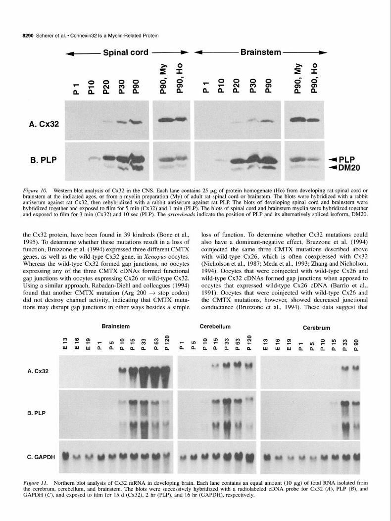
tein in different CNS regions by Western blot analysis. Protein homogenates of spinal cord, brainstem, cerebellum, and cere- brum were prepared from rats of various ages, from Pl to P90. The blots were probed with a rabbit antiserum against Cx32, then the same blots were reprobed with a rabbit antiserum against PLP Cx32 and PLP were more abundant in the brainstem and spinal cord (Fig. 10) than in the cerebrum and cerebellum (not shown), and the amount of Cx32 and PLP in each brain region increased from Pl to P90 (Fig. 10). We also made a myelin preparation of adult cerebrum, brainstem, and spinal cord, to enrich for membrane-related proteins, in order to com- pare the amount of Cx32 in the myelin fractions to that in ho- mogenates of the corresponding brain regions. Western blot analysis demonstrated that in each brain region, the myelin frac- tion contained relatively more Cx32, and that PLP was enriched to a similar extent (Fig. 10). These results demonstrate that Cx32
accumulates in parallel with other myelin proteins during de- velopment.
Cx32 mRNA is expressed in concert with other myelin-related genes in oligodendrocytes
Like the myelin proteins, the myelin-related mRNAs accumulate in parallel during the development of the CNS, with modest differences between different brain regions (Kanfer et al., 1989; Scherer et al., 1994). Thus, we compared the expression of Cx32 mRNA in the cerebrum, cerebellum, and brainstem to that of PLP (Fig. 11). In each region, the levels of Cx32 and PLP mRNA were parallel, with a similar onset and peak. Prolonged overexposure of the blot demonstrated, however, a low level of Cx32 mRNA expression prior to the onset of PLP mRNA ex- pression (data not shown), consistent with the idea that neurons express Cx32 prior to oligodendrocytes.
The above findings indicate that most of the Cx32 mRNA in the CNS originates from oligodendrocytes, which is consistent with the in situ hybridization localization of Cx32 mRNA in adult rat CNS (Micevych and Abelson, 1991). To further sub- stantiate this idea, we analyzed Cx32 mRNA expression in my- elin-dejicient rats and jimpy mice, which have mutations in the PLP gene, and drastically reduced levels of all myelin-related mRNAs (Hudson, 1990). Northern blot analysis of affected male myelin-dejcient rats and their age-matched normal male litter- mates revealed that affected males had much lower levels of Cx32 mRNA at every age (Fig. 12). The levels of Cx32 mRNA in the brains of jimpy mice were also much lower than those in the brains of age-matched normal mice as well as shiverer mice, which have a recessively inherited mutation in the MBP gene (Hudson, 1990). Reprobing the blots for PLP mRNA demon- strated that the levels of PLP mRNA were much lower at every age in both myelin-de$cient rats and jimpy mice than in non- mutant animals and shiverer mice (Fig. 12). The changes in myelin-related mRNA expression in the mutant animals were not confined to Cx32 and PLP, as MBP and MAG are similarly affected (data not shown; see also Roth et al., 1985; Gardinier and Macklin, 1988; Kumar et al., 1988; Kumar et al., 1990; Macklin et al., 1991). Thus, the parallel reduction in Cx32 mRNA and other myelin-related mRNAs in these PLP mutants provides strong evidence that most Cx32 mRNA in the CNS is derived from oligodendrocytes.
Discussion
The finding that mutations in Cx32 cause CMTX (Bergoffen et al., 1993) led us to investigate the role of Cx32 in myelinating glia. In this article, we have shown that Cx32 is expressed in myelinating Schwann cells and oligodendrocytes, but is local- ized to different parts of each cell. In the PNS, Cx32 is found mainly in the incisures and paranodal regions of myelinating Schwann cells, whereas in the CNS, it is found in oligodendro- cyte cell bodies and processes. In both Schwann cells and oli- godendrocytes, Cx32 protein and mRNA are expressed in a co- ordinate manner with those of other myelin genes. In Schwann cells, the expression of Cx32 mRNA and protein is develop- mentally regulated and depends on the integrity of axon- Schwann cell interactions. In oligodendrocytes, Cx32 is ex- pressed in concert with other myelin genes, both in normal de- velopment and in jimpy mice and myelin-dejicient rats. Thus, Cx32 is expressed as part of the myelinating phenotype of both Schwann cells and oligodendrocytes.

The Journal of Neuroscience, December 1995, 75(12) 8289
Figure 9. Immunohistochemical analysis of Cx32 in the ventral funiculus of adult rat spinal cord. These are photomicrographs of transverse (A,B) and longitudinal sections (C,D), double-labeled with a rabbit antiserum against Cx32 (A,C) and a mouse monoclonal antibody against MOG (B,D), and visualized with rhodamine-/and fluorescein-coupled secondary antibodies, respectively. Cx32-immunoreactivity is found in oligodendrocytes (0) and their processes, some of which are indicated by UTYOWS. MOG-immunoreactivity is found on the external/abaxonal surface of the myelin sheath, around a few oligodendrocyte processes (arrowheads), but not in the oligodendrocytes themselves (0). Some myelinated axons are indicated (a in A and B and 1,2,3 in C and D); the myelin sheath is unlabeled by Cx32 or MOG. Scale bar, 10 pm.
Gap junctions in Schwann cells
Since Cx32 forms gap junctions in many tissues, its localization in the incisures and paranodal regions indicates that gap junc- tions should be found at these locations. Small collections of hexagonally packed particles of the appropriate size to be gap junctions have been found in the incisures and paranodes of the PNS myelin sheath by freeze-fracture electron microscopy (San- dri et al., 1982). As these collections of particles seen by freeze- fracture are characteristic of gap junctions, and Cx32 is found at these same sites, it is reasonable to propose that these gap junctions contain Cx32. Since they link apposed layers of the same cell, these could be called “reflexive” gap junctions, which have also been described in the kidney and other tissues (Majack and Larsen, 1980). Why typical gap junctions have not been noted in incisures and paranodes is unclear, as these struc- tures have been extensively studied by transmission electron mi- croscopy (Thomas and Ochoa, 1984; Peters et al., 1991).
More typical gap junctions, between different Schwann cells, have also been described. Electrophysiological recordings and tracer studies have revealed gap junctions between nonmyeli-
nating Schwann cells (but not between myelinating Schwann cells) in peripheral nerve (Konishi, 1990). In peripheral nerve undergoing Wallerian degeneration, gap junctions between ad- jacent Schwann cells have been observed by transmission elec- tron microscopy (Tetzlaff, 1982), and there existence has also been inferred by recent electrophysiological observations (Bru- net and Jirounek, 1994). Finally, gap junctions have been ob- served between rat Schwann cells cultured in the absence of forskolin (Chanson et al., 1993). The connexin(s) that forms gap junctions between nonmyelinating, denervated, and cultured rat Schwann cells has not been identified. Since Cx32 appears to be expressed by nonmyelinating Schwann, it may form the gap junctions in these cells. The connexin expressed by Schwann cells cultured in the absence of forskolin is unlikely to be Cx32, which we find to be expressed at very low levels under these conditions, and which has different electrophysiological prop- erties than those described by Chanson et al. (1993).
How do CMTX mutations cause neuropathy? Many different mutations in the Cx32 gene cause CMTX. To date, 33 different mutations, affecting nearly every portion of
8290 Scherer et al. * Connexin32 Is a Myelin-Related Protein
r+------- Spinal cord - - Brainstem -
A. Cx32
B. PLP
Figure 10. Western blot analysis of Cx32 in the CNS. Each lane contains 25 (*g of protein homogenate (Ho) from developing rat spinal cord or brainstem at the indicated ages, or from a myelin preparation (My) of adult rat spinal cord or brainstem. The blots were hybridized with a rabbit antiserum against rat Cx32, then rehybridized with a rabbit antiserum against rat PLP The blots of developing spinal cord and brainstem were hybridized together and exposed to film for 5 min (Cx32) and 1 min (PLP). The blots of spinal cord and brainstem myelin were hybridized together and exposed to film for 3 min (Cx32) and 10 set (PLP). The arrowheads indicate the position of PLP and its alternatively spliced isoform, DM20.
the Cx32 protein, have been found in 39 kindreds (Bone et al., 1995). To determine whether these mutations result in a loss of function, Bruzzone et al. (1994) expressed three different CMTX genes, as well as the wild-type Cx32 gene, in Xenopus oocytes. Whereas the wild-type Cx32 formed gap junctions, no oocytes expressing any of the three CMTX cDNAs formed functional gap junctions with oocytes expressing Cx26 or wild-type Cx32. Using a similar approach, Rabadan-Diehl and colleagues (1994) found that another CMTX mutation (Arg 200 + stop codon) did not destroy channel activity, indicating that CMTX muta- tions may disrupt gap junctions in other ways besides a simple
Brainstem
loss of function. To determine whether Cx32 mutations could also have a dominant-negative effect, Bruzzone et al. (1994) coinjected the same three CMTX mutations described above with wild-type Cx26, which is often coexpressed with Cx32 (Nicholson et al., 1987; Meda et al., 1993; Zhang and Nicholson, 1994). Oocytes that were coinjected with wild-type Cx26 and wild-type Cx32 cDNAs formed gap junctions when apposed to oocytes that expressed wild-type Cx26 cDNA (Barrio et al., 1991). Oocytes that were coinjected with wild-type Cx26 and the CMTX mutations, however, showed decreased junctional conductance (Bruzzone et al., 1994). These data suggest that
Cerebellum Cerebrum
B. PLP
C. GAPDH
Figure II. Northern blot analysis of Cx32 mRNA in developing brain. Each lane contains an equal amount (10 p,g) of total RNA isolated from the cerebrum, cerebellum, and brainstem. The blots were successively hybridized with a radiolabeled cDNA probe for Cx32 (A), PLP (B), and GAPDH (C), and exposed to film for 15 d (Cx32), 2 hr (PLP), and 16 hr (GAPDH), respectively.

The Journal of Neuroscience, December 1995, 15(12) 8291
A. Cx32
B. PLP
C. GAPDH
Figure 12. Northern blot analysis of Cx32 mRNA expression in the brains of myelin mutants. Each lane contains an equal amount (10 pg) of total RNA isolated from the whole brains (cerebrum, cerebellum, and brainstem) of individual animals of the indicated ages-myelin-dejicient rats and their normal littermates, shiverer mice, and jimpy mice. RNA from cerebra of normal mice is shown for comparison. The blots were successively hybridized together with a radiolabeled cDNA probe for Cx32 (A), PLP (B), and GAPDH (C), and exposed to film for 14 d (Cx32), 1 d (PLP), and 16 hr (GAPDH), respectively. Note that in rats, the major PLP transcripts in rats are 3.2 and 1.6 kb; in mice, 3.2 and 2.8 kb are main transcripts.
many CMTX mutations cause a loss of functional Cx32 protein, and that at least some CMTX mutations can also have a domi- nant-negative effect. Yet, in spite of the large number of differ- ent mutations, and the evidence that different mutations may have different functional effects, there is no compelling evidence that different mutations cause different clinical phenotypes.
The localization of Cx32 in the Schwann cell myelin sheath, and the loss of function of CMTX mutations in oocytes, strongly suggest that at least some CMTX mutations cause a loss of func- tional gap junctions in the incisures and paranodes. Why these gap junctions are critical to the normal health of myelinated axons, however, remains to be determined. One potentially im- portant consideration is that the geometry of the myelin sheath itself. Myelin is a fundamental adaptation of jawed vertebrates, and is formed by the enormous expansion of Schwann cell and oligodendrocyte membrane (Peters et al., 1991). Gap junctions in the paranodes and incisures would allow ions and small mol- ecules to diffuse radially, directly tranversing the myelin sheath, instead of circumferentially through the Schwann cell cytoplasm, which would be a much longer pathway. In the largest myelin sheaths of mammals, this potential radial pathway would be more than lOOO-fold shorter than the circumferential pathway, as the unrolled myelin sheath is more than 4 mm long, whereas the compact myelin sheath is less than 4 pm thick (Friede and Bischhausen, 1980). If CMTX mutations interrupt the function of these gap junctions, then this radial pathway would be abol- ished. At this time, we can only speculate as to what are the crucial molecules that pass through these gap junctions, or even
whether the interruption of diffusion towards or away from the axon is more important.
A related issue is why mutations of Cx32 selectively affect myelinating Schwann cells. Patients who have CMTX have not been reported to have significant abnormalities in other tissues that express Cx32. The level of Cx32 expression does not seem to be critical, as several tissues, such as the brain and spleen, express levels of Cx32 mRNA that are comparable to peripheral nerve, and liver expresses much higher levels (Nishi et al., 1991; Bergoffen et al., 1993; Meda et al., 1993). Myelinating Schwann cells may be uniquely susceptible to Cx32 mutations because Cx32 is the only connexin they express. In support of this idea, we have not detected Cx43, Cx40, or Cx26 mRNA in rat Schwann cells treated with forskolin (Fig. 8 and data not shown). Cultured sciatic fibroblasts express Cx43 mRNA (Fig. 8), and adult sciatic nerve expresses a low level of Cx43 mRNA that is not modulated by axotomy (data not shown), indicating Cx43 mRNA in nerve is derived from fibroblasts and not mye- linating Schwann cells. On the other hand, there is a growing body of evidence that most cell types express more than one connexin. The liver, for example, expresses Cx32 and Cx26 (Nicholson et al., 1987; Meda et al., 1993; Kuraoka et al., 1993), and epidermal cells express four different connexins (Goliger and Paul, 1994).
Cx32 in CNS myelin
Patients who have CMTX are not known to have CNS abnor- malities, although oligodendrocytes, the only other myelin-form-

8292 Scherer et al. * Connexin32 Is a Myelin-Related Protein
ing cells, have been reported to express Cx32 mRNA and protein (Dermietzel et al., 1989; Micevych and Abelson, 1991). In agreement with previous reports (Naus et al., 1990; Belliveau et al., 1991), we found regional and developmental differences in the expression of Cx32 mRNA. We extended these observations by demonstrating a close association between Cx32 mRNA ex- pression and that of other myelin genes, both in normal devel- opment and in PLP mutants. Western blot analysis of Cx32 also demonstrated regional and developmental differences, as well as decreased expression in PLP mutants. Cx32 protein was more abundant in the pons and spinal cord than in the cerebrum and cerebellum, and the amount of protein accumulated in parallel with PLP These results differ from those of Belliveau et al. (1991) who did not find a developmental increase in Cx32 pro- tein in the cerebrum or hindbrain. The good agreement between the levels of Cx32 mRNA and protein both in development and in PLP mutants, nevertheless, indicates that most of the Cx32 mRNA and protein in the CNS is derived from oligodendro- cytes.
The localization of Cx32 in oligodendrocytes has received relatively little attention. Several workers have examined the lo- calization of Cx32 in the neurons of the spinal cord without mentioning oligodendrocyte labeling (Carr et al., 1991; Yama- moto et al., 1991). Dermietzel et al. (1989) reported Cx32-im- munoreactivity in the perinuclear region of oligodendrocytes, but did not systemically evaluate the localization of Cx32 during development. We found that in older animals, Cx32 is predom- inately found in cell bodies and processes, and was not found in compact myelin, in contrast to the recent report of Spray and Dermietzel (1995). These observations generally agree with the localization of gap junctions in by freeze-fracture electron mi- croscopy (Massa and Mugnaini, 1982; Sandri et al., 1982; Rob- inson et al., 1993). Whether Cx32 is also found in the paranodal regions of CNS myelin sheaths, which contains gap junctions by freeze-fracture, remains to be determined.
References
Aguayo AJ, Epps J, Charron J, Bray GM (1976) Multipotentiality of Schwann cells in cross-anastomosed and grafted unmyelinated nerves-quantitative microscopy and radioautography. Brain Res 104:1-20.
Apostolski S, Sadiq SA, Hays A, Corbo M, Suturkova L, Chaliff & Stefansson K, LeBaron RG, Ruoslahti E, Hays AP Latov N (1994) Identification of Gal@ 1-3)CalNAc bearing glycoproteins at the nodes of Ranvier in peripheral nerve. J Neurosci Res 38:134-141.
Archelos JJ, Roggenbuck K, Schneider-Schaulies J, Linington C, Toyka KV, Hartung HP (1993) Production and characterization of mono- clonal antibodies to the extracellular domain of PO. J Neurosci Res 35:46-53.
Baron P, Kamholz J, Scherer S, Honda H, Shy M, Scarpini E, Scarlato G, Pleasure D (1993) Appearance of PLP mRNA in specific regions of the developing rat lumbosacral spinal cord as revealed by in situ hybridization. Exp Neurol 121 :I 399147.
Barrio LC, Suchyna T, Bargiello T, Xu LX, Roginski RS, Bennett MVL, Nicholson BJ (I 99 1) Gap junctions formed by connexins 26 and 32 alone and in combination are differently affected by applied voltage. Proc Nat1 Acad Sci USA 88:8410-8414.
Belliveau DJ, Kidder GM, Vaus CCG (1991) Expression of gap junc- tion genes during postnatal neural development. Dev Genet 12:308- 317.
Bennett MVL, Barrio LC, Bargiello TA, Spray DC, Hertzberg E, Saez JC (1991) Gap junctions: new tools, new answers, new questions. Neuron 6:305-320.
Bergoffen J, Scherer SS, Wang S, Oronzi-Scott M, Bone L, Paul DL, Chen K, Lensch MW, Chance P, Fischbeck K (1993) Connexin mu- tations in X-linked Charcot-Marie-Tooth disease. Science 262:2039- 2042.
Bone LJ, Dahl N, Lensch MW, Chance PF, Kelly T, Le Guern E, Magi S, Parry G, Shapiro H, Wang S, Fischbeck KH (1995) New con- nexin32 mutations associated with X-linked Charcot-Marie-Tooth disease. Neurology, in press.
Brockes JP, Fields P Raff MC (1979) Studies on cultured rat Schwann cells. I. Establishment of purified populations from cultures of pe- ripheral nerve. Brain Res 165:105-l 18.
Brockes JP, Lemke GE, Balzer DR Jr (1980) Purification and prelim- inary characterization of a glial growth factor from the bovine pitu- itary. J Biol Chem 255:8374-8377.
Brunet PC, Jirounek P (1994) Long-range intercellular signalling in glial cells of the peripheral nerve. Neuroreport 5:635-638.
Brunner C, Lassmann H, Waehneldt TV, Matthieu J-M, Linington C (1989) Differential ultrastructural localization of myelin basic pro- tein, myelin/oligodendrocyte glycoprotein, and 2’,3’-cyclic nucleotide 3’-phosphodiesterase in the CNS of adult rats. J Neurochem 52:296- 304.
Bruzzone R, White TW, Scherer SS, Fischbeck KH, Paul DL (1994) Null mutations of connexin32 in patients with X-linked Charcot-Ma- rie-Tooth disease. Neuron 13: 1253-l 260.
Carr PA, Yamamoto T, Karmy G, Nagy JI (1991) Cytochemical rela- tionships and central terminations of a unique population of primary afferent neurons in rat. Brain Res Bull 26:825-843.
Chance PE Pleasure D (1993) Charcot-Marie-Tooth syndrome. Arch Neurol 50: 1180-l 183.
Chanson M, Chandross KJ, Rook MB, Kessler JA, Spray DC (1993) Gating characteristics of a steeply voltage-dependent gap junction channel in rat Schwann cells. J Gen Phvsiol 102:925-946.
Chirgwin JM, Przbyla AE, MacDonald RJ, Rutter RJ (1979) Isolation of biologically active ribonucleic acid from sources enriched in ri- bonuclease. Biochemistry 18:5294-5299.
Cohen R, Guarnieri M (1976) Immunochemical measurement of my- elin basic protein in developing rat brain: an index of myelin syn- thesis. Dev Biol 49:294-299.
Curtis R, Stewart HJS, Hall SM, Wilkin GP, Mirsky R, Jessen KR (1992) GAP-43 is expressed by nonmyelin-forming Schwann cells of the peripheral nervous system. J Cell Biol 116:1455-1464.
DeFerra E Engh H, Hudson L, Kamholz J, Puckett C, Molineaux S, Lazzarini RA (1985) Alternative splicing accounts for the four forms of myelin basic protein. Cell 43:721-727.
Dermietzel R, Hwang TK, Spray DS (1990) The gap junction family: structure, function and chemistry. Anat Embryo1 182:5 17-528.
Dermietzel R, Traub 0, Hwang TK, Beyer E, Bennett MVL, Spray DC, Willecke K (1989) Differential expression of three gap junction pro- teins in developing and mature brain tissues. Proc Nat1 Acad Sci USA 86:10148-10152.
D’Urso D, Brophy PJ, Staugaitis SM, Gillespie CS, Frey AB, Stempak JG, Colman DR (1990) Protein zero of peripheral nerve myelin: biosynthesis, membrane insertion, and evidence for homotypic inter- action. Neuron 4:449460.
Fannon AM, Sherman DL, Ilyina-Gragerova G, Brophy PJ, Friedrich VL, Colman DR (1995) Novel E-cadherin mediated adhesion in pe- ripheral nerve: Schwann cell architecture is stabilized by autotypic adherens junctions. J Cell Biol 129: 189-202.
Fort P Marty L, Piechaczyk M, Sabrouty SE, Dani C, Jeanteur P, Blan- chard JM (1985) Various rat adult tissues express only one major mRNA species from the glyceraldehyde-3-phosphate-dehydrogenase multigenic family. Nucleic Acids Res 13: 1431-1442.
Friede RL, Bischhausen R (1980) The precise geometry of large in- ternodes. J Neurol Sci 48:367-381.
Friedman B, Zaremba S, Hockfield S (1990) Monoclonal antibody rat 401 recognizes Schwann cells in mature and developing peripheral nerve. J Comp Neurol 295:43-57.
Gardinier MV, Macklin WB (1988) Myelin proteolipid protein gene expression in jimpy and jimpyInqd mice. J Neurochem 5 1:360-364.
Goliger JA, Paul DL (1994) Expression of gap junction proteins Cx26, Cx31.1, Cx37, and Cx43 in developing and mature rat epidermis. Dev Dynam 200:1-13.
Goodenough DA, Paul DL, Jesaitis L (1988) Topological distribution of two connexin32 antigenic sites in intact and split rodent hepatocyte gap junctions. J Cell Biol 107:1817-1824.
Hahn AE Brown WE Koopman WJ, Feasby TE (1990) X-Linked dom- inant hereditary motor and sensory neuropathy. Brain 113:151 l- 1525.

The Journal of Neuroscience, December 1995, 15(12) 8293
Hudson L (1990) Molecular biology of myelin proteins in the central and peripheral nervous systems. Semin Neurosci 2:487+96.
Ionasescu VV, Trofatter J, Haines JL, Summers AM, Ionasescu R, Sear- bv C (I 992) X-Linked recessive Charcot-Marie-Tooth neurooathv- chnicaj and’genetic study. Muscle Nerve 15:368-373. I ’
Jacobson S (1963) Sequence of myelination in the brain of the albino rat. J Comp Neurol 121:5-29.
Kanfer J, Parenty M, Goujct-Zalc C, Munge M, Bernier L, Campagnoni AT, Dautigny A, Zalc B (1989) Developmental expression of myelin proteolipid protein, myelin basic protein, and 2’,3’-cyclic nucleotide 3’ phosphodiesterase transcripts in different rat brain regions. J Mel Neurosci 1:39-46.
Koeppen AH, Barron KD, Csiza CK, Greenfield EA (1988) Compar- ative immunohistochemistry of Pelizaeus-Merzbacher disease, the jimpy mouse, and the myelin-deficient rat. J Neurol Sci 84:315-327.
Konishi T (1990) Dye coupling between mouse Schwann cells. Brain Res 508:85-92.
Kumar NM, Gilula N (1992) Molecular biology and genetics of gap junction channels. Semin Cell Biol 3:3-16.
Kumar NM, Gilula NB (1986) Cloning and characterization of human and rat liver cDNAs coding for a gap junction protein. J Cell Biol 103:767-776.
Kumar S, Gordon MN, Espinosa de 10s Monteros MA, de Vellis J (1988) Developmental expression of neural cell type-specific mRNA in the myelin-deficient mutant rat brain: inhibition of oligodendrocyte differentiation. J Neurosci Res 21:268-274.
Kumar S, Macklin WB, Gordon MN, Espinosa de1 10s Monteros A, Cole R, Scully SA, de Vellis J (1990) Transcriptional regulation studies of myelin-associated genes in myelin-deficient mutant rats. Dev Neurosci 12:3 16-325.
Kuraoka A, Iida H, Hatae T Shibata Y, Itoh M, Kurita T (1993) Lo- calization of gap junction proteins, connexin-32 and connexin-26, in rat and guinea pig liver as revealed by quick-freeze, deep-etch im- munoelectron microscopy. J Histochem Cytochem 41:971-980.
Lemke G (1992) Myelin and myelination. In: Molecular neurobiology (Hall ZW, ed), pp 281-312. Sunderland, MA: Sinauer.
Lemke G (1993) The molecular genetics of myelination-an update. Glia 7:263-27 1.
Lemke G, Axe1 R (1985) Isolation and sequence of a cDNA encoding the major structural protein of peripheral myelin. Cell 40:501-508.
Lemke G, Chao M (1988) Axons regulate Schwann cell expression of the major myelin and NGF receptor genes. Development 102:499- 504.
Linnington C, Webb M, Woodhams PL (1984) A novel myelin-asso- ciated glycoprotein defined by a mouse monoclonal antibody. J Neu- roimmunol 6:387-396.
Macklin WB, Gardinier MV, Obeso ZO, King KD, Wight PA (1991) Mutations in the myelin proteolipid protein gene alter oligodendro- cyte gene expression in jimpy and jimpym‘d mice. J Neurochem 56: 163-171.
Majack RA, Larsen WJ (1980) The bicellular and reflexive membrane junctions of renomedullary interstitial cells: functional implications of reflexive gap junctions. Am J Anat 157:181-189.
Massa PT, Mugnaini E (1982) Cell junctions and intramembrane par- ticles of astrocytes and oligodendrocytes: a freeze-fracture study. Neuroscience 7:523-538.
Meda P Pepper MS, Traub 0, Willecke K, Gros D, Beyer E, Nicholson B, Paul D, Orci L (1993) Differential expression of gap junction connexins in endocrine and exocrine glands. Endocrinology 133: 2371-2378.
Micevych PE, Abelson L (1991) Distribution of mRNAs coding for liver and heart gap junction proteins in the rat central nervous system. J Comp Neural 305:96-l 18.
Mirsky R, Jessen KR (1990) Schwann cell development and the reg- ulation of myelination. Semin Neurosci 2:423-435.
Morgan L, Jessen KR, Mirsky R (1991) The effects of CAMP on dif- ferentiation of cultured Schwann cells: progression from an early phe- notype (04+) to a myelin phenotype (PO+, GFAP-, N-CAM-, NGF- receptor-) depends on growth inhibition. J Cell Biol ll2:457-467.
Morgan L, Jessen KR, Mirsky R (1994) Negative regulation of the PO gene in Schwann cells: suppression of PO mRNA and protein in cul- tured Schwann cells by FGF2 and TGFB 1, TGFB2 and TGFB3. De- velopment 120:1399-1409.
Naus CG, Celliveau DJ, Bechberger JF (1990) Regional differences in
connexin32 and connexin43 messenger RNAs in rat brain. Neurosci Lett 111:297-302.
Nicholson B, Dermietzel R, Teplow D, Traub 0, Willecke K, Revel J-P (1987) Two homologous protein components of hepatic gap junc- tions. Nature 329:732-734.
Nishi M, Kumar NM, Gilula NB (1991) Developmental regulation of gap junction gene expression during mouse embryonic development. Dev Biol 146:117-130.
Norton WT, Poduslo SE (1973) Myelination in rat brain. Method of myelin isolation. J Neurochem 21:749-758.
Paul DL (1986) Molecular cloning of cDNA for rat liver gap junction protein. J Cell Biol 103:123-134.
Pedraza L, Owens GC, Green LAD, Salzer JL (1990) The myelin- associated glycoproteins: membrane disposition, evidence of a novel disulfide linkage between immunoglobulin-like domains, and post- translational palmitylation. J Cell Biol 111:265 1-2661.
Peters A, Palay SL, Webster HD (1991) The fine structure of the ner- vous system. New York: Oxford UP
Phillips LH, Kelly TE, Schnatterly P Parker D (1985) Hereditary mo- tor-sensory neuropathy (HMSN): possible X-linked dominant inher- itance. Neurology 35:-502.
Porter S, Clark MB, Glaser L, Bunge RP (1986) Schwann cells stim- ulated to proliferate in the absence of neurons retain full functional capability: J Neurosci 6:3070-3078.
Rabadan-Diehl C. Dahl G. Werner R (1994) A connexin-32 mutation associated with Charcot-Marie-Tooth disease does not affect channel formation in oocytes. FEBS Lett 351:90-94.
Radeke MJ, Misko TP Hsu C, Herzenberg LA, Shooter EM (1987) Gene transfer and molecular cloning of the rat nerve growth factor receptor. Nature 325:593-597.
Reale E, Luciano L, Spitnas M (1975) Freeze-fracture faces of the perineurial sheath of the rabbit sciatic nerve. J Neurocytol 4:261- 270.
Robinson SR, Hampson ECGM, Munro MN, Vaney DI (1993) Uni- directional coupling of gap junctions between neuroglia. Science 262: 1072-1074.
Roth HJ, Hunkeler MJ, Campagnoni AT (1985) Expression of myelin basic protein genes in several dysmyelinating mouse mutants during early postnatal brain development. J Neurochem 45:572-580.
Rozear MP, Pericak-Vance MA, Fischbeck K, Stajich JM, Gaskell PC Jr, Krendel DA, Graham DG, Dawson DV, Roses AD (1987) He- reditary motor and sensory neuropathy, X-linked: a half century fol- low-up. Neurology 37: 1460-1465.
Sambrook J. Fritsch EE Maniatis T (1989) Molecular clonina. Cold Spring Harbor, NY: Cold Spring Harbor Laboratory. -
Sandri C, Van Buren JM, Akert K (1982) Membrane morphology of the vertebrate nervous system. Prog Brain Res 46:201-265.
Scherer SS, Asbury AK (1993) Inherited axonal neuropathies. In: The molecular and genetic basis of neurological disease (Rosenberg RN, Prusiner SB, DiMauro S, Barchi RL, Kunkle LM, eds), pp 899-921. Butterworth-Heinemann.
Scherer SS, Braun PE, Grinspan JG, Collarini E, Wang D-y, Kamholz J (1994) Differential regulation of the 2’,3’-cyclic nucleotide 3’- phosphodiesterase gene in oligodendrocyte development. Neuron 12: 1363-1375.
Scherer SS, Xu Y-T Roling D, Wrabetz L, Fehri ML, Kamholz J (1994) Expression of growth-associated protein-43 kD in Schwann cells is regulated by axon-Schwann cell interactions and CAMP J Neurosci Res 38:575-589.
Schiavinato A, Morandin AR, Guidolin D, Lini E, Nunzi MG, Fiori MG (1991) Perineurium of sciatic nerve in normal and diabetic ro- dents-freeze-fracture study of intercellular junctional complexes. J Neurocytol 20:459-470.
Schwab ME, Schnell L (1989) Region-specific appearance of myelin constituents in the developing rat spinal cord. J Neurocytol 18:161- 164.
Seamon KB, Padgett W, Daly JW (1981) Forskolin: unique diterpene activator of adenylate cyclase in membranes and in intact cells. Proc Nat1 Acad Sci USA 78f336333367.
Snioes GJ. Suter U (1995) Glial cell lineage and develooment. Annu kev Neurosci 18:45-75.’
Snipes GJ, Suter U, Welcher AA, Shooter EM (1992) Characterization of a novel peripheral nervous system myelin protein (PMP-22/SR13). J Cell Biol 117:225-238.
Sobue G, Pleasure D (1984) Schwann cell galactocerebroside induced
8294 Scherer et al. + Connexin32 Is a Myelin-Related Protein
by derivatives of adenosine 3’,5’-monophosphate. Science 224:72- 14
Spray DC, Dermietzel R (1995) X-linked dominant Charcot-Marie- Tooth disease and other potential gap-junction diseases of the nervous system. Trends Neurosci 18:256-262.
Stahl N, Harry J, Popko B (1990) Quantitative analysis of myelin pro- tein gene expression during development in the rat sciatic nerve. Mol Brain Res 8:209-212.
Sternberger NH, Quarles RH, Itoyama Y, Webster HD (1979) Myelin- associated glycoprotein demonstrated immunocytochemically in my- elin and myelin-forming cells of developing rat. Proc Nat1 Acad Sci USA 76:1510-1514.
Stoll G, Trapp BD, Griftin JW (1989) Macrophage function during Wallerian degeneration of rat optic nerve: clearance of degenerating myelin and la expression. J Neurosci 9:2327-2335.
Suter U, Welcher AA, Snipes GJ (1993) Progress in the molecular understanding of hereditary peripheral neuropathies reveals new in- sights into the biology of the peripheral nervous system. Trends Neu- rosci 1650-56.
Tetzlaff W (1982) Tight junction contact events and temporary gap junctions in the sciatic nerve fibres of the chicken during Wallerian degeneration and subsequent regeneration. J Neurocytol 11:839-358.
Thomas PK, Ochoa J (1984) Microscopic anatomy of peripheral nerve fibers. In: Peripheral neuropathy (Dyck PJ, Thomas PK, Lambert EH, Bunge R, eds), pp 39-96. Philadelphia: Saunders.
Trapp BD, Quarles RH (1982) Presence of the myelin-associated gly- coprotein correlates with alterations in the periodicity of peripheral myelin. J Cell Biol 92:877-882.
Trapp BD, Quarles RH (1984) Immunocytochemical localization of the myelin-associated glycoprotein. Fact or artefact? J Neuroimmunol 6:231-249.
Trapp BD, Quarles RH, Griffin JW (1984) Myelin-associated glyco- orotein and mvelinating Schwann cell-axon interaction in chronic B,B’-iminodiprbprionitrie neuropathy. J Cell Biol 98:1272-1278.
Traoo BD. Itovoma Y, Sternberger NH, Quarles RH, Webster HD (1’981) Imm&rohistochemical localization of PO protein in Golgi complex membranes and myelin of developing rat Schwann cells. J Cell Biol 90:1-6.
Trapp BD, Andrews SB, Wong A, O’Connell M, Griffin JW (1989) Co-localization of the myelin-associated glycoprotein and the micro- filament components, F-actin and spectrin, in Schwann cells of my- elinated nerve fibers. J Neurocytol 18:47-60.
Webster HD, Favilla JT (1984) Development of peripheral nerve fibers. In: Peripheral neuropathy (Dyck PJ, Thomas PK, Lambert EH, Bunge R, eds), pp 329-359. Philadelphia: Saunders.
Yamamoto T, Hertzberg EL, Nagy JI (1991) Subsurface cisterns in (Y- motoneurons of the rat and cat: immunohistochemical detection with antibodies against connexin32. Synapse 8: 1 19-l 36.
Zhang J-T, Nicholson BJ (1994) The topological structure of connexin 26 and its distribution compared to connexin 32 in hepatic gap junc- tions. J Membr Biol 139:15-29.