congenital diaphragmatic hernia: the impact of ... · 18 fig. 6 a and b dynamics of post- hepatic...
TRANSCRIPT
Pediatr Surg Int (1995) 10:16-22 © Springer-Verlag 1995
Dietrich Kluth • Burak Tander • Martin von Ekesparre Dick Tibboel • Wolfgang Lambrecht
Congenital diaphragmatic hernia: the impact of embryological studies
Abstract In recent years, a substantial research effort within the specialty of pediatric surgery has been devoted to improving our knowledge of the natural history and pathophysiology of congenital diaphragmatic hernias (CDH) and pulmonary hypoplasia (PH). However, the embryological background has remained elusive because certain events of normal diaphragmatic development were still unclear and appropriate animal models were lacking. Most authors assume that delayed or inhibited closure of the diaphragm will result in a diaphragmatic defect that is wide enough to allow herniation of the gut into the fetal thoracic cavity. However, we feel that this assumption is not based on appropriate embryological observations. To clar- ify whether it was correct, we restudied the morphology of pleuroperitoneal openings in normal rat embryos. Shortly before, a model for CDH and PH had been established in rats using nitrofen (2,4-di-chloro-phenyl-p-nitrophenyl ether) as teratogen. We used this model in an attempt to answer the following questions: (1) When does the dia- phragmatic defect appear? (2) Are the pleuroperitoneal canals the precursors of the diaphragmatic defect? (3) Why is the lung hypoplastic in babies and infants with CDH?
In our study we made following observations: (1) The typical findings of CDH and PH cannot be explained by inhibited closure of the pleuroperitoneal "canals." In normal development, the pleuroperitoneal openings are always too small to allow herniation of gut into the thoracic cavity. (2) The maldevelopment of the diaphragm starts rather early in the embryonic period (5th week). The
D. Kluth (~) • B. Tander • M. yon Ekesparre • W. Lambrecht Department of Pediatric Surgery, University Hospital of Hamburg, Martinistrasse 52, D-20246 Hamburg, Germany
T. Tibboel Department of Pediatric Surgery, Erasmus University Rotterdam, Rotterdam, The Netherlands
B. Tander Research Fellow, Sisli Children's Hospital, Istambul, Turkey Supported in part by "Werner Otto Stiftung", Hamburg, Germany
lungs of CDH rats are significantly smaller than those of control rats at the end of the embryonic period (8th week). (3) The maldevelopment of the lungs in rats with CDH is "secondary" to the defect of the diaphragm. (4) The defect of the lungs is "structural" rather than "functional." Com- plete spontaneous correction of these lung defects is unlikely even after fetal intervention. (5) The "fetal lamb model" does not completely mimic the full picture of CDH, because the onset of the defect lies clearly in the fetal period. We believe that our rat model is better. It is especially useful for describing the abnormal embryology of this lesion.
Key words Congenital diaphragmatic hernia Embryology • Rat embryos • Nitrofen Abnormal diaphragmatic development
Introduction
Due to general advances in pediatric surgery, neonatology, and intensive care medicine, the mortality of most con- genital malformations has dropped continuously over the last decades, while the prognosis of some is unfavorable despite all efforts. Congenital diaphragmatic hernia (CDH) is a typical example of this type of malformation. At first sight, the anatomy and embryology of this lesion appear simple [3, 4, 8, 9, 15, 22, 26, 27]: gut is pushed through a defect in the posterolateral region of the diaphragm, causing compression of the ipsilateral lung as well as shifting of the heart and mediastinum to the contralateral side. Although surgical correction of the defect is easy [14], most babies die because their lungs are too hypoplastic to allow life outside the uterus [14].
As mortality in CDH seems to be closely correlated to the degree of lung hypoplasia (PH), numerous attempts were made to define the pathophysiological relationship between the defect and the degree of the lung lesion [ 1, 6, 7, 10-13, 16, 18]. Harrison concluded from studies in the
EP FP
. . . . , ~ : ~ : ~: ~ . ~ ~ ; ~ s --a * ~ : ~ ~: ,,,~ ~ ,~a~,~ ~,;5-.~ ~, ,~,~.
!~Creat:,o, ~t-c-~;il r=- ~ w : : - - I , t ~ o r r e c t l o n o ! l~elect [(day 6 ~ - 7 0 ) ~ . _ _ _ . . J i n the Fetal Period . . . . ~ ( 1 ) I ] ( d a y 1 0 0 ) ]
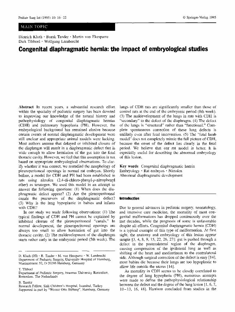
Fig. 1 Concept of pathogenesis (A) and experimental design of sheep model for CDH and PH (B). B Diaphragmatic defect artificially created in fetal period (1) is followed by development of a secondary lung defect (2), PH. In many cases, impaired development in the fetal period (FP) will result in a "functional" defect. (Note the developmental sequence of CDH and PH as sketched in A has never been observed in either humans or animals)
17
EP FP
Fig. 2 A - C Theoretically, lung hypoplasia could result after early disturbed lung development. In B, both defects (PH and CDH) are primary; in (2, PH is followed by disturbed diaphragmatic development [17]. A Popular theory of development of CDH and PH (comp. Fig. 1)
fetal lamb model [15] that the severity of the PH depends on the volume and timing of herniation of viscera into the chest. He was also able to show that in-utero reduction of the volume at the right time could result in nearly-normal lung development in these lambs [12]. The timing of the herniation, which has a considerable impact on the degree of PH in this model, is well known (Fig. 1). This is not the case in humans with CDH, however. For fuman fetuses with CDH, it is generally assumed that the diaphragmatic defect appears because the pleuroperitoneal canals fail to close at 8 to 10 weeks of gestation [15]. It is further speculated that the "returning" intestines later herniate through this defect into the thoracic cavity and compress the developing lung [3, 8, 9, 15]. Typically, the number of airway branches in hypoplastic lungs of babies with CDH is reduced. This observation led to the conclusion that herniation of the gut must take place before the 17th week of gestation, which corresponds to the "pseudoglandular" stage of lung devel- opment [2, 5, 15].
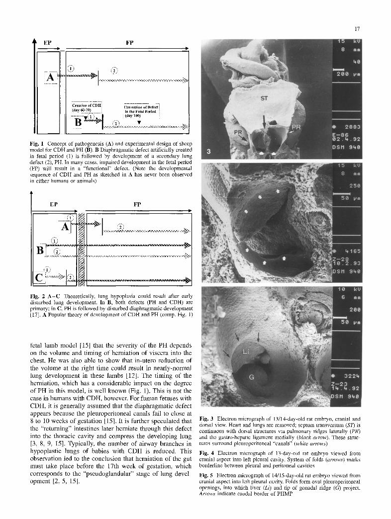
Fig. 3 Electron micrograph of 13/14-day-old rat embryo, cranial and dorsal view. Heart and lungs are removed; septum transversum (ST) is continuous with dorsal structures via pulmonary ridges laterally (PR) and the gastro-hepatic ligament medially (black arrow). These struc- tures surround pleuroperitoneal "canals" (white arrows)
Fig. 4 Electron micrograph of 13-day-old rat embryo viewed from cranial aspect into left pleural cavity. System of folds (arrows) marks borderline between pleural and peritoneal cavities
Fig. 5 Electron micrograph of 14/15-day-old rat embryo viewed from cranial aspect into left pleural cavity. Folds form oval pleuroperitoneal openings, into which liver (Li) and tip of gonadal ridge (G) project. Arrows indicate caudal border of PHMP
18
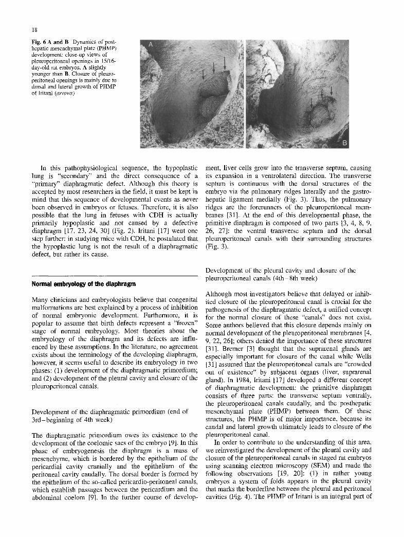
Fig. 6 A and B Dynamics of post- hepatic mesenchymal plate (PHMP) development: close-up views of pleuroperitoneal openings in 15/16- day-old rat embryos, A slightly younger than B. Closure of pleuro- peritoneal openings is mainly due to dorsal and lateral growth of PHMP of Iritani (arrows)
In this pathophysiological sequence, the hypoplastic lung is "secondary" and the direct consequence of a "primary" diaphragmatic defect. Although this theory is accepted by most researchers in the field, it must be kept in mind that this sequence of developmental events as never been observed in embryos or fetuses. Therefore, it is also possible that the lung in fetuses with CDH is actually primarily hypoplastic and not caused by a defective diaphragm [17, 23, 24, 30] (Fig. 2). Iritani [17] went one step further: in studying mice with CDH, he postulated that the hypoplastic lung is not the result of a diaphragmatic defect, but rather its cause.
Normal embryology of the diaphragm
Many clinicians and embryologists believe that congenital malformations are best explained by a process of inhibition of normal embryonic development. Furthermore, it is popular to assume that birth defects represent a "frozen" stage of normal embryology. Most theories about the embryology of the diaphragm and its defects are influ- enced by these assumptions. In the literature, no agreement exists about the terminology of the developing diaphragm, however, it seems useful to describe its embryology in two phases: (1) development of the diaphragmatic primordium; and (2) development of the pleural cavity and closure of the pleuroperitoneal canals.
Development of the diaphragmatic primordium (end of 3rd-beginning of 4th week)
The diaphragmatic primordium owes its existence to the development of the coelomic sacs of the embryo [9]. In this phase of embryogenesis the diaphragm is a mass of mesenchyme, which is bordered by the epithelium of the pericardial cavity cranially and the epithelium of the peritoneal cavity caudally. The dorsal border is formed by the epithelium of the so-called pericardio-peritoneal canals, which establish passages between the pericardium and the abdominal coelom [9]. In the further course of develop-
ment, liver cells grow into the transverse septum, causing its expansion in a ventrolateral direction. The transverse septum is continuous with the dorsal structures of the embryo via the pulmonary ridges laterally and the gastro- hepatic ligament medially (Fig. 3). Thus, the pulmonary ridges are the forerunners of the pleuroperitoneal mem- branes [31]. At the end of this developmental phase, the primitive diaphragm is composed of two parts [3, 4, 8, 9, 26, 27]: the ventral transverse septum and the dorsal pleuroperitoneal canals with their surrounding structures (Fig. 3).
Development of the pleural cavity and closure of the pleuroperitoneal canals (4th-8th week)
Although most investigators believe that delayed or inhib- ited closure of the pleuroperitoneal canal is crucial for the pathogenesis of the diaphragmatic defect, a unified concept for the normal closure of these "canals" does not exist. Some authors believed that this closure depends mainly on normal development of the pleuroperitoneal membranes [4, 9, 22, 26]; others denied the importance of these structures [31]. Bremer [3] thought that the suprarenal glands are especially important for closure of the canal while Wells [31] assumed that the pleuroperitoneal canals are "crowded out of existence" by subjacent organs (liver, suprarenal gland). In 1984, Iritani [17] developed a different concept of diaphragmatic development: the primitive diaphragm consists of three parts: the transverse septum ventrally, the pleuroperitoneal canals caudally, and the posthepatic mesenchymal plate (PHMP) between them. Of these structures, the PHMP is of major importance, because its caudal and lateral growth ultimately leads to closure of the pleuroperitoneal canal.
In order to contribute to the understanding of this area, we reinvestigated the development of the pleural cavity and closure of the pleuroperitoneal canals in staged rat embryos using scanning electron microscopy (SEM) and made the following observations [19, 20]: (1) in rather young embryos a system of folds appears in the pleural cavity that marks the borderline between the pleural and peritoneal cavities (Fig. 4). The PHMP of Iritani is an integral part of

19
this system; (2) in later stages these folds form the oval pleuroperitoneal openings. Into these openings the liver and the tip of the gonadal ridge project (Fig. 5); (3) Closure of the pleuroperitoneal openings is due mainly to dorsal and lateral growth of the PHMP (Fig. 6); and (4) a pleuroperi- toneal "canal" in the actual sense could not be found.
Most authors assume that delayed or inhibited closure of the diaphragm will result in a diaphragmatic defect that is wide enough to allow herniation of gut into the fetal thoracic cavity. However, this assumption is not the result of appropriate embryological observations, but rather the result of interpretations of anatomic- pathologic findings. In a series of normal staged embryos, we measured the width of the pleuroperitoneal openings and the transverse diam- eter of gut loops (Fig. 7). On the basis of these measure- ments, we estimated that a single embryonic gut loop requires an opening of at least 450 g to herniate into the fetal pleural cavity. However, in none of our embryos were the observed pleuroperitoneal openings of appropriate dimensions, indicating that delayed or inhibited closure of the pleuroperitoneal canal cannot result in a diaphragmatic defect of sufficient size. Herniation of gut through these
Fig. 7 Electron micrograph of 14-day-old rat embryo showing transverse diameter of a gut loop (arrows). On the basis of this measurement, a single embryonic gut loop requires an opening of at least 450 g to herniate into fetal pleural cavity
Fig. 8 Electron micrograph of 14-day,old rat embryo (nitrofen group), dorsal view with lungs removed. Large defect seen in right diaphragm (black arrows), which ends abnormally high, leaving parts of liver (Li) uncovered (This stage corresponds to a human embryo 5-6 weeks of age)
Fig. 9 Electron micrograph of a large diaphragmatic defect, typically in close contact with inferior vena cava (IVC) and definitely larger than pleuroperitoneal opening (arrows). Closure impaired by ingrowth of liver (Li)
Fig. 10 Electron micrograph of 15-day-old rat embryo (nitrofen group), lateral view. Small part of liver (Li) protrudes through diaphragmatic defect (D) into thoracic cavity. Note close contact between liver and lung (Lu), which is not yet altered
openings is therefore impossible in later stages. The proposed theory of the pathogenetic mechanisms of CDH development thus lacks any embryological evidence; furthermore, the proposed timing of this process is highly questionable.
20
Abnormal embryology of the diaphragm
Obviously, abnormal development can only be studied successfully in abnormal embryos. Progress in this field has been hampered by the lack of an appropriate animal model. However, for CDH and PH an animal model has been recently established in rats using nitrofen (2,4-di- chloro-phenyl-p-nitrophenyl ether) as a teratogen [21, 29]. We employed this model to study the timing and mechan- isms of abnormal diaphragmatic development. The follow- ing questions should be answered: (1) When does the diaphragmatic defect appear? (2) Are the pleuroperitoneal canals the precursors of the diaphragmatic defect? and (3) Why is the lung hypoplastic in cases of CDH?
As already mentioned, most authors speculate that the diaphragmatic defect appears in the late embryonic period (8th week) when the pleuroperitoneal canals fail to close [3, 4, 8, 9, 15, 22, 26, 27]. In rats, this developmental stage is equivalent to that of 16 or 17 days of gestation. In contrast to this assumption, in our study fully developed diaphragmatic defects were already visible in 14-day-old embryos (Fig. 8). This age in rat embryos is equivalent to human embryos at the end of the 5th week. However, the first observation of a defect is not equivalent with the onset of a malformation. Careful morphologic studies revealed that in nitrofen- treated rats CDH starts to develop in 13- day-old animals. These findings are in accordance with observations made by Iritani [17], who found signs of disturbed diaphragmatic development in I l-day-old mouse embryos exposed to nitrofen continuously from day 5 to 11 after conception. Thus, we conclude that in rat embryos the onset of the diaphragmatic defect clearly lies in the early embryonic period. This phase is compara- ble to that of a human embryo at the beginning of the 5th gestational week.
It is generally assumed that the defects in CDH are identical with the regions of the so-called pleuroperitoneal canals. In our study, all defects were situated in the dorsal part of the diaphragm in close connection to the dorsal wall of the inferior vena cava (Fig. 9). This region is identical with the PHMP described by Iritani. Some defects were small; in these cases the pleuroperitoneal openings closed properly. In others, the defects were definitely larger than the pleuroperitoneal openings (Fig. 9). However, both findings indicate that the defect is not identical with the region of the pleuroperitoneal canals.
Some investigators believe that the lungs in newborns with CDH are primarily hypoplastic [17, 23, 24, 30]. This means that compared to controls, the lungs in these cases are too small to begin with and a "hernia" does not exist. In our study, we found no evidence for this assumption. It is a well-known problem in embryology that it is nearly impossible to find two "identical" embryos, even in one litter, which is why embryologists group their embryos into age-groups [28], developmental "horizons" [28], or "stages" [25]. In humans, the sizes of embryos (and organs) often do not correlate with age; to estimate the normal size of organs in embryos is therefore difficult.
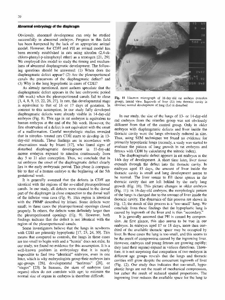
Fig. 11 Electron micrograph of 16-day-old rat embryo (nitrofen group), lateral view. Ingrowth of liver (Li) into thoracic cavity is obvious; normal development of lung (Lu) is disturbed
In our study, the size of the lungs of 13- or 14-day-old rad embryos from the nitrofen group was not obviously different from that of the control group. Only in older embryos with diaphragmatic defects and liver inside the thoracic cavity were the lungs obviously reduced in size. Thus, using SEM techniques we found no evidence for primarily hypoplastic lungs (recently, a study was started to evaluate the pattern of lung growth in rat embryos and fetuses with CDH by calculating the mitotic index).
The diaphragmatic defect appears in rat embryos at the 14th day of development. A short time later, liver tissue expands through the defect into the thoracic cavity. In embryos aged 15 days, the amount of liver inside the thoracic cavity is small and lung development seems to be normal. The liver seems to fill those spaces in the thoracic cavity that are left behind by the early lung growth (Fig. 10). This picture changes in older embryos (Fig. 11): in 16-day-old embryos, the morphologic pattern of the lungs is changed due to the amount of liver inside the thoracic cavity. The dynamics of this process are shown in Fig. 12; the result of this process is a "too-small" lung. We conclude from these findings that the hypoplastic lung is caused by ingrowth of the liver and is thus "secondary."
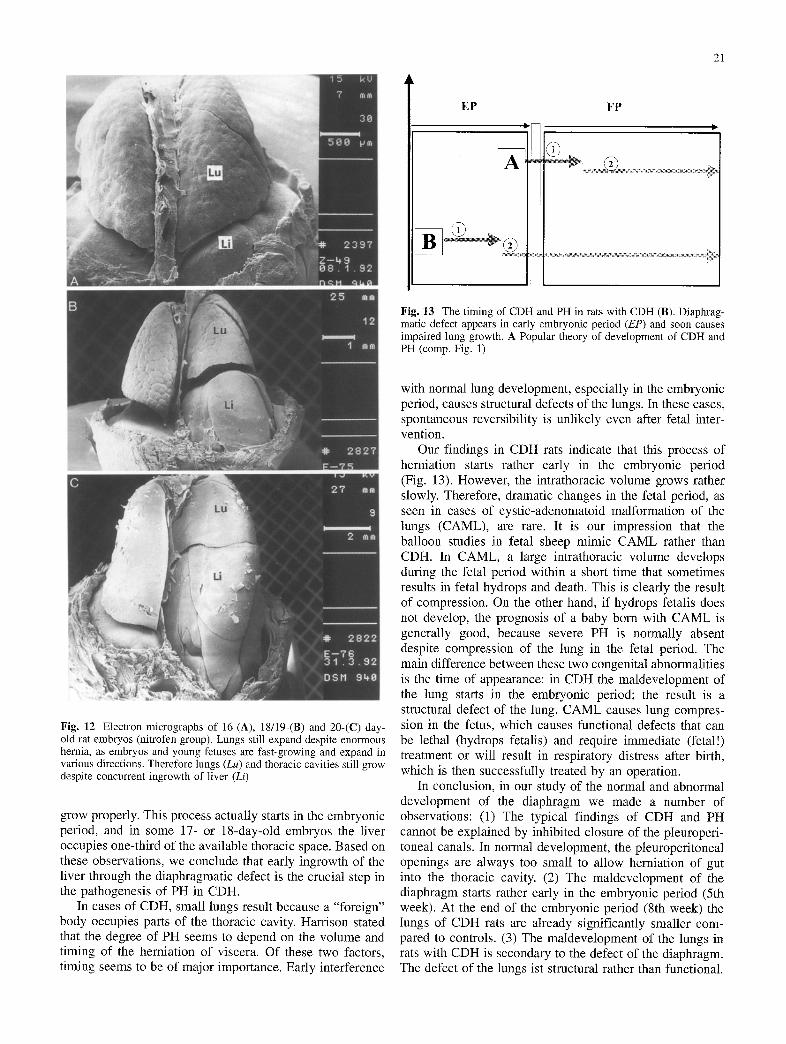
It is generally assumed that PH is caused by compres- sion. At first glance, this also seems to be true in our embryos. In embryos aged 17 or 18 days, more than one- third of the available thoracic space may be occupied by liver. In these cases the lung is too small, and this seems to be the result of compression caused by the ingrowing liver. However, embryos and young fetuses are growing rapidly; they (and their organs) expand in various directions. There- fore, it is not surprising that comparison of two embryos in different age groups reveals that the lungs and thoracic cavities still grow despite the concurrent ingrowth of liver (Fig. 12). Our study thus indicates that too-small, hypo- plastic lungs are not the result of mechanical compression, but rather the result of reduced spatial proportions. The ingrowing liver reduces the available space for the lung to
21
EP
A
@_
FP
Fig. 13 The timing of CDH and PH in rats with CDH (B). Diaphrag- matic defect appears in early embryonic period (EP) and soon causes impaired lung growth. A Popular theory of development of CDH and PH (comp. Fig. 1)
Fig. 12 Electron micrographs of 16-(A), 18/19-(B) and 20-(C) day- old rat embryos (nitrofen group). Lungs still expand despite enormous hernia, as embryos and young fetuses are fast-growing and expand in various directions. Therefore lungs (Lu) and thoracic cavities still grow despite concurrent ingrowth of liver (Li)
grow properly. This process actually starts in the embryonic period, and in some 17- or 18-day-old embryos the liver occupies one-third of the available thoracic space. Based on these observations, we conclude that early ingrowth of the liver through the diaphragmatic defect is the crucial step in the pathogenesis of PH in CDH.
In cases of CDH, small lungs result because a "foreign" body occupies parts of the thoracic cavity. Harrison stated that the degree of PH seems to depend on the volume and timing of the herniation of viscera. Of these two factors, timing seems to be of major importance. Early interference
with normal lung development, especially in the embryonic period, causes structural defects of the lungs. In these cases, spontaneous reversibility is unlikely even after fetal inter- vention.
Our findings in CDH rats indicate that this process of herniation starts rather early in the embryonic period (Fig. 13). However, the intrathoracic volume grows rather slowly. Therefore, dramatic changes in the fetal period, as seen in cases of cystic-adenomatoid malformation of the lungs (CAML), are rare. It is our impression that the balloon studies in fetal sheep mimic CAML rather than CDH. In CAML, a large intrathoracic volume develops during the fetal period within a short time that sometimes results in fetal hydrops and death. This is clearly the result of compression. On the other hand, if hydrops fetalis does not develop, the prognosis of a baby born with CAML is generally good, because severe PH is normally absent despite compression of the lung in the fetal period. The main difference between these two congenital abnormalities is the time of appearance: in CDH the maldevelopment of the lung starts in the embryonic period; the result is a structural defect of the lung. CAML causes lung compres- sion in the fetus, which causes functional defects that can be lethal (hydrops fetalis) and require immediate (fetal!) treatment or will result in respiratory distress after birth, which is then successfully treated by an operation.
In conclusion, in our study of the normal and abnormal development of the diaphragm we made a number of observations: (1) The typical findings of CDH and PH cannot be explained by inhibited closure of the pleuroperi- toneal canals. In normal development, the pleuroperitoneal openings are always too small to allow herniation of gut into the thoracic cavity. (2) The maldevelopment of the diaphragm starts rather early in the embryonic period (5th week). At the end of the embryonic period (8th week) the lungs of CDH rats are already significantly smaller com- pared to controls. (3) The maldevelopment of the lungs in rats with CDH is secondary to the defect of the diaphragm. The defect of the lungs ist structural rather than functional.

22
Complete spontaneous correction of these lung defects is unlikely even after fetal intervention. (4) The fetal lamb model does not completely mimic the full picture of CDH, because the onset of the defect clearly lies in the fetal period. We believe that our rat model is the better model; it is especially useful in describing the abnormal embryology of this lesion.
References
1. Adzick NS, Outwater KM, Harrison MR, Davies R Glick PL, de Lorimier AA, Reid LM (1985) Correction of congenital diaphragmatic hernia in utero. IV. An early gestational fetal lamb model for pulmonary vascular morphometric analysis. J Pediatr Surg 20:73-680
2. Areechon W, Reid L (1963) Hypoplasia of the lung with con- genital diaphragmatic hernia. Br Med J I: 230-233
3. Bremer JL (1943) The diaphragm and diaphragmatic hernia. Arch Pathol 36:539-549
4. Broman I (1902) f2ber die Entwicklung des Zwerchfells beim Menschen. Verb Anat Ges 16: 9-17
5. Davies G, Reid L, (1970) Growth of the alveoli and pulmonary arteries in childhood. Thorax 25:669-681
6. de Lorimier AA, Tierney DE Parker HR (1967) Hypoplastic lungs in fetal lambs with surgically produced congenital diaphragmatic hernia. Surgery 62: 12-17
7. de Lorimier AA, Manus AG, Tyler WS (1969) Morphometry of normal and hypoplastic lungs in newborn lambs. Curr Top Surg Res 1:431-442
8. Gray SW, Skandalakis JE (1972) Embryology for surgeons. WB Saunders, Philadelphia, pp 359-385
9. Grosser O, Ortmann R (1970) Grundril3 der Entwicklungs- geschichte des Menschen, 7th edn. Springer Berlin Heidelberg New York, pp 124-127
10. Haller JA Jr, Signer RD, Golladay ES, Inon AE, Harrington DR Shermeta DW (1976) Pulmonary and ductal hemodynamics in studies of simulated diaphragmatic hernia of fetal and newborn lambs. J Pediatr Surg 11:675-680
11. Harrison MR, Jester JA, Ross NA (1980) Correction of congenital diaphragmatic hernia in utero. I. The model: intrathoracic balloon produces fatal pulmonary hypoplasia. Surgery 88: 174-182
12. Harrison MR, Bressack MA, Churg AM (1980) Correction of congenital diaphragmatic hernia in utero. II. Simulated correction permits fetal lung growth with survival at birth. Surgery 88:260-268
13. Harrison MR, Ross NA, de Lorimier AA (1981) Correction of congenital diaphragmatic hernia in utero. III. Development of a successful surgical technique using abdominoplasty to avoid compromise of umbilical blood flow. J Pediatr Surg 16:934-942
14. Harrison MR (1989) Fetal diaphragmatic hernia. In: Puri P (ed) Congenital diaphragmatic hernia. Modern problems in paediatrics. Karger, Basel, pp 130-142
15. Harrison MR (1990) The fetus with a diaphragmatic hernia: pathophysiology, natural history, and surgical management. In: Harrison MR, Golbus MS, Filly RA (eds) The unborn patient. Fetal diagnosis and treatment, 2nd edn. WB Saunders, Philadel- phia, pp 295-319
16. Hashimoto EG, Pringle KC, Soper RT, Brown CK (1985) The creation and repair of diaphragmatic hernia in fetal lambs: morphology of the type II alveolar cell. J Pediatr Surg 20: 354-356
17. Iritani I (1984) Experimental study on embryogenesis of congeni- tal diaphragmatic hernia. Anat Embryol 169: 133-139
18. Kent GM, Olley PM, Creighton RE, Dobbinson T, Bryan MH, Symchych R Zingg W, Cummings JN (1972) Hemodynamic and pulmonary changes following surgical creation of a diaphragmatic hernia in fetal lambs. Surgery 72:427-433
19. Kluth D, Petersen C, Zimmermann HJ (1987) The developmental anatomy of congenital diaphragmatic hernia. Pediatr Surg Int 2:322-326
20. Kluth D, Petersen C, Zimmermann HJ, Mt~hlhaus K (1989) The embryology of congenital diaphragmatic hernia. In: Puri P (ed) Congenital diaphragmatic hernia. Modern problems in paediatrics. Karger, Basel, pp 7-21
21. Kluth D, Kangah R, Reich R et al. (1990) Nitrofen-induced diaphragmatic hernia in rats: An animal model. J Pediatr Surg 25:850-854
22. Moore KL (1985) K6rperh6hlen, Mesenterien, Zwerchfell. In: Embryologie, Lehrbuch und Atlas der Entwicklungsgeschichte des Menschen, 2nd ed. Schattauer, Stuttgart, pp 189-201
23. Nakao Y (1983) Establishment of animal models for congenital diaphragmatic hernia and its pathogenesis. J Nagoya City Univ Med Assoc 34:318-339
24. Nakao Y, Ueld R, Nakao Y, Fumkawa M, Nomura M, Tada T, Mild M, Kishimoto H (1986) Teratogenic effects of maternal exposure to nitrofen in mice: relationship between congenital diaphragmatic hernia and other congenital anomalies. Nagoya Med J 31:115-125
25. O'Rahilly R, Muller F (1984) Chevalier Jackson Lecture. Respi- ratory and alimentary relations in staged human embryos. New embryological data and congenital anomalies. Ann Otol Rhinol Laryngol 93:421-429
26. Patten BM (1965) Human embryology. McGraw-Hill, New York, pp 499-523
27. Starck D (1975) Embryologie, 3rd edn. Thieme, Stuttgart, pp 488-500
28. Streeter GL (1945) Descriptions of age group XIII, embryos 4 or 5 mm long, and age group XIV, period of indentation of the lens vesicle. Contrib Embryol Carnegie Inst 31:27-64
29. Tenbrinck R, Tibboel D, Gaillard JLJ, et al. (1990) Experimentally induced congenital diaphragmatic hernia in rats. J Pediatr Surg 25:426-429
30. Ueki R, Nakao Y, Nishida T, Nakao Y, Wakabayashi T (1990) Lung hypoplasia in developing mice and rats induced by maternal exposure to nitrofen. Cong Anom 30: 133-143
31. Wells LJ (1954) Development of the human diaphragm and pleural sacs. Contrib Embryol Carnegie Inst 35: 107-137