confidential report on the oral part 2 frcophth ... · examination report november 2015 part 2...
TRANSCRIPT

1
Examination Report November 2015 Part 2 FRCOphth Oral Examination
Contents:
1. Summary 2 2. Candidates 3
3. The Structured Vivas 3
3a. Results and analysis 4
4. The OSCE 5 4a. Results and analysis 5
5. The Examination Overall
5a. Final results 6 5b. Breakdown of oral exam 6 5c. Comparison to previous examinations 9
6. Appendices Appendix 1 Hofstee 11 Appendix 2 Candidate evaluation 12 Appendix 3 Patients seen in OSCE 25
Michael Nelson BSc (Hons) FRCOphth MAEd Education Adviser

2
1. Summary This is the third time that the Part 2 FRCOphth oral examination has been taken by candidates since it was de-coupled from the written examination. The OSCE consists of 5 clinical stations at which candidates are required to examine 3 patients (15 in total). The medicine/neurology station is a neuro-ophthalmology station. The communication station remains unchanged. The total number of marks available for the oral examination has increased from 256 to 318 and as a result of these changes there is weighting towards the OSCE of 62% compared with 38% for the structured viva (SV). 72 candidates sat the examination, which is the smallest cohort to date. The reliability of the oral examination is high at 0.80 (SV) and 0.80 (OSCE), but slightly lower than previous years. The pass rate in OST was the highest of any sitting at 86%, which exceeded the pass rate for candidates who were not in OST (40%). There were statistically significant differences in the success of candidates based upon OST, and UK graduates. Candidates who were in OST were more likely to pass, as were UK graduates. There was no statistically significant difference in success based upon ethnicity, gender, or those who spoke English as a first language. Trainees in OST stages 5 and 6 were more successful than those in ST4 and ST7.
3
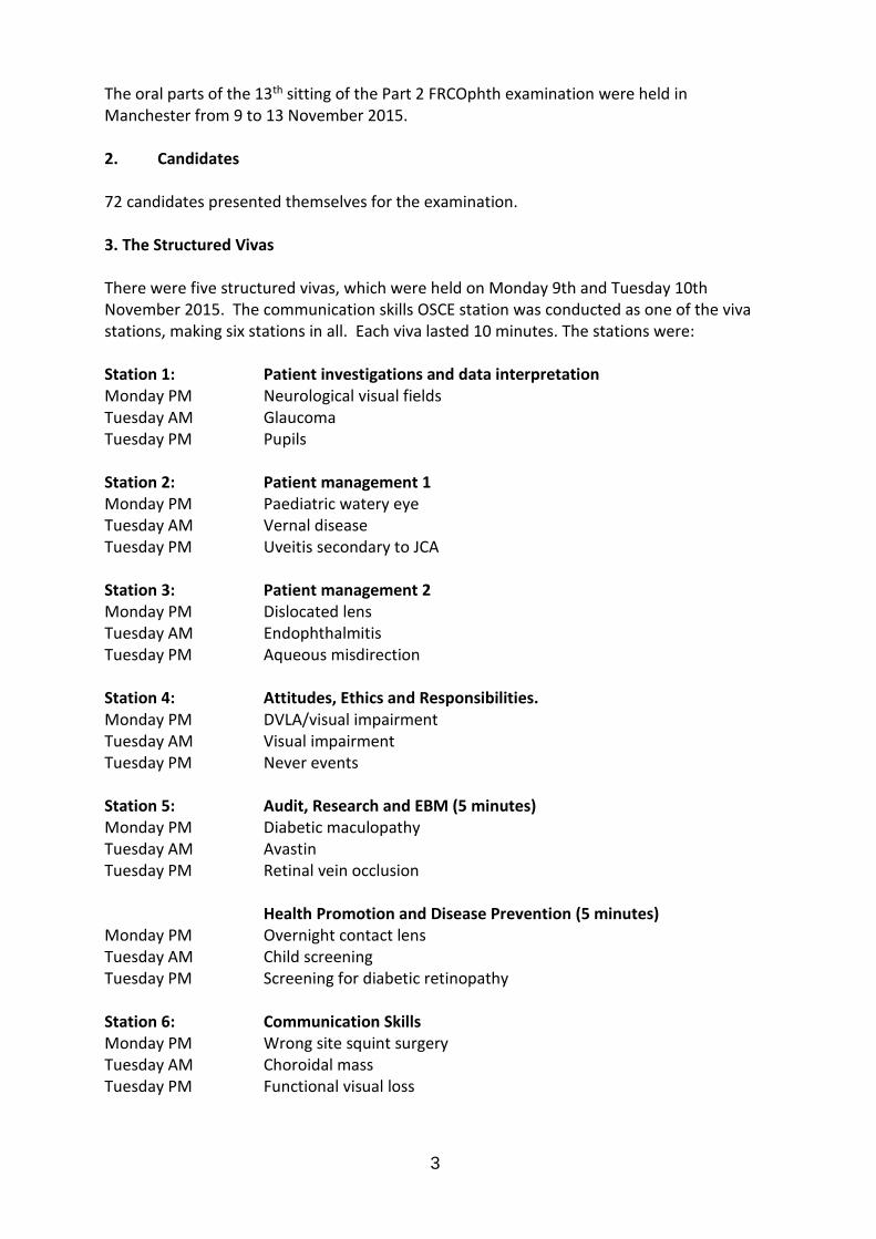
The oral parts of the 13th sitting of the Part 2 FRCOphth examination were held in Manchester from 9 to 13 November 2015. 2. Candidates 72 candidates presented themselves for the examination. 3. The Structured Vivas There were five structured vivas, which were held on Monday 9th and Tuesday 10th November 2015. The communication skills OSCE station was conducted as one of the viva stations, making six stations in all. Each viva lasted 10 minutes. The stations were: Station 1: Patient investigations and data interpretation Monday PM Neurological visual fields Tuesday AM Glaucoma Tuesday PM Pupils Station 2: Patient management 1 Monday PM Paediatric watery eye Tuesday AM Vernal disease Tuesday PM Uveitis secondary to JCA Station 3: Patient management 2 Monday PM Dislocated lens Tuesday AM Endophthalmitis Tuesday PM Aqueous misdirection Station 4: Attitudes, Ethics and Responsibilities. Monday PM DVLA/visual impairment Tuesday AM Visual impairment Tuesday PM Never events Station 5: Audit, Research and EBM (5 minutes) Monday PM Diabetic maculopathy Tuesday AM Avastin Tuesday PM Retinal vein occlusion
Health Promotion and Disease Prevention (5 minutes) Monday PM Overnight contact lens Tuesday AM Child screening Tuesday PM Screening for diabetic retinopathy Station 6: Communication Skills Monday PM Wrong site squint surgery Tuesday AM Choroidal mass Tuesday PM Functional visual loss

4
3a) Results: Maximum mark (5 stations, 10 examiners, 12 marks per station): 120 Pass mark (using borderline candidate method): 66/120 (55%) Mean score: 82/120 (68%) Median score: 83/120 (69%) Range*: 48-110 (40%-92%) Reliability: (Cronbach alpha) 0.8 SEM: 7 Final adjusted pass mark (+ 1 SEM) 73/120 (61%) Pass rate before adjustment (pass mark 66/120) 63/72 (88 %) Pass rate after adjustment (pass mark 73/120) 57/72 (79%) Table 1 Distribution of scores
Score Distribution Total
21-30
31-40
41-50 // 2
51-60 //// 4
61-70 /////// 7
71-80 ///// ///// ///// /// 18
81-90 ///// ///// ///// ///// // 22
91-100 ///// ///// // 12
101-110 ///// / 6
111-120 0
Total 72
The pass mark for the structured viva was increased by 1 SEM from 65.5/120 (55%) to 73/120 (61%).
5
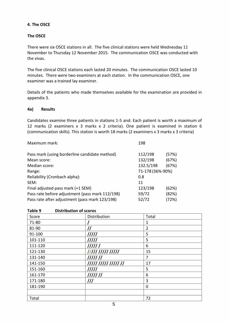
4. The OSCE The OSCE There were six OSCE stations in all. The five clinical stations were held Wednesday 11 November to Thursday 12 November 2015. The communication OSCE was conducted with the vivas. The five clinical OSCE stations each lasted 20 minutes. The communication OSCE lasted 10 minutes. There were two examiners at each station. In the communication OSCE, one examiner was a trained lay examiner. Details of the patients who made themselves available for the examination are provided in appendix 3. 4a) Results Candidates examine three patients in stations 1-5 and. Each patient is worth a maximum of 12 marks (2 examiners x 3 marks x 2 criteria). One patient is examined in station 6 (communication skills). This station is worth 18 marks (2 examiners x 3 marks x 3 criteria) Maximum mark: 198 Pass mark (using borderline candidate method) 112/198 (57%) Mean score: 132/198 (67%) Median score: 132.5/198 (67%) Range: 71-178 (36%-90%) Reliability (Cronbach alpha): 0.8 SEM: 11 Final adjusted pass mark (+1 SEM) 123/198 (62%) Pass rate before adjustment (pass mark 112/198) 59/72 (82%) Pass rate after adjustment (pass mark 123/198) 52/72 (72%) Table 9 Distribution of scores
Score Distribution Total
71-80 / 1
81-90 // 2
91-100 ///// 5
101-110 ///// 5
111-120 ///// / 6
121-130 ///// ///// ///// 15
131-140 ///// // 7
141-150 ///// ///// ///// // 17
151-160 ///// 5
161-170 ///// // 6
171-180 /// 3
181-190 0
Total 72
6
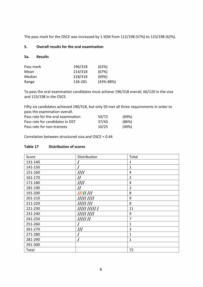
The pass mark for the OSCE was increased by 1 SEM from 112/198 (57%) to 123/198 (62%). 5. Overall results for the oral examination 5a. Results Pass mark 196/318 (62%) Mean 214/318 (67%) Median 218/318 (69%) Range 136-281 (43%-88%) To pass the oral examination candidates must achieve 196/318 overall, 66/120 in the viva and 123/198 in the OSCE. Fifty-six candidates achieved 190/318, but only 50 met all three requirements in order to pass the examination overall. Pass rate for the oral examination 50/72 (69%) Pass rate for candidates in OST 37/43 (86%) Pass rate for non-trainees 10/25 (40%) Correlation between structured viva and OSCE = 0.44 Table 17 Distribution of scores
Score Distribution Total
131-140 / 1
141-150 / 1
151-160 //// 4
161-170 // 2
171-180 //// 4
181-190 // 2
191-200 ///// /// 8
201-210 ///// //// 9
211-220 ///// /// 8
221-230 ///// ///// / 11
231-240 ///// //// 9
241-250 ///// // 7
251-260 / 1
261-270 /// 3
271-280 / 1
281-290 / 1
291-300
Total 72

7
5b) Breakdown of Oral Examination Table 18 Breakdown of results by training
Failed Passed (%) Total
In OST 6 37 (86%) 43
Not in OST 15 10 (40%) 25
Total 21 47(69%) 68
Candidates in OST performed better than those in non-training posts. These differences are statistically significant (p = 0.0001) Table 19 Breakdown of results by gender
Failed Passed (%) Total
Female 8 16 24
Male 14 34 48
Total 22 50 72
Unknown 2 These differences are not statistically significant (p = 0.79) Table 20 Breakdown of results by deanery
Failed Passed Total
East Midlands 0 0 0
East of England 0 2 2
East Scotland 0 0 0
London 0 13 13
Mersey 1 0 1
North Scotland 0 1 1
North Western 0 1 1
Northern 1 0 1
Northern Ireland 0 1 1
Oxford 0 1 1
Peninsula 0 0 0
South East Scotland 0 0 0
West Scotland 1 1 2
Severn 0 2 2
Wales 0 3 3
Wessex 0 2 2
West Midlands 0 8 8
Yorkshire 3 2 5
6 37 43
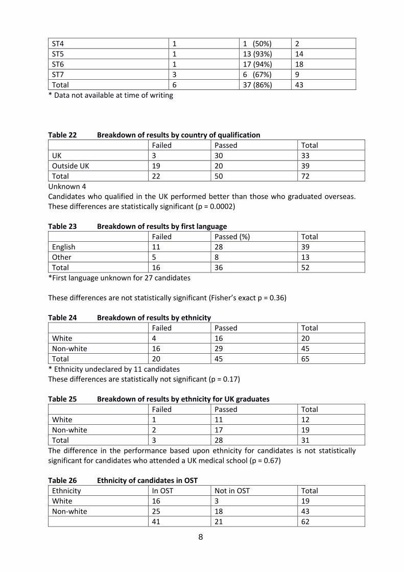
Table 21 Breakdown of results by level of training
Failed Passed Total
ST3 0 0 0
8
ST4 1 1 (50%) 2
ST5 1 13 (93%) 14
ST6 1 17 (94%) 18
ST7 3 6 (67%) 9
Total 6 37 (86%) 43
* Data not available at time of writing Table 22 Breakdown of results by country of qualification
Failed Passed Total
UK 3 30 33
Outside UK 19 20 39
Total 22 50 72
Unknown 4 Candidates who qualified in the UK performed better than those who graduated overseas. These differences are statistically significant (p = 0.0002) Table 23 Breakdown of results by first language
Failed Passed (%) Total
English 11 28 39
Other 5 8 13
Total 16 36 52
*First language unknown for 27 candidates These differences are not statistically significant (Fisher’s exact p = 0.36) Table 24 Breakdown of results by ethnicity
Failed Passed Total
White 4 16 20
Non-white 16 29 45
Total 20 45 65
* Ethnicity undeclared by 11 candidates These differences are statistically not significant (p = 0.17) Table 25 Breakdown of results by ethnicity for UK graduates
Failed Passed Total
White 1 11 12
Non-white 2 17 19
Total 3 28 31
The difference in the performance based upon ethnicity for candidates is not statistically significant for candidates who attended a UK medical school (p = 0.67) Table 26 Ethnicity of candidates in OST
Ethnicity In OST Not in OST Total
White 16 3 19
Non-white 25 18 43
41 21 62
9
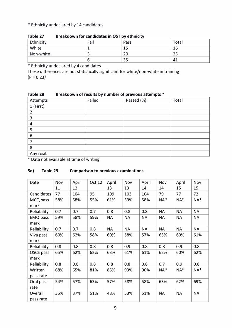
* Ethnicity undeclared by 14 candidates Table 27 Breakdown for candidates in OST by ethnicity
Ethnicity Fail Pass Total
White 1 15 16
Non-white 5 20 25
6 35 41
* Ethnicity undeclared by 4 candidates These differences are not statistically significant for white/non-white in training (P = 0.23) Table 28 Breakdown of results by number of previous attempts *
Attempts Failed Passed (%) Total
1 (First)
2
3
4
5
6
7
8
Any resit
* Data not available at time of writing
5d) Table 29 Comparison to previous examinations
Date Nov 11
April 12
Oct 12 April 13
Nov 13
April 14
Nov 14
April 15
Nov 15
Candidates 77 104 95 109 103 104 79 77 72
MCQ pass mark
58% 58% 55% 61% 59% 58% NA* NA* NA*
Reliability 0.7 0.7 0.7 0.8 0.8 0.8 NA NA NA
EMQ pass mark
59% 58% 59% NA NA NA NA NA NA
Reliability 0.7 0.7 0.8 NA NA NA NA NA NA
Viva pass mark
60% 62% 58% 60% 58% 57% 63% 60% 61%
Reliability 0.8 0.8 0.8 0.8 0.9 0.8 0.8 0.9 0.8
OSCE pass mark
65% 62% 62% 63% 61% 61% 62% 60% 62%
Reliability 0.8 0.8 0.8 0.8 0.8 0.8 0.7 0.9 0.8
Written pass rate
68% 65% 81% 85% 93% 90% NA* NA* NA*
Oral pass rate
54% 57% 63% 57% 58% 58% 63% 62% 69%
Overall pass rate
35% 37% 51% 48% 53% 51% NA NA NA

10
Oral pass rate in OST
46% 43% 63% 56% 64% 65% 70% 80% 86%
* The MCQ examination is now de-coupled from the oral examination Table 30 Cumulative results by deanery (September 2010 to date)
Deanery Number of passes Number of candidates
Pass rate %
East Scotland 5 5 100
Oxford 22 26 85
Severn 16 21 76
Northern Ireland 9 12 75
London KSS 108 153 71
North Scotland 6 9 67
Northern 16 24 67
Mersey 18 28 64
East Midlands 17 27 63
South East Scotland 8 13 62
West Midlands 35 58 60
Wales 17 32 53
Peninsula 11 21 52
West Scotland 11 21 52
North Western 23 45 51
Yorkshire 32 66 48
Wessex 8 17 47
East of England 13 28 46
TOTAL 375 606 62

11
Appendix 1: Hofstee pass mark The Hofstee method of standard setting is based upon the examiner’s opinion on the maximum and minimum credible pass marks and maximum and minimum credible fail rates for the examination. These parameters can then be used to identify a pass mark from a plot of pass mark against fail rate derived from the examination results. Using the following parameters*:
Maximum pass mark 65% (207/318)
Minimum pass mark 50% (159/318)
Maximum fail rate 20%
Minimum fail rate 70% The pass mark for the oral examination using this method would be 200/318 (63%), which is similar to the pass mark derived from the borderline candidate method (62%). (It should be noted that this result is based upon the total marks for the oral examination with complete cross compensation between OSCE and viva results.) * These parameters do not necessarily represent the values that would be chosen by the part 2 examinations sub-committee.
0
10
20
30
40
50
60
70
80
90
100
40 50 60 70 80 90 100
Fail
rate
%
Pass mark %
Hofstee

12
Candidate Feedback – Part 2 FRCOphth Oral Examination Structured Viva The following feedback is from 15 candidates who took part in the structured vivas/comms skills out of 72 (21% response) Viva Station 1 Patient Investigations & Data Interpretation Were you treated in a courteous manner by the examiners in this station? Yes 100% No 0% Comments:
Well managed
The examiners were very nice
Were the questions appropriate for the station? Yes 100% No 0% Comments:
Again very well managed and fair
There was a small issue in the case history and optic nerve photograph were of one eye and there was single visual field of the same patient was of their contralateral eye which meant there was some confusion as to where the field defect was as I wasn’t sure which eye the examiners were asking about – by the examiners responses I felt I probably wasn’t the only candidate who had this difficulty.
Were the questions of an appropriate standard for an exit examination? Yes 93% No 0% Yes & No 7% Comments:
Interpreting an MRI to detect carotid dissection is likely to be beyond most consultants who would rely on their radiologist.
As above
One of the examiners was unaware that the term “benign intracranial hypertension” has been changed for some time now to “idiopathic intracranial hypertension”
Viva station 2 Patient Management 1 Were you treated in a courteous manner by the examiners in this station? Yes 100% No 0% Comments:
The corneal photo presented on an iPad was atrocious! I would struggle to find a worse and more blurred photo of a cornea if I tried. Made diagnosing the condition virtually impossible. Without being able to diagnose he condition it was futile trying to answer all the follow-up questions (e.g. How would you treat this condition?).

13
Examiners were stern but fair Were the questions appropriate for the station? Yes 93% No 7% Comments:
The questions were inappropriate because they were all based on the assumption that one could actually see the rubbish photo on the iPad.
The pictures were a bit misleading – the first showed a swollen bruised eye not consistent with the history and the second was so grainy it was difficult to interpret
Were the questions of an appropriate standard for an exit examination? Yes 80% No 20% Comments:
The quality of the photographs used for the 7-year-old paediatric patient were abysmal – highly pixillated and impossible to use as the basis for a diagnosis. It is essential that the problem of poor image reproduction (which has arisen so often both in the written paper and the oral) should finally be resolved.
My run had all PM components on glaucoma surgery (particularly complications of trabeculectomy). That makes it difficult/unfair to compare with other PM stations that other candidates had.
See above.
I thought that to know the generic names of Vexol and Lotemax was a bit tough!
One of the scenarios was of a rare paediatric congenital condition which is unlikely/unrealistic to come across. If we came across this in practice, we would refer to our paediatric colleagues. Thought it was a bit of a waste of a scenario. Would have been better to ask about management of a more common/sight-threatening condition.
Viva station 3 Patient Management 2 Were you treated in a courteous manner by the examiners in this station? Yes 100% No 0% Comments:
Efficient and fair Were the questions appropriate for the station? Yes 100% No 0% Comments:
I was asked to describe the two methods of performing a vitreous biopsy. I answered by saying that the same needle could be used for biopsy and intravitreal antibiotics, or alternatively the biopsy needle could be used and a separate needle used for
14
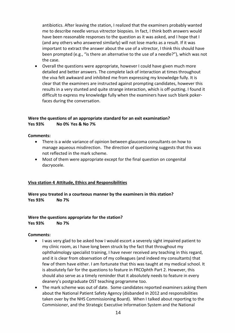
antibiotics. After leaving the station, I realized that the examiners probably wanted me to describe needle versus vitrector biopsies. In fact, I think both answers would have been reasonable responses to the question as it was asked, and I hope that I (and any others who answered similarly) will not lose marks as a result. If it was important to extract the answer about the use of a vitrector, I think this should have been prompted (e.g., “is there an alternative to the use of a needle?”), which was not the case.
Overall the questions were appropriate, however I could have given much more detailed and better answers. The complete lack of interaction at times throughout the viva felt awkward and inhibited me from expressing my knowledge fully. It is clear that the examiners are instructed against prompting candidates, however this results in a very stunted and quite strange interaction, which is off-putting. I found it difficult to express my knowledge fully when the examiners have such blank poker-faces during the conversation.
Were the questions of an appropriate standard for an exit examination? Yes 93% No 0% Yes & No 7% Comments:
There is a wide variance of opinion between glaucoma consultants on how to manage aqueous misdirection. The direction of questioning suggests that this was not reflected in the mark scheme.
Most of them were appropriate except for the final question on congenital dacryocele.
Viva station 4 Attitude, Ethics and Responsibilities Were you treated in a courteous manner by the examiners in this station? Yes 93% No 7% Were the questions appropriate for the station? Yes 93% No 7% Comments:
I was very glad to be asked how I would escort a severely sight impaired patient to my clinic room, as I have long been struck by the fact that throughout my ophthalmology specialist training, I have never received any teaching in this regard, and it is clear from observation of my colleagues (and indeed my consultants) that few of them have either. I am fortunate that this was taught at my medical school. It is absolutely fair for the questions to feature in FRCOphth Part 2. However, this should also serve as a timely reminder that it absolutely needs to feature in every deanery’s postgraduate OST teaching programme too.
The mark scheme was out of date. Some candidates reported examiners asking them about the National Patient Safety Agency (disbanded in 2012 and responsibilities taken over by the NHS Commissioning Board). When I talked about reporting to the Commisioner, and the Strategic Executive Information System and the National
15
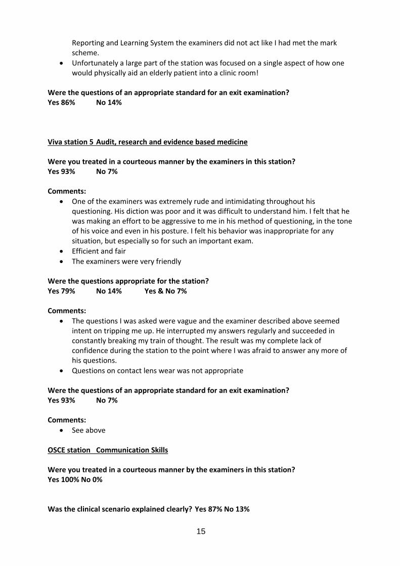
Reporting and Learning System the examiners did not act like I had met the mark scheme.
Unfortunately a large part of the station was focused on a single aspect of how one would physically aid an elderly patient into a clinic room!
Were the questions of an appropriate standard for an exit examination? Yes 86% No 14% Viva station 5 Audit, research and evidence based medicine Were you treated in a courteous manner by the examiners in this station? Yes 93% No 7% Comments:
One of the examiners was extremely rude and intimidating throughout his questioning. His diction was poor and it was difficult to understand him. I felt that he was making an effort to be aggressive to me in his method of questioning, in the tone of his voice and even in his posture. I felt his behavior was inappropriate for any situation, but especially so for such an important exam.
Efficient and fair
The examiners were very friendly Were the questions appropriate for the station? Yes 79% No 14% Yes & No 7% Comments:
The questions I was asked were vague and the examiner described above seemed intent on tripping me up. He interrupted my answers regularly and succeeded in constantly breaking my train of thought. The result was my complete lack of confidence during the station to the point where I was afraid to answer any more of his questions.
Questions on contact lens wear was not appropriate Were the questions of an appropriate standard for an exit examination? Yes 93% No 7% Comments:
See above OSCE station Communication Skills Were you treated in a courteous manner by the examiners in this station? Yes 100% No 0% Was the clinical scenario explained clearly? Yes 87% No 13%
16
Comments:
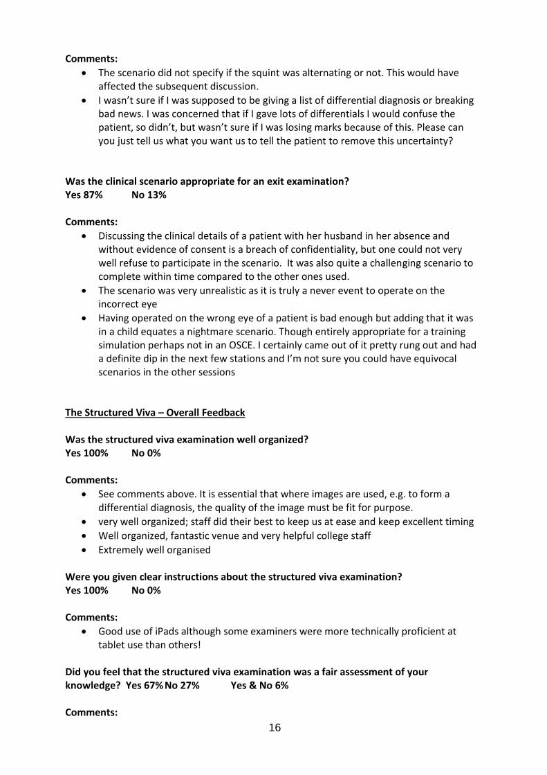
The scenario did not specify if the squint was alternating or not. This would have affected the subsequent discussion.
I wasn’t sure if I was supposed to be giving a list of differential diagnosis or breaking bad news. I was concerned that if I gave lots of differentials I would confuse the patient, so didn’t, but wasn’t sure if I was losing marks because of this. Please can you just tell us what you want us to tell the patient to remove this uncertainty?
Was the clinical scenario appropriate for an exit examination? Yes 87% No 13% Comments:
Discussing the clinical details of a patient with her husband in her absence and without evidence of consent is a breach of confidentiality, but one could not very well refuse to participate in the scenario. It was also quite a challenging scenario to complete within time compared to the other ones used.
The scenario was very unrealistic as it is truly a never event to operate on the incorrect eye
Having operated on the wrong eye of a patient is bad enough but adding that it was in a child equates a nightmare scenario. Though entirely appropriate for a training simulation perhaps not in an OSCE. I certainly came out of it pretty rung out and had a definite dip in the next few stations and I’m not sure you could have equivocal scenarios in the other sessions
The Structured Viva – Overall Feedback Was the structured viva examination well organized? Yes 100% No 0% Comments:
See comments above. It is essential that where images are used, e.g. to form a differential diagnosis, the quality of the image must be fit for purpose.
very well organized; staff did their best to keep us at ease and keep excellent timing
Well organized, fantastic venue and very helpful college staff
Extremely well organised Were you given clear instructions about the structured viva examination? Yes 100% No 0% Comments:
Good use of iPads although some examiners were more technically proficient at tablet use than others!
Did you feel that the structured viva examination was a fair assessment of your knowledge? Yes 67% No 27% Yes & No 6% Comments:
17
Yes and no – there was nothing unfair about the knowledge domains being tested, but occasionally the questions were posed in an unclear manner, which made it hard to work out what was being asked. Mostly, it was a fair assessment, but lack of clarity in the questions meant that sometimes I failed to answer a question, not because of gaps in my knowledge, but because the question was unclear or ambiguous.
Too much glaucoma, no retina, plastics, cornea or strabismus.
Overall the questions were appropriate, however I could have given much more detailed and better answers. The complete lack of interaction with the examiners at times throughout the viva felt awkward and inhibited me from expressing my knowledge fully. It is clear that the examiners are instructed against prompting candidates, however this results in a very stunted and quite strange interaction, which is off-putting. I found it difficult to express my knowledge fully when the examiners have such blank poker-faces during the conversation.
It is very distracting to try to answer questions while one examiner is scribbling comments and the other one is trying to read from an answer sheet. Neither examiner has the time to make eye contact, which in my opinion is an important part of answering questions.
In your opinion should the structured viva examination be included in the exit examination? Yes 80% No 20% Comments:
I think there is certainly a role for the viva in this examination – without it, it would be hard to examine areas that do not lend themselves to an OSCE, e.g. evidence based medicine, communication skills, paediatric ophthalmology, knowledge of surgical procedures, knowledge of guidelines etc. This is not to say that the format of the examination is beyond improvement, and I think several changes could usefully be made, but it would certainly not be appropriate to eliminate this component from the exit examination.
This is a test of performance not knowledge. Knowledge is best tested in the written. There is massive variability in the extent to which examiners ask leading questions, help the candidates to answer and the time they allow you to reach the correct answer when you are struggling to think of it.
However, I feel that a candidate should only retake the OSCE if he/she passes the viva.
Please write any other comments you have about the structured viva examination below:
Very crowded rooms
One of the examiners I had in viva Station 5 had an appalling manner and poor diction. He should not be allowed to examine in future.
For some stations, the pictures on the tablet were not clear and it was difficult to describe and thus further discuss the findings.
18
I think that the structured viva would be better served by short answer questions. The examiners seemed to be waiting for me to say certain sentences or phrases, this type of interrogation could be carried out on paper, with time to consider the responses. Also, the other candidates had a different set of scenarios, if we had short answer questions there would be no need for this, which would make the exam fairer. It would also save 2 days of examiners time, and I should imagine be cheaper to conduct. If it is a requirement of the GMC that we undergo a viva we could count the questions at the end of the OSCE as the viva.
The following feedback is from 16 candidates who took part in the OSCEs out of 72 (22% response) OSCE station 1 Cataract and Anterior Segment Were you treated in a courteous manner by the examiners in this station? Yes 100% No 0% Comments:
Courteous and fair Were the patients you were asked to examine appropriate for the station? Yes 100% No 0% Were the questions of an appropriate standard for an exit examination? Yes 100% No 0% OSCE station 2 Glaucoma and eyelid Were you treated in a courteous manner by the examiners in this station? Yes 88% No 12% Comments:
Courteous and fair
The male examiner in this station was abrupt and discourteous. From speaking to other candidates in my cycle after the examination, it was apparent that they had had similar experiences.
Were the patients you were asked to examine appropriate for the station? Yes 88% No 12%

19
Comments
There was one patient with mainly an orbital problem
Trauma patient with previous surgery Were the questions of an appropriate standard for an exit examination? Yes 81% No 19% Comments:
Some of the questions were vague.
However, I felt that the questioning was vague for all 3 cases and there could have been some more guidance as to what aspects of work-up/management etc. that the examiners wanted me to discuss. Over the course of the 5 OSCE stations, there was huge variation in the amount of guidance that examiners gave. It was unfortunate that in this station I felt that if I was not giving the examiners exactly what they were thinking, then there was little guidance to help get me on the right track. I believe that this will be reflected in a poor mark for this station.
OSCE station 3 Posterior Segment Were you treated in a courteous manner by the examiners in this station? Yes 94% No 0% Yes & No 6% Comments:
Courteous and fair
20
Were the patients you were asked to examine appropriate for the station? Yes 100% No 0% Comments:
The indirect examination was in a pseudophakic with a small pupil and though that is
a necessary skill I’m 6’ 1” and had to stand on my tip toes to get the appropriate distance from the patient who was already reclined fully, I raised this at the time with the examiners but I’m not sure how anyone shorter candidate would have coped.
Were the questions of an appropriate standard for an exit examination? Yes 100% No 0% OSCE station 4 Strabismus and Orbit Were you treated in a courteous manner by the examiners in this station? Yes 88% No 12% Comments:
Very courteous and reassuring
Courteous and fair
One of the examiners was very rude and intimidating. He kept asking me to hurry up, which is most inappropriate in an ocular motility station.
Since one of the cases was very difficult the examiners were very understanding and helpful
I felt I wasn’t allowed to examine the patients without interruption, I felt the patients were ill at ease as was I. This was in stark contrast to the excellent courtesy and manner of the other examiners in all of the other stations.
Were the patients you were asked to examine appropriate for the station? Yes 88% No 6% Yes & No 6% Comments
If it is known that a procedure is tender for a particular patient, please do not ask a
candidate to perform it. My patient clearly had misgivings about having exophthalmometry done. The examiner said, “It is tender for this patient, but go ahead quickly.” One patient had an artificial eye on one side. This should have been clearly mentioned at the start, with some sort of background given to me. It is in very poor taste to ask someone to do a cover test on a patient with one artificial eye.
21
Valuable time was wasted by me thinking “This cannot be an artificial eye, can it?” In a usual clinic, this background knowledge would already be there, or the patient can be examined at the slit lamp instead of my having to ask the patient during an exam, “Is that an artificial eye?”
I was not given an opportunity to examine the second patient. The eye was hypotropic and exotropic. I was asked to give a likely palsy: said third. But told with previous history of trauma it was a 4th. Unfair
One of the patients was very complex (multiple cranial nerve palsies and INO) ophthalmologist we have to make diagnosis without the help of orthoptists
Were the questions of an appropriate standard for an exit examination? Yes 94% No 6% OSCE station 5 Neuro-Ophthalmology Were you treated in a courteous manner by the examiners in this station? Yes 81% No 19% Comments:
Although examiners were very stern
Very understanding and helpful examiners
The examiners adopted a very aggressive "examination style". This made me feel more nervous and reduced my performance during that station and the following OSCE stations.
Unfortunately both examiners were really very aggressive in their questioning, interestingly I had the very same complaint against one of the examiners from my previous attempt in Swansea who then was also examining the Neuro Ophthalmology station.
I found the examiners on this station to be very aggressive. This was my first station and it really put me off for and was a bad start to the whole OSCE. Please could these examiners either be removed or separated for future exams? I definitely don’t think they should both be on the same station again.
Were the patients you were asked to examine appropriate for the station? Yes 100% No 0% Were the questions of an appropriate standard for an exit examination? Yes 75% No 19% Yes & No 6% Comments:
Slightly odd to have detailed questioning on the mechanism of traumatic optic neuropathy – which is poorly understood and controversial.
All of the examiners in the OSCE stations went out of their way to put me at ease. I did not do very well in some stations, which was not the fault of the examiners. However, in the strabismus and orbit station, I did not perform to my full potential due to a nasty examiner, who kept interrupting and asked me to perform a test that
22
was clearly painful for the patient. The second examiner in this station kept a low profile and did not speak much at all to support me.
When asked for differential for cerebropontine angle after the likely acoustic neuroma, I was told you cannot get a meningioma at the cerebro-pontine angle as a cause for 5, 6, 7 palsy.
Some questions were inappropriate i.e. in a patient with a 6th I was asked which one of the other cranial nerves is the most important to be examined. The answer was supposed to be the 5th cranial nerve but why should a 3rd, 4th, 7th or 8th cranial nerve have a lower priority? I feel that all cranial nerves should be examined in this situation.
The aggressive manner in which questions were asked was a problem.
I was quite confused by the examiners questions. The first patient had an orbital apex lesion and I was asked to discuss pathology of the cavernous sinus instead of this. The second patient had a pupil involving 3rd nerve palsy, I was asked the causes of a third and the examiners seemed to get very upset that I started from the brainstem and not with compressive as this was pupil involving. They said that they wanted me to discuss “this” patient – which wasn’t what they had wanted with the first patient, where they had essentially invented pathology asking me to “imagine that her vision was normal”. This seems like a small point, but since it was the first station and they were both quite aggressive towards me it was really difficult to perform well and put me off to a very bad start for the rest of the exam. This was a shame as the rest of the examiners were very encouraging.
The OSCE overall Was the OSCE well organized? Yes 100% No 0% Comments:
• Very well run and wonderful variety of cases; several diagnoses that I had never seen before Were you given clear instructions about the OSCE? Yes 93% No 0% Yes & No 7% Comments
23
Well organized with good timing Did you feel that the OSCE was a fair assessment of your knowledge? Yes 67%No 27% Yes & No 6% Comments:
Not given opportunity to examine the orbit/strabismus patients
Having done this exam before, the patient case-mix is completely random – it appeared to be very skewed towards trauma/orbital cases. Some of the examiners had clearly just examined the patients 5 minutes before we did as candidates and were themselves a bit unsure of what was required.
Trying to establish the diagnosis based on the findings without a complete history does not represent the daily clinical practice. This is a major flaw of the exam.
Please see above comments regarding neuro station In your opinion should the OSCE be included in the exit examination? Yes 93% No 7% Comments:
Very difficult to ensure fairness and consistency with each candidate only seeing 15
patients with necessarily very disparate pathology.
It is impossible to completely standardize this portion of the exam. Unless each candidate sees exactly the same patients as their fellow candidates, it is not possible to be completely objective.
Please write any other comments you have about the OSCE below:
24
Excellent venue. No facility for cleaning the slit lamps between patients – if I was a patient I would want that.
Examining styles varied vastly between different examiners. Some sat back and said nothing – expecting you to talk non-stop from the start. Others were keen to prompt you in the right direction from the get-go, but then it’s not clear whether we would be penalized for requiring prompting. The viva is much more objective – each candidate gets the same scenario and same questions. It is hard to understand, having successfully gained the MRCOphth, why at this late stage in the training programme, why an OSCE is necessary. The old style exam was viva only.
In my Strabismus and Orbit station, I felt like the examiner was imposing his technique on me as opposed to letting me detect findings with my own technique.
The assessment of surgical skills should also be part of the exit exam in a surgical specialty.

25
Appendix 3 Wednesday AM Station 1– Anterior Segment
Station Diagnosis Carousel
Anterior Segment congenital glaucoma B
Anterior Segment corneal graft B
Anterior Segment aphakia, right prosthetic eye B
Anterior Segment left hzvc keratouveitis B
Anterior Segment duanes right aphakia C
Anterior Segment keratoconus, BE PK, Glaucoma C
Anterior Segment FED C
Anterior Segment right PK for HSV keratitis,
keratoconus, RP C
Anterior Segment bilateral herpetic uveitis C
Anterior Segment right neovascular glaucoma, PRP BE C
Anterior Segment aphakia glaucoma D
Anterior Segment Right ICE, Right tube, Right PK D
Anterior Segment FED D
Anterior Segment previous penetrating injury, left iris
defect, retinal tear, LASIK D
Station 2 – Glaucoma and Lid
Station Diagnosis Carousel
Glaucoma and Lid punctal stenosis B
Glaucoma and Lid NAG B
Glaucoma and Lid L Bells B
Glaucoma and Lid uveitic glaucoma B
Glaucoma and Lid congenital glaucoma, right trab, right
PK B
Glaucoma and Lid glaucoma, right ptosis, left ectropion B
Glaucoma and Lid Tarsorrhapy, loss of blink, etc C
Glaucoma and Lid bilateral ptosis and dermatochalosis,
melonoma right eye C
Glaucoma and Lid poag C
Glaucoma and Lid right ptosis/aniridia/ aphakia/ stem
cell dysfunction C

26
Glaucoma and Lid PXF, bilateral pseudophakia, bilateral
trabs C
Glaucoma and Lid POAG D
Glaucoma and Lid bilateral brow ptosis, left
dermatochalosis D
Glaucoma and Lid PDS, PI, cataracts D
Glaucoma and Lid left facial palsy secondary to
cerebellopotine low grade glioma D
Station 3 – Posterior Segment
Station Diagnosis Carousel
Posterior Segment bilateral wet AMD, POAG, PSC B
Posterior Segment PDR B
Posterior segment wet AMD B
Posterior Segment right nasal RD, left prev RD/ peel B
Posterior Segment pseudoanthoma elasticum B
Posterior segment adult vitelliform dystrophy B
Posterior segment FEVR B
Posterior segment DMO C
Posterior segment vasoproliferative tumour, chronic
uveitis C
Posterior Segment wet amd, right AE C
Posterior segment right wAMD C
Posterior Segment right aphakia, right iridectomy
following penetrating injury C
Posterior Segment pseudoanthoma elasticum D
Posterior segment left erm D
Posterior Segment glaucoma, AMD D
Posterior segment left PRE PDR, right mac laser D
Posterior segment RP, pseudophakia D
Posterior Segment Vitelliform D
Station 4 – Strabismus and Orbit
Station Diagnosis Carousel
Strabismus and Orbit Presumed TED B
Strabismus and Orbit left 4th B
Strabismus and Orbit left AE B
Strabismus and Orbit age related restriction of abduction B

27
Strabismus and Orbit left spheno-orbital meningioma C
Strabismus and Orbit Right AE C
Strabismus and Orbit TED C
Strabismus and Orbit bilateral 4th C
Strabismus and Orbit right superior oblique palsy D
Strabismus and Orbit TED, right orbital decompression D
Strabismus and Orbit RTA/ head injury D
Strabismus and Orbit left AE D
Station 5 – Neuro ophthalmology
Station Diagnosis Carousel
Neuro left adies pupil B
Neuro Left sup VF defect & RAPD B
Neuro Bilateral optic atrophy B
Neuro suspected pituitary lesion B
Neuro left hemianopia C
Neuro right carotid artery aneurysm C
Neuro Cavernous sinus meningioma C
Neuro right VI from brain stem meningioma
removal C
Neuro right crao, left articficial eye C
Neuro bilateral optic atrophy with unknown
cause D
Neuro left pseudoptosis, ?marcus
Gunn,right congenital SO palsy D
Neuro bilateral plateau iris and glaucoma D
Neuro Cerebellar strokes D
Neuro Left CRVO. BE Glaucoma D

28
Wednesday PM Station 1 – Anterior Segment
Station Diagnosis Carousel
Anterior Segment right ciliary epithelial cyst B
Anterior Segment Right DSAEK, left PK B
Anterior Segment right traumatic iridodialysis B
Anterior Segment aphakia, right prosthetic eye B
Anterior Segment left herpetic keratouveitis B
Anterior Segment NF1 B
Anterior Segment MMP, ischaemic CRVO, left YAG PI,
right AMD B
Anterior Segment congenital glaucoma C
Anterior Segment macular corneal dystrophy C
Anterior Segment FED C
Anterior Segment artisan lens right eye C
Anterior Segment NF1 C
Anterior Segment bilateral chronic uveitis C
Anterior Segment linear IgA disease D
Anterior Segment FED D
Anterior Segment D
Anterior Segment corneal macular dystrophy D
Anterior Segment previous penetrating injury, left iris
defect D
29
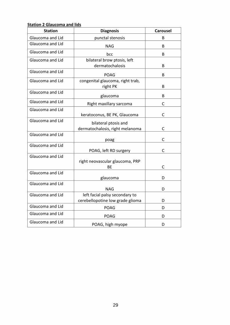
Station 2 Glaucoma and lids
Station Diagnosis Carousel
Glaucoma and Lid punctal stenosis B
Glaucoma and Lid NAG B
Glaucoma and Lid bcc B
Glaucoma and Lid bilateral brow ptosis, left dermatochalosis B
Glaucoma and Lid POAG B
Glaucoma and Lid congenital glaucoma, right trab, right PK B
Glaucoma and Lid glaucoma B
Glaucoma and Lid Right maxillary sarcoma C
Glaucoma and Lid keratoconus, BE PK, Glaucoma C
Glaucoma and Lid bilateral ptosis and
dermatochalosis, right melanoma C
Glaucoma and Lid poag C
Glaucoma and Lid POAG, left RD surgery C
Glaucoma and Lid right neovascular glaucoma, PRP
BE C
Glaucoma and Lid glaucoma D
Glaucoma and Lid NAG D
Glaucoma and Lid left facial palsy secondary to cerebellopotine low grade glioma D
Glaucoma and Lid POAG D
Glaucoma and Lid POAG D
Glaucoma and Lid POAG, high myope D

30
Station 3 – Posterior Segment
Station Diagnosis Carousel
Posterior Segment birdshot chorioretinopathy, right
glaucoma, pseudophakia, B
Posterior segment wet AMD B
Posterior segment vasoproliferative tumour, chronic
uveitis B
Posterior Segment right nasal RD, B
Posterior Segment pseudoanthoma elasticum B
Posterior segment adult vitelliform dystrophy B
Posterior segment FEVR B
Posterior segment DMO C
Posterior segment retinal vasculitis C
Posterior segment Right choroidal naevus C
Posterior Segment PDR C
Posterior Segment L wet amd, R AE C
Posterior segment right wAMD C
Posterior segment PIC/ MFC/ Myopic C
Posterior Segment pseudoanthoma elasticum D
Posterior segment birdshot retinopathy D
Posterior segment bilateral AMD D
Posterior Segment bilateral RD D
Posterior segment bilateral wet AMD D
Posterior Segment Right cryobuckle for RD D
Posterior Segment Vitelliform D
Posterior segment right wet amd D
Station 4 – Strabismus and Orbit
Station Diagnosis Carousel
Strabismus and orbit Presumed TED B
Strabismus and orbit left 4th B
Strabismus and orbit double blowout fracture B
Strabismus and orbit left AE B
Strabismus and orbit age related restriction of abduction B
Strabismus and orbit left spheno-orbital meningioma C

31
Strabismus and orbit amblyopia, ET, KCN, dry eyes C
Strabismus and orbit Right AE C
Strabismus and orbit TED C
Strabismus and orbit bilateral 4th C
Strabismus and orbit right superior oblique palsy D
Strabismus and orbit TED, right orbital decompression, D
Strabismus and orbit Right XT D
Strabismus and orbit RTA/ head injury D
Strabismus and orbit left AE D
Station 5 Neuro-ophthalmology
Station Diagnosis Carousel
Neuro optic disc pit, hypoplastic disc B
Neuro left adies pupil B
Neuro Left sup VF defect & RAPD B
Neuro Bilateral optic atrophy B
Neuro
suspected pituitary lesion B
Neuro
Right Horners B
Neuro
left hemianopia C
Neuro
right carotid artery aneurysm C
Neuro Cavernous sinus meningioma C
Neuro
meningioma C
Neuro
right crao, left articficial eye C
Neuro bilateral optic atrophy with unknown cause D
Neuro left pseudoptosis, ?marcus Gunn,right congenital SO palsy D
Neuro bilateral plateau iris and glaucoma D
Neuro Left AION left altitudinal defect D

32
Neuro
CRAO D
Neuro Cerebellar strokes D
Neuro Intracranial bleed D
Neuro Left CRVO. BE Glaucoma D
Thursday AM Station 1 - Anterior Segment
Station Diagnosis Carousel
Anterior Segment cicatricial conjunctivitis B
Anterior Segment aphakia, right prosthetic eye B
Anterior Segment FED B
Anterior Segment chronic glaucoma B
Anterior Segment left herpetic keratouveitis B
Anterior Segment right DSAEK C
Anterior Segment left herpetic keratitis C
Anterior Segment uveitis, glaucoma C
Anterior Segment FEDS, right DSAEK, Left PK C
Anterior segment right aphakia, right iridectomy
following penetrating injury C
Anterior Segment Fuch heterochromic uveitis D
Anterior Segment bilateral chronic uveitis, right
secondary glaucoma, left cataract D
Anterior Segment right PK, Left DSAEK, right optic
atrophy D
Anterior Segment OCP D

33
Station 2 – Glaucoma and lids
Station Diagnosis Carousel
glaucoma/ lids POAG B
glaucoma/ lids NAG B
glaucoma/ lids left facial palsy secondary to
cerebellopotine low grade glioma B
glaucoma/ lids POAG B
glaucoma/ lids congenital glaucoma, right trab,
right PK B
glaucoma/ lids PXF C
glaucoma/ lids POAG C
glaucoma/ lids NAG C
glaucoma/ lids CPEO + ptosis C
glaucoma/ lids LEFT Fuch heterochromic cyclitis, C
glaucoma/ lids anterior segment dysgenesis,
glaucoma, failed right trab C
glaucoma/ lids poag D
glaucoma/ lids bilateral brow ptosis, left
dermatochalosis D
glaucoma/ lids PDS, PI, cataracts D
glaucoma/ lids POAG D Station 3 – Posterior Segment
Station Diagnosis Carousel
Posterior Segment bilateral wet AMD, POAG, PSC B
Posterior segment wet AMD B
Posterior segment vasoproliferative tumour, chronic
uveitis B
Posterior Segment right nasal RD, B
Posterior Segment juvenile XL retinoschisis B
Posterior Segment pseudoanthoma elasticum B

34
Posterior segment FEVR B
Posterior segment DMO C
Posterior segment myopic degeneration C
Posterior segment Right choroidal naevus C
Posterior segment RP, PSCLO C
Posterior segment right wAMD C
Posterior segment left erm D
Posterior segment presumed sarcoidosis D
Posterior segment pseudoanthoma elasticum D
Posterior segment RP, pseudophakia D
Posterior segment Vitelliform D
Station 4 – Strabismus and orbit
Station Diagnosis Carousel
strab/ orbit Presumed TED B
strab/ orbit left 4th B
strab/ orbit age related restriction of abduction B
strab/ orbit longstanding IR/ SO underaction B
strab/ orbit left AE B
strab/ orbit left spheno-orbital meningioma C
strab/ orbit amblyopia, ET, KCN, dry eyes C
strab/ orbit right fibrous dysplasia C
strab/ orbit TED C
strab/ orbit bilateral 4th C
strab/ orbit right superior oblique palsy D
strab/ orbit Right ocular trauma, left 6th D
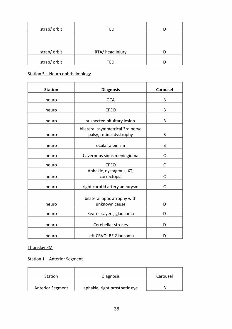
35
strab/ orbit TED D
strab/ orbit RTA/ head injury D
strab/ orbit TED D
Station 5 – Neuro ophthalmology
Station Diagnosis Carousel
neuro GCA B
neuro CPEO B
neuro suspected pituitary lesion B
neuro bilateral asymmetrical 3rd nerve
palsy, retinal dystrophy B
neuro ocular albinism B
neuro Cavernous sinus meningioma C
neuro CPEO C
neuro Aphakic, nystagmus, XT,
correctopia C
neuro right carotid artery aneurysm C
neuro bilateral optic atrophy with
unknown cause D
neuro Kearns sayers, glaucoma D
neuro Cerebellar strokes D
neuro Left CRVO. BE Glaucoma D
Thursday PM Station 1 – Anterior Segment
Station Diagnosis Carousel
Anterior Segment aphakia, right prosthetic eye B

36
Anterior Segment corneal macular dystrophy B
Anterior Segment corneal macular dystrophy D
Anterior Segment FED B
Anterior Segment FED B
Anterior Segment FEDS, left DSEK, D
Anterior Segment iris cyst D
Anterior Segment left herpetic keratitis C
Anterior Segment left herpetic keratouveitis B
Anterior Segment Left PK for HSV, right Artifical eye B
Anterior Segment macular corneal dystrophy C
Anterior Segment NF1 C
Anterior Segment OCP D
Anterior Segment Osteogenesis imperfecta B
Anterior Segment right iris naevus B
Anterior Segment
Right lipid keratopathy, SJS, cicatrizing conjunctivitis, PK C
Anterior Segment right PK, Left DSAEK, right optic
atrophy D
Anterior Segment right PXF, right failed DSAEK, right
baerveldt tube C
Anterior Segment uveitis, glaucoma D

37
Station 2 – Glaucoma and lid
glaucoma/ lids bilateral brow ptosis, left
dermatochalosis C
glaucoma/ lids bilateral narrow angles C
glaucoma/ lids Congenital glaucoma B
glaucoma/ lids congenital glaucoma, right trab,
right PK B
glaucoma/ lids glaucoma B
glaucoma/ lids left facial palsy secondary to
cerebellopotine low grade glioma D
glaucoma/ lids NTG D
glaucoma/ lids POAG B
glaucoma/ lids POAG B
glaucoma/ lids POAG C
glaucoma/ lids POAG D
glaucoma/ lids POAG, high myope D
glaucoma/ lids POAG, ptosis B
glaucoma/ lids R Neovascular glaucoma C
glaucoma/ lids Right ice, right tube, right pk D
glaucoma/ lids right trab, right pxf, right cataract C
Station 3 – Posterior Segment
Posterior Segment Adult Best C
Posterior segment bilateral AMD D
Posterior Segment birdshot chorioretinopathy, right
glaucoma, pseudophakia, B

38
Posterior segment choroidaemia C
Posterior segment FEVR B
Posterior segment myopic degeneration C
Posterior Segment PDR D
Posterior Segment presumed sarcoid D
Posterior Segment pseudoanthoma elasticum B
Posterior Segment pseudoanthoma elasticum D
Posterior segment Right choroidal naevus C
Posterior Segment Right cryobuckle for RD B
Posterior Segment right nasal RD, B
Posterior segment right RRD, bilateral dry AMD, Left
choroidal naevus D
Posterior segment Right toxoplasma retinitis C
Posterior segment right wAMD C
Posterior segment vasoproliferative tumour, chronic
uveitis B
Posterior Segment Vitelliform D
Posterior segment wet AMD B
Station 4 – Strabismus and orbit
Strab/ orbit age related restriction of abduction B
Strab/ orbit bilateral 4th C
Strab/ orbit bilateral VI, left VII, left III, botox
right MR D
Strab/ orbit double blowout fracture B
Strab/ orbit Grave's orbitopathy B
Strab/ orbit left 4th B
Strab/ orbit left AE B

39
Strab/ orbit left amblyopia D
Strab/ orbit left phisical eye/ chronic uveitis C
Strab/ orbit left spheno-orbital meningioma C
Strab/ orbit longstanding IR/ SO underaction B
Strab/ orbit Presumed TED B
Strab/ orbit right fibrous dysplasia C
Strab/ orbit TED C
Strab/ orbit TED D
Strab/ orbit TED D
Station 5 – Neuro ophthalmology
Station Diagnosis Carousel
neuro Aphakic, nystagmus, XT,
correctopia C
neuro bilateral optic atrophy with
unknown cause D
neuro cavernous sinus meningioma C
neuro Cerebellar strokes / Nystagmus D
neuro CPEO B
neuro CPEO C
neuro GCA B
neuro Intracranial bleed D
neuro Left AION left altitudinal defect D
neuro Left CRVO. BE Glaucoma D
neuro Left Horners: Ptosis B
neuro ocular albinism B

40
neuro R CRVO C
neuro right carotid artery aneurysm C
neuro suspected pituitary lesion B