complications of diabetes mellitus -...
TRANSCRIPT

Complications of Complications of
diabetes mellitusdiabetes mellitusdiabetes mellitusdiabetes mellitus

Complications of Diabetes Mellitus
� Chronic Complications of Diabetes Mellitus� Microvascular
� Retinopathy (nonproliferative/proliferative)
� Nephropathy � Neuropathy
� Acute Complications of Diabetes Mellitus�� Hyperglycemia crisisHyperglycemia crisis
� Diabetic ketoacidosis� Hyperglycemia hyperosmolar State
� Lactic acidosis� Neuropathy� Sensory and motor (mono-and polyneuropathy)
� Autonomic
� Macrovascular� Coronary artery disease� Peripheral vascular disease� Cerebrovascular disease
� Lactic acidosis
� Hypoglycemia


Microvascular Microvascular
ComplicationsComplicationsComplicationsComplications


Increased Polyol Pathway Flux
Aldose Reductase Function
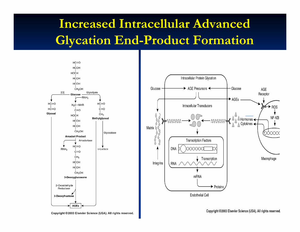
Increased Intracellular Advanced
Glycation End-Product Formation

Activation of Protein Kinase C

Increased Hexosamine Pathway

Diabetic retinopathyDiabetic retinopathy

Hyperglycemia
Pericyteloss
Hyperperfusion Capillary/Endothelial
damage
Loss ofautoregulation
Capillaryocclusion
Vasoactive
Pathophysiology of diabetic retinopathyPathophysiology of diabetic retinopathy
Vasoactivefactors
Loss of tight junction
Retinal ischemia
New vessels-Low resistance
- No pericyte/autoregulation
Growth factors
Macularoedema

Advanced diabetic eye diseaseAdvanced diabetic eye disease
Retinal ischemia
Pericyte
Neovascularitation
Preretinal Pericyteloss
Preretinal haemorrhage
Neovascular glaucoma
Vitrous haemorrhage
Retinal detachment
Blindness

Diabetic retinopathyDiabetic retinopathy
� Blindness is primarily the result of progressive diabetic retinopathy and clinically significant macular edema.
� Diabetic retinopathy is classified into two stages: nonproliferative and proliferative.
� Nonproliferative diabetic retinopathy : marked by retinal vascular Nonproliferative diabetic retinopathy : marked by retinal vascular microaneurysms, blot hemorrhages, and cotton wool spots
� The appearance of neovascularization in response to retinal hypoxia is the hallmark of proliferative diabetic retinopathy.
� Duration of DM and degree of glycemic control are the best predictors of the development of retinopathy; hypertension is also a risk factor
� The most effective therapy for diabetic retinopathy is prevention.

Diabetic nephropathyDiabetic nephropathy
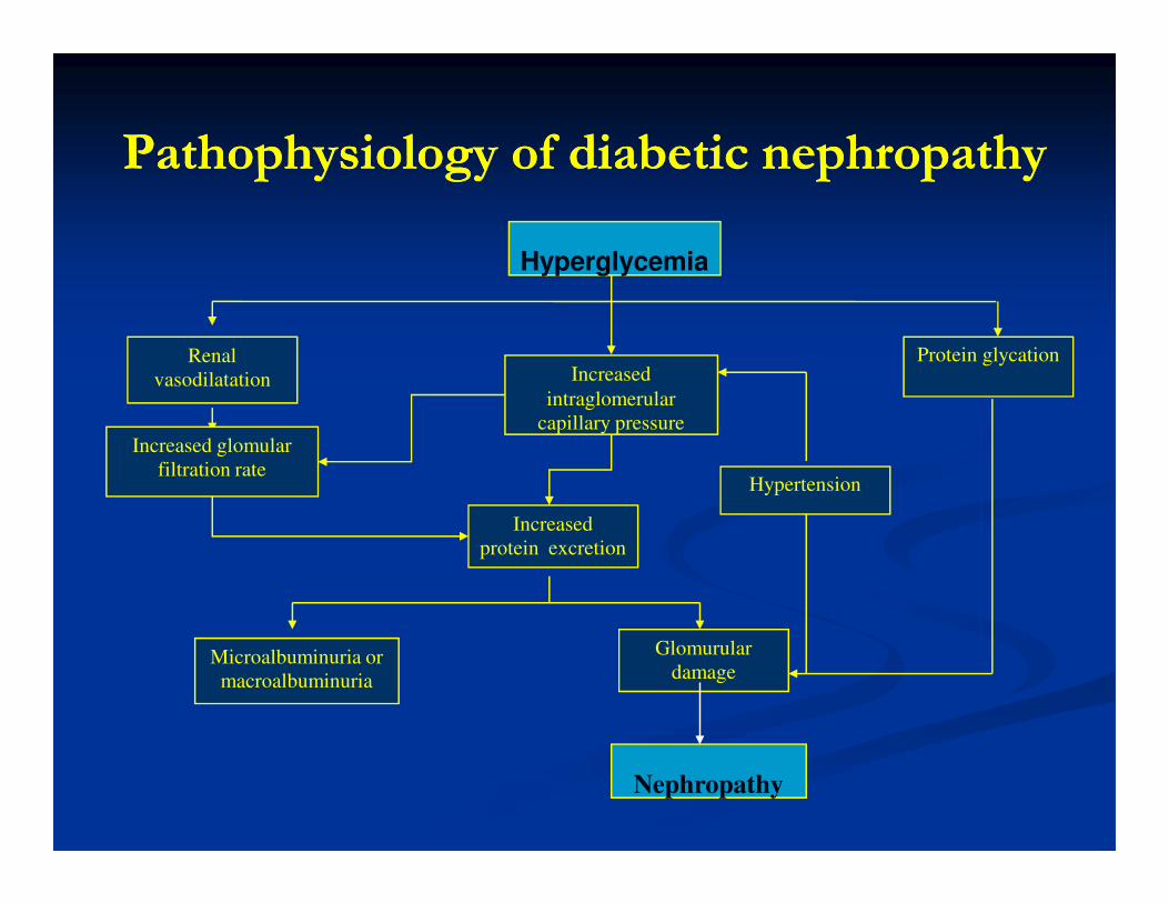
Pathophysiology of diabetic nephropathyPathophysiology of diabetic nephropathy
Hyperglycemia
Renal
vasodilatation Increased
intraglomerular
capillary pressure
Protein glycation
Increased glomular Increased glomular
filtration rateHypertension
Increased
protein excretion
Microalbuminuria or
macroalbuminuria
Nephropathy
Glomurular
damage

Diabetic nephropathy
� Diabetic nephropathy is the leading cause of ESRD in the US.
� Individuals with diabetic nephropathy almost always have diabetic retinopathy.
� The stages of diabetic nephropathy are :� Hyperfiltration
� Microalbuminuria
� Overtproteinuria
Declining GFR� Declining GFR
� End stage renal failure
� Microalbuminuria is defined as 30 to 300 mg/d in a 24-h collection or 30 to 300 g/mg creatinine in a spot collection (preferred method).
� The appearance of microalbuminuria (incipient nephropathy) in type 1 DM is an important predictor of progression to overt proteinuria (300 mg/d) or overt nephropathy.
� Hypertension more commonly accompanies microalbuminuria or overt nephropathy in type 2 DM

Diabetic nephropathy - treatment
� The optimal therapy for diabetic nephropathy is prevention.
� Interventions effective in slowing progression from microalbuminuria to overt nephropathy include: � near normalization of glycemia,� strict blood pressure control, and � administration of ACE inhibitors or ARBs, and � treatment of dyslipidemia.� treatment of dyslipidemia.
� Blood pressure should be maintained at 130/80 mmHg in diabetic individuals without proteinuria.
� A slightly lower blood pressure (125/75) should be considered for individuals with microalbuminuria or overt nephropathy
� A consensus panel of the ADA suggests modest restriction of protein intake in diabetic individuals with microalbuminuria (0.8 g/kg per day) or overt nephropathy (<0.8 g/kg per day)

Diabetic neuropathyDiabetic neuropathy

Mechanism of nerve damage in diabetesMechanism of nerve damage in diabetes
METABOLIC VASCULAR
glucose
sorbitol
myoinositol
Arterial Slow nerve
Altered membrane
potensial
sorbitol
H2O
nerve oedema NO
production
AGEformation
vasoconstriction
Arterial narrowing
Vesselocclusion
Slow nerveconduction
Impairingaxonal transport

Diabetic neuropathy
� Diabetic neuropathy occurs in approximately 50% of individuals with long-standing type 1 and type 2 DM.
� The development of neuropathy correlates with the duration of diabetes and glycemic control; both myelinated and unmyelinated nerve fibers are lost.
� Several stage :
� Intraneural biochemical abnormalities; sorbitol accumulation, � Intraneural biochemical abnormalities; sorbitol accumulation, myoinositol depletion
� Impairement of electrophysiological measurement; decreased nerve conduction velocity; asymptomatic
� Clinical neuropathy; detectable using clinical methods; maybe symptomatic. Histological changes evident
� End stage complications. Exp are ulceration and Charcot neuroarthropathy; major derangements of neural structure and function.

Clinical features symmetrical Clinical features symmetrical
sensorimotor neuropathysensorimotor neuropathy
SymptomsSymptoms
�� Loss of sensation ;Loss of sensation ;�� Anaesthesia;’numbness’Anaesthesia;’numbness’
�� Loss of pain perceptionLoss of pain perception
�� Altered sensation:Altered sensation:
SignsSigns
�� Sensory lossSensory loss
�� Diminished/absent tendon Diminished/absent tendon
reflexsreflexs�� Altered sensation:Altered sensation:
�� ParaesthesiaeParaesthesiae
�� DysaesthesiaeDysaesthesiae
�� PainPain�� BurningBurning
�� Hyperalgesia/allodyniaHyperalgesia/allodynia
�� Neuralgia Neuralgia –– lancinating painlancinating pain
�� Cramps ; restless legCramps ; restless leg
reflexsreflexs
�� Muscle wasting and Muscle wasting and
weaknessweakness
�� Autonomic dysfunctionAutonomic dysfunction
�� Foot ulerationFoot uleration
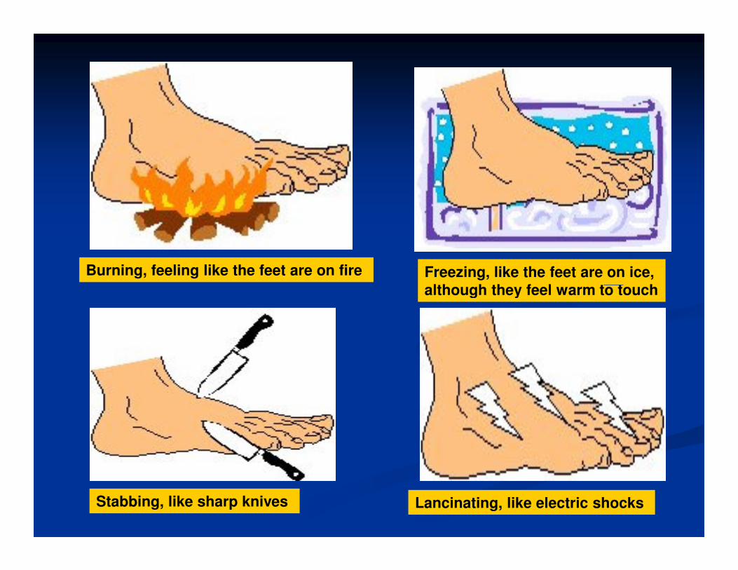
Burning, feeling like the feet are on fire Freezing, like the feet are on ice,
although they feel warm to touchalthough they feel warm to touch
Stabbing, like sharp knives Lancinating, like electric shocks

Treatment of Symmetric Treatment of Symmetric
NeuropathyNeuropathy
�� Glucose controlGlucose control
�� Pain controlPain control�� Tricyclic antidepressants Tricyclic antidepressants
�� Amitriptyline,desipramin, nortriptilin, trazodoneAmitriptyline,desipramin, nortriptilin, trazodone
�� AnticonvulsantsAnticonvulsants�� Carbamazepine, gabapentinCarbamazepine, gabapentin
�� Topical creamsTopical creams�� capsaicincapsaicin
�� Foot careFoot care

Autonomic Neuropathy
� DM-related autonomic neuropathy can involve multiple systems, including the cardiovascular, gastrointestinal, genitourinary, sudomotor, and metabolic systems.
� Autonomic neuropathies affecting the cardiovascular system cause a resting tachycardia and orthostatic hypotension.
� Gastroparesis and bladderemptying abnormalities are often caused by the autonomic neuropathy seen in DM (discussed caused by the autonomic neuropathy seen in DM (discussed below).
� Hyperhidrosis of the upper extremities and anhidrosis of the lower extremities result from sympathetic nervous system dysfunction.
� Anhidrosis of the feet can promote dry skin with cracking, which increases the risk of foot ulcers.
� Autonomic neuropathy may reduce counterregulatory hormone release, leading to an inability to sense hypoglycemia appropriately ((hypoglycemia unawareness)

Macrovascular Macrovascular
complicationscomplicationscomplicationscomplications

Macrovascular complication Macrovascular complication
� Macrovascular complications of diabetes mellitus are condition characterized by atherosclerotic occlusive disease of cerebral, myocard and lower extremities.
� Atherothrombosis is the most common cause of macrovascular complications
� Atherothrombosis is characterized by a sudden (unpredictable) � Atherothrombosis is characterized by a sudden (unpredictable) atherosclerotic plaque disruption (rupture or erosion) leading to platelet activation and thrombus formation
� Atherothrombosis is the underlying condition that results in events leading to myocardial infarction, ischemic stroke, amputation and vascular death

Atherogenesis – A Complex And Progressive Process1
Initiation:
Accumulation of lipids at vascular junctions
experiencing high shear forces
Macrophages bind to and enter intima wall
Macrophages
Inflammatory cytokines induce
expression of adhesion molecules
Pathology of Atherogenesis
Adapted from: P Libby, The Vascular Biology of Atherosclerosis, in: Braunwald
E, Zipes DP & Libby P 6th Edition, Heart Disease: a Textbook of Cardiovascular
Medicine 2001: London: WB Saunders. 2. Davies MJ. Heart 2000;83:361-66, with permission from the BMJ Publishing Group
Result: Atherosclerotic plaque2
Macrophages become foam cells & fatty streak formed
Smooth muscle cells (SMCs) migrate into the intima
Uptake of Lipids by Macrophages
Chemo-attractants such as PDGF released from activated macrophages

Atherothrombosis Has Multiple
Manifestations
Transient ischemic attack
Angina:
• Stable
Ischemic stroke
Myocardial infarction
Adapted from: Drouet L. Cerebrovasc Dis 2002;13(suppl 1):1–6
• Stable
• Unstable
Peripheral arterial disease:
• Intermittent claudication
• Rest pain
• Gangrene
• Necrosis

Macrovascular disease in diabetes Macrovascular disease in diabetes
mellitusmellitus
� Cardiovascular and cerebrovascular disease account for up 70% of death in patients with type 2 DM
� All patients with type 2 diabetes have greater predipostition to macrovascular disease, often having a constellation of risk factors, which have been term insulin resistance.
� It has been hypotethesized that insulin resistance and � It has been hypotethesized that insulin resistance and hyperinsulinemia (environmental and genetic factors), are central to development :� Glucose intolerance� Hypertension� Dyslipidemia� Coagulopathy
� These factors promote accelerated atherosclerosis, explaining the increased risk of macrovascular disease.


Diabetes and Macrovascular
Disease
Libby and Plutsky. Circulation. 2002.

Strategies for reducing macrovascular Strategies for reducing macrovascular
complicationscomplications
�� Prevention proven intervention trialsPrevention proven intervention trials�� Hyperglycemia Hyperglycemia
�� DyslipidemiaDyslipidemia�� DyslipidemiaDyslipidemia
�� HypertensionHypertension
�� Antiplatelet therapiesAntiplatelet therapies
�� Prevention suggested by epidemiologic analysisPrevention suggested by epidemiologic analysis�� Disorders of thrombolysisDisorders of thrombolysis
�� Endothelial disordersEndothelial disorders

The diabetic footThe diabetic foot

Diabetic foot diseaseDiabetic foot disease
� Approximately 15% of individuals with DM develop a foot ulcer, and a significant subset will ultimately undergo amputation (14 to 24%risk with that ulcer or subsequent ulceration).
� Syndrome of diabetic foot disease
� Peripheral neuropathy, peripheral vascular disease and tissue infection
� Risk factors for foot ulcers or amputation include: male sex, � Risk factors for foot ulcers or amputation include: male sex, diabetes 10 years’ duration, peripheral neuropathy, abnormal structure of foot (bony abnormalities,callus, thickened nails), peripheral arterial disease, smoking, history of previous ulcer or amputation, and poor glycemic control.
� The plantar surface of the foot is the most common site of ulceration.
� Ulcers may be primarily neuropathic (no accompanying infection) or may have surrounding cellulitis or osteomyelitis.

Pathophysiology of diabetic foot Pathophysiology of diabetic foot
Neuropathy
Motor dysfunction
Neuropathy Neuropathy
Abnormal
Reduced pain
Sensation and
proprioception
Microvascular disease
Abnormal
Foot posture
Cheiroarthropathy
proprioception
Increased foot prssure
Callus
Poor tissue
nutrition and
oxygenation
Ulcer
Macrovascular disease
Ischemia
Dry, cracked
skin
Arteriovenous
shunting
TraumaMechanical,
thermal,
chemical

Thank your for your attentionThank your for your attention