complications and associated conditions of celiac …
TRANSCRIPT

Sachs’ Children’s Hospital, Department of Clinical Science and Education, Södersjukhuset,
Karolinska Institutet, Stockholm, Sweden
COMPLICATIONS AND ASSOCIATED CONDITIONS
OF CELIAC DISEASE
Ola Olén
Stockholm 2008

All previously published papers were reproduced with permission from the publisher. Published by Karolinska Institutet. Printed by Universitetsservice AB © Ola Olén, 2008 [email protected] ISBN 978-91-7409-139-7

1
“Where all think alike, no one thinks very much”
Walter Lippmann
To my family

2
SUMMARY The aim of this thesis was to explore possible complications and associated conditions of celiac disease (CD) in order to shed new light on the burden of illness related to CD and to identify groups at high risk of CD, where screening for CD may be considered. We also assessed the effects of a gluten-free diet on the risk of lymphoma, an important complication of CD. Throughout the thesis, Swedish population-based registers have been used. To investigate the risk of urinary tract infections (UTI) in CD we linked the Swedish Hospital discharge register and the Medical Birth Register. We studied the risk of UTI in 829 women who had received a diagnosis of CD before they had UTI and in 895 pregnancies to women diagnosed with CD after they had UTI and compared them with 1.7 million women without a diagnosis of CD. We found a moderately increased (but not statistically significant) risk of UTI (Adjusted Odds Ratio (AOR) = 1.37; 95% CI = 0.78-2.43; p= 0.276) in women with undiagnosed CD and no increased risk of UTI in women with diagnosed CD (AOR = 1.02; 95% CI = 0.79-1.32; p = 0.864).
To assess the risk of Immune Thrombocytopenic Purpura (ITP) in CD and vice versa we used the Swedish national Inpatient Register to identify 14,347 individuals with CD (1964-2003) and 69,967 matched reference individuals. We found that individuals with CD were at increased risk of both subsequent ITP of any type (Hazard ratio (HR) = 1.91; 95% = 1.19-3.11; p = 0.008) and subsequent chronic ITP (HR 2.77; 1.09-7.04; p = 0.033). There was also a positive association between CD and prior ITP of any type (Odds ratio (OR) = 2.96; 95% CI = 1.60-5.50; p = 0.001) or with prior chronic ITP (OR = 6.00; 95% CI = 1.83 -19.66; p = 0.003). To examine the risk of subsequent sepsis in individuals with CD we used the Swedish Inpatient register to identify 15,325 individuals with a diagnosis of CD (1964-2003) and 75,249 matched reference individuals from the general population. This study showed a modestly increased risk of sepsis in patients with CD (HR = 2.6; 95% CI = 2.1-3.0; p <0.001) with the highest risk for pneumococcal sepsis (HR = 3.9; 95% CI = 2.2-7.0; p < 0.001). To study the relationship between body mass index (BMI) and CD we identified all individuals with diagnosed or undiagnosed (at time of BMI measurement) CD in the Swedish Medical Birth register and the Swedish Conscript register. In some 800,000 women and 8000 men we found that underweight was associated with undiagnosed CD (future diagnosis of CD) in both women (HR = 2.5; 95% CI = 1.6-3.7) and men (OR = 2.4; 95% CI = 1.2- 4.9). But we also found that many individuals with undiagnosed CD are overweight (9.2% of women, 14.3% of men).

3
In the last study we assessed the relationship between compliance to a gluten free diet and risk of lymphoma in individuals with CD. We linked the Swedish national inpatient register with the Swedish Cancer Registry and thus identified 59 cases of CD and of incident malignant lymphomas. In a nested case-control design, 137 controls with CD, but without lymphoma were matched to the cases by sex, age at diagnosis (± 3 years), follow up time and calendar year of diagnosis (± 3 years). We studied the medical records of all cases and controls, blinded to the case-control status and found that poor compliance was associated with a marked increase in risk of B-cell lymphoma (OR 4.74, CI 0.89-25.33) and extraintestinal lymphoma (OR 2.85, CI 0.68-11.91), whereas risk of T-cell lymphoma (OR 1.01, CI 0.32-3.15) and intestinal lymphoma (OR 0.66, CI 0.17-2.56) remained unelevated.

4
LIST OF ABBREVIATIONS CD Celiac disease HR Hazard Ratio OR AOR
Odds Ratio Adjusted Odds Ratio
AGA tTGA EMA
Gliadin antibodies Tissue Transglutaminase antibodies Endomysial antibodies

5
LIST OF PAPERS This thesis is based upon the following papers, which will be referred to by their Roman numerals:
I. Olén O, Montgomery SM, Ekbom A, Bollgren I, Ludvigsson JF. Urinary tract infections in pregnant women with celiac disease – a population cohort study. Scand J Gastro, 2007; 42: 186-193
II. Olén O, Montgomery SM, Elinder G, Ekbom A, Ludvigsson JF. Increased risk of immune thrombocytopenic purpura among inpatients with coeliac disease. Scand J Gastroenterol. 2008;43(4):416-22.
III. Ludvigsson JF. Olén O, Ekbom A, Bell M, Montgomery SM.
Coeliac disease and risk of sepsis. Gut. 2008 Aug;57(8):1074-80. Epub 2008 Feb 12
IV. Olén O, Montgomery SM, Marcus C, Ekbom A, Ludvigsson JF. Celiac disease
and Body Mass Index: A study of two Swedish general population based registers. Manuscript submitted.
V. Olén O, Askling J, Ludvigsson JF, Hildebrand H, Ekbom A, Ekstrom Smedby K. Celiac disease, compliance to a gluten free diet and risk of Lymphoma by subtype. Manuscript submitted.
6
CONTENTS 1. Introduction 2. Background
2.1. History of celiac disease 2.2. Descriptive Epidemiology
2.2.1. Prevalence of celiac disease in unselected populations 2.2.2. Prevalence of celiac disease in at risk populations
2.3. Pathogenesis 2.3.1. Genetics 2.3.2. Gluten 2.3.3. Immunological pathways 2.3.4. The multi factorial etiology of celiac disease 2.3.5. Autoimmunity, allergy, both or neither?
2.4. Clinical presentation 2.5. Diagnostics of celiac disease
2.5.1. Diagnostic criteria 2.5.2. Serologic screening tools 2.5.3. Genetic testing 2.5.4. Small intestinal biopsy
2.6. Treatment 2.6.1. Dietary guidelines 2.6.2. Why be compliant to a gluten free diet? 2.6.3. Achieving compliance… 2.6.4. Assessing compliance 2.6.5. Other treatment strategies
2.7. Screening 2.8. Complications to and conditions associated with celiac disease studied in this thesis
2.8.1. Urinary tract infections (UTI) 2.8.2. Immune Thrombocytopenic Purpura (ITP) 2.8.3. Sepsis 2.8.4. Body Mass Index (BMI) 2.8.5. Compliance to a gluten free diet and risk of lymphoma by subtype
3. Aims 4. Subjects and Methods
4.1. Setting 4.2. Data sources
4.2.1. The Swedish National Registration Number 4.2.2. Swedish Hospital Discharge (Inpatient) Register 4.2.3. Swedish Medical Birth Register 4.2.4. Swedish Conscript Register 4.2.5. Swedish Cancer Register 4.2.6. Swedish register of population and population changes

7
4.3. Study design 4.3.1. Study I (Cohort study) 4.3.2. Study II and III (Cohort study and Case-control study) 4.3.3. Study IV (Case-control study and Cohort study) 4.3.4. Study V (Nested case-control study)
4.4. Statistical analyses 4.4.1. Cox proportional hazards model 4.4.2. Logistic regression
4.4.2.1. Unconditional logistic regression 4.4.2.2. Conditional logistic regression
4.4.3. Linear regression 4.4.4. Significance testing
5. Results 5.1. Paper I (CD and UTI) 5.2. Paper II (CD and ITP) 5.3. Paper III (CD and Sepsis) 5.4. Paper IV (CD and BMI) 5.5. Paper V (CD, lymphoma and dietary compliance)
6. Discussion 6.1. Methodological considerations
6.1.1. Study design 6.1.1.1. Cohort studies 6.1.1.2. Case-control studies
6.1.2. Internal validity 6.1.2.1. Selection bias 6.1.2.2. Recall bias 6.1.2.3. Surveillance bias/detection bias 6.1.2.4. Misclassification
6.1.2.4.1. Celiac disease 6.1.2.4.2. Urinary tract infections 6.1.2.4.3. Immune thrombocytopenic purpura 6.1.2.4.4. Sepsis 6.1.2.4.5. Compliance to a gluten free diet
6.1.2.5. Confounding 6.1.2.6. Random error/Precision
6.1.3. External validity 6.2. Findings and implications 6.3. Future research
7. Conclusions 8. Acknowledgements 9. Sammanfattning på svenska 10. References
Original papers/submitted manuscripts

8
1 INTRODUCTION Celiac Disease (CD) is one of the most common lifelong disorders, affecting some 1% of both children and adults in the Western world1. CD - also known as celiac sprue, non-tropical sprue, idiopathic sprue, idiopathic steatorrhoea and gluten-sensitive enteropathy - can be defined as a permanent intolerance to the storage proteins from wheat, rye and barley (referred to as gluten) and occurs in HLA-DQ2/DQ8-positive individuals2. CD is characterized by complex adaptive and innate immune reactions that result in the characteristic chronic inflammation and atrophy of small intestinal villi, but also of general inflammation with deposition of disease specific autoantibodies in many parts of the body3. Intestinal mucosa heals when gluten is excluded from the diet. CD can manifest with a previously unsuspected range of clinical presentations, including the typical malabsorption syndrome (chronic diarrhea, weight loss, abdominal distention) and a spectrum of symptoms potentially affecting any organ or body system4, 5. CD thus resembles a multisystemic disorder with the intestine as the primary site of the disease. CD has been linked to a number of complications including malignancy6, autoimmune disorders7 and adverse pregnancy outcome8. It is however a widely under-recognized condition, and at present as few as 1/5-1/2 of individuals with CD actually have a correct diagnosis9-11. Not only cases of atypical or even clinically silent CD, but also classic cases of CD go undiagnosed and may thus be exposed to the risk of long-term complications12. Even if inexpensive, highly sensitive and specific serologic screening for CD has been available since the 1990s, the use of mass screening for CD is still under debate10, 13-15, and instead of mass screening increased alertness in high-risk groups is recommended14-16. Identification of groups at high risk of CD is therefore important. The present thesis includes some of the results from a broad epidemiological research programme, regarding CD. Specifically this thesis explores possible complications and conditions associated with CD in order to shed new light on the burden of illness related to CD and in order to identify groups at high risk of CD, where screening for CD may be considered. The effect of a gluten-free diet on the risk of lymphoma by subtype of CD is also assessed.

9
2 BACKGROUND 2.1 HISTORY Accounts of CD date back to the first century A.D.17. However, it was not until the 1940s that the link to gluten ingestion was established; Dicke, a Dutch pediatrician, observed that the condition of children with CD improved during the food shortages of World War II, only to relapse after cereal supplies were restored.18 Originally considered a rare malabsorption syndrome of childhood, CD is now recognized as a common condition that may be diagnosed at any age and that affects many organ systems19, 20. 2.2 DESCRIPTIVE EPIDEMIOLOGY 2.2.1 Prevalence of CD in unselected populations
CD is a common disease with a screening detected prevalence of approximately 1% in Western populations1. CD is common in many other parts of the world with the exception of Japan21-29. There are clear regional differences in CD prevalence both between and within countries. Except for the children of Saharawi (Arab-Berber origin) with a prevalence of CD as high as 5.6%30, the highest prevalence rates have been seen in Ireland, the United Kingdom, Sweden and Finland (1-1.5%)1. In the most recent screening study in Sweden the CD prevalence in children born during the Swedish epidemic of CD31 (who are now twelve years of age) was investigated. In 7,567 children, 2.7% were found to have CD and 0.2% were found to have latent CD32 (positive serology and increased intraepithelial lymphocytosis, but no villous atrophy). Only 1/3 of children with CD had been clinically diagnosed before the study32. After the introduction of inexpensive, highly sensitive and specific serologic screening for CD during the 1990s the prevalence of Swedish diagnosed CD has gone from 0.1-0.2% in both children and adults33, 34 in the 1980s to 0.1% in adults9 and 0.9% in children32 at present. During this period it has become evident that the “atypical” or “silent” forms of CD are more common than the “classic” form of CD with diarrhea and malabsorption. Moreover, the screening detected (not biopsy confirmed) prevalence of CD in Sweden and Finland has also increased from 0.5-1.0 % in the 1980-90s to 2-3% at present9, 11, 32, 35, 36, the reasons of which are not clear. 2.2.2 Prevalence of CD in at risk populations CD is associated with a number of different types of conditions. Disorders sharing the same HLA-type as CD such as Diabetes Mellitus type 1, disorders that arise due to malnutrition because of CD such as osteoporosis or depression and disorders that likely arise because of an increased “autoimmune pressure” such as Thyroid diseases are all associated with CD. Groups with higher prevalence of CD than the general not-at-risk population are often screened for CD. Well known high risk groups that currently undergo screening for CD in clinical practice in Sweden are for instance patients with type-1 diabetes, relatives of individuals with CD, patients with iron deficiency anemia, individuals

10
with Down’s syndrome, Turner syndrome or Williams syndrome and selective IgA deficiency37. To be able to discuss the possibility of screening new at risk populations identified in this thesis it is important to know the prevalence of CD in groups who already undergo screening. Overall, the prevalence of CD in type-1 diabetes is likely between 3% and 7%38. These findings appear to be consistent across age groups, and by the screening method. Although the magnitude of the risk for CD among patients with diabetes type 1 varies to some degree from study to study, many of these differences can be explained by issues of study design. The prevalence of CD in relatives of patients with CD is increased, both in first-degree and second-degree relatives. That prevalence varies between 2.8% and 17.2% in first-degree relatives and between 2.6% and 19.5% in second-degree relatives and the prevalence of CD appears to be generally higher in families with multiple known cases1, 38-40. There is an increased risk of CD in a number of autoimmune diseases and vice versa (e.g. thyroid disease 41-45, Addison46, 47, Diabetes type 148, 49, Sjögren’s syndrome50 and glomerulonephritis51). CD is associated with liver disease52, hypertransaminasaemia is frequently seen in CD53 and in one study some 10% of individuals with hypertransaminasaemia had CD54. There is also evidence for an increased risk of neuro-psychiatric disease in CD and vice versa (peripheral neuropathy55, mood disorders56, psychosis57 and epilepsy58). Patients with iron deficiency anemia are often screened for CD – and for good reasons. Even if the prevalence of CD varies greatly within the group of patients with iron deficiency anemia it is clearly higher than in the general not-at-risk population in all subgroups of iron deficiency anemia59. In asymptomatic patients with iron deficiency anemia evaluated by serologic testing, the prevalence of CD ranged from 2.3% to 5.0%60, 61. In contrast, the prevalence of CD in patients with iron deficiency anemia and gastrointestinal symptoms ranged from 10.3% to 15%62. CD appears also to be common in premenopausal women with iron deficiency anemia, both with and without heavy periods63. Some researchers report of an increased risk of CD in infertility64, 65, even if this association has been disputed66, 67. It is estimated that 4.6% to 13% of children with Down’s Syndrome have CD68-70. When seeking medical advice, abdominal pain, bloating, and altered bowel habit may occur in the absence of malabsorption and this picture may be indistinguishable from irritable bowel syndrome. Patients satisfying the Rome II criteria for Irritable Bowel Syndrome have a 5% risk for having undiagnosed CD as the cause of their symptoms71 and screening for CD in this group is deemed reasonable72. 2.3 PATHOGENESIS 2.3.1 Genetics
CD does not develop unless a person has alleles that encode for HLA-DQ2 or HLA-DQ8 proteins, products of two of the HLA genes2. However, one third of individuals in Western populations carry these alleles40, 73and only a fraction (3% of individuals

11
carrying DQ2) develops CD1. Thus HLA-DQ2/DQ8 is necessary, but not sufficient to develop the disease. HLA-DQ2/DQ8 have been estimated to account for up to 40% of the genetic load in CD73. Between 90 and 95% of individuals with CD express human leukocyte antigen HLA-DQ2, and the remaining 5–10% express HLA-DQ82. Identical twins have a 75% concordance rate for the disease74, whereas siblings and dizygotic twins are at the second highest risk at 7% to 20% concordance rate38, 40, 74. The strong HLA restriction in CD is related to the characteristics of gluten. HLA-DQ2/DQ8 molecules are responsible for presenting gluten peptides to T cells, thus activating gluten-specific CD4+ T cells. Gluten is rich in proline and glutamines. Proline residues help confer resistance to proteolysis by digestive enzymes, and glutamine is targeted by the ubiquitous enzyme tissue transglutaminase (tTG) for deamidation. This deamidation of glutamine to glutamic acid results in a modified gluten peptide that effectively binds DQ2 and enhances T-cell recognition, resulting in gluten-specific T cells. 2.3.2 Gluten
Storage proteins in wheat and closely related proteins in rye and barley that are responsible for activating CD are collectively called “gluten”. Gluten is the protein fraction of wheat, rye, and barley that confers the properties of stickiness and thus allows the baking of bread75. The scientific names for the gluten proteins in wheat are gliadin (an alcohol soluble prolamine) and glutenin (water soluble), both of which are disease activating. Corresponding proteins in rye and barley that activate disease are called secalin (rye) and hordein (barley). Wheat, rye and barley are closely related in the big family of grasses and they all have a high content of proline and glutamine. The high proline/glutamine content makes them resistant to degradation by gastric, pancreatic, and intestinal brush-border membrane proteases in the human intestine, which enables disease activating protein sequences to reach the proximal jejunum76. Distantly related grasses (Figure 1) such as rice and corn have low levels of proline and glutamine and never activate CD, whereas oats (with its prolamine called avenin) have an intermediate composition and only anecdotally activates CD77-81. Figure 1, Taxonomic relationships of major cereal grains82

12
2.3.3 Immunological pathways
Mucosal injury in CD follows a process of humoral and cell-mediated immune responses, a mixture between innate and adaptive immunity83, 84 (Figure 2):
Figure 2, Interaction of Gluten with environmental, immune and genetic factors in CD (Green PH, Cellier C. Celiac disease. N Engl J Med 2007;357:1731-43.).

13
A) The undigested molecules of gliadin in wheat and corresponding subfractions of proteins from rye (secalin) and barley (hordein) pass through the epithelial barrier of the intestine85, possibly during intestinal infections86 or when there is an increase in intestinal permeability for other reasons. The mechanism by which immunoreactive derivatives breach the mucosal epithelium is however poorly understood. B) Gluten peptides are deamidated by tissue Transglutaminase-2, creating epitopes with increased immunostimulatory potential87-89. The gluten peptides may also become covalently linked to tissue Transglutaminase-2 or other proteins through the enzymatic activity of tissue Transglutaminase-2. C) Deamidated peptides are presented by antigen-presenting cells (that express DQ2 or DQ8) such as dendritic cells, macrophages, or B cells to CD4+ T cells, which become activated and release mediators that ultimately lead to tissue damage90, 91. D) Help from gluten-specific T cells leads to B cell clonal expansion and release of anti-gluten antibodies. Tissue Transglutaminase-2-specific B cells might also become activated by gluten-specific T cells through intermolecular help. E) Expression of pro-inflammatory cytokines by activated T cells promotes the release of matrix metalloproteinases that cause epithelial cell damage and tissue remodeling92. F) The response to gluten also involves the innate immune system, as epithelial cells secrete IL-1593, 94 and express nonclassic MHC class I molecules in response to the stress of gluten exposure. This in turn activates CD8+ cytotoxic T cells expressing the natural killer receptors, which can target and destroy epithelial cells that carry the stress-induced molecules. The CD autoantigen, tissue Transglutaminase-2, is a ubiquitous cellular protein. Several physiological functions of tissue Transglutaminase-2 have been proposed involving receptor-mediated endocytosis, cell differentiation, apoptosis, cell adhesion and stabilization of extra–cellular matrix95. As celiac antibodies have been shown to influence the protein cross linking enzymatic activity of tissue Transglutaminase-2, their presence may interfere with the normal performance of the enzyme in the turnover of the extracellular matrix and cytokines. 2.3.4 The multi factorial etiology of CD CD is strongly associated with the HLA-alleles DQ2 and DQ8. CD is however not present (pathological expression of tissue Transglutaminase autoantibodies) at birth96 or indeed before introduction of gluten-containing foods in the diet20, and usually does not manifest before the age of 2 years97, 98. Large Finnish screening studies of CD have found a prevalence of positive tTG-titres among children of 1.5%99 and a prevalence of positive tTG-titres among adults of 2%11, indicating increasing development of autoantibodies throughout life. Moreover, the total prevalence of screening detected CD seems to have doubled in Finland during the last two decades. Improved screening methods cannot explain the increase. The environmental factors responsible for the increasing prevalence of CD remain to be identified11. The environmental factors most commonly suggested to influence risk of CD are: time of gluten introduction 100(introduction of gluten at 4-6 months of age is favorable compared to both earlier and later introduction), amount of gluten at time of gluten introduction101 (smaller amounts of gluten better than greater amounts), duration of

14
breastfeeding (better if the child is breastfed at time of gluten introduction)101, intrauterine growth restriction and perinatal infections102. Smoking is probably negatively associated with CD35, 103 (or not associated104). 2.3.5 Autoimmunity, allergy, both or neither?
CD can be considered an autoimmune disease because there is HLA restriction, autoantibodies exist (tTG), and there is a strong association with other autoimmune diseases. However, even if gluten reactive T cells have been identified in CD, the existence of autoreactive T cells (typical of autoimmune disorders) has never been demonstrated. No T cell autoantigen has ever been found. CD can also be considered a food allergy: The disease comes with gluten intake and goes with a gluten free diet, but does not induce IgE-mediated reactions. According to the nomenclature for allergic and related reactions defined by the European Academy of Allergology and Clinical Immunology that was revised and accepted by the World Allergy Organization in 2004, CD is classified as a non-IgE mediated allergic (because of the immunologic reaction) hypersensitivity105, 106. In summary, CD is a T cell-mediated disorder with a known environmental trigger that occurs in genetically predisposed individuals. Even if the academic definition of CD balances between autoimmunity and allergy, the consequences for the individual patient are fairly clear cut: CD is to be considered a food allergy (with potentially serious complications) with exclusion of gluten as the paramount mode of treatment. 2.4 CLINICAL PRESENTATION
Celiac disease can be diagnosed in both men and women, at any age, irrespective of body mass index and with almost any type of symptom. In short: He who seeks celiac disease shall find it. Symptoms and signs commonly ascribed to CD are diarrhea, weight loss, fatigue, iron deficiency anemia and other signs of malabsorption. However, while this description may be true for some individuals with CD, the majority presents with subtle gastrointestinal symptoms, extraintestinal symptoms or even no symptoms4, 5, 72. The traditional classification of CD is therefore somewhat confusing, since several screening studies have demonstrated that the so called classic form (diarrhea predominant) is not as common as the atypical form (subtle gastrointestinal symptoms or extraintestinal symptoms) or the silent form9, 10, 12, 25, 99, 107, 108s. In addition, there is the latent or potential form of CD (positive serological screening, but only minimal or no changes in the intestinal mucosa). In recent years, widespread serological testing (and improved sensitivity in serological markers) and increasing awareness of the different forms of CD has lead to a shift in the pattern of clinical presentation and an ever increasing number of cases being diagnosed 109-111. The small intestine has a considerable functional reserve and this explains why many individuals have few or no symptoms and frequently no evidence of malabsorption72. Symptomatology in CD seems to be related to the length of affected bowel, and not to the severity of the mucosal lesion112, 113. Thus, insults compromising the inherent

15
compensatory ability of the small bowel—such as worsening extent of disease, infection, ischaemia and short bowel, among other things—may suffice to unmask previously compensated CD. In patients with CD, immune responses to gluten promote an inflammatory reaction both locally in the small intestine90 and extraintestinally3, 114. Since there is also evidence of the occurrence of extraintestinal manifestations, CD should be regarded as a systemic disease and not solely involving the intestinal tract20. Since the intestinal manifestations of celiac disease are not specific for CD and are common in the general population, lists of the positive predictive value of different symptoms and signs would be very helpful when trying to assess the risk of CD in any given patient. For example, 5% of individuals fulfilling the Rome II criteria of irritable bowel syndrome have CD71. 2.5 DIAGNOSTICS OF CELIAC DISEASE
Duodenal biopsy examination remains the gold standard for diagnosis of CD. Correlation of clinical, serologic and histologic features is essential in the diagnosis and management of CD.
2.5.1 Diagnostic criteria
The European Society of Paediatric Gastroenterology, Hepatology and Nutrition established the first diagnostic criteria for CD already in 1969. In the first criteria the diagnosis was based on morphological assessment of the small intestinal mucosa, obtained on three separate biopsy occasions (Initial flat mucosa when the patient ingested gluten, clear improvement of the small intestinal mucosa on a gluten free diet and deterioration of the mucosa during gluten challenge). In the revised criteria from 1990115 the main criterion for both children37, 116 and adults117 is still a diagnostic intestinal biopsy showing typical histopathological morphology consistent with CD when the individual is on a gluten containing diet (Total villous atrophy, subtotal villous atrophy or partial villous atrophy, whereas solely increased rate of intraepithelial lymphocytes is not yet regarded as CD). There should be full clinical response when gluten is excluded from the diet. A positive serological test that reverts to normal after treatment has started, adds weight to the diagnosis, but is not sufficient to make the diagnosis without an initial small bowel biopsy. Histological improvement on a gluten free diet (GFD) is frequently sought and is recommended in adults because villous atrophy may persist despite a clinical response to the diet and despite normalized serology20, 118. A biopsy after gluten provocation should still be considered in children who are less than two years of age at diagnosis and in individuals were the diagnosis is uncertain, before introducing a GFD for life.

16
2.5.2 Serologic screening tools
Since the 1990s antibodies are important diagnostic tools in CD. In clinical practice Endomysial antibodies (EMA), tissue Transglutaminase antibodies (tTGA) and gliadin antibiodies (AGA) of the IgA-class are widely available of which EMA and tTGA have excellent performance119 (Table 1). IgA-deficiency is common in CD (2-3%)120, 121 and there is a high prevalence of CD among individuals with IgA-deficiency (8%)122. In individuals with IgA-deficiency, testing of IgG EMA and tTGA offers an alternative62,
123-125. Antibodies against gliadin (AGA) is measured by quantitative enzyme-linked immuosorbent assay and available in clinical practice. However, both EMA and tTG have superseded the use of antigliadin antibodies, which although of some use have subsequently been shown to have inferior diagnostic accuracy119, 126 (Table 1). Endomysium is a connective tissue protein found in the collagenous matrix of for instance mucosal cells. Antibodies to endomysium (EMA) can be measured in serum using an immunofluorescence technique with monkey esophagus or human umbilical cord as substrate. The stained substance is manually viewed under a microscope. As a consequence, the test is labor intensive and so requires money, time, and expertise to perform62. Transglutaminase is an enzyme located intra- and intercellularly in the intestine. In CD, tissue Transglutaminase performs targeted deamidation of gluten that results in binding to DQ2/DQ8 with higher affinity. Tissue Transglutaminase antibodies are measured using enzyme linked immunosorbent assay with guinea pig liver or human recombinant tTG as the substrate – a quantitative method that is a cheaper approach than EMA62.
Table 1, Performance of serologic screening tools in CD119. H = Significant heterogeneity by Pearson’s χ2
Analysis Sensitivity (95% CI)
Specificity (95% CI)
Prevalence of CD in tested populations
IgA EMA-ME, adult 0.974 (0.957-0.985) 0.996 (0.988-0.999) ≈40%
IgA EMA-ME, child 0.961 (0.945-0.973) 0.974 (0.963-0.982) ≈40%
IgA tTG-HR, adult 0.981 (0.901-0.997) 0.981 (0.958-0.991) ≈16%
IgA tTG-HR, child 0.957 (0.903-0.981) 0.990 (0.946-0.998) ≈53%
IgA AGA, adult 0.75-0.90 (H) 0.80-0.90 (H) ≈36%
IgA AGA, child 0.80-0.95 (H) 0.80-0.95 (H) ≈36%

17
In a review of the diagnostic accuracy of serologic tests for CD119 it was concluded that, below a CD prevalence of approximately 35% to 40% (note that the CD prevalence is ≈1% in the general population1 and that the CD prevalence seldom is more than 20%, even in groups with high risk of the disease38), the positive predictive value of IgA-EMA and tTG-based tests tends to drop from approximately 90% - 100% to approximately 80% or less. Two recent studies, conducted in clinical settings with a CD prevalence of 3.5% and 3.9% respectively, found positive predictive values of a positive EMA or tTGA of only 29-76%118, 127. Hence, positive serologic test results should be confirmed by intestinal biopsy before making a diagnosis of CD and before instituting lifelong dietary changes. The negative predictive value on the other hand (proportion of individuals that have a negative test result that do not have the disease) is very high (95-100%) up to a CD prevalence of approximately 45%, then dropping of119. Seronegative CD still occurs. For this reason duodenal biopsy in patients with a high suspicion of CD still is recommended even if the serologic testing is negative128. There are no clear guidelines as to the optimal means to monitor adherence to a GFD. Symptom improvement alone may not be enough, especially since increasing numbers of atypical or silent CD cases are being diagnosed. Moreover, multiple studies have shown that the sensitivity of EMA, tTGA or AGA is related to the grade of histological damage in CD, with decreased sensitivity in less severe histological grades129, 130. E.g. Hopper et al showed that in 48 individuals with CD on a “strict GFD” for >1 year, 16 patients had persisting villous atrophy, of which as many as 44% had normalized serology118. Optimally, remision should be based on repeat duodenal biopsy, symptom response, dietary questioning, and serologic status as a composite assessment118, 131. 2.5.3 Genetic testing
The strong association between HLA-DQ2/DQ8 has opened for genetic testing, involving typing of HLA, which has been available for some time. Since the majority of HLA-DQ2/DQ8 carriers do not develop CD, a positive test for DQ2 or DQ8 in an unselected population has a positive predictive value for CD of only 3%132. The value of the test is its high negative predictive value132-134 Correctly used, HLA-typing can contribute in defining a population not needing repeated testing over time to identify development of EMA or tTGA132. At present, HLA typing of individuals with suspected CD is not routinely used in Sweden in order to decrease the need for small bowel biopsies135. 2.5.4 Small intestinal biopsy
The small intestinal biopsy is still regarded as the gold standard when diagnosing CD116, 117, 135, 136. It is true that serological tests for CD such as EMA or tTGA are both highly sensitive and specific, and consequently some researchers have even suggested abandoning duodenal biopsies altogether137 when diagnosing CD. However, the poor positive predictive value (proportion of individuals that have a positive test result that actually have the disease, defined as villous atrophy) of serological tests in most clinical settings does not support such a development62, 118, 128. Instead, in recent years, a number of researchers have advocated an increased use of duodenal biopsies in the diagnosis and management of CD rather than the opposite118, 138.

18
Histopathologically, CD displays a range in severity. Several scoring systems for the histological evaluation of the intestinal mucosa damage have been suggested. The classification according to Marsh is usually applied130:
- Type 0: Normal small bowel mucosa (also referred to as preinfiltrative). Normal villous architecture and < 30 IEL per 100 enterocytes. Patients in this group are identified based on serologic criteria only and may never develop CD.
- Type 1: In the infiltrative lesion the mucosa has a normal villous architecture, a normal height of the crypts and the epithelium is infiltrated by an increased number of IEL representing >30 IEL per 100 enterocytes139. In earlier days >40 IEL per 100 enterocytes have been considered abnormal140, but as reflected by revised classification schemes139, there is a trend towards a lower “normal” number of IEL. Intraepithelial lymphocytosis is however relatively non-specific. With a clinical or family history and serological evidence of CD, this observation is suggestive, but not diagnostic, of the disease141.
- Type 2: The hyperplastic lesion is characterized by unaltered villous architecture, increased IEL and crypt hyperplasia. The presence of a type 2 lesion alone is sufficiently non-specific not to immediately elicit the diagnosis of CD without other factors suggestive of the diagnosis.
- Type 3a: Partial villous atrophy with minor villous blunting. - Type 3b: Subtotal villous atrophy with moderate villous blunting. - Type 3c: Total villous atrophy, i.e. no visible villi.
Although different degrees of villous atrophy can be induced by numerous diseases, villous atrophy is suggestive of CD, even without symptoms or positive serology. Figure 3: a. Normal mucosa. b. Intraepithelial lymphocytosis. c. Partial villous atrophy. d. Total villous atrophy. (Photomicrographs obtained from Prof. Åke Öst, earlier chairman of the Swedish National Steering Group for Small Intestinal Pathology)
19
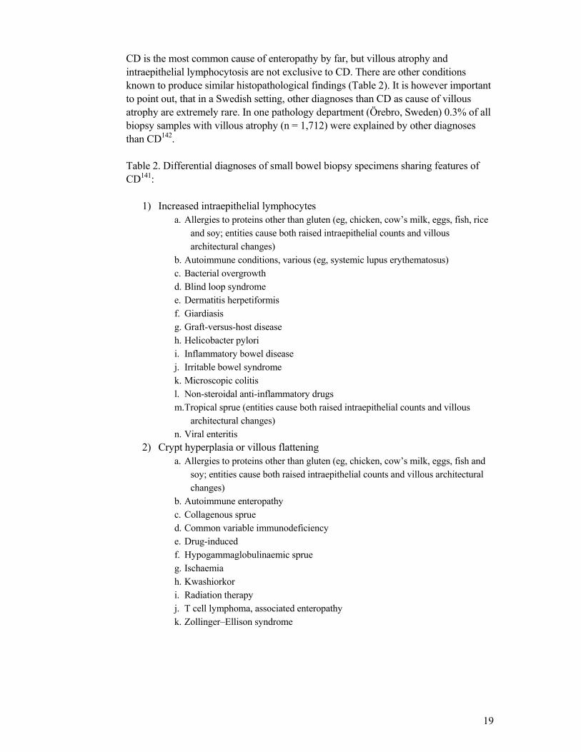
CD is the most common cause of enteropathy by far, but villous atrophy and intraepithelial lymphocytosis are not exclusive to CD. There are other conditions known to produce similar histopathological findings (Table 2). It is however important to point out, that in a Swedish setting, other diagnoses than CD as cause of villous atrophy are extremely rare. In one pathology department (Örebro, Sweden) 0.3% of all biopsy samples with villous atrophy (n = 1,712) were explained by other diagnoses than CD142. Table 2. Differential diagnoses of small bowel biopsy specimens sharing features of CD141:
1) Increased intraepithelial lymphocytes a. Allergies to proteins other than gluten (eg, chicken, cow’s milk, eggs, fish, rice
and soy; entities cause both raised intraepithelial counts and villous architectural changes)
b. Autoimmune conditions, various (eg, systemic lupus erythematosus) c. Bacterial overgrowth d. Blind loop syndrome e. Dermatitis herpetiformis f. Giardiasis g. Graft-versus-host disease h. Helicobacter pylori i. Inflammatory bowel disease j. Irritable bowel syndrome k. Microscopic colitis l. Non-steroidal anti-inflammatory drugs m. Tropical sprue (entities cause both raised intraepithelial counts and villous
architectural changes) n. Viral enteritis
2) Crypt hyperplasia or villous flattening a. Allergies to proteins other than gluten (eg, chicken, cow’s milk, eggs, fish and
soy; entities cause both raised intraepithelial counts and villous architectural changes)
b. Autoimmune enteropathy c. Collagenous sprue d. Common variable immunodeficiency e. Drug-induced f. Hypogammaglobulinaemic sprue g. Ischaemia h. Kwashiorkor i. Radiation therapy j. T cell lymphoma, associated enteropathy k. Zollinger–Ellison syndrome

20
When interpreting duodenal biopsy specimens a few things are important to keep in mind:
- There is a risk for variability in the interpretation of biopsies, especially if the specimens are inadequately oriented, obliquely cut or not optimally stained143. Optimally two independent reviewers should assess all biopsy specimens in candid communication with the clinician144.
- In assessing villous height and crypt depth, it is necessary to identify at least 3 or 4 intact adjacent villi that are cut perpendicularly. Tangentially cut sections lead to an artificial appearance of villous atrophy and a potential overdiagnosis of CD145.
- Lesions associated with CD sometimes have a patchy distribution, which make false negative duodenal biopsy results possible146, 147.
- There are natural differences in villous architecture across populations that can be dramatic. Variants of villous morphology include141:
1) Finger-like, with a cylindrical core and rounded apex 2) Leaf-like, with a broad flattened base with a tapering apex 3) Tongue-like, with a broad flattened base and rounded apex 4) Ridge-like, with a flat linear base that is less in width than its height.
- In the duodenum it is not unusual to see branched villi or villi containing fused tips. Mixed populations of villi are common.
- Gastric metaplasia, gastric heterotopia and heterotopic pancreas can be observed within the small bowel141.
- Biopsy forceps crush and destroy tissue and thus evaluation of specimen margins should be done with caution; in addition to damaging cells, this mode of tissue procurement may introduce artefactual haemorrhage in the sample. Superficial biopsy specimens lacking a muscularis mucosa can cause artefactual separation of the villous bases, resulting in the appearance of shorter and thicker villi.
Given the heterogeneous distribution of lesions in CD and normal differences in small bowel histology, four to six biopsy specimens are recommended to ensure that decent-sized specimens are obtained for analysis and that patchy changes are less likely to be missed148. It is important that the clinician provides a description of the location and gross appearance of the area sampled147. Despite this practice, false negatives can occur146. If the clinical suspicion is high, repeat duodenal biopsy examination or sampling of more distal small bowel should be considered. Healing of the small bowel mucosa proceeds in a caudal to cephalad direction. This may take anywhere from 6 to 24 months after induction of treatment and in some cases the extent of recovery may remain incomplete149.
21
2.6 TREATMENT 2.6.1 Dietary guidelines
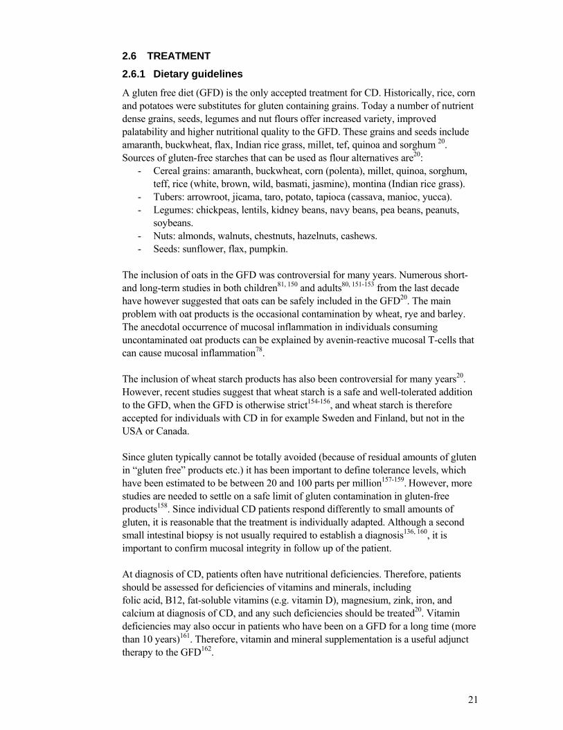
A gluten free diet (GFD) is the only accepted treatment for CD. Historically, rice, corn and potatoes were substitutes for gluten containing grains. Today a number of nutrient dense grains, seeds, legumes and nut flours offer increased variety, improved palatability and higher nutritional quality to the GFD. These grains and seeds include amaranth, buckwheat, flax, Indian rice grass, millet, tef, quinoa and sorghum 20. Sources of gluten-free starches that can be used as flour alternatives are20:
- Cereal grains: amaranth, buckwheat, corn (polenta), millet, quinoa, sorghum, teff, rice (white, brown, wild, basmati, jasmine), montina (Indian rice grass).
- Tubers: arrowroot, jicama, taro, potato, tapioca (cassava, manioc, yucca). - Legumes: chickpeas, lentils, kidney beans, navy beans, pea beans, peanuts,
soybeans. - Nuts: almonds, walnuts, chestnuts, hazelnuts, cashews. - Seeds: sunflower, flax, pumpkin.
The inclusion of oats in the GFD was controversial for many years. Numerous short- and long-term studies in both children81, 150 and adults80, 151-153 from the last decade have however suggested that oats can be safely included in the GFD20. The main problem with oat products is the occasional contamination by wheat, rye and barley. The anecdotal occurrence of mucosal inflammation in individuals consuming uncontaminated oat products can be explained by avenin-reactive mucosal T-cells that can cause mucosal inflammation78. The inclusion of wheat starch products has also been controversial for many years20. However, recent studies suggest that wheat starch is a safe and well-tolerated addition to the GFD, when the GFD is otherwise strict154-156, and wheat starch is therefore accepted for individuals with CD in for example Sweden and Finland, but not in the USA or Canada. Since gluten typically cannot be totally avoided (because of residual amounts of gluten in “gluten free” products etc.) it has been important to define tolerance levels, which have been estimated to be between 20 and 100 parts per million157-159. However, more studies are needed to settle on a safe limit of gluten contamination in gluten-free products158. Since individual CD patients respond differently to small amounts of gluten, it is reasonable that the treatment is individually adapted. Although a second small intestinal biopsy is not usually required to establish a diagnosis136, 160, it is important to confirm mucosal integrity in follow up of the patient. At diagnosis of CD, patients often have nutritional deficiencies. Therefore, patients should be assessed for deficiencies of vitamins and minerals, including folic acid, B12, fat-soluble vitamins (e.g. vitamin D), magnesium, zink, iron, and calcium at diagnosis of CD, and any such deficiencies should be treated20. Vitamin deficiencies may also occur in patients who have been on a GFD for a long time (more than 10 years)161. Therefore, vitamin and mineral supplementation is a useful adjunct therapy to the GFD162.

22
Even if underweight is more common among individuals with yet undiagnosed CD than in the general population163, normal weight and overweight is by far the most common body composition in adult CD163, 164. The small intestine has considerable functional reserve and this explains why many individuals have no evidence of malabsorption165. Moreover, studies of adolescents have indicated an increased risk of obesity when adhering to a GFD166. It has been suggested that when individuals start a GFD the mucosa heals but the total food intake/energy intake remains the same. In addition to this the GFD in itself can be nutritionally imbalanced with high lipid consumption166. Consequently, individuals on a GFD should be monitored with respect to BMI. 2.6.2 Why be compliant to a gluten free diet?
In the vast majority of patients with CD, strict compliance to a GFD in CD results in healing of the intestinal mucosa (malabsorption stops) and likely in decreased general inflammation114. For children and adults with symptomatic gastrointestinal CD, benefits of compliance to a GFD are obvious and often swift clinical improvement with normal bowel habits and normalization of anthropometric values is seen. A GFD will lead to significant improvement in bone density167, 168. It also corrects iron deficiency169 and restores growth in children with CD170. Benefits of a GFD in diabetes type 1 on hemoglobin A1c has not been conclusively demonstrated171, 172. Compliance to a GFD improves quality of life in CD patients with gastrointestinal symptoms173. Studies from different countries have reported positive, negative or no effect173-175 of a GFD on quality of life in asymptomatic, screening detected CD. The long term effects of compliance to a GFD includes reduction of the increased mortality176, risk of lymphoma177 and risk of adverse pregnancy outcomes8 seen in individuals with CD. It is however important to point out that the direct evidence of a protective effect of a gluten-free diet against complications such as non-Hodgkin lymphoma178-180 or autoimmune disease181, 182 has not been formally proven. The assumption that dietary compliance protects against complications is often based on data on duration of gluten exposure47, which per se is very difficult to disentangle from age at CD diagnosis181, 183. In studies that have actually examined the effect of dietary compliance, data on compliance have often been collected retrospectively through patient chart reviews and without the data collector being blinded to the outcome (e.g. cancer vs. not cancer). This increases the risk of bias. Finally, few studies have had sufficiently long follow up to examine the effect of a GFD in CD diagnosed in childhood regarding outcomes commonly seen late in life (e.g. cancer, myocardial infarction, fractures and death). Moreover, the absolute majority of studies assessing the long term effects of CD and a GFD have been restricted to “classic” CD with gastrointestinal symptoms. Information regarding the long term effect of a GFD in “silent” CD, detectable only by screening, is however lacking.

23
2.6.3 Achieving compliance…
Achieving compliance to a GFD is difficult. Reasons for transgressions include poor palpability of gluten free foods, absence of acute symptoms after “cheating”, difficulties finding GFD in social contexts outside the home, high cost of GFD in many countries and contamination of products claimed to be gluten free184. It is recommended that a team approach to the follow-up of the newly diagnosed CD patient include regular supervision by an interested physician, medical nutritional counseling by a dietician and access to local and national support groups184, 185. Since the GFD is complex and can easily overwhelm the patient it is reasonable to complete nutritional education in multiple visits. An ambitious educational program concerning the GFD at the start is important, since dietary compliance and intestinal damage at follow up can be predicted by baseline education186. Compliance of individuals diagnosed in adolescence or adulthood and symptomless individuals diagnosed through screening is often described as low. In a Swedish study, adults diagnosed with CD at an age of <4 years were 80% compliant, whereas adults diagnosed with CD at > 4years of age were 36% compliant). In Italy adolescents with symptomless CD diagnosed through mass screening showed lower compliance in comparison with age-matched patients diagnosed with symptomatic CD187 whereas a Finnish study found good dietary compliance also in adults with screening detected CD188. 2.6.4 Assessing compliance
Once a diagnosis of CD has been made, there is no single method that allows for assessment of compliance: Self-reported compliance is often overrated. Asymptomatic CD patients cannot be followed for symptom response. EMA and tTG titers have been used as proxies for dietary compliance, but there is no agreement in the literature that EMA189 or tTG118 are reliable markers in monitoring compliance or histological response to treatment. In a study by Hopper et al, 48 patients were treated for one year with a GFD. 16/48 patients had persisting villous atrophy and as many as 7 of the 16 patients with villous atrophy (44%) had “false” negative tTG serology118. However, duodenal biopsies also have limitations: Persisting mucosal damage may be a result of refractory CD rather than poor compliance190 (Refractory CD and duodenal cancer are however very rare in comparison with “ordinary CD with low dietary compliance131, 190). Moreover, normal mucosa may mirror the patchy occurrence of mucosal injury in active CD146 rather than a completely healed mucosa. Optimally, compliance should be estimated through a combination of duodenal biopsy 6-12 months after introduction of a GFD, symptom response, dietary questioning, and serologic status as a composite assessment118, 131. 2.6.5 Other treatment strategies
Therapy of CD is usually straightforward with a strict GFD as the “only” medication. However, in the unusual cases of refractory CD, corticosteroids or immunosuppressive drugs can be tried20, 190. Moreover, alternative treatment strategies may well be available in the future: The structure of transglutaminase 2 was recently discovered and

24
may help in designing inhibitors of transglutaminase 2 to treat CD191. Another potential treatment strategy is to ingest enzymes that digest gluten192, thereby increasing the safe threshold for gluten intake157, 158. 2.7 SCREENING
As mentioned earlier, the majority of individuals with CD are undetected (and untreated) and may suffer risks of long term complications. Ever since cheap and highly effective serologic screening for CD has been available, the need for general population screening has been under debate10, 193, 194. At present there is fairly widespread consensus that screening for CD in the general population is NOT warranted – instead increased alertness in high risk groups is called for14, 15. Here follows a brief summary of what to think of when considering screening: The crucial questions in considering general population screening are whether we are doing more harm than good for the individuals being diagnosed and if the public means are spent on the right things. Guides published already 30 to 40 years ago still provide a relevant framework that can be applied to the issue of screening individuals for CD195,
196. Criteria that need to be fulfilled to consider screening for a disease are:
1. The disease should be an important health problem, i.e. high prevalence and/or a serious condition.
2. The diagnostic criteria should be generally accepted. 3. Screening tests should be available and acceptable to the public. 4. The natural history should be understood, with advantage to earlier treatment
and agreement on who needs treatment and when. 5. Treatment for the disease should be accepted and available. 6. Cost-benefit favorable. 7. Needs for repeat testing should be clear. 8. Predictors of response to treatment.
CD is an important health problem with a screening detected prevalence of 1-3% in Sweden, the majority of which remains undiagnosed. Apart from the well documented array of symptoms and decreased quality of life in untreated CD, it is also a condition associated with a number of very serious complications including sepsis, lymphoma, fractures, depression and increased mortality. Diagnostic criteria of CD are clear cut37,
62, 97, 117 (even if algorithms for the detection of CD without a biopsy have been suggested137) and excellent screening tests are available and acceptable to the public119. However, most studies on covering the total burden of CD have included only individuals with symptomatic/clinically diagnosed CD, whereas there is limited data on the natural history of CD detected by screening and consequently on the benefit of early treatment in this group. When asymptomatic patients are encouraged to withdraw gluten from their diet lifelong, this may instead increase the burden of disease and impair quality of life. When it comes to costs, a major concern in screening for CD is that a single positive test for tTGA or EMA has a positive predictive value for biopsy confirmation of CD (villous atrophy) of only approximately 30%–80%118, 127, 128. This means that in a

25
population based screening program thousands of people unnecessarily would have to go through an upper endoscopy, which would be both troublesome for the individual and relatively expensive. The optimal age at which to begin and to repeat testing is undefined. Some reports have suggested that testing before the age of 3 years does not seem warranted197 and that screening school-age children would be likely to detect most, but not all, cases14. The benefits of treatment in individuals with symptomatic CD are obvious and well described. Results from reports assessing short term effects of a GFD in screening detected CD are both reassuring188 and disappointing198. Screening for CD has not been conducted long enough to be able to assess the long term effects of GFD in the group of symptomless CD. In summary, there are limited data on the natural history of CD detected by screening in both children and adults. It is also unclear whether early or preventive treatment alters natural history. Therefore general population screening for CD is NOT warranted at present. Instead increased alertness and screening for CD in high risk populations is recommended. 2.8 COMPLICATIONS TO AND CONDITIONS ASSOCIATED WITH CD
STUDIED IN THIS THESIS
CD is a widely underdiagnosed condition and therefore screening for CD in risk groups of the disease is recommended14, 15, 37. The identification of possible risk groups eligible for screening for CD is therefore important. In this thesis we have explored possible complications and associated conditions of CD in order to shed new light on the burden of illness related to CD and to identify groups at high risk of CD, where screening for CD may be considered. We also assessed the effects of a gluten-free diet on the risk of lymphoma, an important complication of CD. Given the immunological character of CD, we have investigated the possibility that patients with CD are at increased risk of both infections (Paper I and III) and other autoimmune diseases (Paper II). Since CD is a condition characterized by malabsorption it has also been reasonable to investigate the association of BMI and a future diagnosis of CD (Paper IV). The widespread belief that treatment of CD with a GFD may positively affect the risk of long-term complications of CD, such as lymphoma, is based on few, small studies that suffer from a number of limitations7, 178-180. Hence, this association was also studied (Paper V). 2.8.1 Urinary tract infections
Urinary tract infection (UTI) is the most common bacterial infection during pregnancy199. Among pregnant women, some 1-4% will develop acute cystitis for the first time whilst pregnant200 and 1- 2% will develop pyelonephritis201. UTI, especially pyelonephritis, may also be an indicator of immunosuppression202.

26
To the best of our knowledge, only two studies on CD and the risk of UTI have been published203, 204. The former studies are limited by the small sample size and retrospective data collection. We used the Swedish National Registry data to assess the risk of UTI in pregnant women (a) who had a diagnosis of CD prior to infant birth, (b) those who received a diagnosis of CD after infant birth and (c) women without a diagnosis of CD. 2.8.2 Immune Thrombocytopenic purpura
Immune Thrombocytopenic Purpura (ITP) is an autoimmune disease205, with an annual incidence of approximately 5/100,000 person-years206-208. ITP is divided into an acute or a chronic form (more than 6 months’ duration)205. The acute form is usually self-limiting and primarily found in previously healthy children. The chronic form occurs primarily in adults209 and often has an insidious onset209. The primary cause of long-term morbidity and mortality is haemorrhage (serious bleeding occurs in 3-10% of patients)209, 210. Treatment ranges from careful monitoring to oral prednisone, intravenous immune globulin and splenectomy208. A number of case reports support a positive association between CD and (ITP)211-220. Despite these findings, no large study has been undertaken to evaluate the relationship between CD and ITP221. The objective of this study was to examine the relationship between an inpatient diagnosis of CD and an inpatient diagnosis of ITP, and vice versa, through linkage with the Swedish national population- based registers. 2.8.3 Sepsis
Sepsis is an infectious disease characterized by a systemic inflammatory response with tachycardia, fever, hyperventilation and affected white blood cell count222. Several factors may predispose to an increased tendency of severe infection in CD such as increased risk of hyposplenism223-227 and increased mucosal permeability228-231 . With the exception of one mortality study232 (showing a 7-fold increased risk of death from sepsis among individuals with CD), we know of only case-reports of severe infection in CD233-235. In the mortality study, data on sepsis were limited to information from death certificates232. That investigation also failed to consider the underlying etiology of sepsis. For these reasons, we assessed the risk of sepsis in individuals with CD by use of Swedish national inpatient register data. 2.8.4 BMI (Underweight and overweight)
The classic presentation of CD is commonly described as diarrhoea, anaemia and weight loss4, 5, 128 but to the best of our knowledge, there are no studies that assess the association between Body Mass Index (BMI) and undiagnosed CD.

27
The few studies describing the BMI-characteristics in individuals at diagnosis of CD35,
164, 236 and/or after introduction of a gluten-free diet (GFD)164, 188, 236-238 show contradictory results164, 236 or suffer from limitations such as low study power, and lack of reference groups. Another reason to examine the relationship between CD and BMI is that many case reports165, 239-245 and one larger study 246 suggest that a diagnosis of CD may be delayed considerably due to overweight or obesity in patients with CD. The main objective of this study was to examine BMI in individuals with undiagnosed CD in two large, general population-based registers and to assess how BMI may contribute in identifying individuals with undiagnosed CD. 2.8.5 Compliance to a gluten free diet and risk of lymphoma by subtype
CD is associated with a 3 to 6-fold increased risk of malignant lymphoma of any type6,
180, 247-249. There is a widespread belief that treatment of CD with a GFD may positively affect the risk of long-term complications of CD, such as lymphoma250, 251. However, only a few studies have directly evaluated the role of dietary compliance in lymphoma development 7, 178-180. Whereas two of these studies have found an increased risk of lymphoma in individuals with poor compliance to a GFD compared to good dietary compliance178, 180, another study found no such difference179. Previous reports are limited by small sample size7, 178-180 and retrospective data collection that was unblinded to lymphoma status178. To the best of our knowledge, no earlier studies have compared the potential effect of a GFD on lymphoma risk in CD by subgroups of lymphoma20. To assess the relationship between diet compliance and risk of lymphoma overall and lymphoma subtypes in individuals with CD, we conducted a case-control study, nested in a Swedish population-based cohort of 11,650 individuals with an inpatient diagnosis of CD.

28
3 AIMS
The overall aim of this thesis was to identify groups with high risk of CD and vice versa. We also wanted to assess the effect of compliance to a GFD on the risk of lymphoma in CD. The specific aims were: To investigate if there is a difference in risk of UTI during pregnancy or of repeated episodes of UTI before pregnancy between (I) women who had a diagnosis of CD prior to infant birth, (II) women who received a diagnosis of CD after infant birth and (III) women who never received an inpatient diagnosis of CD (Study I). To investigate if there is an increased risk of an inpatient diagnosis of ITP in individuals with an inpatient diagnosis of CD and vice versa. To investigate if there is an increased risk of an inpatient diagnosis of sepsis in individuals with an inpatient diagnosis of CD and vice versa. More specifically we aimed to investigate the risk of an inpatient diagnosis of pneumococcal sepsis in CD.
To investigate the prevalence of underweight, normal weight and overweight in young men and fertile women with an inpatient diagnosis of CD, and to assess the association of underweight, normal weight and overweight with a future inpatient diagnosis of CD. To assess the relationship between diet compliance and risk of lymphoma overall and lymphoma subtypes in individuals with CD, and to explore the potential importance of CD phenotypes at diagnosis for risk of lymphoma.

29
4 SUBJECTS AND METHODS 4.1 SETTING
The studies in this thesis were all conducted in Sweden, a country well suited for epidemiological research252. The most important factors for successful epidemiological research in Sweden have been the use of national registration numbers assigned to all Swedish citizens combined with the existence of nation-wide high-quality health and population registers based on these registration numbers. The public health care system with transparent referral systems, virtually no private institutional care, an ethnically and socio-economically homogenous population and a generally high public acceptance to registration and participation in research projects have ensured that the registers are population-based. 4.2 DATA-SOURCES 4.2.1 The Swedish National Registration Number
Since 1947 every legal resident of Sweden is assigned a national registration number (or personal identification number) as a unique ten-digit (nine-digit at introduction) personal identifier, which is used in a wide variety of contexts, including health care253. The national registration number makes it possible to establish links between different registers. The national registration number consists of six digits for the birth date (year - month - day), followed by three digits that identify the individual and a tenth digit which is a check digit. 4.2.2 The Swedish hospital discharge (Inpatient) register (study I-V)
Individuals with a discharge diagnosis of CD have been the study base in all studies in this thesis. The Swedish hospital discharge (Inpatient) register was established by The National Board of Health and Welfare in Sweden in 1964. The registration is based on individual discharges rather than on individuals and each patient record contains the patient’s unique national registration number, date of admission and discharge, one main diagnosis and up to seven contributory diagnoses (coded according to the International Classification of Diseases, seventh through tenth revision). In addition to the above; surgical procedures, department and hospital of admission are recorded. The coverage of the register was 60% of the Swedish population in 1969, 85% in 1983 and 100% since 1987 and onwards. The accuracy of diagnoses in the Swedish hospital discharge register is generally regarded as high254, but the accuracy naturally differs between diagnoses (and calendar periods for some diseases). In a subset of adults with lymphoma, the diagnosis of CD was correct in 85%255. 4.2.3 The Swedish medical birth register (study I and IV)
The Swedish Medical Birth Registry was established in 1973 to compile information on ante- and perinatal factors, and their importance for the health of the infant. More than 99% of all deliveries in Sweden (85,000-120,000 deliveries per year) from 1973 are accounted for in the registry256. The registry contains individual data on

30
previous gestation, smoking habits, medication, family situation, hospital, length of gestation, type of delivery, diagnoses of mother and the newborn child, operations, type of analgesia, sex, weight, length, size of head, birth-conditions, place of residence, nationality, etc256. Since its start in 1973, the content of the Medical Birth Registry has varied slightly and a revised procedure for recording ante- and perinatal data came into effect in 1982. Data in the registry are collected from standardized medical records completed by medical personnel throughout pregnancy and after delivery. A recent validation of the Medical Birth Register found that most variables in the register are of very high quality256. 4.2.4 The Swedish conscripts’ register (study IV)
The Swedish conscripts’ register (or the National Service Register) contains personal information of all Swedish men (and women volunteering) attending conscription. Variables in the register are national registration number, year (before 1997) or date (after 1997) of conscription, height, weight, cognitive test results and test results of strength (hand, biceps, quadriceps) and endurance (test bicycle) among others. Even though The Military Archives (“Krigsarkivet”, founded in 1805 and keeper of military records from the 16th century and onwards) has been responsible for inspection and control of military records since 1943, easily accessible, computerized records of Swedish conscripts is only available since 1983 and onwards257 (records on microfilm are accessible since the middle of the 1960s and onwards) and administered by The National Service Administration which is also responsible for conscription, entrance assessments, enlistment and reporting on persons serving in the Total Defence. Until the middle of the 1990s virtually all Swedish men attended conscription, which is regulated by law. Since then all men are no longer called to attend conscription because of changed defense political priorities. The proportion of young Swedish men attending conscription has declined between 1995-2000 (98% of all men in 1996, 90% in 1999 and 80% in 2000)257. Both coverage and quality of the register data is excellent until 1995 (<1% data irregularities and <6% data missing in all variables)257. 4.2.5 The Swedish cancer register (study V)
Since 1958 it is mandatory for both hospital departments and histopathological laboratories in Sweden to report all malignancies (with the exception of some diagnoses that have been included in the register later than 1958) to the Swedish Cancer Register at the time of diagnosis. Reports are sent from hospital departments and pathologists to one of six regional oncology centers for quality checks and (re-)coding according to the International Classification of Diseases, seventh revision (ICD-7). Some 99% of the registered cases are morphologically verified258 and the completeness of the registrations has been estimated to be close to 100%258. Since 1958, some 2,500 cases of cancer have been identified through the Cause of Death Register (without having been reported to the Cancer Register). As a comparison, more than 50,000 cases of cancer were reported to the Cancer Register in the year 2005 alone. For malignant

31
lymphoma, detailed information regarding classification within the groups of non-Hodgkin lymphoma and Hodgkin lymphoma has only been available the last few years. No information of stage at diagnosis is recorded. Cancers incidentally detected at autopsy are flagged and cancers only reported on death certificates are not included. 4.2.6 The register of population and population changes (study I-IV)
Statistics Sweden maintains the register of population and population changes since 1960. The register contains official Swedish census data of all residents in Sweden alive at the end of each year (national registration number, name, current address and date of death in subjects recently diseased). Since 1969, the register also collects information on dates of emigration. Data are computerized since 1967 and are collected and updated by the local tax offices. 4.3 STUDY DESIGN
In this section the study design of each study is summarized. The corresponding references are given in the individual papers. 4.3.1 Study I
The first study is a historical cohort study with prospective registration of data. We combined data from two Swedish national medical registries, the Swedish hospital discharge register and the Medical Birth Registry, in order to:
1) analyze the risk of urinary tract infection (UTI) during pregnancy in some 1000 pregnant women with a discharge diagnosis of CD compared to all women in the register without such a diagnosis.
2) analyze the risk of repeated episodes of UTI before pregnancy in some 700 pregnant women with a discharge diagnosis of CD compared to all women in the Medical birth register without a discharge diagnosis of CD.
Information regarding exposure (Discharge diagnosis of CD before first pregnancy, Discharge diagnosis of CD after first pregnancy and No Discharge diagnosis of CD (reference group)) as well as outcome (UTI) has been collected prospectively by “blinded” medical personnel (with no knowledge of the present study). In separate analyses, we adjusted for potential confounders: Diabetes mellitus, maternal age at delivery, parity, nationality and calendar period. From 1982 to 1983 and onwards we also had data on civil status and smoking status (non-smoker versus smoker). Logistic regression was used to calculate odds ratios (ORs) and 95% confidence intervals (95% CI) for UTI in women with CD. 4.3.2 Study II and III
The second and third studies both comprise historical cohort studies and case-control studies with prospectively collected data. In a cohort study of all individuals with discharge diagnoses of CD (some 15,000) and some 70,000 reference individuals (without such a diagnosis) matched for age, gender, calendar year and county. Cox

32
regression was used to estimate the risk of subsequent discharge diagnoses of Immune Thrombocytopenic Purpura (ITP) (study II) and sepsis (study III). In a case control design, conditional logistic regression was used to assess the risk of exposure (diagnosis of ITP or sepsis prior to CD) in 15,000 cases (individuals with diagnoses of CD) and 70,000 matched controls. Diagnoses of CD as well as ITP and sepsis were identified through the Swedish Hospital Discharge Register. 4.3.3 Study IV
In order to investigate the impact of CD on body composition, we studied the prevalence of underweight, normal weight and overweight in cohorts of young men and women at conscription for some 8,000 men and at 10 weeks gestation for some 800,000 women. The differently exposed cohorts studied were individuals that 1) had a discharge diagnosis of CD before the measurement of BMI, 2) had a discharge diagnosis of CD after the measurement of BMI and 3) had no discharge diagnosis of CD. In another part of this study we carried out a cohort study of women to assess the risk of receiving a diagnosis of CD when having a certain BMI. Cox regression was used to assess the risk of CD. We also carried out a case-control study of men to assess the risk of being diagnosed as a celiac dependent on the individual’s BMI. Logistic regression was used to assess the risk of CD in underweight, normal weight and overweight. 4.3.4 Study V
In this nested case-control study, we identified all individuals with a hospital discharge diagnosis of CD between 1964-1995 (n = 11,650) in the Swedish Hospital Discharge Register. Using the National Registration Numbers the cohort was linked to the Swedish Cancer Register. Thus 77 cases of lymphoma and 220 individually matched controls were identified. After exclusion of individuals whose diagnosis of CD could not be confirmed upon review of medical files, 59 cases of incident lymphoma and137 cohort controls remained. Two investigators (OO and HH) reviewed all medical records independently and blinded to the case-control status. The blinding was achieved by removing all information before file review (KES and research assistants) that could give hints of the case or control status. The degree of compliance was evaluated through global assessment of all the prospectively collected information on compliance available from the study. In 21% of study participants the two investigators had differing assessments. Still blinded to case-control status the two investigators reviewed these medical records again and discussed the assessment until consensus was reached. Conditional logistic regression was used to calculate odds rations (OR) as estimates of relative risk.

33
4.4 STATISTICAL ANALYSES 4.4.1 Cox proportional hazards model (studies II-IV)
Cox proportional hazards model is the most commonly applied model in medical time-to-event studies259. Cox regression is a semi-parametric model that implements the proportional hazards model, i.e. constant size of difference between groups over time. One or more predictor variables, called covariates, are used to predict an outcome (event) variable. The classic univariate example is time from diagnosis with a terminal illness until the event of death (hence survival analysis). The central statistical output is the hazard ratio (HR). Hazard is the risk of an outcome in a certain time interval, assuming “survival” to that time. The hazard ratio is the relative hazard, when two groups (exposed and unexposed) are compared and assumes proportional hazards. If a covariate fails this assumption, estimates of relative risk will be inaccurate. For covariates with hazard ratios that increase over time, relative risk will be overestimated and for hazard ratios that decrease over time, relative risk is often underestimated. Hazard Ratios for subsequent ITP or Sepsis (studies II and III) were estimated using an internally stratified Cox regression. This analysis resembles a conditional logistic regression (see below) as individuals with CD are compared with their matched reference subjects (the internal strata or risk-sets) before the estimates are summarized. Follow-up time started at study entry and ended on the date of first discharge diagnosis of ITP or Sepsis, date of emigration, death or the end of the study period (31st December 2003), whichever occurred first. When we estimated the association between BMI and future CD in women (study IV) we also used Cox regression, but without internal stratification. 4.4.2 Logistic regression (studies I -V)
Logistic regression is a form of regression which is used when the independents are of any type (continous and/or categorical) and the dependent is a dichotomy (hence the method of choice in most case-control studies). Continuous variables are not used as dependents in logistic regression. The impact of predictor variables is usually explained in terms of odds ratios. Whereas risk can be defined as the number of patients who develop an outcome divided by the number of patients at risk (risk = p/1, where p is the probability of the event of the study), odds can be defined as the number of patients who develop an outcome divided by the patients who do not develop the disease (odds = p/(1-p)). The odds ratio is simply the cases’ odds of having been exposed to a risk factor divided by the controls’ odds of having been exposed to the same risk factor. 4.4.2.1 Unconditional logistic regression (study I and IV)
Unconditional logistic regression was used to calculate odds ratios and 95% confidence intervals for UTI in women with CD (study I). Unconditional and conditional logistic

34
regressions (separate analyses) estimated the association between BMI and undiagnosed (future diagnosis of) CD in men. 4.4.2.2 Conditional logistic regression (study II-V)
In matched case-control studies, conditional logistic regression is used to investigate the relationship between an outcome of being a case or a control and a set of prognostic factors. In conditional logistic regression every case is compared with his or her individually matched controls, thus minimizing the effect of the matching variables. In matched case-control studies, odds ratios could also be calculated by an unconditional multivariate logistic regression where all the matching variables are included in the analysis. However, if unconditional logistic regression is used instead of conditional, an overestimate will be obtained. In particular for pair-matching, the estimated odds ratio may reach the square of the estimated odds ratio from the conditional logistic regression, the latter being the correct result260. In order to test the relationship between a hospital discharge diagnosis of ITP or Sepsis and a subsequent discharge diagnosis of CD (study II and III) we used conditional logistic regression to assess the risk of exposure in a case-control design. The conditional logistic regression model was also used to estimate the association between BMI and undiagnosed (future diagnosis of) CD in men (study IV). Finally, we used conditional logistic regression to estimate the association of lymphoma (overall and subtypes) and GFD compliance (study V).
4.4.3 Linear regression (study IV)
Linear regression attempts to explain a relationship between two variables with a straight line fit to the data. The regression coefficient gives the change in value of the outcome (dependent variable), per unit change in the exposure (predictor variable). In study IV we restricted our dataset to individuals with a diagnosis of CD and studied the relationship of duration (between diagnosis of CD and measurement of BMI) and actual BMI through linear regression. In these analyses we adjusted for calendar period in men, and for calendar period, age, parity, smoking, and civil status in women. 4.4.4 Significance testing (study IV)
There are an abundance of tests to compare differences between groups in epidemiological research, of which ANOVA (Analysis of variance) and χ2-tests are commonly used parametric tests. In short, ANOVA compares means of two or more samples to see whether or not they come from the same population. The χ2-test measures the difference between actual and expected frequencies. In study IV we examined BMI, weight and height through one-way ANOVA, with Bonferroni post-hoc test for between group comparisons. Prevalence of underweight, normal weight, and overweight was compared between groups of different CD-status using a χ2-test.

35
5 RESULTS 5.1 CD AND UTI (STUDY I)
There was no statistically significant association between undiagnosed CD and UTI during pregnancy (OR 1.34; 95% CI 0.76-2.37; p = 0.313); or between CD diagnosed prior to infant birth and UTI during pregnancy (OR 0.06; 95% CI 0.00-8.98; p = 0.276). Adjusting for potential confounders such as maternal age, parity, nationality, calendar period, civil status, diabetes mellitus and smoking did not change the risk estimates. Among women with undiagnosed CD, the risk of UTI during pregnancy did not differ with time to diagnosis. The OR for UTI during pregnancy in women who received their first hospital-discharge diagnosis of CD within 5 years after infant birth was 1.75 (95% CI 0.56-5.51; p = 0.334; n = 151), and 1.24 in women diagnosed later than 5 years after infant birth (95% CI 0.64-2.41; p = 0.512; n = 635). There was no association between undiagnosed CD and repeated episodes of UTI before the current pregnancy (OR 1.36; 95% CI 0.79-2.34; p = 0.275). The OR for repeated episodes of UTI in women with CD diagnosed prior to infant birth was 1.12 (95% CI 0.88-1.43; p = 0.360). Adjustment for maternal age, parity, nationality, calendar period, civil status, diabetes mellitus and smoking did not influence the risk estimates. Looking specifically at women with undiagnosed CD, there was no association between repeated episodes of UTI and either CD diagnosed within five years after infant birth (OR 1.43; 95% CI 0.70-2.89; p = 0.327; 62 women with CD) or CD diagnosed more than five years after infant birth (OR 1.33; 95% CI = 0.52-3.41; p = 0.553; 39 women). 5.2 CD AND ITP (STUDY II)
CD was associated with an increased risk of later ITP (HR 1.91; 95% CI 1.19-3.11). When we excluded individuals with an ICD code of unspecified thrombocytopenia, the positive association between CD and ITP decreased marginally (HR 1.70; 95% CI 0.85-3.40), but then failed to attain statistical significance. Adjustment for potential confounders such as socio-economic index or DM did not change risk estimates more than marginally. Formal interaction testing, adjusted for the main effects (CD and gender) found that the risk estimate for ITP in males with CD was statistically significantly higher than that in females with CD: p=0.040. Individuals with a diagnosis of CD suffered a 3-fold increased risk of subsequent chronic ITP (HR 2.77; 95% CI 1.09-7.04). When we excluded individuals with an ICD code of unspecified thrombocytopenia, the HR increased slightly (HR 3.42; 95% CI 1.08-10.80). Prior ITP was a positive risk factor for subsequent CD (OR 2.96; 95% CI 1.60-5.50) (based on 16 positive events in 15,382 cases of CD versus 27 positive events in 76,824 matched controls who never had a diagnosis of CD). Individuals with prior chronic ITP carried a 6-fold increased risk for later CD (OR 6.00; 95% CI 1.83-19.66) (based on 6 positive events in 15,382 cases of CD versus 5 positive events in 76,824 matched

36
controls who never had a diagnosis of CD). When we excluded unspecified thrombocytopenia, the OR for CD remained significantly elevated in chronic ITP (HR 15.00; 95% CI 1.56-144.20) but failed to attain statistical significance in ITP of any kind (HR 1.88; 95% CI 0.50-7.07). 5.3 CD AND SEPSIS (STUDY III)
Comparisons with reference individuals from the general population CD was positively associated with subsequent sepsis (HR 2.6; 95% CI 2.1-3.0;). This risk increase was seen in both males and females, as well as in children and adults. A formal interaction test did not indicate that risk estimates differed by age at CD diagnosis (p=0.200). When we stratified individuals by age (five groups), we found an increased risk of sepsis in all age-bands but in the oldest participant quintile (aged ≥ 74 years at CD diagnosis) where the HR for sepsis did not attain statistical significance (HR 1.4; 95% CI 0.8-2.2). The HR for sepsis in individuals diagnosed with CD before the age of three years was 1.9 (95% CI = 1.2-3.0). Comparisons with inpatient reference individuals We found a positive association between CD and subsequent sepsis (HR 1.6; 95% CI 1.2-1.9; based on subsequent sepsis in 221 reference individuals hospitalized for other reasons than CD and 285 individuals with CD), but this risk increase was restricted to CD diagnosed in adulthood (HR = 1.5; 95% CI = 1.1-2.1; p = 0.006)(childhood CD: HR = 1.0; 95% CI = 0.6-1.9; p = 0.908). Sepsis due to specific bacteria In separate analyses we evaluated the association between CD and subsequent sepsis due to specific bacteria. CD was associated with sepsis due to Pneumococci (HR 2.5; 1.2-5.1) and Staphylococci (1.9; 1.2-3.3), but not due to Gram-negative bacteria (1.4; 0.9-2.2), all Streptococci (not specified as Pneumococci)(1.1; 0.6-2.1), and Meningococci (0.4; 0.1-3.6). 5.4 CD AND BMI (STUDY IV)
Cohort study of women In the female part of the study, we identified 174 women with undiagnosed CD, 550 with diagnosed CD (prior to the prenatal health visit) and 787,986 pregnant women who never had an inpatient diagnosis of CD. The proportion of women with low BMI was statistically significantly greater among women with undiagnosed CD (16.7%) compared with reference individuals (5.2%), and women with diagnosed CD (6.4%)(p<0.001). BMI did not differ significantly between individuals with diagnosed CD and reference individuals. As much as 9.2% of women with undiagnosed CD were overweight. Among women with diagnosed CD, there was no statistically significant difference in BMI according to time from diagnosis of CD until prenatal health visit. BMI in women with a diagnosis of CD ≥20 years prior to prenatal health visit had an average BMI of 23.7, while those diagnosed 0-9 years prior to diagnosis had an average BMI of 22.2. Among women with undiagnosed CD, time between prenatal health visit and later diagnosis of CD did not affect BMI (≤7 years: BMI = 21.3; ≥8 years = 21.3).

37
Underweight (BMI<18.5) was associated with undiagnosed (future diagnosis of) CD (HR = 2.5; 95% CI = 1.6-3.7; p < 0.001), a risk estimate that did not change with adjustment for age, parity, calendar period, civil status and smoking. Overweight (BMI≥25) was negatively associated with a future diagnosis of CD. Case-control study of male conscripts Through the Swedish Conscript Register, we identified 70 men with undiagnosed CD and 1,047 with diagnosed CD. In the matched control population, there were 6,887 men with data from the medical examination at conscription. Low BMI was more common in men with undiagnosed CD at conscription (14.3%) than in men with diagnosed CD (9.8%) or reference individuals (6.5%). High BMI was recorded in 14.3% of men with undiagnosed CD. Among men with CD, the highest BMI was found in those with an early diagnosis of CD (diagnosis of CD ≥17 years before conscription: BMI = 22.0). This category of males primarily represented men with CD diagnosed within the first 2 years of life. In men with a diagnosis of CD 16 years or less prior to conscription, and in men with undiagnosed CD, the average BMI was 21.3-21.4. BMI<18.5 in men was positively associated with undiagnosed (future diagnosis of) CD (OR = 2.4; 95% CI = 1.2-4.9; p = 0.011); this estimate did not change notably with adjustment for calendar period. When we used a conditional logistic approach, comparing male conscripts within the same stratum (one individual with CD and his matched reference individuals), the OR for future CD in individuals with low BMI increased somewhat (OR = 3.1; 95% CI = 1.3-7.4; p = 0.011). 5.5 CD, COMPLIANCE AND RISK OF LYMPHOMA (STUDY V)
Compared to good dietary compliance, poor compliance was associated with a non-significantly increased risk of lymphoma overall (OR 1.83, 95% confidence interval (CI) 0.78-4.31). In analyses of lymphoma subtypes, poor compliance was more strongly associated with an increased risk of B-cell lymphoma (OR 4.74, CI 0.89-25.33) and extraintestinal lymphoma (OR 2.85, CI 0.68-11.91)), whereas risk of T-cell lymphoma (OR 1.01, CI 0.32-3.15) and intestinal lymphoma (OR 0.66, CI 0.17-2.56) remained unelevated. The relative risk was independent of latency time. However, when accounting for two different scenarios of the significance of missing (translating missing to either good compliance or poor compliance), an increased risk of lymphoma in association with poor compliance became more evident in the long latency group. Even in the extreme and unlikely scenario that all individuals with missing information regarding compliance in fact had good compliance, the tendency of increased risks of lymphoma in association with poor compliance remained (and actually increased because of the conditional logistic regression model). CD phenotype variants at diagnosis were generally not significantly associated with lymphoma risk.

38
6 DISCUSSION 6.1 METHODOLOGICAL CONSIDERATIONS 6.1.1 Study design
In evidence based medicine, randomized trials are considered to provide the highest degree of evidence261, and thus by many are considered to be the ultimate gold standard for evaluations in health care whereas observational studies based on administrative databases often are labeled “fishing expeditions” and other more or less negative descriptions. However, the need for observational studies to evaluate health care can be justified on several grounds262, 263. Most importantly, not everything is possible to study in an experiment. Moreover, experimental studies may be inappropriate because of limited size or ethical, legal and political reasons. There are two main types of epidemiological studies – the cohort study and the case-control study, and numerous variants of the two259. The choice of study design is typically a trade-off between validity (how well does the study reflect the true associations in the population under study and do the results apply also to other populations?) and efficiency (how do we use time and money the best?). . 6.1.1.1 Cohort studies
Cohort studies, the archetype for all epidemiologic studies, are well suited for studies of rare exposures or multiple outcomes. Cohort studies are typically described as very expensive and time consuming. Even though this might be true for large scale prospective cohort studies like the Framingham heart study264 or the Nurses Health Study265 this is not a problem for historical cohort studies based on registers. In a historical cohort, study exposure and outcome are often prospectively registered which minimizes risk of recall bias. Moreover, exposure and outcome are often registered independently without knowledge of the potential associations to be studied in the future, thus minimizing risk of biased registration. The cost of the study is relatively independent of the size of the study and researchers don’t have to wait for the outcome to happen. The disadvantage with historical cohort studies is that registers seldom are designed specifically for the studies that are being undertaken. The lack of exposure detail therefore puts a limit on exposure assessment and control of confounding. 6.1.1.2 Case-control studies
Case-control studies are convenient for studying rare outcomes and/or multiple exposures259. The shortcomings typically associated with case-control studies are recall bias and inappropriate selection of controls since in contrast to cohort studies, only a sampling from the source population is studied. Controls should be selected independently of the exposure of interest, be representative of the population that generated the cases and have the same person-time exposure experience as the cases266. Given the relative rarity of CD, we have chosen to conduct historical cohort studies (prospective registration of data, n.b.) with a discharge diagnosis of CD as main exposure and other conditions as outcome in study I-IV. In studies II and III we have also used a case-control design to assess the relative risk of different exposures (ITP

39
and Sepsis) and a subsequent diagnosis of CD. Since lymphoma in CD is uncommon in absolute numbers and since assessment of compliance is very labor intensive, we have used a nested case-control design in study V (i.e. all individuals with lymphoma among all individuals with a hospital discharge diagnosis of CD were identified and matched to controls within the cohort of CD). 6.1.2 Internal validity
Internal validity means that the study measured what it set out to267. Epidemiologic studies are afflicted by two broad types of threats to the internal validity: systematic error and random error. Random errors can be defined as errors that would be reduced to zero if a study became infinitely large whereas errors that would remain even in an infinitely large study would be the systematic errors. Another term for systematic errors is bias (Similar validity problems can be described in many ways. The categorization below is a result of personal preference.). 6.1.2.1 Selection bias
Epidemiologists apply the term “selection bias” to many biases, including bias resulting from inappropriate selection of controls in case-control studies, bias resulting from differential loss-to-follow up, volunteer bias, healthy-worker bias, and non response bias. In short: Selection bias stems from an absence of comparability between groups being studied. The common consequence of selection bias is that the association between exposure and outcome among those selected for analysis differs from the association among those eligible259. In the present cohort studies (I-IV) a differential degree of registration of the outcomes (ITP, Sepsis, UTI and BMI) between individuals with or without the exposure (CD) is impossible to rule out, but not very realistic. To minimize the potential differences between theoretical and actual study populations the studies in this thesis were all based on continuously updated and complete population based registers, thus giving all Swedish residents the same probability of being sampled. A specific selection bias problem of study IV is that some may claim that there is a risk that young men with CD would not be included in the conscript register to the same extent as the general population. However, between 1964-1994 all Swedish 18-19-year-old men (approximately 50,000 men/year except approximately 1,500 men/year because of serious handicap, living in institutions etc.) were conscripted and data on height and weight were obtained)( Dr Ahlstrand, personal communication257). Men with a diagnosis of CD prior to conscription were conscripted to the same extent as others (Dr Ahlstrand, personal communication257). 1995-2000 conscription rates started to decline (98% of all men in 1996, 90% in 1999 and 80% in 2000)( Dr Ahlstrand, personal communication257); and we therefore excluded conscripts from 2001 and onwards. Data on weight and height have been compiled from 1983 onwards in the Medical Birth Register. Pre-pregnancy weight (in effect, weight at first antenatal-care visit) data are available for 70 per cent of the women and height is known for about 80 per cent of the mothers, and only relatively few values are invalid. From these data, it is possible to

40
calculate the body mass index (BMI) for about 65 % of the women with a reasonable accuracy256. It is conceivable that women with extreme weight would be less willing to measure weight at prenatal checkup and thus induce selection bias. On the other hand it is just as probable that midwives would make a greater effort in having weight measured when encountering a woman with extreme weight. A rough method of assessing the impact of selection bias in this context is to assess consistency of the association between CD and BMI in the two independent registers in the study. It is reassuring that basically the same results were found in the two parts of the study (IV). Bias in case-control studies can be introduced by differing participation rates between cases and controls. If non-participation among controls is directly or indirectly associated with the investigated exposure, the exposure distribution among participating controls may no longer reflect that of the source population and person-time that gave rise to the cases. In our case-control study (study V) all medical records of cases were found whereas 85% of medical records of controls could be retrieved from the medical institutions where they were stored. It is impossible to know whether this difference mirrors a less problematic CD (primarily managed in primary care) among controls, or that individuals less prone to seek medical advice (with worse dietary compliance as a consequence or because of already very good compliance) are more common among controls. Even if the proportion of controls that could be included is high, they obviously represent a selection from the original sample of controls. 6.1.2.2 Recall bias
In any study that relies on memory of remote exposures, recall bias is pervasive; either as reported in a baseline questionnaire in a cohort study, or in cases that tend to search their memories to identify what might have caused their disease whilst healthy controls have no such motivation. Thus, better recall among cases is common. In case-control studies of the present thesis, information on exposure (CD) did not rely on the memory of the patients or their proxies. Instead, exposure was assessed through pre-existing data-sources (Study I-IV). As opposed to earlier studies on CD and UTI203, 204, our data on exposure (CD) and outcome (UTI) in study I were obtained independently of each other and from different sources (the Hospital Discharge Register and the Medical Birth Register). This non-differential assessment of exposure and outcome is important. In the current study, neither women with undiagnosed CD, nor their midwives were aware of future CD diagnoses when completing the birth questionnaires with respect to UTI. In study V, a case-control study, information of exposure was collected prospectively without knowledge of future case-control status and indeed without either the patient or the doctor knowing of the outcome of the present study. 6.1.2.3 Surveillance bias/detection bias
A potential concern with the use of a Hospital discharge diagnosis of CD as exposure is the risk of surveillance bias. An individual diagnosed with CD in an inpatient setting will undergo a number of investigations and laboratory tests and is likely to come for

41
check up. This will increase the risk that concomitant conditions are detected. In order to minimize risk of detection bias we excluded the first year of follow up in study II and III. Though surveillance bias due to hospital admission may have influenced our risk estimates, it cannot explain our findings. Post-hoc analyses of both ITP and Sepsis found increased risk estimates also when we restricted reference individuals to inpatients. 6.1.2.4 Misclassification
Misclassification (or information bias) can be defined as incorrect measurement of exposure and/or outcome. If information is gathered differentially for one group than for another, then bias results, raising or lowering the relative risk or odds ratio dependent on the direction of the bias. By contrast, non-differential misclassification— i.e., noise in the system—tends to obscure real differences. 6.1.2.4.1 Misclassification - discharge diagnosis of celiac disease
Validation studies of the population-based Hospital Discharge Register have shown that the quality of diagnoses is good254. Unfortunately there are no validation studies of the discharge diagnosis of CD from a sample representative of the whole study population. In the last study of this thesis (study V) the discharge diagnosis of CD was confirmed upon medical record review in 66 cases (CD and lymphoma) and 137 controls (with CD, without lymphoma) out of the initially identified 264 study individuals, corresponding to a positive predictive value of a CD discharge register diagnosis of 77%. Nonspecific ICD-codes used in early study periods were more often incorrect than more recent and more specific ICD-codes (“Sprue”, 269,00 in ICD-8 (used in 1969-1986), was correct in only 29% whereas “Celiac Disease”, 269,98 in ICD-8 and 579A in ICD-9 (used in 1987-1995) was correct in 86% and 92% respectively). The high specificity for CD is not surprising since prior small-intestinal biopsy has been a prerequisite for a diagnosis of CD since 1969. Even if the sensitivity of a discharge diagnosis of CD is likely not as good as the specificity, we believe that we have identified a sizeable proportion of those with a diagnosis of CD. In a Swedish study by Ivarsson et al.9, the prevalence of diagnosed CD was roughly 1/1000. Sweden has 9 million inhabitants and the study population involved some 15,000 individuals with CD. Nevertheless, there will be individuals with false-negative CD in this study. This should not be a major problem since the prevalence of CD is 1%1 and the existence of false-negative CD will only marginally affect the relative risks (and if so, will have resulted in a reduction of differences between groups). A more important aspect of low sensitivity of CD is the external validity of these studies, which will be discussed below. 6.1.2.4.2 Misclassification - Urinary tract infections in the Medical Birth Register
For the years 1973-1982, diagnoses during pregnancy could be noted in the form of ICD codes in the Medical Birth Register. After 1982, this system was replaced with check boxes for certain diagnoses regarded as especially important, among them “repeated UTI”. In these boxes one could mark differently previous diseases from

42
ongoing diseases. The information provided is based on interviews performed by midwives. The check-box method has been shown to be rather poor256, with recorded incidence of diseases with a known prevalence in the general population far from accurate for a number of diagnoses such as diabetes mellitus and epilepsy. Between 1973-1982 the measured prevalence of UTI in the Medical Birth Register was approximately 1%, compared to the expected prevalence of 1-4%201 of cystitis and 1-2% of pyelonephritis268. After 1982 the measured prevalence of repeated episodes of UTI in the Medical Birth Register was roughly 10%, a number that appears reasonable. However, in a validation study from 1998, register data for 548 pregnancies were compared with the original medical records. Out of sixty instances of repeated urinary-tract infections, only nine were recorded in the register256. Moreover, we were not able to confirm the diagnoses of UTI since the Medical birth register does not contain records of urine culture. In summary both sensitivity and specificity of UTI in the Medical Birth Register is likely to be low. Since we do not think there is a difference in misclassification between exposed (CD) and unexposed (no CD) these study weaknesses will possibly lead to an underestimation of the real association between CD and UTI. 6.1.2.4.3 Misclassification – discharge diagnosis of Immune Thrombocytopenic
Purpura
There is a risk that not all individuals with ITP were identified through a hospital-based register. Hospital-based national registers have, however, been used previously to identify ITP in both Denmark and Sweden269. In a Danish incidence study of adult ITP, 1973-95, 89% of all individuals with a diagnosis of ITP had been admitted to hospital209. Although we know of no sensitivity study of adult ITP in the Swedish hospital discharge register, it is reasonable to assume a similar pattern in Sweden (given the similarity of the populations and the health-care systems in the two Scandinavian countries). With few exceptions, children with ITP in both Denmark and Sweden are admitted to hospital and managed by pediatricians270. In order to increase sensitivity for ITP in our study, we chose to define ITP as ICD codes for both ITP and unspecified thrombocytopenia. Since this definition means a lower specificity of ITP, we carried out additional analyses where ITP was defined as an ICD code for ITP only. The Hospital discharge register does not contain any data on laboratory measurements (such as platelet count, examination of blood film, assessment of bone marrow morphology or an estimate of splenic size for the diagnosis of ITP). However, the risk estimates increased when we looked at chronic ITP that required at least two diagnoses of ITP and naturally has a higher specificity. In our study, we found an incidence of ITP (ICD code ITP only) of 3.5/100,000 person-years in individuals entering the study in childhood and 3.7/100,000 among those entering the study in adulthood. Our ITP incidences are hence consistent with earlier data (ITP in childhood: 4.8/ 100,000 person-years207; ITP in adulthood: 3.2-6.6/100,000 person-years208). Besides, misclassification of ITP will only lead to incorrectly increased risk estimates if it differs between those with CD and those without CD. We find such differential misclassification unlikely.

43
6.1.2.4.4 Misclassification, discharge diagnosis of sepsis
The misclassification rate of sepsis is low271, and misclassification will only affect risk estimates if it is differential by CD status. We find such bias unlikely. We calculated the risk of sepsis according to the criteria of Gedeborg et al (with sepsis specificity levels above 90%271), and in the analysis using these criteria there was a more than 2-fold increased risk of sepsis associated with CD. The sensitivity of an inpatient diagnosis of sepsis may be low in our study. A recent validation of the sepsis diagnosis in the Discharge Register showed that only some 50% of those with signs of sepsis in Swedish intensive care units received an inpatient diagnosis of sepsis271 (i.e. low sensisitivity). Instead these patients may have received diagnoses such as meningitis or pneumonia. There is no reason to suspect that differential bias has been introduced because sensitivity for diagnosis of one disease (e.g. sepsis) varies by the presence of the second (e.g. CD). Low sensitivity is likely to have produced more conservative estimates of risk rather than creating spurious associations. 6.1.2.4.5 Misclassification, compliance to a gluten free diet
As mentioned before, there is no single method that allows for assessment of compliance. Optimally, compliance should have been estimated through a combination of duodenal biopsy 6-12 months after introduction of a GFD, symptom response, dietary questioning, and serologic status as a composite assessment118, 131. However, during the study period as a whole, with the majority of study participants diagnosed before 1990, serologic testing was uncommon and duodenal biopsy 6-12 months after start of a GFD was only performed in 33% of study participants. These limitations are however not unique for this study178, 180. Our estimation of compliance was based on available information in medical records. In an earlier study the estimated degree of compliance derived from medical records differed from what patients claimed in interviews by 10%176. Patients may overemphasize their compliance when the dietician or doctor asks about it in a clinical situation and dieticians or doctors may (consciously or unconsciously) “inflate” the description of the strictness of a patient’s diet in the medical records. However, since our assessment of compliance was based on prospective notes taken by the treating dietician or physician, without knowledge of future presence of lymphoma ("case-control-status"), and (most importantly) since our evaluation of these records was blinded to case-control status and carried out by two independent reviewers, any bias due to misclassification is likely to be non-differential, thus leading to an underestimation of the true association. Missing data on compliance among cases (15%) and controls (13%) complicates the interpretation of the results. We do not know if insufficient information regarding compliance is more likely to indicate good or poor compliance, but it is plausible to speculate that missing data represents a mixture of both. However, even in the extreme (and unlikely) scenario of all missing representing good compliance, a doubled risk of lymphoma was indicated in individuals with poor compliance and longer latency times (≥ 3 years), thus supporting our overall conclusions.

44
6.1.2.5 Confounding
Confounding is often described as a mixing or blurring of effects. When a researcher attempts to relate an exposure to an outcome, but actually measures a third factor (to varying extent in different situations), the results are confounded267. By definition, a confounding factor must have an effect (on the outcome) and must be imbalanced between the exposure groups to be compared. Furthermore, a confounder must not be an effect of the exposure259. Confounding can be adjusted for in many ways, e.g. matching, randomization, restriction, stratification and in regression models259. In the present thesis we have used different methods when trying to address possible confounders. In study I and IV (female part of the study) potential confounders such as maternal age at delivery, parity, nationality, calendar period, civil status and smoking were included in the regression models. In the other cohort studies (II, III and male part of IV) all reference individuals were matched to the index persons with CD by sex, age at diagnosis of CD, calendar year at diagnosis of CD and county. In these studies Cox regression was internally stratified and logistic regression was conditional (in the case-control part of the studies), thus eliminating confounding by the matching variables. In studies II and III, we also carried out additional analyses with stratification for sex and age at study entry (≤15 years versus ≥16 years). Since individuals with CD are at increased risk of another immune mediated disorder that always entails hospital admission, i.e. type 1 DM38, 48, we also carried out additional analyzes, adjusted for type 1 DM, to rule out a positive association between CD and ITP being caused by concomitant DM. In the subset with available data on SEI, we also estimated crude and adjusted HRs for the outcomes, since SEI may affect health seeking behaviour272. In the nested case-control study (study V) all controls were matched for sex, age at diagnosis of CD and calendar year. We also stratified on latency time, lymphoma subtype, location of lymphoma etc. 6.1.2.6 Random error/Precision
Positive findings as well as null results can be due to chance. The role of chance can be roughly estimated by p-values and/or width of confidence intervals. Large sample size or power gives narrow confidence intervals and small p-values. It is however crucial to remember that a high degree of precision says nothing about the validity of the study (i.e. does the study really measure what it says it does, and are results clinically relevant and applicable to patients in real life). Therefore, consistency with the a priori hypothesis, biologic plausibility, presence of a dose response relationship and strength of the association must be taken under consideration - irrespective of sample size and confidence intervals. In studies I-IV, the large registry based cohorts enhanced precision and allowed for stable risk estimation. In study V the source population was large, but the cases were few and information of the exposure under study (degree of compliance) were sometimes missing, resulting in wide confidence intervals and non significant results, possibly because of lack of power.

45
6.1.3 External validity
External validity can be defined as the ability to generalize from a study to the reader’s patients267. A potential concern with the use of a hospital based register is the risk that individuals with CD in this study had a more severe type of disease than the average individual with CD. However, in the earlier part of the study period, hospital admission was common as part of the gastrointestinal investigations ultimately leading to a diagnosis of CD. This was especially so in children constituting the majority of our study population. Even today, some individuals are admitted to hospital when undergoing endoscopy with general anesthesia. Our cohort is therefore most likely composed of a mix of patients hospitalized for either diagnostic or therapeutic reasons and/or for concomitant medical conditions, and the results may not be applicable to individuals with atypical CD or even asymptomatic CD diagnosed through screening. Furthermore, studies I and IV are restricted to pregnant women. These results may not be applicable to men or women of other ages (girls or post menopausal women) or infertile women. The male part of study IV is based on male conscripts. These results may not be applicable to boys, older men or women. The association of underweight and undiagnosed CD may not be the same in children or older men. Moreover, the prevalence of underweight, normal weight and overweight in undiagnosed CD compared to diagnosed CD or individuals without CD is likely not the same in countries with prevalence of overweight very different from the Swedish situation (e.g. the United States). 6.2 FINDINGS AND IMPLICATIONS
Previous reports have found an association between CD and UTI203, 204 and have speculated that this is due to impaired immunological defense against infections in active CD, disturbed urinary tract motility in CD or changed bacterial flora in the gut predisposing to contamination of the urinary tract. We could not confirm these findings. Even if our results pointed in the same direction with ORs for UTI in pregnant women with undiagnosed CD consistently above 1 (especially so when CD was first diagnosed within 5 years), ORs failed to attain statistical significance. It is possible that there is no association between CD and UTI, but the sensitivity and specificity of records of UTI in the Medical Birth Register are low and consequently there is an apparent risk of non differential misclassification with falsely decreased risk estimates. Although we cannot exclude the possibility that active CD is associated with UTI in childhood, we feel confident that CD is not a major risk factor for UTI in pregnant women. This is especially pertinent in diagnosed CD. Therefore, screening for CD in pregnant women with UTI is not warranted. The association of CD and numerous autoimmune conditions is well documented. Nevertheless, to the best of our knowledge, study II is the first study (apart from case reports) to report of an association of CD and ITP and vice versa. We found that Individuals with CD were at a two-fold increased risk of subsequent ITP of any type and at a three-fold increased risk of subsequent chronic ITP. There was also a positive

46
association between CD and prior ITP of any type (3-fold increased risk) or with prior chronic ITP (6-fold increased risk). The CD population under study was restricted to inpatients. Individuals with an inpatient diagnosis of CD could possibly be suffering from a more severe form of CD than the average CD-patient and results may therefore not be applicable to all individuals with CD. Since we did not have access to personal identification number of study participants, the diagnosis of ITP could not be confirmed by review of medical records. If there were misclassification regarding the diagnosis of ITP it is likely to be non differential, thus underestimating the association between CD and ITP and vice versa. It is therefore interesting that a recently published study, unfortunately suffering from insufficient sample size to yield statistically significant results, found that AGA- and EMA-positivity was much more common in patients with chronic ITP than in “healthy” controls273. In Study III we found a positive association between CD and subsequent sepsis. Our findings are consistent with earlier case-reports of CD and severe infection233-235 as well as with the increased risk of death from sepsis reported by Peters et al232. We found modestly increased risks of sepsis, with the highest risks seen for pneumococcal sepsis (HR 2.4 with inpatient reference individuals and HR 3.9 with general population reference individuals.). A British study, published after our study, of individuals with an inpatient diagnosis of CD compared to inpatient reference individuals, has confirmed our findings (RR 1.84, 95% CI 1.03–3.04)274. We suggest that hyposplenism in CD may explain the increased risk of pneumococcal sepsis in CD. The underlying idea is that hyposplenism is common in CD226, 227 and the spleen is important in the defense against encapsulated bacteria275. There was evidence that those with CD were at increased risk also of other bacterial forms of sepsis, and it is possible that other mechanisms than hyposplenism may contribute to the increased risk of sepsis in CD (e.g. increased mucosal permeability228-231, altered composition of the intestinal glycocalyx276, difference in the metabolic activity of the intestinal flora in those with CD277, malnutrition etc.). It has been advocated that individuals with CD should be given prophylaxis or vaccinations against Pneumococci225. Our study adds weight to these arguments278. Several studies have described signs of malabsorption at diagnosis of CD4, 5, 237 and others have suggested that presence of overweight does not rule out a diagnosis of CD 117, 164, 236. We were able to confirm these findings in a prospective study, based on two national registers (each study population considerably larger than previous studies on BMI in CD), and (for the first time) where BMI values were recorded independently of the CD diagnosis. This paper (Study IV) found an association between underweight (BMI<18.5) and undiagnosed CD (i.e. diagnosis of CD not yet made at time of BMI measurements) in both men and women. To the best of our knowledge, this is the first study to evaluate the risk of future diagnosis of CD according to BMI-status. It might be argued that our study base are individuals with an inpatient diagnosis of CD, with possibly higher risk of underweight than the average CD patient. However, this study also found that a majority with diagnosed CD have normal weight, and that 9-14% of individuals with undiagnosed CD even are overweight (BMI ≥25). Moreover, prevalence of low BMI differed between undiagnosed CD and diagnosed CD, despite

47
the fact that all individuals with CD had an inpatient diagnosis of CD (female: 16.7% vs. 6.4%; male: 14.3% vs. 9.8%). Nevertheless, underweight might not be a good predictor for otherwise asymptomatic CD (that would only be detected through serologic screening), and the relationship between BMI and CD is therefore applicable in young men and fertile women, but not necessarily in men and women of other ages. In conclusion we suggest that underweight (BMI<18.5) may help physicians identify individuals at increased risk of CD. However, this study also shows that presence of overweight does not rule out a diagnosis of CD. In the present hitherto largest study (Study V) performed with regard to diet compliance in CD and lymphoma risk, our results suggest that GFD compliance may affect the risk of lymphoma in CD. We found a moderately increased risk of lymphoma overall in individuals with poor dietary compliance, that however did not reach statistical significance. We also describe, for the first time, the possibility of a marked heterogeneity in effect by NHL subtype. Our results indicate that poor compliance increases risk of B-cell lymphoma and extraintestinal lymphoma, but not of T-cell lymphoma or intestinal lymphoma in patients diagnosed with CD as adults. The possibility of a differential effect of a GFD on lymphoma by NHL subtype is novel. Apart from associations of weight loss or B12-deficiency and lymphoma, we did not find any significant differences in CD phenotypes at diagnosis or clinical improvement on a GFD between lymphoma cases and controls. We speculate that a strict GFD may lead to a lower systemic inflammatory activity in CD114 and that such a decrease would be enough to reverse or protect from an oncogenic process only in sites far from the intestines. In contrast, a persistent and long-standing local intestinal inflammation may continue to oncogenically stimulate intestinal lymphocytes in spite of a strict GFD. Maybe individuals with classic CD are prone to react with overstimulation of T-cells (and resulting higher risk of T-cell lymphomas) irrespective of dietary treatment. It is also possible that the development of T-cell lymphomas190 passes the “point of no return” many years before the actual diagnosis of lymphoma, but could have been prevented if dietary treatment would have been started much earlier. Hence, the present results may not be valid for individuals where CD is diagnosed in childhood. 6.3 FUTURE RESEARCH
Throughout this thesis, an inpatient diagnosis of CD has been used as a proxy for the disease itself. Future research projects regarding complications and associated disorders of CD should study biopsy confirmed CD as the exposure and also include individuals cared for in an outpatient setting. Unfortunately, in study I-IV, we were not allowed to identify the study participants and thus review of medical records to validate both exposure and outcome was impossible. Future studies should be designed in a way that allows for validation of both exposure and outcome. In clinical practice, there is no consensus on how to manage individuals with latent (or potential) CD, since the natural history of the condition is not known. Studies that compare individuals with CD and with latent CD with regard to complications and associated conditions would be highly interesting.

48
Study V comprises more cases of lymphoma in CD than all previous direct studies combined178-180, but we still did not have the power to formally prove an association between risk of lymphoma and poor diet compliance, even if our results indicate a difference. Hence, large studies with thorough and prospective evaluation of compliance during (very long) clinical follow-up of CD are warranted.

49
7 CONCLUSIONS
I) Although we cannot exclude the possibility that CD is associated with UTI in children, we feel confident that untreated CD is not a major risk factor for UTI in pregnant women. Women with diagnosed CD do not seem to be at increased risk for UTI.
II) We found a positive association between CD and both ITP of any kind and
chronic ITP, irrespective of which disease came first. The evidence is not strong enough to recommend screening for CD in ITP. We do however suggest an increased awareness of CD in patients with ITP, especially in chronic ITP.
III) There is an increased risk of sepsis in patients with CD with the highest risk
for pneumococcal sepsis. Potential explanations include hyposplenism, increased mucosal permeability and an altered composition of the intestinal glycocalyx in individuals with CD. These findings add to arguments promoting vaccination against pneumococcal infections in individuals with CD278.
IV) In young men and women with an inpatient diagnosis of CD, not only
weight loss but also underweight is associated with undiagnosed CD and may help physicians in identifying individuals at high risk of CD. However, in fertile women and male conscripts with undiagnosed CD more than 70% have normal weight and some 10% are overweight – i.e. normal weight or overweight does not exclude the possibility of CD.
V) Compliance to a GFD may affect the risk of malignant lymphoma in
individuals with CD diagnosed in adulthood or adolescence. More specifically, poor compliance seems to increase risk of B-cell lymphomas and extra intestinal lymphomas, but not of T-cell lymphomas or intestinal lymphomas. These findings have to be confirmed in investigations with greater power.

50
8 ACKNOWLEDGEMENTS I wish to express my deepest gratitude to the many people who have contributed to this work. In particular, I would like to thank: Jonas F Ludvigsson, my supervisor, for guiding me during my first steps on the path(s) of epidemiology, scientific writing, funding and medical academia. I could not have wished for a more positive, energetic, skilled, pedagogical, and wonderfully humored tutor. Thank you for your unbelievable devotion, for your never ending generosity, for your patience and unsurpassable flexibility. Thank you for your empathic and inspiring ability and for being a beacon of honesty and goodness! Hallelujah! Anders Ekbom, my co-supervisor, head of the Clinical Epidemiology Unit and creator of the research school for clinicians, for your inspiration, creative ideas, your never ending positive energy and for a treasure of anecdotes. Göran Elinder, my co-supervisor and head of the Department of Clinical Science and Education, for your support, for your constructive feedback and for your constant faith in the importance of observational research. Birger Winbladh, my mentor, for being such a good listener and for being such a role model in your way of combining clinical, scientific and educational skills. Karin Ekström-Smedby, co-author (Study V), for your methodological and linguistic brilliance, for your immense working capacity and flexibility and for pointing out that “the devil is in the detail”. Johan Askling, co-author (Study V), for always producing truly intelligent answers to any kind of question and for seeking perfection. Scott M Montgomery, co-author (Study I-IV) for being a compass, whenever methodological mist would appear, and for teaching scientific modesty when inferences were to be made. Mikael Fored for running the research school for clinicians excellently, for teaching me a lot about scientific writing and for quite simply being a very nice chap. Fredrik Granath, Lena Brandt and Paul Blomqvist, researchers and statisticians at the Clinical Epidemiology unit, for patiently answering all my questions regarding epidemiology, statistics, scientific writing and the pitfalls of medical academia during uncountable inspiring and (nutritionally balanced?) lunches at the “Wheel”. Having the opportunity of conducting research in the same environment as you has been a privilege – and great fun! Gunnar Lilja, Johan Alm and Magnus Wickman for taking their time to discuss my projects, for inspiration and for believing in me. I look forward to future mutual projects.

51
Hans Hildebrand, co-author (Study V), for great collaboration and for combining admirable efficiency, when time limits were tight, with a warm personality. Claude Marcus, co-author (Study IV), for expert guidance in the world of antropometrics. All the fellow doctoral students, researchers and assistants at the Clinical Epidemiology Unit, who have made the coffee brakes and the sharing of laughter and internet cables a pleasure. Ingela Bollgren and Per Thunqvist, my clinical supervisors, for great support. Erik Ingelsson, my friend and colleague, for friendship and long inspiring talks concerning clinical work, epidemiologic research and family life. All my wonderful friends, colleagues and co-workers at the Sachs’ Children’s Hospital. Thanks to you, Sachs is the most wonderful workplace I could imagine. Lars Browald, and Agneta Uusijärvi for welcoming me into the world of gastroenterology, and for many discussions regarding every day care of our patients with gastrointestinal complaints. Per Sandstedt, head of the Sachs’ Children’s hospital and Bodil Schiller, director of studies for believing in the importance of scientifically active clinicians and for giving me the opportunity of spending so much time on research. The Swedish National Registration number, which makes Sweden a paradise for epidemiologists! All my dear friends, for providing an outer world and (in particular Olas Drabanter) for making the world sound better. All of my parents (biological, step-, “plastic”, in-law, in-law-god-), my sister and my brothers for constant love and fantastic support. Ingrid and Karin, our wonderful daughters, for all your love, for all the laughs and for filling my world with a sense of meaning I did not know of before you came. Minna, my east and west, my north and south, my beloved wife, for all your love, patience and “forza”. You’re simply the best!

52
9 SAMMANFATTNING PÅ SVENSKA Celiaki är ett annat ord för glutenintolerans. Patienter med celiaki/glutenintolerans tål inte gluten, vilket finns i vete, korn och råg. Sjukdomen är livslång och karakteriseras av kronisk inflammation och dåligt näringsupptag. Syftet med denna avhandling var att identifiera tillstånd och komplikationer som är kopplade till celiaki. Detta för att belysa sjukdomsbördan vid denna sjukdom och för att försöka identifiera grupper med hög risk för celiaki, i vilka celiakiscreening ("att aktivt leta efter glutenintolerans") skulle kunna övervägas. Ett ytterligare mål med studien var att undersöka hur lymfomrisken vid celiaki påverkas av en glutenfri diet. I alla delarbeten har svenska populationsbaserade register använts. Vi länkade det svenska patientregistret och det medicinska födelseregistret för att studera risken för urinvägsinfektion (UVI) vid celiaki. Risken för UVI jämfördes mellan 829 kvinnor som fått en slutenvårdsdiagnos celiaki före graviditet (då UVI registrerades), 895 kvinnor som fått slutenvårdsdiagnos celiaki efter graviditet och 1.7 miljoner kvinnor som inte fått en slutenvårdsdiagnos celiaki. Vi fann lätt förhöjda (ej statistiskt signifikanta) risker för UVI bland kvinnor som ej ännu fått celiakidiagnos (AOR = 1.37; 95% CI = 0.78-2.43; p= 0.276) och ingen ökad risk för kvinnor som fått celiakidiagnos före graviditet (AOR = 1.02; 95% CI = 0.79-1.32; p = 0.864). Det är inte motiverat att screena alla gravida med UVI för celiaki. Immun Trombocytopen Purpura (ITP) är en blodsjukdom (patienten har låga nivåer av blodplättar, vilket kan ge blödningar). I studie II jämfördes 14,347 individer med en slutenvårdsdiagnos celiaki med 69,967 matchade referensindivider (utan celiaki) med avseende på risk för en slutenvårdsdiagnos ITP och vice versa. Individer med celiaki hade en ökad risk för ITP efter celiakidianos (Hazard ratio (HR) = 1.91; 95% = 1.19-3.11; p = 0.008) samt för kronisk ITP efter celiakidiagnos (HR 2.77; 1.09-7.04; p = 0.033). Det omvända sambandet var ännu starkare. Individer med ITP hade en trefaldigt ökad risk för senare celiaki (Odds ratio (OR) = 2.96; 95% CI = 1.60-5.50; p = 0.001) och individer med kronisk ITP hade en sexfaldigt ökad risk för senare celiaki (OR = 6.00; 95% CI = 1.83 -19.66; p = 0.003). Läkare bör vara medvetna om denna riskökning vid uppföljning av ITP-patienter och tröskeln bör vara låg för att testa tTGA eller EMA i denna grupp. I studie III jämförde vi 15,325 individer med slutenvårdsdiagnos celiaki med 75,325 matchade referensindivider med avseende på risk för sepsis (blodförgiftning/allvarlig infektion med bakterier i blodet). Celiaki var kopplat till en ökad risk för senare sepsis (HR = 2.6; 95% CI = 2.1-3.0; p <0.001) och en fyrdubblad risk för pneumokocksepsis (HR = 3.9; 95% CI = 2.2-7.0; p < 0.001). Dessutom hade individer som haft sepsis en ökad risk att senare få celiaki (OR =2.2, 95% CI =1.7-3.0, p,0.001). Celiakipatienter bör därför vaccineras mot pneumokocker.
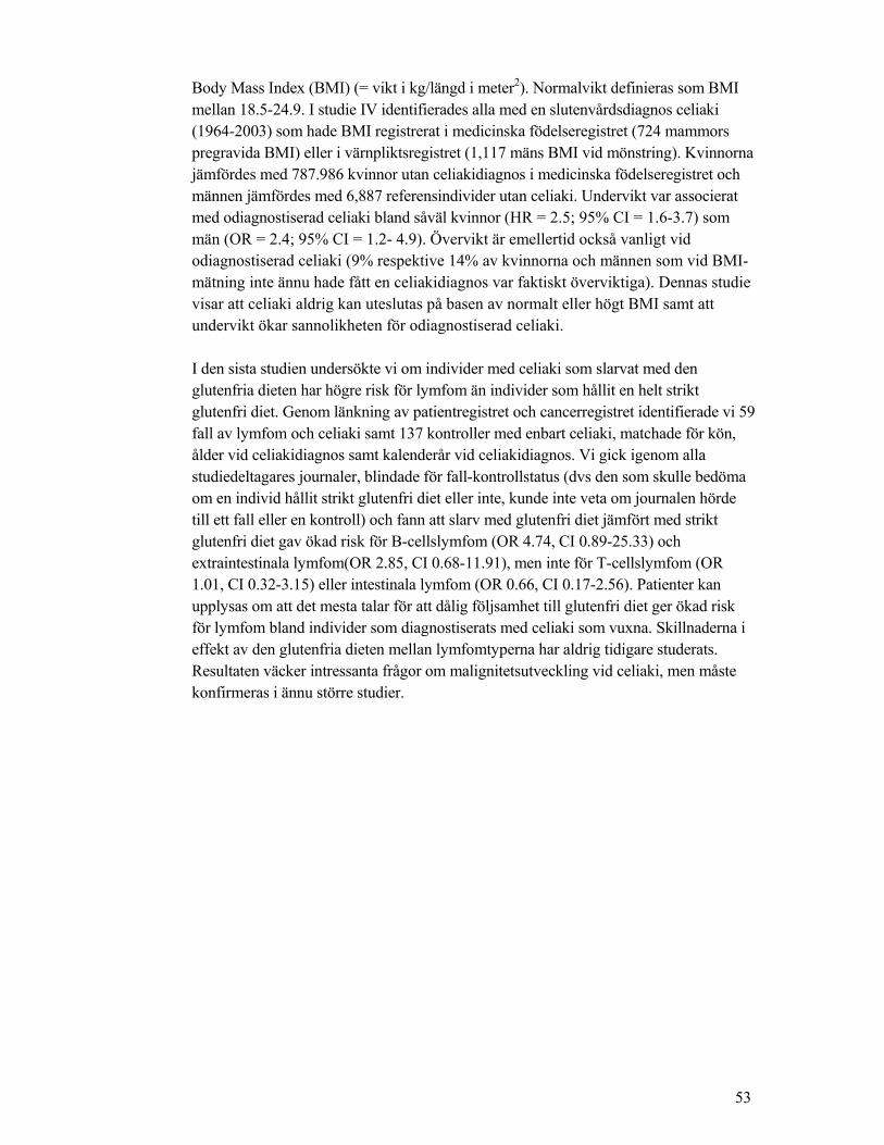
53
Body Mass Index (BMI) (= vikt i kg/längd i meter2). Normalvikt definieras som BMI mellan 18.5-24.9. I studie IV identifierades alla med en slutenvårdsdiagnos celiaki (1964-2003) som hade BMI registrerat i medicinska födelseregistret (724 mammors pregravida BMI) eller i värnpliktsregistret (1,117 mäns BMI vid mönstring). Kvinnorna jämfördes med 787.986 kvinnor utan celiakidiagnos i medicinska födelseregistret och männen jämfördes med 6,887 referensindivider utan celiaki. Undervikt var associerat med odiagnostiserad celiaki bland såväl kvinnor (HR = 2.5; 95% CI = 1.6-3.7) som män (OR = 2.4; 95% CI = 1.2- 4.9). Övervikt är emellertid också vanligt vid odiagnostiserad celiaki (9% respektive 14% av kvinnorna och männen som vid BMI-mätning inte ännu hade fått en celiakidiagnos var faktiskt överviktiga). Dennas studie visar att celiaki aldrig kan uteslutas på basen av normalt eller högt BMI samt att undervikt ökar sannolikheten för odiagnostiserad celiaki. I den sista studien undersökte vi om individer med celiaki som slarvat med den glutenfria dieten har högre risk för lymfom än individer som hållit en helt strikt glutenfri diet. Genom länkning av patientregistret och cancerregistret identifierade vi 59 fall av lymfom och celiaki samt 137 kontroller med enbart celiaki, matchade för kön, ålder vid celiakidiagnos samt kalenderår vid celiakidiagnos. Vi gick igenom alla studiedeltagares journaler, blindade för fall-kontrollstatus (dvs den som skulle bedöma om en individ hållit strikt glutenfri diet eller inte, kunde inte veta om journalen hörde till ett fall eller en kontroll) och fann att slarv med glutenfri diet jämfört med strikt glutenfri diet gav ökad risk för B-cellslymfom (OR 4.74, CI 0.89-25.33) och extraintestinala lymfom(OR 2.85, CI 0.68-11.91), men inte för T-cellslymfom (OR 1.01, CI 0.32-3.15) eller intestinala lymfom (OR 0.66, CI 0.17-2.56). Patienter kan upplysas om att det mesta talar för att dålig följsamhet till glutenfri diet ger ökad risk för lymfom bland individer som diagnostiserats med celiaki som vuxna. Skillnaderna i effekt av den glutenfria dieten mellan lymfomtyperna har aldrig tidigare studerats. Resultaten väcker intressanta frågor om malignitetsutveckling vid celiaki, men måste konfirmeras i ännu större studier.

54
10 REFERENCES 1. Dube C, Rostom A, Sy R, Cranney A, Saloojee N, Garritty C, Sampson M,
Zhang L, Yazdi F, Mamaladze V, Pan I, Macneil J, Mack D, Patel D, Moher D. The prevalence of celiac disease in average-risk and at-risk Western European populations: a systematic review. Gastroenterology 2005;128:S57-67.
2. Sollid LM. Molecular basis of celiac disease. Annu Rev Immunol 2000;18:53-81.
3. Korponay-Szabo IR, Halttunen T, Szalai Z, Laurila K, Kiraly R, Kovacs JB, Fesus L, Maki M. In vivo targeting of intestinal and extraintestinal transglutaminase 2 by coeliac autoantibodies. Gut 2004;53:641-8.
4. Fasano A. Clinical presentation of celiac disease in the pediatric population. Gastroenterology 2005;128:S68-73.
5. Green PH. The many faces of celiac disease: clinical presentation of celiac disease in the adult population. Gastroenterology 2005;128:S74-8.
6. Askling J, Linet M, Gridley G, Halstensen TS, Ekstrom K, Ekbom A. Cancer incidence in a population-based cohort of individuals hospitalized with celiac disease or dermatitis herpetiformis. Gastroenterology 2002;123:1428-35.
7. Collin P, Reunala T, Pukkala E, Laippala P, Keyrilainen O, Pasternack A. Coeliac disease--associated disorders and survival. Gut 1994;35:1215-8.
8. Ludvigsson JF, Montgomery SM, Ekbom A. Celiac disease and risk of adverse fetal outcome: a population-based cohort study. Gastroenterology 2005;129:454-63.
9. Ivarsson A, Persson LA, Juto P, Peltonen M, Suhr O, Hernell O. High prevalence of undiagnosed coeliac disease in adults: a Swedish population-based study. J Intern Med 1999;245:63-8.
10. Catassi C, Ratsch IM, Fabiani E, Rossini M, Bordicchia F, Candela F, Coppa GV, Giorgi PL. Coeliac disease in the year 2000: exploring the iceberg. Lancet 1994;343:200-3.
11. Lohi S, Mustalahti K, Kaukinen K, Laurila K, Collin P, Rissanen H, Lohi O, Bravi E, Gasparin M, Reunanen A, Maki M. Increasing prevalence of coeliac disease over time. Aliment Pharmacol Ther 2007;26:1217-25.
12. Johnston SD, Watson RG, McMillan SA, Sloan J, Love AH. Coeliac disease detected by screening is not silent--simply unrecognized. Qjm 1998;91:853-60.
13. Cranney A, Rostom A, Sy R, Dube C, Saloogee N, Garritty C, Moher D, Sampson M, Zhang L, Yazdi F, Mamaladze V, Pan I, MacNeil J. Consequences of testing for celiac disease. Gastroenterology 2005;128:S109-20.
14. Hoffenberg EJ. Should all children be screened for celiac disease? Gastroenterology 2005;128:S98-103.
15. Collin P. Should adults be screened for celiac disease? What are the benefits and harms of screening? Gastroenterology 2005;128:S104-8.
16. James SP. This month at the NIH: Final statement of NIH Consensus Conference on celiac disease. Gastroenterology 2005;128:6.
17. The extant works of Aretaeus tCAF, trans. London:, Sydenham Society. 18. Dicke W. Coeliakie: een onderzoek naar de nadelige involed van sommige
graansoorten op de lijder aan coeliakie. : Utrecht, the Netherlands: University of Utrecht, , 1950.
19. Maki M, Collin P. Coeliac disease [see comments]. Lancet 1997;349:1755-9. 20. Green PH, Cellier C. Celiac disease. N Engl J Med 2007;357:1731-43. 21. Hovell CJ, Collett JA, Vautier G, Cheng AJ, Sutanto E, Mallon DF, Olynyk JK,
Cullen DJ. High prevalence of coeliac disease in a population-based study from Western Australia: a case for screening? Med J Aust 2001;175:247-50.
22. Csizmadia CG, Mearin ML, von Blomberg BM, Brand R, Verloove-Vanhorick SP. An iceberg of childhood coeliac disease in the Netherlands. Lancet 1999;353:813-4.
23. Gomez JC, Selvaggio GS, Viola M, Pizarro B, la Motta G, de Barrio S, Castelletto R, Echeverria R, Sugai E, Vazquez H, Maurino E, Bai JC.

55
Prevalence of celiac disease in Argentina: screening of an adult population in the La Plata area. Am J Gastroenterol 2001;96:2700-4.
24. Oliveira RP, Sdepanian VL, Barreto JA, Cortez AJ, Carvalho FO, Bordin JO, de Camargo Soares MA, da Silva Patricio FR, Kawakami E, de Morais MB, Fagundes-Neto U. High prevalence of celiac disease in Brazilian blood donor volunteers based on screening by IgA antitissue transglutaminase antibody. Eur J Gastroenterol Hepatol 2007;19:43-9.
25. Fasano A, Berti I, Gerarduzzi T, Not T, Colletti RB, Drago S, Elitsur Y, Green PH, Guandalini S, Hill ID, Pietzak M, Ventura A, Thorpe M, Kryszak D, Fornaroli F, Wasserman SS, Murray JA, Horvath K. Prevalence of celiac disease in at-risk and not-at-risk groups in the United States: a large multicenter study. Arch Intern Med 2003;163:286-92.
26. al-Tawaty AI, Elbargathy SM. Coeliac disease in north-eastern Libya. Ann Trop Paediatr 1998;18:27-30.
27. Rawashdeh MO, Khalil B, Raweily E. Celiac disease in Arabs. J Pediatr Gastroenterol Nutr 1996;23:415-8.
28. Poddar U, Thapa BR, Nain CK, Prasad A, Singh K. Celiac disease in India: are they true cases of celiac disease? J Pediatr Gastroenterol Nutr 2002;35:508-12.
29. Elsurer R, Tatar G, Simsek H, Balaban YH, Aydinli M, Sokmensuer C. Celiac disease in the Turkish population. Dig Dis Sci 2005;50:136-42.
30. Catassi C, Ratsch IM, Gandolfi L, Pratesi R, Fabiani E, El Asmar R, Frijia M, Bearzi I, Vizzoni L. Why is coeliac disease endemic in the people of the Sahara? Lancet 1999;354:647-8.
31. Ivarsson A, Persson LA, Nystrom L, Ascher H, Cavell B, Danielsson L, Dannaeus A, Lindberg T, Lindquist B, Stenhammar L, Hernell O. Epidemic of coeliac disease in Swedish children [see comments]. Acta Paediatr 2000;89:165-71.
32. Myléus A IA, Webb L, et al. Exploring the Iceberg of Celiacs in Sweden – a population-based screening study of twelve-year-old children. 40th annual meeting of ESPGHAN; Barcelona, Spain 9 – 12 May 2007. Oral presentation OP1-06. 2007.
33. Hallert C, Gotthard R, Jansson G, Norrby K, Walan A. Similar prevalence of coeliac disease in children and middle-aged adults in a district of Sweden. Gut 1983;24:389-91.
34. Midhagen G, Jarnerot G, Kraaz W. Adult coeliac disease within a defined geographic area in Sweden. A study of prevalence and associated diseases. Scand J Gastroenterol 1988;23:1000-4.
35. West J, Logan RF, Hill PG, Lloyd A, Lewis S, Hubbard R, Reader R, Holmes GK, Khaw KT. Seroprevalence, correlates, and characteristics of undetected coeliac disease in England. Gut 2003;52:960-5.
36. Murray JA, Van Dyke C, Plevak MF, Dierkhising RA, Zinsmeister AR, Melton LJ, 3rd. Trends in the identification and clinical features of celiac disease in a North American community, 1950-2001. Clin Gastroenterol Hepatol 2003;1:19-27.
37. Fasano A, Araya M, Bhatnagar S, Cameron D, Catassi C, Dirks M, Mearin ML, Ortigosa L, Phillips A. Federation of International Societies of Pediatric Gastroenterology, Hepatology, and Nutrition Consensus report on celiac disease. J Pediatr Gastroenterol Nutr 2008;47:214-9.
38. Murray JA. Celiac disease in patients with an affected member, type 1 diabetes, iron-deficiency, or osteoporosis? Gastroenterology 2005;128:S52-6.
39. Rewers M. Epidemiology of celiac disease: what are the prevalence, incidence, and progression of celiac disease? Gastroenterology 2005;128:S47-51.
40. Hogberg L, Falth-Magnusson K, Grodzinsky E, Stenhammar L. Familial prevalence of coeliac disease: a twenty-year follow-up study. Scand J Gastroenterol 2003;38:61-5.
41. Mankai A, Chadli-Chaieb M, Saad F, Ghedira-Besbes L, Ouertani M, Sfar H, Limem M, Ben Abdessalem M, Jeddi M, Chaieb L, Ghedira I. Screening for celiac disease in Tunisian patients with Graves' disease using anti-endomysium and anti-tissue transglutaminase antibodies. Gastroenterol Clin Biol 2006;30:961-4.

56
42. Elfstrom P, Montgomery SM, Kampe O, Ekbom A, Ludvigsson JF. Risk of Thyroid disease in individuals with Celiac disease. J Clin Endocrinol Metab 2008.
43. Cuoco L, Certo M, Jorizzo RA, De Vitis I, Tursi A, Papa A, De Marinis L, Fedeli P, Fedeli G, Gasbarrini G. Prevalence and early diagnosis of coeliac disease in autoimmune thyroid disorders. Ital J Gastroenterol Hepatol 1999;31:283-7.
44. Spadaccino AC, Basso D, Chiarelli S, Albergoni MP, D'Odorico A, Plebani M, Pedini B, Lazzarotto F, Betterle C. Celiac disease in North Italian patients with autoimmune thyroid diseases. Autoimmunity 2008;41:116-21.
45. Guliter S, Yakaryilmaz F, Ozkurt Z, Ersoy R, Ucardag D, Caglayan O, Atasoy P. Prevalence of coeliac disease in patients with autoimmune thyroiditis in a Turkish population. World J Gastroenterol 2007;13:1599-601.
46. Elfstrom P, Montgomery SM, Kampe O, Ekbom A, Ludvigsson JF. Risk of primary adrenal insufficiency in patients with celiac disease. J Clin Endocrinol Metab 2007;92:3595-8.
47. Ventura A, Magazzu G, Greco L. Duration of exposure to gluten and risk for autoimmune disorders in patients with celiac disease. SIGEP Study Group for Autoimmune Disorders in Celiac Disease. Gastroenterology 1999;117:297-303.
48. Ludvigsson JF, Ludvigsson J, Ekbom A, Montgomery SM. Celiac Disease and Risk of Subsequent Type 1 Diabetes: A general population cohort study of children and adolescents. Diabetes Care 2006;29:2483-8.
49. Cronin CC, Feighery A, Ferriss JB, Liddy C, Shanahan F, Feighery C. High prevalence of celiac disease among patients with insulin-dependent (type I) diabetes mellitus. Am J Gastroenterol 1997;92:2210-2.
50. Szodoray P, Barta Z, Lakos G, Szakall S, Zeher M. Coeliac disease in Sjogren's syndrome--a study of 111 Hungarian patients. Rheumatol Int 2004;24:278-82.
51. Ludvigsson JF, Montgomery SM, Olen O, Ekbom A, Ludvigsson J, Fored M. Coeliac disease and risk of renal disease-a general population cohort study. Nephrol Dial Transplant 2006;21:1809-15.
52. Ludvigsson JF, Elfstrom P, Broome U, Ekbom A, Montgomery SM. Celiac disease and risk of liver disease: a general population-based study. Clin Gastroenterol Hepatol 2007;5:63-69 e1.
53. Bardella MT, Quatrini M, Zuin M, Podda M, Cesarini L, Velio P, Bianchi P, Conte D. Screening patients with celiac disease for primary biliary cirrhosis and vice versa. Am J Gastroenterol 1997;92:1524-6.
54. Volta U, De Franceschi L, Lari F, Molinaro N, Zoli M, Bianchi FB. Coeliac disease hidden by cryptogenic hypertransaminasaemia. Lancet 1998;352:26-9.
55. Chin RL, Sander HW, Brannagan TH, Green PH, Hays AP, Alaedini A, Latov N. Celiac neuropathy. Neurology 2003;60:1581-5.
56. Ludvigsson JF, Reutfors J, Osby U, Ekbom A, Montgomery SM. Coeliac disease and risk of mood disorders--a general population-based cohort study. J Affect Disord 2007;99:117-26.
57. Ludvigsson JF, Osby U, Ekbom A, Montgomery SM. Coeliac disease and risk of schizophrenia and other psychosis: a general population cohort study. Scand J Gastroenterol 2007;42:179-85.
58. Bushara KO. Neurologic presentation of celiac disease. Gastroenterology 2005;128:S92-7.
59. Hin H, Bird G, Fisher P, Mahy N, Jewell D. Coeliac disease in primary care: case finding study. Bmj 1999;318:164-7.
60. Corazza GR, Valentini RA, Andreani ML, D'Anchino M, Leva MT, Ginaldi L, De Feudis L, Quaglino D, Gasbarrini G. Subclinical coeliac disease is a frequent cause of iron-deficiency anaemia. Scand J Gastroenterol 1995;30:153-6.
61. Howard MR, Turnbull AJ, Morley P, Hollier P, Webb R, Clarke A. A prospective study of the prevalence of undiagnosed coeliac disease in laboratory defined iron and folate deficiency. J Clin Pathol 2002;55:754-7.
62. Rostom A, Murray JA, Kagnoff MF. American Gastroenterological Association (AGA) Institute technical review on the diagnosis and management of celiac disease. Gastroenterology 2006;131:1981-2002.

57
63. Annibale B, Lahner E, Chistolini A, Gailucci C, Di Giulio E, Capurso G, Luana O, Monarca B, Delle Fave G. Endoscopic evaluation of the upper gastrointestinal tract is worthwhile in premenopausal women with iron-deficiency anaemia irrespective of menstrual flow. Scand J Gastroenterol 2003;38:239-45.
64. Collin P, Vilska S, Heinonen PK, Hallstrom O, Pikkarainen P. Infertility and coeliac disease. Gut 1996;39:382-4.
65. Meloni GF, Dessole S, Vargiu N, Tomasi PA, Musumeci S. The prevalence of coeliac disease in infertility. Hum Reprod 1999;14:2759-61.
66. Jackson JE, Rosen M, McLean T, Moro J, Croughan M, Cedars MI. Prevalence of celiac disease in a cohort of women with unexplained infertility. Fertil Steril 2008;89:1002-4.
67. Tata LJ, Card TR, Logan RF, Hubbard RB, Smith CJ, West J. Fertility and pregnancy-related events in women with celiac disease: a population-based cohort study. Gastroenterology 2005;128:849-55.
68. Book L, Hart A, Black J, Feolo M, Zone JJ, Neuhausen SL. Prevalence and clinical characteristics of celiac disease in Downs syndrome in a US study. Am J Med Genet 2001;98:70-4.
69. Bonamico M, Mariani P, Danesi HM, Crisogianni M, Failla P, Gemme G, Quartino AR, Giannotti A, Castro M, Balli F, Lecora M, Andria G, Guariso G, Gabrielli O, Catassi C, Lazzari R, Balocco NA, De Virgiliis S, Culasso F, Romano C. Prevalence and clinical picture of celiac disease in italian down syndrome patients: a multicenter study. J Pediatr Gastroenterol Nutr 2001;33:139-43.
70. Carlsson A, Axelsson I, Borulf S, Bredberg A, Forslund M, Lindberg B, Sjoberg K, Ivarsson SA. Prevalence of IgA-antigliadin antibodies and IgA-antiendomysium antibodies related to celiac disease in children with Down syndrome. Pediatrics 1998;101:272-5.
71. Sanders DS, Carter MJ, Hurlstone DP, Pearce A, Ward AM, McAlindon ME, Lobo AJ. Association of adult coeliac disease with irritable bowel syndrome: a case-control study in patients fulfilling ROME II criteria referred to secondary care. Lancet 2001;358:1504-8.
72. Dewar DH, Ciclitira PJ. Clinical features and diagnosis of celiac disease. Gastroenterology 2005;128:S19-24.
73. Bevan S, Popat S, Braegger CP, Busch A, O'Donoghue D, Falth-Magnusson K, Ferguson A, Godkin A, Hogberg L, Holmes G, Hosie KB, Howdle PD, Jenkins H, Jewell D, Johnston S, Kennedy NP, Kerr G, Kumar P, Logan RF, Love AH, Marsh M, Mulder CJ, Sjoberg K, Stenhammer L, Houlston RS, et al. Contribution of the MHC region to the familial risk of coeliac disease. J Med Genet 1999;36:687-90.
74. Greco L, Romino R, Coto I, Di Cosmo N, Percopo S, Maglio M, Paparo F, Gasperi V, Limongelli MG, Cotichini R, D'Agate C, Tinto N, Sacchetti L, Tosi R, Stazi MA. The first large population based twin study of coeliac disease. Gut 2002;50:624-8.
75. Schuppan D. Current concepts of celiac disease pathogenesis. Gastroenterology 2000;119:234-42.
76. Hausch F, Shan L, Santiago NA, Gray GM, Khosla C. Intestinal digestive resistance of immunodominant gliadin peptides. Am J Physiol Gastrointest Liver Physiol 2002;283:G996-G1003.
77. Lundin KE, Nilsen EM, Scott HG, Loberg EM, Gjoen A, Bratlie J, Skar V, Mendez E, Lovik A, Kett K. Oats induced villous atrophy in coeliac disease. Gut 2003;52:1649-52.
78. Arentz-Hansen H, Fleckenstein B, Molberg O, Scott H, Koning F, Jung G, Roepstorff P, Lundin KE, Sollid LM. The molecular basis for oat intolerance in patients with celiac disease. PLoS Med 2004;1:e1.
79. Janatuinen EK, Pikkarainen PH, Kemppainen TA, Kosma VM, Jarvinen RM, Uusitupa MI, Julkunen RJ. A comparison of diets with and without oats in adults with celiac disease [see comments]. N Engl J Med 1995;333:1033-7.

58
80. Janatuinen EK, Kemppainen TA, Julkunen RJ, Kosma VM, Maki M, Heikkinen M, Uusitupa MI. No harm from five year ingestion of oats in coeliac disease. Gut 2002;50:332-5.
81. Hogberg L, Laurin P, Falth-Magnusson K, Grant C, Grodzinsky E, Jansson G, Ascher H, Browaldh L, Hammersjo JA, Lindberg E, Myrdal U, Stenhammar L. Oats to children with newly diagnosed coeliac disease: a randomised double blind study. Gut 2004;53:649-54.
82. Kasarda D. Gluten and gliadin: precipitating factors in coeliac disease. Coeliac disease. Maki M, Collin P, Visakorpi JK, eds. Tampere, Finland: Coeliac Study Group Institute of Medical Technology, 1997:195–212.
83. Stepniak D, Koning F. Celiac disease--sandwiched between innate and adaptive immunity. Hum Immunol 2006;67:460-8.
84. Briani C, Samaroo D, Alaedini A. Celiac disease: From gluten to autoimmunity. Autoimmun Rev 2008.
85. Alaedini A, Green PH. Narrative review: celiac disease: understanding a complex autoimmune disorder. Ann Intern Med 2005;142:289-98.
86. Stene LC, Honeyman MC, Hoffenberg EJ, Haas JE, Sokol RJ, Emery L, Taki I, Norris JM, Erlich HA, Eisenbarth GS, Rewers M. Rotavirus infection frequency and risk of celiac disease autoimmunity in early childhood: a longitudinal study. Am J Gastroenterol 2006;101:2333-40.
87. Molberg O, McAdam SN, Korner R, Quarsten H, Kristiansen C, Madsen L, Fugger L, Scott H, Noren O, Roepstorff P, Lundin KE, Sjostrom H, Sollid LM. Tissue transglutaminase selectively modifies gliadin peptides that are recognized by gut-derived T cells in celiac disease. Nat Med 1998;4:713-7.
88. Molberg O, McAdam SN, Korner R, Quarsten H, Kristiansen C, Madsen L, Fugger L, Scott H, Noren O, Roepstorff P, Lundin KE, Sjostrom H, Sollid LM. Tissue transglutaminase selectively modifies gliadin peptides that are recognized by gut-derived T cells in celiac disease [see comments] [published erratum appears in Nat Med 1998 Aug;4(8):974]. Nat Med 1998;4:713-7.
89. Van De Wal Y, Kooy Y, Van Veelen P, Vader W, Koning F, Pena S. Coeliac disease: it takes three to tango! Gut 2000;46:734-7.
90. Sollid LM. Coeliac disease: dissecting a complex inflammatory disorder. Nat Rev Immunol 2002;2:647-55.
91. Nilsen EM, Jahnsen FL, Lundin KE, Johansen FE, Fausa O, Sollid LM, Jahnsen J, Scott H, Brandtzaeg P. Gluten induces an intestinal cytokine response strongly dominated by interferon gamma in patients with celiac disease. Gastroenterology 1998;115:551-63.
92. Mohamed BM, Feighery C, Kelly J, Coates C, O'Shea U, Barnes L, Abuzakouk M. Increased protein expression of matrix metalloproteinases -1, -3, and -9 and TIMP-1 in patients with gluten-sensitive enteropathy. Dig Dis Sci 2006;51:1862-8.
93. Mention JJ, Ben Ahmed M, Begue B, Barbe U, Verkarre V, Asnafi V, Colombel JF, Cugnenc PH, Ruemmele FM, McIntyre E, Brousse N, Cellier C, Cerf-Bensussan N. Interleukin 15: a key to disrupted intraepithelial lymphocyte homeostasis and lymphomagenesis in celiac disease. Gastroenterology 2003;125:730-45.
94. Di Sabatino A, Ciccocioppo R, Cupelli F, Cinque B, Millimaggi D, Clarkson MM, Paulli M, Cifone MG, Corazza GR. Epithelium derived interleukin 15 regulates intraepithelial lymphocyte Th1 cytokine production, cytotoxicity, and survival in coeliac disease. Gut 2006;55:469-77.
95. Griffin M, Casadio R, Bergamini CM. Transglutaminases: nature's biological glues. Biochem J 2002;368:377-96.
96. Ludvigsson JF, Falth-Magnusson K, Ludvigsson J. Tissue transglutaminase auto-antibodies in cord blood from children to become celiacs. Scand J Gastroenterol 2001;36:1279-83.
97. Hill ID. Management of celiac disease in childhood and adolescence: unique challenges and strategies. Curr Treat Options Gastroenterol 2006;9:399-408.
98. Castano L, Blarduni E, Ortiz L, Nunez J, Bilbao JR, Rica I, Martul P, Vitoria JC. Prospective population screening for celiac disease: high prevalence in the first 3 years of life. J Pediatr Gastroenterol Nutr 2004;39:80-4.

59
99. Maki M, Mustalahti K, Kokkonen J, Kulmala P, Haapalahti M, Karttunen T, Ilonen J, Laurila K, Dahlbom I, Hansson T, Hopfl P, Knip M. Prevalence of Celiac disease among children in Finland. N Engl J Med 2003;348:2517-24.
100. Norris JM, Barriga K, Hoffenberg EJ, Taki I, Miao D, Haas JE, Emery LM, Sokol RJ, Erlich HA, Eisenbarth GS, Rewers M. Risk of celiac disease autoimmunity and timing of gluten introduction in the diet of infants at increased risk of disease. Jama 2005;293:2343-51.
101. Ivarsson A, Hernell O, Stenlund H, Persson LA. Breast-feeding protects against celiac disease. Am J Clin Nutr 2002;75:914-21.
102. Sandberg-Bennich S, Dahlquist G, Kallen B. Coeliac disease is associated with intrauterine growth and neonatal infections. Acta Paediatr 2002;91:30-3.
103. Snook JA, Dwyer L, Lee-Elliott C, Khan S, Wheeler DW, Nicholas DS. Adult coeliac disease and cigarette smoking [see comments]. Gut 1996;39:60-2.
104. Ludvigsson JF, Montgomery SM, Ekbom A. Smoking and celiac disease: a population-based cohort study. Clin Gastroenterol Hepatol 2005;3:869-74.
105. Johansson SG, Hourihane JO, Bousquet J, Bruijnzeel-Koomen C, Dreborg S, Haahtela T, Kowalski ML, Mygind N, Ring J, van Cauwenberge P, van Hage-Hamsten M, Wuthrich B. A revised nomenclature for allergy. An EAACI position statement from the EAACI nomenclature task force. Allergy 2001;56:813-24.
106. Johansson SG, Bieber T, Dahl R, Friedmann PS, Lanier BQ, Lockey RF, Motala C, Ortega Martell JA, Platts-Mills TA, Ring J, Thien F, Van Cauwenberge P, Williams HC. Revised nomenclature for allergy for global use: Report of the Nomenclature Review Committee of the World Allergy Organization, October 2003. J Allergy Clin Immunol 2004;113:832-6.
107. Vilppula A, Collin P, Maki M, Valve R, Luostarinen M, Krekela I, Patrikainen H, Kaukinen K, Luostarinen L. Undetected coeliac disease in the elderly A biopsy-proven population-based study. Dig Liver Dis 2008.
108. Tommasini A, Not T, Kiren V, Baldas V, Santon D, Trevisiol C, Berti I, Neri E, Gerarduzzi T, Bruno I, Lenhardt A, Zamuner E, Spano A, Crovella S, Martellossi S, Torre G, Sblattero D, Marzari R, Bradbury A, Tamburlini G, Ventura A. Mass screening for coeliac disease using antihuman transglutaminase antibody assay. Arch Dis Child 2004;89:512-5.
109. Maki M, Kallonen K, Lahdeaho ML, Visakorpi JK. Changing pattern of childhood coeliac disease in Finland. Acta Paediatr Scand 1988;77:408-12.
110. Ludvigsson JF, Ansved P, Falth-Magnusson K, Hammersjo JA, Johansson C, Edvardsson S, Ljungkrantz M, Stenhammar L, Ludvigsson J. Symptoms and signs have changed in Swedish children with coeliac disease. J Pediatr Gastroenterol Nutr 2004;38:181-6.
111. Lo W, Sano K, Lebwohl B, Diamond B, Green PH. Changing presentation of adult celiac disease. Dig Dis Sci 2003;48:395-8.
112. Goldstein NS. Proximal small-bowel mucosal villous intraepithelial lymphocytes. Histopathology 2004;44:199-205.
113. Marsh MN, Crowe PT. Morphology of the mucosal lesion in gluten sensitivity. Baillieres Clin Gastroenterol 1995;9:273-93.
114. Street ME, Volta C, Ziveri MA, Zanacca C, Banchini G, Viani I, Rossi M, Virdis R, Bernasconi S. Changes and relationships of IGFS and IGFBPS and cytokines in coeliac disease at diagnosis and on gluten-free diet. Clin Endocrinol (Oxf) 2008;68:22-8.
115. ESPGHAN. Revised criteria for diagnosis of coeliac disease. Report of Working Group of European Society of Paediatric Gastroenterology and Nutrition. Arch Dis Child 1990;65:909-11.
116. Hill ID, Dirks MH, Liptak GS, Colletti RB, Fasano A, Guandalini S, Hoffenberg EJ, Horvath K, Murray JA, Pivor M, Seidman EG. Guideline for the diagnosis and treatment of celiac disease in children: recommendations of the North American Society for Pediatric Gastroenterology, Hepatology and Nutrition. J Pediatr Gastroenterol Nutr 2005;40:1-19.
117. National Institutes of Health Consensus Development Conference Statement on Celiac Disease, June 28-30, 2004. Gastroenterology 2005;128:S1-9.

60
118. Hopper AD, Hadjivassiliou M, Hurlstone DP, Lobo AJ, McAlindon ME, Egner W, Wild G, Sanders DS. What is the role of serologic testing in celiac disease? A prospective, biopsy-confirmed study with economic analysis. Clin Gastroenterol Hepatol 2008;6:314-20.
119. Rostom A, Dube C, Cranney A, Saloojee N, Sy R, Garritty C, Sampson M, Zhang L, Yazdi F, Mamaladze V, Pan I, MacNeil J, Mack D, Patel D, Moher D. The diagnostic accuracy of serologic tests for celiac disease: a systematic review. Gastroenterology 2005;128:S38-46.
120. Cataldo F, Marino V, Ventura A, Bottaro G, Corazza GR. Prevalence and clinical features of selective immunoglobulin A deficiency in coeliac disease: an Italian multicentre study. Italian Society of Paediatric Gastroenterology and Hepatology (SIGEP) and "Club del Tenue" Working Groups on Coeliac Disease. Gut 1998;42:362-5.
121. Collin P, Maki M, Keyrilainen O, Hallstrom O, Reunala T, Pasternack A. Selective IgA deficiency and coeliac disease. Scand J Gastroenterol 1992;27:367-71.
122. Meini A, Pillan NM, Villanacci V, Monafo V, Ugazio AG, Plebani A. Prevalence and diagnosis of celiac disease in IgA-deficient children. Ann Allergy Asthma Immunol 1996;77:333-6.
123. Cataldo F, Lio D, Marino V, Picarelli A, Ventura A, Corazza GR, Sigep, Club Del Tenue t W. IgG(1) antiendomysium and IgG antitissue transglutaminase (anti-tTG) antibodies in coeliac patients with selective IgA deficiency. Gut 2000;47:366-9.
124. Sulkanen S, Collin P, Laurila K, Maki M. IgA- and IgG-class antihuman umbilical cord antibody tests in adult coeliac disease. Scand J Gastroenterol 1998;33:251-4.
125. Korponay-Szabo IR, Dahlbom I, Laurila K, Koskinen S, Woolley N, Partanen J, Kovacs JB, Maki M, Hansson T. Elevation of IgG antibodies against tissue transglutaminase as a diagnostic tool for coeliac disease in selective IgA deficiency. Gut 2003;52:1567-71.
126. Rostami K, Kerckhaert J, Tiemessen R, von Blomberg BM, Meijer JW, Mulder CJ. Sensitivity of antiendomysium and antigliadin antibodies in untreated celiac disease: disappointing in clinical practice [see comments]. Am J Gastroenterol 1999;94:888-94.
127. Hadithi M, von Blomberg BM, Crusius JB, Bloemena E, Kostense PJ, Meijer JW, Mulder CJ, Stehouwer CD, Pena AS. Accuracy of serologic tests and HLA-DQ typing for diagnosing celiac disease. Ann Intern Med 2007;147:294-302.
128. Hopper AD, Cross SS, Hurlstone DP, McAlindon ME, Lobo AJ, Hadjivassiliou M, Sloan ME, Dixon S, Sanders DS. Pre-endoscopy serological testing for coeliac disease: evaluation of a clinical decision tool. Bmj 2007;334:729.
129. Rostom A, Dube C, Cranney A, Saloojee N, Sy R, Garritty C, Sampson M, Zhang L, Yazdi F, Mamaladze V, Pan I, McNeil J, Moher D, Mack D, Patel D. Celiac disease. Evid Rep Technol Assess (Summ) 2004:1-6.
130. Marsh MN. Gluten, major histocompatibility complex, and the small intestine. A molecular and immunobiologic approach to the spectrum of gluten sensitivity ('celiac sprue'). Gastroenterology 1992;102:330-54.
131. Kaukinen K, Peraaho M, Lindfors K, Partanen J, Woolley N, Pikkarainen P, Karvonen AL, Laasanen T, Sievanen H, Maki M, Collin P. Persistent small bowel mucosal villous atrophy without symptoms in coeliac disease. Aliment Pharmacol Ther 2007;25:1237-45.
132. Liu E, Rewers M, Eisenbarth GS. Genetic testing: who should do the testing and what is the role of genetic testing in the setting of celiac disease? Gastroenterology 2005;128:S33-7.
133. Margaritte-Jeannin P, Babron MC, Bourgey M, Louka AS, Clot F, Percopo S, Coto I, Hugot JP, Ascher H, Sollid LM, Greco L, Clerget-Darpoux F. HLA-DQ relative risks for coeliac disease in European populations: a study of the European Genetics Cluster on Coeliac Disease. Tissue Antigens 2004;63:562-7.
134. Karell K, Louka AS, Moodie SJ, Ascher H, Clot F, Greco L, Ciclitira PJ, Sollid LM, Partanen J. HLA types in celiac disease patients not carrying the

61
DQA1*05-DQB1*02 (DQ2) heterodimer: results from the European Genetics Cluster on Celiac Disease. Hum Immunol 2003;64:469-77.
135. Stenhammar L, Hogberg L, Danielsson L, Ascher H, Dannaeus A, Hernell O, Ivarsson A, Lindberg E, Lindquist B, Nivenius K. How do Swedish paediatric clinics diagnose coeliac disease? Results of a nationwide questionnaire study. Acta Paediatr 2006;95:1495-7.
136. Revised criteria for diagnosis of coeliac disease. Report of Working Group of European Society of Paediatric Gastroenterology and Nutrition. Arch Dis Child 1990;65:909-11.
137. Barker CC, Mitton C, Jevon G, Mock T. Can tissue transglutaminase antibody titers replace small-bowel biopsy to diagnose celiac disease in select pediatric populations? Pediatrics 2005;115:1341-6.
138. Ludvigsson JF SL. Biopsy is needed in celiac disease. Läkartidningen 2007;104.
139. Green PH, Rostami K, Marsh MN. Diagnosis of coeliac disease. Best Pract Res Clin Gastroenterol 2005;19:389-400.
140. Ferguson A, Murray D. Quantitation of intraepithelial lymphocytes in human jejunum. Gut 1971;12:988-94.
141. Dickson BC, Streutker CJ, Chetty R. Coeliac disease: an update for pathologists. J Clin Pathol 2006;59:1008-16.
142. Ludvigsson JF. Personal communication of unpublished data. Phone +46(0)196023439, 2008.
143. Collin P, Kaukinen K, Vogelsang H, Korponay-Szabo I, Sommer R, Schreier E, Volta U, Granito A, Veronesi L, Mascart F, Ocmant A, Ivarsson A, Lagerqvist C, Burgin-Wolff A, Hadziselimovic F, Furlano RI, Sidler MA, Mulder CJ, Goerres MS, Mearin ML, Ninaber MK, Gudmand-Hoyer E, Fabiani E, Catassi C, Tidlund H, Alainentalo L, Maki M. Antiendomysial and antihuman recombinant tissue transglutaminase antibodies in the diagnosis of coeliac disease: a biopsy-proven European multicentre study. Eur J Gastroenterol Hepatol 2005;17:85-91.
144. Corazza GR, Villanacci V, Zambelli C, Milione M, Luinetti O, Vindigni C, Chioda C, Albarello L, Bartolini D, Donato F. Comparison of the interobserver reproducibility with different histologic criteria used in celiac disease. Clin Gastroenterol Hepatol 2007;5:838-43.
145. Shidrawi RG, Przemioslo R, Davies DR, Tighe MR, Ciclitira PJ. Pitfalls in diagnosing coeliac disease. J Clin Pathol 1994;47:693-4.
146. Bonamico M, Mariani P, Thanasi E, Ferri M, Nenna R, Tiberti C, Mora B, Mazzilli MC, Magliocca FM. Patchy villous atrophy of the duodenum in childhood celiac disease. J Pediatr Gastroenterol Nutr 2004;38:204-7.
147. Oberhuber G, Granditsch G, Vogelsang H. The histopathology of coeliac disease: time for a standardized report scheme for pathologists. Eur J Gastroenterol Hepatol 1999;11:1185-94.
148. Green PH. Celiac disease: how many biopsies for diagnosis? Gastrointest Endosc 2008;67:1088-90.
149. Wahab PJ, Meijer JW, Mulder CJ. Histologic follow-up of people with celiac disease on a gluten-free diet: slow and incomplete recovery. Am J Clin Pathol 2002;118:459-63.
150. Hoffenberg EJ, Haas J, Drescher A, Barnhurst R, Osberg I, Bao F, Eisenbarth G. A trial of oats in children with newly diagnosed celiac disease. J Pediatr 2000;137:361-6.
151. Peraaho M, Collin P, Kaukinen K, Kekkonen L, Miettinen S, Maki M. Oats can diversify a gluten-free diet in celiac disease and dermatitis herpetiformis. J Am Diet Assoc 2004;104:1148-50.
152. Storsrud S, Hulthen LR, Lenner RA. Beneficial effects of oats in the gluten-free diet of adults with special reference to nutrient status, symptoms and subjective experiences. Br J Nutr 2003;90:101-7.
153. Storsrud S, Olsson M, Arvidsson Lenner R, Nilsson LA, Nilsson O, Kilander A. Adult coeliac patients do tolerate large amounts of oats. Eur J Clin Nutr 2003;57:163-9.

62
154. Lohiniemi S, Maki M, Kaukinen K, Laippala P, Collin P. Gastrointestinal symptoms rating scale in coeliac disease patients on wheat starch-based gluten-free diets. Scand J Gastroenterol 2000;35:947-9.
155. Kaukinen K, Collin P, Holm K, Rantala I, Vuolteenaho N, Reunala T, Maki M. Wheat starch-containing gluten-free flour products in the treatment of coeliac disease and dermatitis herpetiformis. A long-term follow-up study. Scand J Gastroenterol 1999;34:163-9.
156. Peraaho M, Kaukinen K, Paasikivi K, Sievanen H, Lohiniemi S, Maki M, Collin P. Wheat-starch-based gluten-free products in the treatment of newly detected coeliac disease: prospective and randomized study. Aliment Pharmacol Ther 2003;17:587-94.
157. Catassi C, Fabiani E, Iacono G, D'Agate C, Francavilla R, Biagi F, Volta U, Accomando S, Picarelli A, De Vitis I, Pianelli G, Gesuita R, Carle F, Mandolesi A, Bearzi I, Fasano A. A prospective, double-blind, placebo-controlled trial to establish a safe gluten threshold for patients with celiac disease. Am J Clin Nutr 2007;85:160-6.
158. Collin P, Maki M, Kaukinen K. Safe gluten threshold for patients with celiac disease: some patients are more tolerant than others. Am J Clin Nutr 2007;86:260; author reply 260-1.
159. Collin P, Thorell L, Kaukinen K, Maki M. The safe threshold for gluten contamination in gluten-free products. Can trace amounts be accepted in the treatment of coeliac disease? Aliment Pharmacol Ther 2004;19:1277-83.
160. Killander A, Arnell H, Hagenas L, Finkel Y. Omitting control biopsy in paediatric coeliac disease: a follow-up study. Acta Paediatr 2007;96:1190-4.
161. Hallert C, Grant C, Grehn S, Granno C, Hulten S, Midhagen G, Strom M, Svensson H, Valdimarsson T. Evidence of poor vitamin status in coeliac patients on a gluten-free diet for 10 years. Aliment Pharmacol Ther 2002;16:1333-9.
162. Kupper C. Dietary guidelines and implementation for celiac disease. Gastroenterology 2005;128:S121-7.
163. Olén O, Montgomery SM, Marcus C, Ekbom A, Ludvigsson JF. Celiac disease and Body Mass Index: A study of two Swedish general population based registers. Manuscript submitted. 2008.
164. Dickey W, Kearney N. Overweight in celiac disease: prevalence, clinical characteristics, and effect of a gluten-free diet. Am J Gastroenterol 2006;101:2356-9.
165. Franzese A, Iannucci MP, Valerio G, Ciccimarra E, Spaziano M, Mandato C, Vajro P. Atypical celiac disease presenting as obesity-related liver dysfunction. J Pediatr Gastroenterol Nutr 2001;33:329-32.
166. Mariani P, Viti MG, Montuori M, La Vecchia A, Cipolletta E, Calvani L, Bonamico M. The gluten-free diet: a nutritional risk factor for adolescents with celiac disease? J Pediatr Gastroenterol Nutr 1998;27:519-23.
167. Mora S, Barera G, Beccio S, Menni L, Proverbio MC, Bianchi C, Chiumello G. A prospective, longitudinal study of the long-term effect of treatment on bone density in children with celiac disease. J Pediatr 2001;139:516-21.
168. Sategna-Guidetti C, Grosso SB, Grosso S, Mengozzi G, Aimo G, Zaccaria T, Di Stefano M, Isaia GC. The effects of 1-year gluten withdrawal on bone mass, bone metabolism and nutritional status in newly-diagnosed adult coeliac disease patients. Aliment Pharmacol Ther 2000;14:35-43.
169. Annibale B, Severi C, Chistolini A, Antonelli G, Lahner E, Marcheggiano A, Iannoni C, Monarca B, Fave GD. Efficacy of gluten-free diet alone on recovery from iron deficiency anemia in adult celiac patients. Am J Gastroenterol 2001;96:132-7.
170. Barera G, Mora S, Brambilla P, Ricotti A, Menni L, Beccio S, Bianchi C. Body composition in children with celiac disease and the effects of a gluten-free diet: a prospective case-control study. Am J Clin Nutr 2000;72:71-5.
171. Amin R, Murphy N, Edge J, Ahmed ML, Acerini CL, Dunger DB. A longitudinal study of the effects of a gluten-free diet on glycemic control and weight gain in subjects with type 1 diabetes and celiac disease. Diabetes Care 2002;25:1117-22.

63
172. Westman E, Ambler GR, Royle M, Peat J, Chan A. Children with coeliac disease and insulin dependent diabetes mellitus--growth, diabetes control and dietary intake. J Pediatr Endocrinol Metab 1999;12:433-42.
173. Nachman F, Maurino E, Vazquez H, Sfoggia C, Gonzalez A, Gonzalez V, Del Campo MP, Smecuol E, Niveloni S, Sugai E, Mazure R, Cabanne A, Bai JC. Quality of life in celiac disease patients Prospective analysis on the importance of clinical severity at diagnosis and the impact of treatment. Dig Liver Dis 2008.
174. Mustalahti K, Lohiniemi S, Collin P, Vuolteenaho N, Laippala P, Maki M. Gluten-free diet and quality of life in patients with screen-detected celiac disease. Eff Clin Pract 2002;5:105-13.
175. Johnston SD, Rodgers C, Watson RG. Quality of life in screen-detected and typical coeliac disease and the effect of excluding dietary gluten. Eur J Gastroenterol Hepatol 2004;16:1281-6.
176. Corrao G, Corazza GR, Bagnardi V, Brusco G, Ciacci C, Cottone M, Guidetti CS, Usai P, Cesari P, Pelli MA, Loperfido S, Volta U, Calabro A, Certo M. Mortality in patients with coeliac disease and their relatives: a cohort study. Lancet 2001;358:356-61.
177. Olen O, Johan Askling, Jonas F Ludvigsson, Hans Hildebrand, Anders Ekbom, Karin Ekström-Smedby. Celiac disease, compliance to a gluten free diet and risk of lymphoma by subtype. submitted 2008.
178. Holmes GK, Prior P, Lane MR, Pope D, Allan RN. Malignancy in coeliac disease--effect of a gluten free diet. Gut 1989;30:333-8.
179. Green PH, Fleischauer AT, Bhagat G, Goyal R, Jabri B, Neugut AI. Risk of malignancy in patients with celiac disease. Am J Med 2003;115:191-5.
180. Silano M, Volta U, Vincenzi AD, Dessi M, Vincenzi MD. Effect of a Gluten-free Diet on the Risk of Enteropathy-associated T-cell Lymphoma in Celiac Disease. Dig Dis Sci 2007.
181. Sategna Guidetti C, Solerio E, Scaglione N, Aimo G, Mengozzi G. Duration of gluten exposure in adult coeliac disease does not correlate with the risk for autoimmune disorders. Gut 2001;49:502-5.
182. Cosnes J, Cellier C, Viola S, Colombel JF, Michaud L, Sarles J, Hugot JP, Ginies JL, Dabadie A, Mouterde O, Allez M, Nion-Larmurier I. Incidence of autoimmune diseases in celiac disease: protective effect of the gluten-free diet. Clin Gastroenterol Hepatol 2008;6:753-8.
183. Viljamaa M, Kaukinen K, Huhtala H, Kyronpalo S, Rasmussen M, Collin P. Coeliac disease, autoimmune diseases and gluten exposure. Scand J Gastroenterol 2005;40:437-43.
184. Pietzak MM. Follow-up of patients with celiac disease: achieving compliance with treatment. Gastroenterology 2005;128:S135-41.
185. Case S. The gluten-free diet: how to provide effective education and resources. Gastroenterology 2005;128:S128-34.
186. Ciacci C, Cirillo M, Cavallaro R, Mazzacca G. Long-term follow-up of celiac adults on gluten-free diet: prevalence and correlates of intestinal damage. Digestion 2002;66:178-85.
187. Fabiani E, Taccari LM, Ratsch IM, Di Giuseppe S, Coppa GV, Catassi C. Compliance with gluten-free diet in adolescents with screening-detected celiac disease: A 5-year follow-up study [In Process Citation]. J Pediatr 2000;136:841-3.
188. Viljamaa M, Collin P, Huhtala H, Sievanen H, Maki M, Kaukinen K. Is coeliac disease screening in risk groups justified? A fourteen-year follow-up with special focus on compliance and quality of life. Aliment Pharmacol Ther 2005;22:317-24.
189. Dickey W, Hughes DF, McMillan SA. Disappearance of endomysial antibodies in treated celiac disease does not indicate histological recovery [In Process Citation]. Am J Gastroenterol 2000;95:712-4.
190. Cellier C, Delabesse E, Helmer C, Patey N, Matuchansky C, Jabri B, Macintyre E, Cerf-Bensussan N, Brousse N. Refractory sprue, coeliac disease, and enteropathy-associated T-cell lymphoma. French Coeliac Disease Study Group. Lancet 2000;356:203-8.

64
191. Pinkas DM, Strop P, Brunger AT, Khosla C. Transglutaminase 2 undergoes a large conformational change upon activation. PLoS Biol 2007;5:e327.
192. Gass J, Bethune MT, Siegel M, Spencer A, Khosla C. Combination enzyme therapy for gastric digestion of dietary gluten in patients with celiac sprue. Gastroenterology 2007;133:472-80.
193. Fasano A. European and North American populations should be screened for coeliac disease. Gut 2003;52:168-9.
194. Kumar PJ. European and North American populations should be screened for coeliac disease. Gut 2003;52:170-1.
195. Wilson JM, Jungner YG. [Principles and practice of mass screening for disease]. Bol Oficina Sanit Panam 1968;65:281-393.
196. Frankenburg WK. Selection of diseases and tests in pediatric screening. Pediatrics 1974;54:612-6.
197. Hoffenberg EJ, MacKenzie T, Barriga KJ, Eisenbarth GS, Bao F, Haas JE, Erlich H, Bugawan Tl T, Sokol RJ, Taki I, Norris JM, Rewers M. A prospective study of the incidence of childhood celiac disease. J Pediatr 2003;143:308-14.
198. Fabiani E, Taccari LM, Ratsch IM, Di Giuseppe S, Coppa GV, Catassi C. Compliance with gluten-free diet in adolescents with screening-detected celiac disease: a 5-year follow-up study. J Pediatr 2000;136:841-3.
199. Cunningham FG, Lucas MJ. Urinary tract infections complicating pregnancy. Baillieres Clin Obstet Gynaecol 1994;8:353-73.
200. North DH, Speed JE, Weiner WB, Morrison JC. Correlation of urinary tract infection with urinary screening at the first antepartum visit. J Miss State Med Assoc 1990;31:331-3.
201. Gilstrap LC, Leveno KJ, Cunningham FG, Whalley PJ, Roark ML. Renal infection and pregnancy outcome. Am J Obstet Gynecol 1981;141:709-16.
202. Tseng CC, Wu JJ, Liu HL, Sung JM, Huang JJ. Roles of host and bacterial virulence factors in the development of upper urinary tract infection caused by Escherichia coli. Am J Kidney Dis 2002;39:744-52.
203. Saalman R, Fallstrom SP. High incidence of urinary tract infection in patients with coeliac disease. Arch Dis Child 1996;74:170-1.
204. Fanos V, Verlato G, Matti P, Pizzini C, Maffeis C. Increased incidence of urinary tract infections in patients with coeliac disease. Pediatr Nephrol 2002;17:570-1.
205. Cines DB, Blanchette VS. Immune thrombocytopenic purpura. N Engl J Med 2002;346:995-1008.
206. Landgren O, Gridley G, Fears TR, Caporaso N. Immune thrombocytopenic purpura does not exhibit a disparity in prevalence between African American and White veterans. Blood 2006;108:1111-2.
207. Zeller B RJ, Hedlund-Treutiger I. Childhood idiopathic thrombocytic purpura in the NOrdic countries: Epidemiology and predictors of chronic disease. Acta Paediatrica 2005;94:178-184.
208. Stasi R, Provan D. Management of immune thrombocytopenic purpura in adults. Mayo Clin Proc 2004;79:504-22.
209. Frederiksen H, Schmidt K. The incidence of idiopathic thrombocytopenic purpura in adults increases with age. Blood 1999;94:909-13.
210. Rosthoj S, Hedlund-Treutiger I, Rajantie J, Zeller B, Jonsson OG, Elinder G, Wesenberg F, Henter JI. Duration and morbidity of newly diagnosed idiopathic thrombocytopenic purpura in children: A prospective Nordic study of an unselected cohort. J Pediatr 2003;143:302-7.
211. Stenhammar L, Ljunggren CG. Thrombocytopenic purpura and coeliac disease. Acta Paediatr Scand 1988;77:764-6.
212. Eliakim R, Heyman S, Kornberg A. Celiac disease and keratoconjunctivitis. Occurrence with thrombocytopenic purpura. Arch Intern Med 1982;142:1037.
213. Mulder CJ, Gratama JW, Trimbos-Kemper GC, Willemze R, Pena AS. Thrombocytopenic purpura, coeliac disease and IgA deficiency. Neth J Med 1986;29:165-6.
214. Mulder CJ, Pena AS, Jansen J, Oosterhuis JA. Celiac disease and geographic (serpiginous) choroidopathy with occurrence of thrombocytopenic purpura. Arch Intern Med 1983;143:842.

65
215. Hauser GJ, Heiman I, Laurian L, Diamant S, Spirer Z. Selective IgA deficiency with multiple autoimmune disorders. J Clin Lab Immunol 1981;6:81-5.
216. Sheehan NJ, Stanton-King K. Polyautoimmunity in a young woman. Br J Rheumatol 1993;32:254-6.
217. Kahn O, Fiel MI, Janowitz HD. Celiac sprue, idiopathic thrombocytopenic purpura, and hepatic granulomatous disease. An autoimmune linkage? J Clin Gastroenterol 1996;23:214-6.
218. Williams SF, Mincey BA, Calamia KT. Inclusion body myositis associated with celiac sprue and idiopathic thrombocytopenic purpura. South Med J 2003;96:721-3.
219. Stene-Larsen G, Mosvold J, Ly B. Selective vitamin B12 malabsorption in adult coeliac disease. Report on three cases with associated autoimmune diseases. Scand J Gastroenterol 1988;23:1105-8.
220. Fisgin T, Yarali N, Duru F, Usta B, Kara A. Hematologic manifestation of childhood celiac disease. Acta Haematol 2004;111:211-4.
221. Halfdanarson TR, Litzow MR, Murray JA. Hematologic manifestations of celiac disease. Blood 2007;109:412-21.
222. Martin GS, Mannino DM, Eaton S, Moss M. The epidemiology of sepsis in the United States from 1979 through 2000. N Engl J Med 2003;348:1546-54.
223. Bullen AW, Hall R, Gowland G, Rajah S, Losowsky MS. Hyposplenism, adult coeliac disease, and autoimmunity. Gut 1981;22:28-33.
224. Robinson PJ, Bullen AW, Hall R, Brown RC, Baxter P, Losowsky MS. Splenic size and function in adult coeliac disease. Br J Radiol 1980;53:532-7.
225. Di Sabatino A, Rosado MM, Cazzola P, Riboni R, Biagi F, Carsetti R, Corazza GR. Splenic hypofunction and the spectrum of autoimmune and malignant complications in celiac disease. Clin Gastroenterol Hepatol 2006;4:179-86.
226. William BM, Corazza GR. Hyposplenism: a comprehensive review. Part I: basic concepts and causes. Hematology 2007;12:1-13.
227. William BM, Thawani N, Sae-Tia S, Corazza GR. Hyposplenism: a comprehensive review. Part II: clinical manifestations, diagnosis, and management. Hematology 2007;12:89-98.
228. Bjarnason I, Marsh MN, Price A, Levi AJ, Peters TJ. Intestinal permeability in patients with coeliac disease and dermatitis herpetiformis. Gut 1985;26:1214-9.
229. Kuitunen M, Savilahti E. Gut permeability to human alpha-lactalbumin, beta-lactoglobulin, mannitol, and lactulose in celiac disease. J Pediatr Gastroenterol Nutr 1996;22:197-204.
230. Thomas KE, Sapone A, Fasano A, Vogel SN. Gliadin stimulation of murine macrophage inflammatory gene expression and intestinal permeability are MyD88-dependent: role of the innate immune response in Celiac disease. J Immunol 2006;176:2512-21.
231. Doig CJ, Sutherland LR, Sandham JD, Fick GH, Verhoef M, Meddings JB. Increased intestinal permeability is associated with the development of multiple organ dysfunction syndrome in critically ill ICU patients. Am J Respir Crit Care Med 1998;158:444-51.
232. Peters U, Askling J, Gridley G, Ekbom A, Linet M. Causes of death in patients with celiac disease in a population-based Swedish cohort. Arch Intern Med 2003;163:1566-72.
233. O'Donoghue DJ. Fatal pneumococcal septicaemia in coeliac disease. Postgrad Med J 1986;62:229-30.
234. Johnston SD, Robinson J. Fatal pneumococcal septicaemia in a coeliac patient. Eur J Gastroenterol Hepatol 1998;10:353-4.
235. Stevens FM, Connolly CE, Murray JP, McCarthy CF. Lung cavities in patients with coeliac disease. Digestion 1990;46:72-80.
236. Murray JA, Watson T, Clearman B, Mitros F. Effect of a gluten-free diet on gastrointestinal symptoms in celiac disease. Am J Clin Nutr 2004;79:669-73.
237. Bardella MT, Fredella C, Prampolini L, Molteni N, Giunta AM, Bianchi PA. Body composition and dietary intakes in adult celiac disease patients consuming a strict gluten-free diet. Am J Clin Nutr 2000;72:937-9.

66
238. West J, Logan RF, Card TR, Smith C, Hubbard R. Risk of vascular disease in adults with diagnosed coeliac disease: a population-based study. Aliment Pharmacol Ther 2004;20:73-9.
239. Conti Nibali S, Magazzu G, De Luca F. Obesity in a child with untreated coeliac disease. Helv Paediatr Acta 1987;42:45-8.
240. Semeraro LA, Barwick KW, Gryboski JD. Obesity in celiac sprue. J Clin Gastroenterol 1986;8:177-80.
241. Czaja-Bulsa G, Garanty-Bogacka B, Syrenicz M, Gebala A. Obesity in an 18-year-old boy with untreated celiac disease. J Pediatr Gastroenterol Nutr 2001;32:226.
242. Owen DA, Thorlakson TK, Walli JE. Celiac disease in a patient with morbid obesity. Arch Intern Med 1980;140:1380-1.
243. Furse RM, Mee AS. Atypical presentation of coeliac disease. Bmj 2005;330:773-4.
244. Sood A, Midha V, Sood N. Nonalcoholic steatohepatitis, obesity and celiac disease. Indian J Gastroenterol 2003;22:156.
245. Oso O, Fraser NC. A boy with coeliac disease and obesity. Acta Paediatr 2006;95:618-9.
246. Dickey W, McConnell JB. How many hospital visits does it take before celiac sprue is diagnosed? [see comments]. J Clin Gastroenterol 1996;23:21-3.
247. Catassi C, Fabiani E, Corrao G, Barbato M, De Renzo A, Carella AM, Gabrielli A, Leoni P, Carroccio A, Baldassarre M, Bertolani P, Caramaschi P, Sozzi M, Guariso G, Volta U, Corazza GR. Risk of non-Hodgkin lymphoma in celiac disease. Jama 2002;287:1413-9.
248. West J, Logan RF, Smith CJ, Hubbard RB, Card TR. Malignancy and mortality in people with coeliac disease: population based cohort study. Bmj 2004;329:716-9.
249. Mearin ML, Catassi C, Brousse N, Brand R, Collin P, Fabiani E, Schweizer JJ, Abuzakouk M, Szajewska H, Hallert C, Farre Masip C, Holmes GK. European multi-centre study on coeliac disease and non-Hodgkin lymphoma. Eur J Gastroenterol Hepatol 2006;18:187-94.
250. Catassi C, Bearzi I, Holmes GK. Association of celiac disease and intestinal lymphomas and other cancers. Gastroenterology 2005;128:S79-86.
251. van Heel DA, West J. Recent advances in coeliac disease. Gut 2006;55:1037-46.
252. Calltorp J, Adami HO, Astrom H, Fryklund L, Rossner S, Trolle Y, Giesecke J. Country profile: Sweden. Lancet 1996;347:587-94.
253. Lunde AS, Lundeborg S, Lettenstrom GS, Thygesen L, Huebner J. The person-number systems of Sweden, Norway, Denmark, and Israel. Vital Health Stat 2 1980;2:1-59.
254. Nilsson AC, Spetz CL, Carsjo K, Nightingale R, Smedby B. [Reliability of the hospital registry. The diagnostic data are better than their reputation]. Lakartidningen 1994;91:598, 603-5.
255. Smedby KE, Akerman M, Hildebrand H, Glimelius B, Ekbom A, Askling J. Malignant lymphomas in coeliac disease: evidence of increased risks for lymphoma types other than enteropathy-type T cell lymphoma. Gut 2005;54:54-9.
256. The Swedish Medical Birth registry, a summary of content and quality. Stockholm, Sweden: National Board of Health and Welfare, 2003.
257. Ahlstrand I. Chief statistitian at the Swedish Conscripts Register (Phone: +46 (0)771-24 40 00 or +46 (0)54-146551), 2007.
258. Cancer Incidence in Sweden 1998. Stockholm: Epidemiologiskt Centrum, Socialstyrelsen, 2000.
259. Rothman K, Greenland S. Modern Epidemiology. Lippincott-Raven Publishers, 1998.
260. Kelinbaum DG KM. Logistic regression - A self learning text. Springer, 2002. 261. Greenhalgh T. Assessing the methodological quality of published papers [see
comments]. Bmj 1997;315:305-8. 262. Hierholzer WJ, Jr. Health care data, the epidemiologist's sand: comments on the
quantity and quality of data. Am J Med 1991;91:21S-26S.

67
263. Black N. Why we need observational studies to evaluate the effectiveness of health care. Bmj 1996;312:1215-8.
264. Ho KK, Anderson KM, Kannel WB, Grossman W, Levy D. Survival after the onset of congestive heart failure in Framingham Heart Study subjects. Circulation 1993;88:107-15.
265. Colditz GA, Winn DM. Criteria for the evaluation of large cohort studies: an application to the nurses' health study. J Natl Cancer Inst 2008;100:918-25.
266. Schulz KF, Grimes DA. Case-control studies: research in reverse. Lancet 2002;359:431-4.
267. Grimes DA, Schulz KF. Bias and causal associations in observational research. Lancet 2002;359:248-52.
268. Finer G, Landau D. Pathogenesis of urinary tract infections with normal female anatomy. Lancet Infect Dis 2004;4:631-5.
269. Landgren O, Engels EA, Pfeiffer RM, Gridley G, Mellemkjaer L, Olsen JH, Kerstann KF, Wheeler W, Hemminki K, Linet MS, Goldin LR. Autoimmunity and susceptibility to Hodgkin lymphoma: a population-based case-control study in Scandinavia. J Natl Cancer Inst 2006;98:1321-30.
270. Treutiger I, Rajantie J, Zeller B, Elinder G, Rosthoj S, Group NIW. Initial management of children with newly diagnosed idiopathic thrombocytopenic purpura in the Nordic countries. Acta Paediatr 2006;95:726-31.
271. Gedeborg R, Furebring M, Michaelsson K. Diagnosis-dependent misclassification of infections using administrative data variably affected incidence and mortality estimates in ICU patients. J Clin Epidemiol 2007;60:155-62.
272. Westin M, Ahs A, Brand Persson K, Westerling R. A large proportion of Swedish citizens refrain from seeking medical care--lack of confidence in the medical services a plausible explanation? Health Policy 2004;68:333-44.
273. Altintas A, Pasa S, Cil T, Bayan K, Gokalp D, Ayyildiz O. Thyroid and celiac diseases autoantibodies in patients with adult chronic idiopathic thrombocytopenic purpura. Platelets 2008;19:252-7.
274. Thomas HJ, Wotton CJ, Yeates D, Ahmad T, Jewell DP, Goldacre MJ. Pneumococcal infection in patients with coeliac disease. Eur J Gastroenterol Hepatol 2008;20:624-8.
275. Kruetzmann S, Rosado MM, Weber H, Germing U, Tournilhac O, Peter HH, Berner R, Peters A, Boehm T, Plebani A, Quinti I, Carsetti R. Human immunoglobulin M memory B cells controlling Streptococcus pneumoniae infections are generated in the spleen. J Exp Med 2003;197:939-45.
276. Forsberg G, Fahlgren A, Horstedt P, Hammarstrom S, Hernell O, Hammarstrom ML. Presence of bacteria and innate immunity of intestinal epithelium in childhood celiac disease. Am J Gastroenterol 2004;99:894-904.
277. Tjellstrom B, Stenhammar L, Hogberg L, Falth-Magnusson K, Magnusson KE, Midtvedt T, Sundqvist T, Norin E. Gut microflora associated characteristics in children with celiac disease. Am J Gastroenterol 2005;100:2784-8.
278. Walters JR, Bamford KB, Ghosh S. Coeliac disease and the risk of infections. Gut 2008;57:1034-5.