complexities of the glomerular basement membrane
TRANSCRIPT

The University of Manchester Research
Complexities of the glomerular basement membrane
DOI:10.1038/s41581-020-0329-y
Document VersionAccepted author manuscript
Link to publication record in Manchester Research Explorer
Citation for published version (APA):Naylor, R. W., Morais, M. R. P. T., & Lennon, R. (2021). Complexities of the glomerular basement membrane.Nature Reviews Nephrology, 17(2), 112-127. https://doi.org/10.1038/s41581-020-0329-y
Published in:Nature Reviews Nephrology
Citing this paperPlease note that where the full-text provided on Manchester Research Explorer is the Author Accepted Manuscriptor Proof version this may differ from the final Published version. If citing, it is advised that you check and use thepublisher's definitive version.
General rightsCopyright and moral rights for the publications made accessible in the Research Explorer are retained by theauthors and/or other copyright owners and it is a condition of accessing publications that users recognise andabide by the legal requirements associated with these rights.
Takedown policyIf you believe that this document breaches copyright please refer to the University of Manchester’s TakedownProcedures [http://man.ac.uk/04Y6Bo] or contact [email protected] providingrelevant details, so we can investigate your claim.
Download date:02. Apr. 2022
Complexities of the glomerular basement membrane
Richard W. Naylor1, Mychel R. P. T. Morais1,2 and Rachel Lennon1,3†
1. Wellcome Centre for Cell-Matrix Research, Division of Cell-Matrix Biology and
Regenerative Medicine, School of Biological Sciences, Faculty of Biology Medicine
and Health, The University of Manchester, Manchester Academic Health Science
Centre, Manchester, UK.
2. Laboratory of Reproduction and Extracellular Matrix Biology, Institute of Biomedical
Sciences, University of São Paulo, São Paulo, Brazil.
3. Department of Paediatric Nephrology, Royal Manchester Children’s Hospital,
Manchester University Hospitals NHS Foundation Trust, Manchester Academic Health
Science Centre, Manchester, UK.
†Email: [email protected]
Abstract
The glomerular basement membrane (GBM) is a key component of the glomerular
capillary wall and is essential for kidney filtration. Major components of the GBM
include laminins, collagen IV, nidogens and heparan sulphate proteoglycans. In
addition, the GBM harbours a number of other structural and regulatory components
and provides a reservoir for growth factors. New technologies have improved our
ability to study the composition and assembly of basement membranes. We now know
that the GBM is a complex macromolecular structure that undergoes key transitions
during glomerular development. Defects in GBM components are associated with a
range of hereditary human diseases such as Alport syndrome, which is caused by
defects in the genes COL4A3, COL4A4 and COL4A5 and Pierson syndrome, which is
caused by variants in LAMB2. In addition, the GBM is affected by acquired
autoimmune disorders and metabolic disease such as diabetes mellitus. Current
treatment for diseases associated with GBM involvement aims to reduce
intraglomerular pressure and to treat the underlying cause where possible. As our
understanding about the maintenance and turnover of the GBM improves, therapies
to replace GBM components or to stimulate GBM repair could translate into new
therapies for patients with GBM-associated disease.

Introduction
Basement membranes are condensed regions of extracellular matrix (ECM) that
underlie continuous sheets of cells. They are found in and around most tissues;
however, the full scope of their function is unknown. The evolutionary appearance of
basement membranes coincided with the emergence of metazoa and as such,
basement membranes are likely to be a fundamental requirement for multi-cellular life1.
In metazoa, basement membranes are integral regulators of tissue shape, size and
health, and when disrupted they cause a range of diseases. Major and conserved
basement membrane components include laminins, type IV collagen [G] , nidogens
[G] , and the heparan-sulphate proteoglycans [G] (HSPGs) agrin, perlecan and
collagen XVIII [G] (Figure 1). These components establish a cell-adhesive sheet that
maintains normal physiology by regulating many facets of cell biology, including cell
polarity, proliferation, apoptosis, survival, migration, differentiation and signalling.
Thus, basement membranes have a vital role in general maintenance of tissue
homeostasis. In addition, they function as selective barriers between adjacent
compartments in various tissues, including in lung alveoli, skin epidermis, gut
epithelium, the placenta and the blood–brain barrier. One of the most studied
basement membranes is in the kidney glomerulus. The glomerular basement
membrane (GBM) separates podocytes from endothelial cells and contributes to the
size and charge-selectivity of the glomerular filtration barrier, which allows water and
small molecules to pass into the urinary space, but retains macromolecules and cells
within the circulation. Indeed, the presence of haematuria or proteinuria is indicative of
glomerular barrier dysfunction. Additional matrix compartments coexist in the
glomerulus, namely the basement membrane of the Bowman’s capsule, which is
produced by the parietal epithelial cells and encapsulates the glomerular tuft; and the
mesangial matrix, which is a loose interstitial matrix produced by mesangial cells that
connect capillary loops in the glomerulus (Figure 2a). Although the mesangial matrix
contains major basement membrane components it does not exist as a sheet-like
structure.
The glomerular filtration barrier and the contribution of the GBM to barrier function was
first investigated using ultrastructural and biochemical analyses2, 3. These studies
determined the three-layered structure of the barrier and identified major components
but could not provide insights into the global composition of the GBM. More recently,
unbiased approaches such as mass spectrometry (MS)-based proteomics have shown
that the composition of basement membranes is tissue specific and revealed that the
GBM is distinct from other basement membranes. For example, we now know that
specific GBM components — including the α3α4α5 network of collagen IV, which
predominates in the mature GBM and differs from the α1α1α2 network found in most
basement membranes — are crucial for glomerular barrier function. Similarly, minor
components of the GBM such as nephronectin and fibulin 1 have restricted tissue
localization and likely provide important glomerular-specific functions. Unravelling the
underlying reasons for these differences will improve our understanding of the diversity
of basement membrane functions and potentially lead to new targeted therapeutic
approaches. In this Review, we describe methods that have enabled investigation of
GBM composition and structure and note how these studies have provided insights
into GBM composition and assembly, its developmental transitions, and its role in
glomerular filtration. We also describe GBM-associated diseases and discuss current
and potential future treatments for these disorders.
[H1] GBM composition and assembly
[H2] Hierarchical assembly
During development, basement membrane assembly proceeds in a hierarchical
fashion. In vitro analyses in embryoid bodies demonstrate that basement membrane
assembly initiates with secretion of laminin [G] heterotrimers by endodermal cells into
the extracellular space4. Polymerisation of this laminin generates an immature
basement membrane, which consists of a lattice-like sheet that binds to specific
receptors on the surface of cells including integrins, discoid domain receptors (DDRs),
α-dystroglycan and syndecans5. Maturation of the basement membrane follows with
the deposition and polymerisation of collagen IV, which establishes a reticular network
on the laminin sheet. Formation of the GBM involves the fusion of two basement
membranes that are independently generated by endothelial cells and podocytes
during glomerulogenesis6. Thus, the mature GBM consists of two laminin sheets on
either side of a thick, central collagenous network. Most basement membranes are 50-
100 nm in width. However, the GBM is much thicker, at 330-460 nm in humans7 and
50-300 nm in rodents8, 9. The next stage in basement membrane assembly involves
deposition of the HSPGs agrin and perlecan. In the mature GBM, agrin is the major
HSPG10 expressed by podocytes and tethers laminins to cell surface receptors to
augment adhesion. Agrin also connects laminin and collagen IV polymers via
nidogen11. Glycosaminoglycan (GAG) side chains, which attach to agrin and the other
major proteoglycans (perlecan and collagen XVIII), can bind water molecules, which
probably contributes to the thickness of basement membranes in living animals12, 13.

Evidence for this hierarchical assembly of basement membranes comes from analysis
of protein localization profiles during development in model organisms and the
observation of embryonic lethality with the loss of basement membrane components.
For example, an elegant genetic study used time-lapse imaging of endogenous GFP-
tagged basement membrane proteins in Drosophila to demonstrate the temporal
hierarchy of basement membrane assembly14. This study showed that laminin α and
β subunits are expressed in the early Drosophila embryo, followed by expression of
collagen IV and perlecan. This sequence of events fits the dynamic expression profiles
of basement membrane orthologs in embryos of C. elegans15, 16 and aligns with our
understanding of basement membrane development from knockout studies in
vertebrate models. In global laminin-deficient mice, basement membrane assembly is
not initiated and embryos do not survive later than embryonic day (E) 5.517. In contrast,
global depletion of collagen IV in mice causes lethality later at E10.5-11.518. This
lethality is not due to a lack of basement membrane assembly, but is rather caused by
impaired stability as evidenced by the irregular appearance and ruptures of basement
membranes throughout the embryo. Taken together, these findings support the
hypothesis that the hierarchy of laminin sheet formation followed by assembly of
collagen IV and then HSPG, as demonstrated in Drosophila, may be conserved in
vertebrates.
[H2] Basement membrane components
[H3] Laminins
Laminins are the major non-collagenous components of basement membranes, and
exist as heterotrimers composed of α, β, and γ chains. In the mature GBM, the major
isoform is laminin α5β2γ1 (also known as termed laminin-521; Figure 1). Globular
laminin (LG) domains exist at the C-terminus of the α-chain of laminin, and bind to the
α-subunit of integrin receptors on the surface of cells. In podocytes, the major integrin
receptor is α3β1, which interacts directly with laminin 5 via LG domains. As the
laminin sheet assembles, the N-terminal arms of the α, β, and γ-chains interact to
enable polymerisation. The laminin chain arms also contain variable numbers of
laminin-type epidermal growth factor-like (LE) domains, which are important for the
binding of laminin to nidogen19, 20 and may act as a bridge to the collagen IV network,
although this link has been disputed21.

[H3] Collagen
Collagen IV is a major component of all basement membranes and is thought to
provide tensile and compressive strength. It is encoded by six genes (COL4A1-6) in
vertebrates, which form three triple helix trimers, α1α1α2, α3α4α5 and α5α5α6. Unlike
laminins, the evolutionary appearance of collagen IV is restricted to metazoa1,
suggesting that they were fundamental in the emergence of multicellularity. Collagen
IV chains are large proteins that consist of a short, N-terminal ~25 amino acid (AA) 7S
domain, a long ~1,400 AA collagenous domain, and a C-terminal ~230 AA non-
collagenous domain. Interestingly, the central collagenous domain (which contains the
Gly-X-Y sequences that permit triple helix formation) also contains about 20 short non-
collagenous (or interruption) domains, which are thought to provide molecular
flexibility22. Once trafficked out of the cell, the collagen IV chains assemble to form a
polymer adjacent to the laminin sheet. The collagen IV network exists as a lattice with
interprotomer binding via homophilic covalent crosslinking either between four
neighbouring N-terminal 7S domains or between two adjoining non-collagenous
domains at the C-terminus. Of note the assembly of collagen IV polymers (specifically
the oligomerisation of NC domains) only occurs outside cells as this process is
dependent on chloride ions, which are more abundant in the extracellular space23. In
the extracellular space, covalent crosslinking at the 7S domains is mediated by lysyl
oxidase-like 2 (LOXL2), creating a dodecamer that is a hallmark of collagen IV
polymerisation24. Covalent crosslinking of opposing non-collagenous domains occurs
via a sulfilimine bond25. Interestingly, formation of this bond is the only known
requirement for ionic bromide in animals and requires the heme peroxidase,
peroxidasin26.
[H3] Heparan sulphate proteoglycans
HSPGs are thought to provide basement membranes with negative charge and
connect laminin and collagen IV networks, although their roles in GBM function seem
to be dispensable as their depletion had no effect on glomerular development or
function27, 28. The three major HSPGs expressed in the GBM during development are
perlecan, agrin and collagen XVIII. Agrin is the primary HSPG synthesised by
podocytes28. The N-terminal domain of agrin binds to laminin 1 to stabilise GAG side
chains in a regular distribution within the GBM whereas its C-terminus binds to cell-
surface receptors such as -dystroglycan and integrin v1 to mediate cell-matrix
adhesion and signalling29. Perlecan and collagen XVIII are abundant in most
glomerular matrix compartments during glomerulogenesis before becoming restricted
to the mesangial matrix and Bowman’s capsule in the mature glomerulus28, 30. Perlecan
is found in the mature GBM, but only on the subendothelial side10 even in agrin-
knockout mice28. Collagen XVIII is a hybrid collagen–HSPG that is found on both sides
of the mature GBM as a polarised molecule but is more abundant in the mesangial
matrix and Bowman’s capsule basement membrane31. Mice deficient in Col18a1 have
impaired renal excretion, and show loss of glomerular stiffness, mild mesangial
expansion and podocyte foot process effacement, but do not show evidence of
ultrastructural GBM defects31, 32. Of note, however, microindentation studies have
shown the GBM in Col18a1-deficient mice is 30% softer than that of control mice29,
suggesting a role for collagen XVIII in the mechanical properties of the GBM.
[H3] Nidogens
Nidogens are dumbbell-shaped sulphated monomeric glycoproteins that bind to
collagen IV, HSPGs and laminin 133, 34. Nidogen-1 and nidogen-2 are both found in
the GBM35, with nidogen-1 predominating in the adult GBM36, 37. Nidogens are localised
close to the collagen IV network38; they were initially thought to act as crosslinkers for
laminins and collagen IV33, 34 and were therefore thought to be essential for basement
membrane assembly. Although nidogens are crucial for basement membrane integrity
in the lungs and heart, one study found that both nidogens were dispensable for kidney
development and GBM assembly in mice, suggesting that nidogens have tissue-
specific roles39. It is possible that other GBM proteins might compensate for the lack
of nidogen in the kidney. Interestingly, in the skin, when collagen IV and laminin supra-
structures were isolated from the dermal–epidermal basement membrane by
immunomagnetic bead purification21, the core protein of perlecan was present in the
laminin network, but the perlecan GAG side chains were present in the collagen IV
network. Nidogens were found in both the laminin and collagen IV networks but did not
form strong molecular bridges between the two polymers, suggesting that HSPGs,
rather than nidogens, might act as the main bridge between laminins and collagen IV.
However, as mentioned earlier deletion of perlecan or agrin did not affect glomerular
architecture or function, suggesting that like nidogens, HSPGs are dispensable for
GBM assembly27. However, support for a role of nidogens in GBM assembly and
function comes from a study in mice lacking the specific binding site for nidogen-1
within laminin 1 (the 1III4 LE module). These mice died immediately after birth with
renal agenesis or non-functioning kidneys associated with defective glomeruli and
focal GBM disruption40. Hence, the role of nidogens in GBM assembly is controversial

and requires further investigation. To date, no genetic variants in nidogen have been
associated with kidney phenotypes in humans.
[H1] Studying GBM composition and structure
[H2] New insights into GBM composition
Greater understanding of the composition of the GBM is important to elucidate
mechanisms of GBM assembly and function in development, ageing and disease.
Over the past 10 years tissue proteomics and genome-based bioinformatic studies
have been used to generate species-specific catalogues of extracellular matrix and
matrix-associated proteins within various healthy and diseased tissues. Collectively
these studies have helped to define the matrisome; the catalogue of >1,000
mammalian genes that encode matrix and matrix-associated proteins..41, 42. A strategy
that involved enrichment of the matrix from isolated glomeruli from human
nephrectomy kidneys combined with unbiased MS-based proteomics37, 43 identified
144 structural and regulatory human matrix proteins, including 25 proteins known to
be associated with the GBM (Figure 3). As it is not possible to isolate the GBM from
other matrix compartments in the glomerulus, these proteins include those within the
mesangial matrix and the Bowman’s capsule basement membrane. Further
investigations are therefore required to determine the abundance and role of specific
proteins in the GBM.
An example of a protein detected in more than one compartment of the glomerular
ECM is fibulin-1, which is a secreted matrix glycoprotein found in basement
membranes, microfibrils and elastic fibres. Fibulin-1 can bind to other basement
membrane proteins such as nidogen-1, and laminin and chains; these interactions
are important for the structural integrity of these supramolecular matrix scaffolds within
tissues44. Fibulin-1 is found in both the mesangial matrix and the GBM, and is required
for normal development of glomerular capillaries44, 45. Mice lacking fibulin-1 develop
distended capillary loops in 10–40% of glomeruli, and capillary wall malformations but
do not show evidence of discrete GBM abnormalities45. In C. elegans, fibulin assembly
is dependent on interaction with hemicentin via epidermal growth factor (EGF)-like
domains at specific sites at which two basement membranes attach in the worm body46,
47. Hemicentin is an evolutionary conserved matrix glycoprotein and a member of the
immunoglobulin superfamily. It is structurally similar to fibulin-1 and has been
implicated in cell–cell and cell–matrix adhesion, and as a mediator of mitotic
cytokinesis48, 49. In C. elegans hemicentin aggregates with integrins and intracellular
plakins [G] to form a specialised linkage system named B-LINK, which mediates the

adhesion of adjacent basement membranes at specific uterine attachment sites50. A
2020 study investigated whether a similar role in GBM assembly exists for hemicentins
in mice; however, hemicentin-1 was not detected in the mature GBM 51. Furthermore,
global deletion of Hmcn1 or Hmcn2 had no apparent effect on glomerulogenesis in
mice or glomerular function at 10 months of age. Although this study detected
hemicentin-1 in the mesangial matrix of mature glomeruli, its role in glomerular function
remains unclear.
Global proteomic analyses in healthy mice also demonstrated that glomerular matrix
composition differs according to genetic background and sex, and correlates with
levels of albuminuria43. For example, levels of netrin-4, a member of the laminin
superfamily, were higher in glomerular matrix extracts from FVB mice than in C57BL/J
mice, which are more resistant to kidney disease. A subsequent study52 demonstrated
that full-length netrin-4 forms high-affinity equimolar complexes with laminin 1 through
laminin N-terminal (LN) domain interactions that block laminin polymerization and
basement membrane assembly. That study also found that netrin-4 disrupts preformed
laminin-111 ternary node complexes in vitro in a non-enzymatic manner, and exerts
anti-angiogenic and anti-tumorigenic roles in vivo through its laminin-binding activity.
A specific role for netrin-4 in GBM integrity is yet to be determined, but these studies
suggest a possible causal relationship between the higher levels of netrin-4 in FVB
mice and higher levels of albuminuria. The global proteomic analysis of healthy mice
also showed a 60% overlap between the mouse and human glomerular matrisome as
well as the existence of a shared set of known basement membrane components
including: collagen IV 345; laminin-521, nidogens 1 and 2, and the HSPGs agrin,
perlecan and type XVIII collagen43, supporting the proposal that major GBM
components are conserved across species (Figure 3).
Podocytes and glomerular endothelial cells are likely to contribute distinct GBM
components during kidney development53-55. Use of proteomics and transmission
electron microscopy to characterise the matrix composition and morphology of
matrices derived from human cultured podocytes and/or glomerular endothelial cells,
showed that only co-culture of these two cell types jointly deposited a matrix that is
molecularly and structurally similar to the glomerular matrix in vivo56. This finding
supports the concept that the development and organisation of the GBM is facilitated
by crosstalk between podocytes and glomerular endothelial cells.

[H2] Methods for visualization
[H3] Electron microscopy of the GBM
Advanced electron microscopy (EM) techniques have provided detailed insights into
the spatial relationship between glomerular cells and the GBM. One form of block-face
scanning electron microscopy (SEM) called serial block-face SEM [G] (SBF-SEM) has
been used to study the complex ultrastructure of the glomerulus and analyse the
spatial configuration of the GBM in health and across a spectrum of kidney diseases
at nanometre scale9, 57, 58. For instance, use of SBF-SEM demonstrated invasion of
podocyte foot processes into damaged areas of GBM in various mouse models of
glomerular disease 9. Low-vacuum SEM [G] (LV-SEM), has been used to show the
presence of spikes on the GBM and subepithelial electron-dense deposits in biopsy
samples from patients with membranous nephropathy59, irregular GBM thickening and
a basket-weave appearance in samples from patients with Alport syndrome60, and
GBM thinning and perforation in samples from patients with thin basement membrane
nephropathy (TBMN)60, 61. Another emerging SEM technique called helium ion
microscopy [G] (HIM) 62 has been used to reveal striking alterations on the surface of
glomeruli from Col4a3-/- mice, including the deposition of long microfilaments,
cytoplasmic bleb-like projections, and increased numbers of prominent podocyte
bridge-like processes63. The use of this technology to investigate basement
membranes is an exciting prospect for the future.
[H3] Super resolution light microscopy
Super resolution light microscopy enables imaging of tissue structures smaller than
the diffraction limit of the illumination source, with EM-level precision64. The best known
examples of Super resolution light microscopy approaches are stimulated emission
depletion [G] (STED) and stochastic optical reconstruction microscopy [G]
(STORM)65. A landmark study used STORM to map the topographical localisation of
GBM components at nanoscale resolution in healthy human and mouse. Based on
antibody staining, the researchers demonstrated that the GBM has a layered structure
in which collagen IV 345 trimers and nidogens are centrally located, collagen IV
121 trimers are located close to endothelial cells, and laminin-521 and agrin have
their N-terminal domains facing the interior of the GBM and C-terminal domains
oriented toward the surface of endothelial cells and podocytes. They also provided
insight into the disruption in GBM organization that occurs in Col4a3-/- (Alport) mice in
which collagen IV 345 trimers were no longer detected and instead 121
trimers are spread across the GBM 38. A subsequent study correlated molecular and
structural changes identified by STORM and SEM imaging, respectively, in the
podocyte actin cytoskeleton and slit diaphragms in mouse models of glomerular
disease. For example, use of this approach identified loss of agrin in the sub-epithelial
region of the GBM in Lamb2-/- mice that also showed podocyte injury and displacement
of slit diaphragm proteins nephrin and podocin away from the GBM 66. STORM was
also used to study the GBM integration of recombinant human laminin-521 following
systemic injection in Lamb2-/-Rag1-/- mice. This study reported accumulation of the
recombinant laminin along the endothelial side of the GBM in these mutant mice,
demonstrating the utility of super resolution light microscopy for defining molecular
localization within the GBM67.
[H3] Tissue expansion
Tissue expansion [G] has also been used to image glomerular structures including the
GBM, slit diaphragms and podocyte foot processes at nanoscale-resolution but using
conventional diffraction-limited light microscopy68. Several protocols exist for
expanding tissue, but the basic principles involve fixation and permeabilisation of
samples, after which they are further embedded within a polymer meshwork and
denatured. The samples are then uniformly expanded up to an average of 4.5 times
their original dimension enabling subsequent visualisation of structures that were
previously unresolved at a resolution of 200-nm 69, 70. Labelling with fluorescent
antibodies can be performed before or after the samples are expanded. A 2018 study71
that performed volumetric analysis and 3D reconstruction of podocyte foot processes
imaged using tissue expansion and STED microscopy obtained a resolution of <20 nm
within the glomerular filtration barrier and thus the GBM.
[H3] Decellularization approaches
Decellularisation through chemical fractionation can also be used to analyse matrix by
3D imaging. In 2019, 72 researchers published a detailed protocol for the in situ
decellularisation of tissues (ISDoT) and 3D imaging of the resulting matrix scaffold.
With the aid of multi-photon confocal microscopy and labelling with multiple antibodies,
ISDoT enabled the interrogation of matrix proteins within decellularised matrix
scaffolds of 33 different murine tissues. Perfusion of mice with decellularisation
reagents (either 0.5% sodium deoxycholate or 0.1% sodium dodecyl sulphate) enabled
the matrix scaffold to be observed in its native 3D context at a submicron level of
resolution, with minimal or no structural damage. The application of such innovative
techniques to the kidney is expected to provide new insights into the complexities of
the GBM.
[H3] Protein tagging and live imaging
Developments in technologies that aid our ability to visualise the GBM has improved
our understanding of GBM composition, organisation and architecture; however, the
imaging approaches that have been applied so far have used fixed tissue and therefore
lack details about the dynamics of the GBM. New approaches that involve labelling of
endogenous basement membrane components and live imaging using model systems
have provided great insights into basement membrane dynamics including hierarchical
assembly14 and turnover. For instance, studies with mCherry-tagged collagen IV and
live imaging have tracked the spatiotemporal incorporation and turnover of collagen IV
in C. elegans 73, 74. Indeed, the utility of C. elegans has been further demonstrated by
a study that used gene editing to tag 29 major basement membrane proteins and
receptors with mNeonGreen, providing new insights into interaction between
basement membrane proteins, their dynamics and change during tissue maturation.
Future approaches that involve labelling of endogenous GBM components and live
imaging in appropriate experimental systems could provide similar insights into GBM
dynamics including turnover and repair in the context of health and disease.
[H1] GBM transitions during development
In metanephric kidney development, glomerular precursors are first observed within
the distal cleft of the S-shaped body75, 76. Secretion of VEGF by these primitive
glomerular cells stimulates haemangioblasts [G] , which are most likely derived from
the neighbouring mesenchyme,77 to form blood vessels that invade the distal cleft. The
proximity of the glomerular precursors to the endothelium invokes a panoply of
changes that gives rise to podocytes78. Mature podocytes are highly arborized and a
key transition in their maturation is the replacement of classical epithelial cell-to-cell
tight junctions with podocyte slit diaphragms composed of proteins such as nephrin,
NEPH1 and podocin79-83. These cell–cell adhesion molecules are first expressed at the
S-shaped body stage and function to establish and maintain the slit diaphragm.
Glomerular maturation creates a closed renal corpuscle encapsulated by a single layer
of parietal epithelial cells (Bowman’s capsule), which surrounds the capillary tuft and
central supportive mesangium.
The GBM is formed from the fusion of two basement membranes that separately
underlie the podocyte layer and the endothelium6. The fusing of two adjacent
basement membranes is not unique to the GBM, and occurs in the optic cup84 and in
lung alveoli85. The initial fusion of the early endothelial and podocyte basement
membranes may require linking molecules. Given its role in basement membrane

fusion in the worm, hemicentin was hypothesised to be a possible linking molecule in
the GBM, though (as described above) recent evidence suggests hemicentin is not
required for GBM formation49. In addition, formation of new GBM also occurs in situ as
the glomerulus matures. The GBM assembly that occurs after the invasion of
haemangioblasts into the distal cleft is unique in that the composition of the matrix
undergoes defined changes that are essential for both filtration and continued
development. These compositional transitions have been most intensely studied in
terms of the contribution of laminins and collagen IV (Figure 2b). Laminin protein
nomenclature labels trimers by their subunits such that laminin-α5β2γ1 is encoded by
LAMA5, LAMB2 and LAMC1 and written as laminin-521. The major laminin
heterotrimers expressed by early glomerular precursors are laminin-111 and laminin-
51186, 87 and these are replaced by laminin-521 as capillaries begin to form within the
distal cleft of the S-shaped body88. The importance of replacing early laminin
heterotrimers with laminin-521 is physiologically relevant as humans with pathogenic
variants in LAMB2 develop Pierson syndrome, which is characterised by kidney,
neurological and ocular defects89. Lama5-/- mice exhibit loss of GBM integrity, which
results in cessation of glomerulogenesis at the stage at which the α1 chain of laminin
is replaced by laminin α5 heterotrimers90. These findings demonstrate that transitions
observed in laminin heterotrimer composition in the early glomerulus are required for
later glomerular development. However, grafting of Lama5-/- metanephroi into wild type
hosts resulted in glomerular vascularisation of the transplanted metanephroi 91,
although the laminin α1 chain was still detected in the GBM (predominantly on the
podocyte side) and the podocyte slit diaphragm did not form. The observation that the
hybrid GBM contained laminin α5 but predominantly on the wild-type endothelial side91
suggests that laminin α5 signalling is necessary for podocyte differentiation and
highlights an essential role for the GBM in glomerulogenesis.
Simultaneously to the replacement of laminin-111 and laminin-511 by laminin-521 in
the GBM, a switch in the collagen IV network occurs92 (Figure 2b). The early
endothelial and podocyte basement membranes are composed of a collagen α1α1α2
network, but upon fusion of the two basement membranes an α3α4α5 network is
deposited92, 93. Whether α1α1α2 is still produced by podocytes remains unclear but
immunolabelling53, super resolution microscopy38 and genetic analyses94 suggest that
the α3α4α5 heterotrimer is exclusively synthesised by podocytes. The reason for this
switch in the composition of the collagen IV network produced by podocytes is poorly
understood; however, a reduction or absence of the α3α4α5 network causes Alport
syndrome, which is characterised by kidney, hearing and ocular phenotypes95, 96.

Although the α1α1α2 collagen IV network persists in human Alport kidneys, GBM
thickness is reduced at early stages of the disease97, indicating that compensatory
α1α1α2 synthesis is insufficient to maintain GBM integrity. In addition, the α3α4α5
heterotrimer is thought to provide additional biomechanical strength relative to that of
the α1α1α2 heterotrimer. This increased strength may be conferred by the greater
number of cysteine residues available to form intermolecular disulphide bonds
between the α3α4α5 chains98. The increased number of intermolecular bonds might
also confer greater resistance to endopeptidase-mediated proteolysis97. Moreover,
unique interactions between the α3α4α5 heterotrimer and cell membrane receptors
(such as collagen IV binding integrins99 or discoidin domain receptors100, 101) or matrix
proteins (such as other α3α4α5 heterotrimers via disulphide bonding, or nidogen, and
HSPGs) might also be important in strengthening and maintaining GBM integrity in
mature glomeruli, but evidence for this role is currently lacking.
Other GBM components may also undergo developmentally timed changes in their
expression and/or composition, but their profiles have not been formally investigated
for functional significance. For example, immunostaining for perlecan shows that it is
highly expressed throughout the immature GBM but is not expressed in the mature
GBM where agrin predominates26. Furthermore, a potentially relevant switch in
glycosaminoglycan (GAG) chain composition occurs in the GBM during
development102. In early stages of development chondroitin sulphate is the dominant
GAG in the GBM, but the mature GBM is composed mainly of heparan sulphate GAG
chains on proteoglycans102. This change may be functionally relevant in disease as the
thickened GBM that is associated with diabetic kidney disease is also abnormally
associated with high levels of chondroitin sulphate proteoglycans103.
[H1] The GBM in glomerular filtration
Glomeruli filter approximately 180 litres of blood per day into the tubules, where 99%
of the primary filtrate is reabsorbed and the remaining 1–2 litres of waste is excreted.
Despite this seemingly simple function, the mechanism of size-selective filtration and
the contribution of the GBM to filtration are not fully understood. The complex
arrangement of podocyte foot processes prompted early modelling studies to describe
the podocyte slit diaphragm as the major determinant of size selectivity. This concept
was supported by measurements showing that the average spacing between podocyte
foot processes is comparable to the size of albumin104, 105. However, more recent
discoveries have questioned this concept.

The endothelial glycocalyx may also contribute to size-selective filtration in the
glomerulus. The glycocalyx is a gel-like layer of proteins and GAG chains (including
heparan sulphate, chondroitin sulphate and hyaluronan). It is positioned within the
fenestrae of the endothelium and anchors down to the underlying GBM109. Studies in
rodents show that enzymatic breakdown of GAGs within the endothelial glycocalyx110
or displacement of the glycocalyx by hypertonic sodium chloride111, induces an
increase in albumin filtration by up to 12-fold. However, the role of the endothelial
glycocalyx in glomerular filtration has been questioned by in vivo experiments that
implicated the GBM as the major component mediating size-selective filtration.
The gel-like resemblance of the GBM 112 led to the proposal that the GBM follows
Ogston’s 1958 gel permeation principle, which describes how permeation into gels is
related to molecular size 113. This principle suggests that the GBM determines the
permeation of macromolecules through the filter in a size-dependent manner through
diffusion, whereas water and ions would pass through the filter by flow generated from
hydraulic pressure112. In this model the fenestrated endothelium and the podocyte slit
diaphragms do not act by size-selectivity, but rather provide resistance to the capillary
fluid flow. Support for this gel permeation–diffusion model came from a 2017 study that
used transmission electron microscopy to follow the fate of variably sized gold
nanoparticles following their injection into the superior mesenteric artery of mice114.
Large nanoparticles (equivalent in size to IgG dimers) permeated into the
subendothelial GBM but did not enter its central region, whereas smaller nanoparticles
(equivalent in size to IgG monomers) could partially permeate into the central region.
Nanoparticles equivalent in size to albumin, which is much smaller than IgG
monomers, could permeate across the GBM. The researchers also showed that
proximal tubules could completely absorb albumin-sized nanoparticles following tail
vein injection at low concentration. However, the injection of high concentrations of
albumin nanoparticles saturated the reabsorption capacity of the proximal tubules
resulting in albuminuria. Interestingly, the gold nanoparticles used in these
experiments frequently aggregated upstream of the slit diaphragms at the approximate
position of the podocyte glycocalyx. They did not accumulate at the endothelial
glycocalyx or at the protein bridges that form the slit diaphragm, leading to the
conclusion that the GBM (acting as a relatively dense gel) and the podocyte glycocalyx
are the two major sites that determine size selectivity in the glomerulus114. However,
the reason why the podocyte glycocalyx acts as a barrier but the endothelial glycocalyx
does not is unclear and requires further experimental insight.
The gel permeation–diffusion model of glomerular filtration is therefore supported by
experimental evidence; however, it is interesting to note that the absence of podocyte
nephrin precludes the formation of the slit-diaphragm and causes massive
proteinuria115, 116 but has no known effect on GBM composition. Rather than providing
evidence for an essential role of the slit diaphragm in size-selectivity, this finding may
imply a requirement for the slit diaphragm in compression and tension of the GBM to
maintain its physiological thickness. Indeed, a gel compression model117, proposes
that podocyte foot process effacement limits GBM compression leading to a subtle,
but physiologically relevant, shift from radial compression to circumferential tension.
This shift reduces compression of the GBM, opening its meshwork and increasing the
average pore size, enabling passage of larger macromolecules. Genetic models of
kidney disease, such as Lamb2-/- or Col4a3-/- mice, show podocyte foot process
effacement and GBM thickening, which is consistent with a requirement for
interdigitating podocyte foot processes in maintenance of GBM compression. In
support of this model, a new study combined morphometric analyses of glomerular
filters prior to and after the onset of albuminuria in a mouse model induced to develop
glomerular barrier dysfunction118. Together with mathematical modelling, the
researchers demonstrated that compression of the GBM by the ‘buttress force’ of
podocyte foot processes facilitates permselectivity, and that when this function is
perturbed capillary dilatation results, the GBM becomes more porous, and there
albuminuria ensues.
The GBM and cellular glycocalyx maintain a net negative charge across the filtration
barrier owing to the negative charge of HSPGs and their GAG side chains. This net
anionic charge led to the suggestion that negatively charged molecules (such as
albumin) would be repelled by the GBM and glycocalyx and selectively retained within
the capillaries. However, this theory has been contested by several studies showing
that depletion of HSPGs in the glomerulus does not affect glomerular function. For
instance, one study showed that deletion of both agrin and perlecan reduced the net
negative charge of the GFB in mice but did not induce proteinuria27. Similarly, removal
of heparan sulphate side chains by glomerular overexpression of heparanase reduced
GAG-associated anionic sites five-fold but did not cause changes in glomerular
ultrastructure or function119. In zebrafish, mutation of the ext2 gene, which encodes the
exostosin glycosyltransferse 2 that polymerizes GAG side chains attached to HSPGs,
led to fewer anionic sites in the GBM but also did not induce proteinuria120. Together,
these findings suggest that the role of charge selectivity in the glomerular filter is likely
to be minor. However, despite the evidence in support of this conclusion, anionic

molecule tracers have more difficulty in passing the glomerular filter than do neutral or
cationic tracers121, suggesting that we do not fully understand the role of charge
selectivity in the glomerulus.
[H1] Human disease with GBM involvement
Overt morphological changes in the GBM are seen in many glomerular diseases
(Figure 4) and are caused by a multitude of molecular and biophysical mechanisms.
Notable changes in GBM composition, arrangement and function occur in genetic
diseases as well as acquired diseases, including autoimmune diseases such as anti-
GBM disease and membranous nephropathy, and metabolic diseases such as diabetic
kidney disease.
[H2] The spectrum of Alport syndrome
Alport syndrome represents a spectrum of phenotypes caused by variants in the
COL4A3, COL4A4 and COL4A5 genes. Patients typically present with early persistent
haematuria followed by progressive proteinuria, and declining kidney function, often
accompanied by hearing loss and ocular complications. These hallmarks are caused
by progressive deterioration in the function of specific basement membranes and the
progression of tissue fibrosis122. Early Alport syndrome is characterized by a thin GBM.
However, as the disease progresses the GBM becomes segmentally thickened, split
and lamellated giving rise to the characteristic basket-weave appearance9, 122, 123.
Podocyte foot process effacement and invasion into the GBM have also been
observed9, 123, 124. The COL4A3, COL4A4 and COL4A5 variants lead to inadequate
incorporation of the collagen IV 345 network into the GBM during
glomerulogenesis, which leads to retention of the collagen IV 112 network. Since
the 112 trimers have about half the number of cysteine residues (and hence fewer
disulfide bonds) than the 345 trimers125, the abnormal GBM might have reduced
mechanical resistance to the hydrostatic pressures that drive glomerular filtration. It
has been proposed that the mechanical load imposed by filtration and the reduced
compression imposed by effaced podocyte foot processes might therefore contribute
to a progressive thickening of the GBM and thereby induce the onset of hematuria and
proteinuria117. The persistent 112 network in Alport syndrome is also thought to be
more susceptible to proteolysis by matrix-degrading enzymes96, 126, 127, and can induce
abnormal outside-in signaling via collagen IV binding receptors such as integrins 1
and 2, and discoidin domain receptor type-1 (DDR1). Deletion or inhibition of these
receptors in Col4a3-/- mice reduces podocyte injury and GBM damage and therefore

delays progression in this model of Alport syndrome128-130. Accumulation of ectopic
laminin 1 in thickened segments of the GBM has also been described in Col4a3-/-
mice, associated with progressive loss of permeability to ferritin 131, and might
contribute to reduced podocyte adhesion132.
COL4A5 is located on the X chromosome and therefore males with X-linked AS (XLAS)
are more severely affected than females. Females with XLAS have a single abnormal
COL4A5 gene and an attenuated kidney phenotype with thin GBMs and some
deposition of 5 subunits in the GBM133. Homozygous or compound heterozygous
variants in COL4A3 or COL4A4 cause autosomal recessive Alport syndrome in both
male and female. However, similar to XLAS in females, heterozygous variants in
COL4A3 or COL4A4 cause thin basement membrane nephropathy (TBMN). GBM
thickness in these individuals uniformly increases from a maximum of 180nm in
children (2-11 years-old) up to 200-264nm in adults in at least 50% of the GBM in most
glomeruli, which is still significantly thinner than that of normal adults (about 320nm in
females and 370nm in males)134, 135. Typically, TBMN manifests as persistent
microscopic haematuria in childhood; however, it is no longer considered benign as it
is associated with progression to proteinuria and impaired kidney function in adults,
with up to a 30% lifetime risk of kidney failure. Unlike XLAS in males and autosomal
recessive AS, the 345 network is present in the mature GBM in TBMN but the
GBM is diffusely thinned and use of LV-SEM has demonstrated the existence of
irregular holes along the GBM surface61. The GBM thinning is likely explained by a
gene dosage effect whereby podocytes that carry one abnormal copy of COL4A3,
COL4A4 or COL4A5 produce fewer 345 trimers than podocytes that carry normal
copies of both genes134. Individuals with heterozygous variants in any of these genes
and poor prognostic indicators (e.g., hypertension, proteinuria) should receive lifelong
renal surveillance122.
[H2] Pierson syndrome
Pierson syndrome is a rare genetic disorder caused by variants in LAMB2, which result
in a complete or partial lack of laminin-521 heterotrimers in the GBM, eye and muscle
tissue. The clinical features of Pierson syndrome include nephrotic syndrome with
progression to kidney failure within the first year of life, neurological defects, and
microcoria [G] 136. Studies of Lamb2-/- mice have contributed enormously to the
pathophysiological underpinnings of GBM dysfunction in Pierson syndrome. These
mice develop normally in utero and assemble a structurally intact GBM in which the
lack of laminin-521 is compensated for by ectopic laminins-111, -211, -332 and more
commonly -511 but not at sufficient levels to secure long-term maintenance of
glomerular permselectivity. Newborn Lamb2-/- mice develop massive proteinuria
shortly after birth that progress to nephrotic syndrome three weeks after birth137-139.
Severe podocyte foot process effacement is detected at two weeks after birth, and
although the GBM is ultrastructurally intact in EM preparations, it displays
disorganization of anionic sites and increased permeability to ferritin139. Notably, since
proteinuria appears before signs of morphological change in the GBM or podocytes, it
has been suggested that proteinuria in LAMB2 deficiency is primarily caused by a
permselectivity defect in the GBM139. This concept is supported by the observation that
the degree of ferritin permeability in the GBM in Lamb2-/- mice is not dependent on
normal podocyte morphology. Interestingly, an ultrastructural analysis of kidney biopsy
samples from patients with a spectrum of glomerular diseases that cause nephrotic
syndrome revealed the presence of tunnels and cavities throughout the nephrotic GBM
apparently large enough to allow unrestricted protein leakage even in the absence of
GBM thickening140. Since laminins are essential for basement membrane assembly, it
has been proposed that the lack of laminin-521 could permit similar fluid flow through
the GBM as a result of altered structural organization due to the defective laminin
network, thereby causing massive early proteinuria138, 139, 141. An alternative
explanation is that the signalling axis from laminin-521 in the GBM via 31 integrins
to the podocyte cytoskeleton is critical for the maintenance of podocyte function and
glomerular permselectivity. Indeed distinct basement membrane ligands have been
shown to influence both cell shape and the assembly of adhesion complexes in
podocytes130, indicating that the matrix ligand matters. Furthermore, forced podocyte-
specific overexpression of Lamb1 in Lamb2-/- mice restored glomerular function,
attenuated GBM damage and podocyte injury in a dose-dependent manner141. This
finding suggests outside-in signalling is also key for intact glomerular function and that
the density of laminin polymers within the GBM may be an important factor.
[H2] Autoimmune disease
[H3] Anti-GBM disease
In addition to genetic disorders, autoimmune disease can also cause abnormalities in
basement membranes. Anti-GBM disease, which is a feature of Goodpasture
syndrome, is a rare, immune-mediated condition caused by autoantibodies against
conformational and cryptic epitopes in collagen IV. The dominant target antigen is in
the NC-1 domain of the 3 chain142, and the disease is characterized by the linear

deposition of anti-3(IV)NC1 IgG (and often complement component C3) along the full
length of the GBM with an absence of electron-dense deposits143-145. Patients with anti-
GBM disease (mostly adults) develop glomerular injury characterised by a crescentic
or focal necrotising glomerulonephritis and a rapid decline in kidney function.
[H3] Membranous nephropathy
GBM defects also occur in autoimmune diseases that target podocytes antigens.
Membranous nephropathy is the most common cause of immune-mediated nephrotic
syndrome in adults and its idiopathic form is associated with autoantibodies against
surface antigens expressed by podocytes, predominantly the phospholipase A2
receptor (70% of cases) and thrombospondin domain 7A (2–5% of cases)146. The GBM
of patients with membranous nephropathy is profoundly thickened with subepithelial
electron-dense deposits and characteristic matrix spikes between the deposits. The
dense deposits contain IgG (mostly IgG4) and complement components (C3, C4 and
C5b–9), which are trapped within the subepithelial surface of the GBM, although the
mechanism for this trapping remains unclear. Spontaneous remission occurs in 30-
40% of cases, but 40% of patients with persistent proteinuria progress to kidney failure
within 10 years 146-148. Of note membranous nephropathy is associated with loss of
GBM heparan sulphate GAGs in patients149 and in a rat model of membranous
nephropathy150. Given that heparan sulphate GAGs regulate complement activation
via interactions with complement regulatory proteins, decreased heparan sulphate
content in the GBM might be critical for glomerular damage induced by in situ activation
of the complement system151.
[H2] Diabetic kidney disease
GBM thickening is an early morphological feature of diabetic kidney disease (DKD)
followed by mesangial matrix expansion and glomerulosclerosis, accompanied by the
development of proteinuria and a progressive decline in kidney function152-155. A 2019
study identified GBM thickness as a predictor of renal survival in patients with DKD156.
Ultrastructural studies of the GBM in patients with advanced DKD have revealed
dramatic changes including denudation and abnormal folding of the GBM, and the
presence of shallow crater-like cavities and tunnels in fragmented segments of the
GBM153, 157. GBM thickening is generally thought to result from matrix dysregulation,
involving overproduction of matrix components and low rates of turnover in response
to the activation of inflammatory and profibrotic pathways linked to high glucose levels.
Diminished expression and activity of matrix-degrading enzymes and their natural
inhibitors might also contribute to the accumulation of matrix in DKD155, 158-160.

Depletion of intracellular ATP in response to high glucose levels also decreases the
de novo synthesis and post-translational processing of HSPGs and heparan sulfate
GAGs, which become scarce in the thickened GBM154, 155, 161. The severity of GBM
damage in DKD is also associated with the formation of advanced glycation end
products [G] (AGEs). For example, collagen IV is highly susceptible to damage by
AGEs at multiple functional sites162, 163, and AGEs can disrupt cell–matrix interactions
and enhance the number of crosslinking bonds within basement membrane proteins
in a way that disturbs their assembly and turnover162-164.
DKD is known to have an underlying genetic component165. In the past few years, two
variants in COL4A3 have been associated with disease progression in DKD. The first,
a low-frequency single nucleotide polymorphism (rs34505188 causing a Arg408His
substitution) was detected in African Americans with DKD and was predicted to be
pathogenic owing to its localisation within a highly conserved region of the gene166.
The second, a common single nucleotide polymorphism (rs55703767 causing a
Asp326Tyr substitution) was detected in Europeans with type 1 diabetes mellitus. This
variant might provide protection against albuminuria and GBM thickening as the amino
acid substitution could destabilise the 345 trimer, resulting in a more flexible
network that resists the forces induced by hyperfiltration and is more accessible to
proteolytic degradation, enabling faster turnover167, 168. Further functional studies of
these COL4A3 variants, in addition to variants in other basement membrane genes
could improve understanding of basement membrane biology and the roles of GBM
components in glomerular disease.
[H1] Therapy for GBM-associated disease
No curative therapies currently exist for GBM-associated disease; however, a number
of treatments can prolong kidney survival (Table 1). Renin-angiotensin-aldosterone
system (RAAS) inhibitors are the current mainstay of therapy for Alport syndrome, and
aim to reduce intraglomerular pressure and thereby mechanical load on glomerular
capillaries, This therapy reduces proteinuria and delays kidney failure by a decade or
more122. New therapy prospects that aim to delay disease progression have also
emerged from preclinical studies (Table 1).
One line of investigation is the assessment of approaches that target fibrosis. Some of
these agents, such as TGF- inhibitors and collagen IV receptor blockers, are still in
the preclinical phase of investigation, whereas others, including a microRNA-21

inhibitor (ClinicalTrials.gov Identifier: NCT02855268)169, are currently in clinical trials.
Future strategies to correct basement membrane defects could also address protein
trafficking (with aid of chemical chaperone) in Alport syndrome170 or deleterious post-
translational modifications such formation of AGEs in diabetes155. In support of this,
studies have shown that attenuation of AGE signalling through the receptor, RAGE,
ameliorated oxidative stress, GBM thickening, glomerulosclerosis and albuminuria in
rodent models of DKD 155, 171.
Approaches to correct the defects in GBM composition to re-establish function and
prevent disease progression are also an avenue of investigation. For example,
transgenic expression of the human COL4A3-COL4A4 locus rescued the Alport
syndrome phenotype of Col4a3-/- mice172. Similarly, transgenic expression of podocyte-
specific Col4a3 after the onset of proteinuria in Col4a3-/- mice partially rescued the
collagen IV network and the ultrastructure of in the GBM173. These findings suggest
that gene replacement therapies might be effective in Alport syndrome. Indeed the
therapeutic potential of gene-targeting strategies is also being investigated for GBM-
associated disease (Figure 5). A 2019 study reported the successful use of CRISPR-
Cas9 gene editing in cultured urine-derived podocytes from patients with Alport
syndrome.174 This approach achieved homologous correction in 59% of variant in
COL4A5 (Gly624Asp) and 44% of variants in COL4A3 (Gly856Glu)174. Exon skipping
therapy [G] can also be induced by CRISPR-Cas9, or with splice-blocking antisense
oligonucleotides. A study in a mouse model of Joubert syndrome [G] showed that
systemic treatment with a splice site masking-antisense oligonucleotide partially
rescued full-length transcript and protein levels of CEP290, and ameliorated cystic
kidney disease175. Exon skipping has also been used to target truncating variants in
exon 21 of COL4A5 in a mouse model of Alport syndrome176. The intervention resulted
in successful collagen IV 345 trimer assembly and GBM localisation and extended
kidney survival in mice. These findings provide optimism for human translation and the
treatment of severe truncating variants in COL4A5.
Recombinant proteins can also be efficiently delivered to the GBM and this approach
represents another potential therapeutic option for GBM-associated disease (Figure
5). Daily injection of pre-albuminuric Lamb2-/- mice with recombinant human laminin-
521 restored GBM permselectivity and delayed proteinuria67. The human laminin-521
stably accumulated on the endothelial side of the GBM but did not accumulate in the
GBM on the podocyte side. Despite this encouraging observation these mice still
developed nephrotic syndrome, perhaps as a consequence of absent laminin-521

engagement with podocyte adhesion receptors. Vascular delivery of smaller GBM-
repair proteins is another potential strategy. Linker proteins, which are small versions
of full-length matrix proteins (for example, LNNd) or chimeric engineered peptide
sequences (for example, miniagrin), are also being used to rescue function and to
identify mechanisms by which amino acid substitutions in laminin LN domains affect
basement assembly (Figure 5). In vitro experiments demonstrated that the linker
protein LNNd can reestablish polymerisation of a non-polymerising laminin-111 that
bears a mutation in its 1 LN domain (LM1Ser68Arg, which is homologous to LM2-
Ser80Arg that leads to Pierson syndrome) and can assemble with collagen IV and
nidogen177. These findings suggest that engineered linker proteins could also be an
effective approach to repair GBM defects in conditions such as Pierson syndrome. The
challenges for this approach include mapping the precise interactions of GBM
components, identifying the location of key binding domains for targeting defective
proteins, and the design and development of strategic and efficient delivery systems.
One goal of future therapy for GBM-associated disease would be to exploit
endogenous mechanisms of GBM repair; however, the regulated maintenance,
turnover and potential for repair of basement membranes is currently poorly
understood. The GBM undergoes turnover, although the exact timescale of this
remodeling is unclear. One of the very few studies of GBM turnover published in 1972
assessed two patients following excessive exposure to silver. In one patient, silver was
identified in the GBM 12 weeks after exposure to silver; in the other case it was absent
from the GBM 14 years following exposure178 and is therefore inconclusive with regard
to the timescale of GBM turnover. Current approaches to label endogenous matrix
proteins74, 179 will ultimately improve our understanding of GBM turnover and the
potential for basement membrane repair.
[H1] Conclusions
The GBM represents a highly specialised macromolecular structure that is uniquely
designed to facilitate filtration. Studies of GBM composition using emerging imaging
and proteomics technologies continue to build a picture of the complexity of the GBM
and provide insights into how composition and structure relate to GBM function. GBM
defects are seen across a wide spectrum of glomerular diseases. As our
understanding of GBM assembly, maintenance and repair improves so will our ability
to conceive new therapeutic strategies to stabilise or even repair defective basement
membranes.

References
1. Fidler, A.L. et al. Collagen IV and basement membrane at the evolutionary dawn of metazoan
tissues. Elife 6 (2017).
2. Farquhar, M.G. Editorial: The primary glomerular filtration barrier--basement membrane or
epithelial slits? Kidney international 8, 197-211 (1975).
3. Timpl, R. Recent advances in the biochemistry of glomerular basement membrane. Kidney
international 30, 293-298 (1986).
4. Li, S., Edgar, D., Fässler, R., Wadsworth, W. & Yurchenco, P.D. The role of laminin in
embryonic cell polarization and tissue organization. Developmental cell 4, 613-624 (2003).
5. Hohenester, E. & Yurchenco, P.D. Laminins in basement membrane assembly. Cell adhesion
& migration 7, 56-63 (2013).
6. Abrahamson, D.R. Origin of the glomerular basement membrane visualized after in vivo
labeling of laminin in newborn rat kidneys. The Journal of cell biology 100, 1988-2000 (1985).
7. Dische, F.E. Measurement of glomerular basement membrane thickness and its application to
the diagnosis of thin-membrane nephropathy. Arch Pathol Lab Med 116, 43-49 (1992).
8. Neumann, K.H., Kellner, C., Kühn, K., Stolte, H. & Schurek, H.J. Age-dependent thickening
of glomerular basement membrane has no major effect on glomerular hydraulic conductivity.
Nephrol Dial Transplant 19, 805-811 (2004).
9. Randles, M.J. et al. Three-dimensional electron microscopy reveals the evolution of glomerular
barrier injury. Scientific reports 6, 35068 (2016).
10. Groffen, A.J. et al. Agrin is a major heparan sulfate proteoglycan in the human glomerular
basement membrane. The journal of histochemistry and cytochemistry : official journal of the
Histochemistry Society 46, 19-27 (1998).
11. Fox, J.W. et al. Recombinant nidogen consists of three globular domains and mediates binding
of laminin to collagen type IV. The EMBO journal 10, 3137-3146 (1991).
12. Candiello, J., Cole, G.J. & Halfter, W. Age-dependent changes in the structure, composition
and biophysical properties of a human basement membrane. Matrix Biol 29, 402-410 (2010).
13. Balasubramani, M. et al. Molecular interactions in the retinal basement membrane system: a
proteomic approach. Matrix Biol 29, 471-483 (2010).
14. Matsubayashi, Y. et al. A Moving Source of Matrix Components Is Essential for De Novo
Basement Membrane Formation. Curr Biol 27, 3526-3534.e3524 (2017).
15. Graham, P.L. et al. Type IV collagen is detectable in most, but not all, basement membranes of
Caenorhabditis elegans and assembles on tissues that do not express it. The Journal of cell
biology 137, 1171-1183 (1997).
16. Huang, C.C. et al. Laminin alpha subunits and their role in C. elegans development.
Development (Cambridge, England) 130, 3343-3358 (2003).
17. Smyth, N. et al. Absence of basement membranes after targeting the LAMC1 gene results in
embryonic lethality due to failure of endoderm differentiation. The Journal of cell biology 144,
151-160 (1999).
18. Poschl, E. et al. Collagen IV is essential for basement membrane stability but dispensable for
initiation of its assembly during early development. Development 131, 1619-1628 (2004).
19. Stetefeld, J., Mayer, U., Timpl, R. & Huber, R. Crystal structure of three consecutive laminin-
type epidermal growth factor-like (LE) modules of laminin gamma1 chain harboring the
nidogen binding site. J Mol Biol 257, 644-657 (1996).
20. Baumgartner, R. et al. Structure of the nidogen binding LE module of the laminin gamma1
chain in solution. J Mol Biol 257, 658-668 (1996).
21. Behrens, D.T. et al. The epidermal basement membrane is a composite of separate laminin- orcollagen IV-containing networks connected by aggregated perlecan, but not by nidogens. The
Journal of biological chemistry 287, 18700-18709 (2012).
22. Xu, T., Zhou, C.Z., Xiao, J. & Liu, J. Unique Conformation in a Natural Interruption Sequence
of Type XIX Collagen Revealed by Its High-Resolution Crystal Structure. Biochemistry 57,
1087-1095 (2018).
23. Cummings, C.F. et al. Extracellular chloride signals collagen IV network assembly during
basement membrane formation. The Journal of cell biology 213, 479-494 (2016).
24. Anazco, C. et al. Lysyl Oxidase-like-2 Cross-links Collagen IV of Glomerular Basement
Membrane. The Journal of biological chemistry 291, 25999-26012 (2016).

25. McCall, A.S. et al. Bromine is an essential trace element for assembly of collagen IV scaffolds
in tissue development and architecture. Cell 157, 1380-1392 (2014).
26. Bhave, G. et al. Peroxidasin forms sulfilimine chemical bonds using hypohalous acids in tissue
genesis. Nature chemical biology 8, 784-790 (2012).
27. Goldberg, S., Harvey, S.J., Cunningham, J., Tryggvason, K. & Miner, J.H. Glomerular filtration
is normal in the absence of both agrin and perlecan-heparan sulfate from the glomerular
basement membrane. Nephrology, dialysis, transplantation : official publication of the
European Dialysis and Transplant Association - European Renal Association 24, 2044-2051
(2009).
28. Harvey, S.J. et al. Disruption of glomerular basement membrane charge through podocyte-
specific mutation of agrin does not alter glomerular permselectivity. Am J Pathol 171, 139-152
(2007).
29. Groffen, A.J., Veerkamp, J.H., Monnens, L.A. & van den Heuvel, L.P. Recent insights into the
structure and functions of heparan sulfate proteoglycans in the human glomerular basement
membrane. Nephrol Dial Transplant 14, 2119-2129 (1999).
30. Saarela, J., Rehn, M., Oikarinen, A., Autio-Harmainen, H. & Pihlajaniemi, T. The short and
long forms of type XVIII collagen show clear tissue specificities in their expression and location
in basement membrane zones in humans. Am J Pathol 153, 611-626 (1998).
31. Kinnunen, A.I. et al. Lack of collagen XVIII long isoforms affects kidney podocytes, whereas
the short form is needed in the proximal tubular basement membrane. J Biol Chem 286, 7755-
7764 (2011).
32. Utriainen, A. et al. Structurally altered basement membranes and hydrocephalus in a type XVIII
collagen deficient mouse line. Hum Mol Genet 13, 2089-2099 (2004).
33. Aumailley, M., Wiedemann, H., Mann, K. & Timpl, R. Binding of nidogen and the laminin-
nidogen complex to basement membrane collagen type IV. Eur J Biochem 184, 241-248 (1989).
34. Aumailley, M. et al. Nidogen mediates the formation of ternary complexes of basement
membrane components. Kidney Int 43, 7-12 (1993).
35. Miner, J.H. The glomerular basement membrane. Exp Cell Res 318, 973-978 (2012).
36. Miosge, N. et al. Ultrastructural colocalization of nidogen-1 and nidogen-2 with laminin-1 in
murine kidney basement membranes. Histochem Cell Biol 113, 115-124 (2000).
37. Lennon, R. et al. Global analysis reveals the complexity of the human glomerular extracellular
matrix. J Am Soc Nephrol 25, 939-951 (2014).
38. Suleiman, H. et al. Nanoscale protein architecture of the kidney glomerular basement
membrane. Elife 2, e01149 (2013).
39. Bader, B.L. et al. Compound genetic ablation of nidogen 1 and 2 causes basement membrane
defects and perinatal lethality in mice. Molecular and cellular biology 25, 6846-6856 (2005).
40. Willem, M. et al. Specific ablation of the nidogen-binding site in the laminin gamma1 chain
interferes with kidney and lung development. Development 129, 2711-2722 (2002).
41. Hynes, R.O. & Naba, A. Overview of the matrisome--an inventory of extracellular matrix
constituents and functions. Cold Spring Harb Perspect Biol 4, a004903 (2012).
42. Naba, A. et al. The matrisome: in silico definition and in vivo characterization by proteomics
of normal and tumor extracellular matrices. Mol Cell Proteomics 11, M111.014647 (2012).
43. Randles, M.J. et al. Genetic Background is a Key Determinant of Glomerular Extracellular
Matrix Composition and Organization. Journal of the American Society of Nephrology : JASN
26, 3021-3034 (2015).
44. de Vega, S., Iwamoto, T. & Yamada, Y. Fibulins: multiple roles in matrix structures and tissue
functions. Cellular and molecular life sciences : CMLS 66, 1890-1902 (2009).
45. Kostka, G. et al. Perinatal lethality and endothelial cell abnormalities in several vessel
compartments of fibulin-1-deficient mice. Mol Cell Biol 21, 7025-7034 (2001).
46. Muriel, J.M., Dong, C., Hutter, H. & Vogel, B.E. Fibulin-1C and Fibulin-1D splice variants
have distinct functions and assemble in a hemicentin-dependent manner. Development 132,
4223-4234 (2005).
47. Muriel, J.M., Dong, C. & Vogel, B.E. Distinct regions within fibulin-1D modulate interactions
with hemicentin. Exp Cell Res 318, 2543-2547 (2012).
48. Xu, X. et al. Specific structure and unique function define the hemicentin. Cell Biosci 3, 27
(2013).
49. Morrissey, M.A. & Sherwood, D.R. An active role for basement membrane assembly and
modification in tissue sculpting. J Cell Sci 128, 1661-1668 (2015).

50. Morrissey, M.A. et al. B-LINK: a hemicentin, plakin, and integrin-dependent adhesion system
that links tissues by connecting adjacent basement membranes. Developmental cell 31, 319-331
(2014).
51. Lin, M.H. et al. Mammalian hemicentin 1 is assembled into tracks in the extracellular matrix of
multiple tissues. Dev Dyn (2020).
52. Reuten, R. et al. Structural decoding of netrin-4 reveals a regulatory function towards mature
basement membranes. Nat Commun 7, 13515 (2016).
53. Abrahamson, D.R., Hudson, B.G., Stroganova, L., Borza, D.B. & St John, P.L. Cellular origins
of type IV collagen networks in developing glomeruli. Journal of the American Society of
Nephrology : JASN 20, 1471-1479 (2009).
54. Abrahamson, D.R., St John, P.L., Stroganova, L., Zelenchuk, A. & Steenhard, B.M. Laminin
and type IV collagen isoform substitutions occur in temporally and spatially distinct patterns in
developing kidney glomerular basement membranes. J Histochem Cytochem 61, 706-718
(2013).
55. St John, P.L. & Abrahamson, D.R. Glomerular endothelial cells and podocytes jointly
synthesize laminin-1 and -11 chains. Kidney Int 60, 1037-1046 (2001).
56. Byron, A. et al. Glomerular cell cross-talk influences composition and assembly of extracellular
matrix. Journal of the American Society of Nephrology : JASN 25, 953-966 (2014).
57. Arkill, K.P. et al. Resolution of the three dimensional structure of components of the glomerular
filtration barrier. BMC Nephrol 15, 24 (2014).
58. Takaki, T., Ohno, N., Saitoh, S., Nagai, M. & Joh, K. Podocyte penetration of the glomerular
basement membrane to contact on the mesangial cell at the lesion of mesangial interposition in
lupus nephritis: a three-dimensional analysis by serial block-face scanning electron microscopy.
Clin Exp Nephrol 23, 773-781 (2019).
59. Miyazaki, H. et al. Application of low-vacuum scanning electron microscopy for renal biopsy
specimens. Pathol Res Pract 208, 503-509 (2012).
60. Okada, S. et al. Morphological diagnosis of Alport syndrome and thin basement membrane
nephropathy by low vacuum scanning electron microscopy. Biomed Res 35, 345-350 (2014).
61. Kajimoto, Y. et al. Pathologic glomerular characteristics and glomerular basement membrane
alterations in biopsy-proven thin basement membrane nephropathy. Clin Exp Nephrol 23, 638-
649 (2019).
62. Joens, M.S. et al. Helium Ion Microscopy (HIM) for the imaging of biological samples at sub-
nanometer resolution. Sci Rep 3, 3514 (2013).
63. Tsuji, K. et al. Ultrastructural Characterization of the Glomerulopathy in Alport Mice by
Helium Ion Scanning Microscopy (HIM). Sci Rep 7, 11696 (2017).
64. Pullman, J.M. New Views of the Glomerulus: Advanced Microscopy for Advanced Diagnosis.
Front Med (Lausanne) 6, 37 (2019).
65. Tam, J. & Merino, D. Stochastic optical reconstruction microscopy (STORM) in comparison
with stimulated emission depletion (STED) and other imaging methods. J Neurochem 135, 643-
658 (2015).
66. Suleiman, H.Y. et al. Injury-induced actin cytoskeleton reorganization in podocytes revealed
by super-resolution microscopy. JCI Insight 2 (2017).
67. Lin, M.H. et al. Laminin-521 Protein Therapy for Glomerular Basement Membrane and
Podocyte Abnormalities in a Model of Pierson Syndrome. J Am Soc Nephrol 29, 1426-1436
(2018).
68. Chozinski, T.J. et al. Volumetric, Nanoscale Optical Imaging of Mouse and Human Kidney via
Expansion Microscopy. Sci Rep 8, 10396 (2018).
69. Angelotti, M.L., Antonelli, G., Conte, C. & Romagnani, P. Imaging the kidney: from light to
super-resolution microscopy. Nephrol Dial Transplant (2019).
70. Wassie, A.T., Zhao, Y. & Boyden, E.S. Expansion microscopy: principles and uses in biological
research. Nat Methods 16, 33-41 (2019).
71. Unnersjö-Jess, D. et al. Confocal super-resolution imaging of the glomerular filtration barrier
enabled by tissue expansion. Kidney Int 93, 1008-1013 (2018).
72. Mayorca-Guiliani, A.E. et al. Decellularization and antibody staining of mouse tissues to map
native extracellular matrix structures in 3D. Nat Protoc 14, 3395-3425 (2019).
73. Jayadev, R. et al. α-Integrins dictate distinct modes of type IV collagen recruitment to basement
membranes. J Cell Biol 218, 3098-3116 (2019).
74. Morrissey, M.A. et al. SPARC Promotes Cell Invasion In Vivo by Decreasing Type IV Collagen
Levels in the Basement Membrane. PLoS Genet 12, e1005905 (2016).
75. Tufro, A., Norwood, V.F., Carey, R.M. & Gomez, R.A. Vascular endothelial growth factor
induces nephrogenesis and vasculogenesis. Journal of the American Society of Nephrology :
JASN 10, 2125-2134 (1999).
76. Kitamoto, Y., Tokunaga, H. & Tomita, K. Vascular endothelial growth factor is an essential
molecule for mouse kidney development: glomerulogenesis and nephrogenesis. The Journal of
clinical investigation 99, 2351-2357 (1997).
77. Ballermann, B.J. Glomerular endothelial cell differentiation. Kidney international 67, 1668-
1671 (2005).
78. Majumdar, A. & Drummond, I.A. Podocyte differentiation in the absence of endothelial cells
as revealed in the zebrafish avascular mutant, cloche. Developmental genetics 24, 220-229
(1999).
79. Holzman, L.B. et al. Nephrin localizes to the slit pore of the glomerular epithelial cell. Kidney
international 56, 1481-1491 (1999).
80. Ruotsalainen, V. et al. Role of nephrin in cell junction formation in human nephrogenesis. The
American journal of pathology 157, 1905-1916 (2000).
81. Liu, G. et al. Neph1 and nephrin interaction in the slit diaphragm is an important determinant
of glomerular permeability. The Journal of clinical investigation 112, 209-221 (2003).
82. Roselli, S. et al. Podocin Localizes in the Kidney to the Slit Diaphragm Area. The American
journal of pathology 160, 131-139 (2002).
83. Reiser, J., Kriz, W., Kretzler, M. & Mundel, P. The glomerular slit diaphragm is a modified
adherens junction. Journal of the American Society of Nephrology : JASN 11, 1-8 (2000).
84. Tsuji, N., Kita, K., Ozaki, K., Narama, I. & Matsuura, T. Organogenesis of mild ocular
coloboma in FLS mice: failure of basement membrane disintegration at optic fissure margins.
Experimental eye research 94, 174-178 (2012).
85. Vaccaro, C.A. & Brody, J.S. Structural features of alveolar wall basement membrane in the
adult rat lung. The Journal of cell biology 91, 427-437 (1981).
86. Miner, J.H. et al. The Laminin α Chains: Expression, Developmental Transitions, and
Chromosomal Locations of α1-5, Identification of Heterotrimeric Laminins 8–11, and Cloning
of a Novel α3 Isoform. The Journal of cell biology 137, 685-701 (1997).
87. Sorokin, L.M., Pausch, F., Durbeej, M. & Ekblom, P. Differential expression of five laminin
alpha (1-5) chains in developing and adult mouse kidney. Developmental dynamics : an official
publication of the American Association of Anatomists 210, 446-462 (1997).
88. Miner, J.H., Lewis, R.M. & Sanes, J.R. Molecular cloning of a novel laminin chain, alpha 5,
and widespread expression in adult mouse tissues. The Journal of biological chemistry 270,
28523-28526 (1995).
89. Zenker, M. et al. Human laminin beta2 deficiency causes congenital nephrosis with mesangial
sclerosis and distinct eye abnormalities. Human molecular genetics 13, 2625-2632 (2004).
90. Miner, J.H. & Li, C. Defective glomerulogenesis in the absence of laminin alpha5 demonstrates
a developmental role for the kidney glomerular basement membrane. Dev Biol 217, 278-289
(2000).
91. Abrahamson, D.R., St John, P.L., Isom, K., Robert, B. & Miner, J.H. Partial rescue of
glomerular laminin alpha5 mutations by wild-type endothelia produce hybrid glomeruli.
Journal of the American Society of Nephrology : JASN 18, 2285-2293 (2007).
92. Miner, J.H. & Sanes, J.R. Collagen IV alpha 3, alpha 4, and alpha 5 chains in rodent basal
laminae: sequence, distribution, association with laminins, and developmental switches. The
Journal of cell biology 127, 879-891 (1994).
93. Miner, J.H. Developmental biology of glomerular basement membrane components. Curr Opin
Nephrol Hypertens 7, 13-19 (1998).
94. Funk, S.D., Bayer, R.H. & Miner, J.H. Endothelial Cell-Specific Collagen IV alpha3 Expression
Does not Rescue Alport Syndrome in Col4a3-/- Mice. American journal of physiology. Renal
physiology (2019).
95. Hudson, B.G., Tryggvason, K., Sundaramoorthy, M. & Neilson, E.G. Alport's syndrome,
Goodpasture's syndrome, and type IV collagen. N Engl J Med 348, 2543-2556 (2003).
96. Hudson, B.G. The molecular basis of Goodpasture and Alport syndromes: beacons for the
discovery of the collagen IV family. J Am Soc Nephrol 15, 2514-2527 (2004).
97. Kalluri, R., Shield, C.F., Todd, P., Hudson, B.G. & Neilson, E.G. Isoform switching of type IV
collagen is developmentally arrested in X-linked Alport syndrome leading to increased
susceptibility of renal basement membranes to endoproteolysis. Journal of Clinical
Investigation 99, 2470-2478 (1997).

98. Gunwar, S. et al. Glomerular basement membrane. Identification of a novel disulfide-cross-
linked network of alpha3, alpha4, and alpha5 chains of type IV collagen and its implications for
the pathogenesis of Alport syndrome. The Journal of biological chemistry 273, 8767-8775
(1998).
99. Gardner, H., Kreidberg, J., Koteliansky, V. & Jaenisch, R. Deletion of integrin alpha 1 by
homologous recombination permits normal murine development but gives rise to a specific
deficit in cell adhesion. Dev Biol 175, 301-313 (1996).
100. Shrivastava, A. et al. An orphan receptor tyrosine kinase family whose members serve as
nonintegrin collagen receptors. Molecular cell 1, 25-34 (1997).
101. Vogel, W., Gish, G.D., Alves, F. & Pawson, T. The discoidin domain receptor tyrosine kinases
are activated by collagen. Molecular cell 1, 13-23 (1997).
102. Reeves, W.H., Kanwar, Y.S. & Farquhar, M.G. Assembly of the glomerular filtration surface.
Differentiation of anionic sites in glomerular capillaries of newborn rat kidney. The Journal of
cell biology 85, 735-753 (1980).
103. McCarthy, K.J., Abrahamson, D.R., Bynum, K.R., St John, P.L. & Couchman, J.R. Basement
membrane-specific chondroitin sulfate proteoglycan is abnormally associated with the
glomerular capillary basement membrane of diabetic rats. The journal of histochemistry and
cytochemistry : official journal of the Histochemistry Society 42, 473-484 (1994).
104. Karnovsky, M.J. & Ainsworth, S.K. The structural basis of glomerular filtration. Advances in
nephrology from the Necker Hospital 2, 35-60 (1972).
105. Rodewald, R. & Karnovsky, M.J. Porous substructure of the glomerular slit diaphragm in the
rat and mouse. The Journal of cell biology 60, 423-433 (1974).
106. Tojo, A. & Endou, H. Intrarenal handling of proteins in rats using fractional micropuncture
technique. The American journal of physiology 263, F601-606 (1992).
107. Russo, L.M. et al. The normal kidney filters nephrotic levels of albumin retrieved by proximal
tubule cells: retrieval is disrupted in nephrotic states. Kidney international 71, 504-513 (2007).
108. Park, C.H. & Maack, T. Albumin absorption and catabolism by isolated perfused proximal
convoluted tubules of the rabbit. The Journal of clinical investigation 73, 767-777 (1984).
109. Dane, M.J. et al. Glomerular endothelial surface layer acts as a barrier against albumin filtration.
The American journal of pathology 182, 1532-1540 (2013).
110. Jeansson, M. & Haraldsson, B. Morphological and functional evidence for an important role of
the endothelial cell glycocalyx in the glomerular barrier. American journal of physiology. Renal
physiology 290, F111-116 (2006).
111. Friden, V. et al. The glomerular endothelial cell coat is essential for glomerular filtration.
Kidney international 79, 1322-1330 (2011).
112. Smithies, O. Why the kidney glomerulus does not clog: a gel permeation/diffusion hypothesis
of renal function. Proceedings of the National Academy of Sciences of the United States of
America 100, 4108-4113 (2003).
113. Ogston, A.G. The spaces in a uniform random suspension of fibres. Transactions of the Faraday
Society (1958).
114. Lawrence, M.G. et al. Permeation of macromolecules into the renal glomerular basement
membrane and capture by the tubules. Proceedings of the National Academy of Sciences of the
United States of America 114, 2958-2963 (2017).
115. Kestila, M. et al. Positionally cloned gene for a novel glomerular protein--nephrin--is mutated
in congenital nephrotic syndrome. Molecular cell 1, 575-582 (1998).
116. Putaala, H., Soininen, R., Kilpelainen, P., Wartiovaara, J. & Tryggvason, K. The murine nephrin
gene is specifically expressed in kidney, brain and pancreas: inactivation of the gene leads to
massive proteinuria and neonatal death. Human molecular genetics 10, 1-8 (2001).
117. Fissell, W.H. & Miner, J.H. What Is the Glomerular Ultrafiltration Barrier? Journal of the
American Society of Nephrology : JASN 29, 2262-2264 (2018).
118. Butt, L. et al. A molecular mechanism explaining albuminuria in kidney disease. Nature
Metabolism 2, 461-474 (2020).
119. van den Hoven, M.J. et al. Reduction of anionic sites in the glomerular basement membrane by
heparanase does not lead to proteinuria. Kidney international 73, 278-287 (2008).
120. Khalil, R. et al. Glomerular permeability is not affected by heparan sulfate glycosaminoglycan
deficiency in zebrafish embryos. American journal of physiology. Renal physiology 317, F1211-
f1216 (2019).
121. Brenner, B.M., Hostetter, T.H. & Humes, H.D. Molecular basis of proteinuria of glomerular
origin. The New England journal of medicine 298, 826-833 (1978).

122. Savige, J. et al. Expert guidelines for the management of Alport syndrome and thin basement
membrane nephropathy. J Am Soc Nephrol 24, 364-375 (2013).
123. Miner, J.H. & Sanes, J.R. Molecular and functional defects in kidneys of mice lacking collagen
alpha 3(IV): implications for Alport syndrome. The Journal of cell biology 135, 1403-1413
(1996).
124. Rumpelt, H.J. Alport's syndrome: specificity and pathogenesis of glomerular basement
membrane alterations. Pediatr Nephrol 1, 422-427 (1987).
125. Khoshnoodi, J., Pedchenko, V. & Hudson, B.G. Mammalian collagen IV. Microsc Res Tech 71,
357-370 (2008).
126. Kalluri, R., Shield, C.F., Todd, P., Hudson, B.G. & Neilson, E.G. Isoform switching of type IV
collagen is developmentally arrested in X-linked Alport syndrome leading to increased
susceptibility of renal basement membranes to endoproteolysis. J Clin Invest 99, 2470-2478
(1997).
127. Funk, S.D., Lin, M.H. & Miner, J.H. Alport syndrome and Pierson syndrome: Diseases of the
glomerular basement membrane. Matrix Biol 71-72, 250-261 (2018).
128. Rubel, D. et al. Collagen receptors integrin alpha2beta1 and discoidin domain receptor 1
regulate maturation of the glomerular basement membrane and loss of integrin alpha2beta1
delays kidney fibrosis in COL4A3 knockout mice. Matrix Biol 34, 13-21 (2014).
129. Gross, O. et al. Loss of collagen-receptor DDR1 delays renal fibrosis in hereditary type IV
collagen disease. Matrix Biol 29, 346-356 (2010).
130. Randles, M.J. et al. Basement membrane ligands initiate distinct signalling networks to direct
cell shape. Matrix Biol (2020).
131. Abrahamson, D.R. et al. Laminin compensation in collagen alpha3(IV) knockout (Alport)
glomeruli contributes to permeability defects. J Am Soc Nephrol 18, 2465-2472 (2007).
132. Cosgrove, D. et al. Integrin alpha1beta1 and transforming growth factor-beta1 play distinct
roles in alport glomerular pathogenesis and serve as dual targets for metabolic therapy. Am J
Pathol 157, 1649-1659 (2000).
133. Nozu, K. et al. A review of clinical characteristics and genetic backgrounds in Alport syndrome.
Clin Exp Nephrol 23, 158-168 (2019).
134. Tryggvason, K. & Patrakka, J. Thin basement membrane nephropathy. J Am Soc Nephrol 17,
813-822 (2006).
135. Savige, J. et al. Thin basement membrane nephropathy. Kidney Int 64, 1169-1178 (2003).
136. Zenker, M. et al. Human laminin beta2 deficiency causes congenital nephrosis with mesangial
sclerosis and distinct eye abnormalities. Human molecular genetics 13, 2625-2632 (2004).
137. Noakes, P.G. et al. The renal glomerulus of mice lacking s-laminin/laminin beta 2: nephrosis
despite molecular compensation by laminin beta 1. Nat Genet 10, 400-406 (1995).
138. Suh, J.H. & Miner, J.H. The glomerular basement membrane as a barrier to albumin. Nat Rev
Nephrol 9, 470-477 (2013).
139. Jarad, G., Cunningham, J., Shaw, A.S. & Miner, J.H. Proteinuria precedes podocyte
abnormalities inLamb2-/- mice, implicating the glomerular basement membrane as an albumin
barrier. The Journal of clinical investigation 116, 2272-2279 (2006).
140. Ota, Z., Shikata, K. & Ota, K. Nephrotic tunnels in glomerular basement membrane as revealed
by a new electron microscopic method. J Am Soc Nephrol 4, 1965-1973 (1994).
141. Suh, J.H., Jarad, G., VanDeVoorde, R.G. & Miner, J.H. Forced expression of laminin beta1 in
podocytes prevents nephrotic syndrome in mice lacking laminin beta2, a model for Pierson
syndrome. Proc Natl Acad Sci U S A 108, 15348-15353 (2011).
142. Saus, J., Wieslander, J., Langeveld, J.P., Quinones, S. & Hudson, B.G. Identification of the
Goodpasture antigen as the alpha 3(IV) chain of collagen IV. J Biol Chem 263, 13374-13380
(1988).
143. Foster, M.H. Basement membranes and autoimmune diseases. Matrix Biol 57-58, 149-168
(2017).
144. Fischer, E.G. & Lager, D.J. Anti-glomerular basement membrane glomerulonephritis: a
morphologic study of 80 cases. Am J Clin Pathol 125, 445-450 (2006).
145. McAdoo, S.P. & Pusey, C.D. Anti-Glomerular Basement Membrane Disease. Clin J Am Soc
Nephrol 12, 1162-1172 (2017).
146. Couser, W.G. Primary Membranous Nephropathy. Clin J Am Soc Nephrol 12, 983-997 (2017).
147. Liu, W. et al. Immunological Pathogenesis of Membranous Nephropathy: Focus on PLA2R1
and Its Role. Front Immunol 10, 1809 (2019).
148. Nangaku, M., Shankland, S.J. & Couser, W.G. Cellular response to injury in membranous
nephropathy. J Am Soc Nephrol 16, 1195-1204 (2005).
149. van den Born, J. et al. Distribution of GBM heparan sulfate proteoglycan core protein and side
chains in human glomerular diseases. Kidney Int 43, 454-463 (1993).
150. Raats, C.J. et al. Reduction in glomerular heparan sulfate correlates with complement
deposition and albuminuria in active Heymann nephritis. J Am Soc Nephrol 10, 1689-1699
(1999).
151. Borza, D.B. Glomerular basement membrane heparan sulfate in health and disease: A regulator
of local complement activation. Matrix Biol 57-58, 299-310 (2017).
152. Tervaert, T.W. et al. Pathologic classification of diabetic nephropathy. J Am Soc Nephrol 21,
556-563 (2010).
153. Kriz, W. et al. Accumulation of worn-out GBM material substantially contributes to mesangial
matrix expansion in diabetic nephropathy. Am J Physiol Renal Physiol 312, F1101-F1111
(2017).
154. Kolset, S.O., Reinholt, F.P. & Jenssen, T. Diabetic nephropathy and extracellular matrix. J
Histochem Cytochem 60, 976-986 (2012).
155. Mason, R.M. & Wahab, N.A. Extracellular matrix metabolism in diabetic nephropathy. J Am
Soc Nephrol 14, 1358-1373 (2003).
156. Stefan, G. et al. Histologic predictors of renal outcome in diabetic nephropathy: Beyond renal
pathology society classification. Medicine (Baltimore) 98, e16333 (2019).
157. Conti, S. et al. Early and late scanning electron microscopy findings in diabetic kidney disease.
Sci Rep 8, 4909 (2018).
158. Xu, X. et al. A glimpse of matrix metalloproteinases in diabetic nephropathy. Curr Med Chem
21, 3244-3260 (2014).
159. Li, Y. et al. C-peptide prevents SMAD3 binding to alpha promoters to inhibit collagen type IV
synthesis. J Mol Endocrinol 61, 47-56 (2018).
160. Geng, X.D. et al. Identification of key genes and pathways in diabetic nephropathy by
bioinformatics analysis. J Diabetes Investig 10, 972-984 (2019).
161. Kanwar, Y.S., Rosenzweig, L.J., Linker, A. & Jakubowski, M.L. Decreased de novo synthesis
of glomerular proteoglycans in diabetes: biochemical and autoradiographic evidence. Proc Natl
Acad Sci U S A 80, 2272-2275 (1983).
162. Chetyrkin, S. et al. Glucose autoxidation induces functional damage to proteins via modification
of critical arginine residues. Biochemistry 50, 6102-6112 (2011).
163. Voziyan, P., Brown, K.L., Chetyrkin, S. & Hudson, B. Site-specific AGE modifications in the
extracellular matrix: a role for glyoxal in protein damage in diabetes. Clin Chem Lab Med 52,
39-45 (2014).
164. Gautieri, A., Redaelli, A., Buehler, M.J. & Vesentini, S. Age- and diabetes-related
nonenzymatic crosslinks in collagen fibrils: candidate amino acids involved in Advanced
Glycation End-products. Matrix Biol 34, 89-95 (2014).
165. Cole, J.B. & Florez, J.C. Genetics of diabetes mellitus and diabetes complications. Nat Rev
Nephrol (2020).
166. Guan, M. et al. Association of kidney structure-related gene variants with type 2 diabetes-
attributed end-stage kidney disease in African Americans. Hum Genet 135, 1251-1262 (2016).
167. Salem, R.M. et al. Genome-Wide Association Study of Diabetic Kidney Disease Highlights
Biology Involved in Glomerular Basement Membrane Collagen. J Am Soc Nephrol 30, 2000-
2016 (2019).
168. Miner, J.H. Type IV collagen and diabetic kidney disease. Nat Rev Nephrol 16, 3-4 (2020).
169. (US National Library of Medicine.
ClinicalTrials.gov. https://clinicaltrials.gov/ct2/show/NCT02855268; 2020).
170. Wang, D. et al. The Chemical Chaperone, PBA, Reduces ER Stress and Autophagy and
Increases Collagen IV α5 Expression in Cultured Fibroblasts From Men With X-Linked Alport
Syndrome and Missense Mutations. Kidney Int Rep 2, 739-748 (2017).
171. Matsui, T. et al. RAGE-Aptamer Blocks the Development and Progression of Experimental
Diabetic Nephropathy. Diabetes 66, 1683-1695 (2017).
172. Heidet, L. et al. A human-mouse chimera of the alpha3alpha4alpha5(IV) collagen protomer
rescues the renal phenotype in Col4a3-/- Alport mice. Am J Pathol 163, 1633-1644 (2003).
173. Lin, X., Suh, J.H., Go, G. & Miner, J.H. Feasibility of repairing glomerular basement membrane
defects in Alport syndrome. J Am Soc Nephrol 25, 687-692 (2014).
174. Daga, S. et al. New frontiers to cure Alport syndrome: COL4A3 and COL4A5 gene editing in
podocyte-lineage cells. Eur J Hum Genet (2019).
175. Ramsbottom, S.A. et al. Targeted exon skipping of a. Proc Natl Acad Sci U S A 115, 12489-
12494 (2018).

176. Yamamura, T. et al. Development of an exon skipping therapy for X-linked Alport syndrome
with truncating variants in COL4A5. Nat Commun 11, 2777 (2020).
177. McKee, K.K., Aleksandrova, M. & Yurchenco, P.D. Chimeric protein identification of
dystrophic, Pierson and other laminin polymerization residues. Matrix Biol 67, 32-46 (2018).
178. Walker, F. Basement-membrane turnover in man. The Journal of pathology 107, 123-125
(1972).
179. Adam Pickard, A.A., Yinhui Lu, Joan Chang, Richa Garva, Nigel Hodson, Karl E. Kadler
Collagen assembly and turnover imaged with a CRISPR-Cas9 engineered Dendra2 tag. biorxiv
(2018).
180. Gross, O. et al. Early angiotensin-converting enzyme inhibition in Alport syndrome delays renal
failure and improves life expectancy. Kidney Int 81, 494-501 (2012).
181. Gomez, I.G. et al. Anti-microRNA-21 oligonucleotides prevent Alport nephropathy
progression by stimulating metabolic pathways. J Clin Invest 125, 141-156 (2015).
182. Richter, H. et al. DNA-Encoded Library-Derived DDR1 Inhibitor Prevents Fibrosis and Renal
Function Loss in a Genetic Mouse Model of Alport Syndrome. ACS Chem Biol 14, 37-49
(2019).
183. Aoki, Y. et al. Highly efficient in vivo delivery of PMO into regenerating myotubes and rescue
in laminin-α2 chain-null congenital muscular dystrophy mice. Hum Mol Genet 22, 4914-4928
(2013).
Acknowledgements
This work was supported by a Wellcome Trust Senior Fellowship awarded
(202860/Z/16/Z) to R.L. and supporting R.W.N., and by a FAPESP fellowship
(2015/03525-2; 2017/26785-5) awarded to M.R.P.T.M.
Author contributions
The authors contributed equally to all aspects of the article.
Competing interests
The authors declare no competing interests.
Peer review information
Nature Reviews Nephrology thanks the anonymous reviewers for their contribution to the
peer review of this work.
Publisher's note
Springer Nature remains neutral with regard to jurisdictional claims in published maps
and institutional affiliations.
Key Points
• The glomerular basement membrane (GBM) is composed of many uniquecomponents that are likely to be important for appropriate GBM function.

• The application of new imaging and proteomics technologies is enablinggreater insight into GBM organisation in health and disease.
• The composition of the GBM changes during glomerulogenesis to permitproper development and filtration function.
• The GBM is a major contributor to the size-selectivity of the glomerular filter.
• Causes of GBM-associated disease include primary genetic defects inbasement membrane components, and damage secondary to autoimmuneand metabolic diseases, leading to abnormal synthesis and/or turnover ofGBM proteins.
• Novel pharmacological and genome editing approaches might facilitatebasement membrane repair and treatment of GBM-associated disease.

Table 1 | Current and investigative therapies for diseases with GBM involvement Target Type Mechanism of action Development
stage References
Available therapies
RAAS RAAS inhibitors (ARBs, aldosterone blockers)
Reduction in intraglomerular pressure
Current standard of care
Gross et al180
Therapies under development
Fibrosis Anti-miR-21 oligonucleotide
Reduces fibrosis in Alport syndrome by silencing miR-21
Preclinical study (Col4a3-/- mouse model of Alport syndrome); clinical trial ongoing
Gomez et al.181;NCT02855268
Collagen IV receptor inhibitors
DDR1 selective inhibitor; reduces DDR1 phosphorylation
Preclinical animal study (Col4a3-/- mouse model of Alport syndrome)
Richter et al.182
RAGE blocker Single-stranded DNA aptamer against RAGE; blocks AGE–RAGE-signalling, oxidative stress, inflammation and ECM accumulation
Rat model of diabetic kidney disease (induced with streptozotocin)
Matsui et al.171
ECM biology Gene replacement therapy
Inducible expression of GBM genes
Preclinical animal study (Col4a3-/- mouse model of Alport syndrome)
Lin et al.173
Chemical chaperone-based therapy
Sodium 4-phenylbutyrate; increases COL4A5 mRNA levels and enhances the deposition of COL4A5
Preclinical in vitro study (fibroblasts from XLAS male patient)
Wang et al.170
AGE, advanced glycation endproducts; ARBs, angiotensin-receptor blockers; DDR1, discoidin domain receptor type-1; ECM, extracellular matrix; GBM, glomerular basement membrane; RAAS, renin–angiotensin–aldosterone system; RAGE, receptor for advanced glycation endproducts; XLAS, X-linked Alport syndrome.

Figure 1. Major components of the glomerular basement membrane. The major
components of all basement membranes include laminins, collagen IV, nidogens and
the heparan sulphate proteoglycans agrin, perlecan and collagen XVIII. The GBM is
enriched in the 345 isoform of collagen IV and also laminin-521, which interacts
with cell surface receptors integrin 31 and -dystroglycan on podocytes and
endothelial cells. Minor components such as fibulin-1 are also present in the GBM.
Figure 2. Glomerular matrix compartments and transition of basement
membrane components during glomerular basement membrane development. a
| The adult glomerulus contains three major distinct matrix compartments (red): the
mesangial matrix, which is synthesized by mesangial cells to support the capillary
loops; the glomerular basement membrane (GBM), which is synthesized by podocytes
and endothelial cells; and the capsule matrix, which synthesized by the parietal cells
of the Bowman’s capsule. b | The composition of the GBM changes throughout
development, with a switch in the composition of early laminin heterotrimers and the
collagen IV network, coinciding with maturation of podocytes and the endothelium.
col(IV), collagen IV; EC, endothelial cell.
Figure 3. Major and minor components of the glomerular basement membrane.
An interactome — that is, a protein-protein interaction network — of the major and
minor components within the glomerular matrix of human and mouse can be generated
with use of mass spectrometry-based proteomics. Major components include the well-
known matrix proteins (for example, collagen IV and laminins), minor components
include proteins more recently identified in the glomerular matrix (e.g., TINAG) and/or
expressed during glomerular development (for example, LAMA1 and LAMB1). The
interactome shown here was constructed using STRING v11. Each node represents a
protein and connecting gray lines illustrate an interaction between connected proteins
that has been experimentally verified and curated from protein interaction databases.
Figure 4. Morphological defects of the glomerular basement membrane
associated with disease. a | The glomerular filtration barrier at the glomerular
capillary wall is composed of interdigitating podocyte foot processes, the protein
bridges of which form the slit diaphragm, and a fenestrated endothelial cell (EC) layer,
separated by a thick central glomerular basement membrane (GBM). The composition
and arrangement of these components are affected in numerous glomerular diseases.
b| In Alport syndrome, the GBM is irregularly thickened and split or lamellated.

Extensive podocyte foot process effacement is observed along with frequent podocyte
protrusions invading the GBM. c | In thin basement membrane nephropathy is
characterized by an abnormally thin GBM, with some degree of podocyte foot process
effacement. d | In anti-GBM disease, anti-collagen IV-3 autoantibodies are typically
found deposited along the thickened GBM. e | The dominant features of membranous
nephropathy include the deposition of large electron-dense immune-complexes on the
subepithelial surface of the GBM, which is extensively damaged and thickened, and
effacement or detachment of podocytes from the capillary wall. f | Advanced diabetic
kidney disease is associated with extensive GBM thickening, podocyte foot process
effacement, retraction and detachment from the GBM. In addition, GBM and podocyte-
derived material is often incorporated into the expanding mesangial matrix.
Figure 5. New technologies to repair defective basement membranes: a | Genetic
approaches to repair damaged basement membranes include gene editing using
systems such as CRISPR-Cas9 and exon skipping, which can be induced by CRISPR-
Cas9, or with splice-blocking antisense oligonucleotides. For example, antisense
oligonucleotide probes have been successfully used to remove mutated exons and
splice sites during splicing and yield truncated mRNA that is translated into functional
truncated proteins that partially restore basement membranes183. b | Protein linkage-
based repair uses engineered linker proteins (such as 1LNNd and LNNd and mini-
agrin) to restore basement membrane assembly, cell-matrix anchorage and signalling.
For example, transgenic expression of 1LNNd subunits in laminin 2-deficient mice
(a model of LAMA2-related muscular dystrophy) can restore polymerization of non-
assembling laminin by providing laminin chains with LN-domain binding sites to
laminin and chains. c | Restorative protein therapy could also be used to replace
missing basement membrane proteins. For example, injection of human recombinant
laminin-521 (hLM-521) effectively replaced the missing laminin-521 network in the
subendothelial aspect of the GBM in Lamb2-/- mice (a model of Pierson syndrome) and
delayed the onset of proteinuria.
Glossary
Type IV collagen
Encoded for by six genes in vertebrates (COL4A1–COL4A6), type IV collagen provides
a scaffold and is present in basement membranes. Type IV collagen proteins form
three heterotrimers (α1α1α2, α3α4α5, α5α5α6) that establish networks in the
extracellular space.
Nidogens
Formerly known as entactin, nidogens are dumbbell-shaped proteins found in all
basement membranes. Nidogen functions to connect collagens and laminins in the
matrix.
Heparan-sulphate proteoglycans
(HSPGs). A large family of extracellular and membrane-attached molecules formed by
a major protein that contains one or more covalently attached heparin sulphate
glycosaminoglycan chains. Examples of HSPGs in basement membranes include
collagen XVIII, agrin and perlecan.
Collagen XVIII
A heparin-sulphate proteoglycan that exists as a homotrimer in basement membranes.
It has an important role in maintaining basement membrane integrity by mediating
signalling (for example canonical Wnt signalling) and cell–matrix interactions (such as
with integrin and VEGF receptors via a C-terminal endostatin domain).
Laminin
A family of glycoproteins that exist as heterotrimers with α, β, and γ subunits. Laminin
heterotrimers polymerise in the extracellular space to form a sheet that is essential for
the formation and function of basement membranes.
Plakins
A family of large binding proteins that link cytoskeletal intermediate filaments to other
filaments and to junctional complexes such as desmosomes and hemidesmosomes.

Serial block-face SEM
(SBF-SEM). An imaging approach that uses a scanning electron microscope equipped
with an automated ultramicrotome that sequentially cuts resin-embedded samples and
scans the block surface in a repetitive manner to yield a stack of aligned images with
transmission EM quality.. This approach provides volumetric imaging data of tissue
structures in the X, Y and Z-axes.
Low-vacuum SEM
(LV-SEM). An EM approach that allows the scanning of non-conductive hydrous
samples achieving EM resolution without prominent charging artifacts
Helium ion microscopy
(HIM). An EM approach in which the beam of electrons used for scanning and surface
imaging in conventional SEM is replaced by a focused beam of helium ions to minimize
sample damage and obtain high magnification images of uncoated soft specimens with
improved sub-nanometer resolution, and high surface focus and contrast.
Stimulated emission depletion
(STED). A super resolution microscopy approach that uses a double laser beam to
scan multiple fluorescent markers at the same time. The first beam stimulates
fluorescence whilst a second creates a light annulus superimposed on top of the first
to deplete fluorescence, creating a small scanning beam below the diffraction limit of
resolution.
Stochastic optical reconstruction microscopy
(STORM). Super resolution microscopy approach that uses stochastic photo-
switchable fluorescent probes that are activated individually by a weak light source to
emit light separately for short time intervals to produce a high-resolution image
constructed point-by-point based on the precise location of each individual fluorophore.
Tissue expansion
A sample preparation technique in which a tissue sample embedded within a polymer
meshwork (for instance a hydrogel) is uniformly expanded to enable nanoscale-
resolution imaging of preserved tissue structures by immunofluorescence staining and
diffraction-limited microscopy.
Haemangioblasts
Multipotent precursor cells that can differentiate into endothelial cells and any cell type
within the haematopoietic lineage.
Microcoria
A congenital disorder of the eye characterised by small pupils with a diameter less
than 2mm.
Advanced glycation end products
(AGEs). Proteins or lipids that are modified by the non-enzymatic binding of a reactive
sugar to basic amino acids such as lysine or arginine.
Exon skipping therapy
A technology that can be used to correct the coding reading frame of a mRNA
transcript from a mutated gene in order to produce a functional protein.
Joubert syndrome
An inherited ciliopathy caused by mutations in more than 30 genes required for normal
cilia function.

Figure 1

Figure 2

Figure 3
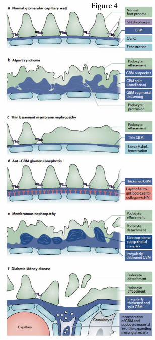
Figure 4

Figure 5