complex anterior asthetic rehabilitation- an
TRANSCRIPT

*Corresponding Author Address: Mrunal Doiphode,Department of Prosthodontics, C.S.M.S.S Dental College and Hospital, Kanchanwadi, Paithan Road, Aurangabad – 431005, Maharashtra. Email:- [email protected]
International Journal of Dental and Health Sciences
Volume 02, Issue 01
Case Report
COMPLEX ANTERIOR ASTHETIC REHABILITATION-
AN INTERDISCIPLINARY APPROACH
Prajkta Khadse1,Mrunal B. Doiphode2,Abhay Kamra3,Kishor M. Mahale4,Babita Yeshwante5,Nazish Baig6
1.P.G. Student, Dept. of Conservative Dentistry and Endodontics, CSMSS Dental College & Hospital, Kanchanwadi, Aurangabad 2.P.G. student, Dept. of Prosthodontics, CSMSS Dental College & Hospital, Kanchanwadi, Aurangabad. 3.Professor & Head of Department, Dept. of Conservative Dentistry and Endodontics, CSMSS Dental College & Hospital, Kanchanwadi, Aurangabad. 4.Professor & Head of Department, Dept. of Prosthodontics, Govt. Dental College & Hospital, Mumbai. 5.Professor & Head of Department, , Dept. of Prosthodontics, CSMSS Dental College & Hospital, Kanchanwadi, Aurangabad. 6.Professor, , Dept. of Prosthodontics, CSMSS Dental College & Hospital, Kanchanwadi, Aurangabad.
ABSTRACT:
Complete rehabilitation of smile of a patient with multiple esthetic challenges involves a multidisciplinary approach and presents a considerable clinical challenge. The interactions between new restorative materials and techniques allow the reproduction of dental structures, restoring form and function in such a way that restorative procedures become imperceptible. The completion of root development and closure of the apex occurs up to 3 years after the eruption of the tooth. Traumatic dental injuries during this period result in endodontic complications. Unfavorable relationships between the residual edentulous ridge, pontic, and gingival papilla may compromise the definitive result of a restoration. Different procedures have been described and developed to improve the relationship between esthetics and functionally acceptable fixed partial dentures. A modified design for ovate pontics is proposed to achieve the esthetic, functional, and hygienic requirements for fixed partial dentures. The purpose of this case is to report successful apexification of non-vital central incisor with open apex using calcium hydroxide and biodentine, followed by an effective and easy technique of tissue sculpturing and long term provisionalization for fabricating modified ovate pontics, for the purpose of achieving optimal esthetics. Key words: Biodentine, Traumatic dental injuries, Apexification, Modified ovate pontic, anterior pontic, aesthetics, ovate pontic, tissue scullpturing INTRODUCTION:
Complex treatment needs can necessitate
oral rehabilitation of patients. Often these
patients will require a multi-disciplinary
approach to correct problems. To return
the patient to optimal function, regain
normal form and address possible
concerns such as esthetics, an integrated
approach that involves various disciplines
needs to be taken.
The biggest endodontic challenge while
treating teeth with associated open apices
Khadse P. et al., Int J Dent Health Sci 2015; 2(1): 230-240
231
is obtaining an apical seal. The main
objective of treatment for these teeth is
apexification. Apexification is defined as
“a method to induce a calcified barrier in
a root with an open apex or the continued
apical development of an incomplete root
in teeth with necrotic pulp.”
Calcium hydroxide has been the
first choice of material for apexification,[1]
with repeated changes over the course of
5 to 20 months to induce the formation of
a calcific barrier.[2] The unpredictable and
often lengthy course of this treatment
modality presents challenges, including
the vulnerability of the temporary coronal
restoration to reinfection.[3] moreover, the
treatment requires a high level of patient
compliance. For these reasons, one visit
apexification has been suggested.[4]
In recent times, synthetic apical barriers
have popularized as alternatives to the
traditional calcium hydroxide
apexification method.[5] Mineral trioxide
aggregate (MTA) has successfully evolved
as a material of choice for this procedure.
MTA creates an apical plug at the root end
and helps to prevent the extrusion of the
obturating materials.[6] A novel material
biodentine was announced in September
of 2010 and made available in January of
2011. Biodentine is similar to MTA in basic
composition and can serve as its
substitute.[7]
The extraction of a tooth in the anterior
region often involves simultaneous local
alveolar ridge deficiencies. In the past,
primarily prosthodontic methods were
used to compensate for these defects.
With advances in periodontology, a
number of techniques have been
developed to preserve the alveolar ridge
and surgically rebuild defective sites.
Today, these techniques are also used in
crown-and bridge prosthetics for ridge
preservation before or directly after
extraction, as well as for buccal crown-
lengthening procedures and ridge
augmentation procedures, leading to an
increased frequency of satisfying ridge
contours.[8]
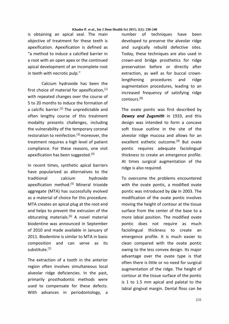
The ovate pontic was first described by
Dewey and Zugsmith in 1933, and this
design was intended to form a concave
soft tissue outline in the site of the
alveolar ridge mucosa and allows for an
excellent esthetic outcome.[9] But ovate
pontic requires adequate faciolingual
thickness to create an emergence profile.
At times surgical augmentation of the
ridge is also required.
To overcome the problems encountered
with the ovate pontic, a modified ovate
pontic was introduced by Liu in 2003. The
modification of the ovate pontic involves
moving the height of contour at the tissue
surface from the center of the base to a
more labial position. The modified ovate
pontic does not require as much
faciolingual thickness to create an
emergence profile. It is much easier to
clean compared with the ovate pontic
owing to the less convex design. Its major
advantage over the ovate type is that
often there is little or no need for surgical
augmentation of the ridge. The height of
contour at the tissue surface of the pontic
is 1 to 1.5 mm apical and palatal to the
labial gingival margin. Dental floss can be

Khadse P. et al., Int J Dent Health Sci 2015; 2(1): 230-240
232
used to push the labial gingival margin
away and cleanse the tissue surface
without any difficulty, in contrast with
other pontic types. The labial gingival
margin rebounds after the dental floss is
removed. Due to the specific design of
this pontic, the illusions of a free gingival
margin and papilla are created, and dark
interproximal spaces, also known as
“black triangles,” are minimized.
However, the modified ovate pontic may
leave a shadow in the apical area of the
tooth-gingival margin if Class I or Class II
ridge defects and a high smile line are
present.[10] Hence, the modified ovate
pontic offers outstanding results with
regards to esthetics, function, and
phonetics, and also, risk of food impaction
is minimal. (Figure 1)
It appears that the presence of adjacent
teeth and the size of gingival embrasure
formed by the teeth is responsible for the
presence and height of the papilla; a
deeper sulcus and overall increased height
of tissue above bone will exist on the
interproximal (5mm) as compared to
facial aspect (3mm). After extaction of a
tooth, a confined embrasure no longer
remains, and the interdental papilla
receded to the same 3mm level above
bone as exists facially, and ginigval scallop
flattens, which results in compromised
gingival esthetics. (Figure 2) [11] One
solution to this is to fabricate a long term
provisional restoration with the same
embrasure volume as existed prior to
extraction, with the application of
controlled pressure in an apical direction,
and the papilla and surrounding gingival
tissues will be subsequently permitted to
reform.
Several authors demonstrated that soft
tissue under pontics is associated with
clinical signs and symptoms of
inflammation such as edema, swelling,
and histological changes. Contrary to
these findings, Silness et al and Tolboe et
al reported that clinically healthy
conditions can be established at pontic
sites if appropriate plaque control is
performed.[12] Jaques et al reported that a
well-controlled hyperpressure applied
with a convex and highly polished pontic,
associated with rigid plaque control,
resulted in only a thinning of the
epithelium and shortening of rete pegs,
without inflammation.[13]
The article highlights an interdisciplinary
approach (Endodontic – Prosthodontic)
for a successful rehabilitation of smile of a
patient with an esthetic challenge. In this
case, we report successful apexification of
non-vital central incisor with open apex
using calcium hydroxide and biodentine,
followed by an effective and easy
technique of tissue sculpturing and long
term provisionalization for fabricating
modified ovate pontics.
CASE DETAIL:
A 49‑year‑old male patient was reported
to the Department of Conservative
Dentistry and Endodontics with the chief
complaint mobile and discolored upper
front teeth with H/O trauma 10 years
back. (Figure 3) The tooth in question did
not respond to both electric and heat pulp
tests. The pre‑operative radiograph

Khadse P. et al., Int J Dent Health Sci 2015; 2(1): 230-240
233
revealed a large blunderbuss canal for
upper central incisors. (Figure 4) Clinical
examination revealed with deep cervical
abrasion with discoloured left maxillary
central incisor and grade III mobility with
right maxillary central incisor and
discoloured right maxillary lateral incisor.
(Figure 3) Due to the poor prognosis and
diminished bone support with respect to
right maxillary central incisor, extraction
of the tooth was advised. Apexification
with biodentine was planned for the other
maxillary central incisor and conventional
endodontic therapy for the lateral incisor.
Labial abrasion was sealed with light cure
composite restoration and standardized
access cavities were prepared and
working length was determined.
Biomechanical preparation was carried
out using 80‑size k‑file in circumferential
manner. Root canal was disinfected using
1% NaOCl and normal saline. Calcium
hydroxide was placed as an intracanal
medicament and patient was recalled
after a week.
At 1‑week recall, canal was irrigated using
1% NaOCl and normal saline. The canals
were dried using paper points and
biodentine was mixed according to
manufacturer’s protocol and it was placed
with a plugger until a thickness of 5 mm. A
sterile cotton pellet was placed in the
canal and the cavity was sealed using
MD‑Temp for 30 min and the root canal
was obturated with thermoplasticized
gutta percha technique. Conventional
root canal cleaning and shaping was done
for the lateral incisor and canal was
obturated with lateral condensation
technique. (Figure 5) The access cavity
was then sealed with the composite
restoration and the patient was referred
to Department of Prosthodontics for full
coverage restoration.
When the patient reported to the
Department of Prosthodontics, the chief
complaint was that of discolored upper
front teeth and a missing tooth. Clinical
examination revealed a missing right
central incisor with adjacent discolored
teeth with composite restorations. The
adjacent discolored teeth were non
tender to percussion and non mobile. The
surrounding gingiva was firm and healthy.
The oral hygiene of the patient was good.
(Figure 6)
The treatment plan was to fabricate a
conventional porcelain-fused-to-metal
fixed partial denture using a modified
ovate pontic.
Scaling and root planing were done before
starting the prosthodontic procedure and
a diagnostic impression of the abutments
upper and lower arches were made. A
wax diagnostic mock up was made on this
diagnostic cast using tooth- colored wax.
A putty impression (Speedex C-Silicone
Impression Material Coltène/Whaledent)
of this diagnostic mock up was obtained.
Tooth preparation was done on both the
abutments adjacent to the tooth to be
extracted. After following all aseptic
protocol and administering local
anaesthesia by local infiltration with 2%
lidocaine containing 1:80,000 epinephrine
(Lignox 2% Adr, Indoco. Remedies, Goa,
India). Ginigivoplasty was also performed

Khadse P. et al., Int J Dent Health Sci 2015; 2(1): 230-240
234
at the same time with a foot ball shaped
diamond according to the procedure as
described by Liu.[10] A 30˚ to 45˚
gingivoplasty was made in the labial
edentulous area and extended apically
and palatally 1 to 1.5 mm from labial
gingival margin. The lingual edentulous
area was prepared to create a shallow
concavity.(Figure 7)
An irreversible hydrocolloid (Tropicalgin;
Zhermack, Badia Polesine, Italy)
impression was made of the upper arch,
and the impression was poured in die
stone (Ultrarock, Kalabhai Karson Pvt.
Ltd., Mumbai, India)
With the help of an acrylic resin trimming
bur the pontic site was minimally
prepared so that the final emergence was
in harmony with the emergence profile of
the natural adjacent teeth. The stone
preparations and ovate pontic site was
coated with separating media.
Autopolymerising provisional crown
material (DPI™ Self – Cure. Tooth Molding
Powder, Dental Products of India) was
mixed and poured it in the putty
impression of the diagnostic mock-up
obtained before. Orient it on the stone
cast, and tie the cast and impression with
rubber bands. The provisional restoration
was trimmed, finished and polished. To
achieve the objective of optimal soft-
tissue levels, the gingival and subgingival
forms of the provisional restoration were
subtly contoured. The provisional
restoration was seated to observe the
adaptation of the surrounding gingiva.
The necessary additions or subtractions
were performed on the provisional
restorations. Thin diamond disks were
used to delineate proximal contact areas
to create the perception of separate
teeth. This contouring process was
continued until the soft-tissue profile of
the available gingival tissue was
optimized. The provisional restoration
was adjusted for correct contour,
emergence profile and occlusion, and final
finishing and polishing was done. At this
stage, the surface of the provisionals was
not kept too convex, as the gingival
tissues had not undergone epithelization
yet. (Figure 8A)
The provisional restoration was cemented
with a non-eugenol temporary cements
cement (TempLute, PrimeDent). Care was
taken to ensure that dental floss could
pass between the pontic and the abuting
tissue. The patient was given oral hygiene
instructions, and the use of mouthwash
and dental floss was prescribed. (Figure 9)
The patient was kept on a regular follow
up at 15 days, 30 days, 45 days and 60
days after soft tissue contouring. (Figure
10 A, B, C & D) The pressure points were
relieved at each appointment and the
pontic site was checked for proper healing
and healthy attached gingiva. At every 15
days interval, 1-mm increment of tooth-
colored acrylic was added to the tissue
surface of the ovate pontic, trimmed,
finished and polished to a convex surface.
(Figure 8B) The convex design of the
pontic was intended to form a concave
soft tissue outline in the site of the
alveolar ridge. After the modifications, the
provisional restoration was reseated.
Some blanching of the gingiva occurred,
Khadse P. et al., Int J Dent Health Sci 2015; 2(1): 230-240
235
which is acceptable and expected upon
initial seating of the provisional
prosthesis. This procedure was repeated
every 15 days. Over a period of 2 months,
the provisional restoration was observed
and further modified to fine-tune the
contours.
At 60 days after soft tissue contouring, the
vertical shaping of the pseudopapillae had
been successfully completed, with a
distally displaced apex and papilla
formation. (Figure 10D) At this stage, it
was decided to finalize the restoration.
Gingival retraction cord (SURE-Cord
Knitted Retraction Cord #000, Sure Endo
products, South Korea) was placed in the
sulcus, and final tooth preparations and
finishing was performed on tooth 12 and
21. (Figure 11) Because of the chances of
tissue rebound, final impression was
made immediately after the removal of
the provisional restoration with vinyl
polysiloxane impression material (Affinis;
Coltène/Whaledent, Inc, Cuyahoga Falls,
Ohio)
Once the soft tissue reached full maturity
and the dentist and patient were satisfied
with the aesthetic results, the approved
provisional restoration provided an
invaluable script for the laboratory
technician to follow. The laboratory was
provided with a cast of the provisional
restoration.
The trial of the metal framework was
done (Figure 12), followed by shade
selection, followed by bisque trial and
required modifications, and finally the
porcelain-fused-to-metal bridge was
cemented using Glass Ionomer Type I
luting cement (GC Gold Label Luting and
Lining Cement, GC Corp., Tokyo, Japan).
The ovate pontic atraumatically seated in
its sulcus already established by the
provisional restoration. (Figure 14 and 15)
At the time of bisque trial of the
restoration, care was taken that the tissue
surface of ovate pontic final prosthesis
was convex, smooth, polished and highly
glazed. (Figure 13)
DISCUSSION:
The artificial apical barrier technique is a
contemporary approach for managing
open apices cases. This technique uses a
barrier material that is placed at the apex
facilitating obturation by confining it
within the canal.
Recently introduced biodentine is similar
to MTA in its basic composition with the
addition of setting accelerators which is
calcium chloride not only results in fast
setting but also improves the handling
properties and strength. Calcium silicate
cements have setting times in the range of
several hours. Decreasing the setting time
was achieved by a combination of
different effects. First particle size greatly
influences the setting time, since the
higher the specific surface, the shorter the
setting. Also, adding calcium chloride to
the liquid component accelerates the
system. Finally, the decrease of the liquid
content in the system decreases the
setting time to harden within 9‑12 min.
Biodentine is superior to MTA like its
consistency is better suited to the clinical
use, ensures a better handling and safety,
does not require a two‑step obturation

Khadse P. et al., Int J Dent Health Sci 2015; 2(1): 230-240
236
and as the setting is faster, there is a
lower risk of bacterial contamination.[7]
Apart from functional and hygienic
requirements, the pontics of fixed partial
dentures in anterior segment have to
satisfy aesthetic function also. Earlier, the
ridge lap pontic was used in the aesthetic
zone, but due to its concave inferior
surface oral hygiene was difficult to
accomplish. For maintenance of good oral
hygiene sanitary pontic and the modified
ridge lap pontic were developed but they
did not satisfy the aesthetic requirements.
Later on, the ovate pontic was developed
which had good emergence profile as
compared to all previously discussed
pontics. Its convex design was intended to
fabricate a concave soft tissue outline in
the edentulous ridge mucosa.[8] Here a
modified ovate pontic was prepared
which had the height of the contour to a
more labial position as compared to an
ovate pontic for which the height of the
contour was at the centre of the
edentulous ridge labiolingually.
This article describes a technique for
preparation of the edentulous site of an
anterior tooth such that it provided a
good emergence profile. An acrylic resin
provisional fixed partial denture with
modified ovate pontic design was used for
this purpose. The technique was
beneficial to the patient because esthetics
was re-established during the period of
temporization comfort, phonetics and
aesthetics were verified. Sufficient time
for tissue healing was provided.
The modified ovate pontic was an
improvement over the conventional ovate
pontic as it had better cleansing ability
and less soft tissue-contact and black
triangles were eliminated. It does not
require as much faciolingual thickness to
create an emergence profile as ovate
pontic, and requires little or no surgical
augmentation of the ridge.[10] The
drawback of this mode of treatment is
that more conservative treatment options
like implant, fibre reinforced composites
or Maryland bridges could be followed.
The period of temporization was also
prolonged.
CONCLUSION:
The necessity for an interdisciplinary
approach to treatments of routine dental
problems has been recognized for a long
time. Restoring a missing, diseased or
discoloured tooth in the anterior quadrant
is a major esthetic challenge. In the
present case, a P.G. student in
Endodontics, and a P.G student in
Prosthodontics participated in the dental
management of a patient with a mobile
and discolored incisors with H/O trauma
10 years back. It is clear that without such
cooperative action the prognosis would
not have been good. In this case report
the pontic design which has been chosen
was a modification over the conventional
ovate pontic. Because of its functional and
aesthetic advantages, this pontic design
can be effectively used to achieve
excellent aesthetics.

Khadse P. et al., Int J Dent Health Sci 2015; 2(1): 230-240
237
REFERENCES:
1. Rafter M. Apexification: A review. Dental Traumatology 2005;21:1-8.
2. Sheehy EC, Roberts GJ. Use of calcium hydroxide for apical barrier formation and healing in nonvital immature permanent teeth: A review. British Dental Journal 1997;183:241-46.
3. Magura ME, Kafrawy AH, Brown CE Jr, Newton C. Human saliva coronal microleakage in obturated root canals: An in vitro study. Journal of Endodontics 1993;25:197-205.
4. Morse DR, O’Larnic J, Yesilsoy C. Apexification: Review of the literature. Quintessence International 1990;21:589-98.
5. Holden DT, Schwartz SA, Kirkpatrick TC, Schindler WG. Clinical outcomes of artificial root‑end barriers with mineral trioxide aggregate in teeth with immature apices. J Endod 2008;34:812‑7.
6. Giuliani V, Baccetti T, Pace R, Pagavino G. The use of MTA in teeth with necrotic pulps and open apices. Dent Traumatol 2002;18:217‑21.
7. Pawar AM, Kokate SR, Shah RA. Management of a large periapical lesion using Biodentine TM as
retrograde restoration with eighteen months evident follow up. J Conserv Dent 2013;16:573-5.
8. Edelhoff D, Spiekermann H, Yildirim M (2002) A review of esthetic pontic design options. Quintessence Int 33(10):736–746
9. Dylina TJ (1999) Contour determination for ovate pontics. J Prosthet Dent 82(2):136–142.
10. Chiun-lin steven liu. Use of a Modified Ovate Pontic in Areas of Ridge Defects: A Report of Two Cases.J Esthet Restor Dent 16:273-283, 2004
11. Spear FM. Maintenance of the interdental papilla following anterior tooth removal. Pract Periodont Aesthet Dent 1999; 11(1): 21-28
12. Kim TH, Domenico Cascione D., and Knezevic A. Simulated tissue using a unique pontic design: A clinical report. J Prosthet Dent 2009;102:205-210
13. Jacques LB, Coelho AB, Hollweg H, Conti PC (1999) Tissue sculpturing: an alternative method for improving esthetics of anterior fixed prosthodontics. J Prosthet Dent 81(5):630–633

Khadse P. et al., Int J Dent Health Sci 2015; 2(1): 230-240
238
FIGURES:
Figure 1. Ovate pontic versus
Modified ovate pontic.
Figure 2. The osseous scallop mimics
the CEJs of the natural dentition, which
is about 3 mm from the height of the
facial to the interproximal of the bone.
The gingival scallop, on the other hand,
averages about 3 mm from the bone on
the facial aspect. Since the tip of the
interproximal papilla averages 4.5mm
above the bone, the total gingival
scallop is 4.5mm from facial to
interproximal.
Figure 3. Initial preoperative
photograph
Figure 4. Preoperative IOPA
radiograph
Figure 5. Post-operative IOPA
radiograph, with 4mm Biodentine
apical plug and root canal obturated
with thermoplasticized gutta percha
in the central incisor and
conventional root canal treatment
completed in lateral incisor.

Khadse P. et al., Int J Dent Health Sci 2015; 2(1): 230-240
239
Figure 6. Post-endodontic therapy
clinical presentation
Figure 7. Tooth preparation and
Ginigivoplasty.
Figure 8. Lateral view of pontic of
provisionals. A. Concave aspect of lingual
surface prevents proper hygiene. B. 1mm
increment added on tissue surface, and
repolished to give a cleansable.
Figure 9. Provisionals in place; luted with
a eugenol free temporary cement
Figure 10. Healing of the modified ovate pontic site 15 days (A), 30 days (B), 45 days
(C), and 60 days (D) after soft tissue contouring, i.e. of controlled pressure applied by
long term provisional restoration. At 60 days, the vertical shaping of the pseudopapillae
has been successfully completed.

Khadse P. et al., Int J Dent Health Sci 2015; 2(1): 230-240
240
Figure 11. Modification of tooth
preparation and gingival retraction.
Figure 12. Trial of metal framework
Figure 13. Tissue surface of final
prosthesis showing the smooth and
polished convex surface of the
modified ovate pontic
Figure 14. Final prosthesis intraorally, on the day of seating. The modified ovate pontic
appears to emerge from the gingiva like a natural tooth A. Front view B. Lateral view
(right)
Figure 15. Final prosthesis
intraorally.