complement inhibition in cynomolgus monkeys by anti factor...
TRANSCRIPT
1521-0103/351/3/527–537$25.00 http://dx.doi.org/10.1124/jpet.114.215921THE JOURNAL OF PHARMACOLOGY AND EXPERIMENTAL THERAPEUTICS J Pharmacol Exp Ther 351:527–537, December 2014Copyright ª 2014 by The American Society for Pharmacology and Experimental Therapeutics
Complement Inhibition in Cynomolgus Monkeys by Anti–Factor DAntigen-Binding Fragment for the Treatment of an AdvancedForm of Dry Age-Related Macular Degeneration s
Kelly M. Loyet, Jeremy Good, Teresa Davancaze, Lizette Sturgeon, Xiangdan Wang,Jihong Yang, Kha N. Le, Maureen Wong, Philip E. Hass, Menno van Lookeren Campagne,Peter C. Haughney,1 Alyssa Morimoto, Lisa A. Damico-Beyer, and Laura E. DeForgeDepartments of Biochemical and Cellular Pharmacology (K.M.L., L.S., L.E.D.), Assay Development and Technologies (J.G., T.D.,M.W., A.M.), BioAnalytical Sciences (X.W., J.Y.), Pharmacokinetics and Pharmacodynamics (K.N.L., P.C.H., L.A.D.-B.), ProteinChemistry (P.E.H.), and Immunology (M.v.L.C.), Genentech, South San Francisco, California
Received April 25, 2014; accepted September 11, 2014
ABSTRACTAnti–factor D (AFD; FCFD4514S, lampalizumab) is a humanizedIgG Fab fragment directed against factor D (fD), a rate-limitingserine protease in the alternative complement pathway (AP).Evaluation of AFD as a potential intravitreal (IVT) therapeutic fordry age-related macular degeneration patients with geographicatrophy (GA) is ongoing. However, it is unclear whether IVTadministration of AFD can affect systemic AP activation andpotentially compromise host-immune responses.We characterizedthe pharmacologic properties of AFD and assessed the effects ofAFD administered IVT (2 or 20 mg) or intravenous (0.2, 2, or 20 mg)on systemic complement activity in cynomolgus monkeys. For theIVT groups, serum AP activity was reduced for the 20 mg dosegroup between 2 and 6 hours postinjection. For the intravenous
groups, AFD inhibited systemic AP activity for periods of timeranging from 5 minutes (0.2 mg group) to 3 hours (20 mg group).Interestingly, the concentrations of total serum fD increased up to10-fold relative to predose levels following administration of AFD.Furthermore, AFD was found to inhibit systemic AP activity onlywhen the molar concentration of AFD exceeded that of fD. Thisoccurred in cynomolgus monkeys at serum AFD levels$2 mg/ml,a concentration 8-fold greater than the maximum serumconcentration observed following a single 10 mg IVT dosein a clinical investigation in patients with GA. Based on thesefindings, the low levels of serumAFD resulting from IVT administrationof a clinically relevant dose are not expected to appreciably affectsystemic AP activity.
IntroductionAge-related macular degeneration (AMD) is a leading cause
of elderly blindness in industrialized nations and is estimatedto affect nearly 6.5% of people over 40 years in the UnitedStates alone (Klein et al., 2011). There are two forms of AMD,termed exudative (wet) and nonexudative (dry). Dry AMDaffects 85–90% of all AMD patients and is a chronic diseasethat can be a precursor to wet AMDand geographic atrophy (GA).In the early stages of AMD, insoluble extracellular aggregatescalled drusen, considered the hallmark lesion of dry AMD,accumulate in the retina (Bird, 2010). A late, advanced formof dry AMD, GA is characterized by loss or atrophy of retinalpigment epithelium and the associated fallout of overlyingphotoreceptors (Sarks et al., 1988; Jager et al., 2008). Althoughtreatment is available for wet AMD, there are no effectivetherapies for dry AMD; treatment of dry AMD is currentlylimited to prophylactic measures, such as vitamin supplements,diet, and lifestyle modifications (Prasad et al., 2010).
This work was supported by Genentech. Support for third-party writingassistance by AnshinBioSolutions Services was provided by Genentech.
Part of this work was presented previously as the following abstracts: LoyetKM, Good J, Sturgeon L, Davancaze T, Wong M, Morimoto A, van LookerenCampagne M, DeForge L, Haughney P, and Damico L (2010) Anti-factor D Fabspecifically inhibits the alternative complement pathway: in vitro character-ization and in vivo effects following administration to cynomolgus monkeys[abstract 2980]; Association for Research in Vision and Ophthalmology AnnualMeeting; 2010 May 4; Fort Lauderdale, FL; AMD Preclinical Studies Session;and Loyet KM, Good J, Sturgeon L, Davancaze T, Wong M, Morimoto A, vanLookeren Campagne M, DeForge L, Haughney P, and Damico L (2010) Anti-factor D Fab specifically inhibits the alternative complement pathway: in vitrocharacterization and in vivo effects following administration to cynomolgusmonkeys [abstract W3067]; American Association of Pharmaceutical ScientistsNational Biotechnology Conference; 2010 May 19; San Francisco, CA.
L.A.D.-B. and L.E.D. contributed equally to this work.1Current affiliation: Department of Pharmacokinetics and Drug Metabo-
lism, Amgen, Seattle, Washington.dx.doi.org/10.1124/jpet.114.215921.s This article has supplemental material available at jpet.aspetjournals.org.
ABBREVIATIONS: AFD, anti–factor D; AH50, 50% maximal hemolysis; AMD, age-related macular degeneration; AP, alternative complementpathway; CP, classical complement pathway; CS, normal cynomolgus monkey serum; ELISA, enzyme-linked immunosorbent assay; Fab, fragmentantigen binding; fB, factor B; FC, flow cell; fD, factor D; fH, factor H; GA, geographic atrophy; GVB, bovine skin gelatin in veronal buffer; HRP,horseradish peroxidase; IVT, intravitreal; mAb, monoclonal antibody; MQC, minimal quantifiable concentration; NRP-1, neuropillin-1; PBS,phosphate-buffered saline; PBS-T, PBS containing 0.05% polysorbate 20; PD, pharmacodynamic; PK, pharmacokinetic; SA, streptavidin; SPR,surface plasmon resonance; TMB, tetramethylbenzidine.
527
http://jpet.aspetjournals.org/content/suppl/2014/09/16/jpet.114.215921.DC1Supplemental material to this article can be found at:
at ASPE
T Journals on A
ugust 23, 2019jpet.aspetjournals.org
Dow
nloaded from

The complement system assists the innate immune responseby recognizing foreign antigens (Sarma and Ward, 2011). Thealternative, classic, and lectin complement pathways consist ofseveral plasma proteins that can be proteolytically activated tokill pathogens and promote inflammation. The rate-limitinginitiator of the alternative complement pathway (AP) iscomplement factor D (fD), a chymotrypsin-like serine proteasespecific for factor B (fB) (Fig. 1A). When associated with C3either in its hydrolyzed or cleaved (C3b) form, fB is a substratefor fD. Cleavage of C3-associated fB to its active form (fBb,another serine protease) yields the AP C3 convertase complex.This complex cleaves additional C3 molecules to generate evenmore C3 convertase, greatly amplifying AP activation (Makrides,1998). C5 associated with fBb and C3b yields the C5 convertasecomplex; this complex cleaves C5 to C5a and C5b. C5b plus C6-9initiates membrane pore formation, contributing to tissuedamage in several diseases, including rheumatoid arthritis andglomerulonephritis (Ricklin et al., 2010; Carroll and Sim, 2011)(Fig. 1A). Because the AP functions in an “always on” mode,a host of negative regulators keeps this pathway’s activity incheck. Beyond its role in peripheral diseases, several lines ofgenetic and pathophysiologic evidence implicate overactivationof AP in AMD (Hageman et al., 2001; Johnson et al., 2001;Anderson et al., 2002, 2010; Edwards et al., 2005; Montezumaet al., 2007; Yates et al., 2007; Francis et al., 2009; Loyet et al.,2012; Ebrahimi et al., 2013; Fritsche et al., 2013). Preclinical
and clinical evidence explicitly implicate fD in AMD. Micelacking functional fD were protected against light-inducedphotoreceptor degeneration (Rohrer et al., 2007) in a photoreceptorinjury model. In addition, circulating fD levels were found to beelevated in patients with AMD as compared with controlindividuals (Scholl et al., 2008; Stanton et al., 2011). Inpostmortem vitreous fluid, fB activation was increased inadvanced AMD patients compared with age-matched controls,although significant increases in fD levels were not seen inthese samples (Loyet et al., 2012).Based on this evidence, local selective inhibition of the AP
may represent a viable approach for the therapeutic treatmentof AMD. Some components of the AP are poor candidates forinhibition due to high plasma concentrations and/or their dual rolein both the alternative and classic complement pathways (CP).For example, protein C5 is crucial for both AP and CP activity.However, because fD has the lowest plasma concentrationamong the complement proteins (Volanakis et al., 1985), isspecific to the AP, and is the rate-limiting AP enzyme, it isdeemed the most suitable AP therapeutic target.Anti–factor D (AFD; FCFD4514S, lampalizumab), derived
frommurine parental monoclonal antibody (mAb) 166-32, is anantigen-binding fragment (Fab) of a humanized mAb directedagainst human fD (Tanhehco et al., 1999; Fung et al., 2001).mAb 166-32 was shown to reduce concentrations of cleavedcomplement components and leukocyte activation in a baboon
Fig. 1. AFD inhibited fD-dependent formation of C3convertase as determined via measurement of C3a desArg generation. (A) A schematic of the AP is shown. The C3convertase reaction with the assay readout is highlighted inyellow. Highlighted in blue is the C5 convertase reactionthat results in membrane attack complex (MAC) formation,which can contribute to tissue damage in several diseases.In the C3 convertase assay, purified protein components,C3, fB, and fD, were combined to spontaneously form the C3convertase, which cleaves C3 into C3a and C3b. C3a wasconverted to C3a des Arg by the addition of rabbit serum,which provided a source of carboxypeptidase N. (B) C3a desArg was quantified by ELISA and plotted as C3 convertaseactivity, percentage of untreated control. Negative control,humanized anti–NRP-1 Fab, and positive control, fH, wereused at 1650 nM. Mean and S.D. values from threeindependent experiments are presented. Data points werecollected in triplicate and analyzed for C3a des Arg induplicate. The mean percentage of maximum C3 convertaseactivity for three independent experiments is plotted. Theuntreated control represented maximum (100%) C3 con-vertase activity and consisted of C3 in the absence ofinhibitors. aHUS, atypical hemolytic uremic syndrome; GN,glomerulonephritis; MS, multiple sclerosis; RA, rheumatoidarthritis; SLE, systemic lupus erythematosus.
528 Loyet et al.
at ASPE
T Journals on A
ugust 23, 2019jpet.aspetjournals.org
Dow
nloaded from

model of cardiopulmonary bypass surgery (Undar et al., 2002).AFD is currently being investigated as a potential intravitreal(IVT) administered therapeutic for GA. In the studies presentedin this work, we characterized the binding and pharmacologicalactivities of AFD. It is highly desirable to inhibit AP only in theeye, sparing systemic APactivity asmuch as possible. Our studiesin cynomolgus monkeys demonstrated that IVT administeredAFD only transiently suppressed systemic AP activity. This isexplained by the minimal systemic concentrations of AFDresulting from IVT administration combined with the pro-gressive neutralization of AFD by a high steady-state pro-duction of fD.
Materials and MethodsAFD (produced at Genentech, South San Francisco, CA) was
provided as a lyophilized powder at 70 mg/vial. Sterile water forinjection, USP (Hospira, Lake Forest, IL), was used to reconstituteAFD to 100 mg/ml. Purified human fD, purified human factor H(fH), human C3a des Arg standard, C1q-depleted human serum,and fB-depleted human serum were obtained from ComplementTechnologies (Tyler, TX). Recombinant cynomolgus fD, anti–neuropillin-1(NRP-1) Fab, anti–huC5, and amousemAb to human fD (clone 4676)wereproduced at Genentech. AnothermousemAb to AFD (clone 242) developedat Genentech binds specifically to AFD and detects AFD, regardless ofwhether it is free or bound to fD. Other materials include native activatedhuman complement component 1s (Calbiochem, EMDMillipore, Billerica,MA), human fD (positive control; Calbiochem), recombinant humaninterferon a2 (negative control; Roche Pharmaceuticals, Nutley, NJ),bovine skin gelatin (Sigma-Aldrich, St. Louis, MO), veronal buffer(BioWhittaker, Walkersville, MD), anti-human C3a des Arg–specificmAb (Genetex, San Antonio, TX), biotinylated anti-human C3aa mAb(Fitzgerald Industries, Concord, MA), streptavidin–horseradish peroxidase(SA-HRP) (GE Healthcare, Piscataway, NJ), tetramethylbenzidine (TMB;KPL, Gaithersburg, MD), rabbit erythrocytes, sheep erythrocytes(Colorado Serum, Denver, CO), pooled male (n 5 5) and female (n 55) naive Chinese origin cynomolgus monkey (Macaca fascicularis)serum (Valley BioSystems, Sacramento, CA), normal cynomolgusmonkey serum (CS; Bioreclamation, Westbury, NY), goat anti-human IgG (H&L)-HRP (Bethyl Laboratories, Montogomery, TX),and mouse IgG (Equitech-Bio, Kerrville, TX).
Measurement of Binding Affinity. Binding kinetics and affinitiesof AFD to native human fD and recombinant cynomolgus fD werecharacterized using a Biacore T100 instrument (GE Healthcare).AFD (10 mg/ml) was immobilized on flow cells (FCs), FC2 and FC4, ofa Series S CM5 sensor chip (GE Healthcare), according to manufacturer’sinstructions, at approximately 80 response units. The two remaining FCs,FC1 and FC3, were used as in-line references. Various concentrationsof fD diluted into HBS-P buffer [0.01 M 4-(2-hydroxyethyl)-1-piperazineethanesulfonic acid, 0.15 M sodium chloride, and 0.05%polysorbate 20] were injected into the four FCs at a flow rate of100 ml/min for 2.5 minutes, followed by a 30-minute dissociation time.The sensor chip was then regenerated by consecutive injection of 4 MMgCl2, at a flow rate of 30 ml/min, for 100 seconds. The experimentswere carried out at 37°C. Sensorgrams of the interaction between AFDand fDwere generated for each experimental run after in-line referencecell correction, followed by buffer sample subtraction. The dissociationrate constant (kd) and association rate constant (ka) were calculatedwith the BIA evaluation software (version 3.2; GE Healthcare) byfitting the sensorgrams with a 1:1 Langmuir binding model with masstransfer correction, and the dissociation equilibrium constant (KD) wascalculated by taking the ratio of kd over ka.
C3 Fluid-Phase Convertase Assay. AFD was tested for its abilityto inhibit the in vitro generation of C3a through the AP C3 convertasecomplex. Carboxypeptidase N in rabbit serum was used (Bokisch andMüller-Eberhard, 1970) to cleave C3a to its inactive form, C3a des Arg.An antibody specific to the des Arg–containing neoepitope was then
used in an enzyme-linked immunosorbent assay (ELISA) to quantitatethe amount of C3a generated by the C3 convertase complex (Fig. 1A).
C3 purified from human serum (Wiesmann et al., 2006) was dilutedto 0.2 mM in 0.5% bovine skin gelatin in veronal buffer (GVB). AFDwas diluted in GVB. Equal volumes (25 ml each) of C3 and AFD orcontrols (fH as positive control, anti–NRP-1 Fab as negative control,and GVB alone as an untreated control) were combined in a 96-wellround-bottom polypropylene plate and preincubated for 10 minutes atroom temperature. Human fB and fD (Complement Technologies)were combined at a concentration of 1.2 mM each in GVB containing0.09 M EGTA and 0.1 M MgCl2; 25 ml was added to each wellcontaining the C3/AFD mixture. After an additional 10-minuteincubation at room temperature, the C3a produced by C3 convertasein the reaction mixtures was cleaved to C3a des Arg by adding rabbitserum (25 ml, diluted 1:2 with GVB); the reactions were quenched byadding 100 ml 0.25 M EDTA. C3a des Arg concentrations weresubsequently measured by ELISA, as described below.
The anti-human C3a des Arg–specific mAb was diluted to 1 mg/mlin phosphate-buffered saline (PBS), pH 7.4, and coated on 384-wellELISA plates (Nunc, Neptune, NJ) through an overnight incubationat 4°C. Plates were washed with PBS containing 0.05% polysorbate 20(PBS-T) and blocked by a 2-hour incubationwith PBS and 0.5% bovineserum albumin. This and all subsequent incubations were performedat room temperature with gentle agitation; washes with PBS-T wereperformed between each assay step. Human C3a des Arg standardand samples from the C3 convertase reaction were diluted in assaybuffer (PBS-T containing 0.5% bovine serum albumin), added towashed plates, and incubated for 2 hours. Plate-bound C3a des Argwas detected following a 2-hour incubation with biotinylated anti-human C3aa mAb and a 30-minute incubation with SA-HRP. TMBwas added, color was developed for 7–8 minutes, and the reaction wasstopped using 1 M phosphoric acid. The plates were read at 450 nmwith a 620 nm reference using a microplate reader (Multiscan Ascent;Thermo Fischer, Waltham, MA). The concentrations of C3a des Arggenerated in the convertase reactions were calculated from a four-parameter fit of the C3a des Arg standard curve using in-house Excel-based software. The minimal quantifiable concentration (MQC) ofC3a des Arg was 390 pg/ml.
AP Hemolysis Assay. As a more physiologically relevant in vitroassay, the ability of AFD to inhibit AP activity was evaluated ina hemolytic assay in which serum (either human or monkey) wascombined with rabbit erythrocytes, as designed and described byPangburn (1988) and Katschke et al. (2009). To ensure complementactivation did not occur through the CP, C1q-depleted human serumwas used, and the buffer included EGTA to chelate calcium, a cationessential for CP activity. fH, a negative regulator of the AP, was used asa positive control in these studies, and an irrelevant humanized Fab(anti–NRP-1) served as the negative control.
C1q-depleted human serum or pooled male (n 5 5) and female (n 5 5)naive Chinese origin cynomolgusmonkey (M. fascicularis) serumwas usedto activate the AP. The concentration of fD present in 10% C1q-depletedhuman serum was 9.6 nM in-well, a value in agreement with previouslyreported fD levels in serum (Barnum et al., 1984; Loyet et al., 2012).
CP Hemolysis Assay. To determine whether the inhibitory activityof AFD was specific for the AP, a CP-mediated hemolysis assay wasdesigned and performed, as described by Mayer (1961) and Katschkeet al. (2009), using sheep erythrocytes sensitized with rat anti-ForssmanIgM antibody (Genentech) (May and Frank, 1973). To ensure complementactivation proceeded through the CP only, fB-depleted human serum andbuffer conditions designed to be optimal for CP activity were used. Thepositive control consisted of anti-human C5; the negative controls were fHand anti–NRP-1 Fab.
Pharmacokinetic/Pharmacodynamic Study in CynomolgusMonkeys. AFDwas administered by a single-dose IVT or intravenousinjection to male cynomolgus monkeys (M. fascicularis) of Chineseorigin to assess the pharmacokinetics (PK) and pharmacodynamics(PD) of the molecule. The animals were aged 2.8 years 6 0.52 years(mean6 S.D.; range 2.3–3.8 years) with an initial body weight of 2.4 kg
PD Effects of Anti–Factor D by IVT and Intravenous Dosing in Cynos 529
at ASPE
T Journals on A
ugust 23, 2019jpet.aspetjournals.org
Dow
nloaded from

6 0.46 kg (mean6 S.D.; range of 1.9–3.8 kg). This study was conductedat Covance Laboratories (Madison, WI). All procedures were conductedin compliance with the US Department of Agriculture Animal WelfareAct Regulations (9 CFR 3), Guide for the Care and Use of LaboratoryAnimals, and the Office of Laboratory Animal Welfare.
The study consisted of five groups. For groups 1 and 2 (n5 10 each),AFD was administered to both eyes, in two 50 ml IVT doses, separatedby 15 minutes. These animals received 1 or 10 mg/eye for a total of 2 or20 mg/animal. Animals in groups 3, 4, and 5 (n5 3 each) received AFDadministered at 0.2, 2, and 20 mg/animal, respectively, by a slowintravenous bolus injection via the saphenous vein.
Predose and postdose serum samples were collected from eachanimal via the femoral vein for PKand PDanalyses. At each time point,whole blood was collected into serum separator tubes, allowed to clot atambient temperature for at least 20 minutes, then centrifuged ina refrigerated centrifuge set at a temperature range of 2°C–8°C. Theserum was harvested within 20 minutes of centrifugation and storedbetween 260°C and 280°C until analysis. For the 2 and 20 mg IVTgroups, blood was collected predose (day 22) and postdose at thefollowing time points: 45 minutes, and 2, 6, 10, 24, 34, 48, 96, 120, 154,192, 288, and 384 hours. For the 0.2, 2, and 20 mg intravenous groups,blood samples were collected predose (day 22) and postdose at thefollowing time points: 5 and 30 minutes, and 1, 2, 3, 5, 8, 24, 34, 96, and168 hours. For IVT group animals only, after blood collections at 24, 48,120, 192, and 384 hours, two animals per group were removed from thestudy and euthanized to collect ocular matrix; the ocular data are notshown in the current study.
Determination of Inhibition of Systemic AP Activity inAFD-Treated Cynomolgus Monkey Serum. To evaluate the timecourse and dose dependency of any potential inhibition of systemic APactivity subsequent to AFD dosing, an ex vivo assay similar to the invitro AP hemolysis assay described above was performed. A keydifference in this assay, however, was that, instead of adding a dilutioncurve of exogenous AFD to the serum samples, the samples themselveswere serially diluted, with any inhibition of hemolytic activity attributedto the injected dose of AFD.
Erythrocytes were prepared, and the assay was performed, asdescribed above, for the AP hemolysis assay with the followingmodifications. To determine the absorbance corresponding to maxi-mum lysis, total lysis controls were prepared with sterile water (80 ml/well), whereas GVB was added to all other wells (50 ml). Cynomolgusmonkey serum samples were serially diluted 1:1.5 over six points andadded along with a negative control (buffer only) to 96-well U-bottompolypropylene plates (30 ml/well). The total lysis controls representedmaximum (100%) hemolysis. Data points were collected in triplicate,and the mean percent maximum hemolysis was plotted against thereciprocal of the final serum dilution in the assay. The 50% maximalhemolysis (AH50) values, defined as 50% maximal hemolysis, weredetermined by nonlinear regression analysis using a four-parameterfit model. For those curves that did not reach saturation, the AH50
was estimated using a curve fit in which the upper asymptote wasfixed at 100%. The percent relative hemolysis was calculated for eachindividual time point as [(postdose AH50 for the individual timepoint)/(predose AH50)] � 100. The AH50 value for serum from eachindividual normal cynomolgus monkey can vary as much as 2-foldfrom the overall average of AH50 values. Therefore, the predose andpostdose samples from each study animal were run on the same assayplate to ensure that postdose changes in AP activity were directlycompared with the individual animal’s baseline complement activity.
Total AFD ELISA. The PK of AFD subsequent to IVT orintravenous dosing was assessed in cynomolgus monkey serumsamples using an ELISA that detects total (free and bound) AFD.Clone 242 was diluted to 1 mg/ml in coating buffer (0.05 M sodiumcarbonate buffer, pH 9.6), added to 96-well ELISA plates (NuncMaxiSorp; Thermo Scientific, Rochester, NY), and coated overnight at4°C. The plates were washed with PBS-T and blocked with assaybuffer; the overall ELISA process was essentially the same as the C3ades Arg ELISA described above. The AFD standard curve was
prepared by serially diluting AFD from 0.25 to 32 ng/ml in assaybuffer containing 10% CS. Assay controls and samples were diluted toa minimum of 1:10 in assay buffer, and subsequent dilutions weremade in assay buffer containing 10% CS. Plate-bound AFD wasdetected with goat anti-human IgG-HRP diluted with assaybuffer containing 100 mg/ml mouse IgG. TMB substrate wasadded, and the reaction was terminated with 1 M phosphoric acidafter an incubation of approximately 15 minutes. Plates wereread, as described above, using a microplate reader (SpectraMax190; Molecular Devices, Sunnyvale, CA), and AFD concentrationswere calculated from a four-parameter fit of the standard curve oneach plate (SoftMax Pro; Molecular Devices). Taking into accountthe minimum serum dilution (1:10), the effective standard curveconcentration range for AFD was 2.5–320 ng/ml and the MQC was15 ng/ml in cynomolgus monkey serum.
Total fD ELISA. Total fD wasmeasured using an ELISA in whichfD was captured on the plate and incubated with an excess of the AFDtherapeutic to ensure complete saturation of available fDbinding sites, andthe fD/AFD complex was detected using the same AFD-specific mAb usedabove in the ELISA for total AFD.
Briefly, clone 4676 was diluted to 1 mg/ml in coating buffer andincubated overnight at 4°C on 96-well ELISA plates. The overall assaymethodology was the same as described for the total AFD ELISA. Thecynomolgusmonkey fD standard curve was prepared by serially dilutingfD from 0.04–5 ng/ml in sample buffer (assay buffer supplemented with500 ng/ml AFD therapeutic, 50 mg/ml mouse IgG, and 1% fD-depletedCS). The assay controls and serum samples were diluted to a minimumof 1:100 in sample buffer. The diluted standards, controls, and sampleswere then incubated on the plates for 2 hours, and plate-bound fD/AFDcomplex was detected using biotin-conjugatedmousemAb to AFD (clone242, 1mg/ml), followed by SA-HRP (3 ng/ml). Color was developed, plateswere read, and concentrations of fD were determined, as describedabove. Taking into account the minimum serum dilution (1:100), theeffective standard curve concentration range of fD was 4–500 ng/ml andthe MQC in cynomolgus monkey serum was 5 ng/ml.
ResultsAFD Binds to fD with High Affinity and Specifically
Inhibits Human and Cynomolgus AP Activity. Surfaceplasmon resonance (SPR)–based Biacore experiments demon-strated that AFD bound to human fD with an average KD of20 pM (Table 1) under the tested conditions. The high kavalue (ka 5 3.6 � 107 M21s21) indicated a very fast process,which might have approached or reached the detection limitsof the Biacore T100 instrument (Onell and Andersson, 2005).However, no significant or systemic discrepancy was observedbetween the experimental data and the fitted curves (data notshown), suggesting the 1:1 binding model is adequate to describethe AFD and fD interaction.AFD was next tested in two functional assays to determine
whether it neutralized the ability of fD to initiate AP activation.
TABLE 1Kinetic analysis and binding affinity of human and recombinantcynomolgus monkey factor D to anti–factor DThe data shown are mean 6 S.D. Mean and S.D. were calculated based on fourindependent experimental runs for human fD binding to AFD and five independentexperimental runs for cynomolgus monkey fD binding to AFD.
Analyte ka kd KD
107 M21s21 1024 s21 pM
Human fD 3.6 6 1.0 7.0 6 1.1 19.7 6 2.9r cyno fD 20.8 6 10.0 23.3 6 9.0 11.7 6 1.6
ka, association rate constant; kd, dissociation rate constant; KD, dissociationequilibrium constant.
530 Loyet et al.
at ASPE
T Journals on A
ugust 23, 2019jpet.aspetjournals.org
Dow
nloaded from
The fluid-phase C3 convertase assay tested the ability of AFDto inhibit fD-dependent activation ofC3 convertase via quantitationof the amount of C3a des Arg produced (Fig. 1A). In this method,the reagent concentrations were optimized to yield an optimalsignal rather than to adhere to physiologic conditions, and fDwas present at 400 nM in well. Under these assay conditions,AFD completely inhibited C3a des Arg production at a concen-tration at and above 333 nM, whereas negligible inhibition wasobserved at 148 nM (Fig. 1B). In contrast, no inhibition of C3convertase activity was detected with a negative control Fabtested at 1650 nM, whereas maximal inhibition was achievedwith a positive control (fH, a specific inhibitor of the AP).AFDwas also tested in vitro in cell-basedAP- andCP-dependent
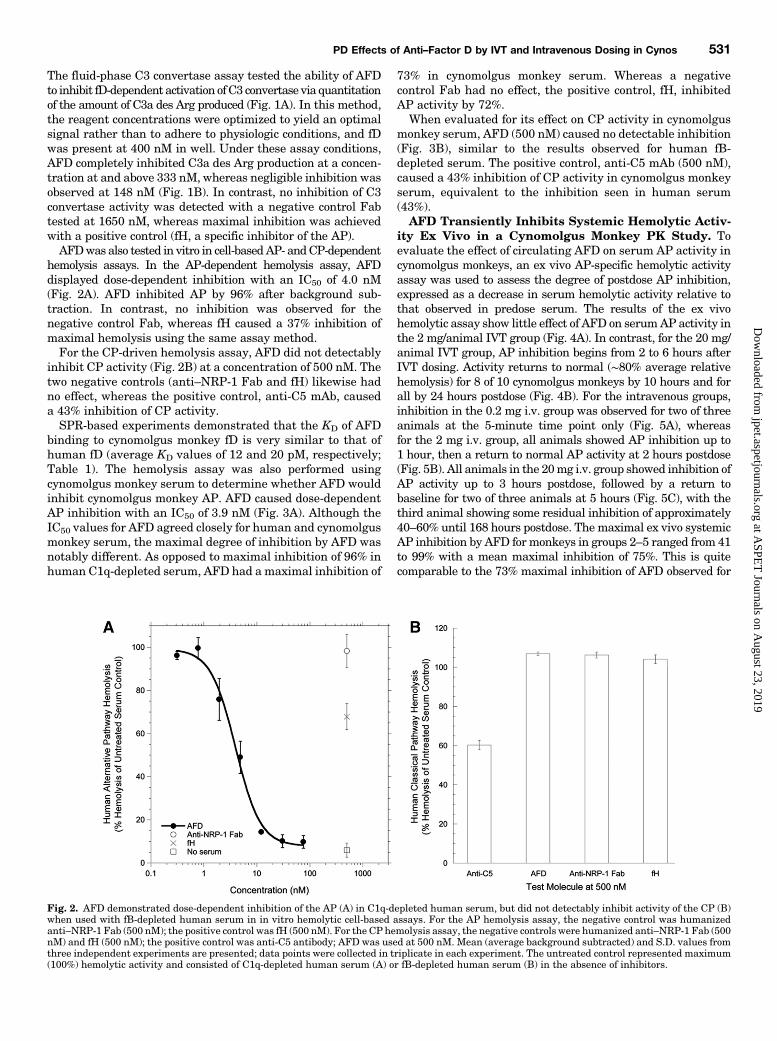
hemolysis assays. In the AP-dependent hemolysis assay, AFDdisplayed dose-dependent inhibition with an IC50 of 4.0 nM(Fig. 2A). AFD inhibited AP by 96% after background sub-traction. In contrast, no inhibition was observed for thenegative control Fab, whereas fH caused a 37% inhibition ofmaximal hemolysis using the same assay method.For the CP-driven hemolysis assay, AFD did not detectably
inhibit CP activity (Fig. 2B) at a concentration of 500 nM. Thetwo negative controls (anti–NRP-1 Fab and fH) likewise hadno effect, whereas the positive control, anti-C5 mAb, causeda 43% inhibition of CP activity.SPR-based experiments demonstrated that the KD of AFD
binding to cynomolgus monkey fD is very similar to that ofhuman fD (average KD values of 12 and 20 pM, respectively;Table 1). The hemolysis assay was also performed usingcynomolgus monkey serum to determine whether AFD wouldinhibit cynomolgus monkey AP. AFD caused dose-dependentAP inhibition with an IC50 of 3.9 nM (Fig. 3A). Although theIC50 values for AFD agreed closely for human and cynomolgusmonkey serum, the maximal degree of inhibition by AFD wasnotably different. As opposed to maximal inhibition of 96% inhuman C1q-depleted serum, AFD had a maximal inhibition of
73% in cynomolgus monkey serum. Whereas a negativecontrol Fab had no effect, the positive control, fH, inhibitedAP activity by 72%.When evaluated for its effect on CP activity in cynomolgus
monkey serum, AFD (500 nM) caused no detectable inhibition(Fig. 3B), similar to the results observed for human fB-depleted serum. The positive control, anti-C5 mAb (500 nM),caused a 43% inhibition of CP activity in cynomolgus monkeyserum, equivalent to the inhibition seen in human serum(43%).AFD Transiently Inhibits Systemic Hemolytic Activ-
ity Ex Vivo in a Cynomolgus Monkey PK Study. Toevaluate the effect of circulating AFD on serum AP activity incynomolgus monkeys, an ex vivo AP-specific hemolytic activityassay was used to assess the degree of postdose AP inhibition,expressed as a decrease in serum hemolytic activity relative tothat observed in predose serum. The results of the ex vivohemolytic assay show little effect of AFD on serumAP activity inthe 2 mg/animal IVT group (Fig. 4A). In contrast, for the 20 mg/animal IVT group, AP inhibition begins from 2 to 6 hours afterIVT dosing. Activity returns to normal (∼80% average relativehemolysis) for 8 of 10 cynomolgus monkeys by 10 hours and forall by 24 hours postdose (Fig. 4B). For the intravenous groups,inhibition in the 0.2 mg i.v. group was observed for two of threeanimals at the 5-minute time point only (Fig. 5A), whereasfor the 2 mg i.v. group, all animals showed AP inhibition up to1 hour, then a return to normal AP activity at 2 hours postdose(Fig. 5B). All animals in the 20mg i.v. group showed inhibition ofAP activity up to 3 hours postdose, followed by a return tobaseline for two of three animals at 5 hours (Fig. 5C), with thethird animal showing some residual inhibition of approximately40–60% until 168 hours postdose. The maximal ex vivo systemicAP inhibition by AFD formonkeys in groups 2–5 ranged from 41to 99% with a mean maximal inhibition of 75%. This is quitecomparable to the 73% maximal inhibition of AFD observed for
Fig. 2. AFD demonstrated dose-dependent inhibition of the AP (A) in C1q-depleted human serum, but did not detectably inhibit activity of the CP (B)when used with fB-depleted human serum in in vitro hemolytic cell-based assays. For the AP hemolysis assay, the negative control was humanizedanti–NRP-1 Fab (500 nM); the positive control was fH (500 nM). For the CP hemolysis assay, the negative controls were humanized anti–NRP-1 Fab (500nM) and fH (500 nM); the positive control was anti-C5 antibody; AFD was used at 500 nM. Mean (average background subtracted) and S.D. values fromthree independent experiments are presented; data points were collected in triplicate in each experiment. The untreated control represented maximum(100%) hemolytic activity and consisted of C1q-depleted human serum (A) or fB-depleted human serum (B) in the absence of inhibitors.
PD Effects of Anti–Factor D by IVT and Intravenous Dosing in Cynos 531
at ASPE
T Journals on A
ugust 23, 2019jpet.aspetjournals.org
Dow
nloaded from

the in vitro hemolysis assay (Fig. 3), which used a cynomolgusmonkey serum pool from a different cohort of animals (n 5 10).Measurement of Total AFD and Total fD in AFD-
Treated Cynomolgus Monkeys. The serum concentrationsof AFD were measured using an assay capable of detectingtotal Fab (free and fD-bound). The concentration profilesshowed that in the IVT groups, the time to reach maximum
AFD concentrations (Tmax) occurred gradually at 6–10 hourspostdose (Fig. 6A), whereas the Tmax was at the first post-injection time point (5 minutes) for the intravenous groups(Fig. 6B).The average baseline (predose) serum fD concentrations
were ∼20–30 nM (Fig. 6). For the IVT groups, as serum totalAFD concentrations rose or fell, so did serum total fD
Fig. 3. AFD demonstrated dose-dependent inhibition of the AP (A), but did not detectably inhibit activity of the CP (B) in cynomolgus monkey serum inin vitro hemolytic cell-based assays. For the AP hemolysis assay, the negative control was humanized anti–NRP-1 Fab (500 nM); the positive control wasfH (500 nM). For the CP hemolysis assay, the negative controls were humanized anti–NRP-1 Fab (500 nM) and fH (500 nM); the positive control wasanti-C5 antibody; AFD was used at 500 nM. Mean (average background subtracted) and S.D. values from three independent experiments are presented;data points were collected in triplicate in each experiment. The untreated control represented maximum (100%) hemolytic activity and consisted ofa cynomolgus monkey serum pool in the absence of inhibitors.
Fig. 4. In vivo inhibition of the AP pathway followingIVT dosing in cynomolgus monkeys was observed in thehighest dose group, but was transient and returned tonormal by 10–24 hours post-AFD administration. Thepercentage of relative inhibition, normalized for eachindividual predose sample (100% relative hemolysis), isshown for all animals (n = 10/group) dosed IVT with1 mg/eye (2 mg total) AFD (A) and 10 mg/eye (20 mgtotal) AFD (B). Percentage of relative hemolysis = (AH50postdose/AH50 predose time point) � 100; baseline (….) =100% relative hemolysis. Note: Data for all time pointswere unavailable for some animals as they were removedfrom the study and used for collecting ocular samples.
532 Loyet et al.
at ASPE
T Journals on A
ugust 23, 2019jpet.aspetjournals.org
Dow
nloaded from

concentrations in a temporally concurrent fashion. Althoughthe serum AFDmaximum serum concentration (Cmax) was 30 nMat 6 hours postdose (2 mg IVT), the average fD concentration hadincreased to 57 nM at this time point, nearly double the AFDaverage molar concentration (Fig. 6A). At no point in time did theAFD concentration exceed the fD concentration, suggesting thatsufficient fD was present in an active, unbound form to initiate theAP in the ex vivo hemolytic activity assay; this is in agreementwiththe ∼100% (baseline) AP activity seen at all time points for theanimals dosed IVT with 2 mg AFD (Fig. 4A).Similarly, at the earliest time point for the 20 mg/animal
IVT group (45 minutes), the average total fD concentrationwas 33 nM. This was more than 3-fold higher than theconcentration of AFD at this time point (9 nM); hence, nosystemic AP inhibition was seen for the 20 mg/animal IVTgroup at 45 minutes postdose (Fig. 4B). However, at 2 hours,the average AFD concentration had increased to 126 nM,whereas there was a lower average fD concentration (92 nM);thus, there was significant inhibition of AP activity for mostanimals. The average serum Cmax for AFD was 260 nM at6 hours postdose, at which time the average total fD concen-tration had increased almost 10-fold from baseline to 221 nM.At 10 hours postdose, the average total fD Cmax (247 nM) was
nearly equimolar to that of the average total serum AFDconcentration (251 nM), suggesting fD predominantly existedas a bound complex form. Furthermore, the AP activity of allanimals did not return to within ∼10% of baseline until96 hours postdose (Fig. 4B), presumably due to neutralizationof AFD by increased total fD concentrations, which was sustainedthrough the last few time points (.100 hours) when the averagemolar AFD again dropped lower than the average molar fD(Fig. 6A).In contrast to the IVT groups in which the AFD and fD Tmax
occurred during the same time range (6–10 hours postdose), theTmax of fD was delayed relative to the Tmax of AFD in theintravenous dosing groups (Fig. 6B). At 0.1 hour, the averageserum Cmax levels of AFD for the 0.2, 2, and 20 mg i.v. groupswere 41, 459, and 5158 nM, respectively. These average AFDconcentrations were higher than the average fD concentration(20–30 nM); therefore, there was AP inhibition at 0.1 hour forall intravenous groups (Fig. 5). For these same groups, theaverage total fD serumCmax levels were 47 nM at 0.5 hour, 138nM at 2 hours, and 215 nM at 5 hours, respectively. At theserespective time points, as the average serum AFD concentra-tion decreased to 31, 109, and 226 nM, concentrations that werebelow or nearly equimolar to the fD Cmax, the AP activity
Fig. 5. In vivo inhibition of the AP pathway followingintravenous dosing in cynomolgus monkeys was ob-served in all dose groups, but was transient andreturned to normal by 45 minutes to 10 hours post-AFD administration. The percentage of relative in-hibition, normalized for each individual predose sample(100% relative hemolysis), is shown for all animals (n =3/group) dosed intravenously with 0.2 mg (A), 2 mg (B),or 20 mg (C) AFD. Percentage of relative hemolysis =(AH50 postdose/AH50 predose time point)� 100; baseline(….) = 100% relative hemolysis.
PD Effects of Anti–Factor D by IVT and Intravenous Dosing in Cynos 533
at ASPE
T Journals on A
ugust 23, 2019jpet.aspetjournals.org
Dow
nloaded from
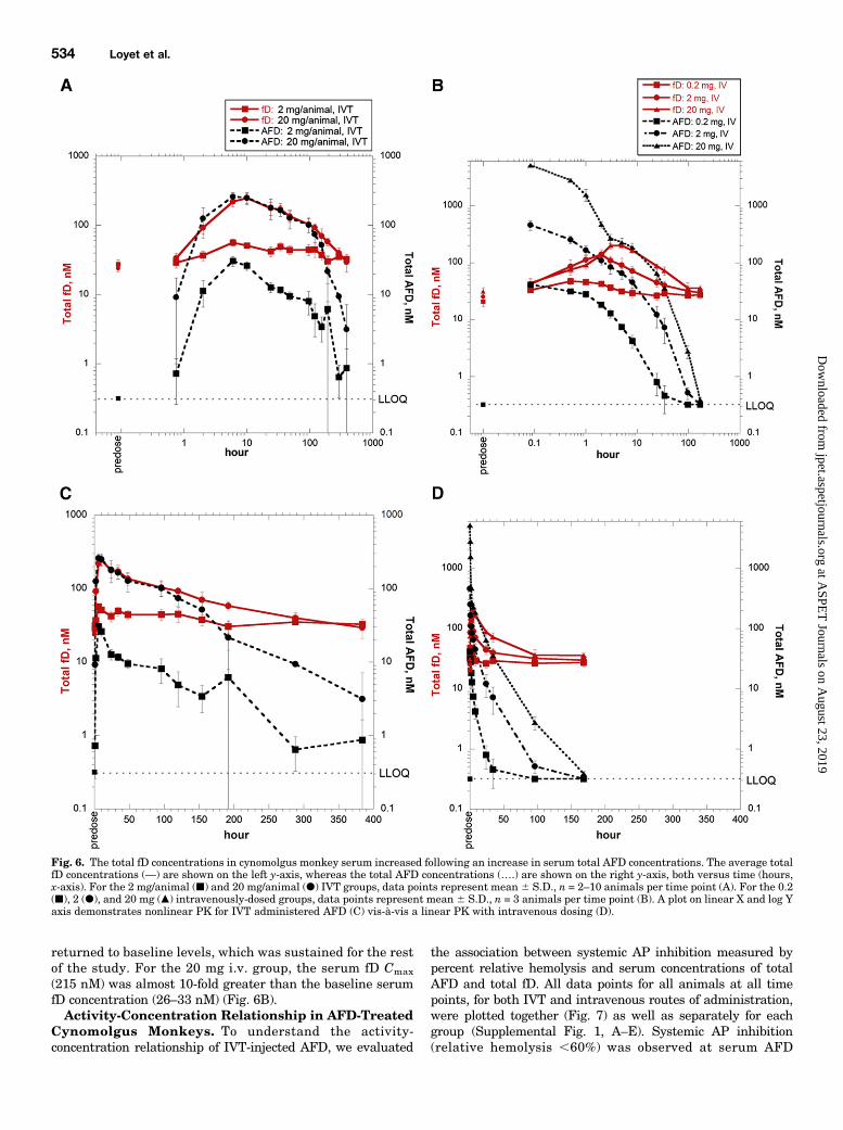
returned to baseline levels, which was sustained for the restof the study. For the 20 mg i.v. group, the serum fD Cmax
(215 nM) was almost 10-fold greater than the baseline serumfD concentration (26–33 nM) (Fig. 6B).Activity-Concentration Relationship in AFD-Treated
Cynomolgus Monkeys. To understand the activity-concentration relationship of IVT-injected AFD, we evaluated
the association between systemic AP inhibition measured bypercent relative hemolysis and serum concentrations of totalAFD and total fD. All data points for all animals at all timepoints, for both IVT and intravenous routes of administration,were plotted together (Fig. 7) as well as separately for eachgroup (Supplemental Fig. 1, A–E). Systemic AP inhibition(relative hemolysis ,60%) was observed at serum AFD
Fig. 6. The total fD concentrations in cynomolgus monkey serum increased following an increase in serum total AFD concentrations. The average totalfD concentrations (—) are shown on the left y-axis, whereas the total AFD concentrations (….) are shown on the right y-axis, both versus time (hours,x-axis). For the 2 mg/animal (j) and 20 mg/animal (d) IVT groups, data points represent mean 6 S.D., n = 2–10 animals per time point (A). For the 0.2(j), 2 (d), and 20 mg (m) intravenously-dosed groups, data points represent mean 6 S.D., n = 3 animals per time point (B). A plot on linear X and log Yaxis demonstrates nonlinear PK for IVT administered AFD (C) vis-à-vis a linear PK with intravenous dosing (D).
534 Loyet et al.
at ASPE
T Journals on A
ugust 23, 2019jpet.aspetjournals.org
Dow
nloaded from

concentrations of 40–5198 nM (2–242 mg/ml). The lowest AFDconcentrations causing.40% inhibition were 40 nM for the 0.2mg i.v. group (Supplemental Fig. 1C); 140 nM for the 2 mg i.v.group (Supplemental Fig. 1D); and 200 nM for the 20 mg i.v.group (Supplemental Fig. 1E) if one animal was excluded(cynomolgus monkey 29, which showed a partial inhibition ofhemolysis through the 96-hour time point; Fig. 5A). As themolar serum concentration of AFD approached and exceededthat of fD (fD/AFD #1; green and blue symbols), the percentrelative hemolysis decreased. For IVT-dosed animals (Supple-mental Fig. 1, A and B), AP inhibition of 40% or more was seenin samples from the higher IVT dose group (20mg/animal), butnot in samples from the lower IVT dose group (2 mg/animal).Both the 20mg IVT and intravenous groups contained sampleswith a wide range of AP inhibition (0–80% AP inhibition)despite a narrow AFD or fD concentration range (∼150–350 nM)at fD/AFD of ∼1 (Supplemental Fig. 1B). For those animals giventhe lowest intravenous dose (0.2 mg), all samples demonstrated,20% AP inhibition, with the exception of those at 5 minutespostdose, wherein up to 80%AP inhibition was observed (Fig. 5A;Supplemental Fig. 1C) because molar concentrations of fDwere lower than those of AFD [fD/AFD,1, AFD Cmax of 40 nM(2 mg/ml)]. This is in contrast to the 2 mg IVT group that hadan AFD Cmax of 35 nM (1.75 mg/ml), fD/AFD .1 at 6 hourspostdose (Fig. 6A), and did not show AP inhibition (Fig. 4A) be-cause molar concentrations of AFD were lower than those of fD.
DiscussionA role of fD in the pathogenesis of AMD is supported by
a murine model in which a genetic fD deficiency providedprotection against oxidative stress–mediated photoreceptor
degeneration (Rohrer et al., 2007). In addition, increasedsystemic activation of complement, including fD, has beendetected in the serum of AMD patients versus controls,suggesting that AMD may be a systemic disease with localmanifestations in the aging macula (Scholl et al., 2008).Many complement pathway inhibitors are currently inclinical or preclinical development for AMD (Ricklin andLambris, 2007); AFD is being evaluated as a potential IVTadministered therapeutic.In vitro studies were performed to characterize the binding
and activity of AFD in human and cynomolgus serum. Becauseanimal studies are necessary as a precursor to clinical trials, itwas important to determine the cross-reactivity of AFD withcynomolgus monkey fD to establish the appropriateness of thisspecies for in vivo testing. AFD was shown to bind to nativehuman fD (20 pM) and recombinant cynomolgus fD (12 pM)with high affinity. AFD specifically inhibited fD activity in theC3 convertase assay. In addition, in vitro serum-based RBChemolysis assays were used as a more physiologically relevantsystem; the results demonstrated that AFD potently inhibitedthe cynomolgus monkey (IC50, 3.9 nM) and human (IC50,4.0 nM) AP, but did not inhibit the CP in these species.The reason for the higher baseline activity (Fig. 3A) remaining
after AFD treatment of cynomolgus monkey serum was unclear,but it was consistent across all assays that were performed usingthis particular cynomolgus monkey serum pool. Unlike thehuman AP hemolysis assay, the cynomolgus monkey serumwasnot depleted of C1q. It is therefore possible that CP activationcould account for the higher baseline activity. However, thispossibility is lessened by including EGTA in the assay buffer tochelate calcium, an essential factor for CP activity. Differences inthe maximal activity of human fH in human versus cynomolgusmonkey serum could be due to a species-dependent difference inC3b concentration or fH binding to C3b; however, this has notbeen tested.Slight differences in AFD affinities for human and cynomolgus
fD may be explained by a single amino acid difference in humanversus cynomolgus fD (Glu178 versus Gln178) in a region near theAFD-binding interface (Katschke et al., 2012). The AP hemolysisassays require nM fD concentrations to achieve adequatehemolysis. Given the 1:1 stoichiometry of AFD:fD, nM AFDconcentrations are required for inhibition in these assaysdespite the pM affinities of AFD binding to fD shown by SPR.The results of the AP hemolysis assay were consistent withthe C3 convertase assay in terms of a complete inhibition of APactivity by AFD. Although a difference in AFD potency wasobserved in the two assay systems, this was expected given thedifferent fD concentrations (9.6 nMversus 400 nM in-well for theAP hemolysis and C3 convertase assays, respectively). Theresults overall indicate that AFD inhibits the AP by blocking fDproteolytic activity. In confirmation, crystallographic studieshave shown that AFD inhibits fD by binding to the surface loopson fD that form the exosite, thereby blocking macromolecularsubstrate access (Katschke et al., 2012).Together, the in vitro results demonstrate a specific in-
teraction of AFD with the AP in both human and cynomolgusmonkey serum; show a lack of effect on the CP in eitherspecies; and support the use of cynomolgus monkeys asa relevant preclinical model for evaluating the PK and PD ofAFD. Evaluating the activity of AFD in cynomolgus monkeyswas limited to assessing its effects on serum complementactivity due to the absence of a nonhuman primate model of
Fig. 7. In vivo inhibition of the AP pathway was generally observed whenthe serum concentration of AFD was greater than 40 nM (2 mg/ml) and fD/AFD ratio was less than 1. The percentage of relative hemolysis data(normalized to individual animals’ predose percentage of relativehemolysis, which was set at 100%) from all time points (n = 263) is shownplotted versus serum AFD concentrations for all animals (n = 29). Datapoints are color-coded for the ratio of fD versus AFD concentrations, withfD/AFD values greater than 1 increasing from greenish-yellow to yellow,orange, and red, and with fD/AFD values less than 1 decreasing fromgreen to turquoise, indigo, and blue. Percentage of relative hemolysis =(AH50 predose/AH50 postdose time point) � 100; baseline (––) = 100%relative hemolysis; AP inhibition (---) ,60% relative hemolysis.
PD Effects of Anti–Factor D by IVT and Intravenous Dosing in Cynos 535
at ASPE
T Journals on A
ugust 23, 2019jpet.aspetjournals.org
Dow
nloaded from

GA. In addition, although laser-induced choroidal neovascula-rization is used as a primate model for wet AMD, our in-housestudies did not generate conclusive evidence for the involve-ment of complement in this model. Lastly, based on poor APinhibition by AFD in dogs or sheep and no AP inhibition byAFD in rabbits, rats, mice, guinea pigs, hamsters, or pigs(US Patent Publication 2002-0081293 A1), these species werenot considered appropriate or relevant for the in vivo evaluationof AFD activity. For these reasons, we were not able to performstudies evaluating the efficacy of AFD in relevant preclinicalanimal models.A single-dose cynomolgus monkey study evaluated the AFD PK
and PD after IVT and intravenous administration. The vitreouselimination half-life was approximately 2.4 days in monkeys,consistent with ranibizumab, and the systemic elimination half-lifewas approximately 2.2 days following IVTadministration and7–20hours following intravenous administration (Do et al., 2014). Thedifference in systemic half-life following IVT compared withintravenous administration suggests that the AFD kineticsfollowing IVT administration was absorption rate-limited.The lowest observed AFD serum concentration causing tran-sient AP inhibitionwas 40 nM (2mg/ml). However, the serumAPactivity assay results for both intravenous and IVT administeredAFD demonstrated that the effect of systemic concentrations ofAFD on the AP activity was not directly related to the serumconcentration of AFD at all time points in cynomolgus monkeys.The wide range of AP inhibition (0–80%) within ∼150 nM to 350nMAFD concentration range (Fig. 7) is not surprising given thatthe fD and AFD concentrations determined by ELISA can beexpected to deviate from the true concentrations by up to620%,as is typical and widely accepted for ELISAs. Even this level oferror can potentially affect the hemolysis assay results becausethe AP can be activated by serum containing 30 nM fD (Biesmaet al., 2001).There is a limited capacity to maintain concentrations of
free (and therefore active) AFD at a level high enough to causeprolonged inhibition of AP activity in the systemic circulationfollowing IVT administration. Metabolism and excretion ofsystemic AFD, combined with neutralization of AFD by targetbinding and a high rate of fD synthesis, contribute to rapidlydecreasing serum concentrations of free AFD. The substantialincreases in serum levels of total fD seen after administeringAFD (Fig. 6) can potentially be explained by less efficient filteringof the higher molecular weight AFD-fD complexes into theextravascular space and/or slower elimination of the AFD-fDcomplexes relative to free fD. However, an increase in thetotal (free and bound) concentration of target following drugadministration is often seen for therapeutic antibodies thatbind soluble endogenous targets, especially when the antibodyaffinity is high and the target turnover is fast (Hayashi et al.,2007; Davda and Hansen, 2010 ). The high rate of fD synthesis,estimated to be 1.33 mg/kg per day in humans (Pascual et al.,1988), was substantiated by the persistently elevated total fDconcentrations, even while total AFD decreased, as seen in the 2and 20mg i.v. groups. fD is primarily expressed in adipose tissue(Cook et al., 1985), although it is also expressed in several othertissues and cell types at a much lower level. Expressed sequencetag distribution data indicate fD expression in the eye is∼40-foldlower than in adipose tissue (http://research-public.gene.com/Research/genentech/genehub-gepis/index.html), and the neuralretina contains relatively few fD transcripts compared withperipheral tissues (Anderson et al., 2010). fD protein levels are
higher in the systemic circulation than in the eye. In cynomolgusmonkeys, 20–31 nM fD was measured in serum as comparedwith 0.52 nM in vitreous fluid (38-fold lower, average of eightnaive cynomolgus monkeys). Similarly, fD concentrations frompostmortem human vitreous samples were lower than in serum(Loyet et al., 2012). Therefore, local inhibition of fD in the eye canbe achieved at lower doses of AFD than would inhibit fDsystemically, and the low systemic concentrations of AFDresulting from IVT dosing are expected to be well below thethreshold concentration required to affect systemic fD activity.A single ascending-dose phase Ia study with IVT adminis-
tration of 0.1–10 mg AFD per eye demonstrated that AFD(FCFD4514S) was safe and well tolerated in patients with GAsecondary to AMD (Do et al., 2014). The serum AFD Cmax
following a single 10mg IVT dose ranged from 4 to 5 nM. Theselevels were 5- to 6-fold lower than the baseline molarconcentrations of serum fD seen in this preclinical study(20–30 nM), suggesting that systemic AP activity wouldremain unaffected. In addition, the highest AFDCmax of 257 ng/ml(5 nM) was still approximately 8-fold lower than 2 mg/ml (40 nM),the lowest concentration causing a temporary effect on systemicAPactivity in cynomolgus monkeys. Evaluating the clinical samplesusing the AH50 assay demonstrated that the systemic concen-trations of IVT administered AFD had no observed adverse effectson the serum AP activity in these GA patients.A phase Ib/II evidence of activity study (ClinicalTrials.gov
Identifier: NCT01229215) showed the potential clinicalbenefits of AFD in patients with GA (Yaspan, 2014). The invitro and in vivo data presented in this work suggest that IVTadministered AFD has the potential to be an important noveltherapeutic for patients with GA and intermediate dry AMD.
Acknowledgments
The authors thank Erich Strauss, Sam Jackson, Hicham Alaoui,Vladimir Bantseev, Kenneth Katschke, Cris Lewis, Tong Lu, Sock-Cheng Lewin-Koh, Michelle Simpson, Micah Steffek, Cecily Sun,Meina Tang, and Yi Zhang for contributions.
Authorship Contributions
Participated in research design: Loyet, Good, van LookerenCampagne, Haughney, Damico-Beyer, DeForge.
Conducted experiments: Loyet, Good, Davancaze, Sturgeon, Wang.Contributed new reagents or analytic tools: Wong, Hass.Performed data analysis: Loyet, Good, Davancaze, Sturgeon,
Wang, Yang, Le, Morimoto.Wrote or contributed to the writing of the manuscript: Loyet, Good,
Wang, Yang, Le, van Lookeren Campagne, Morimoto, Damico-Beyer,DeForge.
References
Anderson DH, Mullins RF, Hageman GS, and Johnson LV (2002) A role for localinflammation in the formation of drusen in the aging eye. Am J Ophthalmol 134:411–431.
Anderson DH, Radeke MJ, Gallo NB, Chapin EA, Johnson PT, Curletti CR, HancoxLS, Hu J, Ebright JN, and Malek G,, et al. (2010) The pivotal role of the comple-ment system in aging and age-related macular degeneration: hypothesis re-visited.Prog Retin Eye Res 29:95–112.
Barnum SR, Niemann MA, Kearney JF, and Volanakis JE (1984) Quantitation ofcomplement factor D in human serum by a solid-phase radioimmunoassay. JImmunol Methods 67:303–309.
Biesma DH, Hannema AJ, van Velzen-Blad H, Mulder L, van Zwieten R, Kluijt I,and Roos D (2001) A family with complement factor D deficiency. J Clin Invest 108:233–240.
Bird AC (2010) Therapeutic targets in age-related macular disease. J Clin Invest 120:3033–3041.
Bokisch VA and Müller-Eberhard HJ (1970) Anaphylatoxin inactivator of humanplasma: its isolation and characterization as a carboxypeptidase. J Clin Invest 49:2427–2436.
Carroll MV and Sim RB (2011) Complement in health and disease. Adv Drug DelivRev 63:965–975.
536 Loyet et al.
at ASPE
T Journals on A
ugust 23, 2019jpet.aspetjournals.org
Dow
nloaded from
Cook KS, Groves DL, Min HY, and Spiegelman BM (1985) A developmentally reg-ulated mRNA from 3T3 adipocytes encodes a novel serine protease homologue.Proc Natl Acad Sci USA 82:6480–6484.
Davda JP and Hansen RJ (2010) Properties of a general PK/PD model of antibody-ligand interactions for therapeutic antibodies that bind to soluble endogenoustargets. MAbs 2:576–588.
Do DV, Pieramici DJ, van Lookeren Campagne M, Beres T, Friesenhahn M, Zhang Y,and Strauss EC; Phase Ia Investigators (2014) A phase ia dose-escalation study ofthe anti-factor D monoclonal antibody fragment FCFD4514S in patients withgeographic atrophy. Retina 34:313–320.
Ebrahimi KB, Fijalkowski N, Cano M, and Handa JT (2013) Decreased membranecomplement regulators in the retinal pigmented epithelium contributes to age-related macular degeneration. J Pathol 229:729–742.
Edwards AO, Ritter R, 3rd, Abel KJ, Manning A, Panhuysen C, and Farrer LA (2005)Complement factor H polymorphism and age-related macular degeneration. Sci-ence 308:421–424.
Francis PJ, Hamon SC, Ott J, Weleber RG, and Klein ML (2009) Polymorphisms inC2, CFB and C3 are associated with progression to advanced age related maculardegeneration associated with visual loss. J Med Genet 46:300–307.
Fritsche LG, Chen W, Schu M, Yaspan BL, Yu Y, Thorleifsson G, Zack DJ, ArakawaS, Cipriani V, and Ripke S,, et al.; AMD Gene Consortium (2013) Seven new lociassociated with age-related macular degeneration. Nat Genet 45:433–439, e1–e2.
Fung M, Loubser PG, Undar A, Mueller M, Sun C, Sun WN, Vaughn WK, and FraserCD, Jr (2001) Inhibition of complement, neutrophil, and platelet activation by ananti-factor D monoclonal antibody in simulated cardiopulmonary bypass circuits.J Thorac Cardiovasc Surg 122:113–122.
Hageman GS, Luthert PJ, Victor Chong NH, Johnson LV, Anderson DH, and MullinsRF (2001) An integrated hypothesis that considers drusen as biomarkers ofimmune-mediated processes at the RPE-Bruch’s membrane interface in aging andage-related macular degeneration. Prog Retin Eye Res 20:705–732.
Hayashi N, Tsukamoto Y, Sallas WM, and Lowe PJ (2007) A mechanism-basedbinding model for the population pharmacokinetics and pharmacodynamics ofomalizumab. Br J Clin Pharmacol 63:548–561.
Jager RD, Mieler WF, and Miller JW (2008) Age-related macular degeneration.N Engl J Med 358:2606–2617.
Johnson LV, Leitner WP, Staples MK, and Anderson DH (2001) Complement acti-vation and inflammatory processes in Drusen formation and age related maculardegeneration. Exp Eye Res 73:887–896.
Katschke KJ, Jr, Stawicki S, Yin J, Steffek M, Xi H, Sturgeon L, Hass PE, Loyet KM,Deforge L, and Wu Y,, et al. (2009) Structural and functional analysis of a C3b-specific antibody that selectively inhibits the alternative pathway of complement.J Biol Chem 284:10473–10479.
Katschke KJ, Jr, Wu P, Ganesan R, Kelley RF, Mathieu MA, Hass PE, Murray J,Kirchhofer D, Wiesmann C, and van Lookeren Campagne M (2012) Inhibitingalternative pathway complement activation by targeting the factor D exosite.J Biol Chem 287:12886–12892.
Klein R, Chou C-F, Klein BEK, Zhang X, Meuer SM, and Saaddine JB (2011) Prevalenceof age-related macular degeneration in the US population. Arch Ophthalmol 129:75–80.
Loyet KM, Deforge LE, Katschke KJ, Jr, Diehl L, Graham RR, Pao L, Sturgeon L,Lewin-Koh S-C, Hollyfield JG, and van Lookeren Campagne M (2012) Activation ofthe alternative complement pathway in vitreous is controlled by genetics in age-related macular degeneration. Invest Ophthalmol Vis Sci 53:6628–6637.
Makrides SC (1998) Therapeutic inhibition of the complement system. PharmacolRev 50:59–87.
May JE and Frank MM (1973) Hemolysis of sheep erythrocytes in guinea pig serumdeficient in the fourth component of complement. J Immunol 111:1661–1667.
Mayer MM (1961) Complement and complement fixation, in Kabat and Mayer’sExperimental Immunochemistry, 2nd ed (Kabat EA and Mayer MM eds) pp133–240, Charles C. Thomas, Springfield, IL.
Montezuma SR, Sobrin L, and Seddon JM (2007) Review of genetics in age relatedmacular degeneration. Semin Ophthalmol 22:229–240.
Onell A and Andersson K (2005) Kinetic determinations of molecular interactionsusing Biacore—minimum data requirements for efficient experimental design. JMol Recognit 18:307–317.
Pangburn MK (1988) Alternative pathway of complement. Methods Enzymol 162:639–653.
Pascual M, Steiger G, Estreicher J, Macon K, Volanakis JE, and Schifferli JA (1988)Metabolism of complement factor D in renal failure. Kidney Int 34:529–536.
Prasad PS, Schwartz SD, and Hubschman J-P (2010) Age-related macular de-generation: current and novel therapies. Maturitas 66:46–50.
Ricklin D and Lambris JD (2007) Complement-targeted therapeutics. Nat Biotechnol25:1265–1275.
Ricklin D, Hajishengallis G, Yang K, and Lambris JD (2010) Complement: a keysystem for immune surveillance and homeostasis. Nat Immunol 11:785–797.
Rohrer B, Guo Y, Kunchithapautham K, and Gilkeson GS (2007) Eliminating com-plement factor D reduces photoreceptor susceptibility to light-induced damage.Invest Ophthalmol Vis Sci 48:5282–5289.
Sarks JP, Sarks SH, and Killingsworth MC (1988) Evolution of geographic atrophy ofthe retinal pigment epithelium. Eye 2:552–577.
Sarma JV and Ward PA (2011) The complement system. Cell Tissue Res 343:227–235.
Scholl HP, Charbel Issa P, Walier M, Janzer S, Pollok-Kopp B, Börncke F, FritscheLG, Chong NV, Fimmers R, and Wienker T,, et al. (2008) Systemic complementactivation in age-related macular degeneration. PLoS One 3:e2593.
Stanton CM, Yates JR, den Hollander AI, Seddon JM, Swaroop A, Stambolian D,Fauser S, Hoyng C, Yu Y, and Atsuhiro K,, et al. (2011) Complement factor D inage-related macular degeneration. Invest Ophthalmol Vis Sci 52:8828–8834.
Tanhehco EJ, Kilgore KS, Liff DA, Murphy KL, Fung MS, Sun WN, Sun C,and Lucchesi BR (1999) The anti-factor D antibody, MAb 166-32, inhibits the al-ternative pathway of the human complement system. Transplant Proc 31:2168–2171.
Undar A, Eichstaedt HC, Clubb FJ, Jr, Fung M, Lu M, Bigley JE, Vaughn WK,and Fraser CD, Jr (2002) Novel anti-factor D monoclonal antibody inhibits com-plement and leukocyte activation in a baboon model of cardiopulmonary bypass.Ann Thorac Surg 74:355–362, discussion 362.
Volanakis JE, Barnum SR, Giddens M, and Galla JH (1985) Renal filtration andcatabolism of complement protein D. N Engl J Med 312:395–399.
Wiesmann C, Katschke KJ, Yin J, Helmy KY, Steffek M, Fairbrother WJ, McCallumSA, Embuscado L, DeForge L, and Hass PE,, et al. (2006) Structure of C3b incomplex with CRIg gives insights into regulation of complement activation. Nature444:217–220.
Yaspan B (2014) A common SNP at the CFI locus is associated with rapid progressionof geographic atrophy (Abstract). Invest Ophthalmol Vis Sci 55:2234.
Yates JR, Sepp T, Matharu BK, Khan JC, Thurlby DA, Shahid H, Clayton DG,Hayward C, Morgan J, and Wright AF,, et al.; Genetic Factors in AMD StudyGroup (2007) Complement C3 variant and the risk of age-related macular de-generation. N Engl J Med 357:553–561.
Address correspondence to: Dr. Kelly M. Loyet, Genentech, 1 DNA Way,MS 98, South San Francisco, CA 94080-4990. E-mail: [email protected]
PD Effects of Anti–Factor D by IVT and Intravenous Dosing in Cynos 537
at ASPE
T Journals on A
ugust 23, 2019jpet.aspetjournals.org
Dow
nloaded from