comparison of whole-body pet/ct, dedicated high...
TRANSCRIPT

Comparison of Whole-Body PET/CT,Dedicated High-Resolution Head and NeckPET/CT, and Contrast-Enhanced CT inPreoperative Staging of Clinically M0Squamous Cell Carcinoma of the Headand Neck
Rosana S. Rodrigues1, Fernando A. Bozza2, Paul E. Christian3, John M. Hoffman4, Regan I. Butterfield3,Carl R. Christensen4, Marta Heilbrun4, Richard H. Wiggins III4, Jason P. Hunt5, Brandon G. Bentz5,Ying J. Hitchcock6, and Kathryn A. Morton4
1Biomaging–INBEB and Department of Radiology, Federal University of Rio de Janeiro, Rio de Janeiro, Brazil; 2Fundacxao OswaldoCruz and National Institute of Science and Technology in Structural Biology and Bioimaging–INBEB, Rio de Janeiro, Brazil;3Huntsman Cancer Institute, University of Utah, Salt Lake City, Utah; 4Department of Radiology, University of Utah, Salt Lake City,Utah; 5Division of Otolaryngology–Head and Neck Surgery, Department of Surgery, University of Utah, Salt Lake City, Utah; and6Department of Radiation Oncology, University of Utah, Salt Lake City, Utah
The purpose of this study was to compare optimized whole-body(WB) and dedicated high-resolution contrast-enhanced PET/CTprotocols and contrast enhanced CT in the preoperative stagingof primary squamous cell carcinoma of the head and neck.Methods: A total of 44 patients with clinically M0 squamouscell carcinoma of the head and neck underwent primary tumor re-section and neck dissection within 6 wk of diagnostic imaging.Imaging consisted of a standard WB PET/CT protocol without in-travenous contrast enhancement, followed by a high-resolutiondedicated head and neck (HN) PET/CT protocol, which includeddiagnostic-quality contrast-enhanced CT (CECT). Imaging re-sults were compared with histopathology. A 5-point scale wasused to designate primary tumor localization and the presenceof lymph node metastasis on a per-patient and per-level basis.For cervical nodes, receiver-operating-characteristic curves weregenerated to determine the differences in performance betweenthe WB and HN PET/CT protocols and CECT. Sensitivity, speci-ficity, positive and negative predictive values, and accuracy werecalculated for primary tumor and cervical nodes. Results: No sta-tistical difference was observed between WB and HN PET/CTprotocols, both of which significantly outperformed CECT, inthe evaluation of the primary tumor. The performance of the HNPET/CT protocol was superior to that of the WB PET/CT in the de-tection of cervical node metastases, achieving statistical signifi-cance on a per-level basis and approaching significance on aper-patient basis, with the greatest advantage in the detectionof small positive lymph nodes (,15 mm). No significant difference
was observed between the WB PET/CT protocol and CECT innodal staging, either on a per-patient or on a per-level basis. Con-clusion: The primary advantage of the dedicated HN PET/CTprotocol over the WB protocol or CECT in the staging of headand neck cancer is in the detection of small lymph node metasta-ses.
Key Words: positron emission tomography; computed tomog-raphy; head and neck cancer; squamous cell carcinoma; tumorstaging
J Nucl Med 2009; 50:1205–1213DOI: 10.2967/jnumed.109.062075
The radiologic evaluation and staging of primary headand neck (HN) cancer have traditionally used both CT andMRI with low to moderate specificity in identifying both theprimary tumor and the nodal involvement (1,2). PET hasbecome a well-established modality for the staging andtherapeutic assessment of HN tumors (3–9). Several reportssuggest that PET may be more sensitive and specific thanMRI or CT in the nodal staging and characterization of theprimary tumor (10). However, considerable debate remainsas to whether the performance of any imaging modality issufficient to supplant surgical or pathologic staging in HNtumors (10,11).
The development of PET/CT, either with off-line fusion orwith dedicated dual-modality scanners, combines tumoralmetabolic assessment with high-resolution structural andanatomic information (1,12). PET/CT is of value in charac-
Received Jan. 11, 2009; revision accepted Apr. 8, 2009.For correspondence or reprints contact: Kathryn A. Morton, Department
of Radiology, University of Utah, 1A71 SOM, 50 N. Medical Dr., SaltLake City, UT 84132.
E-mail: [email protected] ª 2009 by the Society of Nuclear Medicine, Inc.
PET PROTOCOL FOR HEAD AND NECK CANCER • Rodrigues et al. 1205
by on September 7, 2018. For personal use only. jnm.snmjournals.org Downloaded from
terizing the primary lesion, in staging nodes, and in identi-fying distant metastasis, with evidence of an effect in themanagement and outcome (13–17).
Acquisition and processing protocols may affect the per-formance of PET/CT. An increasing number of centers re-cognize the added value of obtaining a diagnostic-quality CTscan for PET/CT, and at least 1 previous report demonstratesbetter performance for a high-resolution dedicated HN PETscanner for the evaluation of patients with suspected HNcancer (18). However, there is no consensus regarding thebest PET/CT protocol for maximizing lesion detectabilityand image quality for HN malignancies.
The overall goal of this project was to optimize theprotocol for PET/CT for staging primary HN cancer. Thespecific aims of this study were to compare the performanceof a high-resolution dedicated HN PET/CT scan using adiagnostic-quality contrast-enhanced CT (CECT), an opti-mized whole-body (WB) PET/CT scan, and a diagnostic-quality CECT scan in staging clinically M0 squamous cellcarcinoma (SCCA) of the head and neck.
MATERIALS AND METHODS
Patient PopulationThe study was approved by the institutional review board at the
University of Utah. From a database of patients who underwentPET/CT between January 2005 and July 2007, the records of 236consecutive patients referred for PET/CT for HN cancer were re-viewed. Patients with SCCA of the head and neck (either in a pri-mary site or in a nodal metastasis) who were considered potentialcandidates for curative surgery were identified. Exclusion criteriaincluded HN malignancies other than SCCA, a delay in surgerylonger than 6 wk from the time of the PET/CT scan, the presence ofknown or suspected metastatic disease at the time of diagnosis, andinability of the patient to undergo both the WB and the HN PET/CTprotocol, which included the intravenous administration of iodin-ated contrast. Patients were also excluded if they had receivedchemotherapy or radiotherapy during the 6 mo before PET/CT orduring the interval between PET/CT and surgery. A total of 44patients met the inclusion criteria.
PET/CT ProtocolAfter patients had fasted for 6 h, their serum blood glucose level
was measured and found to be less than 200 mg/dL in all cases. 18F-FDG (555 MBq [15 mCi]) was injected intravenously 90 min beforethe start of the first PET/CT scan. During the uptake interval,patients rested comfortably in a recliner at 75�F, with head support,and were encouraged to minimize physical activity, talking, andchewing.
All patients were imaged on a commercial combined 16-slicePET/CT scanner (Biograph 16; Siemens). Two sequential image-acquisition protocols were performed for each patient. At 90 minafter injection, a WB PET/CT acquisition protocol was first per-formed from the mid-forehead to the mid-thighs. For this exam-ination, CT was performed using a body-imaging protocol withdiagnostic exposure parameters but without the use of intravenousiodinated or oral contrast media. For the WB PET/CT protocol,patients were imaged in the standard PET/CT bed (cradle) using thebuilt-in head holder, with their arms down at their sides. After theWB protocol, patients were allowed a 15-min break while the cradlewas changed to a flat bed. Patients were repositioned with their chinextended and their head taped into position in a head holder, againwith their arms down at their sides. A high-resolution HN PET/CTacquisition was started at 150 min after an injection of 18F-FDG. TheCT portion of the HN PET/CT protocol was performed according toa HN soft-tissue protocol, using an intravenous bolus of 100-mLiohexol (Omnipaque 300; GE Healthcare) iodinated contrast 1 minbefore the CT acquisition. Imaging acquisition and processingparameters for the WB and HN PET/CT protocols are shown inTable 1.
Surgery and HistopathologyAll 44 patients underwent primary tumor resection and unilat-
eral or bilateral neck dissection (with en bloc removal of lymphnodes) with curative intent within 6 wk of the PET/CT scans. Oneof 3 head and neck surgeons at the University of Utah HuntsmanCancer Hospital performed each surgery. The gold standard for thepresence or absence of tumor was histopathology, confirmed bylight microscopy of frozen or formalin-fixed sections.
Image InterpretationCT and PET/CT images were evaluated on a commercially
available computer workstation (ViewSonic, eSoft version 4.0;
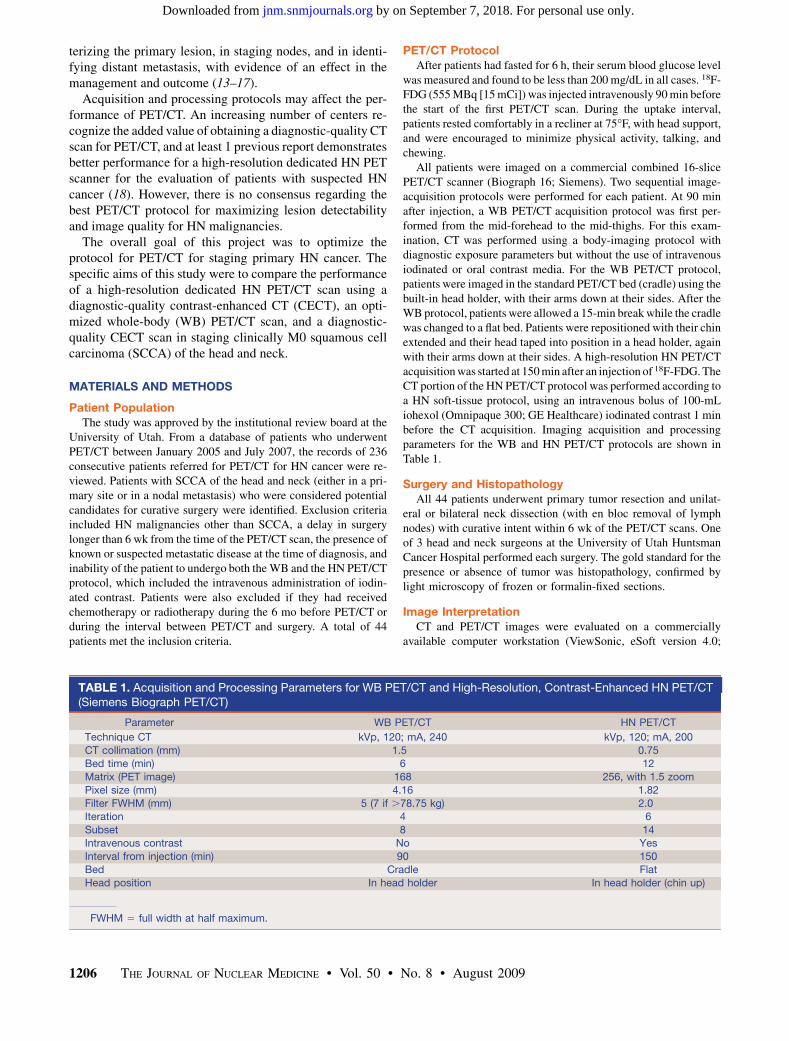
TABLE 1. Acquisition and Processing Parameters for WB PET/CT and High-Resolution, Contrast-Enhanced HN PET/CT(Siemens Biograph PET/CT)
Parameter WB PET/CT HN PET/CT
Technique CT kVp, 120; mA, 240 kVp, 120; mA, 200
CT collimation (mm) 1.5 0.75Bed time (min) 6 12
Matrix (PET image) 168 256, with 1.5 zoom
Pixel size (mm) 4.16 1.82
Filter FWHM (mm) 5 (7 if .78.75 kg) 2.0Iteration 4 6
Subset 8 14
Intravenous contrast No Yes
Interval from injection (min) 90 150Bed Cradle Flat
Head position In head holder In head holder (chin up)
FWHM 5 full width at half maximum.
1206 THE JOURNAL OF NUCLEAR MEDICINE • Vol. 50 • No. 8 • August 2009
by on September 7, 2018. For personal use only. jnm.snmjournals.org Downloaded from
Siemens). The radiologists, without knowledge of the clinical data(including the site of the primary tumor), surgical or pathologicresults, or previous imaging studies, independently reviewed thehigh-resolution CECT and the WB and HN PET/CT scans.
The primary tumor was evaluated by size, metabolic activity, andevidence for infiltration into adjacent structures. Metabolic activitywas defined by a maximum standardized uptake value (SUVmax)and was corrected for body weight. A 5-point scale (1, definitelybenign; 2, probably benign; 3, equivocal; 4, probably malignant; and5, definitely malignant) was first used to designate primary tumoridentification and infiltration into adjacent structures. A positiveexamination was defined as a score of 3–5, and a negative exam-ination was defined as a score of 1–2.
The CECT scans were read independently of the WB and HNPET/CT scans. In interpreting the CT scans, an overall receiver-operating-characteristic (ROC) score (1–5) was applied, as above.In addition, individual CT criteria were evaluated independently.One criterion was nodal size. Nodes were considered enlarged ifthey were greater than 10 mm in maximum short-axis diameter, ex-cept for retrophagyngeal nodes (enlarged if .8 mm) and jugulo-digastric nodes (enlarged if .15 mm). Other criteria independentlyscored as suspected malignancy, regardless of nodal size, includedthe enhancement of lymph nodes by intravenous iodinated contrast(visually appreciable attenuation greater than adjacent muscle), thepresence of necrosis, the absence of a normal fatty hila, a rounded(rather than oval) configuration (except for jugulodigastric nodes),indistinct nodal capsular margins, and asymmetry in size or number.If no enlarged or otherwise suspicious nodes were identified in agiven level by CT, an ROC value of 1 (definitely normal) wasassigned to that level.
By PET/CT criteria, lymph nodes considered suggestive ofmalignancy demonstrated visually appreciable metabolic activityabove that of normal muscle or asymmetric metabolic activitygreater than that of normal-appearing lymph nodes in the same levelin the contralateral neck. For each lymph node level, an ROC scorewas assigned that represented the score of the most metabolicallyactive node within that level. The SUVmax and size of that nodewere recorded. For nodal levels in which no metabolically activenodes were present or in which all nodes were less than 4 mm indiameter, a score of 1 (definitely abnormal) was assigned for thoselevels.
In addition to nodal scores on a per-level basis, a per-patient scorefor nodal metastases was defined for both PET/CT protocols and forCECT based on the highest single score for a nodal level for eachpatient. Discordant scores between the 2 radiologists were reviewedjointly and resolved by consensus.
Statistical AnalysisFor the identification of the primary tumor, x2 analysis was used
to compare the performance of the WB and HN PET/CT protocolswith that of the CECT protocol. A true-positive result was defined ascorrectly identifying the primary site of the tumor. A false-positivefinding was defined as significantly misidentifying the site of thetumor or identifying a tumor at a site in which none was subse-quently found. A P value of less than 0.05 was defined as repre-senting a statistically significant difference.
For nodal disease, ROC curves were generated to determine thedifferences in performance between the HN and WB PET/CT andthe CECT protocols as defined by the 5-point scale, both on a per-level basis and on a per-patient basis. Nodal levels for which patho-logic correlation was available were analyzed. Comparison of the
areas under the curve was made between the modalities usingz statistics. A statistically significant difference between modalitieswas defined as a P value of less than 0.05. Sensitivity, specificity, andpositive and negative predictive values were also calculated, with apositive score defined as an ROC score of 3, 4, or 5 and a negativescore as 1 or 2.
RESULTS
A total of 44 patients met the inclusion criteria. The medianinterval between PET/CT and surgery was 19 d (range, 1–42 d). The median patient age was 65 y (range, 26–97 y). Thepatients included 35 men (80%) and 9 women (20%).
Primary Tumor
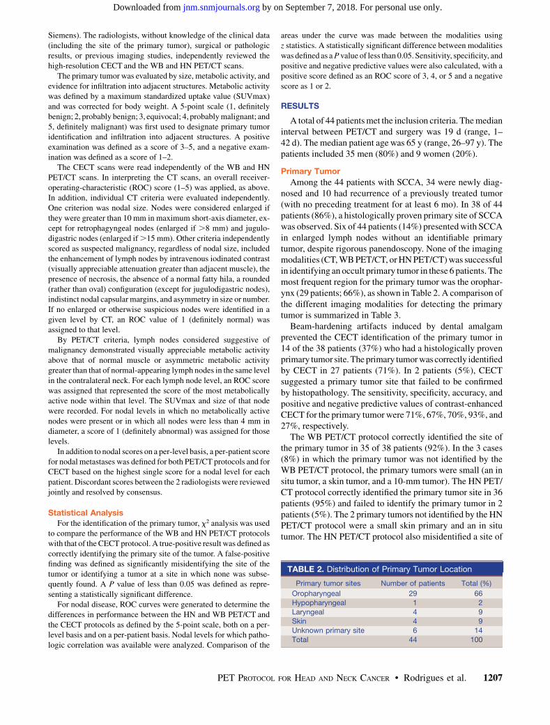
Among the 44 patients with SCCA, 34 were newly diag-nosed and 10 had recurrence of a previously treated tumor(with no preceding treatment for at least 6 mo). In 38 of 44patients (86%), a histologically proven primary site of SCCAwas observed. Six of 44 patients (14%) presented with SCCAin enlarged lymph nodes without an identifiable primarytumor, despite rigorous panendoscopy. None of the imagingmodalities (CT, WB PET/CT, or HN PET/CT) was successfulin identifying an occult primary tumor in these 6 patients. Themost frequent region for the primary tumor was the orophar-ynx (29 patients; 66%), as shown in Table 2. A comparison ofthe different imaging modalities for detecting the primarytumor is summarized in Table 3.
Beam-hardening artifacts induced by dental amalgamprevented the CECT identification of the primary tumor in14 of the 38 patients (37%) who had a histologically provenprimary tumor site. The primary tumor was correctly identifiedby CECT in 27 patients (71%). In 2 patients (5%), CECTsuggested a primary tumor site that failed to be confirmedby histopathology. The sensitivity, specificity, accuracy, andpositive and negative predictive values of contrast-enhancedCECT for the primary tumor were 71%, 67%, 70%, 93%, and27%, respectively.
The WB PET/CT protocol correctly identified the site ofthe primary tumor in 35 of 38 patients (92%). In the 3 cases(8%) in which the primary tumor was not identified by theWB PET/CT protocol, the primary tumors were small (an insitu tumor, a skin tumor, and a 10-mm tumor). The HN PET/CT protocol correctly identified the primary tumor site in 36patients (95%) and failed to identify the primary tumor in 2patients (5%). The 2 primary tumors not identified by the HNPET/CT protocol were a small skin primary and an in situtumor. The HN PET/CT protocol also misidentified a site of
TABLE 2. Distribution of Primary Tumor Location
Primary tumor sites Number of patients Total (%)
Oropharyngeal 29 66
Hypopharyngeal 1 2
Laryngeal 4 9Skin 4 9
Unknown primary site 6 14
Total 44 100
PET PROTOCOL FOR HEAD AND NECK CANCER • Rodrigues et al. 1207
by on September 7, 2018. For personal use only. jnm.snmjournals.org Downloaded from
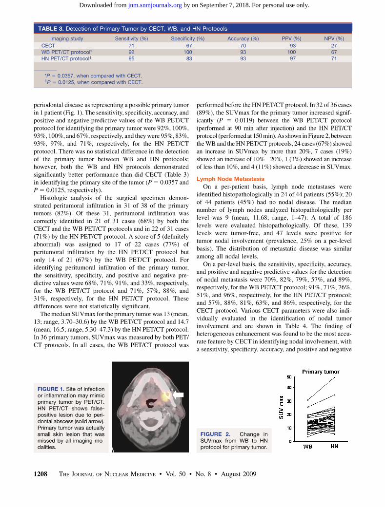
periodontal disease as representing a possible primary tumorin 1 patient (Fig. 1). The sensitivity, specificity, accuracy, andpositive and negative predictive values of the WB PET/CTprotocol for identifying the primary tumor were 92%, 100%,93%, 100%, and 67%, respectively, and they were 95%, 83%,93%, 97%, and 71%, respectively, for the HN PET/CTprotocol. There was no statistical difference in the detectionof the primary tumor between WB and HN protocols;however, both the WB and HN protocols demonstratedsignificantly better performance than did CECT (Table 3)in identifying the primary site of the tumor (P 5 0.0357 andP 5 0.0125, respectively).
Histologic analysis of the surgical specimen demon-strated peritumoral infiltration in 31 of 38 of the primarytumors (82%). Of these 31, peritumoral infiltration wascorrectly identified in 21 of 31 cases (68%) by both theCECT and the WB PET/CT protocols and in 22 of 31 cases(71%) by the HN PET/CT protocol. A score of 5 (definitelyabnormal) was assigned to 17 of 22 cases (77%) ofperitumoral infiltration by the HN PET/CT protocol butonly 14 of 21 (67%) by the WB PET/CT protocol. Foridentifying peritumoral infiltration of the primary tumor,the sensitivity, specificity, and positive and negative pre-dictive values were 68%, 71%, 91%, and 33%, respectively,for the WB PET/CT protocol and 71%, 57%, 88%, and31%, respectively, for the HN PET/CT protocol. Thesedifferences were not statistically significant.
The median SUVmax for the primary tumor was 13 (mean,13; range, 3.70–30.6) by the WB PET/CT protocol and 14.7(mean, 16.5; range, 5.30–47.3) by the HN PET/CT protocol.In 36 primary tumors, SUVmax was measured by both PET/CT protocols. In all cases, the WB PET/CT protocol was
performed before the HN PET/CT protocol. In 32 of 36 cases(89%), the SUVmax for the primary tumor increased signif-icantly (P 5 0.0119) between the WB PET/CT protocol(performed at 90 min after injection) and the HN PET/CTprotocol (performed at 150 min). As shown in Figure 2, betweenthe WB and the HN PET/CT protocols, 24 cases (67%) showedan increase in SUVmax by more than 20%, 7 cases (19%)showed an increase of 10%220%, 1 (3%) showed an increaseof less than 10%, and 4 (11%) showed a decrease in SUVmax.
Lymph Node Metastasis
On a per-patient basis, lymph node metastases wereidentified histopathologically in 24 of 44 patients (55%); 20of 44 patients (45%) had no nodal disease. The mediannumber of lymph nodes analyzed histopathologically perlevel was 9 (mean, 11.68; range, 1–47). A total of 186levels were evaluated histopathologically. Of these, 139levels were tumor-free, and 47 levels were positive fortumor nodal involvement (prevalence, 25% on a per-levelbasis). The distribution of metastatic disease was similaramong all nodal levels.
On a per-level basis, the sensitivity, specificity, accuracy,and positive and negative predictive values for the detectionof nodal metastasis were 70%, 82%, 79%, 57%, and 89%,respectively, for the WB PET/CT protocol; 91%, 71%, 76%,51%, and 96%, respectively, for the HN PET/CT protocol;and 57%, 88%, 81%, 63%, and 86%, respectively, for theCECT protocol. Various CECT parameters were also indi-vidually evaluated in the identification of nodal tumorinvolvement and are shown in Table 4. The finding ofheterogeneous enhancement was found to be the most accu-rate feature by CECT in identifying nodal involvement, witha sensitivity, specificity, accuracy, and positive and negative
TABLE 3. Detection of Primary Tumor by CECT, WB, and HN Protocols
Imaging study Sensitivity (%) Specificity (%) Accuracy (%) PPV (%) NPV (%)
CECT 71 67 70 93 27WB PET/CT protocol* 92 100 93 100 67
HN PET/CT protocoly 95 83 93 97 71
*P 5 0.0357, when compared with CECT.yP 5 0.0125, when compared with CECT.
FIGURE 1. Site of infectionor inflammation may mimicprimary tumor by PET/CT.HN PET/CT shows false-positive lesion due to peri-dontal abscess (solid arrow).Primary tumor was actuallysmall skin lesion that wasmissed by all imaging mo-dalities.
FIGURE 2. Change inSUVmax from WB to HNprotocol for primary tumor.
1208 THE JOURNAL OF NUCLEAR MEDICINE • Vol. 50 • No. 8 • August 2009
by on September 7, 2018. For personal use only. jnm.snmjournals.org Downloaded from
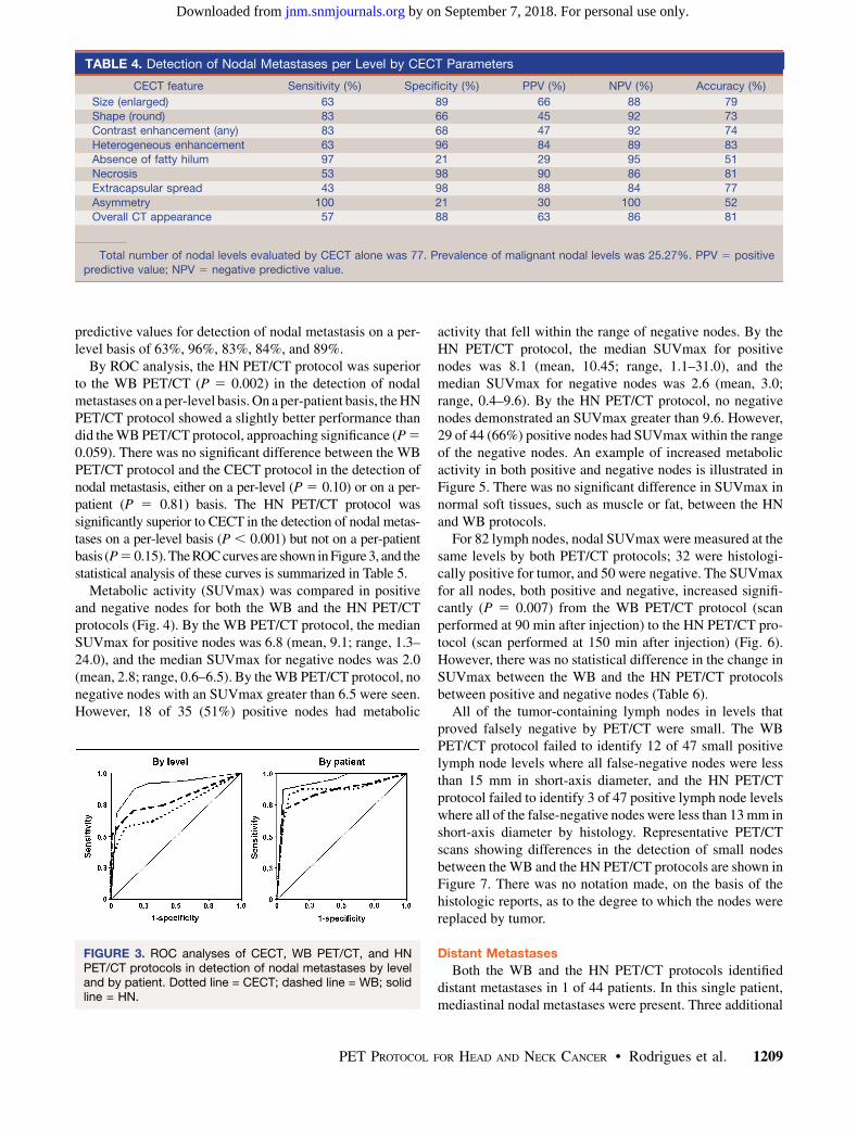
predictive values for detection of nodal metastasis on a per-level basis of 63%, 96%, 83%, 84%, and 89%.
By ROC analysis, the HN PET/CT protocol was superiorto the WB PET/CT (P 5 0.002) in the detection of nodalmetastases on a per-level basis. On a per-patient basis, the HNPET/CT protocol showed a slightly better performance thandid the WB PET/CT protocol, approaching significance (P 5
0.059). There was no significant difference between the WBPET/CT protocol and the CECT protocol in the detection ofnodal metastasis, either on a per-level (P 5 0.10) or on a per-patient (P 5 0.81) basis. The HN PET/CT protocol wassignificantly superior to CECT in the detection of nodal metas-tases on a per-level basis (P , 0.001) but not on a per-patientbasis (P5 0.15). The ROC curves are shown in Figure 3, and thestatistical analysis of these curves is summarized in Table 5.
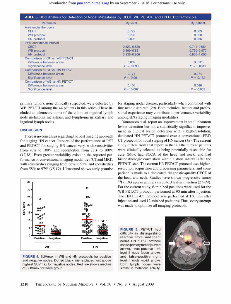
Metabolic activity (SUVmax) was compared in positiveand negative nodes for both the WB and the HN PET/CTprotocols (Fig. 4). By the WB PET/CT protocol, the medianSUVmax for positive nodes was 6.8 (mean, 9.1; range, 1.3–24.0), and the median SUVmax for negative nodes was 2.0(mean, 2.8; range, 0.6–6.5). By the WB PET/CT protocol, nonegative nodes with an SUVmax greater than 6.5 were seen.However, 18 of 35 (51%) positive nodes had metabolic
activity that fell within the range of negative nodes. By theHN PET/CT protocol, the median SUVmax for positivenodes was 8.1 (mean, 10.45; range, 1.1–31.0), and themedian SUVmax for negative nodes was 2.6 (mean, 3.0;range, 0.4–9.6). By the HN PET/CT protocol, no negativenodes demonstrated an SUVmax greater than 9.6. However,29 of 44 (66%) positive nodes had SUVmax within the rangeof the negative nodes. An example of increased metabolicactivity in both positive and negative nodes is illustrated inFigure 5. There was no significant difference in SUVmax innormal soft tissues, such as muscle or fat, between the HNand WB protocols.
For 82 lymph nodes, nodal SUVmax were measured at thesame levels by both PET/CT protocols; 32 were histologi-cally positive for tumor, and 50 were negative. The SUVmaxfor all nodes, both positive and negative, increased signifi-cantly (P 5 0.007) from the WB PET/CT protocol (scanperformed at 90 min after injection) to the HN PET/CT pro-tocol (scan performed at 150 min after injection) (Fig. 6).However, there was no statistical difference in the change inSUVmax between the WB and the HN PET/CT protocolsbetween positive and negative nodes (Table 6).
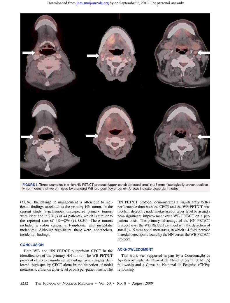
All of the tumor-containing lymph nodes in levels thatproved falsely negative by PET/CT were small. The WBPET/CT protocol failed to identify 12 of 47 small positivelymph node levels where all false-negative nodes were lessthan 15 mm in short-axis diameter, and the HN PET/CTprotocol failed to identify 3 of 47 positive lymph node levelswhere all of the false-negative nodes were less than 13 mm inshort-axis diameter by histology. Representative PET/CTscans showing differences in the detection of small nodesbetween the WB and the HN PET/CT protocols are shown inFigure 7. There was no notation made, on the basis of thehistologic reports, as to the degree to which the nodes werereplaced by tumor.
Distant Metastases
Both the WB and the HN PET/CT protocols identifieddistant metastases in 1 of 44 patients. In this single patient,mediastinal nodal metastases were present. Three additional
TABLE 4. Detection of Nodal Metastases per Level by CECT Parameters
CECT feature Sensitivity (%) Specificity (%) PPV (%) NPV (%) Accuracy (%)
Size (enlarged) 63 89 66 88 79Shape (round) 83 66 45 92 73
Contrast enhancement (any) 83 68 47 92 74
Heterogeneous enhancement 63 96 84 89 83Absence of fatty hilum 97 21 29 95 51
Necrosis 53 98 90 86 81
Extracapsular spread 43 98 88 84 77
Asymmetry 100 21 30 100 52Overall CT appearance 57 88 63 86 81
Total number of nodal levels evaluated by CECT alone was 77. Prevalence of malignant nodal levels was 25.27%. PPV 5 positivepredictive value; NPV 5 negative predictive value.
FIGURE 3. ROC analyses of CECT, WB PET/CT, and HNPET/CT protocols in detection of nodal metastases by leveland by patient. Dotted line = CECT; dashed line = WB; solidline = HN.
PET PROTOCOL FOR HEAD AND NECK CANCER • Rodrigues et al. 1209
by on September 7, 2018. For personal use only. jnm.snmjournals.org Downloaded from
primary tumors, none clinically suspected, were detected byWB PET/CT among the 44 patients in this series. These in-cluded an adenocarcinoma of the colon, an inguinal lymphnode melanoma metastasis, and lymphoma in axillary andinguinal lymph nodes.
DISCUSSION
There is no consensus regarding the best imaging approachfor staging HN cancer. Reports of the performance of PETand PET/CT for staging HN cancer vary, with sensitivitiesfrom 70% to 100% and specificities from 78% to 100%(17,18). Even greater variability exists in the reported per-formance of conventional imaging modalities (CTand MRI),with sensitivities ranging from 36% to 95% and specificitiesfrom 58% to 97% (18,19). Ultrasound shows early promise
for staging nodal disease, particularly when combined withfine-needle aspirate (20). Both technical factors and profes-sional experience may contribute to performance variabilityamong HN staging imaging modalities.
Yamamoto et al. report an improvement in small phantomlesion detection but not a statistically significant improve-ment in clinical lesion detection with a high-resolution,dedicated HN PET/CT protocol over a conventional PET/CT protocol for nodal staging of HN cancer (18). The currentstudy differs from that report in that all the current patientswere clinically selected as being potentially resectable forcure (M0), had SCCA of the head and neck, and hadhistopathologic correlation within a short interval after thePET/CT scan. The current HN PET/CT protocol uses higher-resolution acquisition and processing parameters, and com-parison is made to a dedicated, diagnostic-quality CECT ofthe head and neck. Studies have shown progressive tumor18F-FDG uptake at intervals up to 3 h after injection (21–24).For the current study, 6-min bed positions were used for theWB PET/CT protocol, performed at 90 min after injection.The HN PET/CT protocol was performed at 150 min afterinjection and used 12-min bed positions. Thus, every attemptwas made to optimize all imaging protocols.
FIGURE 4. SUVmax in WB and HN protocols for positiveand negative nodes. Dotted black line is placed just abovehighest SUVmax for negative nodes. Red line shows medianof SUVmax for each group.
TABLE 5. ROC Analysis for Detection of Nodal Metastases by CECT, WB PET/CT, and HN PET/CT Protocols
Parameter By level By patient
Area under the curveCECT 0.722 0.863
WB protocol 0.790 0.850
HN protocol 0.896 0.93695% confidence interval
CECT 0.623–0.820 0.741–0.984
WB protocol 0.699–0.881 0.730–0.970
HN protocol 0.836–0.956 0.860–1.000Comparison of CT vs. WB PET/CT
Difference between areas 0.069 0.0125
Significance level P 5 0.099 P 5 0.0811
Comparison of CT vs. HN PET/CTDifference between areas 0.174 0.074
Significance level P , 0.001 P 5 0.152
Comparison of WB vs HN PET/CT
Difference between areas 0.106 0.086Significance level P , 0.002 P 5 0.059
FIGURE 5. PET/CT haddifficulty in distinguishingreactive from malignantnodes. HN PET/CT protocolshowsprimary tumor (curvedarrow), true-positive leftlevel II node (open arrow),and false-positive rightlevel II node (solid arrow).Both lymph nodes weresimilar in metabolic activity.
1210 THE JOURNAL OF NUCLEAR MEDICINE • Vol. 50 • No. 8 • August 2009
by on September 7, 2018. For personal use only. jnm.snmjournals.org Downloaded from

Metallic beam-hardening artifacts produced by dentalamalgam limited the CECT evaluation of the oral cavityand oral pharynx in 61% of patients. This limitation of theCECT protocol most likely contributed to the significantlybetter performance of both WB and HN PET/CT protocolswhen compared with CECT for identifying the primarytumor. These results are consistent with other reports (25).However, no difference in performance was found betweenWB and HN PET/CT protocols for identifying primaryhead and neck tumors. Tiny, in situ and skin primary lesionsconstituted false-negatives by both the HN and the WBPET/CT scan protocols. The HN PET/CT protocol errone-ously identified a site of periodontal disease as representinga possible site of primary tumor.
There is debate regarding the value of PET or PET/CTin identifying the site of an occult HN primary cancer.Sensitivities of comparable reports range from 15% to 73%(13,26–28). None of the imaging modalities in the currentseries identified the occult primary in the 6 patients whopresented with nodal metastasis but no known primarytumor.
For nodal staging of the neck, the sensitivity and spec-ificity were 70% and 82% for WB PET/CT, 91% and 71%for HN PET/CT, and 57% and 88% for CECT. By ROCanalysis, the HN PET/CT protocol demonstrated a signif-icantly better performance than both the CECT and the WBPET/CT protocols in the detection of nodal metastases on aper-level basis (P , 0.001) and a near-significant improve-ment over WB PET/CT in the detection of nodal disease ona per-patient basis (P 5 0.059). There was no significantdifference between CECT and WB PET/CT in the detectionof nodal metastases on a per-level (P 5 0.10) or a per-patient (P 5 0.81) basis or in the detection of nodalmetastases on a per-patient basis (P 5 0.15). Many studieshave shown that PET/CT is superior to CT in the nodalstaging of HN cancer. However, many of these studiescompared PET to the WB CT scan obtained at the time ofthe PET scan and not to a dedicated CECT of the head andneck, possibly accounting for the relatively better perfor-mance of CECT in the current report.
Others have shown a progressive rise in SUVmax overtime, whereas metabolic activity within inflammatory lesionsmay plateau or drop (21–24). In the current study, however,neither magnitude of SUVmax nor an increase in SUVmaxover time could reliably distinguish benign versus malignantlymph nodes by either the standard WB or the high-resolutionHN PET/CT protocols. Metabolic activity (SUVmax) wassignificantly higher in positive than in negative nodes. Nobenign nodes were identified with an SUVmax greater than9.6 for the HN protocol or greater than 6.5 for the WBprotocol. However, 51% of all malignant nodes for the WBPET/CT protocol and 66% for the HN PET/CT protocol fellwithin the SUVmax range for benign nodes. A progressiveincrease in metabolic activity in both malignant and reactivenodes over time may have contributed to both the improvedsensitivity and the slight reduction in specificity of the HNPET/CT protocol, when compared with the WB protocol.However, it could be argued that the identification of abnor-mal nodes on PET/CT, whether reactive or malignant, definesa vulnerable lymphatic region that must be addressed, eithertherapeutically or surgically.
Small lymph nodes accounted for most false-negativenodes by both the WB and the HN PET/CT protocol. In thisregard, the higher-resolution HN PET/CT protocol had asignificant advantage over the WB protocol and detected4-fold more positive nodes (,15 mm) than did the WBprotocol. The improvement in lesion detection with the HNPET/CT protocol, over the WB PET/CT protocol, could beattributed to a longer interval from injection to imaging andto finer sampling with smaller pixels (1.82 mm for the HNprotocol, which was supported by increasing data density byapplication of a 12-min bed position, as opposed to 4.16-mmpixels for the WB protocol). Smaller pixels reduce the partial-volume effect that reduces the visual intensity of small objectsin addition to falsely lowering the SUVmax.
All of the patients in this series were clinically staged asM0 before PET/CT. Only 1 of 44 patients (2%) proved tohave distant metastatic disease (to mediastinal lymph nodes).This metastatic disease was visible both on the HN PET/CTprotocol (which extends to the carina) and on the WBprotocol. Other published series also report a low incidenceof unsuspected metastatic disease detected by PET/CT in thispopulation, in the range of 7%215% (11,13).
Although the literature reports that PET/CT often altersmanagement in 30%235% of patients with HN cancer
FIGURE 6. Change in SUVmax from WB PET/CT protocol(90 min after injection) to HN PET/CT protocol (150 min afterinjection) for positive and negative nodes.
TABLE 6. Change in SUVmax from WB to HN Protocolfor Positive and Negative Nodes
Change in SUVmax Positive node* Negative node*
Increase $ 10% 23 (71.87) 42 (84.00)
Increase 0%210% 3 (09.38) 5 (10.00)Decrease 6 (18.75) 3 (06.00)
Number of nodes 32 (100.00) 50 (100.00)
*Data are number of nodes, with percentages in parentheses.
PET PROTOCOL FOR HEAD AND NECK CANCER • Rodrigues et al. 1211
by on September 7, 2018. For personal use only. jnm.snmjournals.org Downloaded from
(13,16), the change in management is often due to inci-dental findings unrelated to the primary HN tumor. In thecurrent study, synchronous unsuspected primary tumorswere identified in 7% (3 of 44 patients), which is similar tothe reported rate of 4%28% (11,13,29). These tumorsincluded a colon cancer, a lymphoma, and metastaticmelanoma. Although significant, these were, nonetheless,incidental findings.
CONCLUSION
Both WB and HN PET/CT outperform CECT in theidentification of the primary HN tumor. The WB PET/CTprotocol offers no significant advantage over a highly ded-icated, high-quality CECT alone in the detection of nodalmetastasis, either on a per-level or on a per-patient basis. The
HN PET/CT protocol demonstrates a significantly betterperformance than both the CECT and the WB PET/CT pro-tocols in detecting nodal metastases on a per-level basis and anear-significant improvement over WB PET/CT on a per-patient basis. The primary advantage of the HN PET/CTprotocol over the WB PET/CT protocol is in the detection ofsmall (,15 mm) nodal metastasis, in which a 4-fold increasein nodal detection is found by the HN versus the WB PET/CTprotocol.
ACKNOWLEDGMENT
This work was supported in part by a Coordenacxao deAperfeicxoamento de Pessoal de Nıvel Superior (CAPES)fellowship and a Conselho Nacional de Pesquisa (CNPq)fellowship.
FIGURE 7. Three examples in which HN PET/CT protocol (upper panel) detected small (,15 mm) histologically proven positivelymph nodes that were missed by standard WB protocol (lower panel). Arrows indicate discordant nodes.
1212 THE JOURNAL OF NUCLEAR MEDICINE • Vol. 50 • No. 8 • August 2009
by on September 7, 2018. For personal use only. jnm.snmjournals.org Downloaded from
REFERENCES
1. Schoder H, Yeung HW, Gonen M, Kraus D, Larson SM. Head and neck cancer:
clinical usefulness and accuracy of PET/CT image fusion. Radiology. 2004;
231:65–72.
2. Di Martino E, Nowak B, Hassan HA, et al. Diagnosis and staging of head and neck
cancer: a comparison of modern imaging modalities (positron emission tomog-
raphy, computed tomography, color-coded duplex sonography) with panendo-
scopic and histopathologic findings. Arch Otolaryngol Head Neck Surg. 2000;126:
1457–1461.
3. Kitagawa Y, Nishizawa S, Sano K, et al. Whole-body 18F-fluorodeoxyglucose
positron emission tomography in patients with head and neck cancer. Oral Surg
Oral Med Oral Pathol Oral Radiol Endod. 2002;93:202–207.
4. Goerres GW, Schmid DT, Gratz KW, von Schulthess GK, Eyrich GK. Impact of
whole body positron emission tomography on initial staging and therapy in
patients with squamous cell carcinoma of the oral cavity. Oral Oncol. 2003;
39:547–551.
5. McCollum AD, Burrell SC, Haddad RI, et al. Positron emission tomography
with 18F-fluorodeoxyglucose to predict pathologic response after induction
chemotherapy and definitive chemoradiotherapy in head and neck cancer. Head
Neck. 2004;26:890–896.
6. Goerres GW, Schmid DT, Bandhauer F, et al. Positron emission tomography in
the early follow-up of advanced head and neck cancer. Arch Otolaryngol Head
Neck Surg. 2004;130:105–109.
7. Goshen E, Yahalom R, Talmi YP, Rotenberg G, Oksman Y, Zwas ST. The role of
gamma-PET in the evaluation of patients with recurrent squamous cell cancer of
the head and neck. Int J Oral Maxillofac Surg. 2005;34:386–390.
8. Schwartz DL, Rajendran J, Yueh B, et al. FDG-PET prediction of head and neck
squamous cell cancer outcomes. Arch Otolaryngol Head Neck Surg. 2004;130:
1361–1367.
9. Agarwal V, Branstetter BF IV, Johnson JT. Indications for PET/CT in the head
and neck. Otolaryngol Clin North Am. 2008;41:23–49.
10. Sigg MB, Steinert H, Gratz K, Hugenin P, Stoeckli S, Eyrich GK. Staging of
head and neck tumors: [18F]fluorodeoxyglucose positron emission tomography
compared with physical examination and conventional imaging modalities.
J Oral Maxillofac Surg. 2003;61:1022–1029.
11. Ng SH, Yen TC, Liao CT, et al. 18F-FDG PET and CT/MRI in oral cavity
squamous cell carcinoma: a prospective study of 124 patients with histologic
correlation. J Nucl Med. 2005;46:1136–1143.
12. Kim SY, Roh J-L, Yeo N-K, et al. Combined 18F-fluorodeoxyglucose-positron
emission tomography and computed tomography as a primary screening method
for detecting second primary cancers and distant metastases in patients with head
and neck cancer. Ann Oncol. 2007;18:1698–1703.
13. Branstetter BF IV, Blodgett TM, Zimmer LA, et al. Head and neck malignancy:
is PET/CT more accurate than PET or CT alone? Radiology. 2005;235:580–586.
14. Fleming AJ Jr, Smith SP Jr, Paul CM, et al. Impact of [18F]-2-fluorodeoxy-
glucose-positron emission tomography/computed tomography on previously
untreated head and neck cancer patients. Laryngoscope. 2007;117:1173–1179.
15. Gordin A, Daitzchman M, Doweck I, et al. Fluorodeoxyglucose-positron
emission tomography/computed tomography imaging in patients with carcinoma
of the larynx: diagnostic accuracy and impact on clinical management. Laryngo-
scope. 2006;116:273–278.
16. Gordin A, Golz A, Keidar Z, Daitzchman M, Bar-Shalom R, Israel O. The role
of FDG-PET/CT imaging in head and neck malignant conditions: impact on
diagnostic accuracy and patient care. Otolaryngol Head Neck Surg. 2007;137:
130–137.
17. Scott AM, Gunawardana DH, Bartholomeusz D, Ramshaw JE, Lin P. PET
changes management and improves prognostic stratification in patients with head
and neck cancer: results of a multicenter prospective study. J Nucl Med. 2008;
49:1593–1600.
18. Yamamoto Y, Wong TZ, Turkington TG, Hawk TC, Coleman RE. Head and neck
cancer: dedicated FDG PET/CT protocol for detection—phantom and initial
clinical studies. Radiology. 2007;244:263–272.
19. Hain SF. Positron emission tomography in cancer of the head and neck. Br J
Oral Maxillofac Surg. 2005;43:1–6.
20. De Bondt RB, Nelemans PJ, Hofman PA, et al. Detection of lymph node
metastases in head and neck cancer: a meta-analysis comparing US, USgFNAC,
CT and MR imaging. Eur J Radiol. 2007;64:266–272.
21. Lan XL, Zhang YX, Wu ZJ, Jia Q, Wei H, Gao ZR. The value of dual time point18F-FDG PET imaging for the differentiation between malignant and benign
lesions. Clin Radiol. 2008;63:756–764.
22. Zytoon AA, Murakami K, El-Kholy MR, El-Shorbagy E. Dual time point FDG-
PET/CT imaging: potential tool for diagnosis of breast cancer. Clin Radiol.
2008;63:1213–1227.
23. Hustinx R, Smith RJ, Benard F, et al. Dual time point fluorine-18 fluor-
odeoxyglucose positron emission tomography: a potential method to differentiate
malignancy from inflammation and normal tissue in the head and neck. Eur J
Nucl Med. 1999;26:1345–1348.
24. Zhuang H, Pourdehnad M, Lambright ES, et al. Dual time point 18F-FDG PET
imaging for differentiating malignant from inflammatory processes. J Nucl Med.
2001;42:1412–1417.
25. Veit-Haibach P, Luczak C, Wanke I, et al. TNM staging with FDG-PET/CT in
patients with primary head and neck cancer. Eur J Nucl Med Mol Imaging.
2007;34:1953–1962.
26. AAssar OS, Fischbein NJ, Caputo GR, et al. Metastatic head and neck cancer:
role and usefulness of FDG PET in locating occult primary tumors. Radiology.
1999;210:177–181.
27. Ekberg T, Sorensen J, Engstrom M, Blomquist E, Sundin A, Anniko M. Clinical
impact of positron emission tomography (PET) with (18F)fluorodeoxyglucose
(FDG) in head and neck tumours. Acta Otolaryngol. 2007;127:186–193.
28. Nabili V, Zaia B, Blackwell KE, Head CS, Grabski K, Sercarz JA. Positron
emission tomography: poor sensitivity for occult tonsillar cancer. Am J
Otolaryngol. 2007;28:153–157.
29. Ishimori T, Patel PV, Wahl RL. Detection of unexpected additional primary
malignancies with PET/CT. J Nucl Med. 2005;46:752–757.
PET PROTOCOL FOR HEAD AND NECK CANCER • Rodrigues et al. 1213
by on September 7, 2018. For personal use only. jnm.snmjournals.org Downloaded from

Doi: 10.2967/jnumed.109.062075Published online: July 17, 2009.
2009;50:1205-1213.J Nucl Med. A. MortonChristensen, Marta Heilbrun, Richard H. Wiggins III, Jason P. Hunt, Brandon G. Bentz, Ying J. Hitchcock and Kathryn Rosana S. Rodrigues, Fernando A. Bozza, Paul E. Christian, John M. Hoffman, Regan I. Butterfield, Carl R. Squamous Cell Carcinoma of the Head and NeckPET/CT, and Contrast-Enhanced CT in Preoperative Staging of Clinically M0 Comparison of Whole-Body PET/CT, Dedicated High-Resolution Head and Neck
http://jnm.snmjournals.org/content/50/8/1205This article and updated information are available at:
http://jnm.snmjournals.org/site/subscriptions/online.xhtml
Information about subscriptions to JNM can be found at:
http://jnm.snmjournals.org/site/misc/permission.xhtmlInformation about reproducing figures, tables, or other portions of this article can be found online at:
(Print ISSN: 0161-5505, Online ISSN: 2159-662X)1850 Samuel Morse Drive, Reston, VA 20190.SNMMI | Society of Nuclear Medicine and Molecular Imaging
is published monthly.The Journal of Nuclear Medicine
© Copyright 2009 SNMMI; all rights reserved.
by on September 7, 2018. For personal use only. jnm.snmjournals.org Downloaded from