comparison of the microscan, vitek 2, and crystal gp with 16s rrna sequencing and microseq 500 v2.0...
TRANSCRIPT

BioMed CentralBMC Microbiology
ss
Open AcceResearch articleComparison of the MicroScan, VITEK 2, and Crystal GP with 16S rRNA sequencing and MicroSeq 500 v2.0 analysis for coagulase-negative StaphylococciMiyoung Kim†1, Se Ran Heo2, Soon Hee Choi2, Hyelin Kwon2, Jeong Su Park1, Moon-Woo Seong3, Do-Hoon Lee3, Kyoung Un Park*1,2,4, Junghan Song1,2,4 and Eui-Chong Kim1,4Address: 1Department of Laboratory Medicine, Seoul National University Hospital, 101 Daehang-no, Jongno-gu, Seoul, South Korea, 2Department of Laboratory Medicine, Seoul National University Bundang Hospital, 300 Gumi-dong, Bundang-gu, Seongnam-si, Gyeonggi-do, South Korea, 3Department of Laboratory Medicine, National Cancer Center, 809 Madu-1-dong, Ilsandong-gu, Goyang, Gyeonggi-do, South Korea and 4Department of Laboratory Medicine, Seoul National University College of Medicine, 101 Daehang-no, Jongno-gu, Seoul, South Korea
Email: Miyoung Kim - [email protected]; Se Ran Heo - [email protected]; Soon Hee Choi - [email protected]; Hyelin Kwon - [email protected]; Jeong Su Park - [email protected]; Moon-Woo Seong - [email protected]; Do-Hoon Lee - [email protected]; Kyoung Un Park* - [email protected]; Junghan Song - [email protected]; Eui-Chong Kim - [email protected]
* Corresponding author †Equal contributors
AbstractBackground: Three phenotypic identification systems (MicroScan, VITEK 2, and Crystal GP) wereevaluated for their accuracy to identify coagulase-negative staphylococci (CNS). A total of 120clinical isolates confirmed to be CNS via 16S rRNA sequencing and analysis with the MicroSeq 500v2.0 database were assessed.
Results: The MicroScan, VITEK 2, and Crystal GP systems correctly identified 82.5%, 87.5%, and67.5% of the isolates, respectively. Misidentification was the main problem in MicroScan (10.8%)and Crystal GP (23.3%) systems, whereas the main problem of VITEK 2 was low-leveldiscrimination (7.5%).
Conclusion: None of the 3 phenotypic systems tested could accurately and reliably identify CNSat the species level. Further verifications such as biochemical testing or 16S rRNA sequencingtogether with analysis using a comparable database might be helpful in this regard.
BackgroundBecause of their ubiquity and low virulence, coagulase-negative staphylococci (CNS) have generally been consid-ered to be nonpathogens or simple contaminants.Recently, their clinical significance is being increasinglyrecognized with the elucidation of their pathogenicity.CNS can form biofilms and have been demonstrated to
exhibit antibiotic resistance [1,2]. S. epidermidis, S. haemo-lyticus, and S. lugdunensis, to a lesser extent, are well-known etiological agents of implanted device-mediatedinfections [2-5]. S. saprophyticus can cause community-acquired infections of the uropoietic tract. Hence, species-level identification of CNS is necessary for correct guid-ance of clinicians in terms of appropriate treatment strat-
Published: 23 December 2008
BMC Microbiology 2008, 8:233 doi:10.1186/1471-2180-8-233
Received: 10 June 2008Accepted: 23 December 2008
This article is available from: http://www.biomedcentral.com/1471-2180/8/233
© 2008 Kim et al; licensee BioMed Central Ltd. This is an Open Access article distributed under the terms of the Creative Commons Attribution License (http://creativecommons.org/licenses/by/2.0), which permits unrestricted use, distribution, and reproduction in any medium, provided the original work is properly cited.
Page 1 of 7(page number not for citation purposes)

BMC Microbiology 2008, 8:233 http://www.biomedcentral.com/1471-2180/8/233
egy, and a wide variety of identification methods havebeen proposed.
Many automated phenotypic identification systems arecommercially available, including the MicroScan (DadeBehring, West Sacramento, CA, USA), VITEK 2(bioMérieux, Maray l'Etoile, France), and Crystal GP (Bec-ton Dickinson, Sparks, MD, USA) systems. On the basis ofmetabolic activities and/or morphological features, thesesystems enable microbiologists to identify bacterial iso-lates at the species level with greater ease, accuracy, andrapidity than that previously achieved [6]. However, thesesystems have several potential problems: (i) differentstrains in one species may not exhibit a specific character-istic, (ii) isolates from old cultures may not show theexpected biochemical patterns, (iii) isolates from a hostwho has undergone long-term antimicrobial therapy mayalter their typical biochemical characteristics, (iv) thesame strain may not yield the same results in repeatedtests, (v) the databases have data on a limited number ofspecies, (vi) phenotypic variation may affect the accuracyof species-level identification by automated phenotypicsystems, and (vii) phenotypic systems often suggest 2 ormore designations with comparable probability levels [6-9].
Recently, genotypic methods are emerging as the newstandard for bacterial identification in automated labora-tories [4,6,7,10-14]. 16S ribosomal RNA (rRNA)sequences comprising both variable and conservedregions allow for clear differentiation between organismsnot only at the species level but also at the subspecies level[10]. These sequences permit better identification of rareor phenotypically aberrant species as well as noncultura-ble bacteria. The MicroSeq 500 system (Applied Biosys-tems Inc. [ABI], Foster City, CA, USA) is a commerciallyavailable software system for 16S rRNA analysis.
Despite the increasing clinical significance of CNS and thegrowing use of automated phenotypic systems in clinicallaboratories, few studies have systematically evaluated theaccuracy of these systems [4,9,15]. In the present study,the accuracy of 3 commercial phenotypic systems (Micro-Scan, VITEK 2, and Crystal GP) for CNS identification wascompared. 16S rRNA sequencing and MicroSeq 500 anal-ysis were used as references [10-12,16-19].
ResultsClinical isolatesBy using in-house primers and the MicroSeq 500 v2.0database, we successfully identified all the 120 clinicalisolates with more than 97% matches (data not shown).The identified species were as follows: 16 S. capitis, 4 S.caprae, 3 S. cohnii, 53 S. epidermidis, 4 S. haemolyticus, 25 S.hominis, 6 S. lugdunensis, 1 S. saprophyticus, 2 S. simulans,
and 6 S. warneri. Blood culture specimens primarily con-tained S. epidermidis, whereas a variety of uncommon CNSwere isolated from pitted keratolysis specimens. The resultreflected the typical distribution of staphylococcal speciesdetected routinely in microbiology laboratories.
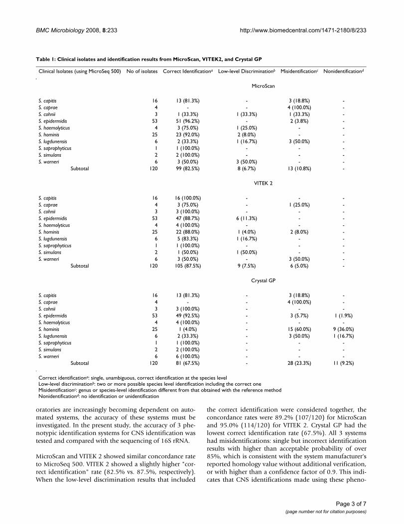
Identification using MicroScanThe overall performances of 3 phenotypic systems aresummarized in Table 1 and the incorrect identificationresults are listed in Table 2. Of 120 isolates, correct iden-tification, low-level discrimination and misidentificationwere 82.5% (99), 6.7% (8) and 10.8% (13), respectively.Nonidentification was not observed. Three S. caprae iso-lates were not correctly identified (isolate number: M211,M215, and M225), because the species S. caprae is notincluded in the MicroScan version 2.0 or 2.1 database.One of the S. epidermidis isolates (M106) was misidenti-fied as S. aureus.
Identification using VITEK 2In the VITEK 2 analysis, correct identification, low-leveldiscrimination and misidentification were 87.5% (105),7.5% (9) and 5.0% (6), respectively. Nonidentificationdid not occur. Of note, the analysis of 6 S. epidermidis iso-lates (6/53, 11.3%) resulted in low-level discriminations– multiple identifications as S. hominis, S. epidermidis, orAerococcus viridans with low percent probabilities, suggest-ing S. hominis with the highest percent probability fol-lowed by S. epidermidis (M123, M124, M125, M126,M127, M137).
Identification using Crystal GPCrystal GP is not as fully automated as the other two, asthe reading of the biochemical reaction depends on man-ual reading. In order to prevent the bias resulting frommanual reading, the biochemical reaction results wereread by two different technicians, followed by repeatedtests on wrongly identified isolates or those with multipleidentifications or nonidentifications. The identificationresults of 81 isolates (67.5%) using the Crystal GP systemagreed with those of the MicroSeq 500 system with confi-dence factors of more than 0.9 (correct identifications). Intotal, 23.3% (28) were misidentified, and 9.2% (11)could not be identified (nonidentification). Low-level dis-crimination was not observed. Misidentifications at thegenus level occurred for 1 S. epidermidis and 1 S. hominisisolate (M112, M216). The Crystal GP system correctlyidentified all the isolates of S. cohnii, S. haemolyticus, S.saprophyticus, S. simulans, and S. warneri.
DiscussionWith the increased attention being given to the clinicalsignificance of CNS, clinical laboratories must correctlyidentify CNS at the species level by using reliable andreproducible methods. Because clinical microbiology lab-
Page 2 of 7(page number not for citation purposes)
BMC Microbiology 2008, 8:233 http://www.biomedcentral.com/1471-2180/8/233
oratories are increasingly becoming dependent on auto-mated systems, the accuracy of these systems must beinvestigated. In the present study, the accuracy of 3 phe-notypic identification systems for CNS identification wastested and compared with the sequencing of 16S rRNA.
MicroScan and VITEK 2 showed similar concordance rateto MicroSeq 500. VITEK 2 showed a slightly higher "cor-rect identification" rate (82.5% vs. 87.5%, respectively).When the low-level discrimination results that included
the correct identification were considered together, theconcordance rates were 89.2% (107/120) for MicroScanand 95.0% (114/120) for VITEK 2. Crystal GP had thelowest correct identification rate (67.5%). All 3 systemshad misidentifications: single but incorrect identificationresults with higher than acceptable probability of over85%, which is consistent with the system manufacturer'sreported homology value without additional verification,or with higher than a confidence factor of 0.9. This indi-cates that CNS identifications made using these pheno-
Table 1: Clinical isolates and identification results from MicroScan, VITEK2, and Crystal GP
Clinical Isolates (using MicroSeq 500) No of isolates Correct Identificationa Low-level Discriminationb Misidentificationc Nonidentificationd
MicroScan
S. capitis 16 13 (81.3%) - 3 (18.8%) -S. caprae 4 - - 4 (100.0%) -S. cohnii 3 1 (33.3%) 1 (33.3%) 1 (33.3%) -S. epidermidis 53 51 (96.2%) - 2 (3.8%) -S. haemolyticus 4 3 (75.0%) 1 (25.0%) - -S. hominis 25 23 (92.0%) 2 (8.0%) - -S. lugdunensis 6 2 (33.3%) 1 (16.7%) 3 (50.0%) -S. saprophyticus 1 1 (100.0%) - - -S. simulans 2 2 (100.0%) - - -S. warneri 6 3 (50.0%) 3 (50.0%) - -
Subtotal 120 99 (82.5%) 8 (6.7%) 13 (10.8%) -
VITEK 2
S. capitis 16 16 (100.0%) - - -S. caprae 4 3 (75.0%) - 1 (25.0%) -S. cohnii 3 3 (100.0%) - - -S. epidermidis 53 47 (88.7%) 6 (11.3%) - -S. haemolyticus 4 4 (100.0%) - - -S. hominis 25 22 (88.0%) 1 (4.0%) 2 (8.0%) -S. lugdunensis 6 5 (83.3%) 1 (16.7%) - -S. saprophyticus 1 1 (100.0%) - - -S. simulans 2 1 (50.0%) 1 (50.0%) - -S. warneri 6 3 (50.0%) - 3 (50.0%) -
Subtotal 120 105 (87.5%) 9 (7.5%) 6 (5.0%) -
Crystal GP
S. capitis 16 13 (81.3%) - 3 (18.8%) -S. caprae 4 - - 4 (100.0%) -S. cohnii 3 3 (100.0%) - - -S. epidermidis 53 49 (92.5%) - 3 (5.7%) 1 (1.9%)S. haemolyticus 4 4 (100.0%) - - -S. hominis 25 1 (4.0%) - 15 (60.0%) 9 (36.0%)S. lugdunensis 6 2 (33.3%) - 3 (50.0%) 1 (16.7%)S. saprophyticus 1 1 (100.0%) - - -S. simulans 2 2 (100.0%) - - -S. warneri 6 6 (100.0%) - - -
Subtotal 120 81 (67.5%) - 28 (23.3%) 11 (9.2%)
Correct identificationa: single, unambiguous, correct identification at the species levelLow-level discriminationb: two or more possible species level identification including the correct oneMisidentificationc: genus or species-level identification different from that obtained with the reference methodNonidentificationd: no identification or unidentification
Page 3 of 7(page number not for citation purposes)

BMC Microbiology 2008, 8:233 http://www.biomedcentral.com/1471-2180/8/233
Table 2: Identification results with percent probability or confidence factor of single wrong identification or multiple identifications in three phenotypic systems
MicroSeq 500 MicroScan VITEK2 Cystal GP
M102 S. epidermidis S. epidermidis 91.36% S. epidermidis 97.10% S. intermediusM104 S. epidermidis S. xylosus 95.12% S. epidermidis 99.00% S. epidermidisM106 S. epidermidis S. aureus 87.13% S. epidermidis 99.00% S. epidermidisM107 S. capitis S. simulans 88.95% S. capitis 98.78% S. capitisM112 S. epidermidis S. epidermidis 99.99% S. epidermidis 99.00% K. sedentariusM117 S. lugdunensis S. haemolyticus 90.39% S. lugdunensis 99.00% NonidentificationM118 S. lugdunensis S. haemolyticus 96.86% S. lugdunensis 90.37% S. haemolyticusM120 S. hominis S. hominis 94.57% S. hominis 95.00% NonidentificationM123 S. epidermidis S. epidermidis 98.20% S. hominis, S. epidermidis, A. viridans
33.82%, 33.09%, 33.09%S. epidermidis
M124 S. epidermidis S. epidermidis 99.25% S. hominis, S. epidermidis 50.27%, 49.73% NonidentificationM125 S. epidermidis S. epidermidis 98.20% S. hominis, S. epidermidis 50.27%, 49.73% S. epidermidisM126 S. epidermidis S. epidermidis 99.99% S. hominis, S. epidermidis, A. viridans
33.71%, 33.33%, 32.96%S. epidermidis
M127 S. epidermidis S. epidermidis 99.99% S. hominis, S. epidermidis 50.28%, 47.92% S. epidermidisM128 S. hominis S. hominis 94.58% S. hominis 94.86% S. saprophyticusM130 S. capitis S. epidermidis 90.20% S. capitis 92.00% S. capitisM137 S. epidermidis S. epidermidis 95.28% S. hominis, S. epidermidis 50.56%, 49.44% S. epidermidisM138 S. capitis S. capitis 95.28% S. capitis 98.86% S. auricularisM139 S. capitis S. capitis 99.60% S. capitis 98.86% S. hominisM141 S. epidermidis S. epidermidis 99.99% S. epidermidis 98.16% S. hominisM149 S. capitis S. epidermidis 90.20% S. capitis 92.00% S. capitisM153 S. hominis S. hominis 97.44% S. hominis 99.00% NonidentificationM154 S. hominis S. hominis 94.62% S. hominis 98.35% NonidentificationM155 S. hominis S. hominis 97.44% S. hominis 97.21% S. warneri, S. saprophyticus 0.5574,
0.4415M156 S. hominis S. hominis 95.00% S. hominis 97.00% S. saprophyticusM157 S. warneri S. warneri, S. cohnii, S. capitis, etc. 60.92%,
15.78%, 12.67%.S. cohnii, S. vitulinus 50.54%, 49.46% S. warneri
M158 S. warneri S. warneri 90.92% S. cohnii 90.54% S. warneriM160 S. hominis S. hominis, S. capitis, S. warneri, etc. 37.58%,
24.33%, 20.21%.S. hominis 93.79% Nonidentification
M161 S. hominis S. hominis 94.57% S. hominis 96.98% S. saprophyticusM162 S. caprae S. hominis 92.35% S. auricularis 90.40% S. saprophyticusM163 S. hominis S. hominis 98.82% S. hominis 98.50% NonidentificationM166 S. hominis S. hominis 85.00% S. hominis 98.82% NonidentificationM167 S. hominis S. hominis 93.05% S. caprae 96.97% S. capraeM176 S. simulans S. simulans 96.77% S. simulans, S. warneri 50.28%, 49.72% S. simulansM17 S. hominis S. simulans 99.09% S. haemolyticus 96.74% S. haemolyticusM179 S. hominis S. epidermidis, S. hominis 68.34%, 31.66% S. hominis 94.88% NonidentificationM180 S. hominis S. hominis 92.77% S. hominis 93.56% S. warneriM181 S. hominis S. hominis 92.77% S. hominis, S. warneri 50.27%, 49.73% S. warneriM182 S. hominis S. hominis 94.57% S. hominis 95.00% NonidentificationM183 S. hominis S. hominis 88.68% S. hominis 91.00% S. saprophyticusM184 S. hominis S. hominis 92.13% S. hominis 99.00% S. intermediusM185 S. hominis S. hominis 96.01% S. hominis 96.81% S. capitisM186 S. hominis S. hominis 98.50% S. hominis 95.00% S. aureus, S. intermediusM187 S. hominis S. hominis 98.50% S. hominis 99.00% NonidentificationM203 S. hominis S. hominis 97.44% S. hominis 97.27% S. cohniiM205 S. warneri S. warneri, S. capitis, S. hominis 86.37%,
7.96%, 5.67%S. saphrophyticus 98.03% S. warneri
M207 S. lugdunensis S. haemolyticus 98.08% S. lugdunensis 99.00% S. lugdunensisM208 S. haemolyticus S. haemolyticus, S. simulans, S. warneri
54.38%, 41.97%, 3.66%S. haemolyticus 94.74% S. haemolyticus
M209 S. hominis S. hominis 97.44% S. hominis 97.76% S. cohniiM210 S. cohnii S. cohnii, S. xylosus 67.28%, 29.12% S. cohnii 99.00% S. cohniiM211 S. caprae S. aureus, S. capitis 69.87%, 29.25% S. caprae 98.60% S. intermidiusM212 S. capitis S. capitis 99.11% S. capitis 98.56% S. vitulusM213 S. warneri S. auricularis, S. hominis, S. warneri 45.13%,
27.55%, 24.55%S. warneri 95.00% S. warneri
Page 4 of 7(page number not for citation purposes)

BMC Microbiology 2008, 8:233 http://www.biomedcentral.com/1471-2180/8/233
typic systems cannot always be considered accurate,regardless of the reported probability.
In MicroScan analysis, none of the S. caprae identifica-tions were correct (3 low-level identifications and 1 misi-dentification). This is because the species S. caprae is notincluded in the MicroScan database, which illustrates theinappropriateness of this database. Ambiguous identifica-tions, including low-level discriminations and misidenti-fications, were observed for 8 out of 10 species (with theexception of S. saprophyticus and S. warneri) with theMicroScan system, compared with 6 ambiguous identifi-cations with the VITEK2 system and 5 with the Crystal GPsystem. This finding was consistent with results reportedby other researchers who found that the MicroScan systemhas greater accuracy in the identification of S. epidermidisand S. saprophyticus than for identification of other species[20-22]. The other notable finding of the present study isthat 1 S. epidermidis isolate from blood culture was misi-dentified as S. aureus with 87.5% probability, which couldhave delayed appropriate treatment for the patient.
In VITEK 2 analysis, low-level discriminations of 6 S. epi-dermidis isolates (11.3% of S. epidermidis isolates)occurred, compared to higher correct identification rateswith the MicroScan and Crystal GP systems (96.2% and92.5%, respectively). Some researchers reported differentsort of difficulties in identifying S. epidermidis with VITEK2 [9,23,24]. However, others reported high accuracy ofVITEK 2 for the identification of staphylocccal species[25,26]. The performance of VITEK 2 in the identificationof CNS including S. epidermidis seems not to be deter-mined yet, and the additional testing such as manual bio-chemical testing or sequencing is required for the accuratediagnosis.
The Crystal GP system provides no guidelines for theinterpretation of the confidence factor, therefore, all iso-lates with confidence factors lower than 0.9 were retested.In spite of repeated testing, this system had the lowest cor-rect identification rate and the highest rate of nonidentifi-cation.
One limitation of this study is that additional biochemi-cal testing was not preformed. 16S rRNA sequencingmight not be a perfect method for interspecies discrimina-tion. However, not all the suggested biochemical tests areavailable in many automated laboratories or based on ourexperience, manual testing could not resolve the ambigu-ity of CNS identification in many cases. To improve relia-bility in this study, all isolates with discrepancies oruncertain results were retested using isolates preserved inskim milk at -70°C.
ConclusionNone of the tested systems (MicroScan, VITEK 2, CrystalGP) are able to accurately identify all of the staphylococ-cal species evaluated in this study. Additional verificationvia molecular testing or suggested biochemical tests mayprove helpful for correct CNS identification.
MethodsClinical isolatesA total of 120 clinical isolates (95 blood culture isolates,25 skin isolates from pitted keratolysis patients) were usedin this study [27]. To exclude simple contaminants, onlyisolates of blood cultures that grew in more than 2 of 3blood cultures were included. The pitted keratolysis spec-imen was obtained after repeated washing of the lesionwith normal saline by scratching the lesion with a sterileblade into a sterile conical tube. The isolates were testedwithin 24 hours. For additional tests or repeated tests, theisolates were suspended in skim milk, stored at -70°C andretested. All of the isolates were confirmed to be CNS byuse of 16S rRNA sequencing with MicroSeq 500 software(version 2.0) with over 97% matches.
Phenotypic identification of CNSThree phenotypic systems were evaluated: MicroScan PosCombo panel type 1A (Dade Behring, West Sacramento,CA, USA), VITEK 2 GP-ID cards (bioMérieux, Marcyl'Etoile, France), and Crystal Gram Positive ID systems(Becton Dickinson, Sparks, MD, USA). Bacterial suspen-sions were prepared from well-isolated colonies. The col-onies were suspended in the broth provided by eachcompany, and the turbidity was adjusted to 0.5 McFar-land. MicroScan Pos Combo panel type 1A was inocu-
M214 S. lugdunensis S. lugdunensis 98.81% S. lugdunensis 95.00% S. simulansM215 S. caprae S. capitis, S. aureus 62.82%, 35.76% S. caprae 99.00% S. cohniiM216 S. hominis S. hominis 97.44% S. hominis 97.76% M. kristinaeM221 S. lugdunensis S. lugdunensis, S. haemoiyticus, S. hominis
62.62%, 28.34%, 9.04%S. lugdunensis, S. warneri 50.00%, 50.00% S. lugdunensis
M222 S. lugdunensis S. lugdunensis 96.30% S. lugdunensis 91.26% S. simulansM224 S. cohnii S. xylosus 91.70% S. cohnii 99.00% S. cohniiM225 S. caprae S. warneri, S. simulans, S. epidermidis 70.19%,
13.84%, 12.32%S. caprae 99.00% S. capitis
Table 2: Identification results with percent probability or confidence factor of single wrong identification or multiple identifications in three phenotypic systems (Continued)
Page 5 of 7(page number not for citation purposes)

BMC Microbiology 2008, 8:233 http://www.biomedcentral.com/1471-2180/8/233
lated according to the manufacturer's recommendationsand processed with a MicroScan Walkaway 96 apparatus.The blood culture isolates were analyzed using a MicroS-can LabPro system (version 2.0) and the skin isolates wereanalyzed with version 2.01. VITEK 2 GP-ID cards wereused in combination with a VITEK 2 system (version 4.02)for all isolates. The Crystal GP results were read manuallyby 2 different technicians using the Crystal GP Identifica-tion Color Chart.
The MicroScan system classifies identification results into4 categories by percent probability: species identified withhigh probability (> 85%), species identified with lowprobability (< 85%), very rare biotype, and group ID (>85% but genus or group identified only). The VITEK 2 sys-tem categorizes the identification results by probability:excellent (96%–99%), very good (93%–95%), good(89%–92%), acceptable (85%–88%), low discrimination(2–3 identifications with 100% probability in total), andnonidentification. The Crystal GP system gives confidencefactors instead of probability; however, there are no suchcriteria in terms of the confidence factor. When the confi-dence factor is low, Crystal GP reports non-identification,which necessitates additional manual testing.
The results of incorrect identifications (not concordant tothat of MicroSeq 500) regardless of probability, identifica-tion with unacceptable probability (lower than 85% forMicroScan and VITEK 2) or low confidence factor (lowerthan 0.9 for Crystal GP), or nonidentification wereretested with colonies that were stored in skim milk at -70°C. Those with incorrect, or consistently unacceptableor nonidentifiable results are listed in Table 2. The confi-dence factor of the Crystal GP system is not included, asthere are no guidelines for these interpretations.
16S rRNA sequencingTo extract bacterial DNA, InstaGene Matrix (Bio-Rad Lab-oratories, Hercules, CA, USA) was used. Two or 3 coloniesof each isolate were suspended in 100 μl sterile distilledwater and centrifuged for 5 min at 15,000 rpm. A 200 μlaliquot of InstaGene Matrix (Bio-Rad Laboratories, Her-cules, CA, USA) was added to the sediment. The mixturewas heat-lysed for 5 min at 100°C, cooled at room tem-perature, and centrifuged for 3 min at 15,000 rpm. Thesupernatant (2.5 μl) of each bacterial extract was used forsuccessive amplification procedures. Polymerase chainreaction (PCR) was conducted in a total volume of 25 μlcontaining 2.5 mM dNTP, 10 pmol of each PCR primer,0.6 units Taq polymerase, 2.5 μl 10 × PCR buffer with 15mM MgCl2 (Takara Bio, Inc., Shiga, Japan), and 2.5 μltemplate. In-house primers were designed using the Light-Cycler Probe Design software (version 2.0) (Roche, Penz-berg, Germany), using published studies as a reference[8,9]. The primers used for amplification were MSQ-F (5'-
TGA AGA GTT TGA TCA TGG CTC AG-3') and MSQ-R (5'-ACC GCG GCT GCT GGC AC-3'). The PCR conditionswere as follows: 10 min of initial denaturation at 95°C,followed by 35 cycles of annealing of 30s at 95°C, 30s at60°C, 45s at 72°C, and a final 10-minute extension at72°C. Gel electrophoresis was used to detect positive PCRsignals and to confirm the length of the amplicon. Prior tosequencing, the PCR products were purified usingExoSAP-IT reagent (USB Corporation, Cleveland, Ohio,USA) according to the manufacturer's instructions. For-ward and reverse sequencing reactions were conducted foreach of the amplified products. The sequencing reactionconsisted of 10 μl MicroSeq 500 sequencing mix (contain-ing 1.6 pmol of MSQ-F and MSQ-R primers), 2.9 μl steriledistilled water, and 1 μl purified PCR product. Sequencingreactions were performed using Big Dye terminator rea-gents on an ABI Prism 3130xl Genetic Analyzer (AppliedBiosystems Inc., Foster City, CA, USA) according to stand-ard automated sequencer protocols.
Sequence analysisMicroSeq 500 version 2.0 software was used for sequenceanalysis. The analysis steps were as follows: (i) the forwardand reverse sequences were assembled into one consensussequence, (ii) the consensus sequence was edited toresolve discrepancies between the 2 strands by evaluatingthe electropherograms, and (iii) the consensus sequencewas compared with sequences in the MicroSeq 500 data-base. The comparison, using the full alignment tool of theMicroSeq 500 software, generated a list of the closestmatches with a distance score, which indicates the per-centage difference between the unknown sequence andthe database sequence. Only when the consensussequence had more than a 97% match with that of theMicroSeq 500 results, the identification results were con-sidered to be acceptable [7,10].
Analysis of the resultsThe results of 16S rRNA sequencing and MicroSeq 500analysis were regarded as the definitive identification [10-12,16-19]. The species-level identification and percentprobability from phenotypic systems were all taken intoconsideration. The concordance of the results of the phe-notypic systems with those of MicroSeq 500 were classi-fied into 4 categories according to the followingdefinitions: (i) correct identification (unambiguous, sin-gle identification identical to that of MicroSeq 500 at thespecies level, with > 85% probability in MicroScan andVITEK 2 or 0.9 confidence factor); (ii) low-level discrimi-nation (2 or more possible species-level identifications,including the correct one, with probability < 85% inMicroScan and VITEK 2 or the low confidence factorunder 0.9 in Crystal GP); (iii) misidentification (genus-level or species-level identification different from thatobtained with the reference method) and (iv) nonidenti-
Page 6 of 7(page number not for citation purposes)

BMC Microbiology 2008, 8:233 http://www.biomedcentral.com/1471-2180/8/233
Publish with BioMed Central and every scientist can read your work free of charge
"BioMed Central will be the most significant development for disseminating the results of biomedical research in our lifetime."
Sir Paul Nurse, Cancer Research UK
Your research papers will be:
available free of charge to the entire biomedical community
peer reviewed and published immediately upon acceptance
cited in PubMed and archived on PubMed Central
yours — you keep the copyright
Submit your manuscript here:http://www.biomedcentral.com/info/publishing_adv.asp
BioMedcentral
fication (isolates that were unable to be identified at thespecies level in spite of the repeated testing).
Authors' contributionsMK designed the study, helped with the phenotypic iden-tification and molecular studies, and wrote the manu-script. SHC performed sample collection and phenotypicidentification. SRH, HK and JSP carried out the moleculargenetic studies, participated in the sequence alignment.MWS, DHL, JS and ECK helped design the study andhelped with technical issues. KUP conceived of study, andparticipated in its design and coordination, helped todraft the manuscript and reviewed the manuscript. All ofthe authors have read and approved the final manuscript.
References1. von Eiff C, Peters G, Heilmann C: Pathogenesis of infections due
to coagulase-negative staphylococci. Lancet Infectious Dis 2000,2:677-685.
2. Longauerova A: Coagulase negative staphylococci and theirparticipation in pathogenesis of human infections. Bratisl LekListy 2006, 107:448-452.
3. Pfaller MA, Herwaldt LA: Laboratory, clinical, and epidemiolog-ical aspects of coagulase-negative staphylococci. Clin MicrobiolRev 1998, 1(3):281-299.
4. Heikens E, Fleer A, Paauw A, Florijn A, Fluit AC: Comparison ofgenotypic and phenotypic methods for species-level identifi-cation of clinical isolates of coagulase-negative staphyloco-cci. J Clin Microbiol 2005, 43:2286-2290.
5. Weinstein MP, Mirrett S, Van Pelt L, McKinnon M, Zimmer BL, KloosW, Reller LB: Clinical importance of identifying coagulase-negative staphylococci isolated from blood cultures: Evalua-tion of MicroScan Rapid and Dried Overnight Gram-PositivePanels versus a conventional reference method. J Clin Microbiol1998, 36:2089-2092.
6. Becker K, Harmsen D, Mellmann A, Meier C, Schumann P, Peters G,von Eiff C: Development and evaluation of quality-controlledribosomal sequence database for 16S ribosomal DNA-basedidentification of Staphylococcus species. J Clin Microbiol 2004,42:4988-4995.
7. Bosshard PP, Zbinden R, Abels S, Böddinghaus B, Altwegg M, BöttgerEC: 16S rRNA gene sequencing versus the API 20 NE Systemand the VITEK 2 ID-GNB Card for identification of nonfer-menting gram-negative bacteria in the clinical laboratory. JClin Microbiol 2006, 44:1359-1366.
8. von Eiff C, Peters G, Becker K: The small colony variant (SCV)concept – the role of staphylococcal SCVs in persistent infec-tions. Injury 2006, 37:S26-S33.
9. von Eiff C, Vaudaux P, Kahl BC, Lew D, Emler S, Schmidt A, Peters G,Proctor RA: Bloodstream infections caused by small-colonyvariants of coagulase-negative staphylococci following pace-maker implantation. Clin Infect Dis 1999, 29:932-4.
10. Clarridge JE 3rd: Impact of 16S rRNA gene sequence analysisfor identification of bacteria on clinical microbiology andinfectious disease. Clin Microbiol Rev 2004, 17:840-862.
11. Woo PC, Ng KH, Lau SK, Yip KT, Fung AM, Leung KW, Tam DM,Que TL, Yuen KY: Usefulness of MicroSeq 500 16S ribosomalDNA-based bacterial identification system for identificationof clinically significant bacterial isolates with ambiguous bio-chemical profiles. J Clin Microbiol 2003, 41:1996-2001.
12. Fontana C, Favaro M, Pelliccioni M, Pistoia ES, Favalli C: Use of theMicroSeq 500 16S rRNA gene-based sequencing for Identifi-cation of bacterial isolates that commercial automated sys-tems failed to identify correctly. J Clin Microbiol 2005,43:615-619.
13. Carretto E, Barbarini D, Couto I, De Vitis D, Marone P, Verhoef J, DeLencastre H, Brisse S: Identification of coagulase-negative sta-phylococci other than Staphylococcus epidermidis by auto-mated ribotyping. Clin Microbiol Infect 2005, 11:177-184.
14. Layer F, Ghebremedhin B, Moder KA, König W, König B: Compar-ative study using various methods for identification of Sta-phylococcus species in clinical specimens. J Clin Microbiol 2006,44:2824-2830.
15. Aldea-Mansilla C, García de Viedma D, Cercenado E, Martín-RabadánP, Marín M, Bouza E: Comparison of phenotypic with genotypicprocedures for confirmation of coagulase-negative staphylo-coccus catheter-related bloodstream infections. J Clin Micro-biol 2006, 44:3529-3532.
16. Hall L, Doerr KA, Wohlfiel SL, Roberts GD: Evaluation of theMicroSeq System for identification of Mycobacteria by 16Sribosomal DNA sequencing and its integration into a routineclinical Mycobacteriology laboratory. J Clin Microbiol 2003,41:1447-1453.
17. Patel JB, Leonard DG, Pan X, Musser JM, Berman RE, Nachamkin I:Sequence-based identification of Mycobacterium speciesusing the MicroSeq 500 16S rDNA Bacterial IdentificationSystem. J Clin Microbiol 2000, 38:246-251.
18. Tang YW, Ellis NM, Hopkins MK, Smith DH, Dodge DE, Persing DH:Comparison of phenotypic and genotypic techniques foridentification of unusual aerobic pathogenic gram-negativebacilli. J Clin Microbiol 1998, 36:3674-3679.
19. Cloud JL, Conville PS, Croft A, Harmsen D, Witebsky FG, Carroll KC:Evaluation of Partial 16S ribosomal DNA sequencing foridentification of Nocardia species by using the MicroSeq 500System with an expanded database. J Clin Microbiol 2004,42:578-584.
20. Hussain Z, Stoakes L, Stevens DL, Schieven BC, Lannigan R, Jones C:Comparison of the MicroScan System with the API Staph-Ident System for species identification of coagulase-negativestaphylococci. J Clin Microbiol 1986, 23:126-128.
21. Skulnick M, Patel MP, Low DE: Evaluation of five commercial sys-tems for identification of coagulase-negative staphylococcito species level. Eur J Clin Microbiol Infect Dis 1989, 8:1001-1003.
22. Stoakes L, Schieven BC, Ofori E, Ewan P, Lannigan R, Hussain Z: Eval-uation of MicroScan Rapid Pos Combo Panels for identifica-tion of staphylococci. J Clin Microbiol 1992, 30:93-95.
23. Ben-Ami R, Navon-Venezia S, Schwartz D, Schlezinger Y, Mekuzas Y,Carmeli Y: Erroneous reporting of coagulase-negative staphy-lococci as Kocuria spp. by Vitek 2 System. J Clin Microbiol 2005,43:1448-1450.
24. Ligozzi M, Bernini C, Bonora MG, De Fatima M, Zuliani J, Fontana R:Evaluation of the VITEK 2 System for identification and anti-microbial susceptibility testing of medically relevant gram-positive cocci. J Clin Microbiol 2002, 40:1681-1686.
25. Eigner U, Schmid A, Wild U, Bertsch D, Fahr A-M: Analysis of theComparative Workflow and Performance Characteristics ofthe VITEK 2 and Phoenix Systems. J Clin Microbiol 2005,43:3829-3834.
26. Delmas J, Chacornac JP, Robin F, Giammarinaro P, Talon R, BonnetR: Evaluation of the Vitek 2 System with a Variety of Staphy-lococcus Species. J Clin Microbiol 2008, 46:311-313.
27. Kim BJ, Park KY, Kim JY, Ahn JY, Won CH, Lee JH, Rho NH, Kim SH,Cho SY, Kwon OS, Huh CH, Youn SW, Kim MN, Ro BY: Compar-ative study of benzoyl peroxide versus clindamycin phos-phate in treatment of pitted keratolysis. Korean J Med Mycol2005, 10:144-150.
Page 7 of 7(page number not for citation purposes)